


Abstract
A systematic study of the structure and optical properties of diamagnetic neutral defects in silicon dioxide is conducted using ab initio quantum-chemical methods. It is found that the =Si---Si= defect, formed by two weakly bonded twofold-coordinated silicon atoms, and the =Si(O2) defect containing a dioxasilirane group are capable of absorbing intense visible-range laser radiation. The potential contribution of these defects to the initiation of laser-induced damage is discussed. It is also shown that bond elongation in certain defects leads to a reduction in excitation energies, shifting them from the ultraviolet to the visible range.
1. Introduction
Multilayer optical coatings are critical components of high-power laser systems [1]. Such coatings are characterized by a high laser-induced damage threshold (LIDT), which defines the maximum radiation intensity at which the coating retains its optical properties [2]. Since the photon energy of high-intensity laser radiation is significantly lower than the optical bandgap of the film-forming materials, radiation absorption leading to coating damage is associated with either defects or multiphoton processes [3]. The latter mechanism occurs under the influence of ultra-high-power laser pulses with durations on the femtosecond level. Laser damage resulting from longer pulses is primarily caused by different defects, including contaminations [4], nanoscale defects [3] and optically active point defects [5,6]. Such defects arise within the bulk of the optical layer, as well as on its surface and in transition regions between the layers of multilayer coatings.
Silicon dioxide is a key material widely used to form low-refractive-index layers in multilayer coatings for high-power laser systems; consequently, investigating the causes of laser-induced damage in SiO2 is of great importance [7]. Optically active intrinsic defects in pure amorphous silicon dioxide (a-SiO2)—such as the oxygen-deficient center (oxygen vacancy, OV) [8,9], the twofold-coordinated silicon atom (=Si) [10], the non-bridging oxygen hole center (NBOHC) [11,12], the peroxy linkage (POL) [13,14], and the peroxy radical (POR) [15,16], as well as other defects—were studied for over half a century [17,18,19].
Reference [20] presents a review of radiation-induced point defects in silica-based optical fibers. In particular, it systematizes the characteristics of optically active intrinsic point defects in silicon dioxide, determined both experimentally (using optical absorption and luminescence, paramagnetic resonance, Raman spectroscopy, etc.) and theoretically via quantum chemical modeling. The luminescence of self-trapped excitons in silica crystals and glasses with a tetrahedral structure was investigated in [20]. Two overlapping luminescence bonds were observed in the 2.5–2.7 eV range. It was suggested in [20] that the luminescence centers formed as a result of the rupture of the Si–O bond and the formation of a bond between non-bridging oxygen and bridging oxygen. Quantum density functional modeling was used in [21] to model defects in β-SiO2. Based on the results of the calculation of the defect formation energy, it was concluded that interstitial intrinsic defects form more easily but are less stable than vacancy defects.
In [22] molecular dynamics simulations were used to model the evolution of point defects in SiO2 glass under neutron irradiation and annealing. It was found that the number of oxygen-deficient centers, peroxy linkages, and peroxy radicals is proportional to the kinetic energy of primary knock-on atoms. Annealing leads to the disappearance of radiation-induced defects. In [23], experimental and computational methods were applied to study the radiation damage in silica glass under γ-ray and vacuum ultraviolet irradiation. It was found that ultraviolet irradiation leads primarily to the formation of surface defects, whereas γ-ray irradiation generates bulk defects. Molecular dynamics simulations demonstrated that irradiation results in the irreversible distortion of Si–O bond lengths and bond angles. In [24], the formation of stoichiometric and oxygen-deficient phases in a-SiO2 was studied using quantum molecular dynamics with a melting–quenching procedure. The formation energies of various intrinsic defects in a-SiO2 were also calculated. It was found that the formation energy of an oxygen vacancy is significantly lower than the formation energy of a twofold-coordinated silicon atom.
Intrinsic point defects in a-SiO2 affect the processes associated with laser-induced damage. It was found in [25] that the probability of laser-induced damage in mechanically processed fused silica increases with the integrated intensity of luminescence associated with surface point defects. Additionally, molecular dynamics simulations were used to model the densification of near-surface layers resulting from mechanical surface processing. Based on the simulation results, the authors of [25] concluded that this densification leads to an increase in the concentration of point defects, thereby lowering the LIDT value. In [26], the correlation between the LIDT value with a 355 nm nanosecond pulse laser and the photoluminescence intensity of subsurface defects in polished fused silica was studied. It was found that the LIDT value decreases linearly with an increasing concentration of photoluminescence defects. In [6], LIDT values for fused silica were determined under exposure to a laser pulse with a wavelength of 355 nm and a duration of 6.4 ns. It was found that the damage threshold increases as the concentration of fluorescent defects and strained Si–O–Si bonds decreases. Furthermore, the fluorescence intensity decreased upon transitioning from the grinding process to etching.
In [27], a link was identified between laser-induced photoluminescence in fused silica and the structural changes leading to material damage under the action of 266 nm nanosecond laser pulses. High-OH fused silica like Suprasil exhibited NBOHC luminescence at 664 nm, whereas low-OH fused silica like Infrasil showed ODC luminescence at 404 nm. It was found that the concentration of laser-induced NBOHCs increased to the point of bulk damage, while the concentration of ODCs decreased. In [26], various point defects were created in fused silica (Suprasil, Infrasil, and EN1027) using ultrashort near-infrared laser pulses. It was found that the induced defects resemble those produced by ultraviolet radiation, despite the significantly lower photon energy of the near-infrared pulses. The appearance of laser-induced =Si centers correlates with the onset of visible optical damage. Furthermore, Raman spectroscopy confirmed that laser pulses induce the rupture of strained bonds and the formation of threefold and fourfold rings [26]. In [28], laser-induced damage was investigated in single-layer SiO2 films deposited via atomic layer deposition onto double-side polished fused silica substrates with a surface roughness of ~0.8 nm. Three samples of varying thicknesses (108 nm, 404 nm, and 535 nm) were prepared. Damage resistance testing was performed using a 355 nm Nd:YAG laser with 8.3 ns pulses. It was found that the damage probability grows with increasing film thickness. Analysis of the damage morphology revealed that laser-induced damage was caused by clusters of point defects that absorbed sufficient laser energy to trigger a micro-explosion within the film. Such defects may include non-bridging oxygen atoms, which were identified in the films using vibrational spectroscopy [28]. Furthermore, it was found that the preliminary laser conditioning of the films at a wavelength of 535 nm increases the LIDT value by approximately a factor of three [28]. In [29], laser-induced damage to polarizers was investigated at a laser wavelength of 535 nm. Based on the experimental results, the authors of [30] suggested that the damage might be caused by subsurface defects in fused silica substrates arising during the polishing process.
Thus, the intrinsic SiO2 structural defects mentioned above play a significant role in initiating laser-induced damage under various conditions. In the present work, the optical parameters of neutral diamagnetic defects in amorphous silicon dioxide (a-SiO2) are calculated using high-level ab initio quantum-chemical modeling. This study focuses on intrinsic defects without impurity atoms and considers both previously known and recently discovered defects [24,29,31]. Defects capable of absorbing radiation in the visible range and potentially influencing the initiation of the laser-induced damage process are discussed. The obtained optical characteristics of the defects are compared with previously published quantum-chemical modeling results.
2. Results
This section presents the results of quantum-chemical modeling, including optimized geometries of defect structures, energies of the first singlet–singlet and singlet–triplet transitions (E(S0 → S1) and E(S0 → T1), respectively), and oscillator strength values. Changes in the E(S0 → S1) and E(S0 → T1) energies upon the stretching of chemical bonds are also calculated.
The initial structures of the oxygen vacancy (OV), the twofold-coordinated silicon atom (=Si), and the peroxy linkage (POL) are taken from [8,10,14], respectively. The structures of =Si=O and =Si(O2) defects, first suggested in [32,33], respectively, are taken from [34,35]. The 2BOC and =Si–O–Si= defects were modeled in [24], so their geometries are adopted from that work. The =Si---Si= defect is proposed in the present work. Based on the initial geometries, the defect structures were optimized using the quantum chemistry methods described in the “Materials and Methods” Section.
The energies and oscillator strengths of singlet–singlet transitions characterize, respectively, the energy of photons absorbed by defects at excitation and the probability of the transitions themselves. Singlet–triplet transitions—which are forbidden in the first order of perturbation theory—are also considered due to the high energy of the radiation-causing laser damage.
2.1. Defects Structure
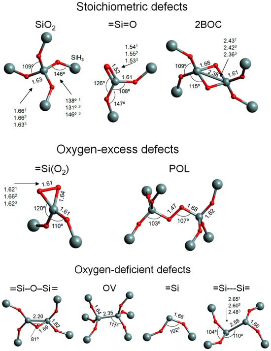
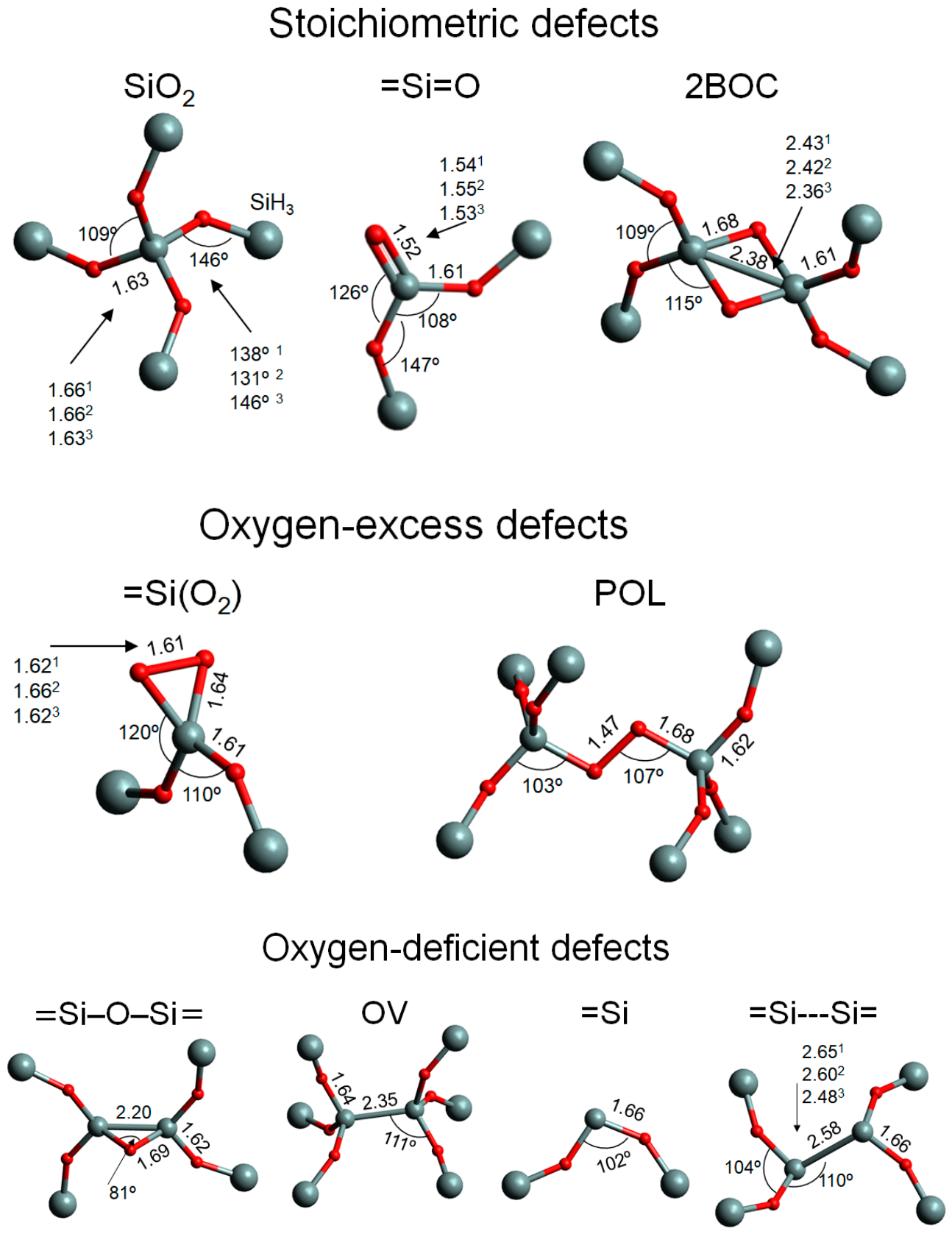
The parameters of the modeled optimized structures are shown in Figure 1. All defects are categorized into three groups: stoichiometric defects, defects with oxygen excess, and defects with oxygen deficiency. To form a defect with oxygen excess (or deficiency) in a defect-free stoichiometric structure, it is necessary to add an oxygen (or silicon) atom. An alternative approach, which does not require the introduction of an additional atom, involves the simultaneous formation of a defect pair: one with excess oxygen and one with oxygen deficiency. A stoichiometric defect can form in a stoichiometric structure without the involvement of additional atoms, as well as in oxygen-deficient and oxygen-excess samples [24].
Figure 1.
The structure of the cluster modeling defects; interatomic distances are given in angstroms, and silicon and oxygen atoms are indicated in gray and red, respectively. The SiH3 terminal groups are depicted as larger gray spheres. The geometry of all clusters is optimized at the B3LYP/cc-pvtz level; values marked with superscripts “1”, “2”, and “3” correspond to cluster geometry optimizations at the B3LYP/cc-pvdz, MP2/cc-pvdz, and MP2/cc-pvtz levels, respectively. Definitions of abbreviations are provided in the text and before the list of references at the end of the manuscript.
The geometric parameters of the structures optimized at the B3LYP/cc-pvtz level are close to those obtained at the MP2/cc-pvtz level, with the exception of the =Si---Si=-type defect. In the defect-free structure, designated as SiO2 in Figure 1, the Si–O bond length (1.63 Å) and the O–Si–O (109°) and Si–O–Si (146°) bond angles are consistent with the corresponding experimental data: 1.62 Å, 109°, and 144°, respectively [36]. In the =Si=O-type defect, which includes a silanone group, the central silicon atom and the three oxygen atoms bonded to it lie in the same plane. The length of the Si=O double bond (1.52 Å) is consistent with the results of quantum-chemical modeling reported in [24,34].
Two silicon atoms and two oxygen atoms constituting the 2BOC defect (or two-membered ring) [31] form a square. The Si–O bond lengths in the 2BOC structure significantly exceed the equilibrium value of 1.62 Å. In contrast, the O–Si–O and Si–O–Si bond angles in the 2BOC defect are approximately 90°, which is lower than their equilibrium values (109° and 146°, respectively). In the =Si(O)2 defect (which includes a dioxasilirane group), the two oxygen atoms and the silicon atom form a nearly equilateral triangle. The O–O bond length (1.61 Å) is significantly greater than the O–O bond length in a peroxide group, which is 1.47 Å (see the POL defect in Figure 1). All bond angles, measuring 60°, are substantially smaller than the equilibrium values (105° for the Si–O–O angle and 109° for the O–Si–O angle). These geometric parameters are consistent with the results of quantum-chemical calculations presented in [34]. The atoms forming the 110° valence angle lie in a plane perpendicular to the equilateral triangle. As the distance between the chemically bonded oxygen atoms increases, the energy of the ground singlet state increases, and a transition to the triplet state occurs upon reaching an O-O distance of 2.3 Å. The height of the energy barrier relative to the ground state is 1.4 eV. In the triplet state, the optimal distance between the oxygen atoms is 2.7 Å.
The =Si–O–Si= defect was recently identified using quantum molecular dynamics simulations [24]. The Si–Si bond in this defect is 0.15 Å shorter than in the OV defect, whereas the Si–O bond is 0.06 Å longer than in the defect-free structure (see SiO2 in Figure 1). The O–Si–Si angle in the =Si–O–Si= defect is 50°, which is approximately half the corresponding angle in OV. The =Si---Si= defect is formed by the coupling of two =Si defects. The bond length between the two Si atoms depends significantly on the level of quantum-chemical modeling; the MP2/cc-pvtz method yields a minimum value of 2.36 Å. The bond energy, calculated as the difference between the energy of the =Si---Si= defect and that of two separate Si defects, depends on the level of theory: 0.11 eV (B3LYP/cc-pvdz), 0.47 eV (MP2/cc-pvtz), and 0.35eV (CCSD/cc-pvtz). MP2 and CCSD methods, unlike B3LYP, explicitly account for electron correlation and therefore provide a more accurate value for the defect formation energy. Thus, the =Si---Si= defect formation energy is 0.4–0.5 eV lower than the formation energy of two =Si defects. Note that, taking into account the small absolute value of the bond energy, the optimization was performed with tight parameters.
Such a low bond energy value is due to the fact that relatively heavy atoms do not form stable π-bonds—unlike carbon atoms, which form strong double bonds with a π-component. For this reason, this defect is represented as =Si---Si=, where “---” denotes a weak chemical bond between two twofold-coordinated Si atoms. Typically, the concentration of =Si---Si= defects in SiO2 is significantly lower than that of other defects, such as OV, POL, and =Si(O2). This is because the formation of the defect =Si is accompanied by the breaking of two chemical bonds, which requires significant energy [24], and the formation of the =Si---Si= defect itself requires two =Si defects. A noticeable concentration of =Si defects can arise in oxygen-deficient films, in transition regions—for example, between the SiO2 layer and the Si substrate—or upon the exposure of the film to high-energy radiation. Under such conditions, the formation of defects =Si---Si= in noticeable concentration is possible. We consider this defect due to its interesting optical properties (see the following subsection).
2.2. Defect Excitation Energy in Equilibrium and Deformed Geometries
Table 1 presents the calculated excitation energies from the ground singlet state to the first excited singlet (S0 → S1) and triplet (S0 → T1) states. Oscillator strength values for the S0 → S1 transitions are also listed. Only vertical transitions from equilibrium ground states to excited states are considered.
Table 1.
Excitation energies E(eV) for the first vertical singlet–singlet (S0→S1) and singlet–triplet (S0→T1) transitions; vdz = cc-pvdz, vtz = cc-pvtz. Oscillator strength values multiplied by 103 are given in parentheses. The corresponding structures are shown in Figure 1. Structures with high oscillator strength and low excitation energy are highlighted in bold. The right column presents data from the literature.
The TD-DFT method systematically underestimates the excitation energies E for both S0 → S1 and S0 → T1 transitions. The latter transition is possible only due to very weak spin–orbit coupling; consequently, the oscillator strength of S0 → T1 is typically several orders of magnitude lower than that for singlet–singlet transitions. The largest energy difference, approximately 1 eV, between TD-DFT and LT-DF-LCC2 is observed for the defect-free structure (SiO2) and the 2BOC (or “two-membered ring”) defect. Expanding the basis set from the cc-pvdz to the cc-pvtz level generally leads to an increase in excitation energy of up to 0.3 eV. For the =Si(O2) defect, modeled using a relatively small cluster, the effect of further expanding the basis set to cc-pvqz is examined; the change in E values is less than 0.1 eV.
For all the structures considered, the transition energy E(S0 → T1) is lower than the transition energy E(S0 → S1). The largest difference between the transition energies to the triplet and singlet excited states (approximately 2 eV) is observed for the =Si and =Si---Si= defects. For the =Si(O2), peroxide linkage (POL), BOC, and OV defects, this difference is approximately 1 eV.
The minimum E values for both the S0 → S1 and S0 → T1 transitions are observed for the =Si---Si= defect. Like the =Si–O–Si=, this defect is characterized by the maximum oscillator strength for the S0 → S1 transition.
For OV and =Si defects, the calculated values of the E(S0 → S1) and oscillator strengths are close to the corresponding experimental values (see the right column of Table 1). The S0→S1 transition energy values for the Si(O2), Si=O, and POL structures, calculated on LT-DF-LCC2/cc-pvtz level, differ from previously reported values by less than 0.3 eV (see the right column). At the same time, the E(S0 → S1) for the defect-free cluster is noticeably lower than the experimental value. Furthermore, the singlet–singlet and singlet–triplet excitation energies are close to those calculated using ab initio methods with different sizes of the cluster, different basis sets, and the CIS method [8].
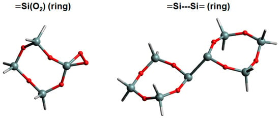
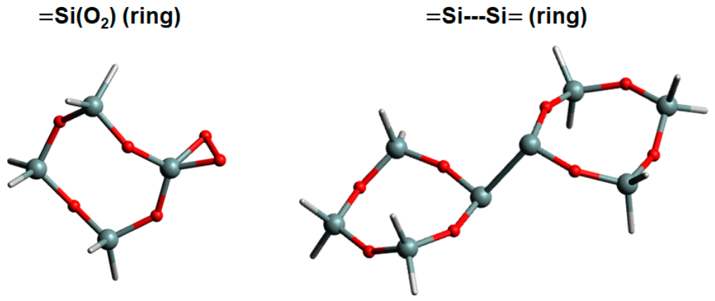
In addition, the optical parameters of the =Si(O2) and =Si---Si= defects were calculated using the ring-like clusters (see Figure 2 and the rows =Si(O2)(ring) and =Si---Si=(ring) in Table 1). The most notable difference, 0.3-0.4 eV, is observed in the E(S0 → T1) and E(S0 → S1) values for the =Si(O2) defect at the LT-DF-LCC2/cc-pvdz level. However, when the basis set is expanded to cc-pvtz, this difference reduces to 0.1 eV. Furthermore, the oscillator strength for the S0 → S1 transition in the =Si---Si= defect in a ring-like cluster is somewhat higher than in a chain-like cluster.
Figure 2.
Structure of the ring-like clusters used in modeling the =Si(O2) and =Si---Si= defects. All atoms constituting the clusters are shown, including the hydrogen atoms that saturate the bonds of the silicon atoms.
The discrepancy of approximately 1 eV between the calculated singlet–singlet transition energy for a defect-free structure (see the SiO2 row) and the experimental value of the fused silica optical band gap is likely due to the fact that larger clusters, larger than those used in this study, are required to accurately reproduce the bulk properties of the material.
The optimization of the geometry of the first singlet excited state of =Si---Si= defects was performed at the TD-B3LYP/cc-pvdz level. During the optimization process, the optimization terminated when the difference in energies between the first excited and ground states, E(S0 → S1), decreased from the initial value of 3.8 eV to 0.8 eV. This suggests a possible crossing of the ground- and excited-state potential energy surfaces, which would necessitate the use of special optimization methods. In any case, the significant reduction in the energy of the excited state during optimization indicates the possibility of absorbed light energy transforming into heat energy. A more precise analysis would require a numerical comparison of the probabilities of radiative and non-radiative recombination.
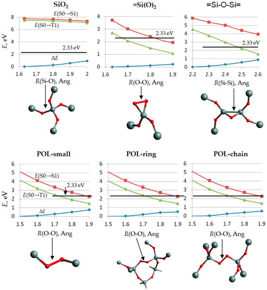
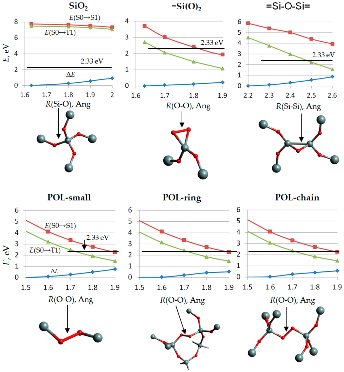
Deformation of chemical bonds leads to changes in excitation energy values. The corresponding dependencies are shown in Figure 3. The observed decrease in excitation energy is attributed to the fact that, as the geometry deviates from the optimal configuration, the energy gap between bonding and nonbonding orbitals and between bonding and antibonding orbitals decreases. Since excited states are formed primarily by nonbonding and antibonding orbitals, a reduction in excitation energy occurs.
Figure 3.
Dependence of the excitation energies of the first singlet–singlet transition E(S0 → S1) and the singlet–triplet transition E(S0 → T1) on bond stretching; ΔE is the deformation energy. The black horizontal line corresponds to the photon energy of the second harmonic of a Nd:YAG laser.
Increasing the Si–O bond length in the defect-free structure (SiO2 in Figure 3) has only a negligible effect on the values of E(S0 → S1) and E(S0 → T1). Indeed, for this structure, the deformation energy ΔE is close to the change in excitation energy. In contrast, for defects, structural deformation leads to a significant reduction in E(S0 → S1) and E(S0 → T1). For the =Si(O2) defect, O–O bond deformation with a ΔE on the order of 0.2 eV results in a decrease in E of approximately 2 eV. This result is consistent with the significant reduction in the excitation energies of the =Si(O2) defect due to O–Si–O bond angle deformation, as described in [34].
A similar dependence of the E(S0 → S1) and E(S0 → T1) energies on the length of the stretched bond is observed for =Si–O–Si=- and POL-type defects. In the latter case (POL), three different clusters containing an O–O bond are used: small, chain-like, and cyclic. The dependencies of E on the O–O bond length are practically identical for all clusters, with one exception: in the small cluster, the deformation energy ΔE is higher for the same values of bond elongation. This is attributed to the fact that, in larger clusters, the weakening of stretched bonds can be partially compensated for by the strengthening of neighboring bonds (alternation of chemical bonds).
3. Discussion
As the results show, the excitation energies for singlet–triplet transitions can fall within the visible range. Let us discuss which defects—and under what conditions—are capable of absorbing visible light with an energy corresponding to the second harmonic E(2ω) = 2.3 eV of Nd:YAG lasers, which are widely used in high-energy facilities [40].
As shown by the data in Table 1, only the =Si---Si= defect has an excitation energy lower than E(2ω). For the =Si(O2) defect, the S0 → T1 transition energy falls within the 2.3–2.6 eV range, which is close to the value of E(2ω). For the other defects listed in Table 1, the energies of vertical transitions from the ground-state equilibrium geometry to the first singlet and triplet excited states significantly exceed E(2ω). It should be noted that only the coupled-cluster calculation results (the LT-DF-LCC2 columns in Table 1) are considered for comparison with E(2ω), as TD-B3LYP calculations systematically underestimate the E(S0 → T1) and E(S0 → S1) values.
The twofold-coordinated silicon atom, =Si or ODC(II), is well known as an optically active center responsible for an absorption band in the ultraviolet region with a maximum at 5.0 eV [41]. The =Si---Si= defect forms through the coupling of two =Si defects; the energy gain from this interaction is approximately 0.4 eV, as noted in the “Results” Section. Another possible mechanism for the =Si---Si= formation involves the detachment of an O2 molecule from a 2BOC-type defect (or “double-ring” defect). In any case, the concentration of =Si---Si= defects in stoichiometric SiO2 films is relatively low, as their formation entails a higher energy cost compared to the other point defects shown in Figure 1. Nevertheless, =Si---Si= defects can potentially affect the initiation of laser damage due to their low excitation energy and large oscillator strength—which is 4–5 times greater than that of =Si and OV defects (Table 1). An oscillator strength of similar magnitude is characteristic only of the BOC defect; however, the E(S0 → S1) value for BOC is approximately 6 eV, which is more than double the photon energy of the second harmonic of a Nd:YAG laser. It should be noted that =Si is one of the defects observed in α-quartz following irradiation by fast particles, particularly neutrons [42,43]. Since high-energy laser systems are used in thermonuclear fusion facilities [1], the appearance of =Si and =Si---Si= defects under the influence of neutron flux radiation is highly probable. It should be noted that the laser damage in amorphous SiO2 thin layers in optical coatings is closely related to the radiation-induced degradation of silica-based optical fibers [44] because the same intrinsic point defects are involved in both processes [45].
The =Si(O2) defect, having an energy of S0 → S1 close to the photon energy of the second harmonic of a Nd:YAG laser, is known as an optically active surface defect [18], associated with adsorption at energies of 3.2 and 5.1 eV [46] and photoluminescence in the green band [34,47]. As noted in the Introduction Section, laser-induced damage in SiO2 layers is associated with subsurface luminescence [25], which can be attributed to the =Si(O2) defect.
The oscillator strength for the S0 → S1 transition at =Si(O2) is approximately 10−3, which is significantly lower than the corresponding value of the =Si defect. The =Si(O2) defect can form from the =Si defect via a simple and energetically favorable reaction: =Si + O2 → =Si(O2). Thus, in porous SiO2 films, an increase in the concentration of =Si defects under neutron irradiation can lead to a rise in the number of =Si(O2) defects due to the diffusion of oxygen molecules from the atmosphere into the film.
Stretching of chemical bonds leads to a decrease in the LIDT value [6] due to a significant reduction in the defect excitation energy. As shown in Figure 3, the values of E(S0 → S1) and E(S0 → T1) can drop below the energy of the second harmonic of the Nd:YAG laser (the black horizontal line in Figure 3). Temperature fluctuations are one of the possible reasons for deviations in the bond length from equilibrium values. The energy equivalent of room temperature, 300 K, is approximately 0.026 eV. The probability of the formation of fluctuations with the deformation energy ΔE is proportional to ν·exp(−ΔE/0.026), where ν is the frequency of the bond vibrations, ~1015 Hz, and ΔE is in eV. So, even at room temperature, the fluctuations ΔE = 0.2 eV and ΔE = 0.3 eV can occur with frequencies ~1012 and ~1011 Hz, respectively, as follows from the expression for the probability of fluctuations. A deformation with energy ΔE= 0.2 eV reduces E(S0 → T1) below the energy of the second harmonic of the Nd:YAG laser (E(2ω) = 2.3 eV) for the POL defect. For the =Si(O2) defect, this energy reduces both E(S0 → T1) and E(S0 → S1) values below 2.3 eV. Significantly stretched O–O bonds with low excitation energy likely arise in clusters composed of several coupled defects (e.g., strained two- and three-membered rings) or at interfaces between layers of different compositions due to steric structural differences. Modeling such structures requires the use of clusters significantly larger than those used in the present study.
Let us estimate the relative concentration of defects that could potentially cause laser-induced damage through the absorption of radiation via singlet–triplet transitions. As mentioned in the Introduction Section, the laser damage induced by a nanosecond Nd:YAG laser can be caused by defects, including non-bridging oxygen atoms [28]. The oscillator strength f for non-bridging oxygen is about 0.05 [48], while the value of f for singlet–triplet transitions is less than 10−3. Absorption is proportional to the product of the defect concentration and the oscillator strength. Thus, if laser-induced damage, as studied in [28], is caused by singlet–triplet transitions in =Si(O2) and =Si---Si= defects, then their concentration must be at least one hundred times higher than that of the non-bridging oxygen atoms. This issue requires further study.
In addition, we can estimate the absolute concentration N of =Si---Si= defects using the relation for the absorption coefficient α [48]:
where e, m, ħ, c and ε0 are the elementary charge, electron mass, Planck constant, vacuum light velocity, and dielectric constant in a Coulomb law. Δ1 = ħΔω is the halfwidth of the absorption band, Γ is a numeric coefficient depending on the shape of the band, n is the refractive index, N is the concentration of the absorbing centers, and f is the oscillator strength. Substituting the fundamental constants, a refractive index of 1.5 for silicon dioxide, Γ = 1.1 for a Gaussian shape of the absorbing band, and a typical value for Δ1, which is on the order of tenths of an eV [48], we obtain:
where N is in 1/m3, and α is in 1/m.
α = (N/n)·((n2 +2)/3)2·(2π2e2ħ/(mcε0))·f/(Δ1Γ),
N ~ 1018α/f,
The value of α(ħω = 2.3 eV) for fused silica irradiated with a neutron flux of 3 · 1015 per cm−2 is about of 102 cm−1 [49]. Assuming an oscillator strength of f ~ 10−3 ÷ 10−4, we obtain N ~ 1023 ÷ 1024 (1/m3) = 1017 ÷ 1018 (1/cm3).
To summarize, =Si---Si= and, likely, =Si(O2) defects containing a dioxysilirane group can absorb laser radiation with an energy of 2.3 eV (the second harmonic of a Nd:YAG laser). Furthermore, bond stretching within the defect structures can lower the excitation energy to values below 2.3 eV; temperature fluctuations may be the cause of such stretching. It should be noted, however, that the potential for absorption at the laser wavelength does not allow for a definitive conclusion regarding the decisive role of such defects in the initiation of laser damage. It is necessary to investigate the stability of these defects—not only within the cluster approximation, as done in the present work, but also by accounting for their interaction with surrounding fragments of the continuous disordered network. The probability of triplet transitions from the ground state to an excited state is low; therefore, for these transitions to contribute significantly to laser radiation absorption, the defect concentration must be high. Classical atomistic modeling methods can be employed to investigate defect stability and estimate their concentration under various film deposition conditions. Calculations of singlet–triplet transition probabilities can be performed using quantum-mechanical methods. Finally, estimating the laser damage threshold requires taking into account the processes of absorbed energy relaxation and thermal transport.
4. Materials and Methods
Quantum chemistry simulations are performed using the cluster approach. The clusters representing the defects are terminated by SiH3 groups, as described in [34]. The manually constructed clusters include at least two coordination spheres surrounding the atoms that form the defect (see Figure 1). The terminal bonds of the Si atoms at the cluster boundaries are saturated with hydrogen atoms. The localization of the molecular orbitals of the ground and excited states is analyzed based on the coefficients of the molecular orbital expansion in terms of atomic orbitals. In all cases examined, the primary contribution to the initial and final states comes from atoms located in the first coordination sphere of the atoms forming the defect. Geometry optimization of the atomistic clusters is carried out using a hybrid functional B3LYP [50] with two correlated consistent basis sets—cc-pvdz and cc-pvtz [51]. All defects in the ground state have closed shells and do not contain unpaired electrons.
Excited states are calculated using Time-Dependent Density Functional Theory (TDDFT) [52] and the Laplace-Transformed Density-Fitted Local Coupled-Cluster approach with single and double excitations (LT-DF-LCC2) [53,54] implemented in the NWChem version 6.6 [55] and Molpro version 2015.1.29 [56,57,58] programs, respectively. The TDDFT calculations are performed with the B3LYP [50] functional. The Avogadro program version 1.2.0 [59] is used for the visualization of the structures. All calculations are performed using the equipment of the shared research facilities of HPC computing resources at Lomonosov Moscow State University [60].
5. Conclusions
Using ab initio quantum chemistry methods within a cluster approach, the optical characteristics of neutral diamagnetic defects potentially associated with laser-induced damage in SiO2 layers are calculated. Since the damage is caused by high-intensity laser radiation, singlet–triplet transitions—forbidden in the first order of perturbation theory—are also considered.
It is determined that the =Si---Si= defect, characterized by a first singlet–triplet excitation energy E(S0→T1) of approximately 1.4–1.5 eV, can absorb high-intensity laser radiation with an energy of 2.3 eV (corresponding to the second harmonic of a Nd:YAG laser). It should be noted, however, that the role of this defect in the initiation of laser-induced damage requires further investigation due to its relatively high formation energy. For the =Si(O2) defect, which contains a dioxysilirane group, the E(S0→T1) value in equilibrium geometry is 2.6 eV; however, this value can drop below 2.3 eV upon slight stretching of the O–O bonds. Stretching the O–O bond in a peroxide bridge or the Si–Si bond in a BOC defect can also lower the excitation energy to 2.3 eV, but in these cases, the energy cost of stretching significantly exceeds that required for the =Si(O2) defect. The probability of such bond stretching due to thermal fluctuations is estimated.
Author Contributions
Conceptualization, F.V.G. and V.B.S.; methodology, F.V.G.; software, V.B.S.; validation, F.V.G. and A.V.T.,; resources, V.B.S.; writing—original draft preparation, F.V.G.; writing—review and editing, V.B.S.; visualization, F.V.G.; supervision, V.B.S.; project administration, V.B.S.; funding acquisition, V.B.S. and A.V.T. All authors have read and agreed to the published version of the manuscript.
Funding
The work was supported by the Ministry of Education and Science of the Russian Federation as part of the program of the Moscow Center for Fundamental and Applied Mathematics under Agreement No. 075-15-2025-345.
Institutional Review Board Statement
Not applicable.
Informed Consent Statement
Not applicable.
Data Availability Statement
The original contributions presented in this study are included in the article. Further inquiries can be directed to the corresponding author.
Conflicts of Interest
The authors declare no conflicts of interest.
Abbreviations
The following abbreviations are used in this manuscript:
| OV | oxygen vacancy |
| MD | molecular dynamics |
| LIDT | laser-induced damage threshold |
| 2BOC | 2-Bridging Oxygen Center |
| POL | peroxy linkage |
References
- Danson, C.; Hillier, D.; Hopps, N.; Neely, D. Petawatt class lasers worldwide. High Power Laser Sci. Eng. 2015, 3, e3. [Google Scholar] [CrossRef] [Scilit]
- Efimov, O.M. Laser-induced damage: 60 years of measurements. J. Opt. Technol. 2021, 88, 409–419. [Google Scholar] [CrossRef] [Scilit]
- Chorel, M. Study of High Damage Threshold Optical Coatings Used in Environment with Very Low Hygrometry for Fusion Class Laser System. Ph.D. Thesis, Université de Bordeaux, Bordeaux, France, 2019. [Google Scholar]
- Cao, X.; Zhang, L.; Qin, Y.; Zhu, H.; Shen, R.; Li, Y.; Zhou, H. Research Progress on Particle Contamination-Induced Damage of Optical Component Surfaces in High-Power Laser Systems. Photonics 2026, 13, 905. [Google Scholar] [CrossRef] [Scilit]
- Mu, Q.; Gao, X.; Wang, Y.; Zhou, P. Bidirectional evolution of NBOHC defects and its effect on laser damage resistance of fused silica during laser conditioning. J. Non. Cryst. Solids 2026, 671, 123853. [Google Scholar] [CrossRef] [Scilit]
- Li, Y.; Yan, H.; Yang, K.; Yao, C.; Wang, Z.; Zou, X.; Yan, C.; Yuan, X.; Ju, X.; Yang, L. Surface molecular structure defects and laser-induced damage threshold of fused silica during a manufacturing process. Sci. Rep. 2017, 7, 17870. [Google Scholar] [CrossRef] [Scilit] [PubMed]
- Tolenis, T.; Grinevičiūtė, L.; Buzelis, R.; Smalakys, L.; Pupka, E.; Melnikas, S.; Selskis, A.; Drazdys, R.; Melninkaitis, A. Sculptured anti-reflection coatings for high power lasers. Opt. Mater. Express 2017, 7, 1249–1258. [Google Scholar] [CrossRef] [Scilit]
- Sulimov, V.B.; Sushko, P.V.; Edwards, A.H.; Shluger, A.L.; Stoneham, A.M. Asymmetry and long-range character of lattice deformation by neutral oxygen vacancy in α-quartz. Phys. Rev. B 2002, 66, 024108. [Google Scholar] [CrossRef] [Scilit]
- Busso, M.; Casassa, S.; Pisani, C.; Sulimov, V.B. Ab initio simulation of the oxygen vacancy bistability in pure and Ge-doped α-quartz. Model. Simul. Mater. Sci. Eng. 2002, 10, 21–33. [Google Scholar] [CrossRef] [Scilit]
- Dianov, E.M.; Sokolov, V.O.; Sulimov, V.B. Semiempirical calculations of point defects in silica. Oxygen vacancy and twofold coordinated silicon atom. J. Non-Cryst. Solids 1992, 149, 5–18. [Google Scholar] [CrossRef] [Scilit]
- Skuja, L.; Kajihara, K.; Grube, J.; Hosono, H. Luminescence of non-bridging oxygen hole centers in crystalline SiO2. AIP Conf. Proc. 2014, 1624, 130–134. [Google Scholar] [CrossRef] [Scilit]
- Skuja, L. Optically active oxygen-deficiency-related centers in amorphous silicon dioxide. J. Non-Cryst. Solids 1998, 239, 16–48. [Google Scholar] [CrossRef] [Scilit]
- Sokolov, V.O.; Sulimov, A.B. Semi-empirical calculation of peroxy linkage in vitreous silicon dioxide. Phys. Status Solidi 1987, 142, K7–K12. [Google Scholar] [CrossRef] [Scilit]
- Wang, W.; Lu, P.; Han, L.; Zhang, C.; Wu, L.; Guan, P.; Su, R.; Chen, J. Structural and electronic properties of peroxy linkage defect and its interconversion in fused silica. J. Non-Cryst. Solids 2016, 434, 96–101. [Google Scholar] [CrossRef] [Scilit]
- Friebele, E.J.; Griscom, D.L.; Stapelbroek, M.; Weeks, R.A. Fundamental Defect Centers in Glass: The Peroxy Radical in Irradiated, High-Purity, Fused Silica. Phys. Rev. Lett. 1979, 42, 1346–1349. [Google Scholar] [CrossRef] [Scilit]
- Edwards, A.H.; Fowler, W.B. Theory of the peroxy-radical defect in a-SiO2. Phys. Rev. B 1982, 26, 6649–6660. [Google Scholar] [CrossRef] [Scilit]
- Skuja, L.; Hirano, M.; Hosono, H.; Kajihara, K. Defects in oxide glasses. Phys. Status Solidi 2005, 2, 15–24. [Google Scholar] [CrossRef] [Scilit]
- Skuja, L.; Kajihara, K.; Hirano, M.; Hosono, H. Oxygen-excess-related point defects in glassy/amorphous SiO2 and related materials. Nucl. Instrum. Methods Phys. Res. Sect. B Beam Interact. Mater. At. 2012, 286, 159–168. [Google Scholar] [CrossRef] [Scilit]
- Griscom, D.L. The Natures of Point Defects in Amorphous Silicon Dioxide. In Defects in SiO2 and Related Dielectrics: Science and Technology; Pacchioni, G., Skuja, L., Griscom, D.L., Eds.; Springer: Dordrecht, The Netherlands, 2000; pp. 117–159. [Google Scholar]
- Trukhin, A.N.; Smits, K.; Jansons, J.; Kuzmin, A. Luminescence of polymorphous SiO2. Radiat. Meas. 2016, 90, 6–13. [Google Scholar] [CrossRef] [Scilit]
- Wang, C.; Guo, X.; Zhang, S.; Qi, Y.; Gao, S. Analysis of the electronic structure of β-SiO2 intrinsic defects based on Density Functional Theory. Mater. Today Commun. 2023, 35, 105554. [Google Scholar] [CrossRef] [Scilit]
- You, Y.-W.; Wei, Y.; Xuan, J.; Zhu, Q.; Li, X.; Hou, K.; Yao, D.; Wang, L.; Li, D. Formation and annihilation of point defects in SiO2 glass during neutron irradiation and annealing. J. Non-Cryst. Solids 2025, 650, 123342. [Google Scholar] [CrossRef] [Scilit]
- Cai, D.; Dong, S.; Zhang, Y.; Liu, H. Molecular dynamics simulation and experimental study on the evolution mechanism of radiation-induced defects in silica glass. J. Non-Cryst. Solids 2026, 676, 123953. [Google Scholar] [CrossRef] [Scilit]
- Sulimov, V.B.; Kutov, D.C.; Sulimov, A.V.; Grigoriev, F.V.; Tikhonravov, A.A. Novel Oxygen-Deficient Centers and Other Intrinsic Defects in Amorphous SiO2: Quantum Molecular Dynamics Simulations. Supercomput. Front. Innov. 2026, 12, 101–124. [Google Scholar] [CrossRef] [Scilit]
- Yang, D.; Cheng, J.; Zhao, L.; Chen, M.; Liu, H.; Wang, J.; Han, C.; Liu, Z.; Wang, S.; Geng, F.; et al. Relationship between the photoluminescence envelope area of surface defects and the laser-induced damage thresholds of mechanically machined fused silica optical surfaces. Ceram. Int. 2023, 49, 22767–22781. [Google Scholar] [CrossRef] [Scilit]
- Zoubir, A.; Rivero, C.; Grodsky, R.; Richardson, K.; Richardson, M.; Cardinal, T.; Couzi, M. Laser-induced defects in fused silica by femtosecond IR irradiation. Phys. Rev. B 2006, 73, 224117. [Google Scholar] [CrossRef] [Scilit]
- Beaudier, A.; Wagner, F.R.; Natoli, J.-Y. Laser-induced modifications in fused silica up to damage initiation caused by multiple UV nanosecond pulses. In Proceedings of the SPIE Laser Damage 2017, Boulder, CO, USA, 24–27 September 2017; Volume 10447, p. 104470B. [Google Scholar]
- Geng, F.; Cheng, H.; Zhang, Q.; Liu, M.; Li, A.Y. Ultraviolet laser damage properties of single-layer SiO2 film grown by atomic layer deposition. Opt. Mater. Express 2020, 10, 1981–1990. [Google Scholar] [CrossRef] [Scilit]
- Sulimov, V.B.; Kutov, D.C.; Sulimov, A.V.; Grigoriev, F.V.; Tikhonravov, A.V. Density functional modeling of structural and electronic properties of amorphous high temperature oxides. J. Non-Cryst. Solids 2022, 578, 121170. [Google Scholar] [CrossRef] [Scilit]
- Liu, X.; Feng, C.; Zhang, W.; Nasibli, H.; Zhao, Y.; Liu, X.; Shuai, K.; Shao, J. Nanosecond laser damage of 532 nm thin film polarizers evaluated by different testing protocols. Opt. Mater. 2024, 149, 115124. [Google Scholar] [CrossRef] [Scilit]
- Sulimov, A.V.; Kutov, D.C.; Grigoriev, F.V.; Tikhonravov, A.V.; Sulimov, V.B. Generation of amorphous silicon dioxide structures via melting-quenching density functional modeling. Lobachevskii J. Math. 2020, 41, 1581–1590. [Google Scholar] [CrossRef] [Scilit]
- Bobyshev, A.A.; Radtsig, V.A. Silanone groups on the surface of mechanically activated silicon dioxide. Kinet. Katal. 1988, 29, 638. [Google Scholar]
- Bobyshev, A.A.; Radtsig, V.A. Formation and physical-chemistry properties of siladioxirane groups on the surface of silica. Khim. Fiz. 1988, 7, 950. [Google Scholar]
- Zyubin, A.S.; Mebel, A.M.; Lin, S.H.; Glinka, Y.D. Photoluminescence of silanone and dioxasilyrane groups in silicon oxides: A theoretical study. J. Chem. Phys. 2002, 116, 9889–9896. [Google Scholar] [CrossRef] [Scilit]
- Raghavachari, K.; Pacchioni, G. Photoabsorption of dioxasilyrane and silanone groups at the surface of silica. J. Chem. Phys. 2001, 114, 4657–4662. [Google Scholar] [CrossRef] [Scilit]
- Mozzi, R.L.; Warren, B.E. The structure of vitreous silica. J. Appl. Crystallogr. 1969, 2, 164–172. [Google Scholar] [CrossRef] [Scilit]
- Itoh, C.; Tanimura, K.; Itoh, N.; Itoh, M. Threshold energy for photogeneration of self-trapped excitons in SiO2. Phys. Rev. B 1989, 39, 11183–11186. [Google Scholar] [CrossRef] [Scilit] [PubMed]
- Stefanov, B.B.; Raghavachari, K. Photoabsorption of the peroxide linkage defect in silicate glasses. J. Chem. Phys. 1999, 111, 8039–8042. [Google Scholar] [CrossRef] [Scilit]
- Girard, S.; Alessi, A.; Richard, N.; Martin-Samos, L.; De Michele, V.; Giacomazzi, L.; Agnello, S.; Di Francesca, D.; Morana, A.; Winkler, B.; et al. Overview of radiation induced point defects in silica-based optical fibers. Rev. Phys. 2019, 4, 100032. [Google Scholar] [CrossRef] [Scilit]
- Meijer, R.A.; Stodolna, A.S.; Eikema, K.S.E.; Witte, S. High-energy Nd:YAG laser system with arbitrary sub-nanosecond pulse shaping capability. Opt. Lett. 2017, 42, 2758–2761. [Google Scholar] [CrossRef] [Scilit] [PubMed]
- Garmysheva, T.; Nepomnyashchikh, A.; Shalaev, A.; Kaneva, E.; Paklin, A.; Chernenko, K.; Kozlova, A.; Pankratov, V.; Shendrik, R. Luminescence of ODC(II) in quartz and cristobalite glasses. J. Non-Cryst. Solids 2022, 575, 121199. Available online: https://www.sciencedirect.com/science/article/abs/pii/S0022309321005627?via%3Dihub (1 January 2022). [CrossRef] [Scilit]
- Azzoni, C.B.; Meinardi, F.; Paleari, A. Trapped-hole centers in neutron-irradiated synthetic quartz. Phys. Rev. B 1994, 49, 9182–9185. [Google Scholar] [CrossRef] [Scilit] [PubMed]
- Corazza, A.; Crivelli, B.; Martini, M.; Spinolo, G.; Vedda, A. Photoluminescence and optical absorption in neutron-irradiated crystalline quartz. Phys. Rev. B 1996, 53, 9739–9744. [Google Scholar] [CrossRef] [Scilit] [PubMed]
- Dianov, E.M.; Kornienko, L.S.; Nikitin, E.P.; Rybaltovskiĭ, A.O.; Sulimov, V.B.; Chernov, P.V. Radiation-optical properties of quartz glass fiber-optic waveguides (review). Sov. J. Quantum Electron. 1983, 13, 274–289. [Google Scholar] [CrossRef] [Scilit]
- Tomashuk, A.L.; Zabezhailov, M.O. Formation mechanisms of precursors of radiation-induced color centers during fabrication of silica optical fiber preform. J. Appl. Phys. 2011, 109, 083103. [Google Scholar] [CrossRef] [Scilit]
- Radzig, V.A. Chapter 7 Point Defects on the Silica Surface: Structure and Reactivity. In Physico-Chemical Phenomena in Thin Films and at Solid Surfaces; Trakhtenberg, L.I., Lin, S.H., Ilegbusi, O.J., Eds.; Academic Press: Cambridge, MA, USA, 2007; Volume 34, pp. 231–345. [Google Scholar]
- León, M.; Martín, P.; Ibarra, A.; Hodgson, E.R. Gamma irradiation induced defects in different types of fused silica. J. Nucl. Mater. 2009, 386–388, 1034–1037. [Google Scholar] [CrossRef] [Scilit]
- Skuja, L. Optical Properties of Defects in Silica. In Defects in SiO2 and Related Dielectrics: Science and Technology; Pacchioni, G., Skuja, L., Griscom, D.L., Eds.; Springer: Dordrecht, The Netherlands, 2000; pp. 73–116. [Google Scholar]
- Guzzi, M.; Martini, M.; Paleari, A.; Pio, F.; Vedda, A.; Azzoni, C.B. Neutron irradiation effects in amorphous SiO2: Optical absorption and electron paramagnetic resonance. J. Phys. Condens. Matter 1993, 5, 8105. [Google Scholar] [CrossRef] [Scilit]
- Becke, A.D. Density-functional thermochemistry. III. The role of exact exchange. J. Chem. Phys. 1993, 98, 5648–5652. [Google Scholar] [CrossRef] [Scilit]
- Dunning, T.H., Jr. Gaussian basis sets for use in correlated molecular calculations. I. The atoms boron through neon and hydrogen. J. Chem. Phys. 1989, 90, 1007–1023. [Google Scholar] [CrossRef] [Scilit]
- Runge, E.; Gross, E.K.U. Density-Functional Theory for Time-Dependent Systems. Phys. Rev. Lett. 1984, 52, 997–1000. [Google Scholar] [CrossRef] [Scilit]
- Kats, D.; Schütz, M. Local Time-Dependent Coupled Cluster Response for Properties of Excited States in Large Molecules. Z. Phys. Chem. 2010, 224, 601–616. [Google Scholar] [CrossRef] [Scilit]
- Freundorfer, K.; Kats, D.; Korona, T.; Schütz, M. Local CC2 response method for triplet states based on Laplace transform: Excitation energies and first-order properties. J. Chem. Phys. 2010, 133, 244110. [Google Scholar] [CrossRef] [Scilit] [PubMed]
- Valiev, M.; Bylaska, E.J.; Govind, N.; Kowalski, K.; Straatsma, T.P.; Van Dam, H.J.J.; Wang, D.; Nieplocha, J.; Apra, E.; Windus, T.L.; et al. NWChem: A comprehensive and scalable open-source solution for large scale molecular simulations. Comput. Phys. Commun. 2010, 181, 1477–1489. [Google Scholar] [CrossRef] [Scilit]
- Werner, H.-J.; Knowles, P.J.; Knizia, G.; Manby, F.R.; Schütz, M. Molpro: A general-purpose quantum chemistry program package. WIREs Comput. Mol. Sci. 2012, 2, 242–253. [Google Scholar] [CrossRef] [Scilit]
- Werner, H.-J.; Knowles, P.J.; Manby, F.R.; Black, J.A.; Doll, K.; Heßelmann, A.; Kats, D.; Köhn, A.; Korona, T.; Kreplin, D.A.; et al. The Molpro quantum chemistry package. J. Chem. Phys. 2020, 152, 144107. [Google Scholar] [CrossRef] [Scilit] [PubMed]
- Werner, H.-J.; Knowles, P.J. MOLPRO, version 2015.1.29. A Package of ab initio Programs. 2015.
- Hanwell, M.D.; Curtis, D.E.; Lonie, D.C.; Vandermeersch, T.; Zurek, E.; Hutchison, G.R. Avogadro: An advanced semantic chemical editor, visualization, and analysis platform. J. Cheminform. 2012, 4, 17. [Google Scholar] [CrossRef] [Scilit] [PubMed]
- Voevodin, V.V.; Antonov, A.S.; Nikitenko, D.A.; Shvets, P.A.; Sobolev, S.I.; Sidorov, I.Y.; Stefanov, K.S.; Voevodin, V.V.; Zhumatiy, S.A. Supercomputer Lomonosov-2: Large Scale, Deep Monitoring and Fine Analytics for the User Community. Supercomput. Front. Innov. 2019, 6, 4–11. [Google Scholar] [CrossRef] [Scilit]

Disclaimer/Publisher’s Note: The statements, opinions and data contained in all publications are solely those of the individual author(s) and contributor(s) and not of MDPI and/or the editor(s). MDPI and/or the editor(s) disclaim responsibility for any injury to people or property resulting from any ideas, methods, instructions or products referred to in the content. |
© 2026 by the authors. Licensee MDPI, Basel, Switzerland. This article is an open access article distributed under the terms and conditions of the Creative Commons Attribution (CC BY) license.