


Abstract
Countercurrent electrophoresis (CCEP) is a technology that forms a steady focus in the separation channel through the coupling of electric field migration and reverse fluid flow, so as to realize the synchronous concentration and separation of substances. This paper reviews the evolution of CCEP from its theoretical origin, device design to microfluidic integration and optimization. Compared with traditional capillary electrophoresis, CCEP has higher processing flux, continuous operation ability and separation resolution, and has been successfully applied to isotope enrichment, protein recovery and complex matrix analysis. This paper further discusses the methods derived from it, such as isoelectric focusing (CACE), conductance gradient focusing (CGF) and electric field gradient focusing (EFGF), which expand the analysis ability and application scenarios of CCEP. Although the technology still faces challenges such as system stability, operation complexity and detection dependence, with the development of microfluidic, intelligent control and new material technology, CCEP shows broad development prospects in biomedicine, environmental monitoring and nuclide separation.
1. Introduction
In the field of chemical separation and analysis, the application of microscale architectures—such as capillaries, microporous chromatographic columns, and microfluidic channels—has become widespread. The capillary electrophoresis (CE) literature also includes a substantial body of review articles detailing various mature separation and identification methodologies [1,2,3,4,5,6]. However, conventional analytical methods are generally limited to characterization and struggle to achieve simultaneous analyte enrichment and preparative-scale purification. As a notable exception, this review focuses on countercurrent electrophoresis (CCEP), an emerging enrichment technology. CCEP offers a practical laboratory method for separating charged analytes in aqueous media based on differences in their electromigration rates. Theoretically, this technique is capable of resolving any pair of substances that exhibit distinct electrophoretic mobilities in an electrolyte.
The evolution of CCEP follows a clear trajectory, spanning theoretical groundwork, methodological development, and practical application. In 1964, Peja [7] laid the essential theoretical foundation. Through careful experiments using Glueckauf’s tangent method, they quantitatively demonstrated for the first time that paper electrophoresis fundamentally couples electrophoretic migration with chromatographic partitioning, leading to a unified mathematical model. Building on this, Carpenter and Hayes [8] combined high voltage with cellulose acetate membranes in 1963 to establish a rapid, high-resolution method for analyzing charge heterogeneity in insulin, highlighting its utility in protein analysis. A conceptual breakthrough came in 1967 with Broome and Raymond [9], who designed an automated prototype that integrated liquid–liquid transfer—adapted from countercurrent distribution—with electrophoretic separation, thereby opening a path to preparative-scale protein isolation.
Later, in 1993, Deshmukh and Bier [10] represented a phase of consolidation and deepening. They developed a one-dimensional computer model of electrophoretic transport that incorporated counterflow, validated it experimentally using recycling isotachophoresis, and systematically revealed the underlying optimization mechanism: during transient processes, counterflow selectively removes slower-migrating components without altering steady-state zone properties. This model was subsequently extended to simulate capillary electrophoresis. Together, these studies map the progression of CCEP from fundamental insight through methodological advances to intelligent separation optimization.
A persistent challenge in electrophoretic separation has been balancing resolution with throughput. CCEP addressed this early on by enabling continuous operation and large-scale preparation, showing clear potential in protein processing. However, its operational complexity and inherent difficulty in separating species with very similar electrophoretic mobilities—such as certain metal ions—revealed practical limitations. This impasse spurred a strategic shift: by concentrating on the isoelectric point (pI) as a key protein attribute, the separation concept behind CCEP gave rise to isoelectric focusing (IEF) [11,12]. IEF abandons continuous-flow operation, instead achieving protein “focusing” within a stable pH gradient. This approach elevates analytical resolution to an extreme, making IEF an indispensable core tool in proteomics.
This review systematically traces the technological evolution from CCEP to IEF, examines their logical connections and complementary roles, and looks ahead to their integration and development within modern bioseparation science. Specifically, it begins by examining the principles, historical development, and challenges of CCEP. It then details how IEF refined and extended the core ideas of CCEP to become a key high-resolution analytical technology. Finally, it discusses further derivatives and advances of IEF, as well as prospects for merging CCEP and IEF concepts in contemporary integrated separation platforms.
2. Research Progress on Counter Current Electrophoresis Separation Method
2.1. Theoretical Introduction
Bethune and Kegeles [13] established a rigorous mathematical theory that bridged discrete countercurrent distribution experimental techniques with continuous moving boundary methods (such as electrophoresis and sedimentation) in 1961. By leveraging early computer simulations, their work became the first theoretical model to simultaneously incorporate chemical reactions, transport phenomena, and diffusion effects, thereby surpassing the mainstream theories of that time.
The electrophoretic velocity acquired by an ion per unit electric field in the direction of migration is generally referred to as mobility or electromigration mobility, denoted as . The mobility measured in an infinitely diluted solution is termed absolute mobility, denoted as [14]. According to electromagnetic theory, the electrostatic force acting on an ion is the product of its charge q and the electric field strength E:
FE = qE.
In fluid dynamics, the frictional drag force on a (spherical) ion is proportional to its effective ionic radius r, velocity v, and solvent viscosity η [15]:
where and are the dielectric constant of the solution and the electromotive force of the ion at infinite dilution, respectively.
Ff = 6πηrv = 6πηrµem0E
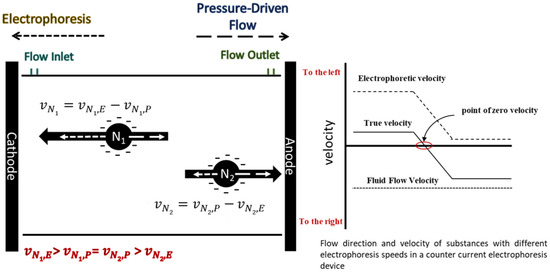
According to the formula, the electric mobility of ions in the same solution environment is mainly related to the radius and charge of the ion cluster. Different charged ions often have different electromigration. By configuring a fluid inlet near the cathode and setting the counterflow velocity between the two electromigration velocities, i.e., , the component with lower electromigration velocity can be swept away from the separation channel, while the component with higher electromigration velocity is retained near the inlet electrode [16].
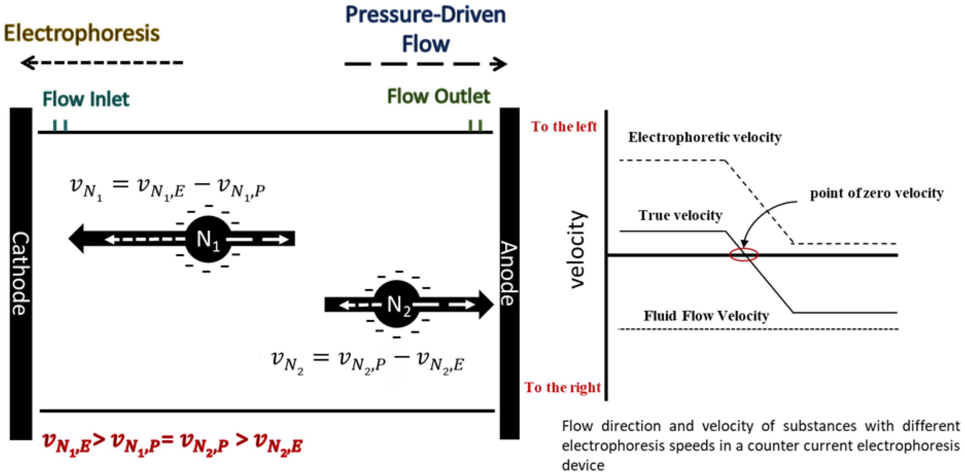
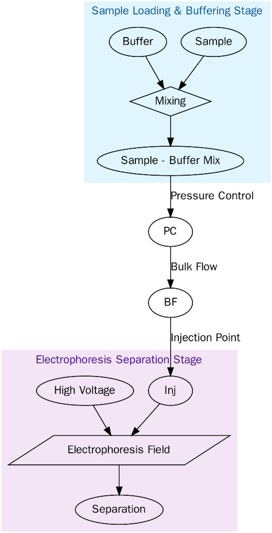
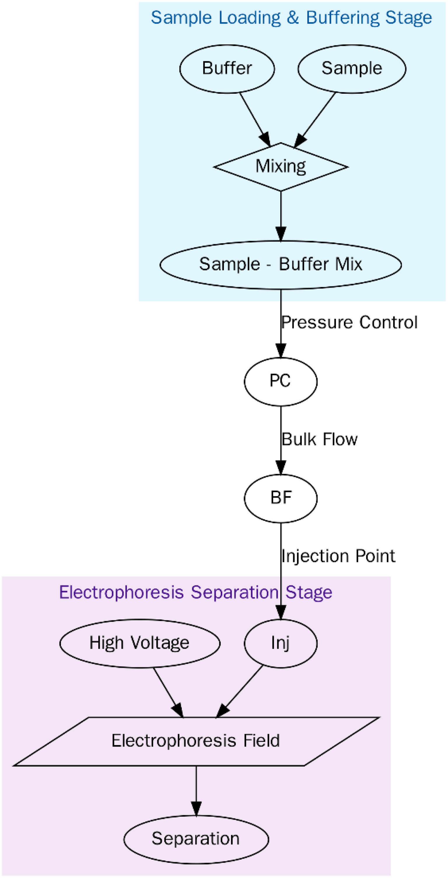
In recent years, the theory of IEF technique has been proposed for simultaneous separation of multiple proteins. The IEF technique, while related to countercurrent electrophoresis, operates on a distinct principle. It requires that analytes meet three key conditions: first, their migration velocity must vary along the separation column; second, this velocity must reach zero at a specific column position (ideally unique for each analyte); and third, their net motion must be directed toward, not away from, this zero-velocity point. When these conditions are satisfied, all analyte molecules will focus into sharp peaks centered at their respective stationary points (Figure 1).
Figure 1.
Schematic diagram of countercurrent electrophoresis separation method.
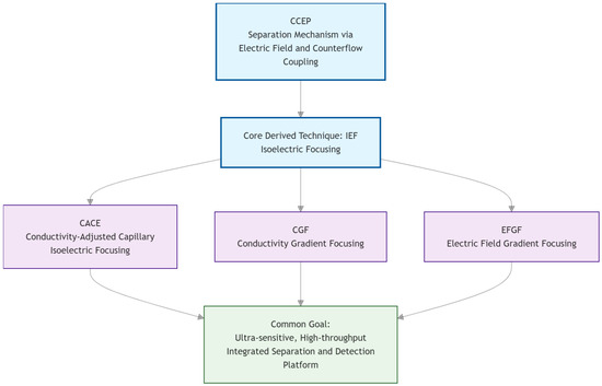
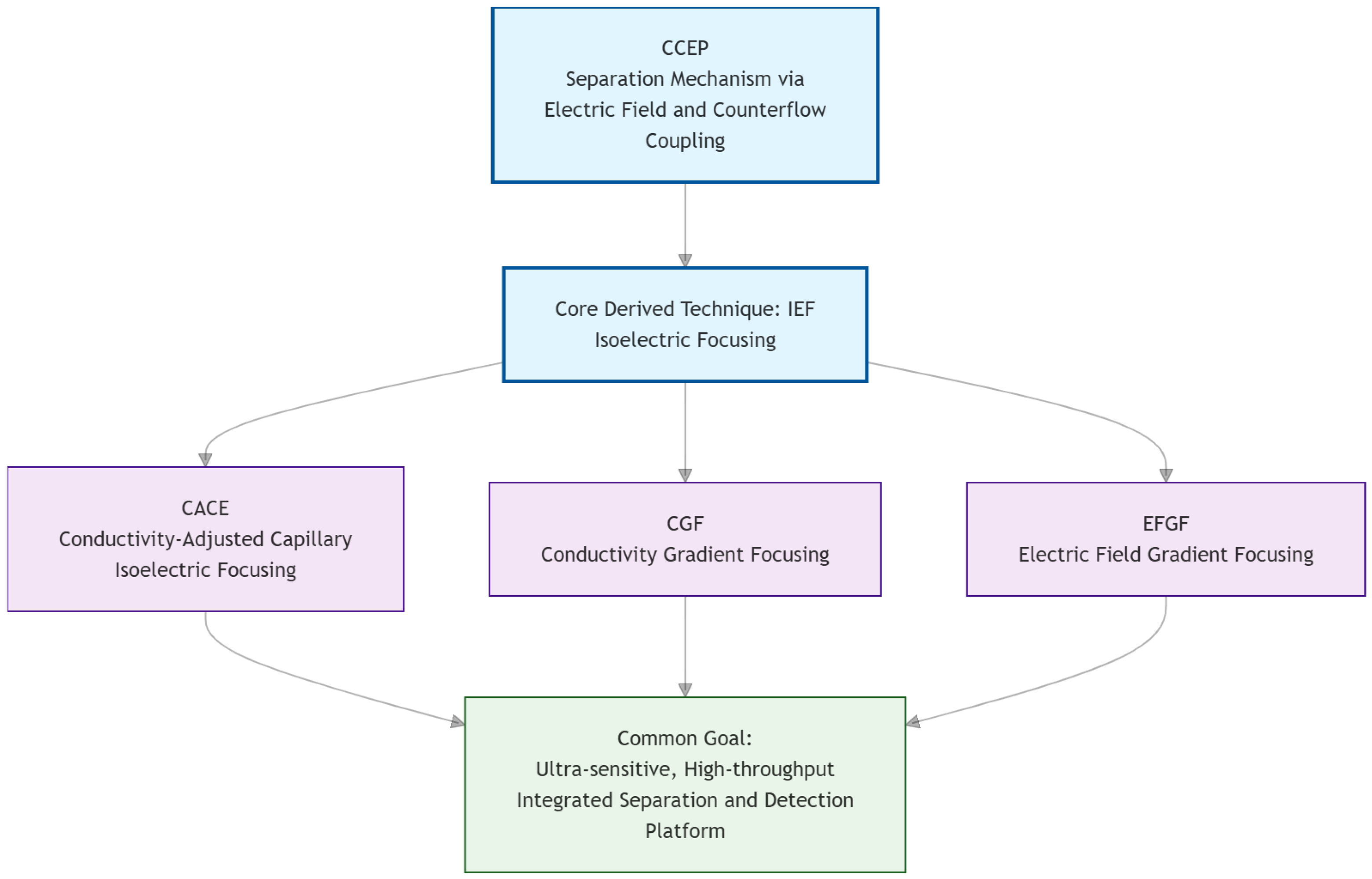
Based on CCEP as the core, three different focusing methods were derived through isoelectric focusing technology, CACE (conductance regulated capillary isoelectric focusing), CGF (conductance gradient focusing), EFGF (electric field gradient focusing), as shown in Figure 2. Take CACE as an example. Generally, the main factors affecting the charge of the sample are the chemical composition of the material and the pH value of the buffer solution. For example, when separating different proteins, if the buffer pH deviates from the isoelectric point (PI) of the protein, the protein will obtain the corresponding charge. In particular, when pH is higher than PI, the dissociated group of protein deprotonates and adsorbs OH- ions, resulting in negative charge; On the contrary, when the pH value is lower than PI, the protein adsorbs H+ and generates a positive charge. The working principle of conductance regulated isoelectric focusing (CACE) theory is to make proteins migrate in a stable pH gradient until they reach the isoelectric point (PI), at which time their net charge becomes zero, so as to realize focusing. However, due to the difficulty in establishing and maintaining a stable artificial pH gradient, its initial development faces challenges.
Figure 2.
Derivation relationship diagram of CCEP technology system.
2.2. Development of Device Design
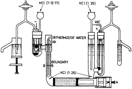
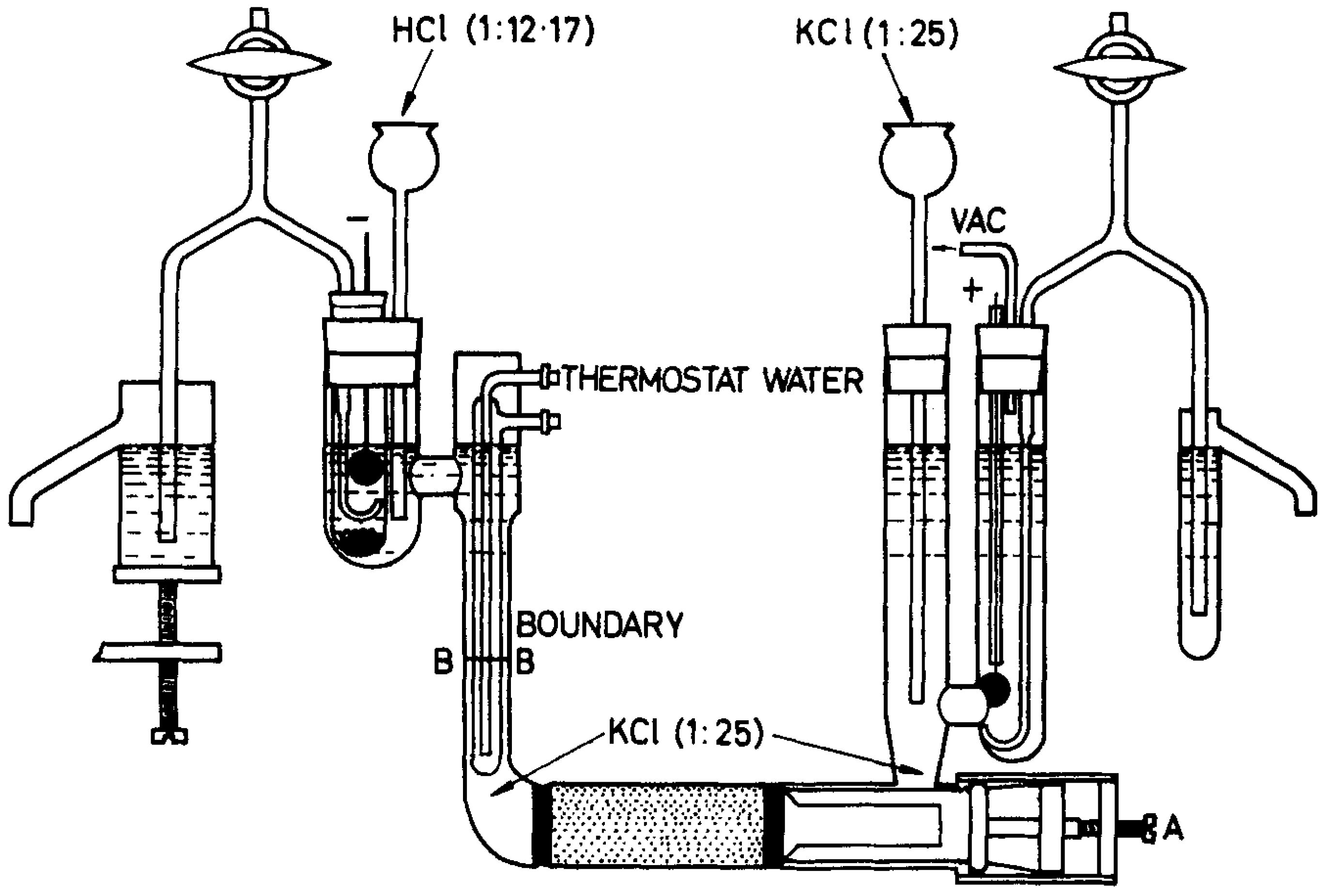
In 1946, Brewer [17] first developed ion migration as a continuous countercurrent separation device. Using countercurrent electrophoresis, they achieved the separation of mixtures containing potassium, chlorine, and copper isotopes. A representative Plexiglas device for potassium isotope enrichment is depicted in Figure 3. However, separation performance was frequently compromised by diffusion and convection. While increasing the electric field strength could mitigate back-diffusion, convection arising from local concentration and temperature gradients proved difficult to suppress. When fillers such as sand, silicon carbide, glass beads, asbestos wool, plates, or capillaries were introduced to curb convection, new challenges emerged: uneven porosity and chemical erosion could lead to bubble formation that disrupted flow, while excessive Joule heating risked causing device deformation.
Figure 3.
Device for enriching potassium isotopes. The anode compartment is continuously flushed with natural KC1 solution. The feed of HCI solution into the cathode compartment is controlled in such a way that the boundary B-B remains constant [17].
Clusius and colleagues [18] achieved a critical breakthrough by adopting a novel strategy that eliminated the need for column packing or active suppression of convective flows. Their apparatus was thus redesigned to restrict convection strictly perpendicular to the field through the incorporation of multiple membranes. It consisted of a main separation tube (20 mm in diameter, 40–100 cm in length) with an axially positioned, 5 mm internal cooling tube for water circulation. Dialysis paper membranes were placed at intervals of 2.5–5 mm along the tube. This architecture facilitated rapid electrolyte concentration equilibration and efficient heat dissipation, while circumventing complications such as channeling and bubble-driven instability. Meanwhile, Martin [19] independently established a countercurrent process in which electrolysis took place within a packed column located between anode and cathode chambers that featured reservoir-like configurations. The control system demanded by this approach, however, proved more complex than that employed in the Brewer and Clusius setups.
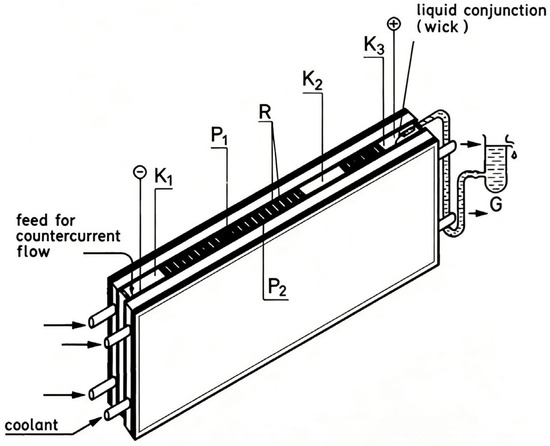
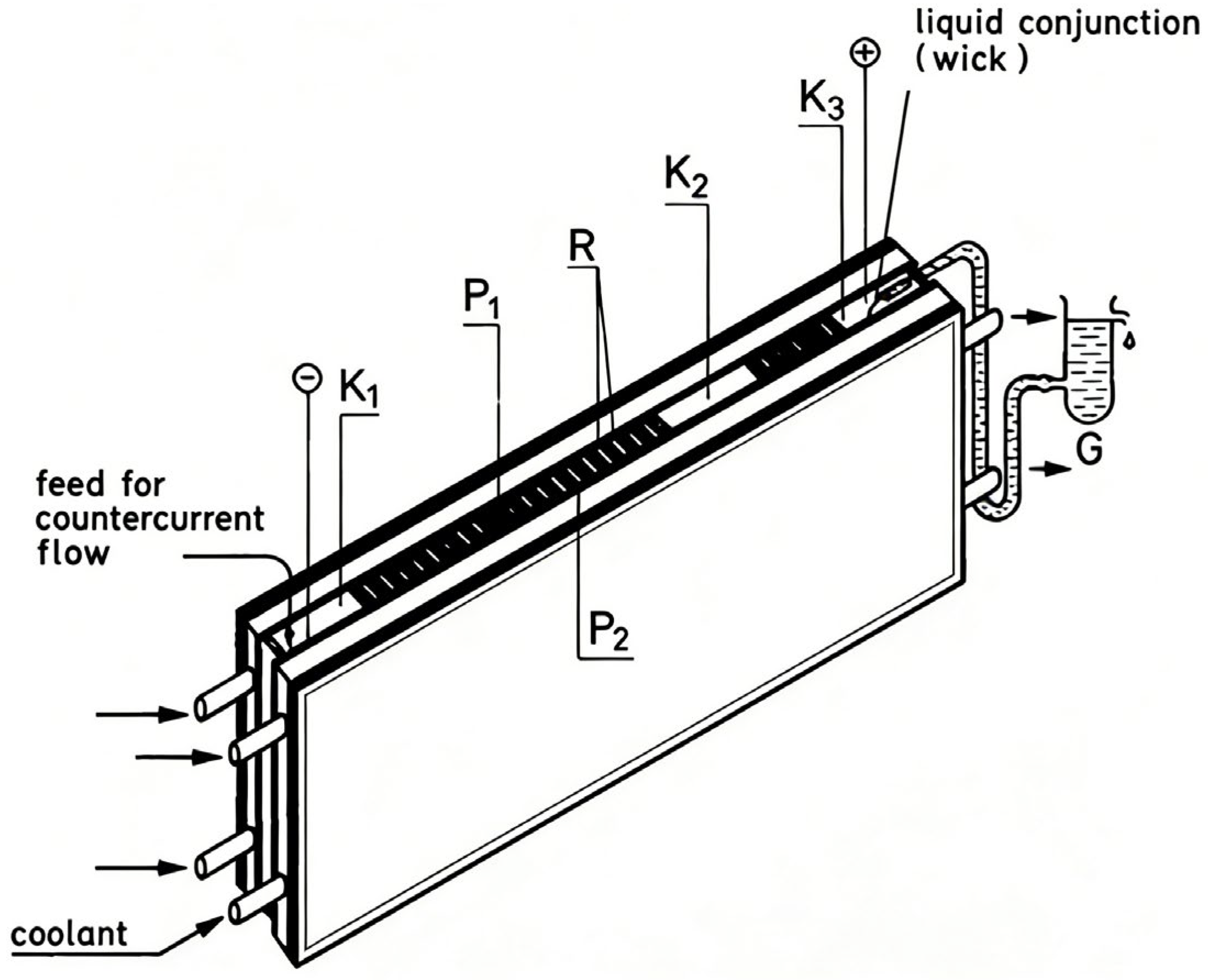
The vertical slit chamber represents a nearly ideal synthesis of the beneficial characteristics of both electrolytic cells and capillaries. As employed by Bilal [20], the chamber consists of two double-walled oxidized ceramic plates cooled by circulating water, with membranes positioned in grooves (Figure 4). The specific parameters and corresponding performance improvements are shown in Table 1. The overall column measures 80 cm in length and 2 mm in width, incorporates 380 membranes, and features a packing height of approximately 5 cm. Operating at a flow rate of 6 mL/h and a current of 200–300 mA under 3000 V, the design achieves excellent cooling performance and sustains stable liquid flow via capillary action, all while providing a relatively large cross-sectional area. Furthermore, unlike in capillary systems, bubble formation does not disrupt the process. To achieve enhanced thermal conductivity, the walls are constructed from oxidized ceramic. In a separate development, Aasted [21] proposed a method to suppress precipitation in countercurrent electrophoresis. Compared to standard (direct) countercurrent electrophoresis, this technique enhances the detection sensitivity for ADV antibodies by at least 32-fold.
Figure 4.
Vertical slit separation device with diaphragm. Two double-walled plates of oxide ceramics (flushed with cooling water) form the vertical chamber. The diaphragms are sitting in the grooves. The electrodes are in the compartments Kl and K3; K2 being an intermediate compartment [20].
Table 1.
Parameters and performance analysis of countercurrent electrophoresis device in vertical slit cavity.
In continuous-flow electrophoresis systems, temperature gradients induced by Joule heating can lead to both stable and unstable convection, thereby compromising separation efficiency. Ostrach [22] conducted a systematic study of this phenomenon, establishing relevant theoretical models and design principles, and elucidated the combined influence of buffer flow velocity, wall cooling, and electric field strength on fluid dynamics. To address the stability challenges arising from such thermal disturbances, membrane fouling, and the complexity of multi-parameter dynamic equilibrium, synergistic innovations across three dimensions—materials, design, and control—are required. At the materials level, constructing permanent covalent coatings (such as cationic copolymer coatings [23] or polypyrrole coatings [24]) is key to enhancing interfacial stability and regulating electroosmotic flow. At the design level, optimizing the flow channel structure is crucial; for example, integrating precisely machined microporous membrane structures can serve as effective spacers to stabilize the separation interface and enable online preconcentration [25]. At the control level, integrating real-time sensors with advanced algorithms is central to achieving precise regulation. Techniques such as machine learning-based model predictive control [26] can effectively achieve adaptive compensation for disturbances, including thermal convection.
A key trend in microfluidics is to enhance separation efficiency through device simplification. The work by Vyas [27] exemplifies this trend by integrating continuous sample injection with a pressure-tunable hydrodynamic counterflow within an extremely short microcolumn (1 cm to 1 mm), as shown in Figure 5. Separation occurs at the sample-microcolumn interface through counterflow scanning. This design eliminates the need for complex injection structures and long separation channels, and has been successfully applied to immunoassays, enzyme kinetics monitoring, and direct ion analysis in complex matrices such as milk and soil. Recent research has further advanced this technology: Liu [28] integrated an on-chip micropump that generates pressure-driven backflow, enabling counterflow regulation without external pumps and pushing device simplification to a new level. Meanwhile, Gebhard [29] provided an in-depth mechanistic exploration of the accumulation, separation, and mixing of charged species using microfluidic electrophoresis, expanding its potential for precise interfacial control. These developments collectively indicate that countercurrent electrophoresis is steadily evolving toward higher integration, deeper mechanistic understanding, and broader practical applicability.
Figure 5.
Design principle of microfluidic device. When the overall flow rate is greater than the electrophoresis speed of the sample analyte, as the applied pressure decreases (at the bottom), the migration rate of the analyte is large enough to enter the micro column, where it can be detected by optical means or by changes in total current [27].
The core experimental design of countercurrent electrophoresis (CCEP) lies in precisely constructing opposing electric field and hydrodynamic forces. By balancing these two forces, components with different electrophoretic mobilities can achieve a net velocity difference, enabling separation. Key parameters such as electric field strength and buffer flow rate require precise control, while disturbances to separation stability from thermal convection and diffusion must be overcome. Its development has formed a complete trajectory from foundational to applied research: a 1967 paper in Nature laid the groundwork for its protein separation apparatus [9]; a 1989 review systematically elaborated theoretical models and proposed an “accumulation criterion” to guide experimental design [30]; subsequent studies delved into practical issues such as axial dispersion [31] and designed user-friendly new devices like the “J-Column” to promote real-world applications. In recent years, its principles have been extended to analytical chemistry pretreatment, exemplified by a 2026 study in Analytical Chemistry that developed it into an efficient sample preparation technique [32]. Notably, the concept of “separation within opposing force fields” inherent in CCEP directly inspired the development of isoelectric focusing (IEF). By establishing a pH gradient to “focus” proteins at their isoelectric points, IEF pushed analytical resolution to its extreme by sacrificing continuous flow and preparative throughput, thereby securing a central position in proteomics.
2.3. Application
2.3.1. Isotope Separation
Several research groups have now explored the feasibility of electrophoretic techniques for lithium isotope separation. Zhang and colleagues [33,34] applied approximately ~6 V across electrodes and demonstrated that electric field-driven separation achieves significantly higher lithium isotope separation efficiency compared to diffusion-only processes, with 6Li accumulating in the cathode chamber. Using computational modeling, they further investigated how electrolysis parameters affect isotopic separation. Their experimental results revealed that separation efficiency depends not only on duration but also on factors including the initial concentration of lithium salts. Building on this foundation, Acosta [35] employed simulation tools to evaluate different electrolytic methods for lithium isotope separation. Their study quantified separation performance under continuous operation and identified that the presence of gallium reduces energy consumption while enhancing lithium isotope fractionation.
Arkenbout [36] systematically developed a countercurrent electrophoresis process for separating lithium isotopes (6Li and 7Li). They introduced an innovative sand-packed column (particle size 0.35–0.5 mm) as the contact device, effectively overcoming limitations of earlier empty columns such as limited interfacial area, short residence time, and channeling effects. By pre-filling the column with mercury and subsequently replacing part of it with electrolyte, stable countercurrent contact between the mercury and electrolyte phases was achieved. After 20–40 h of operation in the 1–2 m long column, the 6Li abundance increased from the natural value of 7.5% to 13% at the bottom while decreasing to 5.3% at the top. This process has achieved an effective separation factor of about 1.5–1.8, demonstrating the method’s effective separation capability. To date, electrophoretic separation of lithium isotopes remains in the exploratory stage, with current research primarily focusing on weak electric field effects and reporting only single-stage separation factors under discontinuous operation.
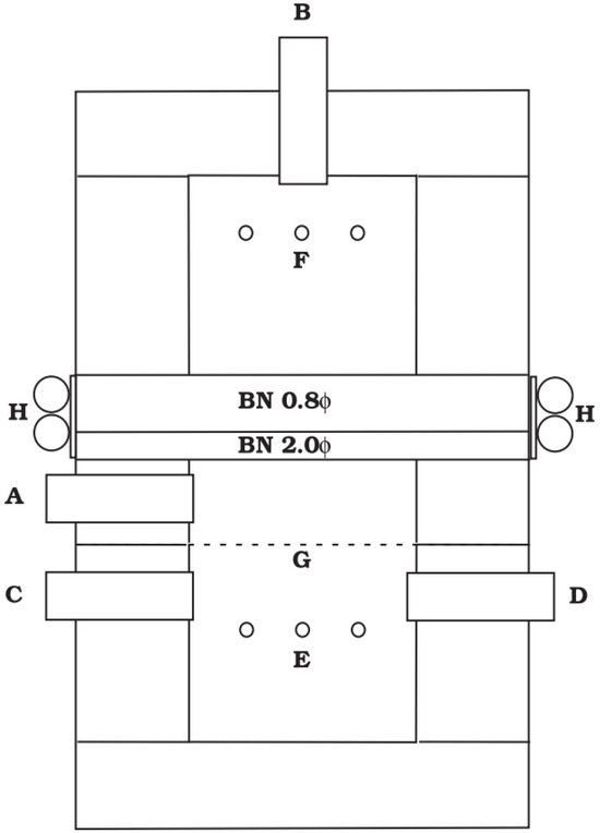
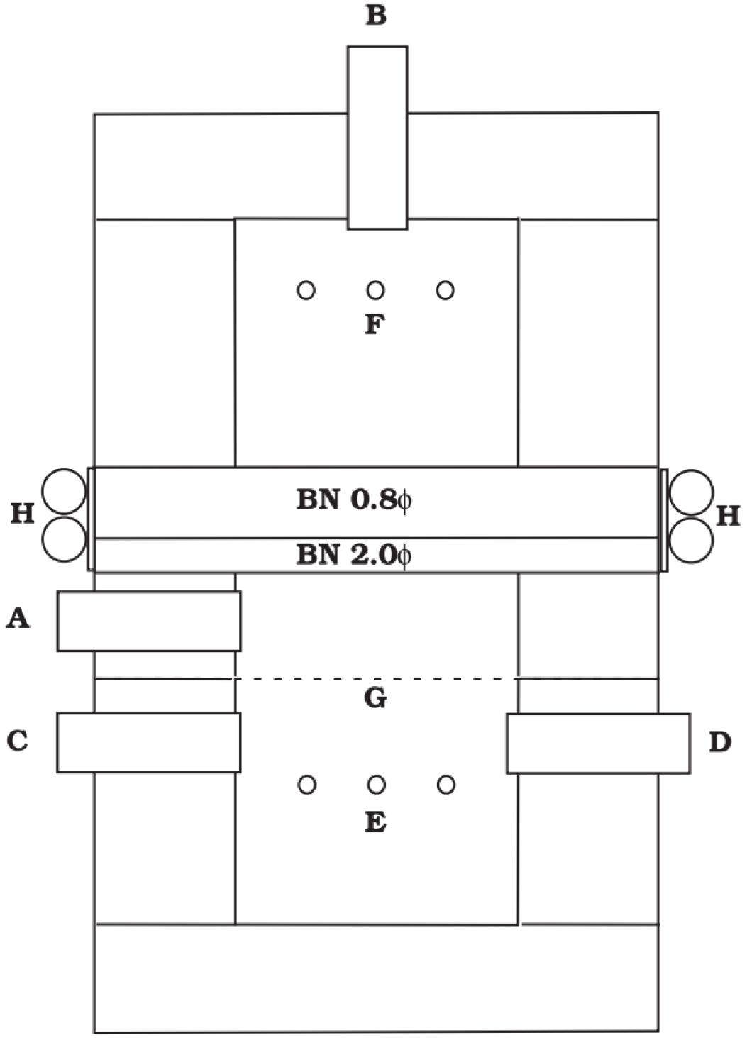
Kishimoto [37] employed a multichannel countercurrent electrophoresis system for calcium isotope enrichment, as shown in Figure 6. By monitoring variations in the 40Ca/48Ca abundance ratio, they determined that 48Ca was enriched relative to 40Ca by a separation factor of 6, which represents a highly significant outcome. A notable finding was that using a peristaltic pump capable of generating pulsed flow, rather than a syringe pump providing steady flow, proved critical for achieving high enrichment. The authors explained that under low Reynolds number conditions, laminar (Hagen-Poiseuille) flow produces a parabolic velocity profile in the channels. This profile tends to obscure separation caused by slight differences in isotopic mobility. In contrast, pulsed or spatially designed oscillatory flow effectively flattens the flow front, resulting in clearer separation.
Figure 6.
Schematic diagram of enrichment device. Made of acrylic resin: A and B are the inlet and outlet of calcium chloride aqueous solution, respectively; HCl aqueous solution is circulated through C and D cycles; E is the cathode; F is an anode made of platinum; G is a cation exchange membrane; H is the copper pipe for cooling water [37].
Table 2 compares CCEP with other mainstream separation technologies. CCEP is more environmentally friendly and safer to operate because it works under mild, room-temperature conditions. Meanwhile, CCEP does have practical limitations, especially in isotope separation. Historically, it involved complex systems, large equipment, slow start-up, high operational demands, and high costs. This led to poor energy efficiency and scalability, hindering large-scale industrial use. These limitations stem from engineering challenges of the past, not a flaw in the core principle. Today, with advances in modern materials (like new membranes and efficient electrodes), precision fluid control, and automation, these issues of complexity, stability, and cost can be re-evaluated and potentially overcome.
Table 2.
Comparison of CCEP with modern mainstream separation technologies.
2.3.2. Protein Separation
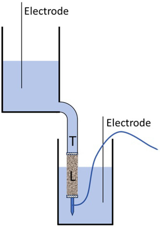
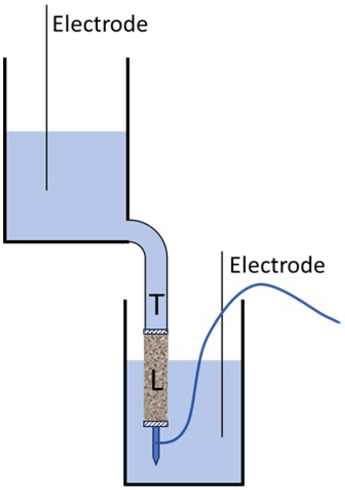
In recent years, the theory of isoelectric focusing (IEF), derived from the principles of countercurrent electrophoresis, has been proposed for simultaneous separation of mul-tiple proteins, with key results summarized in Table 3. Ölverstedt [42] developed an efficient and low loss protein recovery and concentration method, which represents the early and important exploration of protein preparation countercurrent technology. The author uses the rotating quartz tube device originally designed by Hjertén [43], and the instrument is shown in Figure 7, which effectively alleviates the convective disturbance. In addition, coating methylcellulose on the tube wall is very effective in inhibiting electroosmosis. Finally, the method successfully improved the recovery rates of diluted phycoerythrin and hemoglobin to 90% and 93%. However, this method is more suitable for the separation of only two components.
Table 3.
Summary of research results on protein separation.
Figure 7.
A device for protein recovery uses a prepacked Sephadex G-25 column placed between beakers. A T-tube attached to the column’s outlet applies counterflow, with its end sealed by a dialysis membrane. L (leading) and T (terminating) solutions define the initial setup [42].
Locke and Carbonell [30] conducted extensive experimental and theoretical investigations in this field. More recently, in 1998, Chidambara [50] utilized the unique geometry of a J-shaped column to remove electrolytic products from the buffer, thereby achieving more stable separation over several hours of experimentation. Although CACE has been demonstrated to achieve high concentration factors for specific proteins, its application to the separation of protein mixtures is challenging. These practical constraints ultimately precluded its broad adoption. However, its core principle—leveraging counterflow for electrophoretic focusing—served as a conceptual precursor to numerous later focusing techniques.
One of these novel approaches, conductivity gradient focusing (CGF), has been explored in studies [51]. This method utilizes an axial conductivity gradient along the separation column to induce corresponding gradients in electric field strength and electrophoretic velocity. Their apparatus consisted of channels fabricated in a plastic substrate, transversely segmented by dialysis membranes. A high-conductivity buffer was pumped through channels on one side of the membrane, while a low-conductivity buffer was circulated on the opposite side. Ionic diffusion across the membrane established a continuous conductivity gradient along the channel length. Application of an electric field then enabled focusing and separation. Initially, Greenlee and Ivory observed only moderate resolution for protein mixtures containing BSA and hemoglobin. However, by incorporating chromatographic packing materials to suppress vortices and Taylor [52] dispersion in the relatively large channels (1.6 mm × 60.5 mm cross-section), high-resolution separations were achieved. Wang’s research group [53] further refined this methodology by implementing a dialysis tube coaxially fed through a capillary. Subsequent improvements enabled 15,000-fold enrichment of BSA within 4 h and successful separation of ferritin from various BSA isoforms [54].
A further method entails creating electric field gradients more directly in systems of constant conductivity, achieved by varying the current along the separation channel. Implementation requires dialysis membranes or comparable semi-permeable barriers that enable buffer ions to transport current while blocking the migration of the proteins undergoing focusing. This methodology is typically referred to as electric field gradient focusing (EFGF). Koegler and Ivory [55,56] constructed the first EFGF device, which utilized a tapered plexiglass cylinder to generate an electric field gradient and maintained counterflow through a hollow dialysis tube. This device successfully separated different oxidation states of myoglobin, but both resolution and enrichment capacity were limited (only 3-fold enhancement over several hours). Huang and Ivory [57] achieved a major breakthrough by replacing the variable-cross-section design with an electrode array (50 electrodes) to enable real-time control of a nonlinear electric field, accomplishing high-resolution separation of four proteins within 30 min in an 8 cm plastic channel packed with chromatographic media. Subsequently, Myers and Bartle [58] further simplified the setup by using only five electrodes to control a channel filled with PolyHIPE monolithic material, efficiently separating six proteins in 15 min. To advance the integration of the technology, Petsev [59] embedded two independently voltage-controlled dialysis tubes in a PDMS microfluidic chip and successfully demonstrated the focusing and separation of R-phycoerythrin and FITC-labeled bovine serum albumin, showcasing the potential of EFGF technology in miniaturized platforms.
The Ramsey group [60] developed an innovative microfluidic implementation of EFGF. The device incorporates a porous silicate layer—formed in situ during cover-plate bonding via a spin-on glass technique—as a semi-permeable interface extending along the channel, analogous to the approach described by Khandurina [25]. This design enables electrical coupling between an adjacent tapered “field-control” channel and the focusing channel, allowing effective focusing of both small-molecule dyes (sulforhodamine B and dichlorofluorescein) and larger proteins. On the theoretical side, Tolley and Warnick established a systematic model for EFGF [61,62]. Building on this framework, Humble [63] designed and fabricated a miniaturized device that employs a shaped ion-conductive membrane. This system achieved a 10,000-fold enrichment of green fluorescent protein (GFP) within 40 min, and successfully resolved a mixture of GFP, R-phycoerythrin (R-PE), FITC-labeled hemoglobin, and Oregon Green-labeled lysozyme with high resolution. Using a line-embossing fabrication approach, the Wooley team [64] produced a device that attained 150-fold enrichment of protein/peptide mixtures in only 10 min of focusing time. In a separate design, Liu [65] integrated a separation membrane via a weir structure, reaching 4000-fold enrichment of GFP within 60 min. Most recently, Lin [66] reported a tandem EFGF system that connects a dialysis-tube-based EFGF unit in series with a hydrogel-based EFGF channel. This configuration enabled effective desalination and selective separation of myoglobin from a complex matrix containing lysozyme and 0.2 mol/L NaCl.
The evolution from countercurrent electrophoresis to isoelectric focusing marks a fundamental paradigm shift, moving from separation techniques dependent on dynamic equilibrium to focusing methods that utilize intrinsic molecular properties. Countercurrent electrophoresis, as an early experimental approach, achieved separation by opposing buffer flow against electrophoretic migration, yet it was constrained by poor stability and limited resolution. This technological limitation persisted until the development of synthetic carrier ampholytes. These compounds can spontaneously form stable linear pH gradients under an electric field, enabling the practical implementation of isoelectric focusing. This advancement ultimately led to core proteomic technologies such as two-dimensional electrophoresis, completing the transition from rudimentary separation to precise analytical methods.
3. The Advantages of CCEP
3.1. High Sensitivity
In conventional migration-based separation techniques such as capillary electrophoresis (CE) or high-performance liquid chromatography (HPLC), the core mechanism relies on the differential migration velocities of analytes as they move through a separation channel. However, this process is inevitably accompanied by band broadening due to diffusion and uneven mass transfer, leading to signal dilution and thus limiting sensitivity enhancement. In stark contrast, focusing techniques represented by CCEP and IEF operate on the principle of establishing a balanced force field (e.g., a stable pH gradient) that drives different analytes to migrate toward and accumulate at specific positions where their net charge is zero (e.g., the isoelectric point, pI), until a dynamic equilibrium is reached [67]. This fundamental mechanistic difference directly dictates the evolution of analyte peaks: in focusing, analytes continuously concentrate at the focal point, resulting in progressively narrowing peak widths and increasing peak heights; whereas in migration, peaks typically broaden and disperse over time. Consequently, focusing technology inherently integrates high-resolution separation with analyte concentration, elevating the local concentration of analytes to tens or even hundreds of times that of the original sample, thereby providing significant gains in detection. Experimental evidence shows that this effect directly translates into extremely high analytical sensitivity and resolution. For example, in the immunoassay for meat species identification, countercurrent immunoelectrophoresis, which leverages the opposing electrophoretic migration to concentrate antigens and antibodies, demonstrates a sensitivity approximately 2.75 times higher than that of traditional double immunodiffusion [68]. In the field of biopharmaceutical analysis, modern imaging capillary isoelectric focusing (icIEF) has become the “gold standard” for analyzing protein charge heterogeneity, achieving a resolution of up to 0.01 pH units and enabling precise separation of protein variants with minimal differences in isoelectric point [69]. Thus, focusing separation is not merely a separation method but also an effective online enrichment strategy, granting it a distinct advantage over conventional migration-based methods for detecting trace components and complex samples.
3.2. Separate High Salt Components
The analysis of biological samples containing high-salt or complex protein matrices poses distinct challenges for conventional capillary electrophoresis (CE). Protein adsorption onto the capillary wall alters electroosmotic flow and impairs reproducibility, typically requiring dedicated coating strategies to mitigate these effects [70,71]. Meanwhile, high ionic strength can easily induce zone “destacking” during injection, leading to reduced resolution and sensitivity, thus often requiring additional online preconcentration or desalting steps such as transient isotachophoresis (tITP) [5,6]. In sharp contrast, focusing electrophoresis techniques, through their inherent active concentration and stable focusing mechanisms, demonstrate superior matrix tolerance and functional integration. Their empirical advantages are concrete and significant: for instance, they can achieve online desalting and purification of target proteins (e.g., BSA) directly from cell culture media with an ionic strength as high as 2 M NaCl [54]; in chiral drug analysis, effective separation can be accomplished directly by merely performing a 1:1 dilution of complex urine samples with buffer, bypassing cumbersome pretreatment [72]. These examples highlight how focusing methods seamlessly combine high-resolution separation, analyte concentration, and matrix clean-up into a single continuous process, offering a more streamlined and robust workflow for complex real-world samples compared to conventional migration-based separations.
3.3. Integrated and Miniaturized Design
Although capillary electrophoresis (CE) is a well-established microfluidic analytical platform, its miniaturization presents inherent challenges. Attaining high resolution typically demands extended separation lengths, which are difficult to integrate on-chip, while the use of serpentine channels to conserve footprint introduces band-broadening through the “racetrack effect.” Counterflow focusing addresses this limitation by employing a fundamentally distinct separation mechanism [73]. By localizing analytes at stable equilibrium positions rather than relying on extended migration paths, the required channel length is reduced to the spatial width of the focused zones, enabling high-efficiency separations within dramatically shortened channels [74]. This advantage is quantified experimentally: thermal gradient focusing conducted in channels as short as 2 mm and 5 mm has been shown to deliver resolution equivalent to that of conventional CE performed in a 50-cm-long capillary [75,76]. An additional critical benefit is that focusing-based separation does not depend on precise, narrow sample injection. Regardless of the initial analyte distribution, all species migrate toward their respective zero-velocity points, converging into sharp, diffusion-limited peaks. Consequently, the need for dedicated injection reservoirs, complex channel networks, or multi-electrode high-voltage switching is eliminated [77]. In its simplest implementation, the separation device can be reduced to a single straight microchannel or a short capillary segment, greatly simplifying microfluidic chip design and operation.
4. Conclusions
Countercurrent electrophoresis (CCEP) achieves separation and enrichment through the synergistic interaction between electric and counterflow fields, with its development clearly progressing from macroscopic setups toward integrated microfluidic platforms. This technology eliminates the dependence on lengthy separation channels by establishing force field equilibrium points over short distances, combining the potential for high throughput with high-resolution separation. This grants it unique value in areas such as protein purification and the separation of critical isotopes.
However, significant challenges hinder the practical application of CCEP. The system is complex and suffers from stability issues: maintaining precise dynamic balance among multiple parameters—electric field, flow rate, and temperature—is difficult, and minor thermal disturbances or membrane contamination can easily degrade separation efficiency. Miniaturized systems also face a trade-off between throughput and sensitivity: microchannels have limited sample capacity, while high-resolution focusing often requires extended processing time. Furthermore, the high technical threshold and cost—relying on high-performance membrane materials, precision fluid control, and high-sensitivity detection—limit its widespread adoption. Overcoming these bottlenecks requires multidisciplinary collaborative innovation. The key lies in developing novel anti-adsorption materials and intelligent microfluidic systems that integrate online sensing and adaptive control algorithms to achieve operational stability and automation. Simultaneously, CCEP can serve as an efficient pre-processing unit coupled online with analytical techniques like mass spectrometry to form integrated solutions, enhancing the analysis of complex samples.
Currently, CCEP presents significant development opportunities. In the energy sector, it can serve as a green alternative technology for separating critical isotopes. In continuous biopharmaceutical manufacturing, it holds promise for integration into downstream purification processes. In cutting-edge research such as single-cell omics, its online enrichment capability offers an ideal interface for ultratrace analysis. Through deep interdisciplinary integration and engineering innovation, CCEP has the potential to evolve from proof-of-concept to practical engineering application, emerging as a core platform technology for high-end separation and enrichment.
Author Contributions
Z.G. proposed research concepts and designs; Responsible for collecting, organizing, and analyzing research data; Write the initial draft of the paper; Participated in multiple revisions and improvements of the manuscript. H.Z. guided the overall direction and design of the research project; Provide critical academic guidance for data analysis; Conduct critical review of the paper manuscript. Meet all author criteria recommended by the International Committee of Medical Journal Editors (ICMJE). All authors have read and agreed to the published version of the manuscript.
Funding
This research received no external funding.
Data Availability Statement
The original contributions presented in this study are included in the article. Further inquiries can be directed to the corresponding author.
Conflicts of Interest
The authors declare no conflicts of interest.
References
- Stroink, T.; Paarlberg, E.; Waterval, J.C.; Bult, A.; Underberg, W.J. On-line sample preconcentration in capillary electrophoresis, focused on the determination of proteins and peptides. Electrophoresis 2001, 22, 2374–2383. [Google Scholar] [CrossRef] [Scilit]
- Breadmore, C.M.; Haddad, R.P. Approaches to enhancing the sensitivity of capillary electrophoresis methods for the determination of inorganic and small organic anions. Electrophoresis 2001, 22, 2464–2489. [Google Scholar] [CrossRef] [Scilit] [PubMed]
- Urbánek, M.; Křivánková, L.; Boček, P. Stacking phenomena in electromigration: From basic principles to practical procedures. Electrophoresis 2003, 24, 466–485. [Google Scholar] [CrossRef] [Scilit] [PubMed]
- Quirino, J.P.; Terabe, S. Sample stacking of cationic and anionic analytes in capillary electrophoresis. J. Chromatogr. A 2000, 902, 119–135. [Google Scholar] [CrossRef] [Scilit]
- Timerbaev, A.R.; Hirokawa, T. Recent advances of transient isotachophoresis capillary electrophoresis in the analysis of small ions from high-conductivity matrices. Electrophoresis 2006, 27, 323–340. [Google Scholar] [CrossRef] [Scilit]
- Gebauer, P.; Boček, P. Recent progress in capillary isotachophoresis. Electrophoresis 2002, 23, 3858–3864. [Google Scholar] [CrossRef] [Scilit]
- Peja, K.; Dobhalov, L.; Fidler, Z.; Dvoak, J. Countercurrent Electrophoresis on Paper. J. Chromatogr. A 1964, 14, 464–468. [Google Scholar] [CrossRef] [Scilit]
- Carpenter, F.H.; Hayes, S.L. Electrophoresis on Cellulose Acetate of Insulin and Insulin Derivatives: Correlation with Behavior on Countercurrent Distribution and Partition-Column Chromatography. Biochemistry 1963, 2, 1272–1277. [Google Scholar] [CrossRef] [Scilit]
- Broome, J.; Raymond, S. Countercurrent electrophoresis of proteins. Nature 1967, 214, 849–850. [Google Scholar] [CrossRef] [Scilit]
- Deshmukh, R.R.; Bier, M. Counterflow in isotachophoresis: Computer simulation and experimental studies. Electrophoresis 1993, 14, 205–213. [Google Scholar] [CrossRef] [Scilit]
- Shimura, K. Recent advances in capillary isoelectric focusing: 1997–2001. Electrophoresis 2002, 23, 3847–3857. [Google Scholar] [CrossRef] [Scilit] [PubMed]
- Silvertand, L.H.H.; Torano, J.S.; Van Bennekom, W.P.; De Jong, G.J. Recent developments in capillary isoelectric focusing. J. Chromatogr. A 2008, 1204, 157–170. [Google Scholar] [CrossRef] [Scilit] [PubMed]
- Bethune, J.L.; Kegeles, G. Countercurrent distribution of chemically reacting systems. III. Analogs of moving boundary electrophoresis and sedimentation. J. Phys. Chem. 1961, 65, 1761–1764. [Google Scholar] [CrossRef] [Scilit]
- Kodina, G.E.; Korpusov, G.V.; Filyanin, A.T. Production of high-purity 90Y on specially developed centrifugal semicounterflow extractors. Radiochemistry 2002, 44, 64. [Google Scholar] [CrossRef] [Scilit]
- Kanapilly, G.M.; Newton, G.J. A new method for the separation of multicurie quantities of 90Y from 90Sr. Int. Fission Prod. Inhal. Proj. 1969, 11, 75–81. [Google Scholar] [CrossRef] [Scilit]
- Hamaguchi, H.; Ikeda, N.; Kawashima, T. Isolation of Carrier-Free Yttrium from the Radioactive Strontium-Yttrium System by Electrodeposition. Bunseki Kagaku 1958, 7, 243–246. [Google Scholar] [CrossRef] [Scilit]
- Brewer, A.K.; Madorsky, S.L.; Taylor, J.K.; Dibeler, V.H.; Bradt, P.; Parham, O.L.; Britten, R.J.; Gilman, J. Concentration of Isotopes of Potassium by the Counter-current Electromigration Method. J. Res. Natl. Bur. Stand. 1947, 38, 137. [Google Scholar] [CrossRef] [Scilit]
- Clusius, K.; Ramirez, E.R. Zur Trennung der seltenen Erden in wässeriger Lösung durch Ionenwanderung. Helv. Chim. Acta 1953, 36, 1160–1165. [Google Scholar] [CrossRef] [Scilit]
- Martin, H.; Luebke, H.J. Frage Des Masseneffekts Bei der Lonenwanderung in Lösungen. Z. Naturforschung A 1964, 19, 115–126. [Google Scholar] [CrossRef] [Scilit]
- Bilal, B.A. Gegenstromelektrolytische Stofftrennung in dünnen Flüssigkeitsschichten. Z. Naturforschung A 1969, 24, 1791–1794. [Google Scholar] [CrossRef] [Scilit]
- Aasted, B.; Cohn, A. Inhibition of precipitation in counter current electrophoresis. A sensitive method for detection of mink antibodies to Aleutian disease virus. Acta Pathol. Microbiol. Scand. Ser. C Immunol. 1982, 90, 15–19. [Google Scholar] [CrossRef] [Scilit] [PubMed]
- Ostrach, S. Convection in continuous-flow electrophoresis. J. Chromatogr. A 1977, 140, 187–195. [Google Scholar] [CrossRef] [Scilit]
- Shibukawa, M. Multistep pH-Peak-Focusing Countercurrent Chromatography. In Polymer-Based Aqueous-Two Phase Systems: Fundamentals and Applications; Springer: Berlin/Heidelberg, Germany, 2025; pp. 87–105. [Google Scholar] [CrossRef] [Scilit]
- Knob, R.; Gerstmann, S.; Čabala, R.; Frank, H. Polypyrrole-Coated and Polysulfate-Modified CE Capillaries. Chromatographia 2009, 69, 1431–1435. [Google Scholar] [CrossRef] [Scilit]
- Khandurina, J.; Jacobson, S.C.; Waters, L.C.; Foote, R.S.; Ramsey, J.M. Microfabricated porous membrane structure for sample concentration and electrophoretic analysis. Anal. Chem. 1999, 71, 1815–1819. [Google Scholar] [CrossRef] [Scilit]
- Müller-Späth, T.; Aumann, L.; Melter, L.; Ströhlein, G.; Morbidelli, M. Chromatographic separation of three monoclonal antibody variants using multicolumn countercurrent solvent gradient purification (MCSGP). Biotechnol. Bioeng. 2008, 100, 1166–1177. [Google Scholar] [CrossRef] [Scilit]
- Vyas, C.A.; Flanigan, P.M.; Shackman, J.G. Gradient counterflow electrophoresis methods for bioanalysis. Bioanalysis 2010, 2, 815–827. [Google Scholar] [CrossRef] [Scilit]
- Liu, Y.; Xia, L.; Xiao, X.; Li, G. An integrated plastic microchip for enhancing electrophoretic separation using tunable pressure-driven backflows. Electrophoresis 2022, 43, 892–900. [Google Scholar] [CrossRef] [Scilit]
- Gebhard, F. Investigations Toward the Accumulation, Separation, Mixing and Detection of Charged Species Using Microfluidic Electrophoresis. Ph.D. Thesis, Technische Universität Darmstadt, Darmstadt, Germany, 2024. [Google Scholar]
- Locke, B.R.; Carbonell, R.G. A theoretical and experimental study of counteracting chromatographic electrophoresis. Sep. Purif. Methods 1989, 18, 1–64. [Google Scholar] [CrossRef] [Scilit]
- Baratuci, W.B. Counteracting Flow Electrophoresis. Ph.D. Thesis, Case Western Reserve University, Cleveland, OH, USA, 1991. [Google Scholar]
- Ye, L.; Lin, J.; Huang, S.; Huang, X.; Zhang, J.; Xu, H.; Peng, R.; Du, J.; Pan, J. Overcoming Field-Amplification Decay: A Counterflow Electroextraction Strategy for Direct, Fast, and Exhaustive Enrichment of Per- and Polyfluoroalkyl Acids from Cosmetic Matrices. Anal. Chem. 2026. [Google Scholar] [CrossRef] [Scilit]
- Zhang, Z.; Sarswat, P.K.; Free, M.L. Li isotopes concentration flux investigation under conditions of diffusion and electric field assisted migration. Vacuum 2018, 152, 291–300. [Google Scholar] [CrossRef] [Scilit]
- Zhang, Z.; Murali, A.; Sarswat, P.K.; Free, M.L. High-efficiency lithium isotope separation by electrochemical deposition and intercalation with electrochemical isotope effect in propylene carbonate and [BMIM][DCA] ionic liquid. Electrochim. Acta 2020, 361, 137060. [Google Scholar] [CrossRef] [Scilit]
- Acosta, L.N.; Flexer, V. A First Assessment on the Scale-Up Possibilities of Different Electrochemical Techniques for Lithium Isotopic Enrichment. Ind. Eng. Chem. Res. 2018, 57, 11399–11413. [Google Scholar] [CrossRef] [Scilit]
- Arkenbout, G.J.; Boerboom, A.J.; Smit, W.M. Separation of lithium isotopes by countercurrent-exchange-electrolysis. Berichte Bunsenges. Phys. Chem. 1966, 70, 658–664. [Google Scholar] [CrossRef] [Scilit]
- Kishimoto, T.; Matsuoka, K.; Fukumoto, T.; Umehara, S. Calcium isotope enrichment by means of multi-channel counter-current electrophoresis for the study of particle and nuclear physics. Prog. Theor. Exp. Phys. 2015, 2015, 033D03. [Google Scholar] [CrossRef] [Scilit]
- Li, H. Current status and development trend of carbon, nitrogen and oxygen stable isotope production technology. Isotopes 2011, 24, 7–14. [Google Scholar]
- Chai, J.; Li, Z. Polarization selectivity experiment for the separation of gadolinium isotopes by atomic vapor laser. Acta Opt. Sin. 2009, 29, 1596–1600. [Google Scholar] [CrossRef] [Scilit]
- Lin, Z.; Tian, Y.; Su, K.; Wang, L. Electromagnetic isotope separation of high-abundance ~6Li. Isotopes 1998, 11, 20–24. [Google Scholar]
- Xie, X.; Wang, L. A review of the preparation of stable isotopes by centrifugation. Isotopes 2019, 32, 186–194. [Google Scholar]
- Öfverstedt, L.; Johansson, G.; Froman, G.; Hjerten, S. Protein concentration and recovery from gel slabs by displacement electrophoresis (isotachophoresis) and the effects of electroosmosis and counter flow. Electrophoresis 1981, 2, 168–173. [Google Scholar] [CrossRef] [Scilit]
- Hjertén, S.; Jerstedt, S.; Tiselius, A. Apparatus for large-scale preparative polyacrylamide gel electrophoresis. Anal. Biochem. 1969, 27, 108–129. [Google Scholar] [CrossRef] [Scilit]
- Liu, H.; Yang, C.; Yang, Q.; Zhang, W.; Zhang, Y. Construction of a two-dimensional protein separation platform using capillary isoelectric focusing/capillary gel free sieving electrophoresis. Anal. Chem. 2004, 3, 273–277. [Google Scholar]
- Xie, Y.; Yin, K.; Luo, A.; Qu, F.; Deng, Y. A novel immobilized pH gradient capillary isoelectric focusing method for protein separation. Anal. Lab. 2007, 11, 25–28. [Google Scholar] [CrossRef]
- Xie, Y.; Geng, L.; Qu, F.; Luo, A.; Deng, Y. Carbonyl imidazole immobilized pH gradient capillary isoelectric focusing electrophoresis for protein separation. Chem. Bull. 2008, 9, 461–464. [Google Scholar] [CrossRef]
- Wei, J.; Gu, X.; Wang, Y.; Yan, C. Application of Capillary Isoelectric Focusing/Pressure Capillary Electrochromatography Two-Dimensional Separation System in Peptide Separation. Anal. Chem. 2011, 39, 188–192. [Google Scholar]
- Zhong, H.; Fan, H.; Yang, P. Liquid Preparation Isoelectric Focusing two—Dimensional gel Electrophoresis for Enrichment, Separation and Identification of Extremely Basic Proteins. In Proceedings of the Third Chinese Proteomics Conference, Beijing, China, 25–28 October 2004. [Google Scholar]
- Li, X.; Zhao, X.; Ying, W.; Qian, X. Isoelectric focusing pre separation for grading and identification of secreted proteome in liver cancer cells. Chromatography 2013, 9, 831–837. [Google Scholar]
- Chidambara Raj, C.B.; Hunter, J.B. Protein purification by counteracting chromatographic electrophoresis—The focusing window. Bioprocess Eng. 1992, 8, 121–128. [Google Scholar] [CrossRef] [Scilit]
- Greenlee, R.D.; Ivory, C.F. Protein focusing in a conductivity gradient. Biotechnol. Prog. 1998, 14, 300–309. [Google Scholar] [CrossRef] [Scilit]
- Taylor, G. Dispersion of soluble matter in solvent flowing slowly through a tube. Proc. R. Soc. Lond. Ser. A Math. Phys. Sci. 1953, 219, 186–203. [Google Scholar] [CrossRef] [Scilit]
- Wang, Q.; Lin, S.L.; Warnick, K.F.; Tolley, H.D.; Lee, M.L. Voltage-controlled separation of proteins by electromobility focusing in a dialysis hollow fiber. J. Chromatogr. A 2003, 985, 455–462. [Google Scholar] [CrossRef] [Scilit]
- Lin, S.L.; Tolley, H.D.; Lee, M.L. Voltage-controlled electric field gradient focusing with online UV detection for analysis of proteins. Chromatographia 2005, 62, 277–281. [Google Scholar] [CrossRef] [Scilit]
- Koegler, W.S.; Ivory, C.F. Focusing proteins in an electric field gradient. J. Chromatogr. A 1996, 726, 229–236. [Google Scholar] [CrossRef] [Scilit]
- Koegler, W.S.; Ivory, C.F. Field gradient focusing: A novel method for protein separation. Biotechnol. Prog. 1996, 12, 822–836. [Google Scholar] [CrossRef] [Scilit]
- Huang, Z.; Ivory, C.F. Digitally controlled electrophoretic focusing. Anal. Chem. 1999, 71, 1628–1632. [Google Scholar] [CrossRef] [Scilit]
- Myers, P.; Bartle, K.D. Towards a miniaturised system for dynamic field gradient focused separation of proteins. J. Chromatogr. A 2004, 1044, 253–258. [Google Scholar] [CrossRef] [Scilit]
- Petsev, D.N.; Lopez, G.P.; Ivory, C.F.; Sibbett, S.S. Microchannel protein separation by electric field gradient focusing. Lab Chip 2005, 5, 587–597. [Google Scholar] [CrossRef] [Scilit]
- Wang, H.Y.; Foote, R.S.; Jacobson, S.C.; Schneibel, J.H.; Ramsey, J.M. Low temperature bonding for microfabrication of chemical analysis devices. Sens. Actuators B Chem. 1997, 45, 199–207. [Google Scholar] [CrossRef] [Scilit]
- Tolley, H.D.; Wang, Q.; LeFebre, D.A.; Lee, M.L. Equilibrium gradient methods with nonlinear field intensity gradient: A theoretical approach. Anal. Chem. 2002, 74, 4456–4463. [Google Scholar] [CrossRef] [Scilit]
- Warnick, K.F.; Francom, S.J.; Humble, P.H.; Kelly, R.T.; Woolley, A.T.; Lee, M.L.; Tolley, H.D. Field gradient electrophoresis. Electrophoresis 2005, 26, 405–414. [Google Scholar] [CrossRef] [Scilit]
- Humble, P.H.; Kelly, R.T.; Woolley, A.T.; Tolley, H.D.; Lee, M.L. Electric field gradient focusing of proteins based on shaped ionically conductive acrylic polymer. Anal. Chem. 2004, 76, 5641–5648. [Google Scholar] [CrossRef] [Scilit]
- Kelly, R.T.; Li, Y.; Woolley, A.T. Phase-changing sacrificial materials for interfacing microfluidics with ion-permeable membranes to create on-chip preconcentrators and electric field gradient focusing microchips. Anal. Chem. 2006, 78, 2565–2570. [Google Scholar] [CrossRef] [Scilit]
- Liu, J.; Sun, X.; Farnsworth, P.B.; Lee, M.L. Fabrication of conductive membrane in a polymeric electric field gradient focusing microdevice. Anal. Chem. 2006, 78, 4654–4662. [Google Scholar] [CrossRef] [Scilit] [PubMed]
- Lin, S.L.; Li, Y.; Tolley, H.D.; Humble, P.H.; Lee, M.L. Tandem electric field gradient focusing system for isolation and concentration of target proteins. J. Chromatogr. A 2006, 1125, 254–262. [Google Scholar] [CrossRef] [Scilit] [PubMed]
- Ghizzani, V.; Ascione, A.; Gonnella, F.; Massolini, G.; Luciani, F. Exploring imaged capillary isoelectric focusing parameters for enhanced charge variants quality control. Front. Chem. 2025, 13, 1536222. [Google Scholar] [CrossRef] [Scilit] [PubMed]
- Winterø, A.K.; Thomsen, P.D.; Davies, W. A comparison of DNA-hybridization, immunodiffusion, countercurrent immunoelectrophoresis and isoelectric focusing for detecting the admixture of pork to beef. Meat Sci. 1990, 27, 75–85. [Google Scholar] [CrossRef] [Scilit]
- Bo, T.; Yang, F.; Yan, B.; Michels, D.A.; Huang, T.; Pawliszyn, J. Recent developments of imaged capillary isoelectric focusing technology for in-depth biopharmaceutical characterization. TrAC Trends Anal. Chem. 2025, 187, 118212. [Google Scholar] [CrossRef] [Scilit]
- Dolník, V. Wall coating for capillary electrophoresis on microchips. Electrophoresis 2004, 25, 3589–3601. [Google Scholar] [CrossRef] [Scilit]
- Dolník, V. Capillary electrophoresis of proteins 2003–2005. Electrophoresis 2006, 27, 126–141. [Google Scholar] [CrossRef] [Scilit]
- Balss, K.M.; Vreeland, W.N.; Phinney, K.W.; Ross, D. Simultaneous concentration and separation of enantiomers with chiral temperature gradient focusing. Anal. Chem. 2004, 76, 7243–7249. [Google Scholar] [CrossRef] [Scilit]
- Shackman, J.G.; Ross, D. Counter-flow gradient electrofocusing. Electrophoresis 2007, 28, 556–571. [Google Scholar] [CrossRef] [Scilit]
- Malá, Z.; Gebauer, P. Electrophoretic focusing on inverse electromigration dispersion gradient. The fundamental resolution equation and pressure-assisted performance enhancement. J. Chromatogr. A 2018, 1563, 191–197. [Google Scholar] [CrossRef] [Scilit]
- Hoebel, S.J.; Balss, K.M.; Jones, B.J.; Malliaris, C.D.; Munson, M.S.; Vreeland, W.N.; Ross, D. Scanning temperature gradient focusing. Anal. Chem. 2006, 78, 7186–7190. [Google Scholar] [CrossRef] [Scilit]
- Balss, K.M.; Ross, D.; Begley, H.C.; Olsen, K.G.; Tarlov, M.J. DNA hybridization assays using temperature gradient focusing and peptide nucleic acids. J. Am. Chem. Soc. 2004, 126, 13474–13479. [Google Scholar] [CrossRef] [Scilit]
- Gebauer, P. Theory of electrophoretic focusing on an inverse electromigration dispersion profile. Electrophoresis 2020, 41, 471–480. [Google Scholar] [CrossRef] [Scilit]

Disclaimer/Publisher’s Note: The statements, opinions and data contained in all publications are solely those of the individual author(s) and contributor(s) and not of MDPI and/or the editor(s). MDPI and/or the editor(s) disclaim responsibility for any injury to people or property resulting from any ideas, methods, instructions or products referred to in the content. |
© 2026 by the authors. Licensee MDPI, Basel, Switzerland. This article is an open access article distributed under the terms and conditions of the Creative Commons Attribution (CC BY) license.