


Abstract
Reperfusion after acute ischemic stroke (AIS) triggers a series of ferroptosis-related molecular events, including iron dyshomeostasis, oxidative/nitrative stress, antioxidant depletion, and membrane lipid peroxidation. Conventional ferroptosis assays mainly rely on ex vivo or endpoint measurements, limiting their ability to dynamically monitor the spatiotemporal evolution of these events during ischemia–reperfusion. Recent advances in chemical sensing and optical imaging have enabled in situ detection of key ferroptosis-related nodes, such as Fe2+/labile iron pool, ROS/ONOO−, GSH/Cys/GPX4, H2S/Cys–Met metabolism, and lipid peroxidation. In this review, we summarize sensing targets, reaction-based probe design, near-infrared and two-photon imaging, photoacoustic imaging, and multimodal validation strategies for AIS-related ferroptosis. Representative probes for H2O2, ONOO−, H2S, Fe2+, and lipid peroxidation are discussed in the context of cellular models, oxygen-glucose deprivation/reoxygenation, middle cerebral artery occlusion/reperfusion, and in vivo brain imaging. We emphasize that a single probe signal cannot independently confirm ferroptosis and should be interpreted together with GPX4/ACSL4 alterations, MDA/4-HNE levels, tissue injury, neurological outcomes, and Fer-1/Lip-1 rescue experiments. Finally, we discuss current challenges, including limited tissue penetration, blood–brain barrier delivery, quantitative stability, probe safety, and clinical translation, and highlight future directions involving ratiometric, NIR/NIR-II, two-photon, multitarget, and imaging-guided validation strategies.
1. Introduction
Acute ischemic stroke (AIS) remains a major cause of mortality and long-term neurological disability worldwide. Despite substantial advances in reperfusion therapies, including intravenous thrombolysis and endovascular thrombectomy, clinical recovery after vessel recanalization is often incomplete. Restoration of cerebral blood flow can paradoxically initiate a cascade of secondary injury processes collectively referred to as ischemia–reperfusion injury. These events involve excessive production of reactive oxygen species (ROS), disruption of iron homeostasis, mitochondrial impairment, blood–brain barrier (BBB) dysfunction, and microvascular disturbances, all of which contribute to progressive neuronal damage and increase the likelihood of adverse outcomes such as hemorrhagic transformation. Consequently, understanding the molecular mechanisms that evolve during the reperfusion phase has become an important focus in contemporary AIS research. These injury processes are characterized by marked temporal dynamics and spatial heterogeneity. Conventional approaches, such as iron staining, MDA measurement, GSH/GPX4 detection, and histological evaluation, are mostly based on ex vivo or endpoint analyses and are therefore insufficient for continuously tracking the temporal sequence and spatial distribution of key molecular events in living cells, brain slices, or animal models [1,2]. Accordingly, chemical sensing and optical imaging technologies capable of monitoring AIS reperfusion injury-related molecules in situ are becoming important tools linking mechanistic investigation with therapeutic evaluation [1,2].
Ferroptosis is an iron-dependent form of regulated cell death characterized by the accumulation of lipid peroxidation [3,4]. In the context of AIS ischemia–reperfusion, increased Fe2+ release, enhanced ROS generation, glutathione (GSH) depletion, impaired glutathione peroxidase 4 (GPX4) function, and peroxidation of polyunsaturated fatty acid-containing phospholipids collectively drive neurons toward ferroptotic injury [5,6,7]. From the perspective of chemical sensing, ferroptosis should not be regarded as a single endpoint event, but rather as a continuous process composed of multiple detectable molecular nodes. These ferroptosis-related molecular events provide distinct layers of biological information. Alterations in Fe2+ levels primarily reflect disturbances in iron homeostasis and expansion of the labile iron pool, whereas ROS and ONOO− are closely linked to the oxidative and nitrative stress environment that develops after reperfusion. Changes in the GSH/GPX4 system indicate the status of intracellular antioxidant defense, while the accumulation of lipid peroxides and their downstream products, including MDA and 4-HNE, is more indicative of membrane lipid damage during the execution stage of ferroptosis [1,2]. In recent years, fluorescence- and near-infrared-based molecular probes, together with complementary techniques such as photoacoustic imaging (PAI), have provided new opportunities to monitor these molecular processes in a spatially and temporally resolved manner. Such approaches are gradually shifting AIS ferroptosis research beyond conventional endpoint measurements toward dynamic in situ visualization of disease progression [1,2,8,9]. Against this background, it is important to distinguish reviews focusing on molecular imaging from those emphasizing the biological mechanisms of ferroptosis. Accordingly, this review does not aim to provide another comprehensive overview of ferroptosis signaling mechanisms in AIS. Instead, we focus on how ferroptosis-related molecular events can be exploited as sensor-accessible targets for chemical sensing and optical imaging, thereby enabling dynamic visualization and interpretation of ferroptosis-related processes during AIS ischemia–reperfusion.
Beyond Fe2+, ROS, and lipid peroxidation, the cysteine–methionine metabolic axis provides a mechanistically informative entry point for sensing ferroptosis in AIS. Cysteine (Cys) is a key substrate for GSH synthesis, while GSH serves as an essential reducing cofactor for GPX4-mediated detoxification of membrane lipid hydroperoxides [8,9]. Cystine uptake mediated by system Xc−, the transsulfuration pathway involving CBS/CGL, and the Cys-GSH-GPX4 axis jointly determine whether cells can maintain anti-ferroptotic capacity [8,9,10]. During ischemia–reperfusion, enhanced oxidative stress, glutamate dyshomeostasis, and energy metabolic disturbance may alter Cys supply and GSH synthesis, thereby increasing neuronal susceptibility to ferroptosis. Therefore, Cys, GSH, H2S, SLC7A11, CBS/CGL, and GPX4 are not only regulators of ferroptosis but also potential sensing targets for metabolism-oriented imaging [8,9,10,11]. Integrating metabolite sensing with Fe2+, ROS, and lipid peroxidation imaging may further shift the assessment of ferroptosis from “whether ferroptosis occurs” to “why ferroptosis occurs” in a dynamic manner [1,2,8,9,10,11].
In recent years, detection technologies for ferroptosis-related molecules have expanded from conventional biochemical assays to reaction-based fluorescent probes, NIR imaging, genetically encoded sensors, and nanosensing platforms. Meanwhile, functional imaging techniques such as PAI provide complementary information on oxygenation, blood flow, and microcirculatory changes in AIS [1,2,11,12]. Nevertheless, current studies still have several limitations. First, AIS ferroptosis research has mainly focused on pathological mechanisms, whereas how molecular events can be monitored in real time, spatially correlated with brain injury, and applied to intervention evaluation remains insufficiently discussed [5,6,7]. Second, many ferroptosis probes have been developed in tumor cells or general ferroptosis models. Although NIR probes have been used for in vivo stroke-related ferroptosis evaluation, systematic validation in OGD/R, MCAO/R, and in vivo AIS brain imaging models remains relatively limited [1,2,11,12]. It should be emphasized that alterations in ROS, GSH, or Fe2+ levels alone do not constitute definitive evidence of ferroptosis. These molecular readouts represent only individual components of the ferroptotic cascade and should therefore be evaluated in conjunction with lipid peroxidation markers, GPX4 activity, and ferroptosis inhibitor-based rescue studies to establish a more reliable mechanistic interpretation [1,2]. Despite significant advances in molecular sensing and optical imaging, several barriers continue to limit their broader application in AIS research and translation. Light scattering within brain tissue, attenuation by the skull, challenges associated with blood–brain barrier delivery, limited quantitative robustness, and concerns regarding probe biocompatibility and safety all remain important obstacles to the development of clinically relevant imaging strategies [11,12].
Although considerable progress has been made in understanding ferroptosis in AIS, current reviews are generally divided between two perspectives. Disease-oriented reviews primarily discuss iron metabolism, oxidative stress, and ferroptotic signaling pathways in the context of stroke pathology [5,6,7], whereas probe-oriented reviews tend to focus on sensing strategies, fluorescent probe design, or imaging methodologies across multiple disease models [1,2]. As a result, several practical questions remain insufficiently addressed, including which ferroptosis-related molecular events are most informative for monitoring AIS progression, which sensing tools have been validated in stroke models, and which technologies are still confined to cellular or proof-of-concept investigations.
In this review, we approach AIS ferroptosis from a chemical sensing and optical imaging perspective. Our primary objective is to establish a practical framework linking sensor-accessible molecular events with chemical sensing strategies, optical imaging technologies, validation approaches, and translational considerations in AIS. Although the biological mechanisms of ferroptosis are introduced where necessary, they are discussed primarily to facilitate interpretation of probe-derived signals rather than to provide a comprehensive overview of ferroptosis signaling pathways. Key targets that can be monitored in situ include Fe2+/labile iron pool (LIP), ROS/ONOO−, GSH/Cys-related antioxidant defense, H2S-associated sulfur metabolism, and membrane lipid peroxidation. These molecular nodes are discussed in terms of recognition chemistry, signal-generation mechanisms, imaging applicability, and validation requirements. By integrating probe-derived signals with ferroptosis markers, tissue pathology, pharmacological interventions, and functional outcomes, this framework provides a practical perspective for interpreting ferroptosis-related molecular imaging in AIS. AIS ferroptosis is therefore considered not only as a pathological mechanism but also as a dynamic molecular process that can be interrogated through advanced sensing and imaging technologies. Accordingly, mechanistic discussions are included only where necessary to support probe design, molecular recognition, and interpretation of imaging signals. The overall framework of this review and the major sensor-accessible molecular events discussed herein are summarized in Figure 1.
Figure 1.
Overview of sensor-accessible ferroptosis-related molecular events in AIS ischemia–reperfusion.
2. Sensor-Accessible Ferroptosis-Related Molecular Events in AIS
Ferroptosis is not a single endpoint event, but a continuous molecular process driven by iron dyshomeostasis, enhanced oxidative/nitrative stress, impaired antioxidant defense, and uncontrolled membrane lipid peroxidation [1,2,3,4]. In AIS, ischemia–reperfusion can induce Fe2+ release, ROS/ONOO− generation, GSH/Cys depletion, GPX4 dysfunction, and enhanced lipid peroxidation [5,6,7]. Beyond their pathogenic roles in ischemia–reperfusion injury, these molecular alterations also serve as accessible readouts for sensing and imaging studies [1,2]. From a monitoring perspective, not all ferroptosis-related signals provide the same biological information. Some events occur early in the injury process and may indicate the initiation of ferroptotic stress, whereas others are more closely associated with antioxidant failure or the execution stage of lipid peroxidation. Accordingly, the discussion in this section is centered on molecular events that can be detected in situ and that offer meaningful insight into the progression of AIS-associated ferroptosis. Particular emphasis is placed on the biological significance of these signals, their suitability as imaging targets, and the stage of the ferroptotic cascade they are most likely to reflect.
Not all ferroptosis-associated molecular signals convey the same pathological information, and each target reflects a different aspect of the injury cascade. Fe2+/LIP is more suitable as an early readout of upstream iron dyshomeostasis; ROS/ONOO− mainly reflects reperfusion-related oxidative/nitrative stress; GSH/Cys alterations indicate antioxidant substrate availability and intracellular redox status; GPX4 dysfunction represents impaired detoxification of lipid hydroperoxides; and lipid ROS, PUFA-PL-OOH, MDA, and 4-HNE are more closely associated with the execution phase of ferroptosis. Therefore, ferroptosis sensing in AIS should not merely focus on whether a probe signal is enhanced, but should further clarify which step of the ferroptotic cascade the signal corresponds to, which models it is applicable to, and what evidence boundaries should be considered. These signals should also be interpreted together with other validation markers to establish a coherent evidence chain.
2.1. Labile Iron Pool: Early Monitoring Value of Fe2+/LIP
Iron metabolic disorder is a fundamental event in the initiation of ferroptosis. Compared with total iron content, redox-active Fe2+ and the labile iron pool (LIP) are more relevant targets for monitoring ferroptosis-related injury [1,2,3,4]. During AIS ischemia–reperfusion, mitochondrial damage, lysosomal dysfunction, ferritin degradation, and abnormal heme metabolism may all promote the release of active iron [5,6,7].Elevated Fe2+ levels can enhance oxidative stress through Fenton-mediated radical formation and create conditions favorable for membrane lipid oxidation. For this reason, changes in the labile iron pool are frequently viewed as an upstream molecular event and may provide an early indication of ferroptosis-related injury before extensive lipid peroxidation becomes evident [3,4,5,6,7].
Changes in the labile iron pool (LIP) are generally considered among the earliest detectable molecular events associated with ferroptosis. Unlike downstream lipid peroxidation products such as MDA and 4-HNE, elevations in Fe2+ often emerge before extensive membrane oxidative damage becomes apparent, making active iron a potentially informative indicator of early ferroptosis-related stress in AIS. The development of fluorescent and near-infrared probes has enabled direct visualization of Fe2+ dynamics in living biological systems, thereby providing new opportunities to investigate iron dysregulation during ischemia–reperfusion injury [2].
Current sensing strategies for Fe2+ mainly rely on coordination-based fluorescence responses, Fe2+-dependent redox reactions, or N-oxide deoxygenation processes, which can generate turn-on, ratiometric, far-red, or NIR imaging signals. These readouts are particularly useful for monitoring fluctuations in the active iron pool in cells, brain slices, and tissue specimens. Nevertheless, an increase in Fe2+ signal should not be interpreted as definitive evidence of ferroptosis. Its biological relevance becomes more convincing when accompanied by enhanced lipid peroxidation, altered GPX4/ACSL4 expression, and reversal by ferroptosis inhibitors such as ferrostatin-1 or liproxstatin-1.
2.2. Oxidative and Nitrative Stress: Reperfusion-Responsive ROS/ONOO−
Reperfusion reintroduces oxygen into previously ischemic tissue, but this process is frequently accompanied by excessive oxidative stress rather than complete metabolic recovery. Mitochondrial dysfunction, activation of NADPH oxidases, and the recruitment of inflammatory cells collectively contribute to the rapid accumulation of reactive oxygen species (ROS) following cerebral reperfusion [5,6,7,13]. The resulting oxidative environment damages proteins, nucleic acids, and cellular membranes, while simultaneously creating favorable conditions for Fe2+-driven lipid oxidation reactions associated with ferroptotic injury [3,4,5,6,7]. Beyond classical ROS such as H2O2, ·OH, and O2·−, reactive nitrogen intermediates also participate in ischemia–reperfusion pathology. Among them, peroxynitrite (ONOO−) has attracted particular attention because of its involvement in mitochondrial impairment, blood–brain barrier dysfunction, and neuroinflammatory activation during AIS progression [14].
The transient yet pronounced accumulation of ROS and ONOO− following reperfusion makes these species valuable imaging targets for interrogating early oxidative and nitrative stress. Monitoring their dynamic fluctuations may provide insight into the initial stages of ischemia–reperfusion injury before downstream ferroptotic damage is fully established. In particular, NIR fluorescent probes for ONOO− have been used for real-time monitoring in mouse models of cerebral ischemia–reperfusion, indicating relatively clear imaging evidence for oxidative/nitrative stress in stroke models [14]. From the perspective of sensing mechanisms, H2O2 is commonly detected through aryl boronate oxidation and deprotection, resulting in fluorescence enhancement or ratiometric signal changes, whereas ONOO− can trigger NIR signal changes through oxidation, nitration, or hydrazine cleavage. ROS/ONOO− signals are more suitable as rapid readouts of the oxidative/nitrative microenvironment after reperfusion. When used in AIS ferroptosis research, they should be interpreted together with lipid peroxidation, GPX4 alterations, and ferroptosis inhibitor rescue experiments.
2.3. Antioxidant Defense: Functional Status of the GSH/Cys/GPX4 Axis
The GSH/GPX4 axis is a central antioxidant defense system against ferroptosis [1,3,4]. GPX4 relies on GSH as a reducing cofactor to convert membrane phospholipid hydroperoxides into lipid alcohols, thereby blocking lipid peroxidation chain reactions [3,4]. Cys is the key substrate for GSH synthesis; therefore, Cys supply, GSH availability, and GPX4 function collectively determine the ability of cells to detoxify lipid hydroperoxides [8,9,10,15]. Reductions in intracellular GSH are commonly observed during AIS progression and may arise from excessive antioxidant consumption as well as impaired metabolic support for GSH biosynthesis. Such changes weaken cellular redox defenses and create conditions that favor ferroptosis-related injury [5,6,7,9].The causal relationships among System Xc− (SLC7A11), cysteine, GSH, GPX4, and lipid hydroperoxides (Lipid-OOH) constitute the core antioxidant defense axis against ferroptosis. This regulatory cascade, together with the consequences of SLC7A11 inhibition, is illustrated in Figure 2.
Figure 2.
Original schematic illustration of the System Xc− (SLC7A11)-cysteine-GSH-GPX4 antioxidant defense axis and its role in ferroptosis regulation. Under physiological conditions, cystine uptake through System Xc− supports intracellular GSH synthesis and GPX4-mediated reduction of lipid hydroperoxides (Lipid-OOH), thereby suppressing ferroptosis. Inhibition of SLC7A11 decreases cystine uptake and intracellular cysteine availability, resulting in GSH depletion, GPX4 dysfunction, accumulation of lipid hydroperoxides, membrane lipid peroxidation, and ultimately ferroptotic cell death. Blue arrows indicate molecular transport or signaling processes; red arrows indicate physiological metabolic pathways; green arrows indicate inhibition or decreased activity/expression.
Compared with Fe2+ accumulation and ROS generation, which primarily reflect processes driving oxidative injury, the GSH/Cys/GPX4 axis provides information on the capacity of cells to maintain redox homeostasis and resist ferroptotic stress. Disruption of this defense system is often regarded as a critical step that permits the progression of lipid peroxidation and ferroptotic damage. Consequently, monitoring changes in GSH and cysteine availability can offer insight into the status of intracellular antioxidant reserves during AIS.
A variety of sensing strategies have been developed to detect biothiols, including thiol-mediated nucleophilic substitution, Michael addition, disulfide reduction, and cyclization-based reactions. Signals generated from these probes are generally interpreted as indicators of antioxidant substrate availability and cellular redox balance. In contrast, GPX4 is less frequently employed as a direct target for small-molecule imaging probes and is more commonly used as a mechanistic marker to verify impairment of lipid hydroperoxide detoxification. Importantly, decreases in GSH or cysteine should not be viewed as standalone evidence of ferroptosis. Their biological significance is better assessed in conjunction with GPX4 expression or activity, lipid peroxidation status, iron dysregulation, and responses to ferroptosis inhibitors [1].
2.4. Membrane Lipid Peroxidation: A Key Readout of Ferroptotic Execution
Among the molecular events associated with ferroptosis, membrane lipid peroxidation is generally regarded as the most direct indicator of ferroptotic damage. Unlike upstream changes such as iron accumulation or ROS elevation, lipid oxidation occurs at the level of membrane phospholipids and is closely linked to the structural deterioration that ultimately compromises cell viability [1,3,4]. In this process, ACSL4 promotes the incorporation of polyunsaturated fatty acids (PUFAs) into membrane phospholipids, thereby increasing the availability of oxidation-sensitive substrates. Following ischemia–reperfusion, elevated Fe2+ levels, excessive ROS production, and impaired GPX4-dependent detoxification collectively facilitate the conversion of PUFA-containing phospholipids into lipid hydroperoxides, which can subsequently generate reactive aldehydes such as malondialdehyde (MDA) and 4-hydroxynonenal (4-HNE) [16,17]. Progressive accumulation of these products is considered a hallmark of ferroptotic membrane injury in AIS [5,6,7].
From an imaging perspective, lipid peroxidation provides information that differs from conventional oxidative stress markers. While total ROS measurements reflect the overall redox environment, lipid ROS, PUFA-PL-OOH, MDA, and 4-HNE are more directly associated with the execution stage of ferroptosis and therefore offer greater specificity for assessing ferroptotic membrane damage [1,2,16,17,18]. This distinction is particularly important because oxidative stress alone does not necessarily imply ferroptosis. Instead, evidence of phospholipid oxidation provides a stronger link between molecular sensing signals and ferroptotic pathology.
Several fluorescent probes have been developed to visualize lipid oxidation in biological systems. Representative examples include C11-BODIPY 581/591 and Liperfluo, which translate membrane oxidation events into detectable fluorescence changes through spectral shifts or signal enhancement [19]. These probes have become widely used tools for evaluating lipid peroxidation at the cellular level and for distinguishing ferroptosis from other forms of regulated cell death. Indeed, excessive phospholipid oxidation has been proposed as a feature that differentiates ferroptosis from processes such as pyroptosis, emphasizing the diagnostic value of lipid-focused imaging approaches [18].
Nevertheless, interpretation of lipid peroxidation signals still requires caution. Although increased lipid ROS provides stronger evidence for ferroptosis than alterations in Fe2+, ROS, or GSH alone, lipid oxidation may also occur under other pathological conditions characterized by severe oxidative stress. Therefore, imaging results are most informative when integrated with complementary evidence, including ACSL4 expression, GPX4 dysfunction, accumulation of MDA or 4-HNE, and reversal by ferroptosis inhibitors such as ferrostatin-1 (Fer-1) or liproxstatin-1 (Lip-1). Such multidimensional validation remains essential for distinguishing ferroptosis-associated membrane damage from general oxidative injury.
Different lipid peroxidation detection approaches provide complementary information regarding ferroptosis-related membrane damage in AIS models. C11-BODIPY 581/591 is widely used for monitoring lipid reactive oxygen species through a fluorescence shift from red to green after oxidation, making it suitable for evaluating dynamic lipid oxidation changes in OGD/R cells and tissue samples. However, its application for deep brain imaging is limited by insufficient tissue penetration and fluorescence attenuation. Liperfluo exhibits enhanced fluorescence responses toward lipid hydroperoxides and provides sensitive detection of lipid oxidation at the cellular level, but similar to C11-BODIPY, its application in in vivo AIS brain imaging remains challenging. Conventional biochemical assays detecting MDA and 4-HNE provide quantitative evidence of lipid oxidative damage and are frequently used for validation of ferroptosis-related injury; nevertheless, they generally require endpoint sample processing and cannot provide spatially resolved imaging information. Emerging lipid peroxidation-responsive probes aim to overcome these limitations by enabling real-time visualization of lipid oxidation processes, although their selectivity, biological compatibility, and validation in AIS models remain to be established. Therefore, integration of fluorescent lipid peroxidation probes with biochemical markers and ferroptosis rescue experiments is necessary for reliable interpretation of ferroptotic lipid damage [1,16,17,18,19]. Overall, C11-BODIPY and Liperfluo are primarily suitable for dynamic visualization of lipid oxidation at the cellular and ex vivo levels, whereas MDA/4-HNE assays provide quantitative biochemical validation without spatial imaging capability. Novel lipid peroxidation-responsive probes may further enable real-time monitoring of ferroptotic lipid damage; however, their selectivity, quantitative capability biological compatibility, and validation in AIS models require further investigation.
2.5. Metabolic Susceptibility: Sensing Significance of the Cysteine–Methionine Axis
The cysteine–methionine metabolic network has emerged as an important pathway for evaluating ferroptosis-related vulnerability beyond conventional iron- and ROS-centered indicators. Maintenance of intracellular cysteine availability is critical because cysteine serves as the rate-limiting precursor for glutathione (GSH) biosynthesis, while GSH is required for GPX4-dependent detoxification of lipid hydroperoxides [8,9,10,15]. Cellular cysteine homeostasis is supported by two complementary sources: uptake of extracellular cystine through the System Xc− transporter and endogenous production via the methionine-dependent transsulfuration pathway involving CBS and CGL [8,10,20].
Disruption of either pathway can compromise antioxidant capacity. Reduced cystine import, insufficient transsulfuration activity, or increased metabolic demand may collectively lower intracellular cysteine pools, thereby limiting GSH synthesis and weakening GPX4-mediated protection against lipid oxidation. Under these conditions, cells become less capable of maintaining redox balance and more susceptible to ferroptosis-associated damage [8,9,10,15,20].
Alterations within the cysteine–methionine metabolic axis provide insight into the metabolic conditions that predispose neurons to ferroptotic injury. Together, Cys, GSH, SLC7A11, CBS/CGL, H2S, and GPX4 form an interconnected network linking substrate availability, antioxidant defense, and the capacity to detoxify lipid hydroperoxides [8,9,10,15,18,20]. Compared with single-point monitoring of GSH, multi-node sensing around the Cys-GSH-GPX4 axis is more useful for determining whether impaired antioxidant defense originates from insufficient substrate supply, transport dysfunction, or GPX4 impairment. Notably, an H2S-responsive NIR probe has been used for in vivo evaluation of stroke-related ferroptosis, suggesting that sulfur-containing metabolic molecules can also serve as important sensing targets in AIS ferroptosis [11]. From the perspective of pathway-oriented sensing, Cys, GSH, and H2S reflect substrate supply, antioxidant reserve, and sulfur metabolism status, respectively, whereas SLC7A11, CBS/CGL, and GPX4 help explain the origin and functional consequences of probe signals. Therefore, imaging assessment of the cysteine-methionine axis should emphasize multi-node readouts involving Cys, GSH, H2S, SLC7A11, CBS/CGL, and GPX4, rather than relying on a single metabolite signal to interpret the entire ferroptotic process.
Overall, molecular monitoring of AIS ferroptosis should not depend on a single indicator. Fe2+/LIP indicates upstream iron dyshomeostasis, ROS/ONOO− reflects reperfusion-related oxidative/nitrative stress, GSH/Cys/GPX4 evaluates antioxidant defense capacity, lipid peroxidation products are closer to the execution phase of ferroptosis, and the cysteine-methionine metabolic axis provides an explanatory framework for metabolic susceptibility [1,2,3,4,5,6,7]. No single molecular signal is sufficient to establish the presence of ferroptosis in AIS. A more reliable interpretation typically requires converging evidence from GPX4 and ACSL4 alterations, accumulation of MDA or 4-HNE, responses to ferroptosis inhibitors, and corresponding tissue injury outcomes [1]. Representative sensor-accessible molecular events in AIS ferroptosis and their interpretation boundaries are summarized in Table 1.
Table 1.
Sensor-accessible molecular events and their monitoring significance in AIS ferroptosis.
3. Chemical Sensing Strategies for Ferroptosis-Related Molecules: From Molecular Recognition to Signal Conversion
Chemical sensing strategies provide a means to visualize ferroptosis-associated molecular events by coupling specific recognition motifs to detectable optical outputs. Through this approach, biologically relevant targets—including Fe2+, ROS/ONOO−, GSH/Cys, H2S, and lipid peroxidation products—can be translated into fluorescence, near-infrared (NIR), ratiometric, or photoacoustic imaging (PAI) signals. Unlike conventional biochemical assays that primarily provide endpoint information, molecular probes enable dynamic observation of these processes in living cells, brain tissues, and animal models, thereby offering greater insight into the temporal evolution of ferroptosis-related injury [1,2,21].
The performance of a probe is determined not only by its ability to generate a detectable signal but also by the biological information that the signal conveys. In the context of AIS, an effective sensing strategy should clarify which molecular event is being monitored, what stage of the injury process the signal reflects, and what conclusions cannot be drawn from the signal alone. Most ferroptosis-related probes are constructed around three interconnected elements: a recognition motif that selectively reacts with the target analyte, a signal-transduction mechanism that converts molecular recognition into measurable optical responses, and physicochemical properties that determine compatibility with biological models. Depending on probe design, signal generation may involve fluorescence enhancement or quenching, wavelength shifts, ratiometric responses, alterations in NIR absorption, or changes in photoacoustic contrast.
For translational applications in AIS, evaluation of sensing platforms extends beyond analytical performance metrics such as sensitivity and selectivity. Equally important is whether probe-derived signals can be correlated with established ferroptosis indicators and pathological outcomes in OGD/R systems, MCAO/R models, or in vivo brain studies. Consequently, interpretation of imaging results should always consider the biological context in which the signal is obtained, together with complementary evidence from ferroptosis-related molecular and functional markers.
3.1. Reaction-Based Probes: Signal Conversion Driven by Redox and Thiol Reactions
Reaction-based sensing represents one of the most widely used approaches for visualizing ferroptosis-associated molecular events. In these systems, target-responsive moieties are incorporated into fluorescent or near-infrared imaging scaffolds, allowing molecular recognition to be translated into measurable optical outputs. Signal generation may arise from oxidation- or reduction-triggered transformations, nucleophilic reactions, Michael addition processes, bond cleavage events, or intramolecular cyclization, ultimately producing fluorescence activation, spectral shifts, ratiometric responses, or enhanced NIR absorption [1,2].
Because many ferroptosis-related molecules undergo rapid and dynamic fluctuations, reaction-based probes are particularly well suited for monitoring transient molecular changes in living cells, brain slices, and small-animal models [1,2,21]. Their utility lies in the ability to capture specific biochemical events with high temporal resolution, thereby providing insight into the evolution of oxidative stress, iron dysregulation, antioxidant depletion, or lipid oxidation during ischemia–reperfusion injury.
Nevertheless, the biological interpretation of probe-derived signals requires careful consideration. A positive response primarily indicates the presence or alteration of a particular molecular target rather than ferroptosis itself. Consequently, the relevance of these signals to ferroptotic injury is strengthened when they are supported by complementary evidence, including lipid peroxidation measurements, GPX4 or ACSL4 dysregulation, accumulation of MDA and 4-HNE, and reversal by ferroptosis inhibitors such as ferrostatin-1 or liproxstatin-1 [18,19,21].
Aryl boronate derivatives remain among the most widely employed recognition motifs for H2O2 sensing because of their high reactivity toward peroxide-mediated oxidation. Conversion of the boronate group into the corresponding phenol can initiate structural or electronic changes within the probe, resulting in fluorescence activation, spectral shifts, or ratiometric responses. Advances in probe design have progressively expanded H2O2 imaging from conventional single-channel fluorescence measurements to ratiometric and two-photon imaging platforms with improved analytical reliability and tissue penetration. Notably, a ratiometric two-photon fluorescent probe has been applied to visualize H2O2-associated molecular changes during stroke-related ferroptotic injury, demonstrating the potential of advanced optical sensing approaches for investigating ferroptosis in complex biological environments. Its design is based on H2O2-triggered aryl boronate oxidation and enables dynamic observation of H2O2 changes during stroke-related ferroptosis. The advantages of this type of probe include improved local brain tissue imaging capability through two-photon excitation and reduced interference from probe concentration, excitation intensity, and tissue background through ratiometric readout. However, H2O2 mainly reflects oxidative stress status, and its ferroptosis relevance still needs to be interpreted together with lipid peroxidation, GPX4 changes, and Fer-1/Lip-1 rescue results.
ONOO− probes usually exploit the strong oxidative and nitrative reactivity of ONOO−, using hydrazine, alkene, thioether, or aromatic amine groups as responsive units. After reaction with ONOO−, these probes can undergo structural cleavage, altered electron transfer, or NIR fluorescence enhancement. A hydrazine-responsive NIR fluorescent probe has been reported for real-time monitoring of ONOO− in mouse models of cerebral ischemia–reperfusion [14]. This study advanced ONOO− imaging to the CIRI animal model level, indicating that NIR reaction-based probes can be used to dynamically visualize reperfusion-associated oxidative/nitrative stress. The technical progress lies in extending ONOO− detection from cellular or ex vivo analysis to in vivo mouse models. Nevertheless, the signal still represents ONOO−-mediated nitrative stress rather than a ferroptosis-specific readout. When applied to AIS ferroptosis research, ONOO− imaging should therefore be combined with lipid peroxidation, GPX4/ACSL4 alterations, and ferroptosis inhibitor-based validation [14,21].
GSH and Cys probes mainly rely on thiol nucleophilicity for molecular recognition. Common response mechanisms include Michael addition, nucleophilic substitution, disulfide reduction, nitroaromatic substitution, and intramolecular cyclization. Because Cys, Hcy, and GSH all contain thiol groups, selective probe design is critical. Cys probes often distinguish Cys from GSH/Hcy by taking advantage of the smaller molecular size of Cys and its tendency to undergo cyclization after reaction, whereas GSH probes commonly use disulfide reduction or GSH-induced fluorophore release to report cellular reductive reserves [1,2]. For AIS research, GSH/Cys probes are more suitable for addressing whether antioxidant substrate supply is impaired after ischemia–reperfusion, especially when combined with detection of SLC7A11, system Xc−, and GPX4 [8,9,10,15]. At present, Cys/GSH imaging has begun to enter the field of ischemic stroke-related ferroptosis, but it remains an emerging direction requiring further validation [11,15].
Lipid peroxidation probes are more closely related to the execution phase of ferroptosis. C11-BODIPY 581/591 and Liperfluo are representative tools. C11-BODIPY reports membrane lipid oxidation through a red-to-green fluorescence channel shift after oxidation and is commonly used for lipid ROS detection at the cellular and tissue levels. Liperfluo can be oxidized by lipid hydroperoxides to generate enhanced green fluorescence and is suitable for detecting lipid peroxidation in living cells and tissue sections [18,19]. Compared with Fe2+ or total ROS signals, lipid peroxidation probes are more closely associated with the execution phase of ferroptosis and therefore have high practical value in OGD/R cells, brain slices, and tissue validation. Their limitation is insufficient tissue penetration, which usually prevents them from independently supporting deep in vivo brain imaging of ferroptosis. They are more appropriately used as validation tools for the execution stage at the cellular or tissue level [19,21]. In the future, this direction may be further expanded by developing probes targeting early lipid peroxidation reactions, such as probes responsive to lipid radical formation, lipid hydrogen abstraction, or lipid hydroperoxide generation, enabling the initiation of ferroptosis to be captured before the formation of terminal products such as MDA and 4-HNE.
However, despite their advantages in molecular specificity and dynamic visualization, reaction-based probes still face several limitations that restrict their broader application in AIS ferroptosis imaging. First, many reaction-based probes rely on irreversible chemical transformations to generate optical signals. Although such designs often provide high sensitivity and signal amplification, the irreversible nature of these reactions limits repeated measurements and may prevent continuous monitoring of reversible molecular fluctuations. Second, probe responses may be influenced by competing reactive species or complex intracellular environments, resulting in insufficient selectivity under pathological conditions where multiple oxidative and metabolic pathways are simultaneously activated. Third, most reaction-based probes provide relative fluorescence changes rather than absolute quantitative measurements, making calibration and comparison across different biological systems challenging. In addition, photobleaching, limited in vivo stability, nonspecific distribution, and metabolic clearance may further affect signal reliability during long-term or deep-tissue imaging. Therefore, future development of ferroptosis-related probes should focus on improving reversibility, quantitative accuracy, biological stability, and compatibility with complex in vivo environments [1,2,9,10,21].
In addition, cross-reactivity represents another important consideration when interpreting reaction-based probe signals in biological systems. Although many probes are designed with specific recognition motifs, complete selectivity toward a single analyte is difficult to achieve under pathological conditions. For example, ROS-responsive probes may respond to multiple oxidizing species rather than a single ROS subtype, whereas ONOO− probes require careful evaluation against other reactive oxygen and nitrogen species. Similarly, thiol-responsive probes may be affected by abundant intracellular nucleophiles such as GSH, cysteine, and homocysteine, which may complicate discrimination among biologically related sulfur-containing molecules. Such cross-reactivity may influence signal specificity and should therefore be considered together with probe validation experiments, including selectivity tests, complementary molecular markers, and ferroptosis rescue studies [1,2,9,10,21].
3.2. Capturing Active Iron: Applicable Scenarios for Fe2+ Probes
Fe2+ and the labile iron pool are important upstream sensing targets in ferroptosis. Fe2+ probes can generally be divided into coordination-based and reaction-based probes. Coordination-based probes rely on Fe2+ binding to alter photoinduced electron transfer, intramolecular charge transfer, or fluorescence quenching processes of fluorophores, whereas reaction-based probes use the reductive properties of Fe2+ or Fe2+-involved redox reactions to induce irreversible structural conversion, generating turn-on or ratiometric signals [2,21]. However, such irreversible signal-generation mechanisms may complicate the interpretation of temporal Fe2+ imaging, because the detected fluorescence signal reflects accumulated probe activation rather than fully reversible changes in intracellular Fe2+ dynamics. Compared with total iron detection, Fe2+ probes emphasize in situ observation of active iron pool dynamics and are therefore more suitable for analyzing whether iron release participates in the initiation of ferroptosis.
RhoNox-1 is an early representative reaction-based turn-on probe for Fe2+. It uses N-oxide chemistry to achieve selective response to Fe2+ and has been applied to imaging labile Fe2+ in living cells. Based on this principle, FerroOrange and FerroFarRed further expanded the color range and application scenarios of Fe2+ imaging. FerroOrange and FerroFarRed have become representative probes for interrogating intracellular Fe2+ dynamics. While FerroOrange is primarily employed for live-cell imaging of the labile iron pool, FerroFarRed offers advantages for multicolor imaging because signal collection occurs in the far-red spectral window. Importantly, the value of these probes extends beyond simple Fe2+ detection. When used alongside reporters of ROS generation, antioxidant depletion, or lipid oxidation, they provide a framework for evaluating how iron accumulation is coupled to downstream ferroptosis-associated molecular events [21,25].
Among currently available Fe2+ sensing platforms, RhoNox-1, FerroOrange, and FerroFarRed have accumulated substantial evidence for monitoring labile iron dynamics in living cells and ex vivo tissues. By contrast, their application in AIS-related in vivo brain imaging remains relatively limited, and direct validation in stroke models is still lacking. Consequently, these probes are presently most useful for interrogating iron dysregulation at the cellular and tissue levels, while also providing a foundation for the development of next-generation imaging tools capable of visualizing Fe2+ dynamics in the ischemic brain.
Importantly, increases in probe-derived Fe2+ signals should not be interpreted as direct evidence of ferroptosis. Iron accumulation represents only one component of the ferroptotic cascade and gains greater mechanistic relevance when accompanied by lipid peroxidation, impairment of the GSH/GPX4 defense system, and responsiveness to ferroptosis inhibitors such as ferrostatin-1 or liproxstatin-1. Integrating these complementary readouts is therefore essential for distinguishing ferroptosis-associated iron dysregulation from broader disturbances in iron metabolism [21].
3.3. Metabolite-Responsive Probes: Dynamic Observation of the Cys-GSH-H2S Axis
Metabolite sensing focuses more on why cells enter a ferroptosis-susceptible state. The cysteine-methionine metabolic axis regulates Cys supply, GSH synthesis, H2S generation, and GPX4 function, thereby affecting the ability of cells to detoxify lipid peroxides. Therefore, Cys, GSH, and H2S can serve as relatively direct sensing targets within this metabolic axis, whereas upstream metabolic indicators such as SAM/SAH may provide future directions for monitoring methionine cycle dynamics [8,9,10,20,22].
The key to Cys/GSH probe design lies in thiol recognition and selective discrimination. Cys probes commonly use electrophilic groups such as acrylates, aldehydes, cyano groups, and nitrobenzoxadiazole derivatives to react with Cys through addition or substitution reactions, followed by cyclization-induced fluorescence changes. GSH probes usually generate signals through disulfide reduction, chloro-/nitroaromatic substitution, or GSH-induced fluorophore release [1,2]. Cys/GSH probes are suitable for addressing whether antioxidant substrate supply is insufficient after ischemia–reperfusion, particularly when combined with SLC7A11, system Xc−, and GPX4 detection [8,9,10,15]. At present, Cys/GSH imaging has begun to enter ischemic stroke-related ferroptosis research, but overall it remains an emerging direction that requires further validation [11,15].
H2S sensing is an important direction linking the Cys-Met axis with ferroptosis imaging. Common response mechanisms of H2S probes include azide reduction, nitro reduction, nucleophilic addition, disulfide exchange, and metal sulfide formation. HL-H2S is currently one of the representative probes with relatively complete evidence for AIS ferroptosis imaging. This probe adopts an “H2S-triggered–COS-releasing–H2S-regenerating” design. After H2S attacks the responsive group, a 1,6-elimination reaction releases carbonyl sulfide (COS), which is subsequently converted to H2S by carbonic anhydrase, thereby reducing the disturbance of ferroptosis progression caused by H2S consumption by conventional H2S probes [11]. In addition, HL-H2S possesses NIR emission properties. The original study reported an emission wavelength of approximately 670 nm, a detection limit of approximately 1.3 nM, and a response time of approximately 40 min. Its fluorophore also exhibits a viscosity-responsive feature, which may help improve the sensitivity for detecting microenvironmental changes during ferroptosis [11]. This “H2S-triggered–COS-releasing–H2S-regenerating” strategy is summarized in Figure 3.
Figure 3.
H2S-triggered and H2S-releasing mechanism of the near-infrared fluorescent probe HL-H2S for high-fidelity ferroptosis evaluation. Reproduced from Liang et al. [9] (Chem. Sci., 2022, 13, 2992–3001, DOI: 10.1039/D1SC05930K) under the Creative Commons Attribution Licence.
From the perspective of model evidence, HL-H2S has been applied to erastin/RSL3-induced cellular ferroptosis, OGD/R models, and mouse MCAO/R models, and the ferroptotic process has been validated by combining Fe2+, MDA, GSH, GPX4, TTC staining, and neurological function scores [11]. Fer-1/Lip-1 can reverse the related imaging signal changes; therefore, HL-H2S can be discussed as a representative example of molecular imaging of ferroptosis in AIS. Its limitation is that it mainly reflects H2S/CBS-related sulfur metabolism and still needs to be interpreted together with Fe2+ release, GSH depletion, and lipid peroxidation readouts.
SAM/SAH and the methionine cycle represent more upstream directions for metabolic sensing. RNA-based fluorescent sensors have been used for dynamic imaging of SAM in living cells and are suitable for monitoring methionine cycle status from the perspective of metabolic reprogramming [23]. However, SAM/SAH sensing has not yet become a mature tool in AIS ferroptosis research and is more appropriately regarded as a future direction for supplementing upstream metabolic information of the Cys-GSH-GPX4 axis.
3.4. Subcellular Localization Sensing: Mitochondria, Lysosomes, and Membrane Lipid Microenvironments
Ferroptosis-related molecular events do not occur uniformly throughout the entire cell, but are closely associated with subcellular structures such as mitochondria, lysosomes, the endoplasmic reticulum, and the plasma membrane [1,2,21]. Therefore, the value of subcellular localization sensing lies not only in showing whether probe signals are enhanced, but also in clarifying where Fe2+ release, ROS generation, GSH depletion, and lipid peroxidation occur within specific organelles or membrane structures, thereby helping to reveal the spatial origin and temporal sequence of ferroptosis-related signals.
Small-molecule probes usually achieve subcellular localization by introducing targeting groups. Triphenylphosphonium cations can accumulate in mitochondria by exploiting the mitochondrial membrane potential and are commonly used to construct mitochondria-targeted ROS, H2O2, or GSH probes, which are suitable for monitoring mitochondrial redox imbalance after ischemia–reperfusion. Morpholine groups can promote probe accumulation in acidic lysosomes and are useful for analyzing lysosomal Fe2+ release and ferritin degradation-related signals. Long alkyl chains, lipophilic fluorescent scaffolds, or membrane-anchoring structures can enhance the affinity of probes for cell membranes or lipid microenvironments, making them more suitable for monitoring the execution phase of lipid peroxidation [1,2,21]. For example, C11-BODIPY 581/591 and Liperfluo are mainly used to detect membrane lipid oxidation or lipid hydroperoxides and can provide lipid peroxidation readouts at the cellular and tissue levels, although their capability for deep in vivo brain imaging remains limited [18,19,21,24].
For Fe2+ localization imaging, probes such as RhoNox-1, FerroOrange, and FerroFarRed have been used to detect labile Fe2+ in living cells. Among them, RhoNox-1 responds to Fe2+ through N-oxide deoxygenation, FerroOrange is suitable for orange fluorescence imaging of Fe2+ in living cells, and FerroFarRed extends the detection window into the far-red region, which helps reduce visible-light background and improves compatibility with multichannel imaging [25]. These probes are currently more mature in living cells and general ferroptosis models, whereas direct evidence in in vivo AIS brain imaging remains insufficient. Therefore, they are more appropriately positioned as Fe2+/LIP readout tools at the cellular or brain slice level and as technical foundations for developing future in vivo Fe2+ imaging probes for stroke.
Genetically encoded sensors provide another technical route for long-term, repeatable, and spatially localized dynamic monitoring. Grx1-roGFP2 can report the GSH/GSSG redox state in real time and can be targeted to the cytosol, mitochondria, or endoplasmic reticulum through localization sequences. HyPer and its improved variants can be used for dynamic H2O2 monitoring, while RNA-based sensors can be used to observe metabolites such as SAM [23,26]. Compared with small-molecule probes, genetically encoded sensors are more suitable for resolving the dynamic changes and temporal sequence of ferroptosis-related signals in different organelles, such as whether mitochondrial H2O2 elevation precedes GSH/GSSG redox imbalance or whether endoplasmic reticulum redox changes participate in the amplification of lipid peroxidation. However, their application depends on transfection, viral vectors, or transgenic models, and their use in in vivo AIS brain imaging is still limited by delivery efficiency, expression stability, imaging depth, and translational feasibility [26].
Emerging strategies in subcellular localization sensing are likely to emphasize integrated, multiscale approaches rather than single-target probes. Enhancements in mitochondria-, lysosome-, endoplasmic reticulum-, and membrane-specific NIR or two-photon probes may improve imaging resolution and penetration within localized brain regions. Combining multiple readouts, such as Fe2+, ROS/GSH, and lipid peroxidation, can provide insight into the temporal and spatial coordination of ferroptotic events across organelles. Additionally, integration of small-molecule probes, genetically encoded sensors, and functional imaging techniques such as photoacoustic imaging could establish multilayered validation frameworks linking organelle-level molecular changes with tissue oxygenation, microcirculatory background, and regional brain injury. When interpreted alongside colocalization studies, organelle functional assays, ferroptosis markers, and pharmacological rescue experiments, these approaches can significantly enhance the mechanistic understanding of AIS ferroptosis.
3.5. Multitarget Sensing Platforms: Improving Combinatorial Identification of Ferroptosis
A single Fe2+, ROS, GSH, or lipid peroxidation signal can only reflect one molecular node within the ferroptotic cascade and is insufficient to fully determine whether ferroptosis occurs in AIS or to define its progression stage [21]. Therefore, multitarget and multimodal sensing platforms are better suited to address two key questions: first, whether Fe2+ release, oxidative/nitrative stress, impaired antioxidant defense, and lipid peroxidation occur together in a certain temporal sequence; and second, whether these signals are consistent with tissue injury, neurological dysfunction, and intervention responses.
Multitarget sensing can be achieved through dual-responsive probes, ratiometric probes, cascade-responsive probes, and nanoplatforms. Dual-responsive probes are suitable for simultaneously monitoring complementary events, such as ROS elevation and GSH depletion, thereby helping to evaluate whether cells enter a ferroptosis-susceptible state from both injury-enhancing and defense-weakening perspectives. Ratiometric probes can reduce errors caused by probe concentration, excitation intensity, tissue thickness, and local retention differences, making them particularly useful for in vivo or semi-in vivo brain tissue observation. Cascade-responsive probes can be designed according to the sequential relationship among Fe2+, ROS, GSH, and lipid peroxidation in the ferroptotic cascade. Nanoplatforms can integrate targeted delivery, responsive units, signal amplification, and therapeutic modules [1,2,21,27]. For AIS applications, the main advantages of nanoplatforms lie in improving BBB penetration and ischemic-region enrichment; however, particle size control, metabolic clearance, long-term toxicity, batch stability, and heterogeneous brain distribution still restrict their further translation [27].
From the perspective of existing representative tools, different probes and platforms are at different stages of development. H2O2 and ONOO− probes are mainly used to visualize reperfusion-related oxidative/nitrative stress. Among them, ratiometric two-photon H2O2 probes have begun to be applied to dynamic H2O2 imaging during stroke-induced ferroptosis, and ONOO−-responsive NIR probes have entered the stage of real-time monitoring in mouse models of cerebral ischemia–reperfusion [14]. GSH/Cys probes are suitable for evaluating antioxidant substrate reserves and redox status, but systematic validation in AIS ferroptosis models remains relatively limited. HL-H2S is currently a H2S-responsive NIR probe with relatively complete evidence; it exhibits an emission wavelength of approximately 670 nm, a detection limit of 1.3 nM, and a response time of approximately 40 min. It has been applied to erastin/RSL3-induced cellular ferroptosis, OGD/R models, and mouse MCAO/R models, with validation using Fer-1/Lip-1 rescue, Fe2+, MDA, GSH, GPX4, TTC staining, and neurological function scores [11]. C11-BODIPY 581/591 and Liperfluo remain commonly used tools for validating lipid peroxidation at the cellular and tissue levels and are suitable for determining whether ferroptosis has entered the membrane lipid oxidation execution phase [18,19,21,24]. RhoNox-1, FerroOrange, and FerroFarRed have been relatively well established for live-cell Fe2+/LIP imaging, but direct evidence in in vivo AIS brain models remains insufficient [25].
Multimodal sensing emphasizes the complementarity of different imaging modalities. Fluorescence imaging is suitable for mechanistic validation in cells, brain slices, and tissue sections. NIR/NIR-II imaging is advantageous for low-background and deeper-tissue observation. Two-photon imaging is suitable for local brain tissue and subcellular dynamic observation. PAI is more suitable for providing oxygen saturation, hemoglobin, and microcirculatory background information [12]. For AIS ferroptosis research, PAI should not currently be regarded as a mature tool for direct ferroptosis molecular detection, but rather as a functional background complement to fluorescence/NIR molecular probes. For example, when NIR probes show changes in H2S, GSH, or ONOO− signals, PAI can simultaneously provide local oxygen saturation, hemoglobin concentration, and blood flow background, helping to determine whether molecular signals are associated with ischemia, reperfusion, and microcirculatory disturbance [11,12].
Several emerging directions are expected to accelerate the development of ferroptosis-related sensing platforms for AIS. Advances in ratiometric, two-photon, and NIR/NIR-II probe design may improve imaging depth, quantitative robustness, and signal-to-noise performance in complex brain environments. Increasing attention is also being directed toward probes capable of detecting early lipid oxidation events, including lipid radical formation and lipid hydroperoxide generation, which may provide opportunities to visualize ferroptotic injury before the accumulation of downstream products such as MDA and 4-HNE.
Another promising trend involves the integration of multiple molecular readouts, including Fe2+, ROS/ONOO−, GSH/Cys, H2S, and lipid peroxidation, together with physiological information derived from photoacoustic imaging. Such strategies may help establish a more continuous evidence framework linking molecular alterations to tissue microenvironment changes, regional brain injury, and neurological outcomes. Representative probes, model evidence, validation strategies, and major limitations are summarized in Table 2.
Table 2.
Representative probes and sensing strategies for ferroptosis-related molecular events in AIS and related models.
4. Optical Imaging Strategies for Ferroptosis-Related Molecules in AIS
Chemical sensing mainly addresses how molecules are recognized and converted into detectable signals, whereas optical imaging further focuses on how these signals are distributed and dynamically changed in cells, tissues, and the living brain, and whether they correspond to the pathological course of AIS. For AIS ferroptosis research, imaging strategies should not simply be reclassified according to molecular targets such as Fe2+, ROS, GSH, or lipid peroxidation. Instead, they should be discussed according to imaging depth, spatial resolution, model applicability, and validation purpose. Conventional fluorescence imaging is more suitable for mechanistic validation in cells, brain slices, and tissue sections; NIR and two-photon imaging are more appropriate for in vivo or semi-in vivo brain tissue observation; PAI is better suited for providing hemodynamic, hemoglobin, and microcirculatory background information; and functional validation is needed to determine whether molecular signals truly correspond to tissue injury and neurological outcomes [1,2,11,12,21]. Within the framework of this review, Section 4 corresponds to the “imaging level” and mainly addresses how probe signals appear in cells, brain slices, tissue sections, and the living brain, as well as what levels of information different imaging modalities can provide. It should be noted that optical imaging signals mainly reflect specific molecular events or tissue microenvironmental changes, and their mechanistic significance depends on whether they can be validated together with ferroptosis markers, tissue injury, pharmacological rescue, and functional outcomes.
4.1. Fluorescence Imaging: Mechanistic Validation in Cells, Brain Slices, and Tissue Sections
Fluorescence imaging is one of the most widely used optical imaging approaches in ferroptosis research because of its high sensitivity, relatively simple operation, and suitability for dynamic observation in living cells [1,2]. In AIS-related studies, conventional fluorescence imaging is more suitable for OGD/R cell models, ex vivo brain slices, and tissue sections rather than for direct deep-brain in vivo monitoring. Its main value lies in confirming whether ferroptosis-related signals are activated, identifying their cell-type distribution and subcellular localization, and validating them together with protein expression, biochemical assays, and histological results [1,2,25].
At the cellular level, fluorescent probes can be used to observe changes in Fe2+, ROS/ONOO−, GSH/Cys, and lipid peroxidation after OGD/R, erastin, or RSL3 treatment [1,2,14,24,25]. Compared with endpoint methods such as MDA assays, Western blotting, or immunohistochemistry, fluorescence imaging can reveal signal distribution at the single-cell or subcellular level and is suitable for analyzing the temporal sequence of ferroptosis-related molecular events. For example, Fe2+ or ROS signals can first be observed to determine whether upstream injury signals are increased, followed by evaluation of GSH depletion and enhanced lipid peroxidation to determine whether cells have entered the execution phase of ferroptosis [18,19,21,24,25]. These signals should be regarded as spatial readouts of different molecular steps. In particular, Fe2+ or ROS elevation still requires further interpretation together with GPX4, ACSL4, MDA/4-HNE, and Fer-1/Lip-1 rescue experiments.
At the brain slice and tissue section levels, fluorescence imaging can be used to compare signal differences between the ischemic and contralateral hemispheres, as well as between the ischemic core and surrounding regions. At this level, the purpose of fluorescence imaging is not to achieve deep in vivo imaging, but to map molecular signals onto tissue spatial structures. For instance, lipid peroxidation probe signals can be combined with 4-HNE, MDA, ACSL4, or GPX4 immunodetection to determine whether lipid oxidative injury is concentrated in ischemic regions. Fe2+ or ROS signals can also be observed together with neuronal markers, glial markers, or cell death markers to clarify which cell types mainly exhibit these molecular changes [18,28]. When the spatial distribution of fluorescence signals is consistent with the extent of ischemic injury, neuronal damage markers, and ferroptosis-related protein changes, their pathological interpretability is further strengthened.
Another important value of fluorescence imaging is subcellular localization. Mitochondria-targeted ROS/GSH probes can be used to observe mitochondrial redox imbalance after ischemia–reperfusion, lysosome-targeted Fe2+ probes help analyze the source of active iron release, and membrane-localized lipid peroxidation probes are more closely associated with the execution phase of ferroptosis [1,2,18,19,21,24,25]. Through subcellular localization, fluorescence imaging can further reveal the intracellular spatial distribution of ferroptosis-related signals and provide evidence for analyzing the potential sequence of signaling events among mitochondria, lysosomes, the endoplasmic reticulum, and cell membranes. However, subcellular localization results should still be interpreted together with organelle colocalization, cell viability assays, and ferroptosis marker detection to avoid mistaking probe accumulation or organelle retention for specific molecular events.
Nevertheless, conventional fluorescence imaging has several limitations in in vivo AIS applications, including shallow tissue penetration, brain tissue scattering, autofluorescence, skull obstruction, and photobleaching. Therefore, the appropriate positioning of fluorescence imaging in this section is as a mechanistic validation tool at the cellular, brain slice, and tissue levels. When used to support the identification of AIS ferroptosis, fluorescence imaging should be combined with GPX4, ACSL4, MDA/4-HNE, TTC staining, and ferroptosis inhibitor rescue experiments to establish a closed-loop validation system, rather than relying solely on changes in fluorescence signals [18,19,21,29,30,31].
4.2. NIR and Two-Photon Imaging: In Vivo and Semi-In Vivo Brain Tissue Observation
Near-infrared (NIR) imaging and two-photon imaging are important technologies that promote ferroptosis molecular imaging from the cellular level toward animal brain tissue observation. Compared with visible-light fluorescence, NIR imaging has lower tissue autofluorescence, weaker scattering, higher signal-to-noise ratio, and relatively better tissue penetration. NIR-II imaging has further potential for deep-tissue signal acquisition. Two-photon imaging relies on long-wavelength excitation, which can reduce photodamage and improve local deep-tissue imaging capability, making it suitable for observing local brain regions and subcellular microenvironmental changes [1,2].
In AIS research, NIR and two-photon imaging are more suitable for small-animal in vivo imaging, cranial window observation, ex vivo brain tissue, or semi-in vivo brain slices. Their main tasks are to improve imaging depth, reduce background signals, and reveal molecular differences among the ischemic core, penumbra, and reperfused regions as much as possible. For example, NIR probes targeting ONOO−, GSH, Cys, H2S, or lipid peroxidation can be used to observe changes in oxidative/nitrative stress, antioxidant defense, and metabolic status at different stages of ischemia–reperfusion [11,14]. These signals provide a basis for dynamic molecular observation at the brain tissue level, but their interpretation should still consider probe distribution, BBB permeability, and validation with ferroptosis markers.
Near-infrared and two-photon imaging technologies offer clear advantages over conventional fluorescence imaging for studies aimed at in vivo applications, particularly with respect to tissue penetration and reduced background interference. Nevertheless, interpretation of signals obtained from these modalities is often influenced by factors beyond target abundance alone. Following AIS, blood–brain barrier permeability undergoes substantial temporal and spatial changes, which can facilitate probe access to ischemic tissue while simultaneously increasing the likelihood of nonspecific accumulation and signal enhancement.
As a consequence, regional increases in NIR or two-photon imaging signals should be interpreted with caution. Signal intensity may reflect not only changes in the intended molecular target but also alterations in local perfusion, blood–brain barrier integrity, probe retention, or clearance kinetics. Careful experimental design and complementary validation are therefore required to distinguish target-specific responses from imaging artifacts associated with ischemia–reperfusion pathology. For AIS ferroptosis imaging, NIR/two-photon probes should be evaluated in terms of brain distribution, ischemic-region enrichment, background signal, toxicity, and metabolic clearance. At the same time, enhanced NIR or two-photon signals still require interpretation together with ex vivo slice confirmation, ferroptosis marker detection, and pharmacological rescue experiments [11,14,20].
From a technical perspective, NIR and two-photon imaging are suitable for addressing whether ferroptosis-related molecular events can be dynamically observed at the brain tissue level, but they cannot fully replace histological and pharmacological validation. A more reasonable application strategy is to combine them with ex vivo slice confirmation, TTC staining, immunodetection, and ferroptosis inhibitor rescue experiments, thereby forming a continuous evidence chain of “in vivo/semi-in vivo signal–tissue localization–pathological validation” [11,14,21,29,30,31]. Therefore, the advantage of NIR and two-photon imaging lies in improving the ability to dynamically observe molecular events at the brain tissue level, while their conclusions still need to be established on the basis of multi-index validation.
4.3. Photoacoustic Imaging: Evaluation of Oxygenation, Hemoglobin, and Microcirculatory Background
Photoacoustic imaging (PAI) combines optical absorption contrast with ultrasound tissue penetration and is a functional imaging modality with translational potential in AIS research. Its basic principle is that tissues absorb pulsed laser energy, undergo thermoelastic expansion, and generate ultrasonic signals, which are then reconstructed to obtain structural and functional information. Because oxygenated hemoglobin and deoxygenated hemoglobin exhibit different absorption characteristics at different wavelengths, PAI can monitor oxygen saturation, total hemoglobin concentration, and local microvascular perfusion without exogenous probes [12,28,32,33].
For AIS ferroptosis research, the major value of PAI is not direct visualization of ferroptosis-related molecules such as Fe2+, GSH, or lipid peroxidation, but rather depiction of the hemodynamic, oxygenation, and microcirculatory background during ischemia–reperfusion. Large-vessel recanalization does not necessarily indicate complete microcirculatory recovery. Hypoxia, no-reflow, insufficient capillary perfusion, and abnormal oxygen utilization may still exist in the ischemic core, penumbra, and reperfused regions. PAI can dynamically display HbO2, HbR, oxygen saturation, and microvascular changes, thereby providing tissue microenvironmental context for interpreting molecular signals such as ROS elevation, GSH depletion, and enhanced lipid peroxidation [12,28,32,33]. Therefore, in current AIS ferroptosis research, PAI is more appropriately positioned as a functional imaging technique for assessing tissue microenvironment and perfusion background rather than as a direct tool for determining ferroptosis-related molecular events.
A further direction for combining PAI with ferroptosis molecular imaging is the development of molecularly responsive PAI probes. In theory, GSH-, H2S-, Fe2+-, ROS-, or lipid peroxidation-responsive units can be integrated into PAI platforms with strong NIR absorption, enabling simultaneous acquisition of information on both hemodynamic oxygenation status and molecular injury status. However, in the AIS context, PAI probes directly used for detecting ferroptosis-related molecules remain scarce, and most related studies are still at the proof-of-concept stage or have been conducted in tumor models or non-AIS disease models. Therefore, PAI should currently be considered a functional background technology within multimodal imaging systems: endogenous hemoglobin signals can be used to evaluate the ischemia–reperfusion microenvironment, while fluorescence/NIR or other molecular probes can be used to monitor ferroptosis-related molecular events [11,12,28,32,33]. Even if molecularly responsive PAI probes are developed in the future, their signals will still require validation against ferroptosis markers, tissue injury, and pharmacological rescue results.
The advantages of PAI include relatively good imaging depth, the ability to provide oxygenation and microcirculatory information, and complementarity with fluorescence, NIR, MRI, or CTP imaging. Its limitations include the influence of the skull on PAI signals, difficulties in quantitative imaging of deep brain regions, variations in instrumental parameters, and insufficient specificity for ferroptosis-related molecules [12,28,32,33]. If PAI is to be used for monitoring AIS ferroptosis in the future, rigorously validated molecularly responsive PAI probes need to be developed and further combined with GPX4, ACSL4, MDA/4-HNE, lipid peroxidation imaging, and Fer-1/Lip-1 rescue experiments for integrated validation [21]. As a complementary imaging modality, photoacoustic imaging (PAI) provides structural and functional information that can support the interpretation of ferroptosis-related molecular signals in AIS (Figure 4).
Figure 4.
Photoacoustic imaging as a functional background modality for AIS ferroptosis-related molecular imaging. (a) Schematic workflow of photoacoustic imaging in photothrombosis and MCAO models. (b) Oxygen saturation mapping of mouse brain using photoacoustic computed tomography (PACT). (A–C) Ultrasonograms and two-dimensional (2D) and three-dimensional (3D) PA functional images of brain oxygen saturation (sO2) in the photothrombosis model at different time points, respectively. Organ labels indicate (1) scalp, (2) skull, and (3) cerebral cortex. (D–F) Ultrasonograms and 2D and 3D PA functional images of brain sO2 in the MCAO model at different time points, respectively. (G,H) Statistical results of brain sO2 mapping in the photothrombosis and MCAO models (n = 5; error bars represent the standard deviation). An asterisk (*) indicates statistical significance (p < 0.05). Reproduced from Lv et al. [32], Theranostics, 2020, 10, 816–828, under the terms of the Creative Commons Attribution 4.0 International License.
4.4. Functional Validation of Imaging Results: From Molecular Signals to Tissue Injury and Neurological Function
The key issue in AIS ferroptosis imaging research is to demonstrate that molecular signals correspond to tissue injury, changes in ferroptosis markers, and functional outcomes. Therefore, imaging results should be validated within a multilayer framework that includes at least ferroptosis markers, tissue injury, neurological function, and pharmacological rescue [21,29,30,31].
4.4.1. Cross-Validation Between Imaging Signals and Ferroptosis Markers
Imaging results for Fe2+, ROS, GSH, or lipid peroxidation should be interpreted together with indicators such as GPX4, ACSL4, MDA, 4-HNE, SLC7A11, or GSH levels to determine whether the signal is located within a ferroptosis-related pathway. In particular, lipid peroxidation probe signals would have stronger mechanistic significance if they are consistent with decreased GPX4, increased ACSL4, and elevated MDA/4-HNE levels [18,19,21].
4.4.2. Correlation Between Imaging Signals and the Extent of Tissue Injury
TTC staining can be used to evaluate cerebral infarct extent and serves as an important endpoint for determining whether molecular signals correspond to ischemic tissue injury. When calculating infarct volume, the influence of cerebral edema should also be considered [29,30]. If GSH depletion, enhanced lipid peroxidation, or abnormal H2S signals occur in ischemic brain regions and TTC staining shows corresponding infarct enlargement, the imaging signals can be considered to have better pathological relevance. Histological analyses offer important validation of imaging-derived findings by linking molecular signals to tissue-level injury. Assessment of neuronal integrity can be achieved using NeuN staining, while Fluoro-Jade B is frequently employed to identify neurodegeneration. In addition, immunostaining of ferroptosis-related markers helps establish the anatomical context of molecular alterations observed during AIS progression [29].
4.4.3. Association Between Imaging Signals and Neurological Outcomes
Neurological deficit scores, rotarod tests, grip strength tests, balance beam tests, cylinder tests, and adhesive removal tests are commonly used in AIS studies to evaluate motor, sensory, and coordination functions [31]. If a molecular imaging signal is consistent not only with infarct volume and histological injury but also with the severity of behavioral deficits, the corresponding molecular event is more likely to have disease relevance and interventional value.
4.4.4. Pharmacological Rescue for Validating the Ferroptosis Relevance of Imaging Signals
Pharmacological rescue is a critical step for determining whether imaging signals are ferroptosis-related. Fer-1/Lip-1 can inhibit lipid peroxidation-dependent cell death and can therefore be used to validate whether probe signals are associated with the ferroptotic process [19,21]. If a given imaging signal is enhanced in OGD/R or MCAO/R models and can be reversed by Fer-1/Lip-1, accompanied by GPX4 recovery, decreased MDA/4-HNE levels, reduced infarct volume, and improved neurological function, the signal has stronger mechanistic credibility [21,29,30,31].
Therefore, AIS ferroptosis imaging research should not stop at the level of observing changes in probe signals. Instead, it should establish a closed-loop validation system integrating molecular imaging, ferroptosis markers, tissue injury, neurological function, and pharmacological rescue. Only when imaging signals, pathological indicators, and intervention outcomes are mutually consistent can the corresponding probes have mechanistic interpretability and value for therapeutic evaluation [21,29,30,31].
The applicability and limitations of different optical imaging modalities in AIS ferroptosis research are summarized in Table 3.
Table 3.
Application scope and limitations of optical imaging modalities in AIS ferroptosis research.
5. Integrated Applications of AIS Ferroptosis Sensing and Imaging: From Mechanistic Validation to Therapeutic Evaluation
Existing studies suggest that molecular imaging of ferroptosis-related events in AIS can be used not only to observe single-signal changes, such as Fe2+, ROS, GSH, or lipid peroxidation, but also to support mechanistic validation and therapeutic evaluation. However, studies that truly integrate molecular probe signals, regional brain injury background, pharmacological intervention, and functional outcomes into a unified evaluation system remain limited [34,35,36,37]. Therefore, this section discusses potential application pathways from three perspectives: multiscale integration, dynamic monitoring of metabolic axes, and intervention validation. In integrated applications, the value of probe signals depends on whether they can be consistently associated with regional brain perfusion, ferroptosis markers, tissue injury, pharmacological rescue, and neurological outcomes.
5.1. Multiscale Integration: From Molecular Events to Regional Brain Injury
The integrated application of AIS ferroptosis sensing and imaging should not simply determine whether Fe2+, ROS, GSH, or lipid peroxidation changes occur. More importantly, it should address where these signals appear in the brain, at which disease stage they occur, and whether they correspond to the boundaries of tissue injury. AIS ischemia–reperfusion is characterized by marked spatial heterogeneity. Cerebral perfusion, oxygen supply, BBB permeability, mitochondrial function, and antioxidant defense are not synchronized across the ischemic core, penumbra, and reperfused regions [38,39,40,41,42]. Therefore, ferroptosis-related probe signals must be interpreted within specific regional brain contexts rather than simply judging injury severity based on signal enhancement or attenuation [21,38,39,40,41,42].
In the ischemic core, severe hypoperfusion and energy metabolic failure may have already caused irreversible tissue injury. In this region, even if increased Fe2+, GSH depletion, or enhanced lipid peroxidation is observed, these findings may mainly indicate that ferroptosis-related molecular events participate in terminal injury processes, rather than suggesting that the region still has substantial interventional reversibility [38,39,42]. In contrast, the penumbra is characterized by hypoperfusion without complete infarction. If Fe2+/LIP elevation, ROS/ONOO− enhancement, GSH/Cys depletion, and gradual lipid peroxidation accumulation are observed in this region, they may suggest that neurons are shifting from reversible stress toward the execution phase of ferroptosis. Molecular signals in this region may therefore have greater value for risk stratification and intervention timing [38,39,40,41,42].
Signal interpretation in reperfused regions is more complex. After blood flow is restored, increased oxygen supply can induce ROS burst, ONOO− generation, and enhanced lipid peroxidation. Meanwhile, altered BBB permeability may increase probe entry into ischemic regions, resulting in enhanced local fluorescence or NIR signals. Therefore, probe signals in reperfused regions may reflect true molecular events, but may also be influenced by reperfusion, probe leakage, local retention, and impaired clearance [28,32,33,38,39,40,41,42]. In these regions, molecular imaging should be combined with CT/MRI perfusion parameters, PAI-derived oxygenation information, or recanalization status to distinguish between increased target levels and increased probe delivery.
Accordingly, multiscale integration should emphasize spatial registration among molecular probe signals, anatomical regions, perfusion status, and final tissue injury extent. Molecular probe signals should be mapped to the ischemic core, penumbra, reperfused region, and final infarct boundary. Fe2+ or ROS signals may indicate upstream injury activation, GSH/Cys signals may reflect antioxidant reserve, lipid peroxidation signals may reveal the execution phase of ferroptosis, while PAI, CTP, or DWI can provide information on blood flow, oxygenation, and tissue injury background [28,32,33,38,39,40,41,42]. If a region simultaneously exhibits hypoperfusion or reperfusion disturbance, GSH depletion, enhanced lipid peroxidation, and subsequent TTC-confirmed infarct enlargement, it is more likely to represent a high-risk region of ferroptosis-related injury. An increase in probe-derived signal does not necessarily indicate progression of ferroptotic injury. When imaging findings are not accompanied by consistent alterations in perfusion, histopathology, or neurological function, the possibility of nonspecific oxidative responses, variability in probe biodistribution, or short-lived metabolic perturbations should be taken into account.
Interpretation of imaging findings can benefit from consideration of both temporal progression and spatial distribution. Molecular events detected shortly after reperfusion are more likely to reflect iron dysregulation and oxidative/nitrative stress, whereas later-stage signals may provide information on antioxidant failure and ferroptosis susceptibility. Spatially, the coexistence of GSH/Cys depletion, GPX4 dysfunction, and perfusion abnormalities may help identify regions undergoing progressive ferroptotic injury. Late-stage validation can combine lipid peroxidation, MDA/4-HNE, TTC staining, neuronal injury, and behavioral outcomes to determine whether these molecular signals are translated into tissue and functional damage [29,30,31,38,39,40,41,42]. If Fer-1/Lip-1, Nrf2 modulators, or Cys/GSH metabolic interventions reduce lipid peroxidation, decrease infarct volume, and improve neurological function in regions with abnormal signals, the imaging system would have not only observational value but also mechanistic interpretability and therapeutic evaluation significance [35,36,43,44,45,46,47,48,49].
Overall, multiscale integration in AIS ferroptosis imaging should shift from “detecting whether a given molecule changes” to “interpreting the pathological meaning of that signal within a specific brain region”. Only by correlating probe signals with perfusion, oxygenation, BBB status, infarct boundaries, and functional outcomes can we determine whether these signals represent ferroptosis-related injury progression, an interventional risk region, or merely altered probe distribution and tissue permeability [21,26,28,29,30,31,32,33,38,39,40,41,42].
5.2. Metabolic Axis-Oriented Dynamic Monitoring: CBS-Cys-GSH-GPX4
The cysteine–methionine metabolic axis is an important entry point that distinguishes this review from general reviews on ferroptosis imaging. Compared with injury signals such as Fe2+, ROS, or lipid peroxidation, the CBS-Cys-GSH-GPX4 axis places greater emphasis on why cells enter a ferroptosis-susceptible state. CBS and CGL participate in the transsulfuration pathway and can affect endogenous Cys production. Cys is an important substrate for GSH synthesis, while GSH provides reducing equivalents for GPX4-mediated detoxification of membrane lipid hydroperoxides. Therefore, this metabolic axis can be regarded as a continuous regulatory chain linking substrate supply, antioxidant defense, and lipid peroxidation control [15,34,37,48,49,50,51].
Existing studies provide mechanistic support for this concept. CBS-related studies have shown that CBS may act as a negative regulator of ferroptosis, and the transsulfuration pathway may help regulate Cys supply and ferroptosis susceptibility [34,37]. Pharmacological studies on CBS also suggest that although AOAA is commonly used as a CBS inhibitor, its selectivity is limited and it may affect other pyridoxal phosphate-dependent enzymes; therefore, mechanistic interpretation should be cautious [37].
In the AIS-related context, the H2S/CBS pathway also has a research basis. Open-access studies have shown that excessive H2S generation and inflammatory responses participate in acute ischemic brain injury under hyperhomocysteinemia conditions [34]. However, these studies mainly focus on H2S, inflammation, and oxidative stress, and have not directly established a complete evidence chain of “AOAA-Cys/GSH/GPX4-ferroptosis imaging”. Therefore, AOAA is more appropriately regarded as a candidate tool for intervening in the CBS/H2S metabolic axis rather than as a mature tool already validated for AIS ferroptosis imaging [34,37].
Sensing design around this metabolic axis can be divided into three levels. First, at the substrate level, Cys- and GSH-responsive probes can be used to monitor antioxidant substrate reserves. Second, at the pathway level, SLC7A11, CBS/CGL, H2S, and SAM/SAH can be combined to evaluate cystine uptake, transsulfuration compensation, and methionine cycle status. Third, at the functional level, GPX4 activity, lipid peroxidation imaging, and Fer-1 rescue experiments can be used to determine whether metabolic alterations truly affect ferroptosis execution [15,34,36,37,48,49]. Compared with single-point GSH detection, dynamic monitoring of the CBS-Cys-GSH-GPX4 axis can better reflect metabolic reprogramming and changes in antioxidant defense capacity before AIS ferroptosis occurs. It should be noted that Cys, GSH, or H2S signals are still readouts of metabolic status and cannot independently prove that ferroptosis has occurred. Only when these signals are consistent with changes in SLC7A11, CBS/CGL, GPX4, lipid peroxidation, and Fer-1/Lip-1 rescue results can this metabolic axis be considered to participate in AIS ferroptosis.
In terms of imaging technology selection, fluorescence and NIR probes are suitable for dynamic observation of small molecules such as Cys, GSH, and H2S. Genetically encoded sensors can be used for long-term monitoring of intracellular redox status and metabolites, whereas PAI can provide oxygenation and microcirculatory background information. It should be emphasized that mature CBS activity-, GPX4 function-, and SAM/SAH-responsive PAI probes are still scarce, and PAI should not be simply equated with metabolic axis detection. A more reasonable strategy is to use PAI to describe regional brain perfusion and oxygenation status, fluorescence/NIR probes to monitor metabolic molecules such as Cys, GSH, and H2S, and protein expression, biochemical assays, and histological validation to jointly construct a dynamic evaluation system for the metabolic axis [11,15,34,36,37,48,49].
5.3. Imaging-Guided Intervention Validation: CA, AOAA, and Fer-1/Lip-1 as Functional Controls
It should be noted that studies directly combining AIS ferroptosis molecular imaging with pharmacological interventions remain limited, and different intervention tools have different levels of evidence. Fer-1/Lip-1 has been used for specific validation of ferroptosis imaging signals, and HL-H2S probe studies provide relatively direct in vivo stroke imaging evidence. CA mainly has mechanistic evidence related to Nrf2-mediated anti-ferroptotic protection [35], whereas AOAA is more often used as a regulatory tool for the CBS/H2S metabolic axis, and its direct combination with AIS ferroptosis molecular probe imaging still requires further validation [34,37]. Therefore, CA, AOAA, and Fer-1/Lip-1 correspond to three different validation logics: antioxidant protective intervention, metabolic axis regulatory intervention, and ferroptosis-specific positive control.
5.3.1. CA: A Representative Intervention Targeting the Nrf2/GSH/GPX4 Antioxidant Protective Pathway
CA can serve as a representative intervention tool targeting the Nrf2/GSH/GPX4 antioxidant protective pathway and is mainly used to evaluate whether activation of antioxidant defense can alleviate AIS-related ferroptotic injury. Existing studies have shown that CA can reduce cerebral ischemic injury in pMCAO rat models and OGD/R-treated SK-N-SH cells, decrease oxidative damage and neuroinflammation, and resist ferroptosis through the Nrf2 signaling pathway. Meanwhile, CA can downregulate TFR1 and ACSL4 and promote GSH generation, whereas the Nrf2 inhibitor ML385 weakens its protective effects [35]. These findings indicate that CA has relatively clear mechanistic evidence supporting the “Nrf2-GSH-ferroptosis defense” axis.
However, from the perspective of imaging evidence, current CA-related studies mainly rely on biochemical indicators, protein expression, TTC infarct volume, and neurobehavioral evaluation, and have not yet established a complete evidence chain of “CA intervention-ferroptosis molecular probe imaging-pharmacological rescue validation” [35]. Therefore, CA is more suitable as a candidate protective intervention in future imaging validation studies. It can be used to observe whether Fe2+-, ROS-, GSH-, H2S-, or lipid peroxidation-related imaging signals improve synchronously with tissue protection after activation of the Nrf2/GSH/GPX4 antioxidant defense system. It should not be directly regarded as part of a mature existing imaging validation system.
5.3.2. AOAA: A Mechanistic Validation Tool for the CBS/H2S/Cys Metabolic Axis
AOAA can be used as a mechanistic validation tool for regulating the CBS/H2S/Cys metabolic axis, mainly by inhibiting CBS-related H2S generation and transsulfuration pathway activity. This helps determine whether the CBS/H2S/Cys metabolic axis participates in regulating AIS ferroptosis susceptibility. Existing cellular mechanistic studies suggest that CBS inhibition is associated with ferroptosis sensitivity, and the CBS/transsulfuration pathway may influence cellular tolerance to ferroptosis by regulating Cys supply and GSH synthesis [34,37]. In addition, studies related to abnormal H2S generation and ischemic brain injury indicate that the H2S/CBS axis has further research value in AIS-related injury [34].
However, direct studies combining AOAA with Cys/GSH/H2S probe imaging in AIS models and further validating the results with Fer-1/Lip-1 rescue are still lacking. In addition, AOAA is not a highly selective CBS inhibitor and may affect metabolic processes involving other pyridoxal phosphate-dependent enzymes [37]. Therefore, AOAA should be positioned as a tool for exploring metabolic axis mechanisms rather than as a mature therapeutic or imaging validation method. Its reasonable application is to observe whether inhibition of the CBS/H2S/Cys pathway leads to consistent changes in GSH reserves, lipid peroxidation, GPX4 function, and ferroptosis-related imaging signals [34,37].
5.3.3. Fer-1/Lip-1: Positive Controls for Ferroptosis-Specific Validation
Fer-1/Lip-1 mainly act by inhibiting lipid peroxidation-dependent cell death and are commonly used pharmacological validation tools in ferroptosis research [36]. Studies on Fer-1 and its analogues in cerebral ischemia–reperfusion injury also suggest that anti-lipid peroxidation and regulation of Nrf2-related pathways can improve oxidative stress, neuroinflammation, and tissue injury [36,43].
Among the three types of interventions, Fer-1/Lip-1 has the strongest evidence for ferroptosis imaging validation. HL-H2S NIR probe studies have already incorporated Fer-1/Lip-1 into the imaging validation system. After erastin- or RSL3-induced ferroptosis, HL-H2S signals change, while Fer-1/Lip-1 treatment can reverse these signal changes. Together with Fe2+, MDA, GSH, and GPX4 indicators, these findings validate the ferroptotic state. Therefore, Fer-1/Lip-1 is most suitable as a positive control for determining whether imaging signals are ferroptosis-related. If a probe signal changes in AIS or OGD/R models and can be reversed by Fer-1/Lip-1, accompanied by decreased lipid peroxidation, restored GPX4 function, and reduced tissue injury, the association between the signal and the ferroptotic process becomes stronger [11,36,43].
In addition to CA, AOAA, and Fer-1/Lip-1, studies involving Fer-1 analogues, glycyrrhizic acid, electroacupuncture, and natural bioactive compounds also suggest that evaluation of anti-ferroptotic interventions should not rely on a single molecular indicator. Instead, oxidative stress, ferroptosis markers, infarct volume, and neurological outcomes should be observed simultaneously [43,44,45,46,47,50,51]. For sensing and imaging research, these interventions are more appropriately used as tools to validate the pathological relevance of probe signals rather than as independent evidence for therapeutic efficacy.
Based on the above evidence, a stratified framework for imaging-guided intervention validation can be established. The model group is used to show the natural evolution of ferroptosis-related molecular events after AIS ischemia–reperfusion. The CA group is used to validate whether activation of Nrf2/GSH/GPX4 antioxidant defense reduces ferroptosis-related imaging signals. The AOAA group is used to explore whether inhibition of the CBS/H2S/Cys metabolic axis alters GSH reserves, lipid peroxidation, and ferroptosis susceptibility. The Fer-1/Lip-1 group is used to confirm whether lipid peroxidation-, H2S-, or GSH-related imaging signals are truly associated with the ferroptotic process [34,35,36,37]. If regulation of the Nrf2/SLC7A11/GPX4 axis, Fer-1/Lip-1 inhibition, or other anti-ferroptotic interventions induce consistent changes in imaging signals, ferroptosis markers, infarct volume, and neurological outcomes, the sensing/imaging system can be considered to have good mechanistic interpretability and value for therapeutic evaluation [35,36,43,44,45,46,47,48,49].
Intervention results should still be interpreted cautiously. The effects of CA may involve antioxidation, anti-inflammation, Nrf2 activation, and other cytoprotective mechanisms. AOAA affects H2S synthesis and may also alter the activity of other metabolic enzymes. Although Fer-1 is a classical ferroptosis inhibitor, its effects can still be influenced by administration timing, tissue distribution, and pharmacokinetic properties [34,35,36,37]. Therefore, therapeutic interventions should be combined with multi-index imaging, protein expression, biochemical assays, histological validation, and functional evaluation to avoid overinterpretation based solely on changes in a single probe signal [21,35,36,43,44,45,46,47].
CA, AOAA, and ferroptosis inhibitors such as ferrostatin-1 (Fer-1) and liproxstatin-1 (Lip-1) provide complementary information for interpreting ferroptosis-related imaging findings in AIS. Rather than serving identical purposes, these interventions interrogate different levels of the ferroptotic process. CA primarily reflects antioxidant protection, AOAA is useful for probing the functional contribution of the CBS/H2S/cysteine metabolic pathway, whereas Fer-1 and Lip-1 offer stronger mechanistic evidence by directly assessing the ferroptosis dependence of imaging-derived signals [34,35,36,37].
Integration of these validation strategies can substantially strengthen interpretation of molecular imaging results. Instead of relying solely on changes in probe intensity, investigators can examine whether imaging signals are accompanied by intervention-responsive improvements, whether molecular alterations correlate with ferroptosis-associated pathology, and whether these changes are linked to tissue preservation and functional recovery. Such a multidimensional approach provides a more robust framework for evaluating the biological significance of ferroptosis-related sensing signals in AIS [21,35,36,43,44,45,46,47]. Representative pharmacological interventions and their roles in imaging validation are summarized in Table 4.
Table 4.
Evidence positioning of pharmacological interventions in AIS ferroptosis imaging validation.
6. Clinical Translational Challenges and Future Directions
The ability to visualize ferroptosis-associated molecular events has substantially expanded the experimental toolkit available for AIS research. Imaging targets such as Fe2+, ROS/ONOO−, GSH/Cys, H2S, and lipid peroxidation products provide complementary information regarding iron dysregulation, oxidative stress, antioxidant defense capacity, and membrane damage during ischemia–reperfusion injury. Through integration of these molecular readouts, it has become increasingly possible to characterize the temporal evolution and spatial heterogeneity of ferroptosis-related pathology in the injured brain.
Despite these advances, significant challenges remain before ferroptosis imaging can be translated beyond preclinical investigations. Analytical performance alone is unlikely to determine translational success; equally important are issues related to probe biodistribution, blood–brain barrier transport, imaging depth, signal quantification, and biological interpretability within the complex cerebral microenvironment. At present, most sensing strategies have been validated primarily in cellular systems, brain slices, or small-animal models, whereas reliable approaches for noninvasive and quantitative assessment of ferroptosis in the human brain are still lacking.
Future progress will likely depend on the convergence of probe engineering, multimodal imaging technologies, quantitative imaging standards, and rigorous biological validation. Establishing stronger links between molecular imaging signals, ferroptosis-specific mechanisms, and clinically relevant outcomes may ultimately determine whether these approaches can evolve from experimental research tools into practical platforms for diagnosis, therapeutic monitoring, and precision intervention in AIS [27,52,53,54,55].
6.1. Challenges for Clinical Translation
Despite substantial advances in ferroptosis-related sensing and imaging technologies, several barriers continue to hinder their translation into clinically relevant applications for AIS. One of the most important challenges involves effective delivery across the blood–brain barrier (BBB). The permeability of the BBB is highly heterogeneous during ischemia–reperfusion injury and varies according to lesion location, reperfusion status, and disease progression. Although disruption of barrier integrity may facilitate probe accumulation within ischemic tissue, it can also increase nonspecific distribution and complicate interpretation of imaging signals. Consequently, successful probe development requires consideration of factors extending beyond analytical responsiveness, including biodistribution, molecular size, charge characteristics, lipophilicity, metabolic fate, and long-term biocompatibility. Nanotechnology-based delivery platforms have shown promise for improving stability and targeting efficiency, yet questions regarding toxicity, immunogenicity, and translational feasibility remain unresolved [27,52,53,54,55]. In addition, the metabolic clearance and long-term fate of imaging probes require careful evaluation. Rapid renal or hepatic clearance may reduce imaging sensitivity, whereas prolonged retention or accumulation in non-target tissues may raise safety concerns. Therefore, future probe development should balance imaging performance with biocompatibility, biodegradability, and predictable pharmacokinetic behavior [14,52,53,54,55].
Another major limitation arises from the difficulty of imaging molecular events within deep brain structures. Optical imaging approaches have demonstrated considerable value in cellular systems and small-animal models; however, translation to the human brain remains challenging because signal propagation is substantially attenuated by the skull and surrounding tissue. Emerging modalities such as NIR, NIR-II, two-photon imaging, and photoacoustic imaging have improved penetration depth and imaging performance, but none currently provide a practical solution for routine molecular assessment of deep cerebral lesions in clinical settings. Rather than relying exclusively on optical techniques, future development may benefit from integration with established neuroimaging modalities, including magnetic resonance imaging (MRI), computed tomography perfusion (CTP), susceptibility-weighted imaging (SWI), and ultrasound-based approaches [56,57,58].
Interpretation of imaging results is further complicated by the lack of robust quantitative standards. Most current studies continue to rely on relative signal intensity measurements, which can be influenced by probe dosage, administration protocols, imaging timing, instrumentation settings, biological variability, and image-processing procedures. As a result, direct comparison across studies remains difficult, limiting reproducibility and reducing confidence in the clinical significance of molecular imaging findings. Establishment of standardized imaging workflows, quantitative calibration strategies, and harmonized reporting criteria will likely be essential for future clinical translation. Compared with CTP, DWI, or CTA, ferroptosis imaging still lacks standardized acquisition protocols, quantitative thresholds, multicenter validation, and clinical outcome correlation. Therefore, imaging signals should be interpreted together with GPX4, ACSL4, MDA/4-HNE, lipid peroxidation, pharmacological rescue, and functional outcomes [56,57,58,59,60,61].
Fourth, mechanistic specificity and disease heterogeneity continue to constrain clinical interpretation of probe signals. Age, baseline vascular status, hypertension, diabetes, homocysteine levels, therapeutic time window, collateral circulation, and reperfusion quality may all influence iron metabolism, oxidative stress, and antioxidant defense capacity. Meanwhile, AIS injury often involves multiple mechanisms, including apoptosis, necroptosis, pyroptosis, excitotoxicity, inflammation, and BBB disruption. A positive imaging signal should be viewed as one component of the ferroptosis assessment process rather than definitive proof of ferroptotic injury. Robust interpretation is more likely to emerge from the convergence of complementary molecular markers, longitudinal observations, and multimodal imaging evidence, which together improve the reliability of ferroptosis evaluation in AIS [52,58,59,60,61]. From the perspective of technological maturity, current ferroptosis-related molecular imaging approaches are primarily applicable to mechanistic studies, ex vivo validation, and preclinical animal models [25,52,53,54,55]. Fluorescence-based probes provide excellent molecular specificity but remain limited for direct clinical brain imaging because of penetration constraints [56,57,58]. Selected optical approaches may eventually support intraoperative or localized applications after improvements in safety and tissue accessibility, whereas routine noninvasive clinical imaging of ferroptosis in AIS still requires further technological development and validation [25,52,53,54,55].
6.2. Future Directions: Multimodal Validation and Precise Evaluation of AIS Ferroptosis Molecular Events
Progress in ferroptosis-related sensing and imaging is likely to depend on a more integrated view of the ferroptotic process rather than continued optimization of individual probe responses. Because ferroptosis emerges from coordinated alterations in iron metabolism, oxidative stress, antioxidant defense, and lipid oxidation, interpretation of imaging findings should increasingly incorporate multiple molecular dimensions. Changes in Fe2+, ROS/ONOO−, GSH/Cys, GPX4 activity, and lipid peroxidation are best viewed as interconnected components of a dynamic pathological network rather than isolated biomarkers.
A promising direction for future research is the development of temporally resolved imaging strategies capable of capturing different stages of ferroptosis progression. Molecular events associated with iron dysregulation and oxidative stress may be most informative during the early phase of ischemia–reperfusion injury, whereas later observations of antioxidant depletion, GPX4 impairment, and phospholipid oxidation may provide stronger evidence for progression toward ferroptotic damage. Integration of these molecular readouts with infarct evolution, histopathological alterations, and neurological outcomes could ultimately enable a more comprehensive framework for evaluating ferroptosis in AIS than reliance on any single imaging signal. Only by interpreting molecular signals within their temporal sequence and pathological context can the credibility of AIS ferroptosis imaging be improved [52,58,59,60,61].
Multimodal integration should serve the construction of an evidence chain linking molecular events, regional brain injury, and functional outcomes. Fluorescence, NIR, and two-photon probes are suitable for visualizing molecular changes such as Fe2+, H2O2, GSH, Cys, H2S, and lipid peroxidation; PAI is more suitable for providing oxygenation, hemoglobin, and microcirculatory background information; and CTP, DWI, and SWI can reveal the ischemic core, penumbra, reperfusion status, and hemorrhagic risk. A reasonable research pathway is to use molecular probes to visualize ferroptosis-related molecular events, PAI or perfusion imaging to interpret their hemodynamic and oxygenation background, MRI/CT and TTC/histology to validate the extent of brain tissue injury, and neurobehavioral tests or clinical scores to evaluate functional outcomes [56,57,58].
The application of artificial intelligence and radiomics should focus on quantitative integration of ferroptosis imaging rather than general stroke prediction. In the future, ferroptosis probe signals, CT/MRI perfusion parameters, ferroptosis-related blood or tissue biomarkers, and functional outcomes may be jointly incorporated into predictive models to determine whether ferroptosis-related molecular injury participates in AIS progression and to predict its relationship with infarct expansion, neurological impairment, and responses to anti-ferroptotic interventions [56,59,60].
Personalized evaluation should also return to ferroptosis-related molecular events themselves. Different AIS patients may have different dominant injury mechanisms, such as Fe2+ accumulation, GSH/Cys depletion, or enhanced lipid peroxidation. If these injury subtypes can be identified through molecular imaging, conventional imaging, AI-based analysis, and ferroptosis-related biomarkers, more suitable candidates may be selected for ferroptosis inhibitors, Nrf2 modulators, or Cys/GSH metabolic interventions [52,56,59,60]. This molecular stratification concept may help avoid treating AIS ferroptosis as a single uniform pathological process.
Theranostic platforms may represent a long-term direction, but they should still be positioned as preclinical exploratory tools at present. In theory, responsive platforms with both imaging and drug delivery functions can be designed around GSH depletion, H2S changes, ROS burst, or enhanced lipid peroxidation. However, AIS is characterized by acute onset, a narrow therapeutic time window, dynamically changing BBB status, and high safety requirements. Therefore, the prerequisite for translation is to demonstrate that probes or delivery systems have reliable brain region enrichment, low toxicity, controllable clearance, and interpretable imaging information within a short therapeutic window [27,53,54,55,58].
Overall, future evaluation of the translational potential of AIS ferroptosis imaging systems should focus on three key questions. First, can probe signals stably reflect key molecular events such as Fe2+, ROS/ONOO−, GSH/Cys, H2S, or lipid peroxidation? Second, do these molecular signals correspond to blood flow and oxygenation status, tissue injury, and functional outcomes in ischemic brain regions? Third, can these signals be reversed by Fer-1/Lip-1, Nrf2 modulators, or Cys/GSH metabolic interventions? Only when a consistent evidence chain of “molecular sensing–imaging background–pharmacological validation–functional outcome” is established can AIS ferroptosis imaging further evolve from a mechanistic observation tool into an auxiliary technology for risk stratification, therapeutic evaluation, and precision intervention [27,52,53,54,55,56,57,58,59,60,61].
7. Conclusions
Ferroptosis is an important molecular mechanism involved in AIS ischemia–reperfusion injury. However, the core of imaging-based evaluation should not be the increase or decrease in a single indicator, but rather multi-node, dynamic, and verifiable molecular monitoring. Fe2+ release, ROS/ONOO− generation, GSH/Cys depletion, GPX4 dysfunction, and enhanced lipid peroxidation together constitute the ferroptosis-related injury cascade in AIS. No single signal can independently confirm ferroptosis. Only by linking molecular events at different stages with regional brain perfusion, tissue injury, and neurological outcomes can the occurrence and progression of ferroptosis in AIS be more accurately interpreted.
Current evidence indicates that some H2O2-, H2S-, GSH-, and lipid peroxidation-related probes have shown potential applications in stroke or cerebral ischemia–reperfusion-related models. H2O2 probes are mainly used to monitor early oxidative stress after reperfusion. H2S probes, particularly HL-H2S, have provided relatively direct evidence for in vivo ferroptosis imaging during stroke. GSH probes can dynamically reflect antioxidant defense status, whereas lipid peroxidation probes such as C11-BODIPY and Liperfluo are more closely associated with the execution phase of ferroptosis. In contrast, Fe2+ probes are currently more commonly used for live-cell LIP imaging and general ferroptosis models; Cys probes are still in the early stage of expansion toward stroke-related ferroptosis; and PAI mainly serves to evaluate oxygenation, hemoglobin, and microcirculatory background in AIS, with direct evidence for detecting ferroptosis-related molecules still requiring further strengthening. Therefore, different probes and imaging modalities should be appropriately positioned according to their sensing targets, model applicability, and signal interpretation boundaries.
Future progress in ferroptosis-related sensing and imaging is likely to rely on a more integrated understanding of the ferroptotic process rather than continued optimization of individual probe responses. Molecular alterations involving Fe2+, ROS/ONOO−, GSH/Cys, GPX4 activity, and lipid peroxidation represent interconnected components of a dynamic pathological network, and their biological significance is best interpreted in combination rather than in isolation.
Translation of these approaches toward clinically relevant applications will require advances in several areas, including probe delivery, quantitative imaging, biological validation, and multimodal integration. Combining molecular sensing technologies with established neuroimaging modalities may help connect molecular events to tissue injury, perfusion abnormalities, and neurological outcomes. Equally important is the development of validation frameworks that incorporate ferroptosis-associated markers, pharmacological interventions, and functional assessments to strengthen confidence in the interpretation of imaging-derived signals.
The greatest value of ferroptosis imaging may not lie in replacing conventional neuroimaging techniques, but in providing molecular information that cannot be directly obtained from structural or perfusion-based imaging alone. As probe design, imaging technologies, and validation strategies continue to evolve, ferroptosis-related sensing platforms may become increasingly useful tools for mechanistic investigation, therapeutic assessment, and translational research in AIS.
Author Contributions
R.W. and J.L. (co-lead authors) contributed equally to this work: they designed the research framework, collected literature, analyzed data, and drafted the original manuscript. S.H. and Y.L. participated in literature screening and data curation, and revised the manuscript for important intellectual content. N.W. (corresponding author) conceived and supervised the research project, guided the research design and data analysis, critically reviewed and revised the manuscript, and finalized the submission version. Z.H. (corresponding author) provided professional guidance on materials-related research, participated in determining the research direction, reviewed and edited the manuscript, and was responsible for funding acquisition and institutional support. All authors have read and agreed to the published version of the manuscript.
Funding
This research was funded by the 2024 Heilongjiang Provincial Colleges and Universities Fundamental Scientific Research Business Funds Scientific Research Project (No: 2024-KYYWF-0583) and the [Study on the Protective Anti-inflammatory Mechanism of Etanercept (Yisaipu) in Cerebral Ischemia Injury in Rats], grant number [2022-KYYWF-0639]. The APC was funded by the same source.
Institutional Review Board Statement
Not applicable.
Informed Consent Statement
Not applicable.
Data Availability Statement
No new data were generated in this review article. All data and information analyzed in the manuscript are derived from the publicly available published literature and academic databases, which have been appropriately cited throughout the text to ensure traceability and reproducibility of the research content.
Conflicts of Interest
The authors declare no conflicts of interest.
Correction Statement
This article has been republished with a minor correction in this article’s citation information. This change does not affect the scientific content of the article.
References
- Chen, Z.; Lin, H.; Wang, X.; Li, G.; Liu, N.; Zhang, M.; Shen, Y. The application of approaches in detecting ferroptosis. Heliyon 2024, 10, e23507. [Google Scholar] [CrossRef] [Scilit] [PubMed]
- Li, H.; An, Y.; Gao, J.; Yang, M.; Luo, J.; Li, X.; Lv, J.; Li, X.; Yuan, Z.; Ma, H. Recent Advances of Fluorescence Probes for Imaging of Ferroptosis Process. Chemosensors 2022, 10, 233. [Google Scholar] [CrossRef] [Scilit]
- Dixon, S.J.; Lemberg, K.M.; Lamprecht, M.R.; Skouta, R.; Zaitsev, E.M.; Gleason, C.E.; Patel, D.N.; Bauer, A.J.; Cantley, A.M.; Yang, W.S.; et al. Ferroptosis: An Iron-Dependent Form of Nonapoptotic Cell Death. Cell 2012, 149, 1060–1072. [Google Scholar] [CrossRef] [Scilit] [PubMed]
- Stockwell, B.R.; Angeli, J.P.F.; Bayir, H.; Bush, A.I.; Conrad, M.; Dixon, S.J.; Fulda, S.; Gascón, S.; Hatzios, S.K.; Kagan, V.E.; et al. Ferroptosis: A Regulated Cell Death Nexus Linking Metabolism, Redox Biology, and Disease. Cell 2017, 171, 273–285. [Google Scholar] [CrossRef] [Scilit] [PubMed]
- Bu, Z.-Q.; Yu, H.-Y.; Wang, J.; He, X.; Cui, Y.-R.; Feng, J.-C.; Feng, J. Emerging Role of Ferroptosis in the Pathogenesis of Ischemic Stroke: A New Therapeutic Target? ASN Neuro 2021, 13, 17590914211037505. [Google Scholar] [CrossRef] [Scilit] [PubMed]
- Tuo, Q.-Z.; Lei, P. Ferroptosis in ischemic stroke: Animal models and mechanisms. Zool. Res. 2024, 45, 1235–1248. [Google Scholar] [CrossRef] [Scilit] [PubMed]
- Tian, X.; Li, X.; Pan, M.; Yang, L.Z.; Li, Y.; Fang, W. Progress of Ferroptosis in Ischemic Stroke and Therapeutic Targets. Cell Mol. Neurobiol. 2024, 44, 25. [Google Scholar] [CrossRef] [Scilit] [PubMed]
- Lewerenz, J.; Hewett, S.J.; Huang, Y.; Lambros, M.; Gout, P.W.; Kalivas, P.W.; Massie, A.; Smolders, I.; Methner, A.; Pergande, M.; et al. The cystine/glutamate antiporter system xc− in health and disease: From molecular mechanisms to novel therapeutic opportunities. Antioxid. Redox Signal. 2013, 18, 522–555. [Google Scholar] [CrossRef] [Scilit] [PubMed]
- Higashi, Y.; Aratake, T.; Shimizu, T.; Shimizu, S.; Saito, M. Protective Role of Glutathione in the Hippocampus after Brain Ischemia. Int. J. Mol. Sci. 2021, 22, 7765. [Google Scholar] [CrossRef] [Scilit] [PubMed]
- Liu, N.; Lin, X.; Huang, C. Activation of the reverse transsulfuration pathway through NRF2/CBS confers erastin-induced ferroptosis resistance. Br. J. Cancer 2019, 122, 279–292. [Google Scholar] [CrossRef] [Scilit] [PubMed]
- Liang, T.; Qiang, T.; Ren, L.; Cheng, F.; Wang, B.; Li, M.; Hu, W.; James, T.D. Near-infrared fluorescent probe for hydrogen sulfide: High-fidelity ferroptosis evaluation in vivo during stroke. Chem. Sci. 2022, 13, 2992–3001. [Google Scholar] [CrossRef] [Scilit] [PubMed]
- Yang, X.; Chen, Y.-H.; Xia, F.; Sawan, M. Photoacoustic imaging for monitoring of stroke diseases: A review. Photoacoustics 2021, 23, 100287. [Google Scholar] [CrossRef] [Scilit] [PubMed]
- Qin, C.; Yang, S.; Chu, Y.-H.; Zhang, H.; Pang, X.-W.; Chen, L.; Zhou, L.-Q.; Chen, M.; Tian, D.-S.; Wang, W. Signaling pathways involved in ischemic stroke: Molecular mechanisms and therapeutic interventions. Signal Transduct. Target. Ther. 2022, 7, 215. [Google Scholar] [CrossRef] [Scilit] [PubMed]
- Shang, J.; Yang, Y.; Sun, Y.; Gao, W.; Ma, K.; Wang, C.; Yu, X.; Li, L.; Zheng, J.; Zhao, N.; et al. Real-time monitoring of ONOO− in cerebral ischemia-reperfusion injury mouse models using a hydrazine-based NIR fluorescent probe. Redox Biol. 2025, 80, 103494. [Google Scholar] [CrossRef] [Scilit] [PubMed]
- Zhang, R.; Lei, J.; Chen, L.; Wang, Y.; Yang, G.; Yin, Z.; Luo, L. γ-Glutamylcysteine Exerts Neuroprotection Effects against Cerebral Ischemia/Reperfusion Injury through Inhibiting Lipid Peroxidation and Ferroptosis. Antioxidants 2022, 11, 1653. [Google Scholar] [CrossRef] [Scilit] [PubMed]
- Ding, K.; Liu, C.; Li, L.; Yang, M.; Jiang, N.; Luo, S.; Sun, L. Acyl-CoA synthase ACSL4: An essential target in ferroptosis and fatty acid metabolism. Chin. Med. J. 2023, 136, 2521–2537. [Google Scholar] [CrossRef] [Scilit] [PubMed]
- Kagan, V.E.; Mao, G.; Qu, F.; Angeli, J.P.F.; Doll, S.; Croix, C.S.; Dar, H.H.; Liu, B.; Tyurin, V.A.; Ritov, V.B.; et al. Oxidized arachidonic and adrenic PEs navigate cells to ferroptosis. Nat. Chem. Biol. 2017, 13, 81–90. [Google Scholar] [PubMed]
- Wiernicki, B.; Dubois, H.; Tyurina, Y.Y.; Hassannia, B.; Bayir, H.; Kagan, V.E.; Vandenabeele, P.; Wullaert, A.; Berghe, T.V. Excessive phospholipid peroxidation distinguishes ferroptosis from other cell death modes including pyroptosis. Cell Death Dis. 2020, 11, 922. [Google Scholar] [CrossRef] [Scilit] [PubMed]
- Murray, M.B.; Leak, L.B.; Lee, W.C.; Dixon, S.J. Protocol for detection of ferroptosis in cultured cells. STAR Protoc. 2023, 4, 102457. [Google Scholar] [CrossRef] [Scilit] [PubMed]
- Hayano, M.; Yang, W.S.; Corn, C.K.; Pagano, N.C.; Stockwell, B.R. Loss of cysteinyl-tRNA synthetase (CARS) induces the transsulfuration pathway and inhibits ferroptosis induced by cystine deprivation. Cell Death Differ. 2015, 23, 270–278. [Google Scholar] [CrossRef] [Scilit] [PubMed]
- Nghiem, T.-H.T.; Kusuma, F.; Park, J.; Joe, Y.; Chung, H.T.; Han, J. Brief guide to detecting ferroptosis. Mol. Cells 2025, 48, 100276. [Google Scholar] [CrossRef] [Scilit]
- Wang, L.; Cai, H.; Hu, Y.; Liu, F.; Huang, S.; Zhou, Y.; Yu, J.; Xu, J.; Wu, F. A pharmacological probe identifies cystathionine β-synthase as a new negative regulator for ferroptosis. Cell Death Dis. 2018, 9, 1005. [Google Scholar] [CrossRef] [Scilit] [PubMed]
- Kim, H.; Jaffrey, S.R. A Fluorogenic RNA-Based Sensor Activated by Metabolite-Induced RNA Dimerization. Cell Chem. Biol. 2019, 26, 1725–1731.e6. [Google Scholar] [CrossRef] [Scilit] [PubMed]
- Yamanaka, K.; Saito, Y.; Sakiyama, J.; Ohuchi, Y.; Oseto, F.; Noguchi, N. A novel fluorescent probe with high sensitivity and selective detection of lipid hydroperoxides in cells. RSC Adv. 2012, 2, 7894–7900. [Google Scholar] [CrossRef] [Scilit]
- Grubwieser, P.; Brigo, N.; Seifert, M.; Grander, M.; Theurl, I.; Nairz, M.; Weiss, G.; Pfeifhofer-Obermair, C. Quantification of Macrophage Cellular Ferrous Iron Fe2+ Content Using a Highly Specific Fluorescent Probe in a Plate Reader. Bio. Protoc. 2024, 14, e4929. [Google Scholar] [CrossRef] [Scilit] [PubMed]
- Kostyuk, A.I.; Panova, A.S.; Kokova, A.D.; Kotova, D.A.; Maltsev, D.I.; Podgorny, O.V.; Belousov, V.V.; Bilan, D.S. In Vivo Imaging with Genetically Encoded Redox Biosensors. Int. J. Mol. Sci. 2020, 21, 8164. [Google Scholar] [CrossRef] [Scilit] [PubMed]
- Lv, W.; Liu, Y.; Li, S.; Lv, L.; Lu, H.; Xin, H. Advances of nano drug delivery system for the theranostics of ischemic stroke. J. Nanobiotechnology 2022, 20, 248. [Google Scholar] [CrossRef] [Scilit] [PubMed]
- Zhu, X.; Huang, Q.; DiSpirito, A.; Vu, T.; Rong, Q.; Peng, X.; Sheng, H.; Shen, X.; Zhou, Q.; Jiang, L.; et al. Real-time whole-brain imaging of hemodynamics and oxygenation at micro-vessel resolution with ultrafast wide-field photoacoustic microscopy. Light Sci. Appl. 2022, 11, 138. [Google Scholar] [CrossRef] [Scilit] [PubMed]
- Liu, F.; Schafer, D.P.; McCullough, L.D. TTC, Fluoro-Jade B and NeuN staining confirm evolving phases of infarction induced by middle cerebral artery occlusion. J. Neurosci. Methods 2009, 179, 1–8. [Google Scholar] [CrossRef] [Scilit] [PubMed]
- McBride, D.W.; Klebe, D.; Tang, J.; Zhang, J.H. Correcting for Brain Swelling’s Effects on Infarct Volume Calculation After Middle Cerebral Artery Occlusion in Rats. Transl. Stroke Res. 2015, 6, 323–338. [Google Scholar] [CrossRef] [Scilit] [PubMed]
- Ruan, J.; Yao, Y. Behavioral tests in rodent models of stroke. Brain Hemorrhages 2020, 1, 171–184. [Google Scholar] [CrossRef] [Scilit] [PubMed]
- Lv, J.; Li, S.; Zhang, J.; Duan, F.; Wu, Z.; Chen, R.; Chen, M.; Huang, S.; Ma, H.; Nie, L. In vivo photoacoustic imaging dynamically monitors the structural and functional changes of ischemic stroke at a very early stage. Theranostics 2020, 10, 816–828. [Google Scholar] [CrossRef] [Scilit] [PubMed]
- Menozzi, L.; Yang, W.; Feng, W.; Yao, J. Sound out the impaired perfusion: Photoacoustic imaging in preclinical ischemic stroke. Front. Neurosci. 2022, 16, 1055552. [Google Scholar] [CrossRef] [Scilit] [PubMed]
- Ji, Y.; Li, Y.; Zhao, Z.; Li, P.; Xie, Y. Hydrogen Sulfide Overproduction Is Involved in Acute Ischemic Cerebral Injury Under Hyperhomocysteinemia. Front. Neurosci. 2020, 14, 582851. [Google Scholar] [CrossRef] [Scilit] [PubMed]
- Li, X.-N.; Shang, N.-Y.; Kang, Y.-Y.; Sheng, N.; Lan, J.-Q.; Tang, J.-S.; Wu, L.; Zhang, J.-L.; Peng, Y. Caffeic acid alleviates cerebral ischemic injury in rats by resisting ferroptosis via Nrf2 signaling pathway. Acta Pharmacol. Sin. 2023, 45, 248–267. [Google Scholar] [CrossRef] [Scilit] [PubMed]
- Miotto, G.; Rossetto, M.; Di Paolo, M.L.; Orian, L.; Venerando, R.; Roveri, A.; Vučković, A.-M.; Travain, V.B.; Zaccarin, M.; Zennaro, L.; et al. Insight into the mechanism of ferroptosis inhibition by ferrostatin-1. Redox Biol. 2020, 28, 101328. [Google Scholar] [CrossRef] [Scilit] [PubMed]
- Zuhra, K.; Augsburger, F.; Majtan, T.; Szabo, C. Cystathionine-β-synthase: Molecular Regulation and Pharmacological Inhibition. Biomolecules 2020, 10, 697. [Google Scholar] [CrossRef] [Scilit] [PubMed]
- Long, Z.; Zhu, Y.; Zhao, H.; Liao, S.; Liu, C. Ferroptosis in ischemic stroke: Mechanisms, pathological implications, and therapeutic strategies. Front. Neurosci. 2025, 19, 1623485. [Google Scholar] [CrossRef] [Scilit] [PubMed]
- She, X.; Lan, B.; Tian, H.; Tang, B. Cross Talk Between Ferroptosis and Cerebral Ischemia. Front. Neurosci. 2020, 14, 776. [Google Scholar] [CrossRef] [Scilit] [PubMed]
- Tuo, Q.-Z.; Liu, Y.; Xiang, Z.; Yan, H.-F.; Zou, T.; Shu, Y.; Ding, X.-L.; Zou, J.-J.; Xu, S.; Tang, F.; et al. Thrombin induces ACSL4-dependent ferroptosis during cerebral ischemia/reperfusion. Signal Transduct. Target. Ther. 2022, 7, 59. [Google Scholar] [CrossRef] [Scilit] [PubMed]
- Wang, J.; Lv, C.; Wei, X.; Li, F. Molecular mechanisms and therapeutic strategies for ferroptosis and cuproptosis in ischemic stroke. Brain Behav. Immun. Health 2024, 40, 100837. [Google Scholar] [CrossRef] [Scilit] [PubMed]
- Yan, H.-F.; Tuo, Q.-Z.; Yin, Q.-Z.; Lei, P. The pathological role of ferroptosis in ischemia/reperfusion-related injury. Zool. Res. 2020, 41, 220–230. [Google Scholar] [CrossRef] [Scilit] [PubMed]
- Chen, Y.; He, W.; Wei, H.; Chang, C.; Yang, L.; Meng, J.; Long, T.; Xu, Q.; Zhang, C. Srs11-92, a ferrostatin-1 analog, improves oxidative stress and neuroinflammation via Nrf2 signal following cerebral ischemia/reperfusion injury. CNS Neurosci. Ther. 2023, 29, 1667–1677. [Google Scholar] [CrossRef] [Scilit] [PubMed]
- Zhu, K.; Zhu, X.; Liu, S.; Yu, J.; Wu, S.; Hei, M. Glycyrrhizin Attenuates Hypoxic-Ischemic Brain Damage by Inhibiting Ferroptosis and Neuroinflammation in Neonatal Rats via the HMGB1/GPX4 Pathway. Oxidative Med. Cell. Longev. 2022, 2022, 8438528. [Google Scholar] [CrossRef] [Scilit]
- Ye, T.; Zhang, N.; Zhang, A.; Sun, X.; Pang, B.; Wu, X. Electroacupuncture pretreatment alleviates rats cerebral ischemia-reperfusion injury by inhibiting ferroptosis. Heliyon 2024, 10, e30418. [Google Scholar] [CrossRef] [Scilit] [PubMed]
- Yang, X.-C.; Jin, Y.-J.; Ning, R.; Mao, Q.-Y.; Zhang, P.-Y.; Zhou, L.; Zhang, C.-C.; Peng, Y.-C.; Chen, N. Electroacupuncture attenuates ferroptosis by promoting Nrf2 nuclear translocation and activating Nrf2/SLC7A11/GPX4 pathway in ischemic stroke. Chin. Med. 2025, 20, 4. [Google Scholar] [CrossRef] [Scilit] [PubMed]
- Lou, Y.; Ma, M.; Jiang, Y.; Xu, H.; Gao, Z.; Gao, L.; Wang, Y. Ferroptosis: A new strategy for traditional Chinese medicine treatment of stroke. Biomed. Pharmacother. 2022, 156, 113806. [Google Scholar] [CrossRef] [Scilit] [PubMed]
- Deng, X.; Chu, W.; Zhang, H.; Peng, Y. Nrf2 and Ferroptosis: A New Research Direction for Ischemic Stroke. Cell. Mol. Neurobiol. 2023, 43, 3885–3896. [Google Scholar] [CrossRef] [Scilit] [PubMed]
- Yuan, Y.; Zhai, Y.; Chen, J.; Xu, X.; Wang, H. Kaempferol Ameliorates Oxygen-Glucose Deprivation/Reoxygenation-Induced Neuronal Ferroptosis by Activating Nrf2/SLC7A11/GPX4 Axis. Biomolecules 2021, 11, 923. [Google Scholar] [CrossRef] [Scilit] [PubMed]
- Ma, R.; Sun, X.; Liu, Z.; Zhang, J.; Yang, G.; Tian, J.; Wang, Y. Ferroptosis in Ischemic Stroke and Related Traditional Chinese Medicines. Molecules 2024, 29, 4359. [Google Scholar] [CrossRef] [Scilit] [PubMed]
- Yang, K.; Zeng, L.; Yuan, X.; Wang, S.; Ge, A.; Xu, H.; Zeng, J.; Ge, J. The mechanism of ferroptosis regulating oxidative stress in ischemic stroke and the regulation mechanism of natural pharmacological active components. Biomed. Pharmacother. 2022, 154, 113611. [Google Scholar] [CrossRef] [Scilit] [PubMed]
- Chen, G.; Li, L.; Tao, H. Bioinformatics Identification of Ferroptosis-Related Biomarkers and Therapeutic Compounds in Ischemic Stroke. Front. Neurol. 2021, 12, 745240. [Google Scholar] [CrossRef] [Scilit]
- Jiang, C.; Zhou, Y.; Chen, R.; Yang, M.; Zhou, H.; Tang, Z.; Shi, H.; Qin, D. Nanomaterial-Based Drug Delivery Systems for Ischemic Stroke. Pharmaceutics 2023, 15, 2669. [Google Scholar] [CrossRef] [Scilit] [PubMed]
- Ren, J.-X.; Ma, H.-Y.; Yin, W.-J.; Li, Y.-K.; Lei, S.-Y.; Liu, J.-C.; Yang, Y.; Guo, Z.-N. Emerging Targeted Delivery Strategies of Nanosystems for Ischemic Stroke Treatment. Int. J. Nanomed. 2025, 20, 8143–8171. [Google Scholar] [CrossRef] [Scilit]
- Zhang, J.; Chen, Z.; Chen, Q. Advanced Nano-Drug Delivery Systems in the Treatment of Ischemic Stroke. Molecules 2024, 29, 1848. [Google Scholar] [CrossRef] [Scilit] [PubMed]
- Lu, J.; Yassin, M.M.; Guo, Y.; Yang, Y.; Cao, F.; Fang, J.; Zaman, A.; Hassan, H.; Zeng, X.; Miao, X.; et al. Ischemic perfusion radiomics: Assessing neurological impairment in acute ischemic stroke. Front. Neurol. 2024, 15, 1441055. [Google Scholar] [CrossRef] [Scilit] [PubMed]
- Uchida, Y.; Kan, H.; Inoue, H.; Oomura, M.; Shibata, H.; Kano, Y.; Kuno, T.; Usami, T.; Takada, K.; Yamada, K.; et al. Penumbra Detection With Oxygen Extraction Fraction Using Magnetic Susceptibility in Patients With Acute Ischemic Stroke. Front. Neurol. 2022, 13, 752450. [Google Scholar] [CrossRef] [Scilit] [PubMed]
- Wu, F.; Zhang, Z.; Ma, S.; He, Y.; He, Y.; Ma, L.; Lei, N.; Deng, W.; Wang, F. Microenvironment-responsive nanosystems for ischemic stroke therapy. Theranostics 2024, 14, 5571–5595. [Google Scholar] [CrossRef] [Scilit] [PubMed]
- Al-Janabi, O.M.; El Refaei, A.; Elgazzar, T.; Mahmood, Y.M.; Bakir, D.; Gajjar, A.; Alateya, A.; Jha, S.K.; Ghozy, S.; Kallmes, D.F.; et al. Current Stroke Solutions Using Artificial Intelligence: A Review of the Literature. Brain Sci. 2024, 14, 1182. [Google Scholar] [CrossRef] [Scilit] [PubMed]
- Liu, Y.; Wen, Z.; Wang, Y.; Zhong, Y.; Wang, J.; Hu, Y.; Zhou, P.; Guo, S. Artificial intelligence in ischemic stroke images: Current applications and future directions. Front. Neurol. 2024, 15, 1418060. [Google Scholar] [CrossRef] [Scilit] [PubMed]
- Ruscu, M.; Cercel, A.; Kilic, E.; Catalin, B.; Gresita, A.; Hermann, D.M.; Albu, C.V.; Popa-Wagner, A. Nanodrugs for the Treatment of Ischemic Stroke: A Systematic Review. Int. J. Mol. Sci. 2023, 24, 10802. [Google Scholar] [CrossRef] [Scilit] [PubMed]
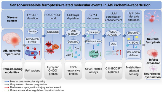
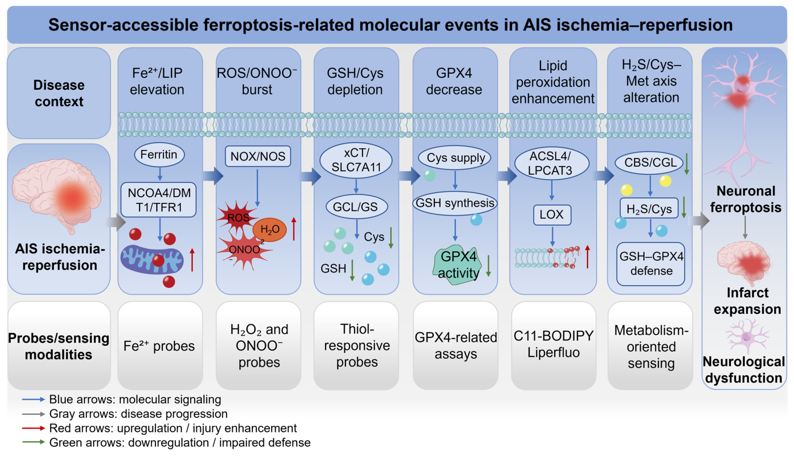
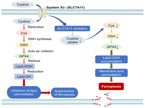
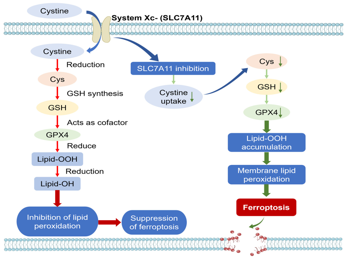
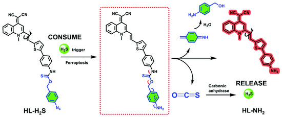
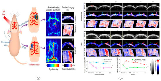
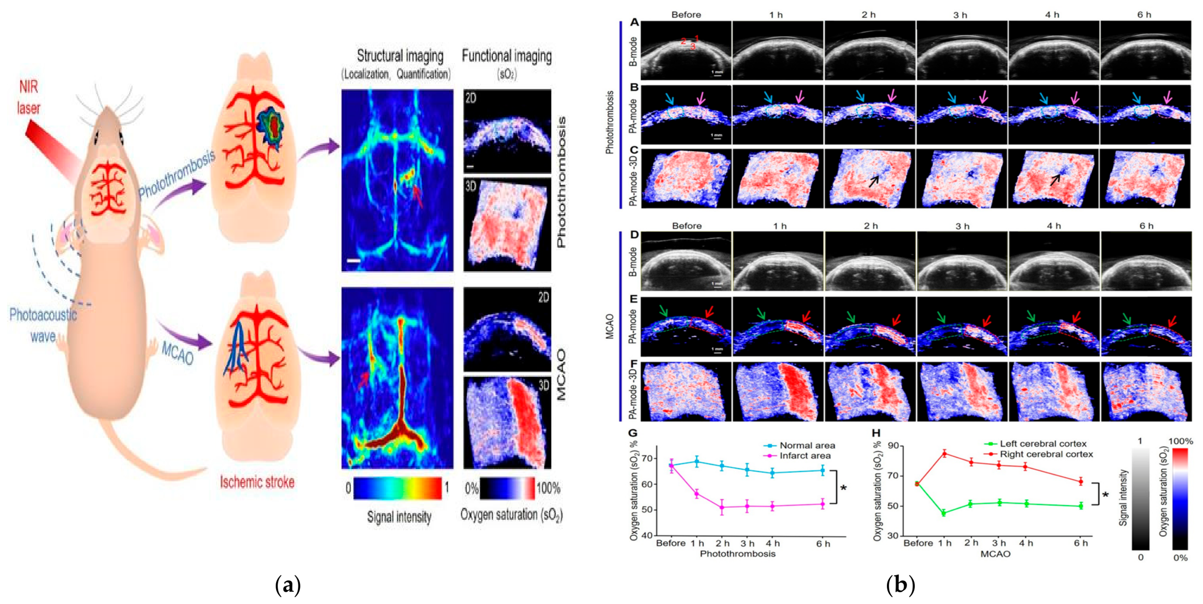

Disclaimer/Publisher’s Note: The statements, opinions and data contained in all publications are solely those of the individual author(s) and contributor(s) and not of MDPI and/or the editor(s). MDPI and/or the editor(s) disclaim responsibility for any injury to people or property resulting from any ideas, methods, instructions or products referred to in the content. |
© 2026 by the authors. Licensee MDPI, Basel, Switzerland. This article is an open access article distributed under the terms and conditions of the Creative Commons Attribution (CC BY) license.