


Abstract
Ocimum gratissimum L. (OGL) is a medicinal plant known for various therapeutic properties, yet its potential in genetic-level skin anti-aging remains underexplored. This study aimed to evaluate the phytochemical profiles, antioxidant activities, and anti-aging potential of OGL leaf extracts obtained via aqueous boiling (OGL-B) and ethanolic maceration (OGL-M). Total phenolic and flavonoid contents were quantified using colorimetric assays, while rutin, catechin, and quercetin were analyzed using HPLC. Biological activities were assessed using antioxidant models, tyrosinase inhibition, and B16F10 anti-melanogenesis assays, while skin regenerative potential was evaluated through collagen biosynthesis and expression of longevity-related genes (SIRT1, FOXO1, TERT, and KLOTHO) in human skin fibroblasts. OGL-B provided a significantly higher extraction yield (14.70%) and superior multifunctional antioxidant capacity than OGL-M. OGL-M showed higher phenolic content and cell-free tyrosinase inhibition, whereas OGL-B demonstrated greater cellular anti-melanogenic efficacy and more than twofold higher collagen stimulatory effects than L-ascorbic acid. OGL-B significantly upregulated mRNA expression (271.25 ± 0.21%), exceeding the reference activator resveratrol, and enhanced fibroblast viability to 121.53 ± 3.85% at 1 mg/mL, indicating high biocompatibility. These findings suggest that OGL-B is a multifunctional extract capable of modulating longevity-related pathways, with potential for advanced anti-aging food supplement and cosmeceutical applications.
1. Introduction
The quest for youthful and radiant skin has propelled the global cosmeceutical industry toward the discovery of potent, plant-derived bioactive compounds. Driven by a paradigm shift toward “Green Beauty” and sustainable innovation, modern formulations are increasingly defined by consumer demand for natural-based skincare. These products are expected to provide high safety profiles and efficacy while maintaining a minimal environmental footprint [1].
Skin aging is primarily mediated by reactive oxygen species (ROS) derived from both intrinsic and extrinsic sources [2]. These ROS trigger inflammatory processes and cellular apoptosis and activate matrix metalloproteinases (MMPs)—proteolytic enzymes responsible for the degradation of structural proteins, specifically collagen and elastin [3,4,5]. Furthermore, oxidative stress disrupts melanocyte homeostasis, culminating in hyperpigmentation and uneven skin tone [3]. To mitigate these age-related changes, the integumentary system utilizes a sophisticated genetic network centered on, a class III histone deacetylase. SIRT1 modulates essential cellular functions, including growth and senescence, primarily by deacetylating the transcription factor FOXO1 [6,7,8]. In tandem, the TERT gene encodes the catalytic subunit of telomerase, which maintains telomere length to decelerate the “cellular clock” and extend the replicative lifespan of dermal cells [9]. Systemic photoprotection and longevity are further supported by the KL (KLOTHO) gene. While its membrane-bound isoform regulates mineral metabolism, its soluble form functions as a circulating hormone that suppresses the pro-aging insulin/IGF-1 signaling pathway [10]. Collectively, the SIRT1-FOXO1-TERT-KL axis constitutes an integrated defense system that promotes DNA repair, stabilizes the extracellular matrix (ECM), and modulates oxidative stress [2]. By shielding cells from cumulative damage and bolstering endogenous repair mechanisms, this genetic synergy is fundamental to maintaining skin resilience and delaying the phenotypic hallmarks of biological aging.
Ocimum gratissimum L., commonly known as clove basil or scent leaf, is an aromatic perennial herb of the Lamiaceae family renowned for a complex phytochemical profile dominated by the phenylpropene eugenol—which can constitute up to 74.2% of its essential oil—alongside thymol, citral, and ethyl cinnamate [11,12]. This herb is also a significant source of polar secondary metabolites, including rosmarinic acid, caffeic acid, and various flavonoids such as quercetin and luteolin, which underpin its potent antioxidant and anti-inflammatory activities [13]. Recent cosmeceutical research highlights its potential for skin preservation, as its bioactive extracts have demonstrated the ability to inhibit tyrosinase, the rate-limiting enzyme in melanin synthesis, and suppress degradative enzymes like collagenase and elastase responsible for wrinkle formation [14]. Furthermore, the high radical scavenging capacity of its extracts protects dermal fibroblasts from oxidative stress and UV-induced senescence, positioning O. gratissimum as a multifunctional natural agent for anti-aging and hyperpigmentation therapy [13].
This study provides a comprehensive evaluation of O. gratissimum L. leaf extracts as multifunctional agents for advanced dermatological applications. By characterizing their antioxidant capacities, tyrosinase inhibitory effects, and anti-melanogenesis properties—alongside their regulatory roles in collagen biosynthesis and anti-aging gene expression—we establish a robust scientific framework for integrating O. gratissimum L. into high-performance food supplement and cosmeceutical formulations.
2. Materials and Methods
2.1. Plant Material and Authentication
Fresh leaves of Ocimum gratissimum L. (OGL) were harvested from Kanchanaburi, Thailand, in January 2025. Collection procedures strictly adhered to the national guidelines and legislation of Thailand. Botanical identification was verified by Assoc. Prof. Dr. Sanong Chomko, a former lecturer and taxonomist at the Biology Department, Faculty of Science, Mahasarahkam University, Thailand. Voucher specimens (RSPG-RMUTT-111) were deposited at the Herbarium of the Faculty of Integrative Medicine, Rajamangala University of Technology Thanyaburi (RMUTT), Pathum Thani, Thailand.
2.2. Preparation and Extraction
The harvested leaves were thoroughly rinsed with deionized water to remove surface impurities and dehydrated in a forced-air oven at 50 °C until a constant moisture content of <5% was achieved. The dried biomass was subsequently pulverized into a fine powder. To evaluate the impact of the extraction solvent and temperature, two distinct methodologies were employed:
Aqueous decoction (OGL-B): The powder was extracted in boiling distilled water (10% w/v; equivalent to 10 g of powder per 100 mL of distilled water) for 2 h.
Ethanolic maceration (OGL-M): The powder was macerated in 95% ethanol (10% w/v; equivalent to 10 g of powder per 100 mL of 95% ethanol) at room temperature for 48 h.
Following extraction, both preparations were filtered and concentrated to a semi-solid state using a rotary evaporator located at Rajamangala University of Technology Thanyaburi, Rangsit Centre, Pathum Thani, Thailand, at 50 °C under reduced pressure. The resulting crude extracts were stored in light-protected amber glass vials at 4 °C until further phytochemical and biological analyses.
2.3. Determination of Total Phenolic and Flavonoid Contents
2.3.1. Total Phenolic Content (TPC)
The TPC of the OGL extracts was determined using the Folin–Ciocalteu colorimetric assay, adapted from previously established protocols [2]. Briefly, 50 µL of each extract (OGL-B and OGL-M; 1.0 mg/mL) was loaded into a 96-well microplate, followed by the addition of 75 µL of Folin–Ciocalteu reagent (Global Chemie, Mumbai, India) and 75 µL of 7.5% (w/v) Na2CO3. The mixture was incubated at room temperature in the dark for 90 min. Absorbance was subsequently measured at 580 nm using a VICTOR® Nivo microplate reader (PerkinElmer, Waltham, MA, USA). TPC values were extrapolated from a gallic acid standard curve (Sigma-Aldrich, St. Louis, MO, USA) and expressed as milligrams of gallic acid equivalents (GAE) per gram of dry extract (mg GAE/g).
2.3.2. Total Flavonoid Content (TFC)
The TFC was quantified via a modified aluminum chloride method [2]. A 50 µL aliquot of each extract (OGL-B and OGL-M; 1.0 mg/mL) was combined with 25 µL of 10% (w/v) AlCl3 (VWR Chemicals BDH, Radnor, PA, USA) and 100 µL of 5% (w/v) NaNO2 (Global Chemie, Mumbai, India) in a 96-well microplate. Following a 5 min incubation, 25 µL of 1 M NaOH (VWR Chemicals BDH, Radnor, PA, USA) was added. The reaction proceeded at room temperature for 10 min, after which the absorbance was recorded at 420 nm. TFC was calculated against a quercetin standard curve (Sigma-Aldrich) and reported as milligrams of quercetin equivalents (QE) per gram of dry extract (mg QE/g).
2.4. Quantification of Rutin, Catechin, and Quercetin via HPLC
The concentrations of rutin, catechin, and quercetin in the OGL extracts were determined using high-performance liquid chromatography (HPLC) according to the method described by [2]. Stock solutions (1000 µg/100 mL methanol) were serially diluted to establish calibration curves. Extract samples were dissolved in HPLC-grade methanol to a concentration of 1 mg/mL. All solutions and mobile phases were filtered through a 0.45 µm membrane and degassed prior to analysis. Analysis was performed on an Agilent 1260 Infinity HPLC system (Agilent Technologies, Waldbronn, Germany) equipped with a photodiode array detector (PDA). Separation was achieved using an Agilent Zorbax C18 column (Agilent Technologies, Wilmington, DE, USA). The isocratic mobile phase consisted of methanol and 0.1% orthophosphoric acid (75:25, v/v) at a flow rate of 0.6 mL/min. The column temperature was maintained at 25 °C with a 10 µL injection volume and a total run time of 8 min. Analytes were detected at a wavelength of 370 nm. Peak identities were confirmed by matching the retention times of sample peaks with those of authentic rutin, catechin, and quercetin standards analyzed under the same chromatographic conditions, and quantification was performed using the corresponding calibration curves.
2.5. Antioxidant Activities
2.5.1. DPPH Radical Scavenging Assay
The free radical scavenging capacity of OGL extracts was evaluated using a modified 2,2-diphenyl-1-picrylhydrazyl (DPPH) assay [15]. Briefly, 100 µL of the extract at varying concentrations was mixed with 100 µL of 0.1 mg/mL DPPH solution (Sigma-Aldrich, St. Louis, MO, USA) in absolute ethanol within a 96-well microplate. The mixture was incubated in the dark at room temperature for 30 min, after which the absorbance was recorded at 515 nm using a microplate reader. L-ascorbic acid (Sigma-Aldrich) served as the positive control.
2.5.2. Inhibition of Lipid Peroxidation
Lipid peroxidation inhibition was determined using a modified ferric thiocyanate (FTC) method [15]. A 50 µL aliquot of the extract was combined with 50 µL of linoleic acid (Sigma-Aldrich) prepared in 50% DMSO (SRL, Mumbai, India) in a 96-well microplate. The reaction was initiated by the addition of 50 µL of 5 mM NH4SCN (Global Chemie, Mumbai, India) and 50 µL of 2 mM FeCl2 (Sigma-Aldrich). The plate was incubated at 37 °C in the dark for 60 min, and the absorbance was measured at 490 nm. α-Tocopherol (Sigma-Aldrich) served as the positive control.
2.5.3. Ferrous Ion-Chelating Activity
The metal-chelating ability was assessed via the FIC method [15]. Briefly, 50 µL of the extract was mixed with FeCl2 (1 mg/mL) and 50 µL of 1 mg/mL ferrozine (TCI, Tokyo, Japan) in 1% HCl. The reaction mixture was incubated in the dark at room temperature for 60 min, followed by absorbance measurement at 515 nm. EDTA (Global Chemie, Mumbai, India) served as the positive control.
All antioxidant experiments were conducted in triplicate. The percentage of inhibition for radical scavenging, lipid peroxidation, and metal chelation was calculated using the following equation: % Inhibition = [(A0 − A1)/A0] × 100, where A0 is the absorbance of the control and A1 is the absorbance of the sample. The half-maximal inhibitory concentrations—specifically the SC50 (DPPH scavenging), LC50 (lipid peroxidation), and MC50 (metal chelation)—were determined by non-linear regression analysis of the plots of inhibition percentage versus sample concentration.
2.6. Tyrosinase Inhibition Activity
Mushroom tyrosinase inhibition was evaluated using a modified dopachrome method with L-tyrosine as the substrate [15]. The reaction was performed in a 96-well microplate, with each well containing: 50 µL of each extract (OGL-B or OGL-M; at various concentrations), 50 µL of 0.1 mg/mL L-tyrosine (Sigma-Aldrich), 50 µL of 0.1 mg/mL mushroom tyrosinase (Sigma-Aldrich), and 50 µL of 0.1 mM phosphate buffer (pH 6.8). The mixture was incubated at 37 °C in the dark for 60 min. Following incubation, the absorbance was recorded at 570 nm using a microplate reader. Kojic acid (SRL, Mumbai, India) was employed as the positive control. To account for the intrinsic color of the plant extracts, the percentage of tyrosinase inhibition was calculated using the following formula: % Tyrosinase inhibition = [(A − B) – (C − D)]/(A – B) × 100, where A is the absorbance of the blank after incubation, B is the absorbance of the blank before incubation, C is the absorbance of the samples after incubation, and D is the absorbance of the samples before incubation. The half-maximal inhibitory concentration (IC50) was determined by non-linear regression analysis of the dose–response curve, plotting the percentage of inhibition against the logarithm of the extract concentration.
2.7. Cell Culture and Maintenance
The human dermal fibroblast line (ATCC CRL-2932) and the murine B16F10 melanoma cell line (ATCC CRL-6166) were obtained from the American Type Culture Collection (ATCC; Manassas, VA, USA). Human dermal fibroblasts were utilized for the assessment of cytotoxicity, stimulation of collagen biosynthesis, and the expression analysis of anti-aging markers, including SIRT1, FOXO1, TERT, and KL. B16F10 melanoma cells were employed specifically for the evaluation of anti-melanogenesis activity. Both cell lines were cultured in Dulbecco’s modified Eagle medium (DMEM) (Gibco-Invitrogen, Carlsbad, CA, USA) supplemented with 10% fetal bovine serum (FBS) and 1% penicillin/streptomycin (100 IU/mL). The cultures were maintained in a humidified incubator at 37 °C under an atmosphere of 5% CO2. Media were replenished every 48–72 h, and cells were passaged upon reaching 80–90% confluence using 0.25% trypsin-EDTA.
2.8. In Vitro Biological Activities
The treatment conditions were selected according to the requirements of each cell-based endpoint. A concentration of 0.1 mg/mL was used as a common working concentration based on the cytotoxicity profile observed in human dermal fibroblasts. The different incubation periods reflect the distinct biological endpoints: 72 h was used to assess melanin accumulation in B16F10 cells, whereas 24 h was used to assess collagen biosynthesis and gene expression responses in human dermal fibroblasts [10,16]. A separate cytotoxicity assay in B16F10 cells was not performed.
2.8.1. Anti-Melanogenesis Assay
The inhibitory effect of the extracts on melanin synthesis was evaluated in B16F10 melanoma cells [16]. Cells (2.5 × 10⁵ cells/well) were seeded in 6-well plates and incubated for 24 h prior to treatment with 0.1 mg/mL of extract for 72 h. Kojic acid was used as the positive control. Post-treatment, cells were washed with PBS and lysed in 200 µL of 10% (w/v) NaOH at 60 °C for 1 h to solubilize melanin. Absorbance was recorded at 450 nm. The percentage of anti-melanogenesis activity was calculated as %Anti-melanogenesis = 100 − [(Asample/Acontrol) × 100], where Acontrol is the absorbance of the control, and Asample is the absorbance of the samples.
2.8.2. Stimulation of Collagen Biosynthesis
Total collagen production in human dermal fibroblasts was quantified using the Sirius Red colorimetric method [10]. Fibroblasts (5 × 10⁵ cells/well) were seeded in 6-well plates and treated with 0.1 mg/mL of the extracts for 24 h; L-ascorbic acid served as the positive control. Following incubation, cells were washed with PBS and stained with 0.1% (w/v) Sirius Red in saturated picric acid for 1 h. After removing excess dye with 10 mM HCl, the bound dye was eluted with 0.1 M NaOH. Absorbance was measured at 540 nm. The relative collagen content was calculated as follows: % Collagen content = [Csample/Ccontrol] × 100, where Ccontrol is the absorbance of the control, and Csample is the absorbance of the samples.
2.8.3. Gene Expression Analysis via qRT-PCR
Total RNA was extracted from treated fibroblasts using the NucleoSpin RNA Plus kit (Macherey-Nagel, Düren, Germany) and quantified via a Qubit fluorometer (Invitrogen, Carlsbad, CA, USA). Relative mRNA expression levels of SIRT1, FOXO1, TERT, and KL were determined using a one-step qRT-PCR system (Toyobo, Osaka, Japan) on an Mx3005P platform (Agilent Technologies, Santa Clara, CA, USA). The sequences of the specific primers were as follows: SIRT1: 5′-TAG CCT TGT CAG ATA AGG AAG GA-3′ (forward), 5′-ACA GCT TCA CAG TCA ACT TTG T-3′ (reverse); FOXO1: 5′-GAC GCC GTG CTA CTC GTT-3′ (forward) and 5′-CGG TTC ATA CCC GAG GTG-3′ (reverse); TERT: 5′-GCA TTG GAA TCA GAC AGC AC-3′ (forward), 5′-CCA CGA CGT AGT CCA TGT TC-3′ (reverse); KL: 5′-GAT AGA GAA AAA TGG CTT CCC TCC-3′ (forward), 5′-GGT CGG TAA ACT GAG ACA GAG TGG-3′ (reverse); and β-actin: 5′TCA TGC AGT GTG ACG TTG ACA TCC GT-3′ (forward), 5′-CCT AGA AGC ATT TGC GGT GCA CGA TG-3′ (reverse). The thermal cycling conditions consisted of reverse transcription at 90 °C (30 s) and 60 °C (20 min), followed by 45 cycles of denaturation (95 °C, 15 s), annealing (60 °C, 15 s), and extension (74 °C, 10 s). Target gene expression was normalized to the internal control β-actin, and relative fold changes were calculated using the 2−ΔΔCT method [2,16,17].
For gene expression analysis, fibroblasts were treated with 0.1 mg/mL of OGL-B or OGL-M for 24 h before RNA extraction.
2.9. Cytotoxicity Assay
Cell viability following extract treatment was assessed using the 3-(4,5-dimethylthiazol-2-yl)-2,5-diphenyltetrazolium bromide (MTT) colorimetric assay [16]. Human dermal fibroblasts were seeded in 96-well plates at a density of 1 × 10⁴ cells/well in 180 µL of complete DMEM. Following a 24 h equilibration period, cells were treated with 20 µL of each extract (OGL-B or OGL-M) at 0.001, 0.01, 0.1, and 1 mg/mL and incubated for an additional 24 h. Post-treatment, the culture medium was aspirated, and the cells were washed three times with 10 mM phosphate-buffered saline (PBS, pH 6.8). Subsequently, 200 µL of 0.5 mg/mL MTT solution (Sigma-Aldrich, St. Louis, MO, USA) was added to each well and incubated at 37 °C for 3 h. The resulting formazan crystals were solubilized by replacing the supernatant with 100 µL of dimethyl sulfoxide (DMSO). The plates were subjected to orbital agitation at 200 rpm for 15 min to ensure complete dissolution. Absorbance was recorded at 570 nm using a microplate reader. The percentage of cell viability was calculated as follows: %Cell viability = [Asample/Acontrol] × 100, where Acontrol is the absorbance of the control and Asample is the absorbance of the samples.
2.10. Statistical Analysis
Data are expressed as the mean ± standard deviation (SD) of three independent experiments. Statistical differences were analyzed using one-way ANOVA followed by Tukey’s post hoc test. p-values < 0.05 were considered statistically significant. For all Figures, statistical comparisons were performed among the experimental groups. The statistical annotations shown in each figure indicate significant differences among groups at p < 0.05; groups with different annotations are significantly different, whereas groups with the same annotation are not significantly different.
2.11. Declaration of Generative AI Use
The authors report that generative AI was not used in their research or preparation of this manuscript.
3. Results and Discussion
3.1. The Extraction Yields and Phytochemical Constituents of the OGL Extracts
The extraction yields, physical characteristics, and phytochemical profiles of O. gratissimum L. leaf (OGL) extracts are summarized in Table 1. The aqueous boiling extract (OGL-B) provided a significantly higher yield (14.70% w/w) compared to the ethanolic extract (OGL-M, 10.15% w/w). Both extracts were viscous semi-solids with distinct odors; however, OGL-B was reddish brown in color, whereas OGL-M exhibited a dark green appearance.
Table 1.
The extraction yields, characteristics, and total phenolics (TPC) and flavonoids (TFC) of the OGL extracts.
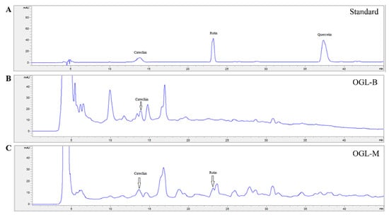
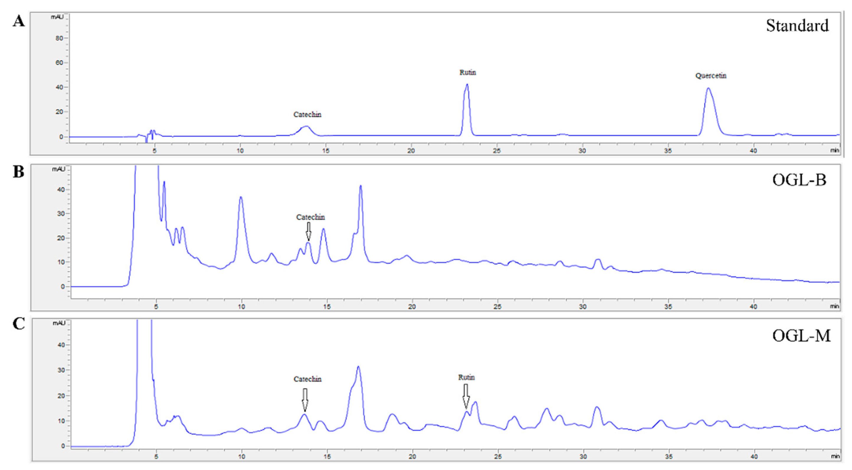
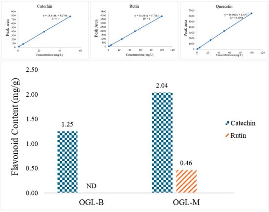
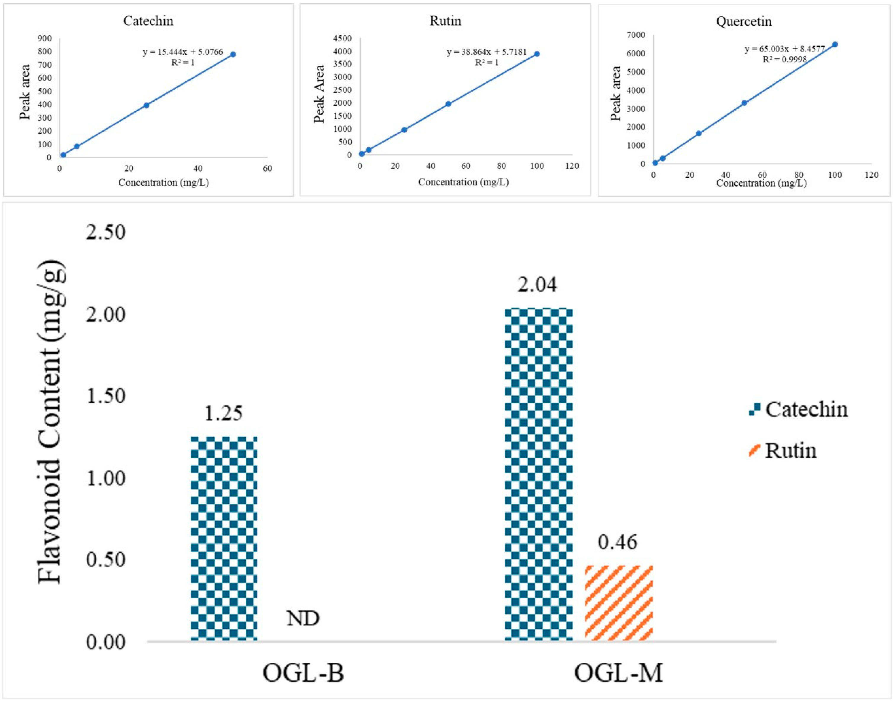
The total phenolic content (TPC) and total flavonoid content (TFC) of Ocimum gratissimum L. (OGL) extracts were quantified using the Folin–Ciocalteu and aluminum chloride colorimetric assays, respectively (Table 1). The maceration process (OGL-M) yielded the highest phenolic recovery (2.574 ± 0.146 g GAE/100 g), while the boiling method (OGL-B) exhibited the superior flavonoid concentration (2.890 ± 0.047 g QE/100 g). High-Performance Liquid Chromatography (HPLC) analysis further identified and quantified specific biomarkers, including rutin, catechin, and quercetin (Figure 1 and Figure 2). Identification of rutin and catechin was based on retention-time matching with authentic standards under the same HPLC conditions. The OGL-M method demonstrated superior extraction efficiency, yielding the highest concentration of catechin (2.04 ± mg/g) and serving as the only process to successfully recover detectable levels of rutin (0.46 ± mg/g). In contrast, the boiling process (OGL-B) resulted in a markedly lower recovery of catechin (1.25 ± mg/g), while rutin remained undetected (ND), suggesting potential thermal degradation of these compounds during heat treatment. Notably, despite the high sensitivity and linearity of the quercetin calibration curve (R2 = 0.9994), this specific flavonoid was not detected in either the OGL-B or OGL-M samples, indicating that its concentration was below the limit of detection for the current experimental conditions.
Figure 1.
HPLC chromatograms of the standard rutin, catechin, and quercetin, and the OGL extracts at a concentration of 1.0 mg/mL. OGL is the O. gratissimum L. extract. B is the boiling extraction with distilled water. M is the maceration process in 95% (v/v) ethanol. Subfigures (A–C) are the HPLC chromatograms of standards and the O. gratissimum L. the boiling extraction with distilled water and O. gratissimum L. is the maceration process in 95% (v/v) ethanol.
Figure 2.
The levels of rutin, catechin, and quercetin in the OGL extracts (1.0 mg/mL) by HPLC. The data are expressed as mean ± SD. Statistical comparisons were performed among all experimental groups using one-way ANOVA followed by Tukey’s post hoc test. The statistical annotations shown in the figure indicate significant differences among groups at p < 0.05; groups with different annotations are significantly different, whereas groups with the same annotation are not significantly different. OGL is the O. gratissimum L. extract. B is the boiling extraction with distilled water. M is the maceration process in 95% (v/v) ethanol. ND is not detected.
The results indicate that the OGL-M extraction method is significantly more effective at preserving and recovering bioactive flavonoids than the boiling method (OGL-B). The higher concentration of catechin and the exclusive presence of rutin in OGL-M suggest that these compounds may be thermolabile (heat-sensitive). These phytochemicals are well-documented for their potent biological activities, particularly in anti-oxidation, anti-inflammation, and photoprotection, which are essential for anti-aging applications. The results suggest that solvent polarity, extraction technique, and temperature significantly influence the recovery of these bioactive compounds from the OGL matrix [18].
3.2. Antioxidant Activities of the OGL Extracts
The antioxidant activities of OGL extracts are summarized in Table 2. The antioxidant potential of OGL extracts was evaluated through three distinct mechanisms. OGL-B exhibited superior antioxidant capacity across all assays compared to OGL-M. Specifically, OGL-B demonstrated the highest free radical scavenging activity (SC50 = 0.65 ± 0.05 mg/mL), although it remained significantly lower than the reference standard, L-ascorbic acid (p < 0.05). Furthermore, OGL-B showed moderate lipid peroxidation inhibition (LC50 = 3.18 ± 5.19 mg/mL) and weak metal-chelating activity (MC50 = 24.03 ± 5.72 mg/mL), whereas OGL-M was inactive in these assays. These results suggest that OGL-B contains a broader range of bioactive compounds capable of neutralizing oxidative stress compared to OGL-M.
Table 2.
Antioxidant activities of the OGL extracts.
The antioxidative potential of the OGL extracts was evaluated through three distinct models—free radical scavenging, lipid peroxidation inhibition, and metal chelation—which are essential for identifying natural compounds capable of mitigating skin aging [2]. While the free radical scavenging model relies on the donation of hydrogen atoms to reduce DPPH radicals [19], the lipid peroxidation model assesses inhibition of linoleic acid oxidation, whereas the metal chelation model measures inhibition of ferrozine-Fe2+ complex formation [20,21].
In this study, OGL-B exhibited superior multifunctional antioxidant activities compared to OGL-M, particularly in lipid peroxidation inhibition ((LC50 = 3.18 ± 5.19 mg/mL) and metal chelation (MC50 = 24.03 ± 5.72 mg/mL), whereas OGL-M remained inactive. Although the potency of both extracts was lower than the reference standards (L-ascorbic acid, α-tocopherol, and EDTA), the observed SC50, LC50, and MC50 values indicate that these extracts possess significant bioactivity. This effectiveness is likely driven by a synergistic effect of phytochemicals such as alkaloids, flavonoids, and tannins. Specifically, rutin and catechin, together with other flavonoid-related constituents, may contribute to the extracts’ ability to protect skin cells from oxidative stress-related damage [22]. Quercetin was not detected in either extract under the present HPLC conditions and therefore cannot be directly assigned to the observed antioxidant effects. The broader activity spectrum of OGL-B suggests its better potential as a cosmeceutical ingredient for skin protection compared to OGL-M.
3.3. Tyrosinase Inhibition and Anti-Melanogenesis of the OGL Extracts
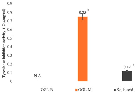
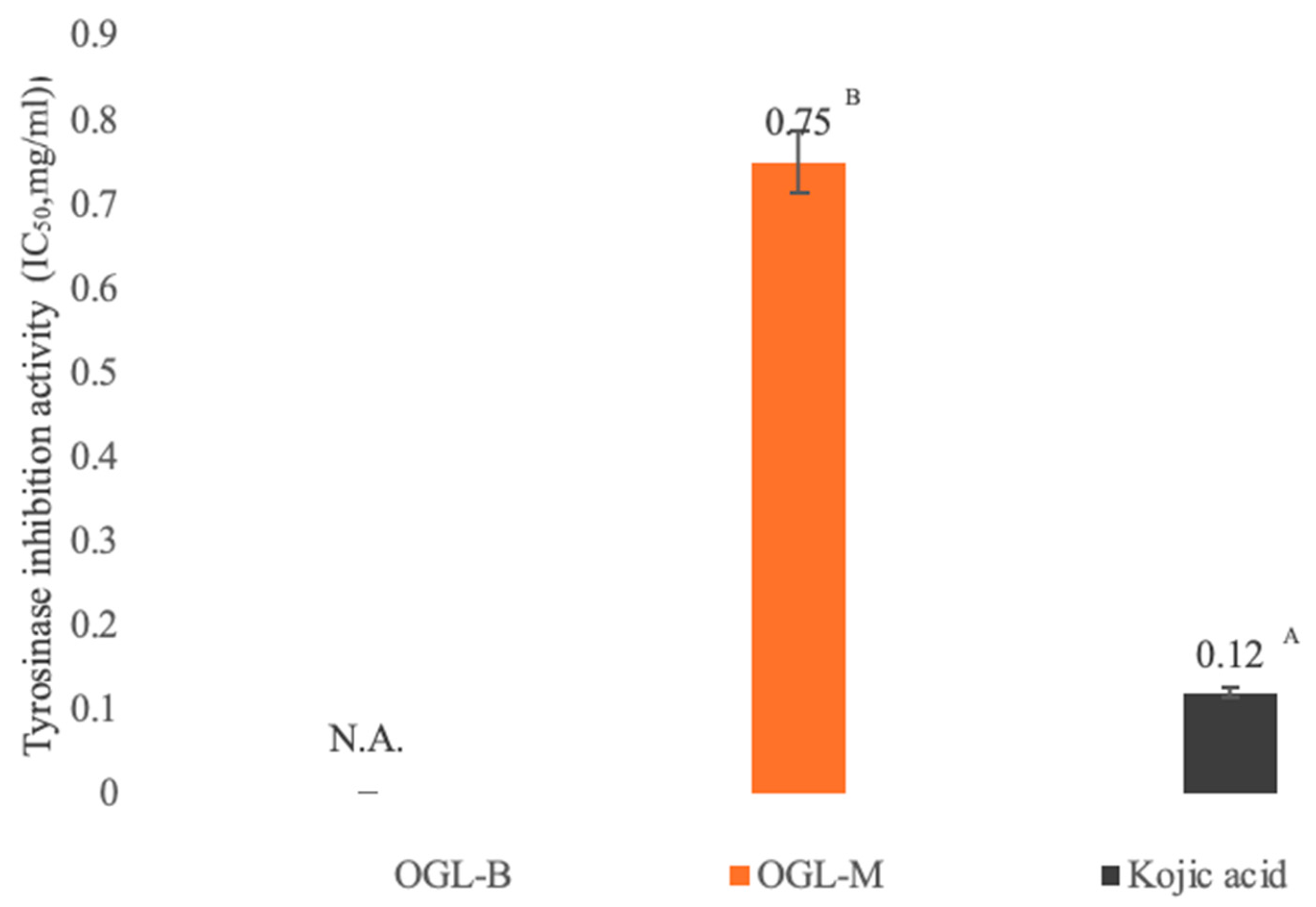
The inhibitory effects of various OGL extracts on tyrosinase activity are illustrated in Figure 3. Among the tested samples, the OGL-M extract exhibited the most potent tyrosinase inhibitory activity with an IC50 value of 0.75 ± 0.02 mg/mL. Although all extracts showed a degree of inhibition, their potency remained significantly lower than that of the positive control, kojic acid, which yielded an IC50 of 0.01 ± 0.00 mg/mL (p < 0.05). The results suggest that while OGL extracts contain bioactive constituents capable of suppressing tyrosinase, their crude nature requires a higher concentration to achieve comparable effects to pure kojic acid.
Figure 3.
Tyrosinase inhibition activities of the OGL extracts. The data are expressed as mean ± SD. Statistical comparisons were performed among all experimental groups using one-way ANOVA followed by Tukey’s post hoc test. Different superscript letters (A,B) The statistical annotations shown in the figure indicate significant differences among groups at p < 0.05; groups with different annotations are significantly different, whereas groups with the same annotation are not significantly different. OGL is the O. gratissimum L. extract. B is the boiling extraction with distilled water. M is the maceration process in 95% (v/v) ethanol.
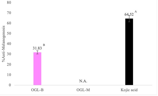
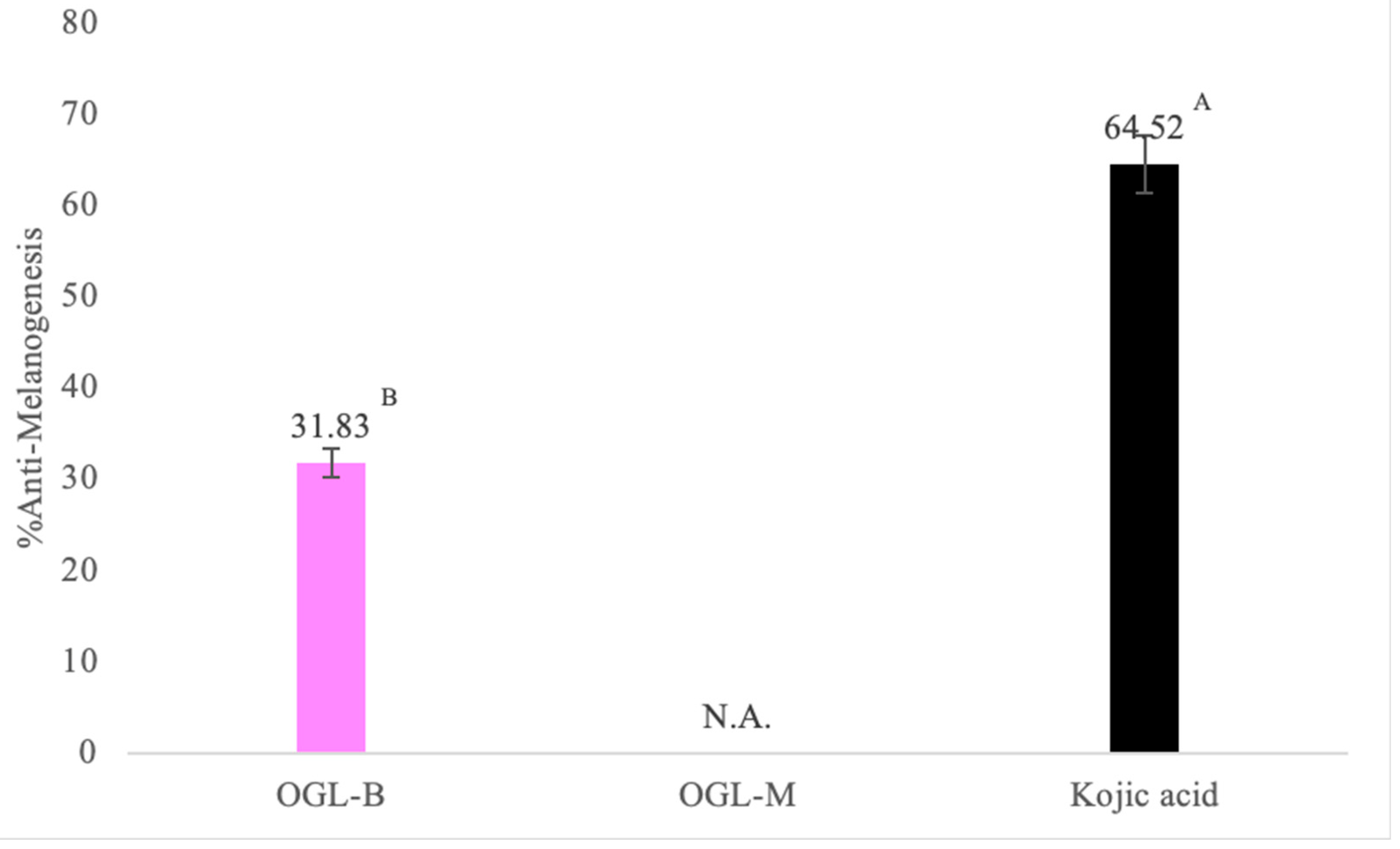
To evaluate the physiological relevance of the extracts, their anti-melanogenic effects were tested on murine B16F10 melanoma cells (Figure 4). At the selected concentration of 0.1 mg/mL, all OGL extracts significantly reduced melanin content compared to the untreated control. The OGL-B (Boiling) extract demonstrated the highest anti-melanogenic efficacy, achieving a 31.83 ± 3.16% reduction in melanin production. However, this effect was significantly less pronounced than the 68.55 ± 1.67% inhibition observed in cells treated with kojic acid (p < 0.05). Notably, the OGL-M extract, which performed best in the cell-free tyrosinase assay, showed relatively lower efficacy in the cellular model.
Figure 4.
Anti-melanogenesis in murine melanoma (B16F10) cell lines by OGL extracts at a concentration of 0.1 mg/mL. The data are expressed as mean ± SD. Statistical comparisons were performed among all experimental groups using one-way ANOVA followed by Tukey’s post hoc test. Different superscript letters (A,B) The statistical annotations shown in the figure indicate significant differences among groups at p < 0.05; groups with different annotations are significantly different, whereas groups with the same annotation are not significantly different. OGL is the O. gratissimum L. extract. B is the boiling extraction with distilled water. M is the maceration process in 95% (v/v) ethanol.
The findings of this study confirm that OGL extracts possess significant anti-hyperpigmentation potential by targeting both tyrosinase activity and cellular melanogenesis. Tyrosinase serves as the rate-limiting enzyme in the melanogenic pathway, orchestrating the hydroxylation of L-tyrosine and the subsequent oxidation of L-dopa into dopaquinone [10].
The potent inhibitory effect of OGL extracts is likely associated with their flavonoid constituents. Quercetin is a well-documented flavonoid that functions as a competitive inhibitor of tyrosinase by chelating essential copper ions at the enzyme active site [23] and has also been reported to downregulate microphthalmia-associated transcription factor (MITF), thereby reducing the transcription of melanogenic enzymes, including TRP-1 and TRP-2 [24]. However, quercetin was not detected in either OGL-B or OGL-M under the present HPLC conditions; therefore, the tyrosinase inhibitory and anti-melanogenic effects observed in this study cannot be directly attributed to quercetin.
Interestingly, a discrepancy was observed between the cell-free tyrosinase assay and the B16F10 cell-based assay, particularly regarding the OGL-M extract. While OGL-M showed the strongest direct tyrosinase inhibition, its anti-melanogenic activity in living cells was limited. This difference may reflect the solubility, membrane permeability, intracellular stability, or cellular transport of flavonoid constituents, which can cause activity in a cell-free enzyme system to differ from that observed in intact cells [25,26].
3.4. Stimulation of Collagen Biosynthesis of the OGL Extracts
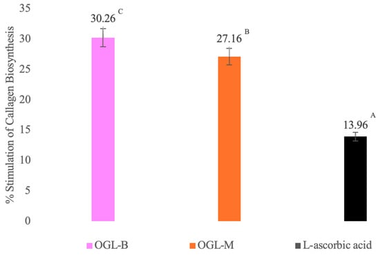
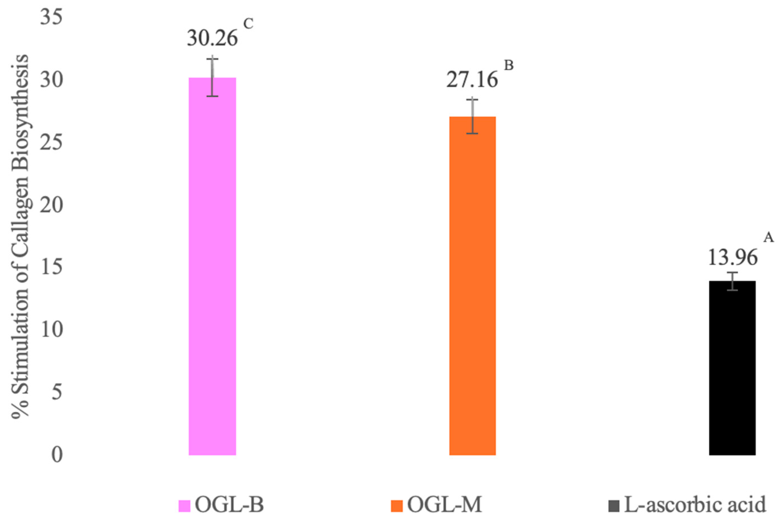
As demonstrated by the Sirius Red assay (Figure 5), all OGL extracts significantly stimulated collagen biosynthesis in human skin fibroblasts. Notably, OGL-B exhibited the most potent effect at 30.26 ± 7.47%, statistically outperforming the positive control, L-ascorbic acid (13.96 ± 3.65; p < 0.05), by more than twofold.
Figure 5.
Stimulation of collagen biosynthesis in human skin fibroblasts by OGL extracts at a concentration of 0.1 mg/mL. The data are expressed as mean ± SD. Statistical comparisons were performed among all experimental groups using one-way ANOVA followed by Tukey’s post hoc test Different superscript letters (A–C) The statistical annotations shown in the figure indicate significant differences among groups at p < 0.05; groups with different annotations are significantly different, whereas groups with the same annotation are not significantly different. OGL is the O. gratissimum L. extract. B is the boiling extraction with distilled water. M is the maceration process in 95% (v/v) ethanol.
As the primary structural protein of the dermis, collagen provides essential strength and support; its degradation is a primary hallmark of cutaneous aging, resulting in reduced elasticity and durability [27]. The biosynthetic process is a sophisticated cellular sequence involving gene transcription, translation, and multiple post-translational modifications [28]. Our findings indicate that the superior stimulatory effect of OGL extracts, especially OGL-B, may be attributed to their flavonoid content and phytochemical profiles, particularly catechin and rutin identified in the OGL extracts. Flavonoids are known to directly promote collagen synthesis and preserve the extracellular matrix by inhibiting matrix metalloproteinases (MMPs) through the chelation of zinc ions, effectively increasing net collagen accumulation [29]. Furthermore, the observed changes in SIRT1 and TERT mRNA expression may be related to the cellular responses associated with the collagen production observed in fibroblasts.
3.5. Activation of the Anti-Aging Gene Expression of the OGL Extracts
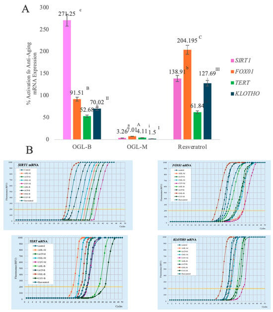
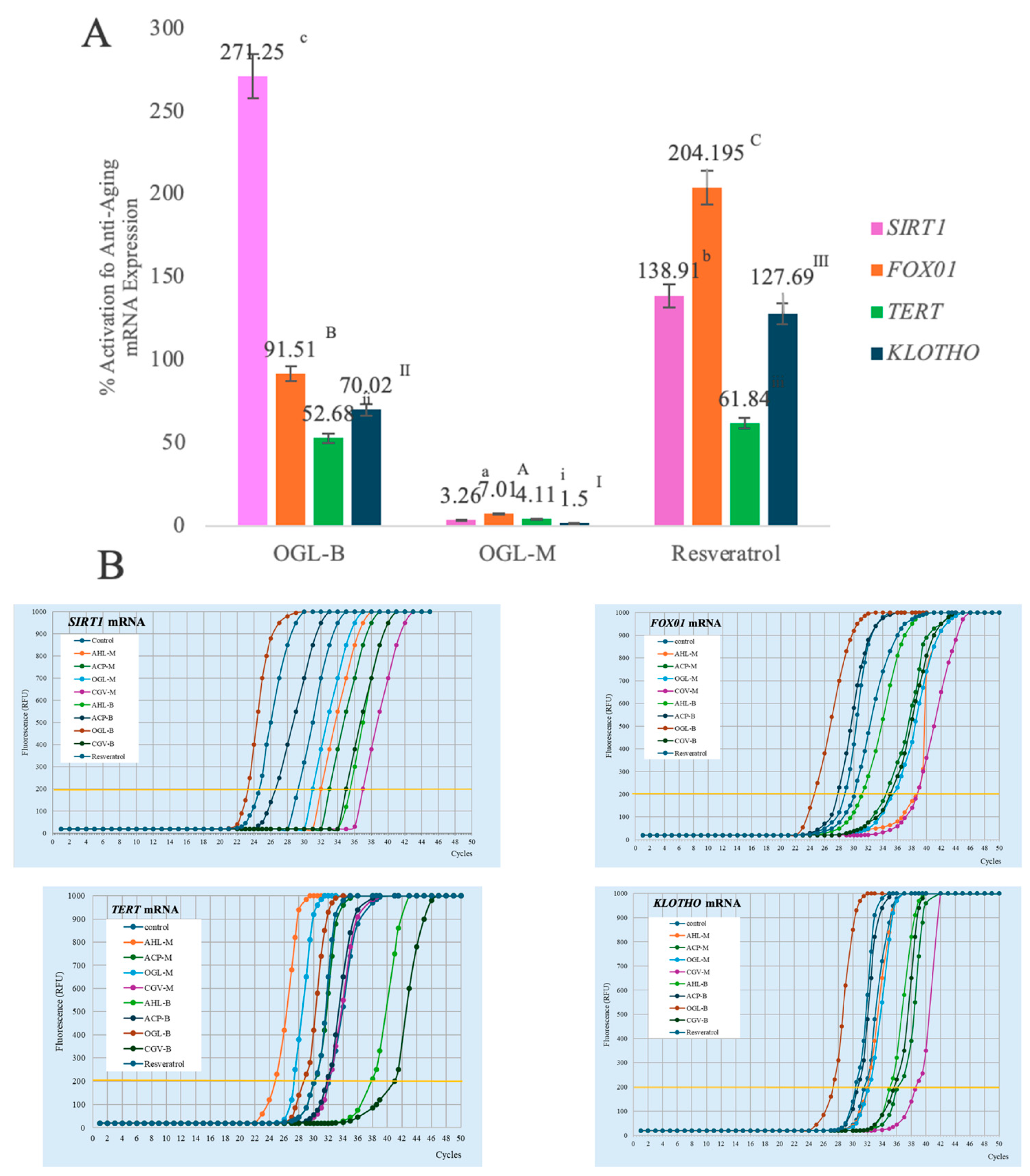
The stimulatory effects of OGL extracts on anti-aging gene expression (SIRT1, FOXO1, TERT, and KLOTHO) were investigated, as shown in Figure 6. OGL-B exhibited a remarkably high activation of SIRT1 mRNA expression at 271.25 ± 0.21%, significantly surpassing the positive control, resveratrol (138.91 ± 1.00%). While resveratrol showed higher induction of FOXO1 (204.19 ± 9.21%), OGL-B maintained potent activity across all tested genes, including TERT (52.68 ± 0.87%) and KLOTHO (70.02 ± 0.19%). In contrast, OGL-M demonstrated minimal activity (all expressions <10%). These findings suggest that OGL-B acts as a potent SIRT1 activator, potentially offering superior anti-aging benefits through the modulation of longevity-related genetic pathways compared to OGL-M. The qRT-PCR results were normalized to β-actin and calculated by the 2−ΔΔCt method, and the corresponding amplification plots are presented in Figure 6B.
Figure 6.
Activation of anti-aging genes in human skin fibroblasts (A) and the amplification plot (B) of the OGL extracts at a concentration of 0.1 mg/mL. The data are expressed as mean ± SD. For each gene, statistical comparisons were performed among the treatment groups using one-way ANOVA followed by Tukey’s post hoc test. (a–c for SIRT1, A–C for FOX1, i–iii for TERT, I–III for KLOTHO). The statistical annotations shown in the figure indicate significant differences among treatment groups within the same gene at p < 0.05; groups with different annotations are significantly different, whereas groups with the same annotation are not significantly different. OGL is the O. gratissimum L. extract. B is the boiling extraction with distilled water. M is the maceration process in 95% (v/v) ethanol.
The antioxidative and anti-aging potential of O. gratissimum L. (OGL) extracts was evaluated through chemical and genetic models. Recent studies have highlighted the capacity of natural plant extracts to upregulate SIRT1 and FOXO1 mRNA expression; for instance, Prunus mume seed and Nephelium lappaceum extracts have been shown to enhance SIRT1 expression and protect against skin aging [2,10,30]. Additionally, compounds like centcyamine and turmeric polysaccharides can activate KLOTHO and TERT, respectively, to maintain cellular longevity [16,17]. While several O. gratissimum L. varieties are documented for cosmetic use, research regarding their influence on longevity-related gene expression remains sparse. To our knowledge, this study is among the first to evaluate the effects of O. gratissimum L. (OGL) leaf extracts on SIRT1, FOXO1, TERT, and KLOTHO mRNA expression. Our results demonstrate that OGL-B possesses superior bioactivity compared to OGL-M, particularly in its remarkable ability to upregulate SIRT1 mRNA (271.25 ± 0.21%), which significantly outperformed the well-established activator resveratrol (138.91 ± 1.00%). This high potency is likely driven by the synergistic effect of its phytochemical constituents, such as rutin and catechin, together with other flavonoid-related compounds, which are known to modulate the SIRT1/AMPK/NF-κB and SIRT1/Nrf2 axes [31]. These polyphenols enhance antioxidant enzyme activity and mitochondrial efficiency, consistent with our findings where OGL-B also showed the highest free radical scavenging and lipid peroxidation inhibition activities. Among the genes evaluated, SIRT1 showed the most pronounced response to OGL-B, whereas the TERT response was comparatively lower (52.68% for OGL-B). This disparity is unsurprising given that TERT expression is tightly controlled by stringent regulatory mechanisms to prevent uncontrolled cell division, unlike the more readily activated cellular stress response genes like SIRT1 and FOXO1 [32]. These findings suggest that the molecular response to OGL-B may be more strongly associated with the sirtuin-related pathway than with telomerase-related regulation under the present experimental conditions. Overall, the broader activity spectrum of OGL-B—from chemical antioxidant capacity to modulation of longevity-related gene expression—positions it as a promising candidate for cosmeceutical applications in skin aging mitigation.
3.6. Cytotoxicity of the OGL Extracts
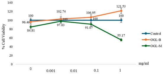
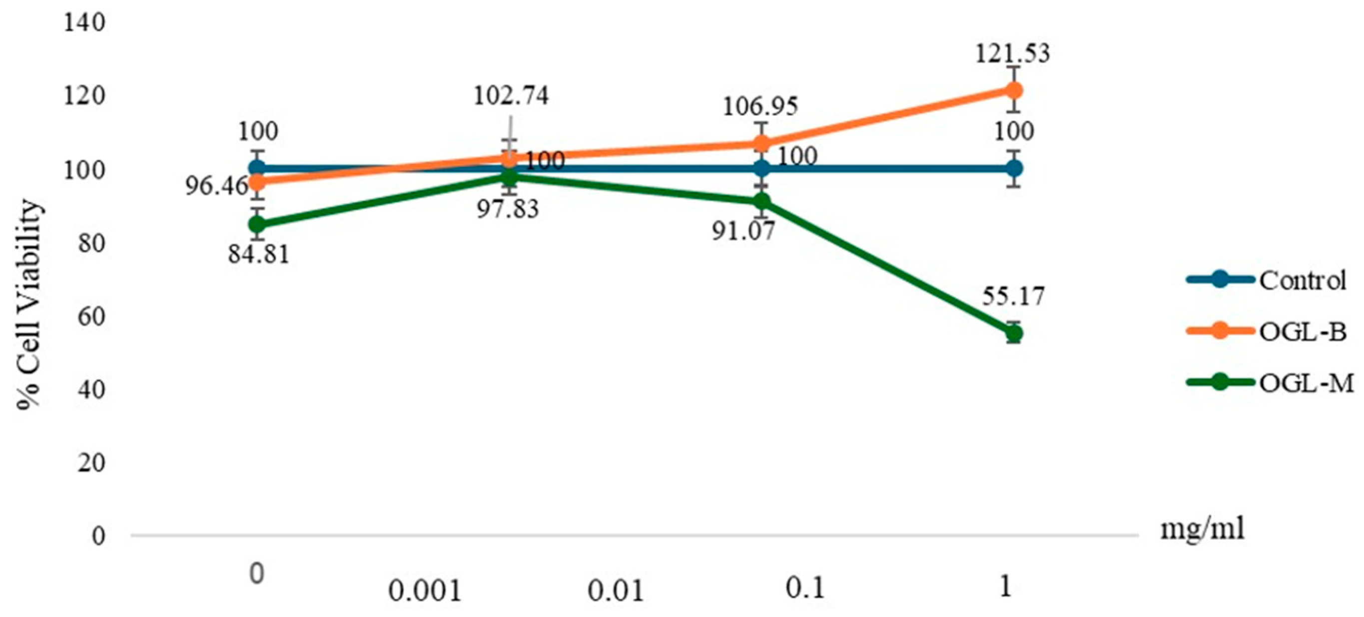
Based on the MTT assay results presented in Figure 7, the cytotoxic profiles of the OGL extracts exhibited a concentration-dependent response. OGL-B demonstrated excellent biocompatibility, maintaining cell viability above 90% across all tested concentrations. Notably, at 1 mg/mL, OGL-B increased cell viability to 121.53%, suggesting a potential stimulatory effect on fibroblast proliferation, which is beneficial for skin regenerative applications. In contrast, OGL-M showed a significant reduction in cell viability to 55.17% at 1 mg/mL, indicating potential cytotoxicity at higher dosages, whereas it remained non-toxic (viability > 80%) at concentrations of 0.1 mg/mL and below.
Figure 7.
Cytotoxicity of the OGL extracts at various concentrations on human skin fibroblasts. The data are expressed as mean ± SD. OGL is the O. gratissimum L. extract. B is the boiling extraction with distilled water. M is the maceration process in 95% (v/v) ethanol.
These findings suggest that while OGL-B is safe for dermal applications up to at least 1 mg/mL, the concentration of OGL-M must be carefully controlled. The observed cytotoxicity of OGL-M at high concentrations may be related to differences in the phytochemical composition of the ethanolic extract; however, the mechanism responsible for this effect was not directly investigated in the present study. Consequently, for subsequent anti-aging gene expression studies, a concentration of 0.1 mg/mL was selected as the optimal non-toxic dose for both extracts to ensure that the observed genetic responses were not influenced by impaired cell viability.
4. Conclusions
This study demonstrates that the extraction method significantly influences the phytochemical and biological profile of Ocimum gratissimum L. (OGL) leaf extracts. The aqueous boiling extract (OGL-B) emerged as the superior candidate, providing higher extraction yields and broader antioxidant capacity compared to the ethanolic extract (OGL-M). Biologically, OGL-B exhibited potent anti-melanogenic effects and significantly outperformed L-ascorbic acid in stimulating collagen biosynthesis. Most notably, OGL-B modulated longevity-related gene expression, with the most pronounced effect observed for SIRT1, whose induction surpassed that of resveratrol. Combined with its excellent biocompatibility and stimulatory effect on fibroblast viability, OGL-B shows high potential as a safe, multifunctional ingredient for anti-aging and skin-protective food supplement and cosmeceutical applications.
Author Contributions
Conceptualization, K.B.; methodology, K.B., T.T., S.P., W.C. and K.S.; validation, K.B., W.R. and R.C.; formal analysis, K.B., N.K. and S.P.; investigation, K.B. and S.P.; resources, K.B.; data curation, K.B.; writing—original draft preparation, K.B. and S.P.; writing—review and editing, K.B., K.S., N.K., W.R., R.C. and S.P.; visualization, K.B. and S.P.; supervision, K.B.; project administration, K.B.; funding acquisition, K.B. All authors have read and agreed to the published version of the manuscript.
Funding
This research was funded by the National Science, Research and Innovation Fund, Thailand Science Research and Innovation (TSRI), through Rajamangala University of Technology Thanyaburi, grant number FRB680045/0168.
Institutional Review Board Statement
Not applicable. This study did not involve human participants or animals. The human dermal fibroblast cell line (ATCC CRL-2932) used in this study was obtained commercially from the American Type Culture Collection (ATCC).
Informed Consent Statement
Not applicable.
Data Availability Statement
The data supporting the findings of this study are included in the tables and figures. The datasets generated and analyzed during the current study are available from the corresponding author upon reasonable request.
Acknowledgments
The authors gratefully acknowledge the Faculty of Integrative Medicine, RMUTT, and C Meda-Plus Co., Ltd., Bangkok, Thailand, for providing the necessary laboratory facilities, equipment, and technical support throughout this study.
Conflicts of Interest
The authors declare no conflicts of interest.
Abbreviations
| AMPK | AMP-activated protein kinase |
| ANOVA | Analysis of variance |
| ATCC | American Type Culture Collection |
| DMEM | Dulbecco’s modified Eagle medium |
| DMSO | Dimethyl sulfoxide |
| DPPH | 2,2-Diphenyl-1-picrylhydrazyl |
| ECM | Extracellular matrix |
| EDTA | Ethylenediaminetetraacetic acid |
| FBS | Fetal bovine serum |
| FIC | Ferrous ion chelating |
| FOXO1 | Forkhead box O1 |
| FTC | Ferric thiocyanate |
| GAE | Gallic acid equivalents |
| HPLC | High-performance liquid chromatography |
| IC50 | Half-maximal inhibitory concentration |
| KL | KLOTHO |
| LC50 | Half-maximal lipid peroxidation inhibitory concentration |
| MC50 | Half-maximal metal-chelating concentration |
| MDR1 | Multidrug resistance protein 1 |
| MITF | Microphthalmia-associated transcription factor |
| MMPs | Matrix metalloproteinases |
| mRNA | Messenger RNA |
| MTT | 3-(4,5-Dimethylthiazol-2-yl)-2,5-diphenyltetrazolium bromide |
| ND | Not detected |
| NF-κB | Nuclear factor kappa B |
| Nrf2 | Nuclear factor erythroid 2-related factor 2 |
| OGL | Ocimum gratissimum L. |
| OGL-B | Aqueous boiling extract of Ocimum gratissimum L. |
| OGL-M | Ethanolic maceration extract of Ocimum gratissimum L. |
| PBS | Phosphate-buffered saline |
| PDA | Photodiode array |
| QE | Quercetin equivalents |
| qRT-PCR | Quantitative reverse transcription polymerase chain reaction |
| RNA | Ribonucleic acid |
| ROS | Reactive oxygen species |
| SC50 | Half-maximal DPPH scavenging concentration |
| SD | Standard deviation |
| SIRT1 | Sirtuin 1 |
| TERT | Telomerase reverse transcriptase |
| TFC | Total flavonoid content |
| TPC | Total phenolic content |
| TRP-1 | Tyrosinase-related protein 1 |
| TRP-2 | Tyrosinase-related protein 2 |
References
- Bom, S.; Jorge, J.; Ribeiro, H.M.; Marto, J. A step forward on sustainability in the cosmetics industry: A review. J. Clean. Prod. 2019, 225, 270–290. [Google Scholar] [CrossRef] [Scilit]
- Boonpisuttinant, K.; Taka, T.; Ruksiriwanich, W.; Chutoprapat, R.; Udompong, S.; Kansawang, R.; Sangsee, J.; Chompoo, W.; Samothai, K.; Srisuttee, R. Assessment of in vitro anti-skin aging activities of Phyllanthus indofischeri Bennet extracts for dermatological and aesthetic applications. Sci. Rep. 2022, 12, 18661. [Google Scholar] [CrossRef] [Scilit]
- Jin, C.K.; Sang, G.L.; Kyoung, M.L.; Hyoung, J.J.; Ju, H.L. Skin-aging pigmentation: Who is the real enemy? Cells 2022, 11, 2541. [Google Scholar] [CrossRef] [Scilit]
- Panwar, P.; Butler, G.S.; Jamroz, A.; Azizi, P.; Overall, C.M.; Brömme, D. Aging-associated modifications of collagen affect its degradation by matrix metalloproteinases. Matrix Biol. 2018, 65, 30–44. [Google Scholar] [CrossRef] [Scilit]
- Feng, C.; Chen, X.; Yin, X.; Jiang, Y.; Zhao, C. Matrix metalloproteinases on skin photoaging. J. Cosmet. Dermatol. 2024, 23, 3847–3862. [Google Scholar] [CrossRef] [Scilit]
- Wątroba, M.; Szukiewicz, D. The role of sirtuins in aging and age-related diseases. Adv. Med. Sci. 2016, 61, 52–62. [Google Scholar] [CrossRef] [Scilit]
- Grabowska, W.; Sikora, E.; Bielak-Zmijewska, A. Sirtuins, a promising target in slowing down the aging process. Biogerontology 2017, 18, 447–476. [Google Scholar] [CrossRef] [Scilit]
- Bernardes de Jesus, M.; Vera, E.; Schneeberger, K.; Tejera, A.M.; Ayuso, E.; Bosch, F.; Blasco, M.A. Telomerase gene therapy in adult and old mice delays aging and increases longevity without increasing cancer. EMBO Mol. Med. 2012, 4, 691–704. [Google Scholar] [CrossRef] [Scilit]
- Prud’homme, G.J.; Kurt, M.; Wang, Q. Pathobiology of the KLOTHO antiaging protein and therapeutic considerations. Front. Aging 2022, 3, 931331. [Google Scholar] [CrossRef] [Scilit]
- Boonpisuttinant, K.; Srisuttee, R.; Khong, H.Y.; Chutoprapat, R.; Ruksiriwanich, W.; Udompong, S.; Chompoo, W.; Boonbai, R.; Rakkaew, R.; Sangsee, J.; et al. In vitro anti-ageing activities of ethanolic extracts from Pink rambutan (Nephelium lappaceum Linn.) for skin applications. Saudi Pharm. J. 2023, 31, 535–546. [Google Scholar] [CrossRef] [Scilit]
- Leonardo de Paiva Belo, L.; Costa de Moraes, F.; Hitomi Yamamoto, C.; Vieira de Sousa, O. Evaluation of antibiotic efficacy of Ocimum gratissimum L. essential oil against Staphylococcus aureus and Pseudomonas aeruginosa. J. Med. Plants Res. 2023, 17, 57–63. [Google Scholar] [CrossRef] [Scilit]
- Ballester, E.L.C.; Costa, W.G.d.S.; Ferreira, C.H.d.N.; Retcheski, M.C.; Cazarolli, L.H.; Schwengber, G.H.; Mauerwerk, M.T.; Pinheiro, C.G.; Heinzmann, B.M.; Baldisserotto, B.; et al. Dietary Ocimum gratissimum essential oil improves the antioxidant status and maintains the performance of Macrobrachium rosenbergii juveniles. Appl. Sci. 2025, 15, 2745. [Google Scholar] [CrossRef] [Scilit]
- Ugbogu, O.C.; Emmanuel, O.; Agi, G.O.; Ibe, C.; Ekweogu, C.N.; Ude, V.C.; Uche, M.E.; Nnanna, R.O.; Ugbogu, E.A. A review on the traditional uses, phytochemistry, and pharmacological activities of clove basil (Ocimum gratissimum L.). Heliyon 2021, 7, e08404. [Google Scholar] [CrossRef] [Scilit]
- Ciganović, P.; Jakupović, L.; Momchev, P.; Nižić Nodilo, L.; Hafner, A.; Zovko Končić, M. Extraction optimization, antioxidant, cosmeceutical and wound healing potential of Echinacea purpurea glycerolic extracts. Molecules 2023, 28, 1177. [Google Scholar] [CrossRef] [Scilit]
- Chutoprapat, R.; Malilas, W.; Rakkaew, R.; Udompong, S.; Boonpisuttinant, K. Collagen biosynthesis stimulation and anti-melanogenesis of Bambara groundnut (Vigna subterranea) extracts. Pharm. Biol. 2020, 58, 1023–1031. [Google Scholar] [CrossRef] [Scilit]
- Abadie, S.; Leduc, C.; Lintner, K.; Bedos, P. The alkaloid centcyamine increases expression of KLOTHO and lamin B1, slowing the onset of skin ageing in vitro and in vivo. Int. J. Cosmet. Sci. 2021, 43, 561–572. [Google Scholar] [CrossRef] [Scilit]
- Pan, M.H.; Wu, J.C.; Ho, C.T.; Badmaev, V. Effects of water extract of Curcuma longa (L.) roots on immunity and telomerase func-tion. J. Complement. Integr. Med. 2017, 14, 20150107. [Google Scholar] [CrossRef] [Scilit]
- Venuprasad, M.P.; Kandikattu, H.K.; Razack, S.; Khanum, F. Phytochemical analysis of Ocimum gratissimum by LC-ESI–MS/MS and its antioxidant and anxiolytic effects. S. Afr. J. Bot. 2014, 92, 151–158. [Google Scholar] [CrossRef] [Scilit]
- Kedare, S.B.; Singh, R.P. Genesis and development of DPPH method of antioxidant assay. J. Food Sci. Technol. 2011, 48, 412–422. [Google Scholar] [CrossRef] [Scilit]
- Bakır, T.; Sönmezoglu, İ.; Apak, R. Quantification of antioxidant ability against lipid peroxidation with an ‘Area Under Curve’ approach. J. Am. Oil Chem. Soc. 2017, 94, 25–35. [Google Scholar] [CrossRef] [Scilit]
- Wong, F.C.; Yong, A.L.; Ting, E.P.S.; Khoo, S.C.; Ong, H.C.; Chai, T.T. Antioxidant, metal chelating, anti-glucosidase activities and phytochemical analysis of selected tropical medicinal plants. Iran. J. Pharm. Res. 2014, 13, 1409–1415. [Google Scholar]
- Panche, A.N.; Diwan, A.D.; Chandra, S.R. Flavonoids: An overview. J. Nutr. Sci. 2016, 5, e47. [Google Scholar] [CrossRef] [Scilit]
- Fan, M.; Zhang, G.; Hu, X.; Xu, X.; Gong, D. Quercetin as a tyrosinase inhibitor: Inhibitory activity, conformational change and mechanism. Food Res. Int. 2017, 100, 226–233. [Google Scholar] [CrossRef] [Scilit]
- Choi, M.-H.; Shin, H.-J. Anti-melanogenesis effect of quercetin. Cosmetics 2016, 3, 18. [Google Scholar] [CrossRef] [Scilit]
- Dobrzynska, M.; Napierala, M.; Florek, E. Flavonoid nanoparticles: A promising approach for cancer therapy. Biomolecules 2020, 10, 1268. [Google Scholar] [CrossRef] [Scilit]
- Filipiak-Duliban, A.; Brodaczewska, K.; Kajdasz, A.; Kieda, C. Spheroid culture differentially affects cancer cell sensitivity to drugs in melanoma and RCC models. Int. J. Mol. Sci. 2022, 23, 1166. [Google Scholar] [CrossRef] [Scilit]
- Baumann, L. Skin aging and its treatment. J. Pathol. 2007, 211, 241–251. [Google Scholar] [CrossRef] [Scilit]
- Onursal, C.; Dick, E.; Angelidis, I.; Schiller, H.B.; Staab-Weijnitz, C.A. Collagen biosynthesis, processing, and maturation in lung ageing. Front. Med. 2021, 8, 593874. [Google Scholar] [CrossRef] [Scilit]
- Stipčević, T.; Piljac-Žegarac, J.; vanden Berghe, D. Effect of different flavonoids on collagen synthesis in human fibroblasts. Plant Foods Hum. Nutr. 2006, 61, 29–34. [Google Scholar] [CrossRef] [Scilit]
- Son, H.U.; Choi, H.J.; Kang, S.S.; Bae, J.S.; Lee, S.H. Prunus mume seed exhibits inhibitory effect on skin senescence via SIRT1 and MMP-1 regulation. Oxid. Med. Cell. Longev. 2021, 2021, 5528795. [Google Scholar] [CrossRef] [Scilit]
- Wiciński, M.; Erdmann, J.; Nowacka, A.; Kuźmiński, O.; Michalak, K.; Janowski, K.; Ohla, J.; Biernaciak, A.; Szambelan, M.; Zabrzyński, J. Natural phytochemicals as SIRT activators—Focus on potential biochemical mechanisms. Nutrients 2023, 15, 3578. [Google Scholar] [CrossRef] [Scilit]
- Zhang, B.; Chen, J.; Cheng, A.S.L.; Ko, B.C.B. Depletion of Sirtuin 1 (SIRT1) leads to epigenetic modifications of telomerase (TERT) gene in hepatocellular carcinoma cells. PLoS ONE 2014, 9, e84931. [Google Scholar] [CrossRef] [Scilit]

Disclaimer/Publisher’s Note: The statements, opinions and data contained in all publications are solely those of the individual author(s) and contributor(s) and not of MDPI and/or the editor(s). MDPI and/or the editor(s) disclaim responsibility for any injury to people or property resulting from any ideas, methods, instructions or products referred to in the content. |
© 2026 by the authors. Licensee MDPI, Basel, Switzerland. This article is an open access article distributed under the terms and conditions of the Creative Commons Attribution (CC BY) license.