


Abstract
Melatonin, a methoxyindole synthesized by the pineal gland, is secreted in response to photoperiodic cues relayed from the retina through an endogenous circadian oscillator within the suprachiasmatic nucleus. Consequently, melatonin secretion regulates the circadian rhythm. Melatonin has been reported to have antioxidant, photoprotective, anti-inflammatory, anticancer, and wound-healing properties. It has also been reported to promote hair growth, although the underlying mechanisms remain unclear. In this study, we explored the potential molecular mechanisms of melatonin-induced hair growth by using an in vivo C57BL/6 mouse model. We observed morphological changes in the dorsal area and changes in the hair cycle and anagen induction were observed through hematoxylin–eosin staining. The molecular mechanisms were explored using Western blotting and immunofluorescence assay. Our findings indicate that topical melatonin promotes anagen entry and hair regrowth in C57BL/6 mice, accompanied by the modulation of Wnt/β-catenin-related signaling proteins. The decrease in grayscale value, increase in hair length and skin thickness and histological change in hair follicles in the melatonin-treated group indicated hair regrowth in the dorsal skin of mice. Moreover, the expression of the Wnt/β-catenin pathway was remarkably regulated. These findings further our understanding of the molecular mechanisms underlying topical melatonin-induced hair growth.
1. Introduction
Hair plays a vital role in humans and animals. The primary functions of human hair include insulation, protection of the skin from external factors, sebum production, and redirection of sweat; it also affects social and sexual interactions [1]. For both men and women, healthy hair denotes youth, health, and vitality [2]. Hair comprises the follicle and the shaft. Hair follicles are periodically regenerated through a continuous cycle that is mainly divided into three stages: anagen, catagen, and telogen [3]. The anagen phase is the period of active growth and lasts approximately 3–6 years. In the catagen phase, which lasts 3–4 weeks, most of the hair follicle undergoes apoptosis. The telogen phase lasts approximately 3–4 months and involves a reduction in hair follicle size, leading to the hair eventually falling out, which is then replaced by new hair [4]. The anagen cycle can be divided into six stages according to the location of the hair follicle, the shape of the dermal papilla cell and hair bulb, and elongation of the keratinocytes and the newborn hair shaft [5]. The Wnt/β-catenin signaling pathway plays a critical role in the anagen cycle [6]. By mediating the release of β-catenin from the cytosol into the nucleus, the Wnt/β-catenin signaling pathway promotes the formation of new hair [7,8].
Melatonin, a pineal hormone synthesized from tryptophan at night, regulates the circadian rhythm and seasonal variations in physiology and neuroendocrine function [9,10]. It has been reported to effectively reduce oxidative stress directly by neutralizing reactive oxygen species or indirectly by activating antioxidant enzymes [11]. It may also exert anticancer properties by inhibiting tumor cell proliferation and promoting cellular turnover to replace tumor cells with healthy cells through apoptosis [12]. Melatonin promotes wound healing through the regulation of VEGF, HO-1 and COX2 and the selective inhibition of iNOS [5]. Although a few studies have indicated that melatonin may promote the hair anagen cycle and induce hair growth in women with early-stage androgenic alopecia [13,14], how it stimulates hair growth remains unclear. Because several factors can influence hair health, alternative treatments for hair loss have been receiving increasing attention in recent years. Emerging preclinical and clinical observations further support this rationale: in human dermal papilla spheroids, melatonin activates AKT/GSK-3β/β-catenin signaling and enhances growth properties in a receptor-dependent manner, suggesting a plausible pathway for follicular activation [15]. In a depilation-synchronized murine model, melatonin supplementation accelerates hair regeneration, and co-administration of the MT1/MT2 antagonist luzindole attenuates this effect, implicating canonical melatonin receptors and Wnt/β-catenin axis engagement [16]. On the clinical side, recent reviews conclude that topical melatonin can increase anagen hair rate and hair density with good tolerability in androgenetic alopecia, while highlighting heterogeneity in concentration, vehicle, and dosing schedules that warrants standardized preclinical evaluation [17,18]. Moreover, the C57BL/6 depilation model provides a robust readout for drug-induced shifts in hair-cycle dynamics, as dorsal skin darkening marks entry into anagen and can be coupled with histologic staging to quantify treatment effects [19,20]. In this study, we compared the effect of melatonin and 5% minoxidil on hair regrowth. Hair regrowth was examined through morphological and histological analyses after topical application of melatonin on the dorsal skin of C57BL/6 mice.
2. Materials and Methods
2.1. Materials
Minoxidil (MXD) was purchased from Tokyo Chemical Inc. (Tokyo, Japan). Melatonin, the anti-Wnt3a, anti-Wnt4, anti-β-catenin, anti-GSK-3β, anti-β-actin, and anti-PCNA antibodies were purchased from Santa Cruz Biotechnology Inc. (Santa Cruz, CA, USA). The fluorescence or horseradish peroxidase-labeled secondary antibodies were purchased from Jackson ImmunoResearch Inc. (Jackson, PA, USA). 4′,6-Diamidino-2-phenylindole (DAPI) was obtained from Beyotime (Shanghai, China).
2.2. Animal Model
We obtained 7-week-old male C57BL/6 mice from the National Laboratory Animal Center (Taipei, Taiwan). The mice were maintained in a 12-h/12-h light/dark cycle, with free access to water and food. All experiments on mice were conducted at the Laboratory Animal Center of Chung Shan Medical University (Taichung, Taiwan) between 2022/10 and 2023/03 and were performed in accordance with the guidelines of the Institutional Animal Ethics Committee of Chung Shan Medical University, No. 2755 (Taichung, Taiwan). The method used has been described previously [21]. In brief, in the telogen phase, the backs of the mice were shaved using clippers. The mice were randomly divided into five groups: the vehicle group, the 25, 50, and 100 mM melatonin groups, and the 5% minoxidil group. Melatonin concentrations of 25, 50, and 100 mM were selected as an exploratory two-fold concentration range to evaluate whether increasing topical concentrations produced greater hair-growth effects. Because a validated optimal or maximum tolerated topical concentration for hair-growth stimulation has not been established, these concentrations were used for proof-of-concept dose-ranging rather than as clinically equivalent doses. Melatonin and minoxidil were separately dissolved in PEG 400/ethanol (7:3, v/v) to prepare melatonin formulations at 25, 50, and 100 mM and a 5% (w/v) minoxidil formulation, respectively. All melatonin formulations contained the same vehicle composition. The vehicle group received the same volume of PEG 400/ethanol (7:3, v/v) without melatonin. A 30 μL aliquot of each formulation was topically applied once daily to an approximately 6 cm2 shaved dorsal area for 14 consecutive days. The dorsal skin was photographed on Days 1, 7, and 14. On Day 14, 10 hair shafts were randomly collected from the central portion of the depilated dorsal region of each mouse. To minimize potential anatomical variation, sampling from the peripheral margins of the depilated area was avoided, and a consistent general sampling region was maintained across all experimental groups. The length of each hair shaft was measured individually, and the mean value of the 10 measurements was calculated for each mouse. The resulting animal-level mean was used as the experimental unit for subsequent statistical analysis. The dorsal skin was photographed on Days 1, 7, and 14. Images were imported into ImageJ software (v. 1.53c, U.S. National Institutes of Health, Bethesda, MD, USA) and converted to 8-bit grayscale images, with pixel values ranging from 0 (black) to 255 (white). The same anatomically defined dorsal region was selected as the region of interest for each animal. The mean gray value was calculated by dividing the sum of the pixel intensity values within the region of interest by the total number of pixels. Lower mean gray values indicated darker dorsal skin pigmentation and were interpreted as an indirect indicator of anagen-associated pigmentation [22].
2.3. Hematoxylin–Eosin Staining
The mice were scarified, and their skin tissues were obtained and fixed with 4% isotonic paraformaldehyde for 24 h, dehydrated with a graded ethanol series, cleared in xylene, and embedded in paraffin [23]. Next, 3 μm thick sections were cut and stained with hematoxylin–eosin. Histological analysis was performed using a Lionheart FX automated microscope (BioTek, Winooski, VT, USA). Anagen staging was performed on longitudinal hematoxylin–eosin-stained sections according to established histomorphometric criteria [5,24], including follicular depth and the morphology of the hair bulb, dermal papilla, matrix, and hair shaft. Follicles classified as anagen I–IIIa, IIIb–IIIc, and IV–VI were assigned scores of 100, 200, and 300, respectively. The anagen induction score for each mouse was calculated by dividing the sum of all follicular scores by the total number of follicles evaluated.
2.4. Immunostaining
The samples were harvested for immunostaining. The 3-μm-thick sections were deparaffinized in Neo-clear and rehydrated in a graded ethanol series. After antigen retrieval and blocking, the sections were incubated with primary antibodies specific for β-catenin for 16 h at 4 °C. The slides were then washed and incubated with Alexa Fluor 488 antirabbit and Cy™3 anti-mouse secondary antibodies for 1 h at room temperature. Next, DAPI was used to counterstain the nuclei. Immunofluorescence was analyzed using a Lionheart FX automated microscope (BioTek, Winooski, VT, USA).
2.5. Western Blotting
The mouse skin tissues were homogenized in a protein extraction solution containing 1% proteinase inhibitor. The fractions of cytosolic and nuclear proteins were isolated from the skin tissues using a cytoplasmic and nuclear protein extraction kit. For the subcellular fractionation experiment, 50 mM melatonin was selected as the intermediate effective concentration among the three concentrations examined. This concentration was chosen to provide a representative assessment of β-catenin subcellular distribution without relying exclusively on either the lowest or highest tested concentration. The protein content of the supernatant was determined using the Bradford assay [25]. For Western blot analysis, dorsal skin tissues obtained from three independent mice per group were analyzed as biological replicates. Equal amounts of protein were separated by SDS-PAGE and transferred onto polyvinylidene difluoride membranes. The membranes were blocked with 5% nonfat milk. Next, the samples were probed with primary antibodies, including Wnt3a, Wnt4, β-catenin, GSK-3β, β-actin, and anti-PCNA, followed by incubation with secondary antibodies. Whole-cell proteins were extracted from dorsal skin tissues for the analysis of Wnt3a, Wnt4, GSK-3β, and β-catenin. β-actin was used as the loading control for whole-cell lysates. For the analysis of β-catenin subcellular distribution, cytosolic and nuclear protein fractions were separately isolated using a cytoplasmic and nuclear protein extraction kit. Wnt3a, Wnt4, GSK-3β, and total β-catenin levels in whole-cell lysates were normalized to β-actin. Cytosolic β-catenin was normalized to β-actin, whereas nuclear β-catenin was normalized to PCNA. Densitometric values were expressed relative to the vehicle group. Finally, the blots were visualized using ECL reagents and quantified with Fusion Solo S and Evolution Capt software (v. 17.03, Vilber Lourmat, Collégien, France).
2.6. Statistical Analysis
Data are presented as the mean ± standard error of the mean (SEM). Each mouse was considered an independent biological replicate. Multiple-group comparisons were performed using one-way analysis of variance followed by Bonferroni’s post hoc multiple comparison test. For measurements obtained from multiple hairs, follicles, or microscopic fields, the values were first averaged for each mouse, and the animal-level mean was used for statistical analysis. Exact p-values were reported where appropriate, and p < 0.05 was considered statistically significant. Given the small sample size, the results of the normality assessment should be interpreted cautiously.
3. Results
3.1. Melatonin Promoted Hair Growth in C57BL/6 Mice
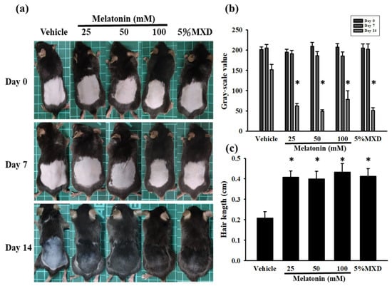
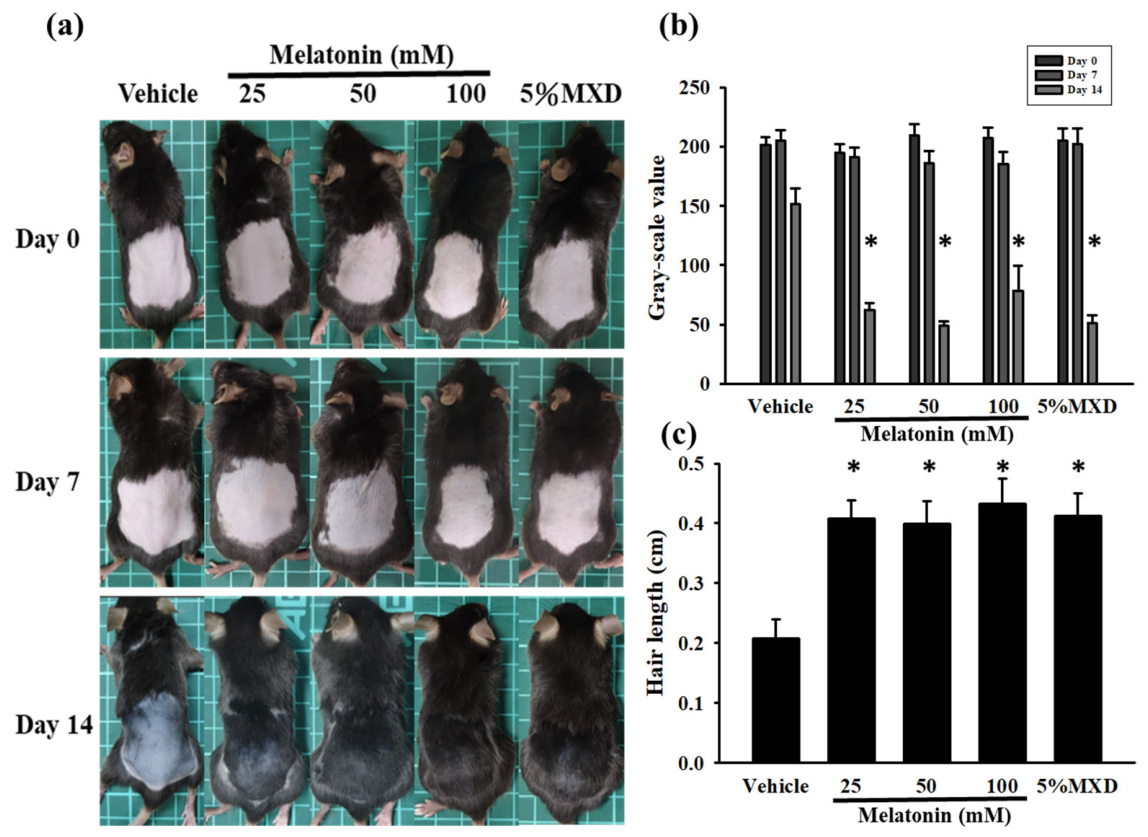
The ability of melatonin to promote hair regrowth was tested in 7-week-old male C57BL/6 mice. After hair removal on Day 1, all mice had pink skin on the dorsal area. The black pigmentation of the skin is a hallmark of the transition of the hair growth cycle from telogen to anagen. Topical application of both melatonin (25, 50, and 100 mM) and 5% minoxidil led to a darker dorsal skin color than that in the vehicle group on Day 7 (Figure 1a). On Day 14, both the melatonin and minoxidil groups exhibited more hair regrowth over the shaved area than that observed in the vehicle group (Figure 1a). The treatment groups also exhibited darker pigmentation than the vehicle group. In C57BL/6 mice, dorsal skin darkening reflects anagen-associated follicular melanogenesis and is commonly used as a surrogate marker of anagen entry. Thus, the reduced grayscale values on Day 7 indicated increased anagen-associated pigmentation, whereas the greater hair coverage and hair length on Day 14 provided direct evidence of hair regrowth. As shown in Figure 1b, the melatonin-treated groups exhibited significantly lower mean grayscale values than the vehicle group, consistent with increased anagen-associated pigmentation (p < 0.05). As shown in Figure 1c, we determined the hair length by randomly harvesting 10 hair shafts. The results revealed that hair length was significantly increased in all melatonin groups (p < 0.05).
Figure 1.
Melatonin promotes hair regrowth in C57BL/6 mice. (a) Representative images of hair growth on the dorsal skin of C57BL/6 mice on Days 1, 7, and 14 following topical treatment with melatonin or 5% minoxidil. (b) Mean gray values of the same anatomically defined dorsal skin region on Days 1, 7, and 14, quantified using ImageJ after conversion to 8-bit grayscale images. Pixel values ranged from 0 (black) to 255 (white); lower mean gray values indicated darker dorsal skin pigmentation associated with anagen entry. (c) Hair length measured on Day 14. Ten hairs were randomly collected from each mouse, and the mean hair length for each mouse was used for statistical analysis. Data are presented as the mean ± SEM; n = 3 independent mice per group. Statistical comparisons were performed using one-way ANOVA followed by Bonferroni’s post hoc multiple-comparison test. * p < 0.05 versus the vehicle group.
3.2. Melatonin Induced the Anagen Cycle
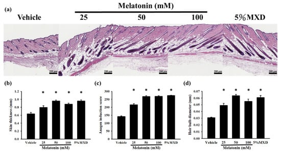
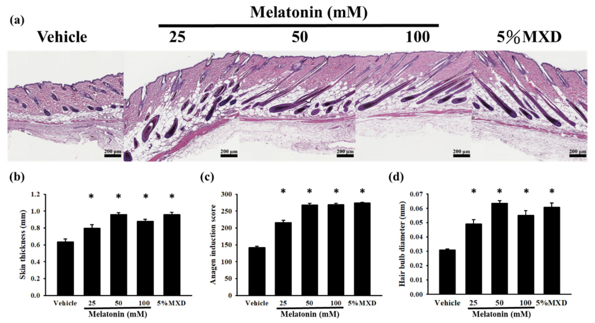
Histological analysis of skin tissue, including skin thickness, anagen induction score, and hair bulb diameter, was performed (Figure 2a) to explore the effect of topical melatonin on the development of skin tissue and hair follicles in mice. In the longitudinal sections of the dorsal skin on Day 14, melatonin significantly increased skin thickness in C57BL/6 mice compared with vehicle mice (Figure 2b, p < 0.05). Next, we observed some changes: the location of the hair follicle was deeper, the shape of the dermal papilla cell and hair bulb was larger, and keratinocytes and the newborn hair shaft were elongated. The anagen induction score was calculated according to these anagen features (Figure 2c). The anagen induction score and hair bulb diameter were significantly higher in the melatonin group than in the vehicle group (Figure 2c,d, both p < 0.05). These results indicated that melatonin promoted hair growth by accelerating the transition from the telogen phase to the anagen phase.
Figure 2.
Histological effect of melatonin treatment in C57BL/6 mice. (a) Histological visualization of the dorsal skin from each group. (b) Skin thickness of each group on Day 14 (measured using ImageJ). (c) The anagen induction score of each group was recorded at Day 14. (d) Hair bulb diameter on Day 14 (measured using ImageJ). Data are presented as the mean ± SEM; n = 3 independent mice per group. Statistical comparisons were performed using one-way ANOVA followed by Bonferroni’s post hoc multiple-comparison test. * p < 0.05 versus the vehicle group.
3.3. β-Catenin Activation Was Regulated by Melatonin in Hair Follicles
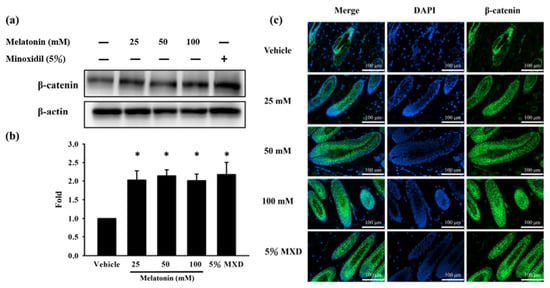
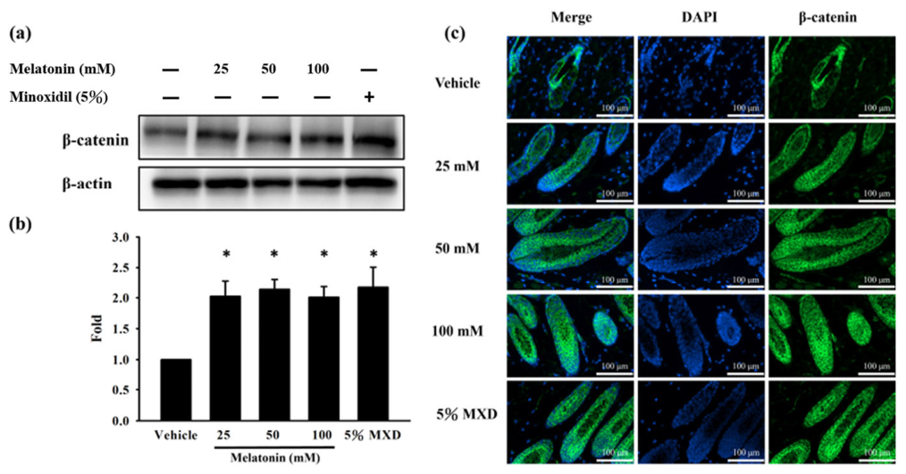
To explain the molecular mechanism of anagen phase induction in the melatonin group, immunofluorescence analysis was performed. To explore whether the Wnt/β-catenin signaling pathway was modulated by melatonin in hair follicles, the β-catenin expression of dorsal skin tissue was analyzed using Western blotting, and the longitudinal sections of the dorsal skin on Day 14 were examined through immunofluorescence staining with anti-β-catenin antibodies (Figure 3a–c). Compared with the vehicle group, both the melatonin and minoxidil groups exhibited upregulated β-catenin expression in the hair follicles. These findings indicate that melatonin-induced hair follicle activation was associated with increased β-catenin expression.
Figure 3.
Expression of β-catenin in hair follicle of C57BL/6 mice. Western blot analysis of β-catenin expression on Day 14 of melatonin/minoxidil treatment. β-actin was used as the loading control for whole-cell lysates. (a) Representative β-catenin expression in dorsal skin tissue samples in the vehicle, melatonin, and minoxidil groups. (b) Comparison of β-catenin expression between the various treatment and vehicle groups. Data are presented as the mean ± SEM; n = 3 independent mice per group. Statistical comparisons were performed using one-way ANOVA followed by Bonferroni’s post hoc multiple-comparison test. * p < 0.05 versus the vehicle group. (c) Immunofluorescence staining displaying β-catenin (green) in hair follicles with DAPI (blue) counterstaining.
3.4. Melatonin Regulated the Wnt/β-Catenin Signaling Pathway
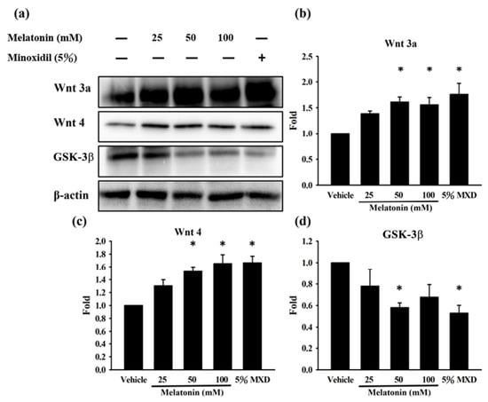
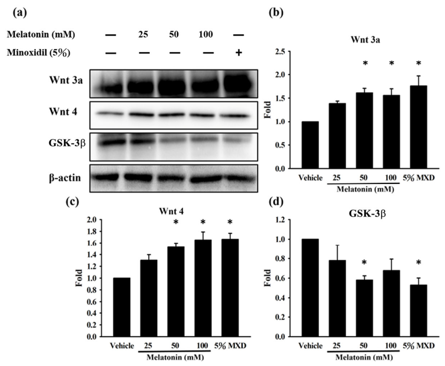
We explored the potential molecular mechanisms underlying the effects of melatonin on the hair cycle. Figure 4 presents the between-group comparisons of the expression levels of other typical proteins involved in the Wnt/β-catenin signaling pathway. The melatonin group exhibited significantly higher wnt3a and wnt4 and lower GSK-3β expression levels than the vehicle group (p < 0.05). At 50 mM, melatonin significantly increased Wnt3a and Wnt4 expression and decreased GSK-3β expression compared with the vehicle group, indicating coordinated modulation of multiple components of the Wnt/β-catenin signaling pathway. Although Wnt4 exhibited the clearest numerical increase across the tested concentrations, increasing the melatonin concentration to 100 mM did not consistently produce a greater effect on all pathway-associated proteins. Therefore, these findings are more consistent with a threshold or plateau-like response than with a linear concentration-dependent relationship. These results indicate that melatonin-induced hair growth was accompanied by modulation of Wnt/β-catenin-related proteins.
Figure 4.
Activity of Wnt/β-catenin signal pathway in hair follicle of C57BL/6 mice. Western blot analysis of Wnt3a, Wnt4, GSK-3β in following 14 days of melatonin and minoxidil treatment. β-actin was used as the loading control for whole-cell lysates. (a) Representative level of proteins in dorsal skin in the vehicle, melatonin, and minoxidil groups. (b) Changes in Wnt3a expression between the treatment and vehicle groups. (c) Changes in Wnt4 expression between the treatment and vehicle groups. (d) Changes in GSK-3β expression between the treatment and vehicle groups. Data are presented as the mean ± SEM; n = 3 independent mice per group. Statistical comparisons were performed using one-way ANOVA followed by Bonferroni’s post hoc multiple-comparison test. * p < 0.05 versus the vehicle group.
3.5. Nuclear Translocation of β-Catenin Was Induced by Melatonin in C57BL/6 Mice
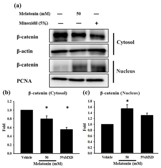
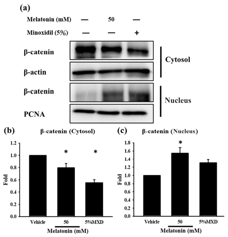
To assess the subcellular distribution of β-catenin, 50 mM melatonin was selected as the intermediate effective concentration for the cytosolic and nuclear fractionation analysis. Nuclear translocation of β-catenin was a crucial step to activating the Wnt/β-catenin signaling pathway. Expression of proteins in the cytosol and nucleus was calculated relative to the vehicle group. As shown in Figure 5a–c, the melatonin-treated group exhibited significantly higher β-catenin expression levels in nucleus and lower β-catenin expression levels in the cytosol. These findings indicate that topical melatonin-induced hair regrowth was accompanied by decreased cytosolic β-catenin and increased nuclear β-catenin, consistent with β-catenin stabilization and nuclear redistribution. Direct statistical comparison showed no significant difference between the 50 mM melatonin and 5% minoxidil groups in both nucleus and cytosolic β-catenin expression.
Figure 5.
Nuclear translocation of β-catenin signal by melatonin in hair follicle of C57BL/6 mice. The 50 mM melatonin concentration was selected as the intermediate effective concentration for the fractionation analysis. Western blot analysis of β-catenin translocation in following 14 days of melatonin and minoxidil treatment. β-actin and PCNA were used as loading controls for the cytosolic and nuclear fractions, respectively. Cytosolic and nuclear β-catenin levels were normalized to β-actin and PCNA, respectively. (a) Cytosolic and nuclear levels of β-catenin in the vehicle group, melatonin and minoxidil group. (b) The change in fold of cytosolic β-catenin expression between the treated and vehicle groups was calculated. (c) The change in fold of nuclear β-catenin expression between the treated and vehicle groups was calculated. Data are presented as the mean ± SEM; n = 3 independent mice per group. Statistical comparisons were performed using one-way ANOVA followed by Bonferroni’s post hoc multiple-comparison test. * p < 0.05 versus the vehicle group.
4. Discussion
Hair follicle development can be divided into three phases: growth (anagen), transitional (catagen), and rest (telogen) [3]. Hair follicle activity is a representative target for exploring the pathways of cell proliferation, cell differentiation, and hair growth. Hair loss or depigmentation can affect social perception and emotional and psychological health, with hair loss diseases including androgenic alopecia, alopecia areata, and anagen effluvium [26]. Hair loss can also occur due to genetic, endocrine, immunological, and inflammatory conditions as well as cancer treatment [2]. The commonly used drugs for hair loss treatment—minoxidil and finasteride—can promote the hair growth cycle but also exert adverse effects, such as allergic reactions and sexual dysfunction, respectively. Therefore, a hair growth-promoting drug with no undesirable side effects is required. Studies have reported that melatonin may directly or indirectly promote hair growth. A clinical study indicated that melatonin was effective in treating androgenic alopecia [13], but its effect on hair growth in other alopecia conditions remains unknown. An in vitro study indicated that melatonin could increase dermal papilla cell growth by activating the AKT/GSK-3β/β-Catenin signaling pathway [27] and may thus promote hair growth. However, no study has investigated how melatonin promotes hair growth and regulates the potential pathways in mice. Our results showed that topical melatonin promoted hair follicle growth and was accompanied by the modulation of Wnt/β-catenin-related signaling proteins in C57BL/6 mice.
Following depilation of the dorsal skin, the hair follicles undergo hair regrowth in C57BL/6 telogen mice [27] by activating the anagen phase within 2 weeks [28,29]. After 2 weeks, rapid hair follicle growth on the dorsal skin confirms the occurrence of the anagen phase [29,30]. Following depilation, dorsal skin darkening in C57BL/6 mice reflects anagen-associated follicular melanogenesis and serves as a surrogate marker of anagen entry [31]. Therefore, the lower grayscale values in melatonin-treated mice indicated accelerated anagen progression rather than a direct pigmentation-driven effect on hair growth. This interpretation was further supported by increased hair length. Together, these results suggest that melatonin promoted hair growth in C57BL/6 mice.
During the anagen cycle, the shape and size of the hair follicles were rounder, larger, and plumper. Moreover, the location of the hair follicle was deeper at anagen IV–VI [22,32]. In the present study, the hair follicles in the dorsal skin of the melatonin-treated groups were longer and narrower compared with the vehicle group, and their hair bulbs were plumper. Moreover, the hair follicles were deeper in all groups. These features indicated that the hair follicles were in anagen IV–VI. Taken together, these morphological and histological characteristics highlight the hair growth-promoting effects of melatonin. During the entire experiment, we did not observe any edema, erythema, or dryness on the dorsal skin. No grossly visible erythema, edema, or dryness was observed during the 14-day treatment period. However, this observation provides only preliminary evidence of short-term local tolerability and does not establish dermal or systemic safety.
Anagen activation is related to various signaling pathways [33,34], especially the Wnt/β-catenin pathway [35,36]. The Wnt family is a group of secreted proteins that are expressed during the development of the embryonic epidermis, hair placodes, and hair follicles [37]. Wnt3a and Wnt4 are involved in hair growth, including regeneration of the hair cycle, upregulation of genes in anagen, and repair of hair damage [6,37,38]. Upon binding to receptors, Wnt3a and Wnt4 can inactivate GSK-3β, which is responsible for β-catenin ubiquitination and phosphorylation [39,40]. Thereafter, the nuclear translocation of stabilized β-catenin occurs, and β-catenin binds to T-cell factor (TCF)/lymphoid enhancer factor, which regulates the anagen cycle [6,41]. β-catenin promotes the transcriptional activation of hair growth-promoting genes [41]. A study indicated that melatonin upregulated β-catenin in rat hair bulbs [42]. Specifically, another study demonstrated in a depilation-synchronized mouse model that melatonin accelerated hair regeneration, whereas the MT1/MT2 receptor antagonist luzindole attenuated this effect; Wnt/β-catenin-related gene expression and β-catenin signaling, particularly in dermal papillae, were also increased [16]. However, receptor antagonists were not used in the present study. Therefore, whether the observed hair-growth effects and Wnt/β-catenin-related changes were mediated through MT1 or MT2 signaling remains undetermined. Our study is the first to analyze β-catenin expression in the hair follicle by using an immunofluorescence assay: β-catenin was upregulated in all treatment groups compared with the vehicle group.
At 50 mM, melatonin significantly increased Wnt3a and Wnt4 expression and reduced GSK-3β expression relative to the vehicle group, indicating coordinated modulation of multiple components of the Wnt/β-catenin pathway rather than selective regulation of Wnt4 alone. Although Wnt4 showed the clearest numerical increase across the tested concentrations, the responses of Wnt3a, Wnt4, and GSK-3β did not consistently increase further at 100 mM. This pattern is therefore more appropriately interpreted as a threshold or plateau-like response rather than a linear concentration-dependent effect. WNT4 has been reported to increase during the telogen-to-anagen transition in human hair follicles, supporting its potential relevance to anagen induction [43]. Nevertheless, because Wnt4-specific inhibition or genetic manipulation was not performed, the present findings identify Wnt4 as a potential contributor within broader Wnt signaling network rather than as the principal or indispensable mediator of melatonin-induced hair regrowth. Taken together, these findings support the biological plausibility that melatonin modulates Wnt/β-catenin signaling during hair-cycle progression [15,16]. However, the concurrent changes observed in the present study demonstrate an association rather than a causal relationship. Because melatonin receptor antagonists, Wnt/β-catenin inhibitors, and genetic pathway-interference approaches were not employed, the necessity of this pathway for melatonin-induced hair growth remains to be established.
The present findings have potential translational relevance to human hair follicle physiology. In human dermal papilla spheroids, melatonin promotes hair-inductive properties through receptor-dependent AKT/GSK-3β/β-catenin signaling [15], while Wnt pathway remodeling also occurs during the human telogen-to-anagen transition [43]. Clinical studies further suggest that topical melatonin may increase the anagen hair rate [14]. Nevertheless, translation from depilation-synchronized C57BL/6 mice remains limited because murine dorsal follicles enter anagen synchronously, whereas human scalp follicles cycle asynchronously and are additionally influenced by androgen sensitivity, follicular miniaturization, age, and scalp location [44]. Therefore, our findings support the biological plausibility of melatonin-associated Wnt/β-catenin modulation in human hair follicles but require further validation in human follicle models and clinical studies.
Although topical melatonin significantly promoted several anagen-associated outcomes across the tested concentration range, most endpoints did not exhibit a consistent linear dose–response relationship. The broadly comparable effects observed at 25–100 mM may indicate that the effective biological threshold had already been reached at the lower end of the tested concentration range. Receptor saturation, restricted follicular penetration, and a ceiling effect associated with depilation-induced synchronization may also contribute to this nonlinear pattern. However, these possibilities were not directly examined in the present study.
Niu et al. used outbred ICR mice as a general-purpose pharmacological model, whereas the present study employed inbred C57BL/6 mice, which are commonly used in hair-growth studies because their dorsal hair follicles exhibit a well-characterized synchronized hair cycle and pigmentation changes associated with anagen entry [16,44]. In the study by Niu et al., melatonin was administered orally at 40 mg/kg/day (1.2 mg/30 g mouse) [16]. In contrast, melatonin was applied topically in the present study at 25, 50, or 100 mM in 30 µL over an approximately 6 cm2 dorsal area, corresponding to nominal applied doses of 5.8, 11.6, and 23.2 mg/kg/day, respectively, or 29, 58, and 116 µg/cm2. However, direct dose comparisons between oral and topical administration are limited because cutaneous absorption, follicular bioavailability, and systemic exposure were not determined. Therefore, although topical administration may facilitate localized delivery to hair-bearing skin, its pharmacokinetic and efficacy advantages over oral administration remain to be established.
The concentrations of 25, 50, and 100 mM were selected as an exploratory range to evaluate whether increasing topical melatonin concentrations produced greater hair-growth effects. These concentrations correspond to approximately 0.58%, 1.16%, and 2.32% (w/v), respectively, and are relatively high compared with concentrations previously evaluated clinically, such as a randomized placebo-controlled trial that used a 0.1% topical melatonin once daily for 6 months [14]. Importantly, the 100 mM formulation represented only the highest concentration examined in the present study and should not be interpreted as a maximum tolerated or maximum non-toxic concentration. Moreover, several morphological, histological, and molecular endpoints did not exhibit a consistent monotonic concentration–response relationship across the 25–100 mM range, suggesting that higher concentrations may not necessarily provide additional efficacy. Because concentrations above 100 mM were not evaluated, the present study cannot define the minimally effective, optimal, or maximum tolerated concentration. Accordingly, these findings should be regarded as proof-of-concept evidence rather than as a basis for direct clinical dosing. Although no grossly visible erythema, edema, or dryness was observed during the 14-day treatment period, standardized dermal irritation, systemic exposure, follicular drug concentrations, long-term dermal safety, and organ toxicity were not assessed. Therefore, the present findings do not establish that topical melatonin is safer or more effective than oral or systemic administration. Further studies using lower and clinically relevant concentrations, together with pharmacokinetic and comprehensive toxicological evaluations, are warranted.
Converging evidence indicates that hair follicle regeneration and anagen initiation depend on Wnt-mediated stabilization and nuclear accumulation of β-catenin. In the epidermis, Wnt-coupled mechanotransduction drives nuclear β-catenin enrichment and regeneration [45]. The myokine irisin promotes the telogen-to-anagen transition by inhibiting GSK-3β and activating β-catenin signaling [46]. In a murine model of androgenetic alopecia, mesenchymal stem cell therapy restores follicular β-catenin signaling and enhances hair growth [47]. Collectively, β-catenin nuclear translocation emerges as a key event enabling anagen entry and proliferative maintenance. Consistent with this mechanism, Western blotting showed that topical melatonin (50 mM) decreased cytoplasmic β-catenin and increased nuclear β-catenin compared with the vehicle group; a similar pattern was observed with 5% minoxidil (Figure 5). These data indicate β-catenin translocation from the cytoplasm to the nucleus in response to topical melatonin treatment in C57BL/6 mice. Nuclear accumulation of β-catenin is closely associated with anagen entry and follicular proliferation, plausibly facilitating the telogen-to-anagen transition and accounting for the pro-anagen effects observed in our model.
Several limitations should be considered when interpreting the present findings. First, the sample size was limited to three mice per group, which reduced statistical power and the sensitivity of normality and variance assessments. Complete investigator blinding was not implemented throughout all experimental procedures. Although standardized quantitative methods were used, potential observer bias cannot be completely excluded. In addition, a precise anatomical positioning template and fully standardized photographic acquisition conditions were not prospectively applied. Therefore, minor regional variations in hair sampling and possible effects of lighting or exposure on gray-scale measurements cannot be excluded. The present findings should consequently be regarded as exploratory proof-of-concept evidence and confirmed in larger, prospectively powered, and fully blinded studies. Second, the mechanistic evidence remains associative. Although coordinated changes in Wnt3a, Wnt4, GSK-3β, total β-catenin, and β-catenin subcellular distribution support the involvement of Wnt/β-catenin-related signaling, no melatonin receptor antagonist, pathway inhibitor, or genetic interference approach was used. Therefore, the requirement of this pathway and the specific involvement of MT1 or MT2 receptors remain unconfirmed. Furthermore, independent fraction-purity markers were not evaluated. Although β-actin and PCNA were used as loading controls for the cytosolic and nuclear fractions, respectively, minor cross-contamination cannot be completely excluded, and the β-catenin redistribution data should be interpreted as supportive rather than definitive evidence. Third, the study was not designed to establish a formal concentration–response relationship. Only three relatively high topical concentrations, a 14-day treatment period, and a single terminal molecular time point were evaluated. Consequently, the minimally effective, optimal, and maximum tolerated concentrations remain undetermined, as does the durability of the hair-growth response after treatment discontinuation. Finally, safety assessment was limited to gross visual examination of the treated skin. Standardized dermal irritation scoring, body-weight monitoring, systemic exposure, serum biochemical analysis, histopathological toxicity, and organ toxicity were not evaluated. Additional efficacy endpoints, including follicle density, hair shaft diameter, and follicular proliferation markers such as Ki-67, were also not assessed. Future studies should incorporate longer treatment and follow-up periods, broader efficacy measurements, clinically relevant concentrations, and comprehensive pharmacokinetic and toxicological evaluations.
5. Conclusions
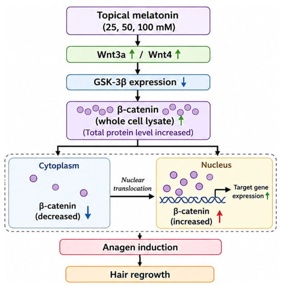
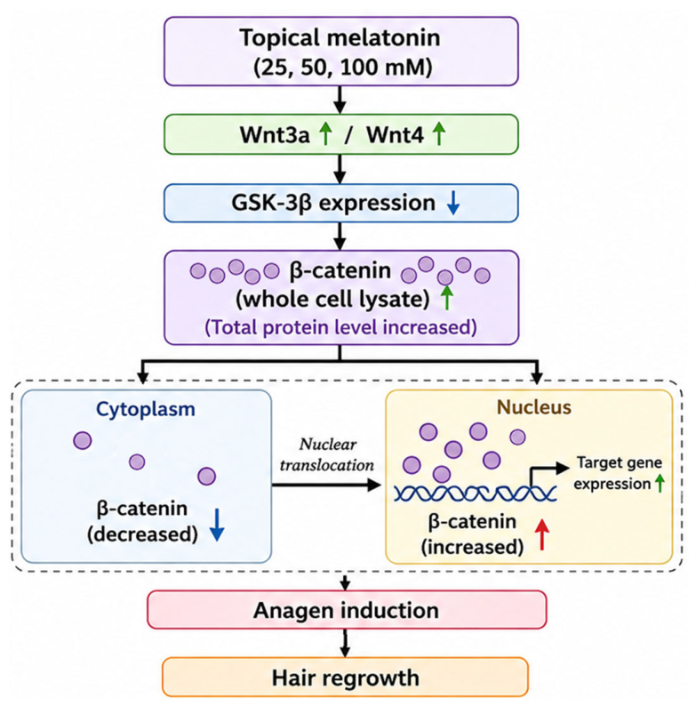
In conclusion, topical melatonin promoted anagen entry and hair regrowth in C57BL/6 mice and was accompanied by the modulation of Wnt/β-catenin-related signaling proteins (Figure 6). Western blotting showed that β-catenin expression was significantly increased at all tested melatonin concentrations (25–100 mM) compared with the vehicle group, with expression levels numerically similar to those observed in the 5% minoxidil group. Immunofluorescence analysis further demonstrated stronger follicular β-catenin signals. Melatonin also increased Wnt3a and Wnt4 expression while decreasing GSK-3β expression. In addition, melatonin treatment was associated with increased nuclear accumulation of β-catenin, consistent with canonical Wnt/β-catenin signaling during anagen. Collectively, these findings support the potential involvement of the Wnt/β-catenin pathway in melatonin-induced hair regrowth, although further mechanistic studies are warranted.
Figure 6.
Proposed association between topical melatonin treatment, Wnt/β-catenin-related signaling, anagen induction, and hair regrowth in C57BL/6 mice. Topical melatonin treatment was associated with increased Wnt3a and Wnt4 expression, decreased GSK-3β expression, and elevated total β-catenin in whole cell lysates. Consistent with these changes, cytosolic β-catenin decreased, while nuclear β-catenin increased, accompanied by upregulation of target gene expression, anagen induction, and subsequent hair regrowth. Arrows indicate changes directly in the present study. Upward and downward arrows indicate increased and decreased protein expression, respectively, compared to the vehicle group.
Author Contributions
Conceptualization, M.-W.L., Y.-H.K. and M.-K.H.; Funding Acquisition, W.-Y.C. and Y.-H.K.; Investigation, M.-W.L., S.-C.L., W.-Y.C., C.-J.C., Y.-H.K. and M.-K.H.; Methodology, M.-W.L., S.-C.L., W.-Y.C. and C.-J.C.; Project Administration, Y.-H.K. and M.-K.H.; Validation, Y.-H.K. and M.-K.H.; Writing—Original Draft, M.-W.L., S.-C.L., W.-Y.C. and C.-J.C.; Writing—Review & Editing, Y.-H.K. and M.-K.H. All authors have read and agreed to the published version of the manuscript.
Funding
This study received financial support from the National Science and Technology Council (NSTC), Taiwan (NSTC 113-2320-B-040-014-MY3). The authors would like to thank the National Chung Hsing University and Chung Shan Medical University (NCHU-CSMU 11408).
Institutional Review Board Statement
All experiments on mice were conducted at the Laboratory Animal Center of Chung Shan Medical University (Taichung, Taiwan) were performed in accordance with the guidelines of the Institutional Animal Ethics Committee of Chung Shan Medical University, No. 2755, dated 18 October 2022 (Taichung, Taiwan).
Informed Consent Statement
Not applicable.
Data Availability Statement
Data will be made available on request. The data that support the findings of this study are available from the corresponding author upon reasonable request.
Conflicts of Interest
The authors declare no conflict of interest.
References
- Hoover, E.; Alhajj, M.; Flores, J.L. Physiology, Hair. In StatPearls; StatPearls Publishing: Treasure Island, FL, USA, 2023. [Google Scholar]
- Wolff, H.; Fischer, T.W.; Blume-Peytavi, U. The Diagnosis and Treatment of Hair and Scalp Diseases. Dtsch. Arztebl. Int. 2016, 113, 377–386. [Google Scholar] [CrossRef] [Scilit] [PubMed]
- Owecka, B.; Tomaszewska, A.; Dobrzeniecki, K.; Owecki, M. The Hormonal Background of Hair Loss in Non-Scarring Alopecias. Biomedicines 2024, 12, 513. [Google Scholar] [CrossRef] [Scilit] [PubMed]
- Houschyar, K.S.; Borrelli, M.R.; Tapking, C.; Popp, D.; Puladi, B.; Ooms, M.; Chelliah, M.P.; Rein, S.; Pförringer, D.; Thor, D.; et al. Molecular Mechanisms of Hair Growth and Regeneration: Current Understanding and Novel Paradigms. Dermatology 2020, 236, 271–280. [Google Scholar] [CrossRef] [Scilit] [PubMed]
- Wang, W.-H.; Ramos, R.; Tai, K.-Y.; Wu, Y.-S.; Chang, T.-Y.; Yan, J.-Y.; Plikus, M.V.; Oh, J.W.; Lin, S.-J. Studying Hair Growth Cycle and Its Effects on Mouse Skin. J. Investig. Dermatol. 2023, 143, 1638–1645. [Google Scholar] [CrossRef] [Scilit] [PubMed]
- Choi, B.Y. Targeting Wnt/β-Catenin Pathway for Developing Therapies for Hair Loss. Int. J. Mol. Sci. 2020, 21, 4915. [Google Scholar] [CrossRef] [Scilit] [PubMed]
- Lim, X.; Tan, S.H.; Yu, K.L.; Lim, S.B.; Nusse, R. Axin2 Marks Quiescent Hair Follicle Bulge Stem Cells Maintained by Autocrine Wnt/β-Catenin Signaling. Proc. Natl. Acad. Sci. USA 2016, 113, E1498–E1505. [Google Scholar] [CrossRef] [Scilit] [PubMed]
- Liu, J.; Xiao, Q.; Xiao, J.; Niu, C.; Li, Y.; Zhang, X.; Zhou, Z.; Shu, G.; Yin, G. Wnt/β-Catenin Signalling: Function, Biological Mechanisms, and Therapeutic Opportunities. Signal Transduct. Target. Ther. 2022, 7, 3. [Google Scholar] [CrossRef] [Scilit] [PubMed]
- Claustrat, B.; Gronfier, C. Melatonin: Some Aspects of Biochemistry, Physiology, and Pharmacology. Adv. Tech. Stand. Neurosurg. 2026, 54, 49–78. [Google Scholar] [CrossRef] [Scilit] [PubMed]
- Cruz-Sanabria, F.; Carmassi, C.; Bruno, S.; Bazzani, A.; Carli, M.; Scarselli, M.; Faraguna, U. Melatonin as a Chronobiotic with Sleep-Promoting Properties. Curr. Neuropharmacol. 2023, 21, 951–987. [Google Scholar] [CrossRef] [Scilit] [PubMed]
- Reiter, R.J.; Mayo, J.C.; Tan, D.X.; Sainz, R.M.; Alatorre-Jimenez, M.; Qin, L. Melatonin as an Antioxidant: Under Promises but Over Delivers. J. Pineal Res. 2016, 61, 253–278. [Google Scholar] [CrossRef] [Scilit] [PubMed]
- Ozdemir-Sanci, T.; Sanci, A.; Alimogulları, E.; Karakoyunlu, A.N. Effects of Melatonin on Different Stages of Bladder Cancer Survival. Bull. Urooncol. 2024, 23, 100–105. [Google Scholar] [CrossRef] [Scilit]
- Fischer, T.W.; Trüeb, R.M.; Hänggi, G.; Innocenti, M.; Elsner, P. Topical Melatonin for Treatment of Androgenetic Alopecia. Int. J. Trichology 2012, 4, 236–245. [Google Scholar] [CrossRef] [Scilit] [PubMed]
- Fischer, T.W.; Burmeister, G.; Schmidt, H.W.; Elsner, P. Melatonin Increases Anagen Hair Rate in Women with Androgenetic Alopecia or Diffuse Alopecia: Results of a Pilot Randomized Controlled Trial. Br. J. Dermatol. 2004, 150, 341–345. [Google Scholar] [CrossRef] [Scilit] [PubMed]
- Bae, S.; Yoon, Y.G.; Kim, J.Y.; Park, I.C.; An, S.; Lee, J.H.; Bae, S. Melatonin Increases Growth Properties in Human Dermal Papilla Spheroids by Activating AKT/GSK3β/β-Catenin Signaling Pathway. PeerJ 2022, 10, e13461. [Google Scholar] [CrossRef] [Scilit] [PubMed]
- Niu, Y.-L.; Li, Y.-K.; Gao, C.-X.; Li, W.-W.; Li, L.; Wang, H.; Shen, W.; Ge, W. Melatonin Promotes Hair Regeneration by Modulating the Wnt/β-Catenin Signalling Pathway. Cell Prolif. 2024, 57, e13656. [Google Scholar] [CrossRef] [Scilit] [PubMed]
- Greco, G.; Di Lorenzo, R.; Ricci, L.; Di Serio, T.; Vardaro, E.; Laneri, S. Clinical Studies Using Topical Melatonin. Int. J. Mol. Sci. 2024, 25, 5167. [Google Scholar] [CrossRef] [Scilit] [PubMed]
- Babadjouni, A.; Reddy, M.; Zhang, R.; Raffi, J.; Phong, C.H.; Mesinkovska, N.A. Melatonin and the Human Hair Follicle. J. Drugs Dermatol. 2023, 22, 260–264. [Google Scholar] [CrossRef] [Scilit] [PubMed]
- Curtis, A.; Calabro, K.; Galarneau, J.R.; Bigio, I.J.; Krucker, T. Temporal Variations of Skin Pigmentation in C57BL/6 Mice Affect Optical Bioluminescence Quantitation. Mol. Imaging Biol. 2011, 13, 1114–1123. [Google Scholar] [CrossRef] [Scilit] [PubMed]
- Lin, X.; Zhu, L.; He, J. Morphogenesis, Growth Cycle and Molecular Regulation of Hair Follicles. Front. Cell Dev. Biol. 2022, 10, 899095. [Google Scholar] [CrossRef] [Scilit] [PubMed]
- Choi, M.; Choi, S.-J.; Jang, S.; Choi, H.-I.; Kang, B.-M.; Hwang, S.T.; Kwon, O. Shikimic Acid, a Mannose Bioisostere, Promotes Hair Growth with the Induction of Anagen Hair Cycle. Sci. Rep. 2019, 9, 17008. [Google Scholar] [CrossRef] [Scilit] [PubMed]
- Zhang, Y.; Ni, C.; Huang, Y.; Tang, Y.; Yang, K.; Shi, X.; Zhang, Y.; Li, Z.; Wang, J.; Zhu, Y.; et al. Hair Growth-Promoting Effect of Resveratrol in Mice, Human Hair Follicles and Dermal Papilla Cells. Clin. Cosmet. Investig. Dermatol. 2021, 14, 1805–1814. [Google Scholar] [CrossRef] [Scilit] [PubMed]
- Lin, F.-C.; Lee, S.-S.; Li, Y.-C.; Ho, Y.-C.; Chen, W.-Y.; Chen, C.-J.; Lee, M.-W.; Yeh, K.-L.; Tsai, S.C.-S.; Kuan, Y.-H. Protective Effects of Kirenol against Lipopolysaccharide-Induced Acute Lung Injury through the Modulation of the Proinflammatory NF-κB Pathway and the AMPK2-/Nrf2-Mediated HO-1/AOE Pathway. Antioxidants 2021, 10, 204. [Google Scholar] [CrossRef] [Scilit] [PubMed]
- Ohn, J.; Kim, K.H.; Kwon, O. Evaluating Hair Growth Promoting Effects of Candidate Substance: A Review of Research Methods. J. Dermatol. Sci. 2019, 93, 144–149. [Google Scholar] [CrossRef] [Scilit] [PubMed]
- Yang, L.-C.; Chang, Y.-C.; Chiang, C.-Y.; Huang, F.-M.; Su, N.-Y.; Kuan, Y.-H.; Lee, S.-S. Protective Effect of Wogonin on Inflammatory Responses in BisGMA-Treated Macrophages through the Inhibition of MAPK and NF-κB Pathways. Environ. Toxicol. 2022, 37, 3007–3012. [Google Scholar] [CrossRef] [Scilit] [PubMed]
- Nestor, M.S.; Ablon, G.; Gade, A.; Han, H.; Fischer, D.L. Treatment Options for Androgenetic Alopecia: Efficacy, Side Effects, Compliance, Financial Considerations, and Ethics. J. Cosmet. Dermatol. 2021, 20, 3759–3781. [Google Scholar] [CrossRef] [Scilit] [PubMed]
- Li, C.; Feng, C.; Ma, G.; Fu, S.; Chen, M.; Zhang, W.; Li, J. Time-Course RNA-Seq Analysis Reveals Stage-Specific and Melatonin-Triggered Gene Expression Patterns during the Hair Follicle Growth Cycle in Capra hircus. BMC Genom. 2022, 23, 140. [Google Scholar] [CrossRef] [Scilit] [PubMed]
- Zhang, N.N.; Park, D.K.; Park, H.J. Hair Growth-Promoting Activity of Hot Water Extract of Thuja orientalis. BMC Complement. Altern. Med. 2013, 13, 9. [Google Scholar] [CrossRef] [Scilit] [PubMed]
- Park, S.; Lee, J. Modulation of Hair Growth Promoting Effect by Natural Products. Pharmaceutics 2021, 13, 2163. [Google Scholar] [CrossRef] [Scilit] [PubMed]
- Kim, Y.E.; Choi, H.C.; Nam, G.; Choi, B.Y. Costunolide Promotes the Proliferation of Human Hair Follicle Dermal Papilla Cells and Induces Hair Growth in C57BL/6 Mice. J. Cosmet. Dermatol. 2019, 18, 414–421. [Google Scholar] [CrossRef] [Scilit] [PubMed]
- Slominski, A.; Paus, R.; Costantino, R. Differential expression and activity of melanogenesis-related proteins during induced hair growth in mice. J. Investig. Dermatol. 1991, 96, 172–179. [Google Scholar] [CrossRef] [Scilit] [PubMed]
- Miyachi, K.; Yamada, T.; Kawagishi-Hotta, M.; Hasebe, Y.; Date, Y.; Hasegawa, S.; Arima, M.; Iwata, Y.; Kobayashi, T.; Numata, S.; et al. Extracellular Proteoglycan Decorin Maintains Human Hair Follicle Stem Cells. J. Dermatol. 2018, 45, 1403–1410. [Google Scholar] [CrossRef] [Scilit] [PubMed]
- Lee, J.H.; Choi, S. Deciphering the Molecular Mechanisms of Stem Cell Dynamics in Hair Follicle Regeneration. Exp. Mol. Med. 2024, 56, 110–117. [Google Scholar] [CrossRef] [Scilit] [PubMed]
- Kim, S.; Kim, J.Y.; Leem, J.W.; Jo, H.J.; Leem, J.W.; Kang, H.Y.; Han, E.-H.; Lee, B.-G.; Jiang, H.-H.; Roh, E.; et al. Hydrolyzed Keratin Peptide Enhances Hair Condition and Promotes Hair Regeneration. J. Med. Food 2026, 29, 555–566. [Google Scholar] [CrossRef] [Scilit] [PubMed]
- Oak, A.S.W.; Bagchi, A.; Brukman, M.J.; Toth, J.; Ford, J.; Zheng, Y.; Nace, A.; Yang, R.; Hsieh, J.-C.; Hayden, J.E.; et al. Wnt Signaling Modulates Mechanotransduction in the Epidermis to Drive Hair Follicle Regeneration. Sci. Adv. 2025, 11, eadq0638. [Google Scholar] [CrossRef] [Scilit] [PubMed]
- Wu, Z.; Zhu, Y.; Liu, H.; Liu, G.; Li, F. Wnt10b Promotes Hair Follicles Growth and Dermal Papilla Cells Proliferation via Wnt/β-Catenin Signaling Pathway in Rex Rabbits. Biosci. Rep. 2020, 40, BSR20191248. [Google Scholar] [CrossRef] [Scilit] [PubMed]
- Mäkelä, O.J.M.; Mikkola, M.L. Mesenchyme Governs Hair Follicle Induction. Development 2023, 150, dev202140. [Google Scholar] [CrossRef] [Scilit] [PubMed]
- Xiang, F.; Wang, P.; Gong, H.; Luo, J.; Zhou, X.; Zhan, C.; Hu, T.; Wang, M.; Xing, Y.; Guo, H.; et al. Wnt4 Increases the Thickness of the Epidermis in Burn Wounds by Activating Canonical Wnt Signalling and Decreasing the Cell Junctions between Epidermal Cells. Burn. Trauma 2023, 11, tkac053. [Google Scholar] [CrossRef] [Scilit] [PubMed]
- Hu, L.; Chen, W.; Qian, A.; Li, Y.-P. Wnt/β-Catenin Signaling Components and Mechanisms in Bone Formation, Homeostasis, and Disease. Bone Res. 2024, 12, 39. [Google Scholar] [CrossRef] [Scilit] [PubMed]
- Shi, X.; Liu, Y.; Huang, L.; Ou, Y.; Chen, X.; Ling, Y.; Zhang, Y.; Lin, Q. Minoxidil Ameliorates Myelodysplastic Syndrome by Targeting Wnt4 While Sparing Normal Hematopoiesis. Cell Commun. Signal. 2025, 23, 544. [Google Scholar] [CrossRef] [Scilit] [PubMed]
- Ma, X.; Xiang, F.; Pei, Z.; Miao, J.; Wu, P.; Song, X.; Li, Y.; Zhang, Y. Circ-Smad5 Retards the G1/S Transition of Cell Cycle via Inhibiting the Activity of Wnt/Lef/Cyclin D1 Signaling in JB6 Cells. Genes Dis. 2021, 8, 364–372. [Google Scholar] [CrossRef] [Scilit] [PubMed]
- Uslu, S.; Oktem, G.; Uysal, A.; Soner, B.C.; Arbak, S.; Ince, U. Stem Cell and Extracellular Matrix-Related Molecules Increase following Melatonin Treatment in the Skin of Postmenopausal Rats. Cell Biol. Int. 2014, 38, 924–932. [Google Scholar] [CrossRef] [Scilit] [PubMed]
- Hawkshaw, N.J.; Hardman, J.A.; Alam, M.; Jimenez, F.; Paus, R. Deciphering the molecular morphology of the human hair cycle: Wnt signalling during the telogen–anagen transformation. Br. J. Dermatol. 2020, 182, 1184–1193. [Google Scholar] [CrossRef] [Scilit] [PubMed]
- Castro, A.R.; Portinha, C.; Logarinho, E. The Emergent Power of Human Cellular vs Mouse Models in Translational Hair Research. Stem Cells Transl. Med. 2022, 11, 1021–1028. [Google Scholar] [CrossRef] [Scilit] [PubMed]
- Fu, J.; Liu, X.; Tan, L.; Cui, Z.; Liang, Y.; Li, Z.; Zhu, S.; Zheng, Y.; Yeung, K.W.K.; Chu, P.K.; et al. Modulation of the Mechanosensing of Mesenchymal Stem Cells by Laser-Induced Patterning for the Acceleration of Tissue Reconstruction through the Wnt/β-Catenin Signaling Pathway Activation. Acta Biomater. 2020, 101, 152–167. [Google Scholar] [CrossRef] [Scilit] [PubMed]
- Kim, Y.; Lee, J.M.; Jang, Y.N.; Park, A.Y.; Kim, S.-Y.; Kim, B.J.; Lee, J.O. Irisin Promotes Hair Growth and Hair Cycle Transition by Activating the GSK-3β/β-Catenin Pathway. Exp. Dermatol. 2024, 33, e15155. [Google Scholar] [CrossRef] [Scilit] [PubMed]
- Yan, W.; Liu, J.; Xie, X.; Jin, Q.; Yang, Y.; Pan, Y.; Zhang, Y.; Zhang, F.; Wang, Y.; Liu, J.; et al. Restoration of Follicular β-Catenin Signaling by Mesenchymal Stem Cells Promotes Hair Growth in Mice with Androgenetic Alopecia. Stem Cell Res. Ther. 2024, 15, 439. [Google Scholar] [CrossRef] [Scilit] [PubMed]
Disclaimer/Publisher’s Note: The statements, opinions and data contained in all publications are solely those of the individual author(s) and contributor(s) and not of MDPI and/or the editor(s). MDPI and/or the editor(s) disclaim responsibility for any injury to people or property resulting from any ideas, methods, instructions or products referred to in the content. |
© 2026 by the authors. Licensee MDPI, Basel, Switzerland. This article is an open access article distributed under the terms and conditions of the Creative Commons Attribution (CC BY) license.