


Abstract
The telomere is one of the biological hallmarks of skin aging. Sodium acetylated hyaluronate (SAH) is an acetylated derivative of sodium hyaluronate, produced by a grafting technology that confers higher resistance to hyaluronidase. This study aimed to explore whether SAH is associated with telomere-related endpoints and with clinical signs of skin aging. Two vitro studies were performed, assessing telomerase activity in human adult keratinocytes and telomere length in human normal fibroblasts by quantitative PCR (qPCR) assay kits, with or without SAH. A randomized, split-face, double-centered, double-blind, placebo-controlled clinical trial was then conducted on 44 Caucasian and Asian female volunteers, comparing a facial cream containing 0.1% SAH with a placebo cream over 28 days; crow’s feet wrinkle depth, area, and volume, under-eye roughness (Rz), elasticity (R2), and firmness (F4) were measured by PRIMOS 3D and Cutometer® MPA 580 at baseline, day 7, and day 28. In vitro, only the highest SAH concentration (0.15%) was associated with a significant increase in telomerase activity and in telomere length versus control, whereas lower concentrations had no significant effect. Clinically, the SAH cream significantly reduced crow’s feet wrinkle volume and area, improved skin elasticity, and reduced under-eye roughness versus placebo, with non-significant trends for wrinkle depth and firmness. These exploratory findings make SAH an interesting active ingredient for anti-aging skincare cosmetics, combining a curative dimension (clinical improvement of skin-aging parameters) with a preventive dimension (telomere preservation observed in vitro for the first time), and warrant further mechanistic investigation.
1. Introduction
Skin aging is a progressive and universal biological process that produces visible changes such as wrinkles, loss of elasticity, increased roughness, and reduced firmness [1,2]. It results from the combination of intrinsic (chronological) aging and extrinsic factors, in particular chronic exposure to ultraviolet radiation and other environmental stressors [1]. These changes are highly prevalent and accumulate steeply with age: in a community-based prospective cohort of 777 adults, the prevalence of severe photoaging rose from 42% to 88% over a 15-year follow-up [3]. Because the visible signs of facial aging strongly influence self-perception and quality of life, and because the proportion of older individuals in the population continues to grow, the prevention and management of skin aging remain an important biological concern and a central objective of the cosmetic industry. There is therefore a continuing need for cosmetic active ingredients whose anti-aging benefits can be linked, where possible, to well-defined biological mechanisms.
In recent years, the telomere has been recognized as one of the hallmarks of aging [4,5,6]. As an essential genetic element [7], the telomere is a specialized structure located at the end of eukaryotic chromosomes, made up of numerous copies of some simple G-rich repeats such as TTAGGG in humans [8]. Telomeres protect the ends of chromosomes from degradation or fusion and allow their complete replication [9]. Without telomeres, genetic instability occurs [8]. As cell division proceeds, telomeres are expected to become shorter in most normal cells due to the end-replication problem [10]. In humans, telomere length decreases with age, a phenomenon supported by evidence from both in vivo and in vitro studies [9,11]. A study conducted in the United Kingdom (UK) found that telomeres shortened steadily with age at an average rate of 27 base pairs (bp) per year in a large group of Caucasian women [12]. Acting as a mitotic clock [8], the shortening of telomeres to a critical or threshold length may initiate cell replicative senescence [7].
Telomere structure and length can be maintained by an enzyme complex called telomerase [8]. Telomerase consists of a protein component, the reverse transcriptase known as TERT (telomerase reverse-transcriptase), and an RNA component called TERC (telomerase RNA component), which is used by TERT as a template to add telomeric repeats to the chromosome end [8,13]. It has been suggested that the reduced efficiency of telomerase activity in humans with age leads to the shortening of the telomeres [9].
Telomeres and telomerase activity are thought to play an important role in the aging process of the skin [14,15]. Telomeres have been found to be shorter in skin fibroblasts and keratinocytes obtained from patients with dyskeratosis congenita (DC) [8]. Another study reported that mutations in telomerase components (e.g., TERT, TERC, dyskerin) in patients led to telomere shortening as well as skin atrophy [16]. Two complementary aspects of this biology can be evaluated experimentally: telomerase activity, which reflects the capacity of cells to counteract telomere attrition, and telomere length, which reflects the cumulative replicative history of the cells. At the tissue level, the progressive shortening of telomeres and the decline of telomerase activity are associated with the replicative senescence of dermal fibroblasts and epidermal keratinocytes; this senescence contributes to a reduced synthesis of extracellular matrix components and, ultimately, to the visible signs of skin aging such as wrinkle formation, loss of elasticity, and increased roughness [1,17]. Based on these observations, it is of interest to investigate the effects of cosmetic active ingredients on telomeres and/or telomerase, in order to provide more fundamental scientific evidence supporting modern skincare strategies for aging skin in the cosmetic industry.
Hyaluronic acid (HA) is a non-sulfated glycosaminoglycan (GAG) composed of repeating polymeric disaccharides of D-glucuronic acid and N-acetyl-D-glucosamine [18,19]. HA is broadly distributed in the extracellular matrix (ECM), especially in soft connective tissues [20]. In humans, HA is most abundant in the skin, which accounts for 50% of the total HA content in the body [21]. HA can bind up to 1000 times its weight in water and may help the skin retain and maintain moisture [1]. HA is considered an important molecule against skin aging [21]. HA and its salt form, sodium hyaluronate, are popular active ingredients used in cosmetic products.
More recently, an acetylated derivative of sodium hyaluronate, sodium acetylated hyaluronate (SAH), has been developed in the cosmetic industry. Natural HA is degraded by hyaluronidase, and the expression of this enzyme increases during the skin aging process [21]. SAH has been reported to exhibit higher resistance to hyaluronidase compared with classic sodium hyaluronate [22]. This enhanced enzymatic resistance was the main rationale for selecting SAH as the active ingredient in the present work: in contrast to unmodified HA, hydrolyzed HA, or low-molecular-weight HA, which are rapidly susceptible to hyaluronidase, the acetylated form is expected to remain functional in the skin for a longer period, implying a more durable anti-aging effect. SAH was therefore considered a suitable candidate for exploring whether an HA-derived molecule could be associated with telomere-related biological endpoints.
Beyond intrinsic and environmental factors, skin aging is also shaped by ethnic background, since skin structure, pigmentation, and the clinical presentation of aging differ across populations [23]. There is growing recognition of the importance of diversity, equity, and inclusion (DEI) in dermatology and skincare, with calls to ensure that research and product development reflect the full range of skin types and ethnicities rather than a single reference population [24]. To date, however, skin-aging research in general, and telomere biology in skin in particular, have been conducted predominantly in Caucasian populations [25,26,27,28]. Evaluating the anti-aging efficacy of cosmetic actives in more than one ethnic group is therefore valuable, both for the generalizability of the findings and for the goal of inclusive skincare.
To date, no study has investigated the effects of SAH on telomeres, which are one of the hallmarks of aging as described above. The objective of the present work was therefore to explore whether SAH is associated with telomere-related biological endpoints and with measurable clinical signs of skin aging. More specifically, the present work was designed to evaluate the ability of a SAH-containing cosmetic product to act through two complementary modes: a “curative” mode, reflected in the clinical improvement of wrinkles, skin elasticity, and skin firmness; and a “preventive” mode, reflected in the activation of telomerase and the inhibition of telomere shortening. To this end, three studies were conducted. Two in vitro studies were performed: the first evaluated the effect of SAH on telomerase activity in human adult keratinocytes, and the second evaluated the effect of SAH on telomere length in human normal fibroblasts. In addition, a randomized, split-face, double-centered, double-blind, placebo-controlled clinical trial was carried out to evaluate the anti-aging performance of a facial cream containing 0.1% SAH versus a placebo cream, measuring crow’s feet wrinkle depth, area, and volume, under-eye skin roughness (Rz), skin elasticity (R2), and skin firmness (F4). To embrace inclusiveness in skincare, this clinical trial enrolled both Caucasian and Asian female volunteers, so that the anti-aging performance of SAH could be evaluated in more than one population group rather than in a single reference population.
2. Materials and Methods
2.1. Sodium Acetylated Hyaluronate
The SAH (HyaInno™ ACE), produced by Shandong Tiansheng Biotechnology Co., Ltd. (TS-Biotech) (Linqu, China), was used for the present studies. Sodium hyaluronate obtained from a bio-fermentation process was used as the starting material, and a green acetylation process described in patent CN117586434A was performed to obtain SAH.
2.2. Human-Derived Skin Cells and Ethical Statement
Primary human epidermal keratinocytes and normal human dermal fibroblasts were isolated from anonymized discarded human skin obtained from abdominoplasty procedures. The skin samples were collected by French hospitals after prior informed consent had been obtained from the donors for the secondary use of surgical tissue for research purposes, in accordance with applicable French ethical and legal requirements, including the French Public Health Code, Articles L.1243-3 to L.1243-5.
The tissues were anonymized before transfer to the investigators through an authorized French human tissue/cell provider operating in compliance with applicable French regulations for the collection, preparation, conservation, and transfer of human biological material for scientific use. The investigators had no direct contact with the donors and had no access to any personal or identifying information at any stage.
As only anonymized discarded surgical tissue was used, and as no identifiable personal data were collected or accessible to the investigators, no specific ethics committee approval was required for the in vitro use of these samples under the applicable French regulatory framework.
All available donor and cell information is provided in the relevant experimental sections below. When detailed donor-level information was not available, this was due to donor anonymization and provider policy.
2.3. Telomerase Activity Study In Vitro
The aim of this study was to evaluate the effect of SAH on telomerase activity in a model composed of adult human keratinocytes at a low passage number in monolayer culture.
2.3.1. Procedure
For this experiment, the human middle-aged keratinocytes were used at a low passage number (i.e., passage 2 from cell isolation). The cells were cultivated in a monolayer until reaching approximately 75% confluency before being used in the test.
Romidepsin, also known as FK228, is a histone deacetylase inhibitor. In order to validate the study model, FK228 at 100 ng/mL was used as a reference inducer of telomerase activity (reference product), based on a previous study [29]. As FK228 has very low solubility in water [30], the versatile solvent dimethyl sulfoxide (DMSO) was used as the vehicle for FK228 in the test.
The cells at passage 2 were incubated for 24 h: (1) in the absence of any active ingredient (control group); (2) in the presence of the reference product and its vehicle, namely, DMSO at 0.01% (v/v) alone and DMSO at 0.01% (v/v) with FK228 at 100 ng/mL, respectively (reference group); or (3) in the presence of various SAH concentrations: 0.015%, 0.05%, and 0.15% (w/v), respectively (active group). The SAH, originally in powder form, was diluted directly in the incubation medium in order to reach the different concentrations described above.
At the end of the incubation period, telomerase was extracted from the cells by following the protocol detailed in the assay kit [31], and its activity was determined by quantitative polymerase chain reaction (qPCR) using a Telomerase Activity Quantification qPCR Assay Kit (TAQ) (catalog No. 8928, ScienCell Research Laboratories Inc., Carlsbad, CA, USA). The principle of this commercial kit couples a PCR step (in which telomerase performs elongation) with a qPCR step that allows the semi-quantitative determination of the amount of telomerase elongation product. The qPCR runs were performed using a QuantStudio™ qPCR system (Thermo Fisher Scientific Inc., Waltham, MA, USA). The telomerase activity is expressed as relative units, as mean ± standard deviation (S.D.). For each condition (control, reference, and each SAH concentration), three independent cultures were prepared and processed in parallel as separate experimental replicates ( per group).
2.3.2. Statistical Analysis
The telomerase activity is expressed as relative units, defined as the fold change in telomerase activity relative to the untreated control group (mean ± S.D.; per group). The level of statistical significance between the vehicle-only group and the reference-product group, used for model validation, was assessed using Student’s t-test. The control group and the three SAH concentrations of the active group (0.015%, 0.05%, and 0.15%, w/v) were then compared by a one-way analysis of variance (ANOVA) followed by Tukey’s multiple comparisons test, in which every group mean was compared with every other group mean (all pairwise comparisons among the four data groups). This unified analysis allowed both the comparison of each SAH concentration with the control and the comparison of the three SAH concentrations with one another. Statistical significance is based on a Tukey-corrected p-value: * pcorrected < 0.05; ** pcorrected < 0.01; *** pcorrected < 0.001; **** pcorrected < 0.0001; ns, not significant. These comparisons are additionally summarized by lowercase letters in the bar charts: groups sharing at least one common letter do not differ significantly, whereas groups sharing no common letter differ significantly (pcorrected < 0.05).
2.4. Telomere Length Study In Vitro
Previous experiments showed that telomere shortening could not be observed in human normal keratinocytes when these cells were used between passages 1 and 5, as human normal keratinocytes tend to dedifferentiate at higher passages. Human normal fibroblasts were therefore selected for this part of the study.
The aim of this study was to evaluate the effect of SAH on telomere shortening in a model composed of human normal fibroblasts in monolayer culture.
2.4.1. Procedure
A telomere model was developed by monitoring changes in telomere length with increasing cell passage. The experimental window was defined as three consecutive passages from passage 2 to passage 5, which allowed a significant variation of telomere length within a reasonable experiment duration. Telomere length was measured by qPCR after passage 2 (baseline) and after passage 5 (endpoint), and the two measurements were compared.
Starting after passage 2, the human middle-aged fibroblasts were carried through three consecutive passages until passage 5: (1) in the absence of any active ingredient (control group) or (2) in the presence of increasing SAH concentrations: 0.015%, 0.05%, and 0.15% (w/v), respectively (active group).
At the end of the incubation, the cells were trypsinized. DNA was extracted from the cells using a dedicated DNA extraction kit, and quantified with a Nanodrop™ spectrophotometer (Thermo Fisher Scientific Inc., Waltham, MA, USA). Telomere length was measured using an Absolute Human Telomere Length Quantification (AHTLQ) qPCR Assay Kit (catalog No. 8918, ScienCell Research Laboratories Inc., Carlsbad, CA, USA), following the standard protocol supplied with the kit [32]. For each sample, the variation in telomere length was measured by relative quantification using a single copy reference (SCR) gene as the reference gene. One qPCR run was performed for each sample with a telomere primer set that recognizes and amplifies telomere sequences. A second qPCR was then performed using an SCR primer set that recognizes and amplifies a 100-bp-long region located on human chromosome 17, and the result served as the reference for data normalization (kit reference control). The qPCR run was carried out using a QuantStudio™ qPCR system (Thermo Fisher Scientific Inc., Waltham, MA, USA). Telomere length is expressed as relative units (mean value ± S.D.), corresponding to telomere length compared with the kit reference control of known telomere length, in kilobase pairs (kbp). For each condition, three independent cultures were prepared and carried through the passages in parallel as separate experimental replicates ( per group).
2.4.2. Statistical Analysis
The telomere length is expressed as relative units, defined as the telomere length relative to the kit reference control of known telomere length (mean ± S.D.; per group). The control group at passage 2, the control group at passage 5, and the three SAH concentrations at passage 5 (0.015%, 0.05%, and 0.15%, w/v) were analyzed together by a one-way ANOVA followed by Tukey’s multiple comparisons test, with all pairwise comparisons among the five data groups. This single analysis encompassed the passage-dependent change in the untreated control (passage 2 versus passage 5), the comparison of each SAH concentration with the passage-5 control, and the comparison of the three SAH concentrations with one another. Statistical significance is based on a Tukey-corrected p-value: * pcorrected < 0.05; ** pcorrected < 0.01; *** pcorrected < 0.001; **** pcorrected < 0.0001; ns, not significant. These comparisons are additionally summarized by lowercase letters in the bar charts: groups sharing at least one common letter do not differ significantly, whereas groups sharing no common letter differ significantly (pcorrected < 0.05).
2.5. Anti-Aging Clinical Study
2.5.1. Ethical and Regulatory Framework
The clinical study was conducted by Complife at two testing sites, Complife Italia S.r.l., San Martino Siccomario, Italy and Complife (Beijing) Testing Technology Co., Ltd., Beijing, China, as a controlled, placebo-comparative, non-invasive cosmetic efficacy study in healthy adult volunteers. The study evaluated topical cosmetic products and did not involve any medicinal product, medical device, invasive procedure, biological sampling, diagnosis, or therapeutic intervention.
All participants were selected under the supervision of a dermatologist according to predefined inclusion and exclusion criteria, informed of the study procedures and possible risks, and provided written informed consent before any study-related procedure. The informed consent forms and technical records are kept on file by Complife.
According to the Complife study report, the tested cosmetic product complied with Cosmetic Regulation (EC) No 1223/2009 and applicable Chinese cosmetic regulations and was assessed as safe for use in human volunteers under normal or reasonably foreseeable conditions of use. The study was conducted in accordance with the Declaration of Helsinki and applicable local regulations governing cosmetic efficacy evaluations in Italy and China.
Based on the applicable local regulatory framework and Complife’s regulatory assessment, prior approval by an independent Ethics Committee/IRB and clinical trial registration were not mandatory for this non-invasive cosmetic efficacy evaluation in healthy volunteers.
2.5.2. Protocol
The clinical study was performed at two geographic sites—San Martino Siccomario, Italy, and Beijing, China—representing a Caucasian panel and an Asian panel, respectively. In total, 44 female subjects aged from 40 to 70 years old (mean age: 58.9 ± 6.6), including 22 in the Caucasian panel and 22 in the Asian panel, participated in this study. All participating subjects read and voluntarily signed the informed consent form (ICF). No adverse event, whether related or unrelated to the use of test products, was reported, and no drop-out occurred during the study. The measurement data of one subject in the Asian panel (No. 16) were aberrant, i.e., far outside the expected distribution of the results (most plausibly because the products were self-applied at home, where correct use cannot be fully controlled) and were therefore excluded; the final analysis was performed on the remaining 43 subjects.
Two types of facial cream were prepared for the test. A base cream was developed as the placebo, and the active cream contained 0.1% SAH (a concentration selected mainly on the basis of formulation feasibility and economic considerations) in addition to the base cream formula. The full composition of both creams, together with the International Nomenclature Cosmetic Ingredient (INCI) names and the preparation procedure, is detailed in Table 1.
Table 1.
Composition of the active cream (containing 0.1% SAH) and the placebo (base) cream used in the anti-aging clinical study. The two creams were identical except for the addition of 0.1% SAH in the active cream, which was compensated by the aqueous phase in the placebo cream.
Following a double-blind, split-face randomization scheme, the active cream and the base cream were assigned and applied by each subject on each half face twice a day (in the morning and in the evening) for 28 consecutive days.
Measurements were taken at baseline (D0, before first application), day 7 (D7), and day 28 (D28) in a dedicated test room (temperature: 20–22 °C, relative humidity: 40–60%). The depth, volume, and area of the subjects’ crow’s feet wrinkles were determined by image analysis based on pictures obtained with PRIMOS 3D (GFMesstechnik GmbH, Teltow, Germany). The index of skin roughness on under-eye wrinkles, the Rz parameter, was also measured by PRIMOS 3D. Skin elasticity and firmness on the cheeks, the R2 and F4 parameters, were measured using a Cutometer® MPA 580 (Courage+Khazaka electronic GmbH, Köln, Germany).
2.5.3. Statistical Analysis
The active-treated and placebo-treated hemifaces were compared using the percentage variation relative to baseline (D0) at each checkpoint (D7 and D28, respectively); for each subject and parameter, this variation was calculated as the difference between the value at the checkpoint and the baseline value, divided by the baseline value, expressed as a percentage (mean ± S.E.M.; ). The two pre-specified per-timepoint comparisons (active versus placebo at D7 and at D28) were analyzed using paired Student’s t-tests, with associated mean differences, two-sided 95% confidence intervals (CIs), and Cohen’s dz effect sizes; the p-values of these two comparisons were adjusted using the Šidák correction over a family of two. As noted above, subject No. 16 was excluded, leaving 43 subjects in the final statistics. Statistical significance is based on Šidák-corrected p-value: * pcorrected < 0.05; ** pcorrected < 0.01; *** pcorrected < 0.001; **** pcorrected < 0.0001; ns, not significant.
3. Results
3.1. Beneficial Effects on Telomerase Activity
The telomerase activity in this study model was first evaluated using the reference product and its vehicle. DMSO tested at 0.01% (v/v) (vehicle-only group) showed no significant effect on telomerase activity. The reference product FK228, tested at 100 ng/mL with DMSO, resulted in a 2.5-fold significant increase in telomerase activity compared with the vehicle-only group (p < 0.001). These results can be considered validation of the study model.
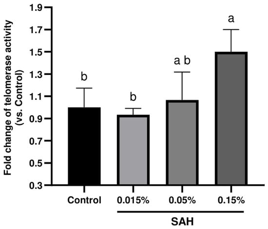
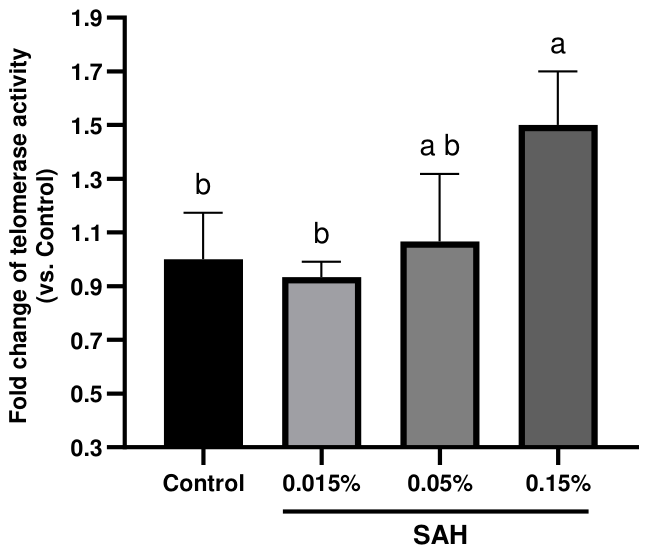
SAH tested at 0.015% and 0.05% (w/v) did not significantly modify telomerase activity relative to the untreated control group (Tukey-adjusted p = 0.969 for both comparisons; Figure 1 and Table 2). In contrast, SAH tested at 0.15% (w/v) was associated with a 1.5-fold increase in telomerase activity that reached statistical significance compared with the control group (mean difference relative units; 95% CI to ; pcorrected = 0.043). The pairwise comparisons among the three SAH concentrations, performed to characterize the concentration–response relationship, showed that the 0.15% concentration was also significantly higher than the lowest concentration (0.15% versus 0.015%: mean difference ; 95% CI to ; pcorrected = 0.023), whereas the difference between 0.15% and 0.05% did not reach significance (pcorrected = 0.080) and the two lower concentrations did not differ from each other (pcorrected = 0.814). This pattern is summarized by the compact letter display in Figure 1, in which only the 0.15% group (letter a) is statistically separated from the control and the 0.015% group (letter b), with the 0.05% group (letter ab) statistically intermediate.
Figure 1.
Effect of SAH on telomerase activity in a model composed of human adult keratinocytes in monolayer culture. Cells were incubated with SAH at 0.015%, 0.05%, or 0.15% (w/v); the control group consisted of keratinocytes incubated under identical conditions without SAH (untreated control). Telomerase activity was quantified by qPCR and is expressed as the fold change relative to the mean of the control group. Bars represent the mean ± S.D. of replicates. Groups were compared by one-way ANOVA followed by Tukey’s multiple comparisons test (all pairwise comparisons among the four groups); statistical significance is indicated above the bars by lowercase letters: bars sharing at least one common letter do not differ significantly, whereas bars sharing no common letter differ significantly (pcorrected < 0.05). The detailed statistical output is reported in Table 2.
Table 2.
One-way ANOVA with Tukey’s multiple comparisons test for the in vitro telomerase activity study ( per group). All six pairwise comparisons among the untreated control and the three SAH concentrations are reported. The mean difference is calculated as the first minus the second group of each comparison; a positive value therefore indicates higher telomerase activity in the first group.
3.2. Beneficial Effects on Telomere Length
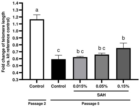
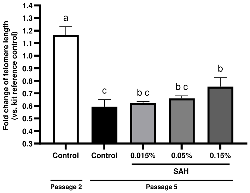
When the two control groups were compared, telomere length was markedly shortened between passage 2 and passage 5 (mean difference relative units; 95% CI to ; Tukey-adjusted p < 0.0001; Figure 2 and Table 3), confirming a clear passage-dependent telomere attrition in the untreated fibroblasts. This variation was in line with the findings reported in previous studies [33,34].
Figure 2.
Effect of SAH on telomere length in a model composed of human normal dermal fibroblasts in monolayer culture. Two untreated control groups are shown: an early-passage control measured at passage 2 (baseline, before passage-dependent shortening), and a late-passage control measured at passage 5 (fibroblasts carried to passage 5 without SAH). The three SAH groups (0.015%, 0.05%, and 0.15%, w/v) were treated and measured at passage 5. Telomere length was quantified by qPCR and is expressed as the fold change relative to the kit reference control. Bars represent the mean ± S.D. of replicates. All five groups were compared by one-way ANOVA followed by Tukey’s multiple comparisons test (all pairwise comparisons among the five groups); statistical significance is indicated above the bars by lowercase letters: bars sharing at least one common letter do not differ significantly, whereas bars sharing no common letter differ significantly (pcorrected < 0.05). The detailed statistical output is reported in Table 3.
Table 3.
One-way ANOVA with Tukey’s multiple comparisons test for the in vitro telomere length study ( per group). All ten pairwise comparisons among the passage-2 control, the passage-5 control, and the three SAH concentrations (measured at passage 5) are reported. The mean difference is calculated as the first minus the second group of each comparison; a positive value therefore indicates a longer relative telomere length in the first group.
Among the passage-5 groups, SAH tested at 0.015% and 0.05% (w/v) did not significantly attenuate the telomere shortening of normal human fibroblasts relative to the passage-5 control (pcorrected = 0.946 and 0.526, respectively). SAH tested at 0.15% (w/v) was associated with a 1.3-fold higher relative telomere length than the passage-5 control, reaching statistical significance (mean difference relative units; 95% CI to ; pcorrected = 0.021; Figure 2). When the three SAH concentrations were compared with one another, none of the pairwise differences reached significance (0.015% versus 0.05%, pcorrected = 0.897; 0.015% versus 0.15%, pcorrected = 0.064; 0.05% versus 0.15%, pcorrected = 0.238); the concentration–response trend across the SAH range therefore did not achieve statistical resolution at the present sample size, as reflected in the overlapping letters of the SAH groups (b and bc) in Figure 2. None of the three SAH groups restored telomere length to that of the early-passage (passage 2) control. This observation suggests a beneficial potential of SAH against telomere shortening; as noted for the telomerase study, the effect was observed only at the highest concentration, in cells from a single donor and over a limited passage range, and should therefore be interpreted with caution and confirmed in further experiments.
3.3. Clinical Anti-Aging Improvement
The anti-aging improvement associated with the topical application of SAH was assessed through six skin parameters in this placebo-controlled clinical study: the depth, area, and volume of crow’s feet wrinkles, the under-eye skin roughness (Rz), and the skin elasticity (R2) and firmness (F4) parameters evaluated on the cheeks.
3.3.1. Per-Timepoint Comparison of Active Versus Placebo
The pre-specified comparisons of active versus placebo at D7 and at D28 were performed for each parameter using paired t-tests; the two timepoint comparisons were further Šidák-corrected as a family of two. The mean differences (active − placebo), 95% confidence intervals, raw and Šidák-corrected p-values, and Cohen’s dz effect sizes are reported in Table 4.
Table 4.
Per-timepoint comparison of active versus placebo (paired t-tests, df = 42) for the percentage variation from baseline. Each parameter is expressed as the variation relative to baseline (D0). The two pre-specified comparisons (D7, D28) were adjusted using the Šidák correction over a family of two. Mean differences are reported as active − placebo.
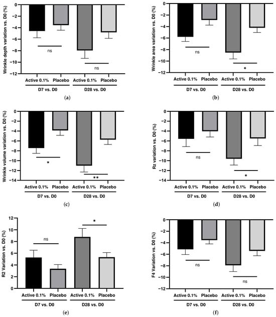
Among the three crow’s feet parameters, crow’s feet wrinkle depth (Figure 3a) showed a per-timepoint difference at D28 that was statistically significant before correction (praw = 0.044) but did not survive the Šidák correction over the two timepoints (pcorrected = 0.086; not significant; see Table 4); the effect size (dz = ) was nevertheless of a similar order of magnitude to those of the other parameters, and the direction of effect was consistent with an improvement on the active-treated side. Crow’s feet wrinkle area (Figure 3b) showed a significant active-versus-placebo difference after Šidák correction at D28 (mean difference %; 95% CI: , ; pcorrected = 0.018; dz = ), with the D7 comparison falling just short of the corrected significance threshold (pcorrected = 0.055; not significant; see Table 4). The strongest and most consistent effect was observed on crow’s feet wrinkle volume (Figure 3c): the active group differed significantly from the placebo group already at D7 (mean difference %; 95% CI: , ; pcorrected = 0.040; dz = ), and the effect grew at D28 (mean difference %; 95% CI: , ; pcorrected = 0.002; dz = ).
Figure 3.
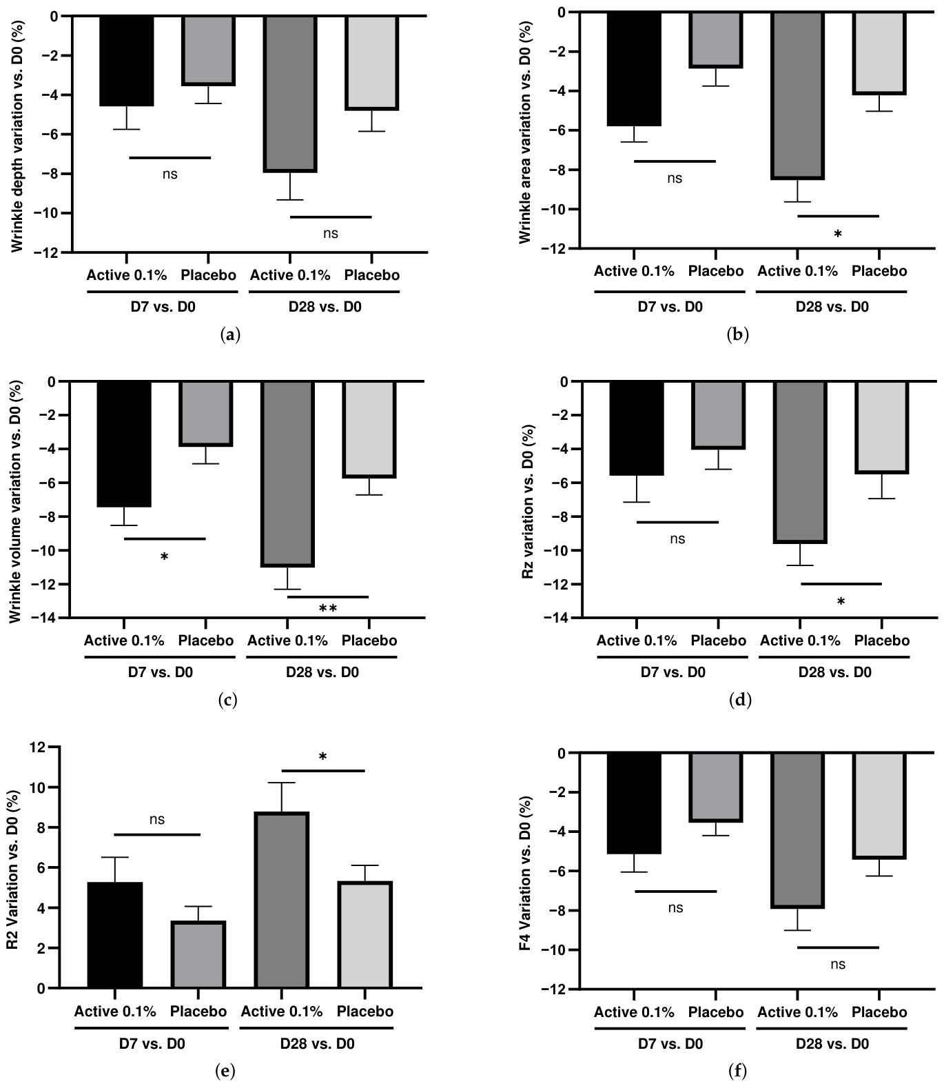
Comparative effects of SAH formulated at 0.1% in an active cream versus the placebo cream on six skin parameters. The results are expressed as mean value of the percentage variation from baseline at D7 and D28 for the active group and the placebo group, respectively, where D0, D7, and D28 represent the measurement values at each checkpoint (mean ± S.E.M.; ). (a) Crow’s feet wrinkle depth; (b) crow’s feet wrinkle area; (c) crow’s feet wrinkle volume; (d) skin roughness Rz parameter on under-eye wrinkles (a more negative variation indicates smoother skin); (e) skin elasticity R2 parameter (a more positive variation indicates better elasticity); (f) skin firmness F4 parameter (a more negative variation indicates better firmness). Statistical significance markers shown on the figure correspond to the Šidák-corrected per-timepoint comparison between the active group and the placebo group: * p < 0.05; ** p < 0.01; ns, not significant. The detailed statistical results are reported in Table 4.
Under-eye roughness Rz (Figure 3d) was significantly reduced in the active group at D28 (mean difference %; 95% CI: , ; pcorrected = 0.050; dz = ); because the corrected p-value lies just below the 0.05 threshold (0.04996 before rounding to three decimals), this effect is statistically significant but of modest magnitude and is best regarded as a just-significant improvement in skin smoothness. Skin elasticity R2 (Figure 3e) improved significantly in the active group at D28 (mean difference %; 95% CI: , ; pcorrected = 0.041; dz = ), whereas the D7 comparison did not reach significance. Skin firmness F4 (Figure 3f), like crow’s feet wrinkle depth, was statistically significant at D28 before correction (praw = 0.040) but did not survive the Šidák correction (pcorrected = 0.078; not significant; see Table 4), with an effect size (dz = ) and a direction of effect again consistent with an improvement on the active-treated side.
Overall, four of the six parameters reached statistical significance after correction: crow’s feet wrinkle volume at both D7 and D28, and crow’s feet wrinkle area, under-eye roughness Rz, and skin elasticity R2 at D28. Crow’s feet wrinkle depth and skin firmness F4 are best described as showing a trend toward improvement that the present study was not adequately powered to confirm at the level of the individual timepoint comparison.
3.3.2. Visual Observations
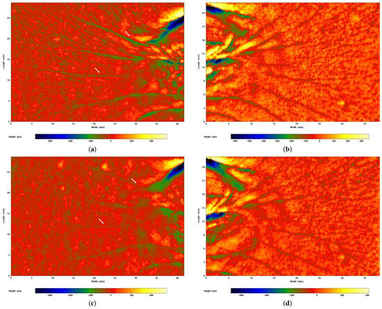
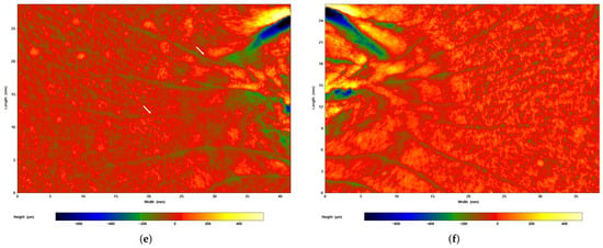
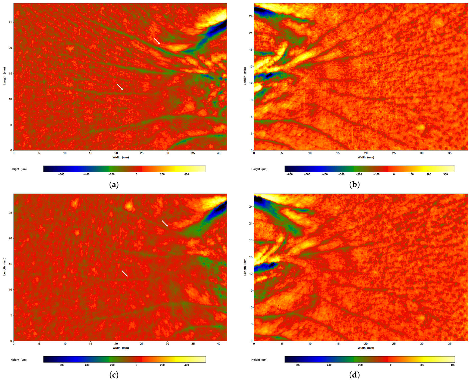
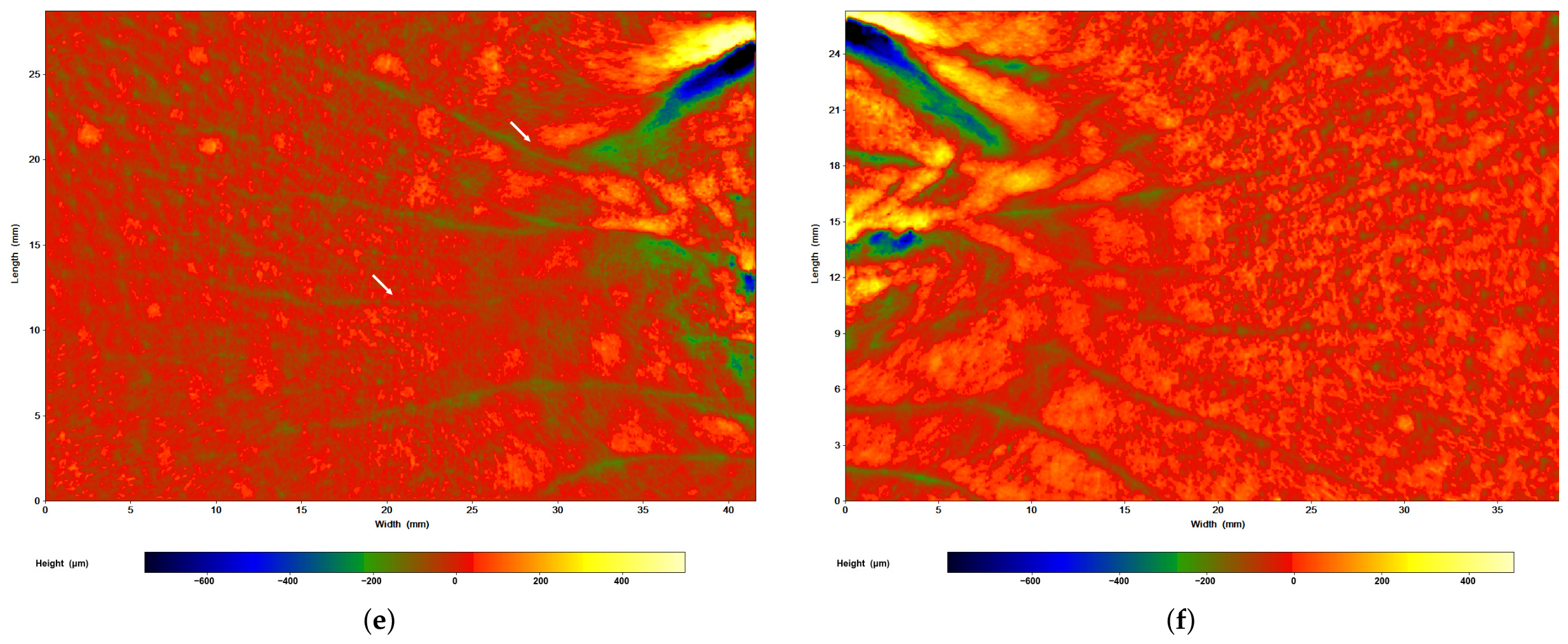
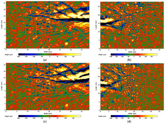
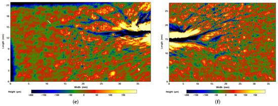
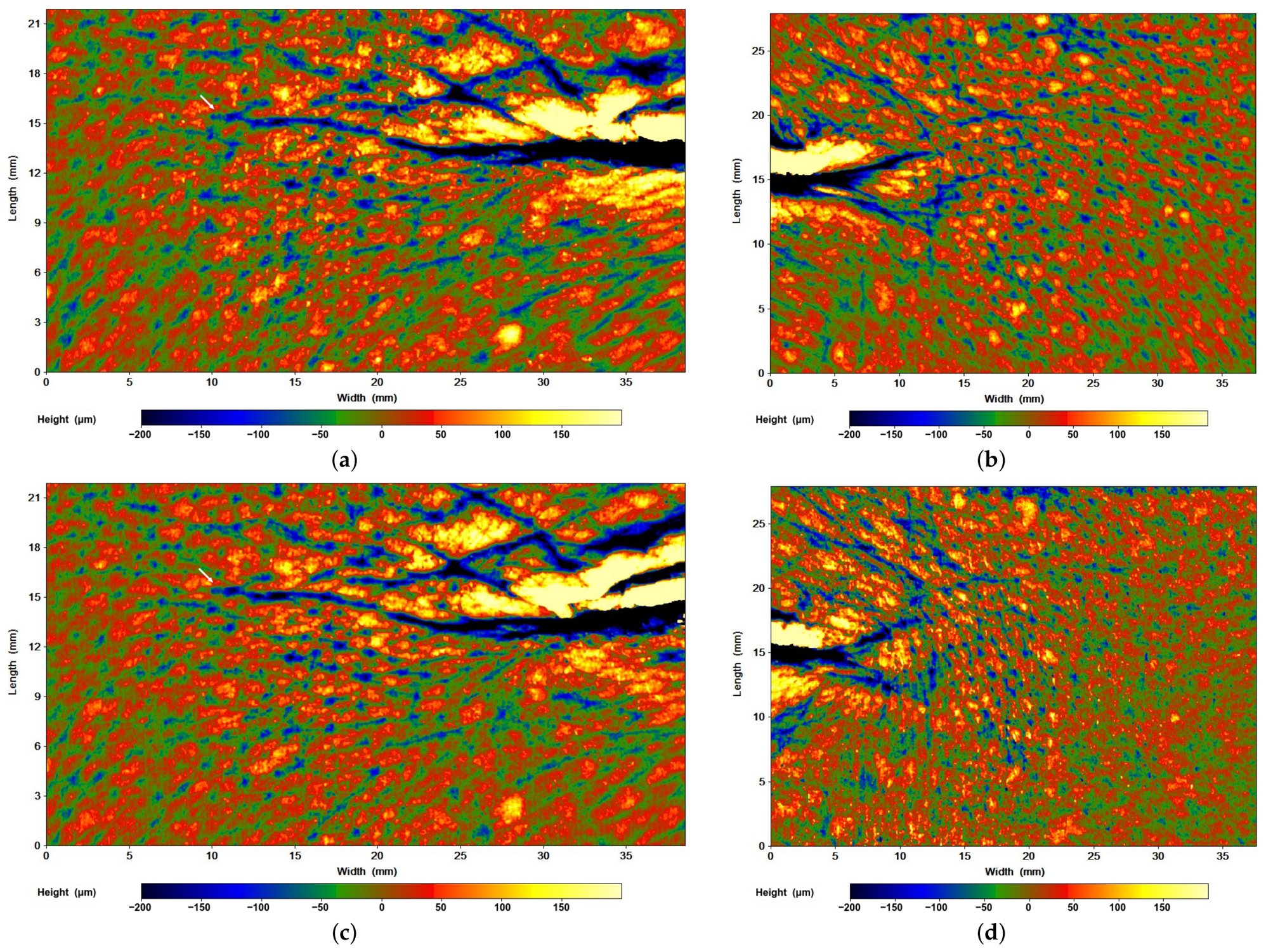
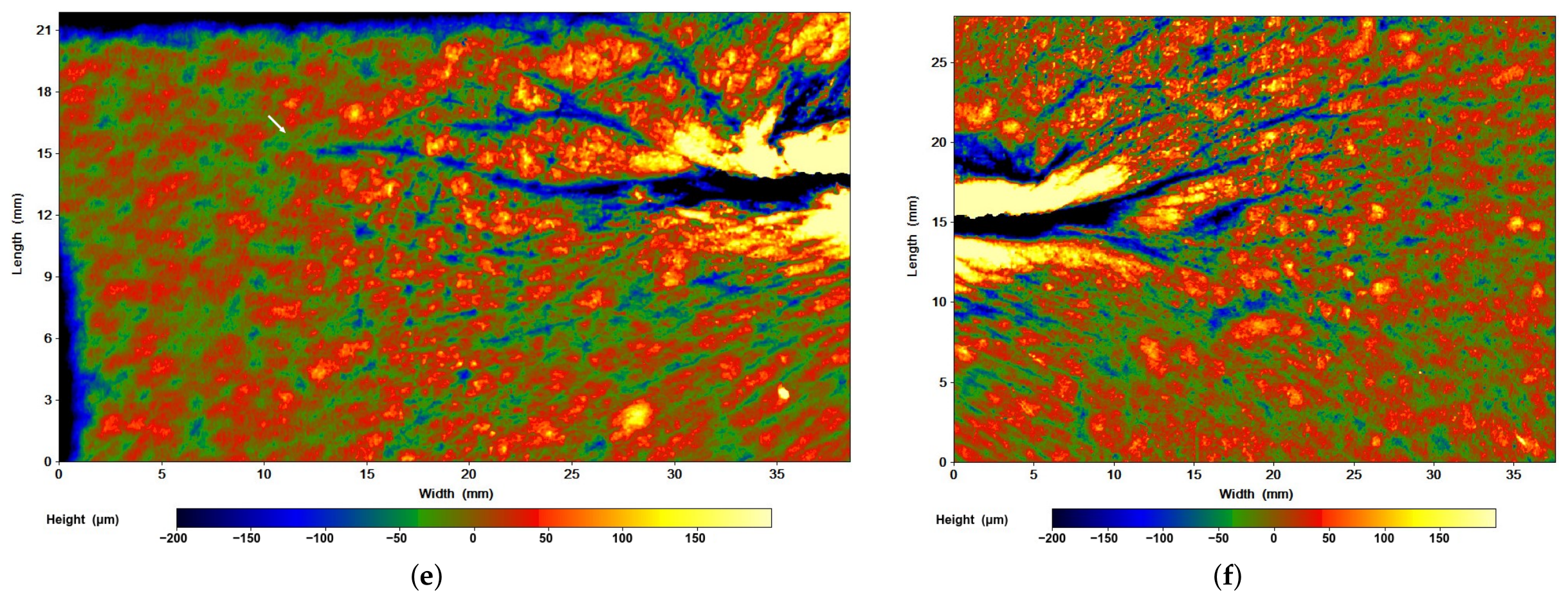
The corrected analysis above is complemented by direct visual observations of the crow’s feet area, presented as rainbow contour maps. As shown in Figure 4 with an example of one Caucasian panellist, milder crow’s feet wrinkles were observed after 7 and 28 days of active cream application compared with the contralateral, placebo-treated hemiface measured at the same checkpoint. Milder crow’s feet wrinkles were also observed in the Asian panel, with an example shown in Figure 5, after 28 days of active cream application, compared with the other hemiface measured at the same checkpoint.
Figure 4.
Representative visual assessment of crow’s feet wrinkles in one participant from the Caucasian panel, shown by rainbow contour where the colors represent heights in m (see the legend below each measurement). The left column (a,c,e) shows the active-treated hemiface and the right column ((b,d), ERROR: Failed to execute system command: (f)) the placebo-treated hemiface, measured at baseline (D0; (a,b)), day 7 (D7; (c,d)), and day 28 (D28; (e,f)). White arrows in (a,c,e) mark representative crow’s feet wrinkle areas on the active-treated hemiface, where a progressive attenuation is visible from D0 to D28.
Figure 5.
Representative visual assessment of crow’s feet wrinkles in one participant from the Asian panel, shown by rainbow contour where the colors represent heights in m (see the legend below each measurement). The left column (a,c,e) shows the active-treated hemiface and the right column (b,d,f) the placebo-treated hemiface, measured at baseline (D0; (a,b)), day 7 (D7; (c,d)), and day 28 (D28; (e,f)). White arrows in (a,c,e) mark representative crow’s feet wrinkle areas, where an attenuation is visible from D0 to D28.
Taken together, the clinical results indicate that the 0.1% SAH cream produced a measurable improvement over placebo on several skin aging parameters within the 28-day study period, with the strongest and most robust effect observed on crow’s feet wrinkle volume and area, and more modest but significant improvements in skin elasticity and under-eye roughness. For the remaining parameters (wrinkle depth and firmness), the active-versus-placebo difference did not reach statistical significance at the corrected level, and the corresponding results should be interpreted as exploratory trends rather than as confirmed effects.
4. Discussion
The in vitro telomerase results indicate a concentration-dependent association between SAH and telomerase activity in keratinocytes, in which a threshold concentration appears to be required to detect an effect. A candidate, literature-based signaling route that might connect an HA-derived molecule such as SAH to telomerase activity is considered together with the telomere length findings below. Because a significant effect was detected at a single concentration in cells from one donor, the present data are best interpreted as hypothesis-generating: confirmation in multi-donor experiments, ideally with additional intermediate concentrations, would be required to resolve the shape of the concentration–response curve and to establish whether the effect is robust.
The attenuation of telomere shortening by SAH is consistent with the work of Tian et al. [35] that hyaluronic acid can activate the TGF-/BMP signaling pathway, which has been reported to modulate telomerase activity [36]. This provides a tentative, literature-based route by which an HA-derived molecule such as SAH might influence telomere maintenance. The reported direction of TGF-/BMP effects on telomerase is, however, context-dependent, so this link remains hypothetical; dedicated investigations to clarify it are currently under way in our laboratory.
It has been recognized that telomeres play an important signaling role during the lifetime of skin cells, including in skin aging. Telomerase, the tightly regulated enzyme complex that maintains telomere length in cells, has been implicated as a vital element in the maintenance of skin cell function and proliferation [8,15]. These considerations motivated the inclusive, placebo-controlled clinical evaluation of SAH, whose anti-aging effects on facial skin are interpreted in the following paragraphs.
The improvement of crow’s feet wrinkles and roughness observed in this study is consistent with the well-documented properties of HA and its derivatives. HA is able to bind a large amount of water and to retain moisture in the skin [1,21,37,38]; adequate skin hydration directly reduces the appearance of fine lines and contributes to a smoother surface relief. Beyond this immediate hydration effect, HA-based ingredients have also been associated with preservation of the dermal extracellular matrix, including protection of type I collagen against degradation [22]. As a complementary, hypothetical route, the SAH effects observed in the clinical study could arise in part from the reported ability of hyaluronic acid to activate TGF- [35], a well-known stimulator of collagen synthesis; because collagen is the main structural constituent of the dermis, such an effect would be consistent with the observed improvements in wrinkles, firmness, and elasticity. The higher resistance of SAH to hyaluronidase [22] may prolong these effects over the 28-day application period. These mechanisms, related to hydration and to extracellular matrix preservation, are likely to act in parallel with, rather than as a direct consequence of, the telomere-related effects observed in vitro. At the population level, a recent systematic review and meta-analysis of HA-based interventions for skin aging reported significant improvements in skin hydration and radiance, whereas the effect on skin elasticity was more variable and did not reach significance across the pooled studies [39]; this is consistent with the view that the hydration-related benefits of HA are the most robust, while effects on deeper matrix properties such as elasticity depend on additional structural factors.
Beyond the crow’s feet wrinkle parameters, the improvements in under-eye roughness (Rz) and cheek elasticity (R2), together with the consistent (albeit non-significant) trend in firmness (F4), are coherent with these same hydration- and matrix-related mechanisms: a smoother skin surface is the expected macroscopic correlate of improved hydration, whereas the gains in elasticity and firmness are consistent with better preservation of the dermal extracellular matrix. The convergence of these secondary endpoints with the crow’s feet results reinforces the overall anti-aging profile of SAH observed in the clinical study.
The present clinical trial included both a Caucasian panel and an Asian panel, and some representative improvements of SAH versus placebo were observed within each panel. It should be emphasized, however, that the study was not designed or statistically powered to compare the two ethnic groups: no formal group-specific (Caucasian versus Asian) statistical analysis was performed, and the sample size of each panel was limited. The results should therefore be interpreted as applying to the study population as a whole, and no conclusion can be drawn regarding inter-ethnic differences in the response to SAH. A previously reported clinical evaluation using SAH as the main active was single-centered and covered Caucasian women only [22]; the present study extends the observation of an anti-aging effect to an additional Asian panel. While several epidemiological studies have discussed leukocyte telomere length across different ethnic groups [40,41,42,43,44], telomere biology in the context of skin aging has been studied predominantly in Caucasian populations [8,25,26,27,28]. Dedicated, adequately powered studies covering a wider diversity of volunteers, including men and other racial groups, would be required to evaluate whether the response to SAH differs between populations.
Several limitations should be borne in mind when interpreting these findings. The two in vitro studies were each based on cells from a single donor and on a limited number of replicates, so inter-individual biological variability was not captured, and the telomere length analysis covered a restricted passage window (passage 2 to passage 5). In addition, telomerase activity and telomere length were necessarily assessed in two different cell types (keratinocytes and fibroblasts, respectively), so the two in vitro endpoints are not directly comparable and were treated as separate, parallel observations rather than paired measurements of a single mechanism, with no quantitative cross-endpoint comparison performed. No unmodified HA or other HA derivative was tested in parallel, so no claim of superiority of SAH over HA can be made from the present data. The clinical trial enrolled female volunteers only, and its findings therefore cannot be generalized to men. Finally, the study was not designed to identify the underlying molecular pathway: the in vitro telomere-related observations and the short-term clinical improvements are best regarded as complementary rather than causally linked, and the mechanisms proposed above are working hypotheses to be tested in dedicated future studies.
5. Conclusions
In summary, the present research provides the first exploratory evidence that SAH, an acetylated derivative of sodium hyaluronate, may be associated with telomere-related biological endpoints. In two in vitro studies based on human keratinocytes and fibroblasts, SAH at 0.15% (w/v) was associated with an increase in telomerase activity and a higher relative telomere length compared with control. In a randomized, split-face, double-centered, double-blind, placebo-controlled clinical trial conducted in both a Caucasian and an Asian panel of female volunteers, a facial cream formulated with 0.1% SAH significantly reduced crow’s feet wrinkle volume (from day 7) and wrinkle area (at day 28), significantly improved skin elasticity (at day 28), and significantly reduced under-eye roughness (at day 28) compared with placebo, with consistent, non-significant trends for wrinkle depth and firmness. As working hypotheses, these observations may be explained by the hydration and extracellular-matrix-preserving properties of HA, together with an HA-mediated activation of TGF-, a known stimulator of collagen synthesis; none of these mechanisms is demonstrated by the present data. Given the exploratory design of the study, in particular the use of single-donor cells and the absence of direct mechanistic evidence, these findings should not be interpreted as a definitive demonstration of a telomere-protective effect; the precise biological mechanism linking SAH to telomere biology remains to be elucidated, and the in vitro and clinical observations are best regarded as complementary rather than causally linked. Overall, these results identify SAH as an interesting active ingredient for anti-aging skincare cosmetics, combining a curative dimension—the clinical improvement of several skin-aging parameters—with a preventive dimension suggested by the telomere-related effects observed in vitro, the latter remaining to be confirmed.
Author Contributions
Conceptualization, Y.J., A.D., L.W., T.S. and J.J.; methodology, Y.J., A.D. and T.S.; writing—original draft preparation, J.W.; writing—review and editing, J.W. and A.D. All authors have read and agreed to the published version of the manuscript.
Funding
These studies were financially supported by TS-Biotech Group. Funding numbers: 24.0034 for in vitro studies, and TS2201_22-CB-11075_S220195G1 for clinical study.
Institutional Review Board Statement
The in vitro studies used human keratinocytes and dermal fibroblasts derived from anonymized discarded surgical skin collected after donor consent for secondary research use; therefore, no specific ethics committee approval was required under applicable French regulations. The clinical study was conducted by Complife as a non-invasive cosmetic efficacy study in healthy volunteers, in accordance with the Declaration of Helsinki and applicable local regulations. Based on Complife’s regulatory assessment, independent Ethics Committee/IRB approval and clinical trial registration were not mandatory for this type of cosmetic efficacy evaluation.
Informed Consent Statement
Written informed consent for participation was obtained from all participants before inclusion in the clinical study. For the in vitro studies, the discarded surgical skin samples used for cell isolation were obtained after prior informed consent from the donors for secondary research use and were anonymized before transfer to the investigators. No personally identifiable participant information is included in the revised manuscript. As individual age and sex details have been removed from the representative figure captions, specific informed consent for publication was not required.
Data Availability Statement
The data presented in this study are available on request from the corresponding author due to commercial confidentiality.
Acknowledgments
During the preparation of this manuscript, the authors used Opus 4.8 for the purpose of polishing the language of the manuscript. The authors have reviewed and edited the output and take full responsibility for the content of this publication.
Conflicts of Interest
Authors Yuhua Jiang and Luyan Wang are employed by the company Shandong Tiansheng Biotechnology Co., Ltd. (TS-Biotech China). Authors Jason Wu and Junjie Jiang are employed by the company TS Biotech SAS (TS-Biotech France). Authors Aline Delobelle and Thibaut Saguet are external consultants, contracted by CosmeLink SARL and TS Consulting. The authors declare that this study received funding from TS-Biotech Group. The funder had the following involvement with the study: discussion of all the study protocols, supply of the SAH testing samples for in vitro studies, and preparation of the active and placebo creams for clinical study. The funder did not affect the experimental process and results in the study. The funding relationship does not affect the scientific quality of the manuscript.
References
- Baumann, L. Skin ageing and its treatment. J. Pathol. 2007, 211, 241–251. [Google Scholar] [CrossRef] [Scilit] [PubMed]
- Naharro-Rodriguez, J.; Bacci, S.; Hernandez-Bule, M.L.; Perez-Gonzalez, A.; Fernandez-Guarino, M. Decoding Skin Aging: A Review of Mechanisms, Markers, and Modern Therapies. Cosmetics 2025, 12, 144. [Google Scholar] [CrossRef] [Scilit]
- Hughes, M.C.B.; Williams, G.M.; Pageon, H.; Fourtanier, A.; Green, A.C. Dietary Antioxidant Capacity and Skin Photoaging: A 15-Year Longitudinal Study. J. Investig. Dermatol. 2021, 141, 1111–1118.e2. [Google Scholar] [CrossRef] [Scilit] [PubMed]
- Chakravarti, D.; LaBella, K.A.; DePinho, R.A. Telomeres: History, health, and hallmarks of aging. Cell 2021, 184, 306–322. [Google Scholar] [CrossRef] [Scilit] [PubMed]
- López-Otín, C.; Blasco, M.A.; Partridge, L.; Serrano, M.; Kroemer, G. The Hallmarks of Aging. Cell 2013, 153, 1194–1217. [Google Scholar] [CrossRef] [Scilit] [PubMed]
- López-Otín, C.; Blasco, M.A.; Partridge, L.; Serrano, M.; Kroemer, G. Hallmarks of aging: An expanding universe. Cell 2023, 186, 243–278. [Google Scholar] [CrossRef] [Scilit] [PubMed]
- Allsopp, R.C.; Harley, C.B. Evidence for a Critical Telomere Length in Senescent Human Fibroblasts. Exp. Cell Res. 1995, 219, 130–136. [Google Scholar] [CrossRef] [Scilit] [PubMed]
- Buckingham, E.M.; Klingelhutz, A.J. The role of telomeres in the ageing of human skin. Exp. Dermatol. 2011, 20, 297–302. [Google Scholar] [CrossRef] [Scilit] [PubMed]
- Lindsey, J.; McGill, N.I.; Lindsey, L.A.; Green, D.K.; Cooke, H.J. In vivo loss of telomeric repeats with age in humans. Mutat. Res. DNAging 1991, 256, 45–48. [Google Scholar] [CrossRef] [Scilit] [PubMed]
- Jacczak, B.; Rubiś, B.; Totoń, E. Potential of Naturally Derived Compounds in Telomerase and Telomere Modulation in Skin Senescence and Aging. Int. J. Mol. Sci. 2021, 22, 6381. [Google Scholar] [CrossRef] [Scilit] [PubMed]
- Allsopp, R.C.; Vaziri, H.; Patterson, C.; Goldstein, S.; Younglai, E.V.; Futcher, A.B.; Greider, C.W.; Harley, C.B. Telomere length predicts replicative capacity of human fibroblasts. Proc. Natl. Acad. Sci. USA 1992, 89, 10114–10118. [Google Scholar] [CrossRef] [Scilit] [PubMed]
- Valdes, A.M.; Andrew, T.; Gardner, J.P.; Kimura, M.; Oelsner, E.; Cherkas, L.F.; Aviv, A.; Spector, T.D. Obesity, cigarette smoking, and telomere length in women. Lancet 2005, 366, 662–664. [Google Scholar] [CrossRef] [Scilit] [PubMed]
- Cao, C.; Gong, W.; Shuai, Y.; Rasouli, S.; Ge, Q.; Khan, A.; Dakic, A.; Putluri, N.; Shvets, G.; Zheng, Y.L.; et al. Canonical and non-canonical functions of the non-coding RNA component (TERC) of telomerase complex. Cell Biosci. 2025, 15, 30. [Google Scholar] [CrossRef] [Scilit] [PubMed]
- Jin, S.; Li, K.; Zong, X.; Eun, S.; Morimoto, N.; Guo, S. Hallmarks of skin aging: Update. Aging Dis. 2023, 14, 2167–2176. [Google Scholar] [CrossRef] [Scilit] [PubMed]
- Liu, Z.; Sun, C.; Zhang, Z.; Jiang, Y.; Zhao, C. Telomeres in skin aging. Biogerontology 2025, 26, 83. [Google Scholar] [CrossRef] [Scilit] [PubMed]
- Liu, N.; Yin, Y.; Wang, H.; Zhou, Z.; Sheng, X.; Fu, H.; Guo, R.; Wang, H.; Yang, J.; Gong, P.; et al. Telomere dysfunction impairs epidermal stem cell specification and differentiation by disrupting BMP/pSmad/P63 signaling. PLoS Genet. 2019, 15, 1–26. [Google Scholar] [CrossRef] [Scilit] [PubMed]
- Zhang, J.; Yu, H.; Man, M.Q.; Hu, L. Aging in the dermis: Fibroblast senescence and its significance. Aging Cell 2024, 23, e14054. [Google Scholar] [CrossRef] [Scilit] [PubMed]
- Bukhari, S.N.A.; Roswandi, N.L.; Waqas, M.; Habib, H.; Hussain, F.; Khan, S.; Sohail, M.; Ramli, N.A.; Thu, H.E.; Hussain, Z. Hyaluronic acid, a promising skin rejuvenating biomedicine: A review of recent updates and pre-clinical and clinical investigations on cosmetic and nutricosmetic effects. Int. J. Biol. Macromol. 2018, 120, 1682–1695. [Google Scholar] [CrossRef] [Scilit] [PubMed]
- Zanchetta, C.; Scandolera, A.; Reynaud, R. Hyaluronic Acid in Topical Applications: The Various Forms and Biological Effects of a Hero Molecule in the Cosmetics Industry. Biomolecules 2025, 15, 1656. [Google Scholar] [CrossRef] [Scilit] [PubMed]
- Laurent, T.C.; Fraser, J.R.E. Hyaluronan. FASEB J. 1992, 6, 2397–2404. [Google Scholar] [CrossRef] [Scilit]
- Papakonstantinou, E.; Roth, M.; Karakiulakis, G. Hyaluronic acid: A key molecule in skin aging. Dermato-Endocrinology 2012, 4, 253–258. [Google Scholar] [CrossRef] [Scilit] [PubMed]
- Meunier, M.; Scandolera, A.; Chapuis, E.; Lapierre, L.; Sandré, J.; Brunner, G.; Lovchik, M.; Reynaud, R. The anti-wrinkles properties of sodium acetylated hyaluronate. J. Cosmet. Dermatol. 2022, 21, 2749–2762. [Google Scholar] [CrossRef] [Scilit] [PubMed]
- Fink, B.; Campiche, R.; Shackelford, T.K.; Voegeli, R. Effects of under-eye skin and crow’s feet on perceived facial appearance in women of five ethnic groups. Int. J. Cosmet. Sci. 2025, 47, 510–522. [Google Scholar] [CrossRef] [Scilit] [PubMed]
- Desai, S.R.; Khanna, R.; Glass, D.; Alam, M.; Barrio, V.; French, L.E.; Gohara, M.; McKinley-Grant, L.; Harvey, V.; Heath, C.; et al. Embracing diversity in dermatology: Creation of a culture of equity and inclusion in dermatology. Int. J. Womens Dermatol. 2021, 7, 378–382. [Google Scholar] [CrossRef] [Scilit] [PubMed]
- Kosmadaki, M.; Gilchrest, B. The role of telomeres in skin aging/photoaging. Micron 2004, 35, 155–159. [Google Scholar] [CrossRef] [Scilit] [PubMed]
- Sugimoto, M.; Yamashita, R.; Ueda, M. Telomere length of the skin in association with chronological aging and photoaging. J. Dermatol. Sci. 2006, 43, 43–47. [Google Scholar] [CrossRef] [Scilit] [PubMed]
- Song, N.; Cui, Y.; Xi, W. Association Between Telomere Length and Skin Cancer and Aging: A Mendelian Randomization Analysis. Front. Genet. 2022, 13, 931785. [Google Scholar] [CrossRef] [Scilit] [PubMed]
- Ribero, S.; Mangino, M.; Bataille, V. Skin phenotypes can offer some insight about the association between telomere length and cancer susceptibility. Med. Hypotheses 2016, 97, 7–10. [Google Scholar] [CrossRef] [Scilit] [PubMed]
- Krunic, D.; Moshir, S.; Greulich-Bode, K.M.; Figueroa, R.; Cerezo, A.; Stammer, H.; Stark, H.J.; Gray, S.G.; Nielsen, K.V.; Hartschuh, W.; et al. Tissue context-activated telomerase in human epidermis correlates with little age-dependent telomere loss. Biochim. Biophys. Acta Mol. Basis Dis. 2009, 1792, 297–308. [Google Scholar] [CrossRef] [Scilit] [PubMed]
- Harrison, S.J.; Bishton, M.; Bates, S.E.; Grant, S.; Piekarz, R.L.; Johnstone, R.W.; Dai, Y.; Lee, B.; Araujo, M.E.; Prince, H.M. A Focus on the Preclinical Development and Clinical Status of the Histone Deacetylase Inhibitor, Romidepsin (Depsipeptide, Istodax®). Epigenomics 2012, 4, 571–589. [Google Scholar] [CrossRef] [Scilit] [PubMed]
- Herbert, B.S.; Hochreiter, A.E.; Wright, W.E.; Shay, J.W. Nonradioactive detection of telomerase activity using the telomeric repeat amplification protocol. Nat. Protoc. 2006, 1, 1583–1590. [Google Scholar] [CrossRef] [Scilit] [PubMed]
- Cawthon, R.M. Telomere measurement by quantitative PCR. Nucleic Acids Res. 2002, 30, e47. [Google Scholar] [CrossRef] [Scilit] [PubMed]
- Figueroa, R.; Lindenmaier, H.; Hergenhahn, M.; Nielsen, K.V.; Boukamp, P. Telomere Erosion Varies during in Vitro Aging of Normal Human Fibroblasts from Young and Adult Donors. Cancer Res. 2000, 60, 2770–2774. [Google Scholar] [PubMed]
- Martens, U.M.; Chavez, E.A.; Poon, S.S.; Schmoor, C.; Lansdorp, P.M. Accumulation of Short Telomeres in Human Fibroblasts Prior to Replicative Senescence. Exp. Cell Res. 2000, 256, 291–299. [Google Scholar] [CrossRef] [Scilit] [PubMed]
- Tian, Y.B.; Wang, N.X.; Xu, Y.; Yu, C.Y.; Liu, R.M.; Luo, Y.; Xiao, J.H. Hyaluronic acid ameliorates the proliferative ability of human amniotic epithelial cells through activation of TGF-β/BMP signaling. PeerJ 2020, 8, e10104. [Google Scholar] [CrossRef] [Scilit] [PubMed]
- Buckley, S.; Shi, W.; Driscoll, B.; Ferrario, A.; Anderson, K.; Warburton, D. BMP4 signaling induces senescence and modulates the oncogenic phenotype of A549 lung adenocarcinoma cells. Am. J. Physiol.-Lung Cell. Mol. Physiol. 2004, 286, L81–L86. [Google Scholar] [CrossRef] [Scilit] [PubMed]
- Chylińska, N.; Maciejczyk, M. Hyaluronic Acid and Skin: Its Role in Aging and Wound-Healing Processes. Gels 2025, 11, 281. [Google Scholar] [CrossRef] [Scilit] [PubMed]
- Nobile, V.; Roveda, G.; Spartà, E.; Tursi, F. Oral Intake and Topical Application of Hyaluronic Acid Ameliorates Skin Aging Signs: Efficacy Results of a Placebo-Controlled In&Out Trial. Cosmetics 2025, 12, 52. [Google Scholar] [CrossRef] [Scilit]
- Zhou, R.; Yu, M. The Effect of Local Hyaluronic Acid Injection on Skin Aging: A Systematic Review and Meta-Analysis. J. Cosmet. Dermatol. 2025, 24, e16760. [Google Scholar] [CrossRef] [Scilit] [PubMed]
- Brown, L.; Needham, B.; Ailshire, J. Telomere Length Among Older U.S. Adults: Differences by Race/Ethnicity, Gender, and Age. J. Aging Health 2017, 29, 1350–1366. [Google Scholar] [CrossRef] [Scilit] [PubMed]
- Diez Roux, A.V.; Ranjit, N.; Jenny, N.S.; Shea, S.; Cushman, M.; Fitzpatrick, A.; Seeman, T. Race/ethnicity and telomere length in the Multi-Ethnic Study of Atherosclerosis. Aging Cell 2009, 8, 251–257. [Google Scholar] [CrossRef] [Scilit] [PubMed]
- Hamad, R.; Tuljapurkar, S.; Rehkopf, D.H. Racial and Socioeconomic Variation in Genetic Markers of Telomere Length: A Cross-Sectional Study of U.S. Older Adults. EBioMedicine 2016, 11, 296–301. [Google Scholar] [CrossRef] [Scilit] [PubMed]
- Rewak, M.; Buka, S.; Prescott, J.; De Vivo, I.; Loucks, E.B.; Kawachi, I.; Non, A.L.; Kubzansky, L.D. Race-related health disparities and biological aging: Does rate of telomere shortening differ across blacks and whites? Biol. Psychol. 2014, 99, 92–99. [Google Scholar] [CrossRef] [Scilit] [PubMed]
- Chae, D.H.; Nuru-Jeter, A.M.; Adler, N.E.; Brody, G.H.; Lin, J.; Blackburn, E.H.; Epel, E.S. Discrimination, Racial Bias, and Telomere Length in African-American Men. Am. J. Prev. Med. 2014, 46, 103–111. [Google Scholar] [CrossRef] [Scilit] [PubMed]

Disclaimer/Publisher’s Note: The statements, opinions and data contained in all publications are solely those of the individual author(s) and contributor(s) and not of MDPI and/or the editor(s). MDPI and/or the editor(s) disclaim responsibility for any injury to people or property resulting from any ideas, methods, instructions or products referred to in the content. |
© 2026 by the authors. Licensee MDPI, Basel, Switzerland. This article is an open access article distributed under the terms and conditions of the Creative Commons Attribution (CC BY) license.