


Abstract
Background: The skin barrier plays a fundamental role in preventing transepidermal water loss (TEWL), regulating immune responses, and protecting against pathogen colonization. Disruption of this barrier underlies xerosis, sensitive skin, and clinical cracking. Heat-treated probiotics (postbiotics) represent a stable and biologically active approach to topical formulation. Objective: To evaluate the safety, molecular mechanisms, and clinical efficacy of heat-treated Lactiplantibacillus plantarum Skinbac™ SB14 (SB14) in improving skin barrier function, hydration, and the appearance of dry and cracked skin. Methods: In vitro studies assessed cell viability (MTT assay) and cytotoxicity (LDH release assay), Aquaporin-3 (AQP3) expression, Claudin-1 expression recovery following UV-induced damage (post-damage treatment model), cytokine modulation in Normal Human Epidermal Keratinocytes (NHEK) and Peripheral Blood Mononuclear Cells (PBMCs), and antipathogen activity against Staphylococcus aureus biofilm. A 30-day open-label, placebo-controlled clinical study (n = 20 healthy volunteers, both sexes, age > 18 years) evaluated an emulsion containing 1% SB14 versus placebo using instrumental measurements of superficial hydration (Corneometer® CM825) and TEWL (Tewameter® TM300), and clinical scoring of skin hydration (Kligman scale 1–4) and skin cracking (ODS Overall Dry Skin Score 1–5) via C-Cube imaging. Results: In vitro testing confirmed the safety of SB14 (full cell viability by MTT assay; no cytotoxicity by LDH release assay) and demonstrated significant AQP3 upregulation (p < 0.05), partial Claudin-1 recovery in UV-damaged cells following post-damage SB14 application (p < 0.1 vs. UV damage), significant reduction in pro-inflammatory IL-8 and IL-23 in NHEK (p < 0.01 and p < 0.05), strong innate immune activation in PBMCs (TNF-α and IL-6, p < 0.001), and 21% inhibition of S. aureus biofilm at 72 h. Clinically, the SB14 formulation significantly increased superficial skin hydration by +46.1% at T14 (p = 0.0451) and +33.6% at T30 (p = 0.0144) versus baseline, while TEWL decreased by −14.5% at T14 (p = 0.0205). Clinical hydration scores improved significantly from a median of 3.0 (moderate dry skin) at baseline to 2.0 (slightly dry skin) at T30 (p = 0.0073). Cracking scores improved significantly from a median of 3.5 at baseline to 2.0 at T30 (p = 0.0037), with 80% of subjects showing improvement at T30. All parameters remained non-significant in the placebo group. No adverse events were reported. Conclusions: SB14 is safe, biologically active across multiple barrier-relevant mechanisms, and clinically effective in improving hydration and reducing visible skin cracking in subjects with dry, barrier-compromised skin.
1. Introduction
The skin constitutes the body’s primary interface with the external environment, performing essential roles in mechanical protection, immune surveillance, thermoregulation, and the regulation of transcutaneous water exchange [1,2]. Its structural and functional integrity depends on a stratified epithelium in which keratinocytes progressively differentiate toward the surface, forming a compact stratum corneum reinforced by intercellular lipids, tight junctions (TJs), and cytoskeletal proteins [3]. Critical indicators of barrier competence include stratum corneum hydration, transepidermal water loss (TEWL), and the integrity of junctional proteins such as Claudins and Occludin [4,5]. When barrier function is impaired, TEWL increases, hydration decreases, and the skin becomes vulnerable to irritants, allergens, and microbial colonization, hallmarks of conditions such as xerosis, atopic-prone skin, and clinical cracking [6,7].
The skin microbiome plays an integral role in supporting barrier homeostasis. A balanced cutaneous microbiota contributes to immune education, competitive exclusion of pathogens, and maintenance of an acidic surface pH that favors barrier lipid organization [8,9]. Dysbiosis, particularly involving Staphylococcus aureus, is strongly associated with barrier dysfunction, local inflammation, and impaired barrier repair [10,11]. Strategies that modulate the skin microenvironment, including topical application of probiotic-derived ingredients, have therefore gained increasing interest as approaches to support barrier integrity [12,13].
Among the structural determinants of barrier function, Aquaporin-3 (AQP3) is the predominant aquaglyceroporin expressed in basal and suprabasal keratinocytes, facilitating transcellular movement of water and glycerol and playing key roles in stratum corneum hydration, cellular proliferation, and barrier repair [14,15]. Its expression declines in barrier-compromised conditions, and its upregulation is associated with improved hydration and barrier recovery [16]. Tight junction proteins, particularly Claudin-1, are equally critical: they seal the paracellular space, limit antigen permeation, and protect against UV-induced damage [17]. Additionally, the inflammatory cytokine landscape—including IL-8 as a neutrophil-recruiting chemokine and IL-23 as a driver of Th17 responses—is intimately linked to barrier dysfunction and the chronicity of dry skin conditions [18,19].
The field of postbiotics has emerged as a scientifically robust and formulation-practical approach to topical bioactives [20,21]. Heat-treated bacteria retain structural components—cell wall fragments, surface proteins, and metabolites—that preserve immunomodulatory and barrier-supporting activities while eliminating viability concerns, enhancing formulation stability, and simplifying regulatory classification [22,23]. Lactiplantibacillus plantarum is among the most studied probiotic species for skin applications, with documented effects on hyaluronic acid synthesis, tight junction reinforcement, UV protection, and antimicrobial defense [24,25,26].
The present study evaluates Skinbac™ SB14 (SB14), a heat-treated strain of Lactiplantibacillus plantarum, for its potential as a cosmetic active ingredient targeting skin barrier dysfunction and dry/cracked skin. We conducted a comprehensive in vitro characterization—encompassing safety, AQP3 modulation, Claudin-1 recovery following UV induced damage, cytokine profiling in both keratinocytes and immune cells, and antipathogen activity against S. aureus biofilm—and contextualized these findings within a placebo-controlled clinical study assessing the effects of a 1% SB14 emulsion on TEWL, superficial hydration, and the clinical appearance of dry and cracked skin over 30 days. Our primary objectives were to: (I) establish the safety profile of SB14 in a human keratinocyte model; (II) characterize molecular mechanisms relevant to barrier repair and immune modulation; and (III) evaluate clinical efficacy through instrumental and clinical measurements in volunteers with dry, barrier-compromised skin on elbows and heels.
2. Materials and Methods
2.1. Cell Culture
Normal Human Epidermal Keratinocytes (NHEK-Ad, Cat. No. 00192627) were obtained from Lonza (Basel, Switzerland) and cultured in supplier-recommended Keratinocyte Growth Medium (Cat. No. 00192060) at 37 °C in a 5% CO2 humidified atmosphere. For cytotoxicity and molecular assays, cells were seeded in 24-well plates at 5 × 10⁴ cells/well and allowed to adhere overnight prior to treatment. Peripheral Blood Mononuclear Cells (PBMCs) were obtained from healthy donors for cytokine profiling experiments, as described below.
2.2. Preparation of the Heat-Treated Probiotic Strain
Lactiplantibacillus plantarum was subjected to thermal inactivation at temperatures exceeding 75 °C for 30–90 min, followed by spray-drying and quantification by flow cytometry using Thiazole Orange/Propidium Iodide staining (TO/PI, Cat. No. 349483, BD Biosciences, San Jose, CA, USA), yielding a standardized powder of 109 total fluorescent units per gram (TFU/g), stored at 4 °C until use. Working concentrations of 107 TFU/mL were prepared fresh in the appropriate cell culture medium for in vitro experiments. For the clinical study, SB14 was incorporated into the test emulsion at 1% w/w (corresponding to 107 TFU/mL).
2.3. In Vitro Testing
The experimental platform employed in this study is consistent with the standardized methodology previously validated for the Skinbac™ postbiotic series [17].
2.3.1. Safety Assessment
Cell viability and cytotoxicity were assessed in NHEK cells using the MTT tetrazolium reduction assay and the Lactate Dehydrogenase (LDH) release assay, respectively. Cells were treated with 107 TFU/mL of SB14 for 24 h at 37 °C. LDH release was quantified using the CytoTox 96® Non-Radioactive Cytotoxicity Assay (Promega, Madison, WI, USA; Cat. No. G7891). MTT assay was performed with 0.5 mg/mL MTT (Sigma-Aldrich, St. Louis, MO, USA; Cat. No. M-5655) for 4 h; formazan crystals were dissolved in DMSO (Sigma-Aldrich; Cat. No. D5879) and absorbance measured at 570 nm using a VarioskanLUX microplate reader (Thermo Fisher Scientific, Waltham, MA, USA). Sodium dodecyl sulfate (SDS) served as positive control. All experiments were performed in triplicate.
2.3.2. AQP3 Expression and UV-Induced Claudin-1 Assessment
For AQP3 quantification, NHEK cells were treated with 107 TFU/mL of SB14 and incubated at 37 °C for 24 h. Cell lysates were analyzed for AQP3 protein levels by ELISA (Human AQP3 ELISA Kit, Assay Genie, Dublin, Ireland; Cat. No. HUFI00733). Results are expressed as percentage relative to untreated control cells. To assess the capacity of SB14 to promote barrier recovery following UV-induced tight junction damage, NHEK cells were first exposed to a UVC lamp (30 J/cm2) to induce epithelial stress, and subsequently treated with SB14 (107 TFU/mL) for 24 h. Claudin-1 protein expression was then quantified by ELISA (Assay Genie). Results are expressed as percentage of expression relative to unirradiated, untreated control cells. AQP3 was evaluated under basal conditions to assess the intrinsic hydration-modulating capacity of SB14. Claudin-1 was assessed in a post-UV-damage model to evaluate barrier recovery potential, using UVC irradiation as a standardized stress model consistent with the methodology previously employed for this strain series [17].
2.3.3. Cytokine Modulation
Cytokine modulation was evaluated in NHEK (epithelial model) and PBMCs (peripheral immune model). NHEK cells were stimulated with 107 TFU/mL of SB14 combined with UVC irradiation and incubated for 24 h [17]. PBMCs were stimulated with 107 TFU/mL of SB14 alone for 24 h. Supernatants were collected and concentrations of TNF-α, IL-6, IL-8, and IL-23 were quantified by ELISA (R&D Systems, Minneapolis, MN, USA). Results are expressed as fold change relative to untreated controls (basal level = 1). These four cytokines were selected for their established relevance to cutaneous immune homeostasis: TNF-α and IL-6 as key regulators of innate immunity and the Th1/Th2 balance, IL-8 as a neutrophilic chemokine implicated in barrier-disrupted skin inflammation, and IL-23 as a central mediator of Th17 polarization [19].
2.3.4. Antipathogen Activity Against Staphylococcus Aureus
S. aureus was inoculated at OD600 = 0.05 in a 48-well plate and immediately treated with SB14. After 72 h, planktonic cells were separated from biofilm, and biofilm metabolic activity was quantified using the AlamarBlue assay (resazurin sodium salt at 0.015% final concentration; Sigma-Aldrich). Fluorescence was expressed as relative fluorescence units (RFU) and reported relative to untreated SA control.
2.4. Clinical Study
2.4.1. Study Design and Population
An open-label, placebo-controlled, parallel-group clinical study was conducted by Abich S.r.l. (Clinical and Cosmetological Trials Center, Vimodrone, Milan, Italy; Report N° REL/0142/2026) between 15 December 2025 and 16 January 2026. Twenty healthy volunteers of both sexes, aged over 18 years, were enrolled: 10 presented with dry or very dry and cracked skin on the elbows and 10 with dry/very dry and cracked heels. Ten volunteers used the active product (COSM25_008 Emulsion with SB14) and 10 used the placebo emulsion (COSM24_001). Volunteers were recruited from the Abich S.r.l. volunteer database and were deemed eligible after medical assessment confirming the absence of any active pathology at the test sites. All volunteers provided written informed consent. Data processing was conducted in accordance with Italian privacy law (EU Regulation 679/2016, GDPR).
Exclusion criteria included: pregnancy or nursing; local or systemic pharmacological treatments that could affect skin response; signs of skin irritation at the test site; any active skin disease interfering with study objectives; simultaneous participation in other studies; or failure to obtain medical approval. The study was conducted in accordance with the Declaration of Helsinki and applicable national regulations governing non-invasive cosmetic product testing; formal Ethics Committee approval was not required. Participant characteristics are summarized in Table 1.
Table 1.
Demographic and study characteristics.
2.4.2. Product Application and Assessments
Both active and placebo products were applied to the designated test areas (elbows or heels) at least twice daily for 30 consecutive days. Volunteers were instructed to refrain from using similar products on the test areas throughout the study. Measurements were performed at T0 (before application), T14, and T30 in standard environmental conditions with monitored and controlled temperature and humidity. The following instruments and methods were used:
- –
- Superficial skin hydration: Corneometer® CM825 (Multiprobe Adapter Systems MPA®, Courage-Khazaka GmbH, Cologne, Germany), based on capacitance measurement of the superficial stratum corneum at approximately 15 µm depth; values expressed in arbitrary units (a.u.), range 0–130; measurement accuracy ±3%.
- –
- Transepidermal water loss (TEWL): Tewameter® TM300 (Courage-Khazaka GmbH, Cologne, Germany), based on open-chamber evaporimetry following Fick’s diffusion law; values expressed in g/h/m2.
- –
- Clinical evaluation of skin hydration: images acquired with C-Cube (Pixience, Toulouse, France), evaluated on a Kligman scale 1–4 (1 = hydrated skin; 2 = slightly dry skin; 3 = moderately dry skin; 4 = very dry skin). Performed at T0, T14, and T30 by trained Abich S.r.l. personnel.
- –
- Clinical evaluation of skin cracking: images acquired with C-Cube, evaluated on the ODS (Overall Dry Skin Score) scale 1–5 (1 = hydrated skin; 2 = slightly dry skin; 3 = moderately dry skin with flaking; 4 = dry skin with slight cracking; 5 = very dry skin with deep cracking). Performed at T0, T14, and T30 by trained Abich S.r.l. personnel.
The INCI composition of the active formulation (COSM25_008) was as follows: Aqua, Isoamyl Laurate, Glycerin, Sodium Polyacrylate, Dicaprylyl Carbonate, Polyglyceryl-3 Caprate, Lactobacillus, Maltodextrin, Sodium Benzoate, Potassium Sorbate, Citric Acid. The placebo formulation (COSM24_001) was identical except for the omission of Lactobacillus and Maltodextrin.
2.5. Statistical Analysis
In vitro data are presented as mean ± standard deviation (SD) of three independent experiments. Normality was assessed using the Kolmogorov–Smirnov test; paired or independent t-tests were used as appropriate for normally distributed in vitro comparisons versus untreated control. For clinical endpoints, intra-group comparisons (T14 and T30 vs. T0) were performed using paired t-tests (Corneometer, Tewameter) or Friedman test (non-parametric, for ordinal clinical scores on Kligman and ODS scales). Statistical significance was set at p < 0.05. As this was an exploratory pilot study, no correction for multiple comparisons was applied; results should be interpreted as hypothesis-generating. Statistical analyses of clinical outcomes were performed by Abich S.r.l. using certified laboratory software; p-values reported in the text correspond to these certified analyses. Claude (Anthropic, San Francisco, CA, USA) was used to assist with language editing and manuscript revision. All AI-assisted content was reviewed and validated by the authors, who take full responsibility for the scientific accuracy of the manuscript.
3. Results
3.1. In Vitro Safety Assessment
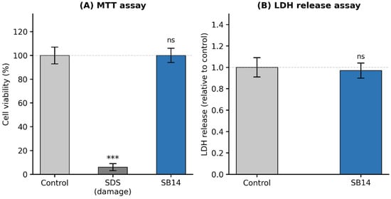
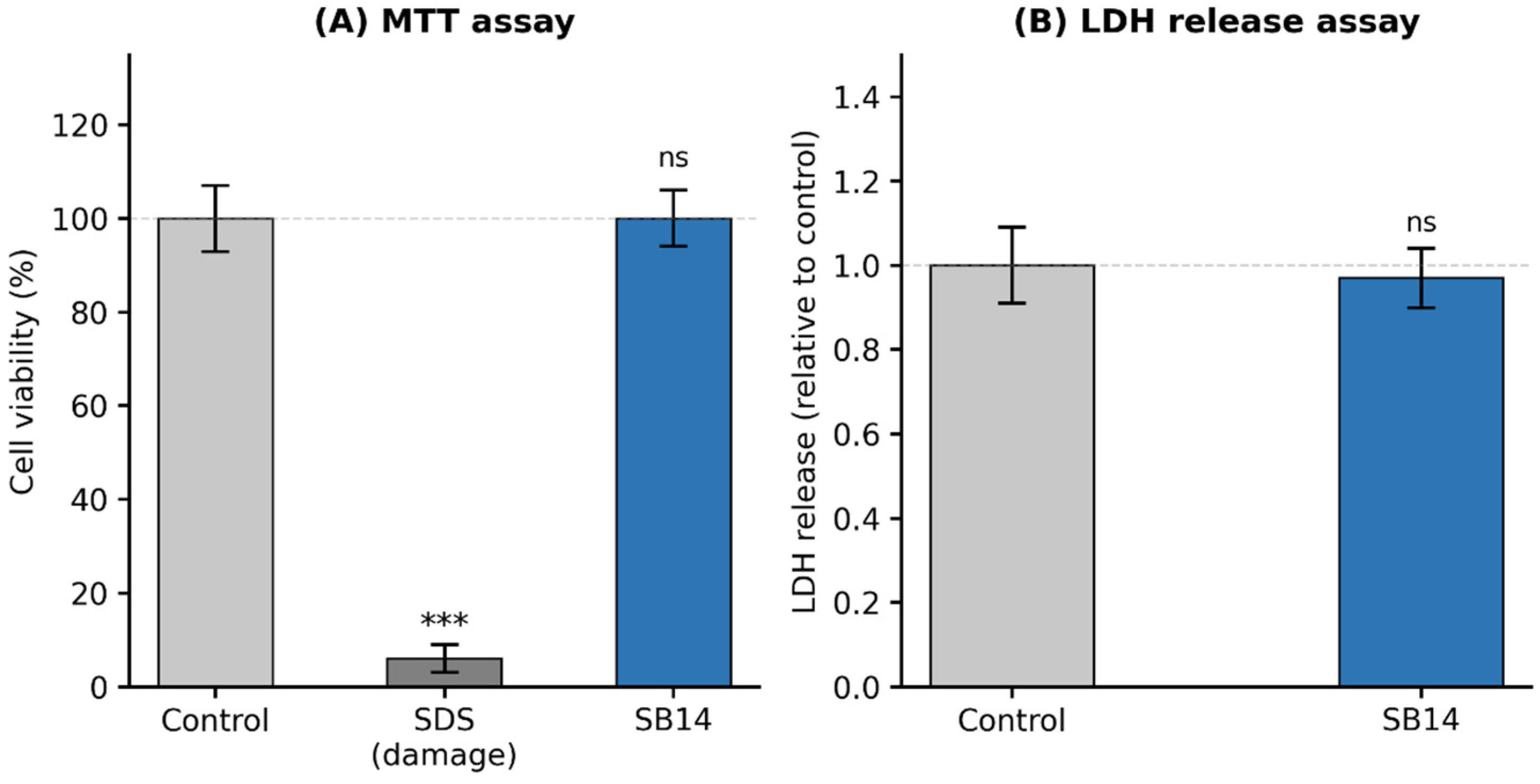
The cytotoxic safety of SB14 was evaluated in NHEK cells using complementary assays (Figure 1). MTT assay, based on mitochondrial respiration, demonstrated maintained cell viability (approximately 100% relative to untreated control) following 24 h treatment at 107 TFU/mL, with no statistically significant difference versus control. LDH release into the culture medium was similarly equivalent between SB14-treated and untreated cells. The positive control (SDS) produced marked cytotoxicity, validating assay sensitivity. These results establish the absence of cytotoxic effects of SB14 at the tested concentration.
Figure 1.
Safety assessment of SB14 in NHEK cells. (A) Cell viability by MTT assay after 24 h treatment (107 TFU/mL); SDS was used as positive control. (B) Cytotoxicity by LDH release assay under identical conditions. Data represent mean ± SD of three independent experiments. ns = not significant vs. untreated control. *** p < 0.001 vs. negative control.
3.2. Mechanistic Analysis
3.2.1. AQP3 Expression and Claudin-1 Recovery Following UV-Induced Damage
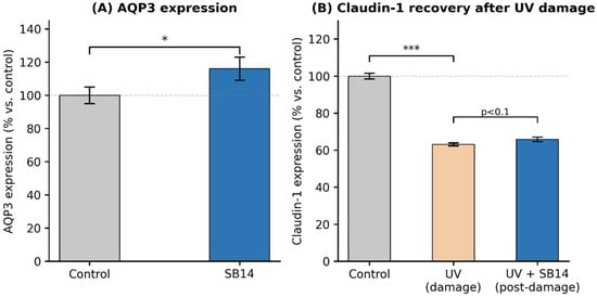
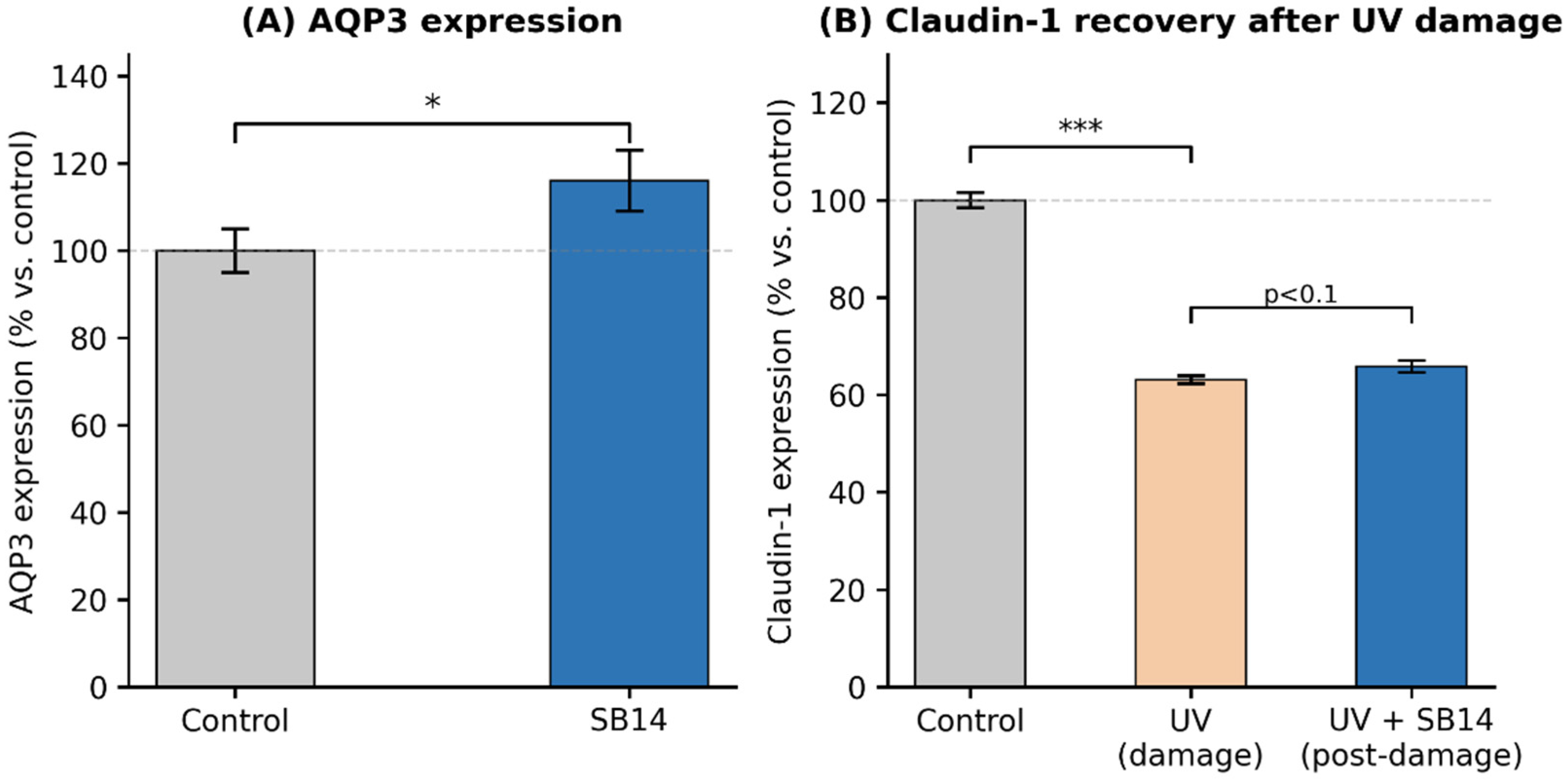
Treatment of NHEK cells with SB14 resulted in a statistically significant increase in AQP3 protein expression versus untreated controls (p < 0.05), suggesting modulation of epidermal water transport pathways relevant to stratum corneum hydration and barrier homeostasis. (Figure 2A).
Figure 2.
Molecular effects of SB14 on NHEK cells. (A) AQP3 protein expression by ELISA after 24 h treatment (p < 0.05 vs. control). (B) Claudin-1 expression in unirradiated control, UV-irradiated control, and cells treated with SB14 following UV damage (post-damage treatment model). UV irradiation significantly reduced Claudin-1 (p < 0.001 vs. control); subsequent SB14 treatment showed partial recovery (p < 0.1 vs. UV damage). Data represent mean ± SD of three independent experiments. Statistical symbols: * p < 0.05 vs. untreated control; *** p < 0.001 vs. unirradiated control. p < 0.1 vs. UV-damaged cells indicates a directional trend not reaching conventional statistical significance.
To evaluate whether SB14 could support barrier recovery following UV-induced tight junction damage, NHEK cells were first irradiated with UVC (30 J/cm2) and subsequently treated with SB14 for 24 h. UV irradiation alone significantly reduced Claudin-1 to 63.17 ± 0.82% of control (p < 0.001 vs. unirradiated control). Subsequent treatment with SB14 resulted in a partial recovery of Claudin-1 expression to 65.9 ± 1.22% (p < 0.1 vs. UV-damaged cells). While this trend did not reach conventional statistical significance, the directional recovery of Claudin-1 following post-damage SB14 application is consistent with a barrier-restorative activity and warrants further investigation in adequately powered studies. (Figure 2B).
3.2.2. Cytokine Modulation in NHEK and PBMCs
SB14 demonstrated a distinct immunomodulatory cytokine profile in both cell models, summarized in Table 2. In NHEK cells, SB14 significantly downregulated IL-8 (0.2 ± 0.1-fold, p < 0.01) and IL-23 (0.2 ± 0.03-fold, p < 0.05) versus control, while significantly increasing IL-6 (2 ± 0.01-fold, p < 0.05). TNF-α was not significantly altered in NHEK (1.1 ± 0.1-fold, p > 0.05). In PBMCs, SB14 strongly induced TNF-α (13.3 ± 0.03-fold, p < 0.001) and IL-6 (6.34 ± 0.18-fold, p < 0.001), consistent with innate immune activation, and significantly reduced IL-8 (0.93 ± 0.007-fold, p < 0.01). IL-23 was unchanged in PBMCs (1 ± 0.01-fold, p > 0.05).
Table 2.
Cytokine modulation by SB14. Results expressed as fold change relative to untreated control (basal = 1 ± SD). ns = not significant.
3.2.3. Antipathogen Activity Against Staphylococcus Aureus
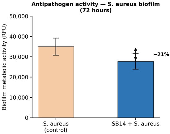
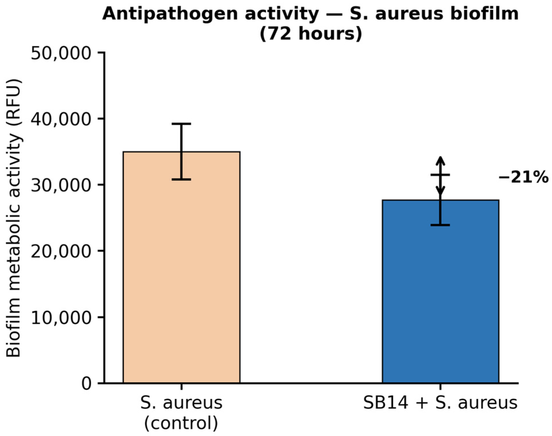
SB14 demonstrated notable inhibitory activity against S. aureus biofilm formation. After 72 h of co-incubation, SA biofilm metabolic activity was reduced by approximately 21% compared to the untreated SA control, as quantified by AlamarBlue fluorescence (Figure 3). This suggests that SB14-derived components can interfere with SA biofilm establishment, a finding potentially relevant to skin conditions characterized by S. aureus colonization.
Figure 3.
Antipathogen activity of SB14 against S. aureus biofilm (AlamarBlue assay, RFU, after 72 h). SB14 reduced SA biofilm by approximately 21% vs. SA control.
3.3. Clinical Efficacy Assessment
All 20 volunteers completed the 30-day study without adverse events or drop-outs. Instrumental and clinical assessments demonstrated improvements in the SB14-treated group across all four measured parameters at both T14 and T30, while the placebo group showed no statistically significant changes in any parameter at any timepoint.
3.3.1. Superficial Skin Hydration
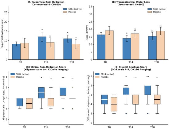
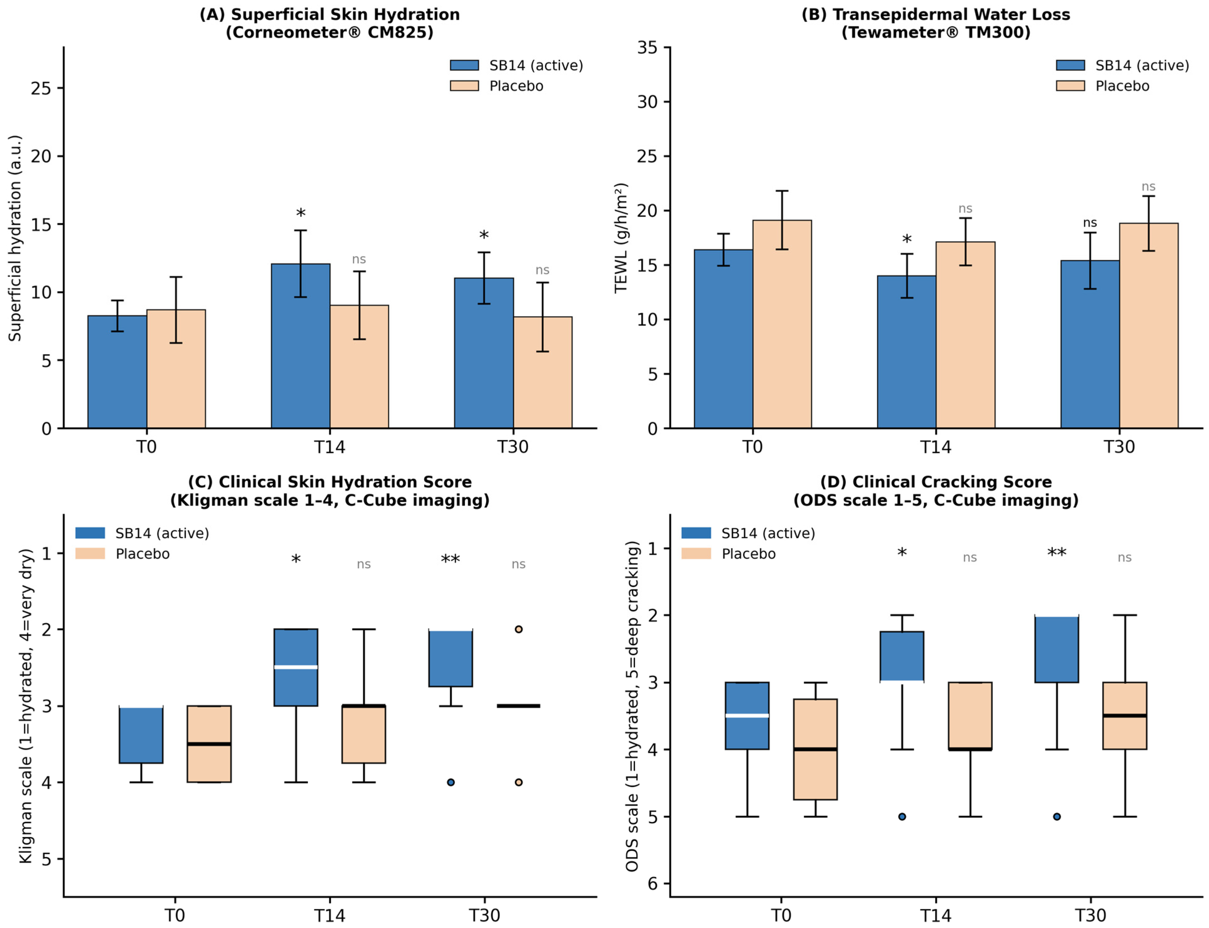
In the active group, mean superficial hydration (Corneometer) increased from 8.3 ± 3.6 a.u. at baseline to 12.1 ± 7.8 a.u. at T14 (+46.1%, p = 0.0451) and 11.0 ± 5.9 a.u. at T30 (+33.6%, p = 0.0144 vs. T0). The improvement remained statistically significant at T30 despite being numerically lower than at T14, indicating sustained hydration over the treatment period. In the placebo group, mean hydration values were 8.7 ± 7.7 a.u. at T0, 9.0 ± 7.9 a.u. at T14 (+3.9%, p = 0.7203), and 8.2 ± 8.0 a.u. at T30 (−6.0%, p = 0.4993), confirming the absence of any spontaneous hydration effect (Figure 4A).
Figure 4.
Clinical efficacy of SB14 versus placebo over 30 days. (A) Mean superficial skin hydration (Corneometer, a.u.). (B) Mean TEWL (Tewameter, g/h/m2). (C) Median clinical skin hydration score (Kligman scale 1–4). (D) Median cracking score (ODS scale 1–5). Active group (n = 10) shown in filled bars; placebo group (n = 10) shown in lighter-shaded bars. Statistical significance for active group: * p < 0.05, ** p < 0.01 versus baseline (T0). Paired t-test for Corneometer and Tewameter; Friedman test for clinical scores. All placebo comparisons were non-significant (ns).
3.3.2. Transepidermal Water Loss (TEWL)
In the active group, mean TEWL decreased from 16.4 ± 4.7 g/h/m2 at baseline to 14.0 ± 6.4 g/h/m2 at T14 (−14.5%, p = 0.0205). At T30, TEWL was 15.4 ± 8.2 g/h/m2 (−6.1% vs. T0; p = 0.4930, ns), indicating that the reduction in TEWL was most pronounced at the early timepoint and showed a partial regression by day 30, although directional improvement was maintained. In the placebo group, TEWL showed no significant changes at either timepoint (T14: −10.4%, p = 0.1393; T30: −1.5%, p = 0.8755), confirming the specificity of the observed effect in the active group (Figure 4B).
3.3.3. Clinical Evaluation of Skin Hydration (Kligman Scale)
Clinical hydration was assessed by trained Abich S.r.l. personnel using C-Cube imaging and graded on the Kligman 1–4 scale (higher scores = drier skin). In the active group, median hydration score improved progressively from 3.0 (moderate dry skin) at T0 to 2.5 at T14 (p = 0.0442) and 2.0 (slightly dry skin) at T30 (p = 0.0073 vs. T0), demonstrating a statistically significant and clinically meaningful shift toward a better-hydrated skin condition. At T14, 60% of treated volunteers showed improvement; this proportion increased to 80% at T30. In the placebo group, median scores remained unchanged (T0: 3.5; T14: 3.0, p = 0.3711; T30: 3.0, p = 0.1461), and improvement was noted in only 30% at T14 and 40% at T30, consistent with natural variability (Figure 4C).
3.3.4. Clinical Evaluation of Skin Cracking (ODS Scale)
Cracking severity was assessed by trained Abich S.r.l. personnel using C-Cube imaging and graded on the ODS 1–5 scale (higher scores = more severe cracking). In the active group, median cracking score improved from 3.5 (intermediate between moderate dry skin with flaking and dry skin with slight cracking) at T0 to 3.0 at T14 (p = 0.0336) and 2.0 (slightly dry skin) at T30 (p = 0.0037 vs. T0), representing a statistically significant and progressive improvement in skin cracking appearance over the treatment period. At T14, 70% of treated volunteers showed improvement; this increased to 80% at T30. In the placebo group, cracking scores remained consistently elevated (T0: 4.0; T14: 4.0, p = 0.5023; T30: 3.5, p = 0.1797), with improvement in only 20% of subjects at T14 and 40% at T30 (Figure 4D).
4. Discussion
This study provides converging in vitro and clinical evidence for the safety and biological activity of SB14 as a cosmetic active ingredient targeting dry, barrier-compromised skin. The use of a heat-treated (postbiotic) format confers practical formulation advantages—including enhanced stability, extended shelf life, and elimination of viable organism concerns—while preserving the bioactive properties of the parent strain [20,22,23].
The combined use of the MTT viability assay and the LDH cytotoxicity assay confirmed the safety profile of SB14 at 107 TFU/mL: full cell viability was maintained (MTT) and no significant LDH release was detected, providing complementary evidence of cellular health that constitutes an essential prerequisite for any topical cosmetic ingredient [27,28]. This safety profile, consistent with prior characterizations of heat-treated lactobacilli [22,23], enables confident translation of in vitro bioactivity findings to clinical development.
The significant upregulation of AQP3 in SB14-treated keratinocytes is mechanistically coherent with the clinical improvements in superficial hydration observed in this study. AQP3 is the predominant epidermal aquaglyceroporin, facilitating water and glycerol transport across keratinocyte membranes and contributing directly to stratum corneum water content and cellular proliferation capacity [14,15]. Its expression declines in barrier-compromised skin, and its upregulation is associated with improved hydration and barrier recovery [16]. The directional, though not fully significant, recovery of Claudin-1 in UV-damaged cells subsequently treated with SB14 further supports a possible barrier-restorative effect at the paracellular level. This post-damage experimental design—where SB14 is applied after UV insult rather than concurrently—better reflects a realistic topical use scenario and highlights the potential of SB14 to support tight junction recovery following environmental stress, warranting confirmation in adequately powered mechanistic studies.
The cytokine modulation profile of SB14 reveals a nuanced immunological action particularly well-suited to dry and barrier-disrupted skin. The marked downregulation of IL-8 in NHEK is of particular clinical relevance: IL-8 is a potent neutrophilic chemokine whose excess production at the epidermal level drives neutrophil recruitment, amplifies oxidative damage, and perpetuates the inflammatory cycle in barrier-disrupted skin [18]. The concurrent downregulation of IL-23 in NHEK—a cytokine pivotal in driving Th17-mediated inflammation—further supports an anti-inflammatory orientation of SB14 in the epithelial compartment [19]. The moderate but significant increase in IL-6 in NHEK may reflect autocrine keratinocyte proliferative signaling rather than pro-inflammatory activity, in the absence of parallel IL-8 or IL-23 induction [29]. In PBMCs, the strong induction of TNF-α and IL-6 is consistent with pattern recognition of bacterial cell wall components (peptidoglycans, lipoteichoic acids) and reflects innate immune priming rather than pathological inflammation [22,30]. Notably, IL-8 was also reduced in PBMCs, further suggesting a consistent anti-chemotactic signal across cell types. The absence of IL-23 alteration in PBMCs indicates that SB14 does not drive Th17 polarization at the systemic level, an important safety consideration for repeated topical application.
The 21% reduction in S. aureus biofilm formation by SB14 at 72 h adds a further clinically relevant dimension: S. aureus colonization of xerotic, cracked skin is a recognized driver of barrier deterioration, local inflammation, and impaired repair [10,11]. Postbiotic-mediated inhibition of pathogen biofilm—likely through the release of antimicrobial peptides and biosurfactants during thermal inactivation—could contribute to restoring a healthier microbial skin environment conducive to barrier recovery [23].
The clinical results provide strong support for the formulation efficacy of SB14 in this indication. The significant and sustained improvement in superficial hydration at both T14 (+46.1%, p = 0.0451) and T30 (+33.6%, p = 0.0144) in the active group, absent in the placebo group, demonstrates a genuine treatment effect attributable to the postbiotic active. This persistent hydration benefit is consistent with the AQP3-mediated mechanism identified in vitro: stable upregulation of this channel could support ongoing glycerol and water movement into the stratum corneum well beyond immediate emollient effects.
The TEWL results present a more nuanced picture: a significant barrier-protective effect was evident at T14 (−14.5%, p = 0.0205), with partial regression but persistent directional improvement at T30 (−6.1%, ns). This kinetic pattern may reflect an early, robust barrier-repair response during the acute treatment phase, followed by a maintenance phase where barrier improvement is sustained but less pronounced instrumentally. The non-significance at T30 is consistent with high intra-individual variability in TEWL measurements—inherent to open-chamber evaporimetry, particularly on body sites such as heels and elbows subject to biomechanical and environmental variation—and should not be interpreted as a loss of effect, given the consistent directional improvement and the significant parallel improvements in clinical hydration and cracking scores at the same timepoint.
The clinical scoring data represent the most compelling evidence of the formulation’s cosmetic benefit. The progressive improvement in Kligman hydration scores from 3.0 to 2.0 over 30 days (p = 0.0073), and the parallel improvement in ODS cracking scores from 3.5 to 2.0 (p = 0.0037), correspond to meaningful shifts in clinical skin condition—from moderate dry skin with early cracking to slightly dry skin. The fact that 80% of treated volunteers showed improvement in both hydration and cracking at T30, compared to 40% for the placebo in both assessments, further underscores the clinical relevance of the active ingredient’s contribution. The complete absence of significant changes in the placebo group across all four parameters strengthens causal attribution to the SB14 ingredient.
Study Limitations
This study has several limitations. The small group size (n = 10 per arm) limits statistical power, particularly for secondary endpoints such as TEWL at T30, and reduces the precision of effect size estimates. The open-label design, while mitigated by objective instrumental measurements and blinded clinical scoring by CRO personnel, could theoretically introduce participant behavior bias. The heterogeneous anatomical distribution (elbows vs. heels) within the active group may have introduced variability, as these sites differ in skin thickness, sebaceous activity, and biomechanical stress exposure; future studies should stratify by body site. The 30-day duration, while standard for exploratory cosmetic trials, may be insufficient to fully characterize the kinetics and durability of barrier recovery, particularly for TEWL which showed non-significant improvement at T30. No correction for multiple testing was applied; results are hypothesis-generating and require confirmation in larger, adequately powered, randomized controlled trials.
5. Conclusions
This study demonstrates that SB14 is safe for topical application and biologically active across multiple skin barrier-relevant pathways: it significantly upregulates AQP3, demonstrates partial Claudin-1 recovery following UV-induced damage, downregulates pro-inflammatory IL-8 and IL-23 in keratinocytes, activates innate immune defense in PBMCs, and inhibits S. aureus biofilm formation. These converging in vitro activities are mechanistically coherent with the statistically significant clinical improvements observed over 30 days in subjects with dry and cracked skin: sustained superficial hydration (+33.6% at T30, p = 0.0144), early TEWL reduction (−14.5% at T14, p = 0.0205), progressive normalization of the clinical hydration score (median from 3.0 to 2.0, p = 0.0073), and significant improvement in cracking severity (median from 3.5 to 2.0, p = 0.0037)—with 80% of treated subjects showing improvement at T30 in both parameters, compared to non-significant changes in the placebo group. The postbiotic format of SB14 offers practical formulation advantages including microbiological safety and enhanced stability. Additional randomized, double-blind, placebo-controlled studies involving larger and geographically diverse populations will be needed to confirm and expand these findings, characterize responder profiles, and establish optimal formulation strategies for different skin types and body sites.
Author Contributions
Conceptualization, M.P. and A.A.; methodology, A.V. and G.D.; investigation, G.D. and A.V.; data curation, G.D. and A.V.; writing, original draft preparation, G.D.; writing—review and editing, G.D., A.V. and A.A.; supervision, M.P. and A.A.; project administration, M.P. and A.A. All authors have read and agreed to the published version of the manuscript.
Funding
This research was supported by Probiotical S.p.A.
Institutional Review Board Statement
Ethical review and approval were not required for this study pursuant to applicable national regulations governing non-invasive cosmetic product testing in adult volunteers. The study was conducted in accordance with the Declaration of Helsinki and Good Clinical Practice (GCP) principles. Data were processed in compliance with EU Regulation 679/2016 (GDPR). This study does not meet the ICMJE definition of a clinical trial, as it evaluates cosmetic endpoints (skin hydration, TEWL, and cracking appearance) in healthy volunteers using a cosmetic emulsion in accordance with EU Regulation (EC) No 1223/2009 on cosmetic products. No medicinal product, medical intervention, or health outcome in the clinical sense was investigated. Prospective registration in a public trial registry is therefore not applicable.
Informed Consent Statement
Written informed consent was obtained from all participants prior to enrolment.
Data Availability Statement
The original contributions presented in this study are included in the article. Further inquiries can be directed to the corresponding author.
Acknowledgments
The in vivo study was conducted by ABICH S.r.l. (Clinical and Cosmetological Trials Center, Verbania and Vimodrone, Italy), whose contribution to clinical data acquisition is gratefully acknowledged. The authors would like to thank Paolo Saronni for his technical support and valuable contributions throughout the project. During the preparation of this work the authors used Claude (Anthropic, San Francisco, CA, USA) for language editing and manuscript revision assistance. The authors reviewed and edited all AI-assisted content and take full responsibility for the final manuscript.
Conflicts of Interest
G.D., A.V., A.A. and M.P. are employees of Probiotical Research Srl. The authors declare that, despite these financial and commercial relationships, the research was conducted with complete scientific autonomy and independence. The funders had no role in study design; collection, analyses, or interpretation of data; writing; or the decision to publish.
References
- Rajkumar, J.; Chandan, N.; Lio, P.; Shi, V. The Skin Barrier and Moisturization: Function, Disruption, and Mechanisms of Repair. Ski. Pharmacol. Physiol. 2023, 36, 174–185. [Google Scholar] [CrossRef] [Scilit] [PubMed]
- Fluhr, J.W.; Muguet, V.; Christen-Zaech, S. Restoring Skin Hydration and Barrier Function: Mechanistic Insights into Basic Emollients for Xerosis Cutis. Int. J. Dermatol. 2025, 64, 5–12. [Google Scholar] [CrossRef] [Scilit] [PubMed]
- Günzel, D.; Yu, A.S. Claudins and the modulation of tight junction permeability. Physiol. Rev. 2013, 93, 525–569. [Google Scholar] [CrossRef] [Scilit] [PubMed] [PubMed Central]
- Honari, G.; Maibach, H. Chapter 1—Skin Structure and Function. In Applied Dermatotoxicology; Academic Press: Cambridge, MA, USA, 2014; pp. 1–10. [Google Scholar] [CrossRef] [Scilit]
- Fluhr, J.W.; Moore, D.J.; Lane, M.E.; Lachmann, N.; Rawlings, A.V. Epidermal barrier function in dry, flaky and sensitive skin: A narrative review. J. Eur. Acad. Dermatol. Venereol. 2024, 38, 812–820. [Google Scholar] [CrossRef] [Scilit] [PubMed]
- Lee, H.J.; Kim, M. Skin Barrier Function and the Microbiome. Int. J. Mol. Sci. 2022, 23, 13071. [Google Scholar] [CrossRef] [Scilit] [PubMed] [PubMed Central]
- Rawlings, A.V.; Harding, C.R. Moisturization and skin barrier function. Dermatol. Ther. 2004, 17, 43–48. [Google Scholar] [CrossRef] [Scilit] [PubMed]
- Schuetz, R.; Claypool, J.; Sfriso, R.; Vollhardt, J.H. Sunscreens can preserve human skin microbiome upon erythemal UV exposure. Int. J. Cosmet. Sci. 2024, 46, 71–84. [Google Scholar] [CrossRef] [Scilit] [PubMed]
- Ashkanani, A.; Ashkanani, G.; Yousef, M.; Rob, M.; Al-Marri, M.; Naseem, N.; Laws, S.; Chaari, A. Microbiome and Skin Health: A Systematic Review of Nutraceutical Interventions, Disease Severity, Inflammation, and Gut Microbiota. Microorganisms 2026, 14, 63. [Google Scholar] [CrossRef] [Scilit] [PubMed]
- Woo, Y.R.; Kim, H.S. Interaction between the microbiota and the skin barrier in aging skin: A comprehensive review. Front. Physiol. 2024, 15, 1322205. [Google Scholar] [CrossRef] [Scilit] [PubMed] [PubMed Central]
- Ratanapokasatit, Y.; Laisuan, W.; Rattananukrom, T.; Petchlorlian, A.; Thaipisuttikul, I.; Sompornrattanaphan, M. How Microbiomes Affect Skin Aging: The Updated Evidence and Current Perspectives. Life 2022, 12, 936. [Google Scholar] [CrossRef] [Scilit] [PubMed]
- Habeebuddin, M.; Karnati, R.K.; Shiroorkar, P.N.; Nagaraja, S.; Asdaq, S.M.B.; Khalid Anwer, M.; Fattepur, S. Topical Probiotics: More Than a Skin Deep. Pharmaceutics 2022, 14, 557. [Google Scholar] [CrossRef] [Scilit] [PubMed] [PubMed Central]
- Yu, Y.; Dunaway, S.; Champer, J.; Kim, J.; Alikhan, A. Changing our microbiome: Probiotics in dermatology. Br. J. Dermatol. 2020, 182, 39–46. [Google Scholar] [CrossRef] [Scilit] [PubMed]
- Bollag, W.B.; Aitkens, L.; White, J.; Hyndman, K.A. Aquaporin-3 in the epidermis: More than skin deep. Am. J. Physiol. Physiol. 2020, 318, C1144–C1153. [Google Scholar] [CrossRef] [Scilit] [PubMed] [PubMed Central]
- Qin, H.; Zheng, X.; Zhong, X.; Shetty, A.K.; Elias, P.M.; Bollag, W.B. Aquaporin-3 in keratinocytes and skin: Its role and interaction with phospholipase D2. Arch. Biochem. Biophys. 2011, 508, 138–143. [Google Scholar] [CrossRef] [Scilit] [PubMed] [PubMed Central]
- Karimi, N.; Ahmadi, V. Aquaporin Channels in Skin Physiology and Aging Pathophysiology: Investigating Their Role in Skin Function and the Hallmarks of Aging. Biology 2024, 13, 862. [Google Scholar] [CrossRef] [Scilit] [PubMed]
- Mondadori, G.; Amoruso, A.; Visciglia, A.; Deusebio, G.; Pinto, D.; Pane, M.; Rinaldi, F. Heat-Treated Probiotics’ Role in Counteraction of Skin UVs-Induced Damage In Vitro. Cosmetics 2025, 12, 121. [Google Scholar] [CrossRef] [Scilit]
- Chilicka, K.; Dzieńdziora-Urbińska, I.; Szyguła, R.; Asanova, B.; Nowicka, D. Microbiome and Probiotics in Acne Vulgaris-A Narrative Review. Life 2022, 12, 422. [Google Scholar] [CrossRef] [Scilit] [PubMed] [PubMed Central]
- Lunjani, N.; Ahearn-Ford, S.; Dube, F.S.; Hlela, C.; O’Mahony, L. Mechanisms of microbe-immune system dialogue within the skin. Genes Immun. 2021, 22, 276–288. [Google Scholar] [CrossRef] [Scilit] [PubMed] [PubMed Central]
- Salminen, S.; Collado, M.C.; Endo, A.; Hill, C.; Lebeer, S.; Quigley, E.M.M.; Sanders, M.E.; Shamir, R.; Swann, J.R.; Szajewska, H.; et al. The International Scientific Association of Probiotics and Prebiotics (ISAPP) consensus statement on the definition and scope of postbiotics. Nat. Rev. Gastroenterol. Hepatol. 2021, 18, 649–667, Erratum in Nat. Rev. Gastroenterol. Hepatol. 2021, 18, 671. https://doi.org/10.1038/s41575-021-00481-x. Erratum in Nat. Rev. Gastroenterol. Hepatol. 2022, 19, 551. https://doi.org/10.1038/s41575-022-00628-4. PMID: 33948025; PMCID: PMC8387231. [Google Scholar] [CrossRef] [Scilit] [PubMed]
- De Almeida, C.V.; Antiga, E.; Lulli, M. Oral and Topical Probiotics and Postbiotics in Skincare and Dermatological Therapy: A Concise Review. Microorganisms 2023, 11, 1420. [Google Scholar] [CrossRef] [Scilit] [PubMed] [PubMed Central]
- Piqué, N.; Berlanga, M.; Miñana-Galbis, D. Health Benefits of Heat-Killed (Tyndallized) Probiotics: An Overview. Int. J. Mol. Sci. 2019, 20, 2534. [Google Scholar] [CrossRef] [Scilit] [PubMed] [PubMed Central]
- Lopes, E.G.; Moreira, D.A.; Gullón, P.; Gullón, B.; Cardelle-Cobas, A.; Tavaria, F.K. Topical application of probiotics in skin: Adhesion, antimicrobial and antibiofilm in vitro assays. J. Appl. Microbiol. 2017, 122, 450–461. [Google Scholar] [CrossRef] [Scilit] [PubMed]
- Gruber, J.V.; Holtz, R. Living, quiescent Lactobacillus plantarum Lp90 probiotic, delivered topically to full thickness tissues in vitro via a just-add-water cream delivery system, stimulates the expression of elastin protein. J. Cosmet. Dermatol. 2023, 22, 2852–2860. [Google Scholar] [CrossRef] [Scilit] [PubMed]
- Hashemi, S.S.; Rafati, A.; Roohinejad, S.; Yaghoobi, F.; Salehi, A. Postbiotics as Emerging Therapeutics for Skin Wound Healing and Dermatological Care: Clinical Trends and Mechanistic Insights. Int. Wound J. 2025, 22, e70799. [Google Scholar] [CrossRef] [Scilit] [PubMed] [PubMed Central]
- Santos, Y.R.; Andréo-Filho, N.; Lopes, P.S.; Leite-Silva, V.R. A review of skin microbiome and new challenges to cosmetic microbiome-friendly formulations. Int. J. Cosmet. Sci. 2026. Epub ahead of printing. [Google Scholar] [CrossRef] [Scilit] [PubMed]
- Mosmann, T. Rapid colorimetric assay for cellular growth and survival: Application to proliferation and cytotoxicity assays. J. Immunol. Methods. 1983, 65, 55–63. [Google Scholar] [CrossRef] [Scilit] [PubMed]
- Decker, T.; Lohmann-Matthes, M.L. A quick and simple method for the quantitation of lactate dehydrogenase release in measurements of cellular cytotoxicity and tumor necrosis factor (TNF) activity. J. Immunol. Methods. 1988, 115, 61–69. [Google Scholar] [CrossRef] [Scilit] [PubMed]
- Klimitz, F.J.; Shen, Y.; Repetto, F.; Brown, S.; Knoedler, L.; Ko, C.J.; Abu Hussein, N.; Crisler, W.J.; Adams, T.; Kaminski, N.; et al. Keratinocytes as active regulators of cutaneous and mucosal immunity: A systematic review across inflammatory epithelial disorders. Front. Immunol. 2025, 16, 1694066. [Google Scholar] [CrossRef] [Scilit] [PubMed]
- Mehta, J.P.; Ayakar, S.; Singhal, R.S. The potential of paraprobiotics and postbiotics to modulate the immune system: A Review. Microbiol. Res. 2023, 275, 127449. [Google Scholar] [CrossRef] [Scilit] [PubMed]

Disclaimer/Publisher’s Note: The statements, opinions and data contained in all publications are solely those of the individual author(s) and contributor(s) and not of MDPI and/or the editor(s). MDPI and/or the editor(s) disclaim responsibility for any injury to people or property resulting from any ideas, methods, instructions or products referred to in the content. |
© 2026 by the authors. Licensee MDPI, Basel, Switzerland. This article is an open access article distributed under the terms and conditions of the Creative Commons Attribution (CC BY) license.