


Abstract
The application of natural bioactive compounds in cosmeceutical products, particularly as skin-lightening agents, has received increasing interest. Caulerpa racemosa, a green macroalga of the Chlorophyta division, contains beneficial nutrients that are applicable as food and cosmeceutical ingredients. This study investigated the in vitro effect of the ethanolic extract of C. racemosa (CR) on regulation of melanogenic-related signaling and gene expression in SK-MEL-5 human melanoma-derived cells. Identification of bioactive components revealed that catechin, rutin, and quercetin as flavonoid contents were found in CR extract, analyzed using HPLC. The expressions of microphthalmia-associated transcription factor (MITF), extracellular signal-regulated kinase (ERK) signaling molecules, and melanogenic-related molecules were analyzed via Western blotting and qPCR. The CR extract treatment applied to SK-MEL-5 cells decreased the MITF protein expression level, which correlated with increased pERK expression, and no cytotoxic effect was observed. The subsequent treatment reduced the expression of melanogenesis-related genes (TYR, TYRP1, MC1R, and DCT) that were downstream targets of MITF. This study provides preliminary evidence that CR extract may modulate melanogenesis-related signaling. However, the specific bioactive compounds responsible for the observed effects remain to be identified, as the extract contains a complex mixture of phytochemicals. Further fractionation studies are needed to pinpoint the active constituents. The variability of extract composition due to seasonal and geographical factors should be considered for future standardization.
1. Introduction
Melanin pigments are a product of the melanogenesis process and are produced by melanocytes; they can be divided into two types: eumelanin and pheomelanin. Tyrosinases, the primary melanogenesis enzymes, are highly similar to those found by [1]. The α-MSH (alpha-melanocyte-stimulating hormone)—MC1R (melanocortin 1 receptor) signal acts as the principal pathway in melanogenesis. In this pathway, the extracellular signal-regulated kinase (ERK) acts as the most important signal driving the melanogenic mechanism [2,3,4]. On the other hand, MEK/ERK signaling activation has been reported to inhibit melanogenesis by phosphorylating MITF, which leads to proteasomal degradation [5].
MITF, or microphthalmia-associated transcription factor, acts as a master regulator in melanogenesis, the process of melanin production, by promoting the transcription of melanogenic enzymes, such as tyrosinase (TYR), tyrosinase-related protein 1 (TYRP1), and tyrosinase-related protein-2 (TYRP2) [6], and the melanosome trafficking machinery, including CDC42, RAB17, RAC1, and RAB27A, leading to increased melanin production and accumulation [7,8]. MITF also influences melanocyte proliferation and melanosome maturation, biogenesis, transport, and survival by regulating Bcl-2 expression [9]. Additionally, the expression of these melanogenic enzymes is regulated by MITF through direct interactions with an E-box/M-box in their promoters [10,11], and the most pro-pigmentation pathway involves α-MSH (α-melanocyte-stimulating hormone) binding on its MC1R, which is expressed by melanocytes. Mechanistically, α-MSH binding to MC1R triggers cAMP elevation and activation of the Protein Kinase A/cAMP-response element-binding protein (PKA/CREB) signaling pathway that stimulates the transcription of a variety of downstream targets, including MITF [12]. MITF suppression may subsequently downregulate the expression of melanogenic enzymes, resulting in reduced melanin production, as a key beneficial effect of whitening substances.
Marine algae are applicable as an important ingredient in cosmetic products, as they are potential natural sources of vitamins, minerals, and other nutrients [13,14]. Bioactive compounds from seaweeds are considered to possess beneficial health effects, such as antioxidants, anticancer, anticoagulants, antidiabetic, and anti-inflammation properties [15,16,17,18]. Seaweed has gained attention as a natural antioxidant and skin protectant for pharmaceutical and cosmeceutical production because it is extremely safe and can be processed for long-term use [19]. Several tropical seaweeds are considered as sources of whitening products. Caulerpa racemosa is tropical algae in Chlorophyta, cultivated in the Southern Pacific Ocean and is typically distributed along the coasts of Thailand [15,16]. It contains bioactive components such as proteins, amino acids, fatty acids, minerals, and vitamins, and various bioactive components including flavonoids, tannins, phenols, saponins, phytosterol, terpenoids, sulphated polysaccharides, secondary metabolites, caulerpin, caulerpenyne, caulersin, and racemosin C [16,17,18,19,20,21,22,23]. The ethanolic extracts of the seaweeds C. racemosa, U. intestinalis, and L. challengeriae showed tyrosinase-inhibiting activity and reduced the melanin contents in B16F10 murine melanoma cells treated with α-melanocyte-stimulating hormone [16]. This study examined the in vitro mechanistic effect of C. racemosa extracts on MITF expression as the master upstream molecule for melanogenic regulation and related downstream molecular expression in human melanoma-derived cells.
2. Materials and Methods
2.1. Extract Preparation
Algal cultivation of C. racemosa was provided by Phetchaburi coastal fisheries research and development center, Thailand, during summer season (March–May, 2023). Dried whole crudes were soaked in absolute ethanol at room temperature for 3 days, evaporated using a rotary evaporator (BÜCHI Labortechnik AG, Flawil, Switzerland), and stored at −20 °C to prevent extract deterioration.
2.2. High-Performance Liquid Chromatography Analysis
The phytochemical profile in the flavonoid content of the ethanolic extract of C. racemosa (CR) was analyzed via rapid high-performance liquid chromatography. A reversed-phase column and a Spectra System UV-200 by Spectra System P-4000 (Thermo Separation Products-TSP, Riviera Beach, CA, USA) were used, and the column’s temperature was not maintained (ambient temperature). A gradient run was performed using two mobile phases: (A) a formic acid solution at pH 2.5 and (B) methanol. The method was as follows: from 0 to 20 min, linear gradient from 80% to 30% solvent A, and from 20.1 to 22 min, isocratic at 90% solvent B. Additionally, a re-equilibration period of 8 min with 80% solvent A was used between individual runs. The injection volume was 10 μL, and the flow rate was 1 mL/min. Semi-quantitative profiling of flavonoid contents was reported and compared with whole weight of lyophilized extracts. The LOD and LOQ values were 0.4 and 1.2 μg/mL for catechin, 0.1 and 0.5 μg/mL for rutin, and 0.8 and 2.6 μg/mL for quercetin, respectively.
2.3. Cell Culture
SK-MEL-5 human melanoma-derived cells were purchased from the American Type Culture Collection (American Type Culture Collection (ATCC), Manassas, VA, USA) and cultured in a minimum essential medium (MEM, Gibco, Thermo Fisher Scientific, Waltham, MA, USA) supplemented with 10% fetal bovine serum (FBS, Gibco, Thermo Fisher Scientific, Waltham, MA, USA), 1% sodium pyruvate (Merck), and 1% penicillin–streptomycin (Merck, Rahway, NJ, USA) in a 37 °C incubator with a humidified atmosphere of 5% CO2. The aforementioned medium was renewed every 2 days.
2.4. Cell Viability Assay
SK-MEL-5 cells were seeded in 96-well plates at 8 × 10⁴ cells/well and cultured until 85–90% confluence and were treated with the CR extract at final concentrations of 10, 50, 100, 250, and 500 µg/mL for 24 h. After treatment, the 3-(4,5-dimethylthiazol-2-yl)-2,5-diphenyl tetrazolium bromide (MTT) solution (Bio Basic Inc., Markham, ON, Canada) was added into the wells at a final concentration of 0.15 mg/mL, incubated for 1h, and then dissolved with DMSO. The reaction was measured using a spectrophotometer (Cytation 5, BioTek Instruments, Winooski, VT, USA), and the cell viability percentage was calculated and compared with that of the untreated cells.
2.5. Western Blotting
Cells were plated in 60 mm2 dishes at a density of 4 × 10⁵ cells/dish. After 48 h, the cells were treated with CR at concentrations of 50 and 100 µg/mL for 30 min. The ERK inhibitor U0126 at a final concentration of 10 µM was added alongside CR to investigate ERK expression. Cells were washed with cold phosphate-buffered saline (PBS) before being lysed with a lysis buffer (20 mM Tris, 1% octyl phenoxypolyethoxylethanol (NP-40), and 50 mM NaCl) containing a 0.1% protease inhibitor cocktail (Calbiochem, Sigma-Aldrich, St. Louis, MO, USA). The concentration of crude proteins was determined using the Bradford reagent (Bio-Rad Laboratories, Hercules, CA, USA) and was then used for Western blotting analysis. The proteins were separated on 10% SDS-PAGE gels and transferred to nitrocellulose membranes. The membranes were blocked with 5% skimmed milk in tris-buffered saline (TBS) containing 0.1% tween 20 and probed with rabbit anti-human MITF (Cell Signaling Technology, Danvers, MA, USA, 1:1000 dilution), rabbit anti-human p44/42 MAPK (Cell Signaling, USA, dilution 1:2000 dilution), rabbit anti-human phospho-p44/42 MAPK (Cell Signaling, USA, dilution 1:4000 dilution), and mouse anti-human ꞵ-actin (Cell Signaling, USA, 1:10,000 dilution), followed by anti-rabbit or anti-mouse antibodies conjugated with horseradish peroxidase (HRP) enzymes. The chemiluminescent signal was visualized using an enhanced chemiluminescence substrate (Bio-Rad Laboratories, Hercules, CA, USA), and the band density of proteins was determined and normalized using Image J software (version 5.0), with β-actin as the internal loading control.
2.6. Quantitative PCR
SK-MEL-5 cells were seeded and treated for 30 min. Total RNA was extracted using the Total RNA extraction kit (Geneaid Biotech Ltd., New Taipei City, Taiwan) according to the manufacturer’s protocol. cDNA synthesis was performed using superscript III reverse transcriptase (Invitrogen, Thermo Fisher Scientific, Waltham, MA, USA). qPCRs were performed using SensiFAST SYBR No-ROX (Bioline, Meridian Bioscience, Cincinnati, OH, USA), and the results were determined using a C1000 Touch Thermal cycler (Bio-Rad Laboratories, Hercules, CA, USA). All primers are listed in Table 1. The Cq value of each target gene was normalized with Glyceraldehyde-3-phosphate dehydrogenase (GAPDH), and relative fold change (RCF) was calculated and compared against untreated cells using the 2−∆∆Cq method.
Table 1.
Reverse transcription quantitative PCR primers.
2.7. Statistical Analysis
Data were presented as the mean ± SEM, and data analysis was performed using GraphPad Prism version 8.1.0 (GraphPad Software, San Diego, CA, USA). The statistical significance of differences between the values was evaluated via analysis of variance (ANOVA) followed by Tukey’s post hoc test for multiple-group comparisons or an independent sample t-test. In all comparisons, differences were considered significant when p-values were less than 0.05 (p ≤ 0.05).
3. Results
3.1. HPLC Analysis of the CR Extract
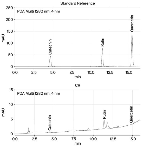
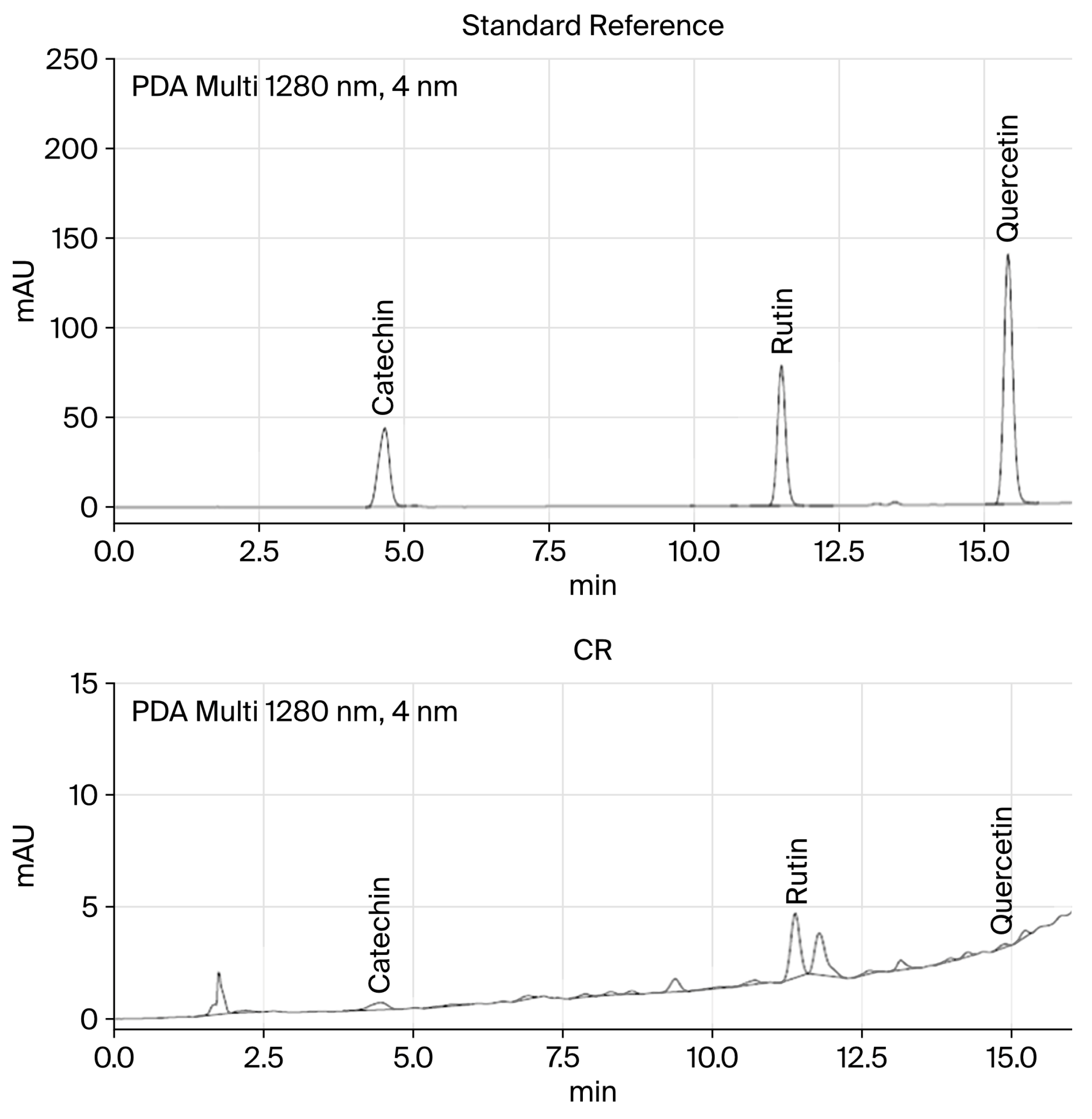
Total flavonoid contents presented in CR extract was 5.43 ± 0.54 mg of quercetin equivalent. Three compounds from the flavonoid group were found in the CR extract (Figure 1). These constituents included rutin as the highest value, followed by catechin, and quercetin. Detailed identification results were revealed in Table 2.
Figure 1.
High-performance liquid chromatogram of flavonoid compounds in the CR extract compared with reference standards.
Table 2.
Flavonoid compound identification.
3.2. Viability of CR-Treated Human Melanoma-Derived Cells
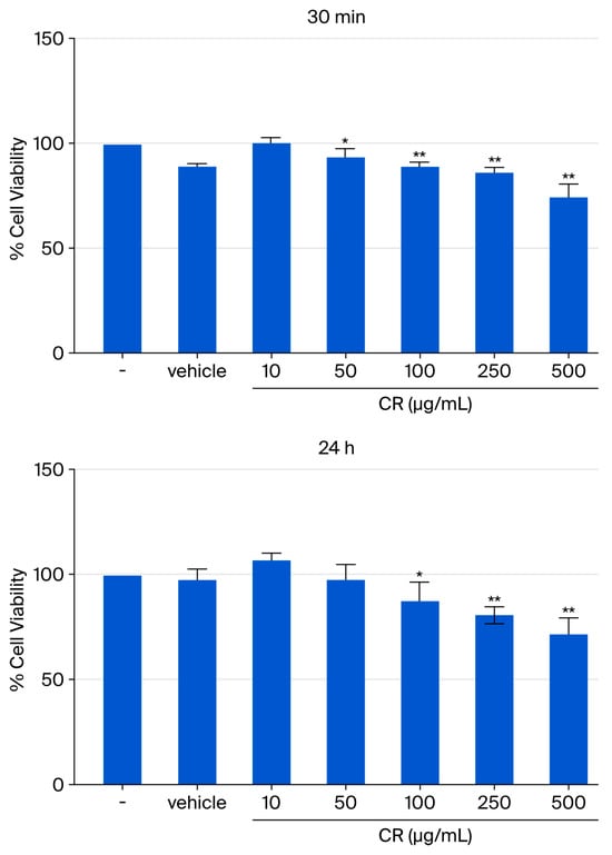
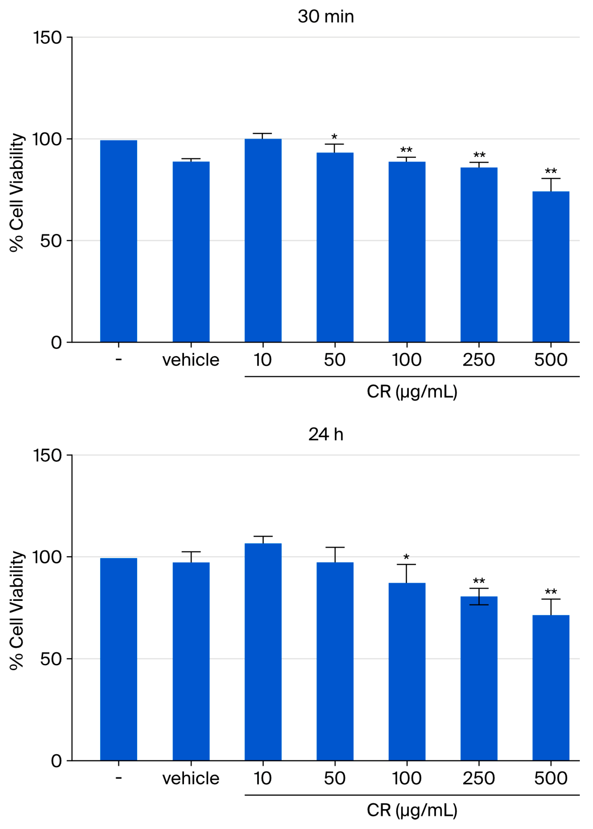
SK-MEL-5 cells were exposed to CR at the final concentrations of 10, 50, 100, 200, and 500 μg/mL for 24 h, and cell viability was measured using the MTT assay and normalized relative to the untreated group, which was at 100%. Under normal-culture conditions, the viability of the CR-treated groups at 10 and 50 µg/mL was nearly equivalent to that of the control. However, the groups at 100–500 µg/mL had slightly reduced viability at 30 min and 24 h (Figure 2).
Figure 2.
The viability of SK-MEL-5 cells treated with the indicated doses of CR (10–500 μg/mL) for 30 min and 24 h was measured using the MTT assay. Data were presented as the mean ± SEM. *: p < 0.05 and **: p < 0.01 in comparison with the untreated group. The vehicle (0.2% DMSO) was used to dissolve the crude extract. N = 3.
3.3. CR Decreases MITF Expression and Is Associated with the Activation of ERK Signaling
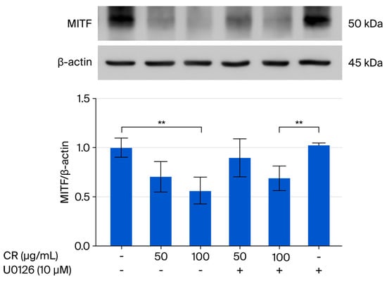
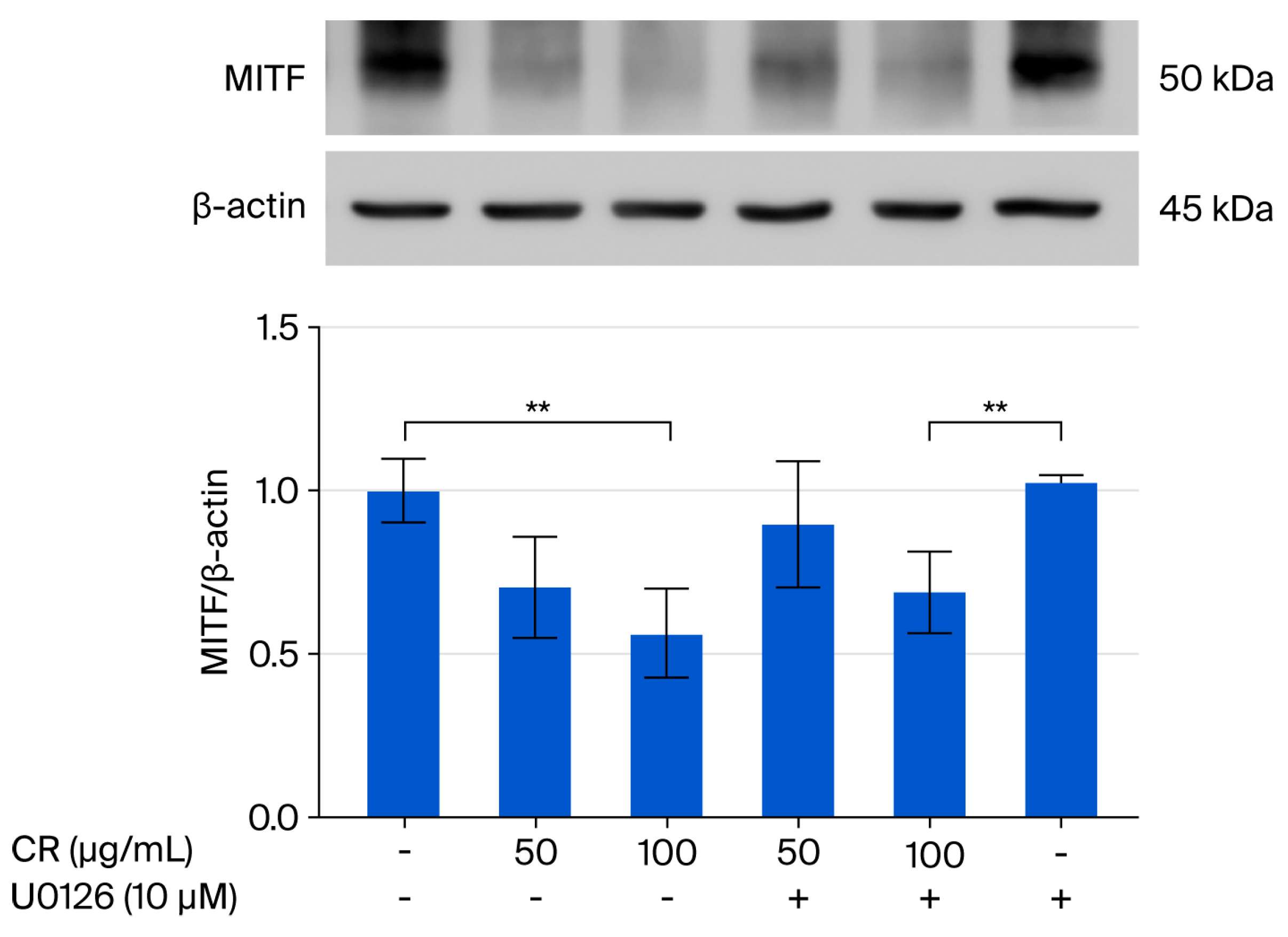
The expression of MITF, a melanogenesis-related protein, in CR-treated SK-MEL-5 cells was analyzed via Western blotting. The CR extract at the final concentration of 50 and 100 μg/mL decreased MITF protein expression after treatment for 30 min in a dose-dependent manner. On the other hand, U0126 significantly restored MITF expression compared with the CR extract (Figure 3).
Figure 3.
MITF expression in SK-MEL-5 cells treated with the indicated doses of CR (50 and 100 μg/mL) or co-treated with the ERK1/2-specific inhibitor U0126 (10 μM) for 30 min was analyzed via Western blotting. Data were presented as the mean ± SEM. **: p < 0.01 in comparison with the untreated group. +: treatment; −: non-treatment; N = 3.
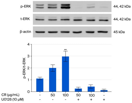
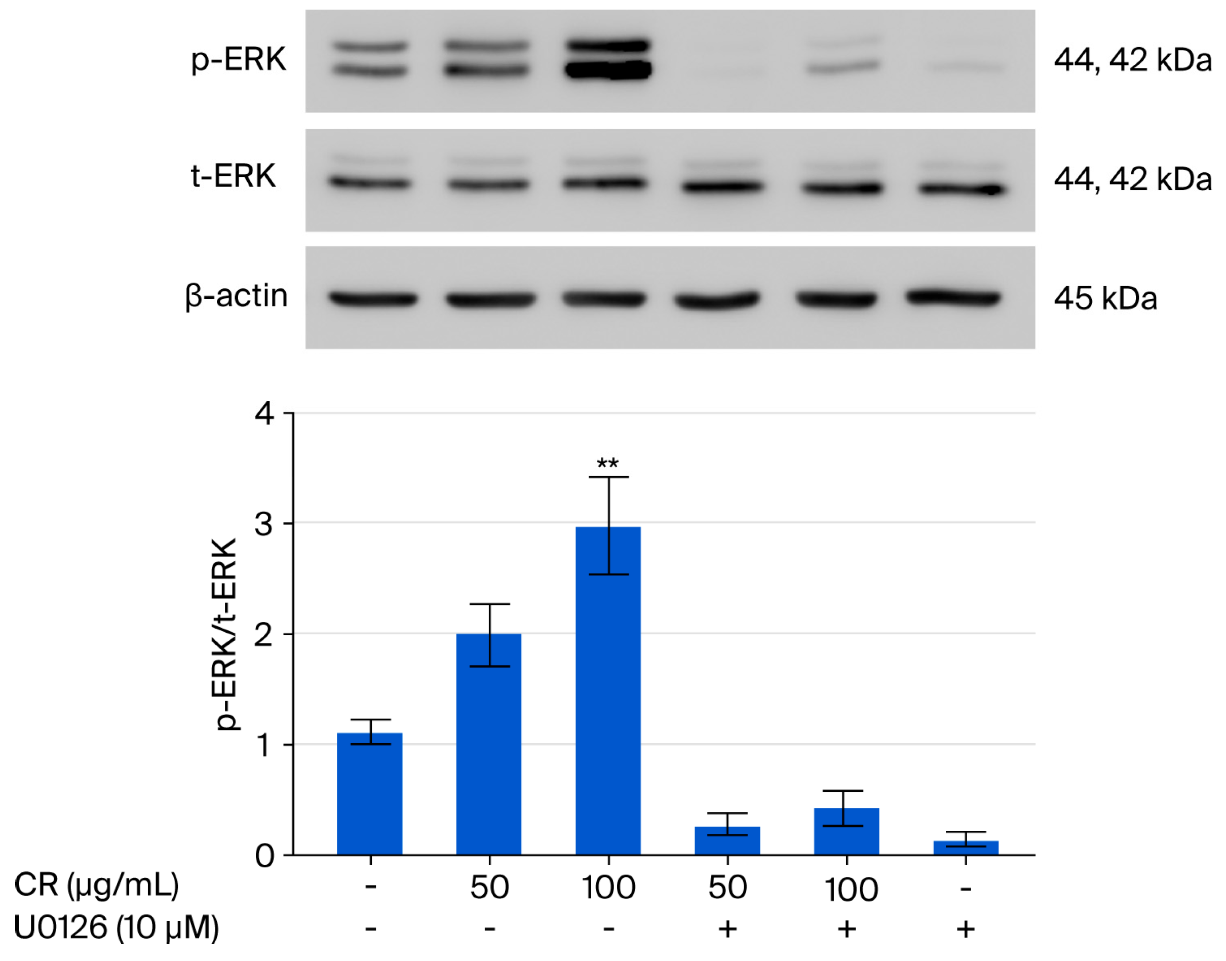
The expression of ERK, a regulatory protein of melanogenesis, in the α-MSH–MC1R signaling pathway following CR treatment was also analyzed via Western blotting. The CR treatment increased ERK expression in CR-treated SK-MEL-5 cells, as shown by the ratio between the p-ERK and t-ERK proteins (Figure 4). To confirm the relationship of this phenomenon with ERK signaling, cells were co-treated with CR and the ERK1/2-specific inhibitor U0126. ERK expression was significantly reduced by U0126, while co-treatment with CR and U0126 resulted in a greater ratio of ERK than the single treatment with U0126 (Figure 5). In addition, U0126 decreased the MITF level when co-treated with CR, compared with cells treated with U0126 only, which was significantly lower than that of the untreated control (Figure 4). This implied that CR decreased MITF expression in SK-MEL-5 cells that might be associated with the activation of the ERK signaling pathway.
Figure 4.
p-ERK, t-ERK, and β-actin protein expression levels in SK-MEL-5 cells treated with the indicated doses of CR (50 and 100 μg/mL) or co-treated with the ERK-specific inhibitor U0126 (10 μM) for 30 min were analyzed using Western blotting. Data were presented as the mean ± SEM. **: p < 0.01 in comparison with the untreated group. +: treatment; −: non-treatment. N = 3.
Figure 5.
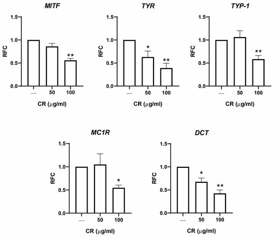
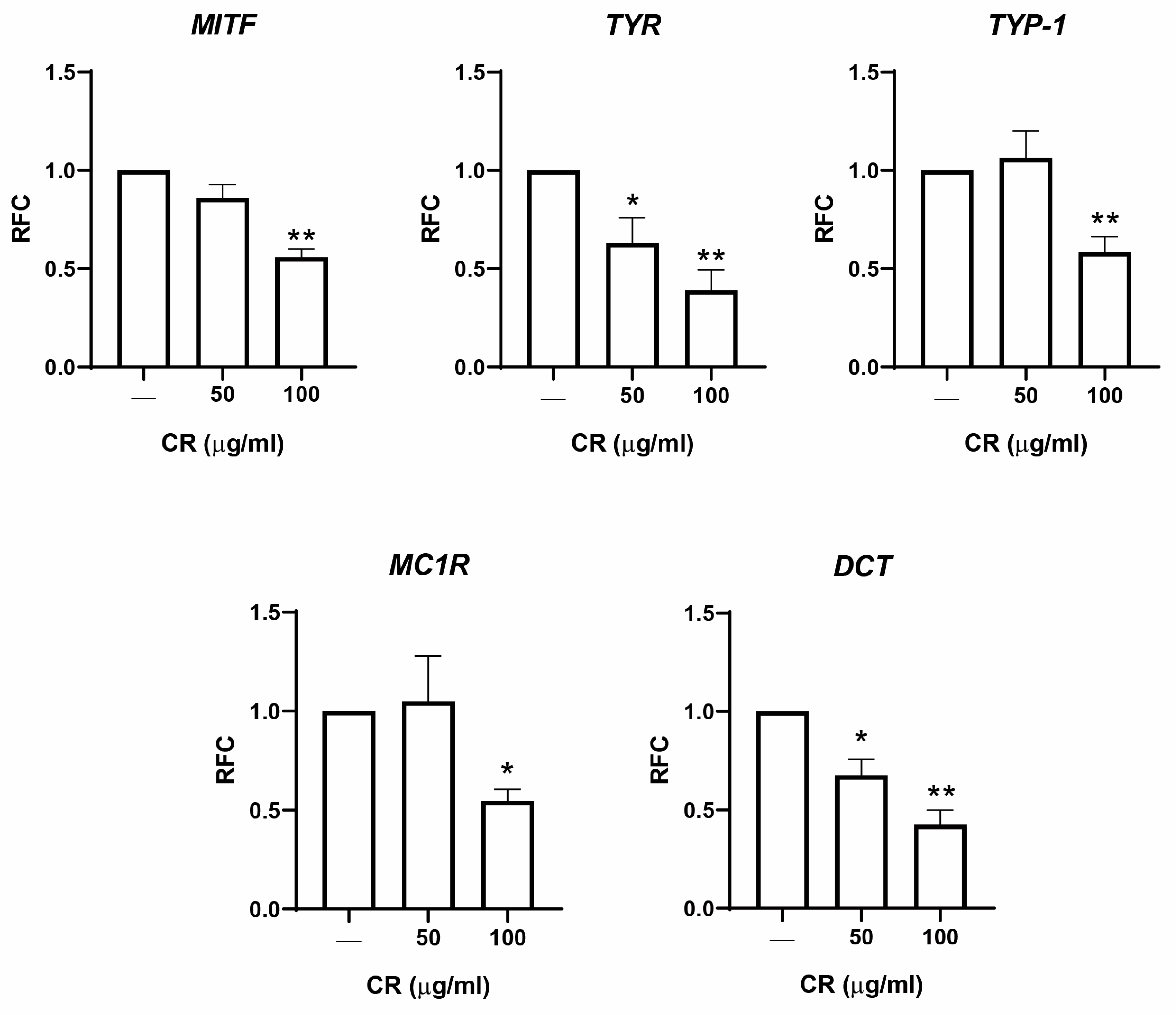
The expression of melanogenesis-related genes in SK-MEL-5 cells treated with the indicated doses of CR (50 and 100 μg/mL) for 30 min was measured by qPCR. Data were presented as the mean ± SEM. * and ** indicate p < 0.05 and p < 0.01, respectively, in comparison with the untreated group. N = 3.
3.4. Expression of Melanogenesis-Related Genes Following CR Treatment
The quantitative expression of the melanogenesis-related genes MITF, TYR, TYP1, MC1R, and dopachrome tautomerase (DCT) in SK-MEL-5 cells after CR treatment was analyzed via qPCR. CR at the final concentrations of 50 and 100 μg/mL decreased MITF, TYR, and DCT expression following treatment for 30 min in a dose-dependent manner compared with the GAPDH expression level. Additionally, TYP-1 and MC1R expression levels were significantly decreased following CR treatment at 100 μg/mL (Figure 5).
4. Discussion
This study investigated the molecular effects of the CR extract in cultured SK-MEL-5 human melanoma-derived cells on targeting MITF expression as a key upstream molecule in controlling melanogenesis. The CR extract was tested for toxicity against SK-MEL-5 cells to ensure its safety when applied to study the in vitro effects. Based on the bioactive compounds identified in the CR, quercetin and rutin comprised the highest proportions of the flavonoid compounds. Quercetin (3,3,4,5,7-pentahydroxyflavone) and rutin (quercetin-3-rutinoside), a glycoside containing a disaccharide covalently attached to the quercetin unit, are widely used flavonoid compounds in cosmetic applications and are presented in many edible plants [24,25,26]. Additionally, quercetin has dual effects on melanogenesis. A review emphasized that the effective concentration of quercetin for anti-melanogenesis is between 20 and 50 μM; however, values exceeding this range may promote melanogenesis [27]. Rutin has been shown to inhibit melanin production by modulating the cellular oxidative status and inhibiting tyrosinase by chelating copper ions essential for tyrosinase function [28]. The quercetin and rutin combination could potentially be used as photoprotective or sunscreen agents, as they possess a sun protection factor (SPF) of about 12 [29]. This result implies that flavonoid contents in CR extract may partly, but not completely, influence biological effects. It should be noted that the composition of the ethanolic extract of CR depends on the season, collection site and extraction parameters. In the present work, we did not perform full standardization of the raw material, which may affect the reproducibility of the effect; further studies should take these factors into account.
MITF is a master regulator of melanocyte development and melanogenesis, as it controls the transcription of downstream enzymes including TYR, TYRP-1, and DCT. Additionally, it is upregulated by the MC1R, a G-protein receptor on the melanocyte surface, which is activated by α-MSH [6,30,31]. Our findings demonstrate that the CR extract could reduce expression of the key melanogenic regulators of MITF, along with its upstream molecule MC1R, in SK-MEL-5 cells following 30 min of treatment. This swift suppression may align with the compound’s potential to disrupt the cAMP/PKA/MAPK signaling axis, which drives melanocyte differentiation and melanin synthesis. Therefore, the observed downregulation of MITF likely leads to a cascade that inhibits melanogenic genes, consistent with previous reports showing that MITF inhibition reduces melanin production [32]. ERK (extracellular signal-regulated kinase) is part of a kinase family in the MAPK pathway that plays a pivotal role in regulating the post-translation of MITF. ERK activation in melanocytes often leads to phosphorylation and subsequent accelerated MITF degradation, resulting in melanogenesis suppression [33,34]. We found that a reduction in MITF expression level following CR treatment in SK-MEL-5 cells was associated with ERK activation, as shown by the increased pERK:total ERK ratio. The results supported CR effects in regulating MITF expression in human melanoma-derived cells through ERK signaling activation.
As evidenced by qPCR result, the CR extract treatment in SK-MEL-5 cells could reduce the mRNA levels of downstream melanogenesis-related molecules TYR, TYRP-1, and DCT. The reduction in TYR and TYRP-1 expression might imply its role in melanin production. TYR is the rate-limiting enzyme in melanogenesis, catalyzing the initial steps of melanin production, while TYRP-1 and DCT are involved in eumelanin synthesis and stabilization [35,36]. MC1R is the most well-known receptor on melanocytes; its binding with α-MSH activates adenylate cyclase, increases intracellular cAMP, and activates the PKA /CREB pathway [13,37]. Additionally, it is a major upstream regulator of human pigmentation and is categorized as a melanoma susceptibility gene [38]. MC1R inhibition blocks α-MSH signaling, reducing cAMP levels and downstream MITF activation, which suppresses melanin synthesis and melanoma growth [39]. This implies that CR administration might be associated with melanin production in human melanoma-derived cells, primarily through the ERK/MC1R/MITF pathway, which can lead to reduced transcription of pigmentation genes and TYR/TYP1/DCT cascades that inhibit melanin synthesis. Together with viability analysis, administration of CR at final concentrations between 50 and 100 μg/mL in SK-MEL-5 cells might provide effective results for related molecular expression and retain its viability, which would be proposed for further study.
Other members of Chlorophyta such as Chlorella vulgaris and blue Lagoon microalgae also have anti-melanogenesis properties, as they reduce oxidative stress, a key driver of melanogenesis [40,41]. Other green algae, such as Prasiola japonica, have shown significant activity, with their major component, loliolide, exhibiting antioxidant and tyrosinase-inhibitory effects [6]. Octaphlorethol A, a member of the phenolic group, is isolated from Ishige foliacea and has anti-melanogenesis properties via MITF downregulation mediated by ERK signaling pathway activation [42].
Although the present study demonstrates the in vitro effect of the CR extract on regulating of MITF expression with related signaling molecules in melanoma-derived cells, it may not completely reflect anti-melanogenic activity and should not be interpreted as evidence of cosmetic safety. The experiments were conducted under specific in vitro conditions using a melanoma-derived cell line, which does not adequately represent normal human skin. The study, conducted in a short time period, might not reflect the actual biological phenomenon. Further studies on in-depth bioactive component characterization, bioassays in reconstructed human skin models, skin irritation assays, and long-term safety assessments are necessary to establish the safety and suitability of CR extracts for cosmetic applications.
5. Conclusions
The CR extract reduced MITF expression is primarily associated with activation of ERK signaling and reduced expression of melanogenesis-related genes TYR/TYP1/DCT cascades in SK-MEL-5 cells. The CR extract shows promising activity in vitro, but its application as a cosmetic ingredient requires further characterization of the active components and validation of its safety and efficacy in more complex models. The present study does not establish a direct causative link to specific polyphenolic compounds, and the observed effects may be attributable to other constituents present in the extract. Furthermore, the detailed standardization of the raw material should be addressed in future work.
Author Contributions
Conceptualization, R.S. and T.S.; methodology, R.S., K.S., P.V. and T.S.; software, R.S. and T.S.; validation, K.S., P.V. and T.S.; formal analysis, R.S. and T.S.; investigation, R.S., K.S., P.V., N.L., W.Y., M.T., W.K. and T.S.; resources, N.L., W.Y., M.T., W.K. and T.S.; data curation, R.S. and T.S.; writing—original draft preparation, R.S.; writing—review and editing, K.S., P.V., N.L., W.Y., M.T., W.K. and T.S.; visualization, R.S.; supervision, K.S., P.V. and T.S.; project administration, T.S.; funding acquisition, T.S. All authors have read and agreed to the published version of the manuscript.
Funding
This work was financially supported by Thailand Science Research and Innovation (TSRI), National Science Research and Innovation Fund (NSRF) (Fundamental Fund: Grant no. 21/2566 and 8/2567) and the Wellness Research Unit for Integrative Organ and Movement Sciences (WRU-IOMS): 2568.
Institutional Review Board Statement
Not applicable.
Informed Consent Statement
Not applicable.
Data Availability Statement
The original contributions presented in this study are included in the article. Further data inquiries can be addressed to the corresponding author.
Acknowledgments
We thank Atiporn Jenjadkarn, Faculty of Allied Health Sciences, Burapha University, Chonburi, Thailand, for technical assistance.
Conflicts of Interest
The authors declare no conflicts of interest.
Abbreviations
The following abbreviations are used in this manuscript:
| RT | Retention time |
| MW | Molecular weight |
| qPCR | Quantitative polymerase chain reaction |
References
- Wang, R.; Chen, T.; Zhao, B.; Fan, R.; Ji, K.; Yu, X.; Wang, X.; Dong, C. FGF21 regulates melanogenesis in alpaca melanocytes via ERK1/2-mediated MITF downregulation. Biochem. Biophys. Res. Commun. 2017, 490, 466–471. [Google Scholar] [CrossRef] [Scilit] [PubMed]
- Chung, Y.C.; Hyun, C.G. Inhibitory Effects of Pinostilbene Hydrate on Melanogenesis in B16F10 Melanoma Cells via ERK and p38 Signaling Pathways. Int. J. Mol. Sci. 2020, 21, 4732. [Google Scholar] [CrossRef] [Scilit] [PubMed]
- D’Mello, S.A.; Finlay, G.J.; Baguley, B.C.; Askarian-Amiri, M.E. Signaling Pathways in Melanogenesis. Int. J. Mol. Sci. 2016, 17, 1144. [Google Scholar] [CrossRef] [Scilit] [PubMed]
- Kamilijiang, M.; Zang, D.; Abudukelimu, N.; Aidarhan, N.; Liu, G.; Aisa, H.A. Anti-Melanogenesis Effect of Polysaccharide from Saussurea involucrata on Forskolin-Induced Melanogenesis in B16F10 Melanoma Cells. Nutrients 2022, 14, 5044. [Google Scholar] [CrossRef] [Scilit] [PubMed]
- Lv, J.; Zou, K.; Yin, C.; Xu, W.; Meng, D.; Zhang, H.; Yu, W.; Jiang, P.; Yun, C.; Xue, H.; et al. Daidzin suppresses melanogenesis through ERK and AKT signaling pathways mediated MITF proteasomal degradation. Exp. Mol. Pathol. 2025, 143, 104986. [Google Scholar] [CrossRef] [Scilit] [PubMed]
- Park, H.J.; Byun, K.A.; Oh, S.; Kim, H.M.; Chung, M.S.; Son, K.H.; Byun, K. The Combination of Niacinamide, Vitamin C, and PDRN Mitigates Melanogenesis by Modulating Nicotinamide Nucleotide Transhydrogenase. Molecules 2022, 27, 4923. [Google Scholar] [CrossRef] [Scilit] [PubMed]
- Kim, N.; Kim, S.; Lee, M.W.; Jeon, H.J.; Ryu, H.; Kim, J.M.; Lee, H.J. MITF Promotes Cell Growth, Migration and Invasion in Clear Cell Renal Cell Carcinoma by Activating the RhoA/YAP Signal Pathway. Cancers 2021, 13, 2920. [Google Scholar] [CrossRef] [Scilit] [PubMed]
- Rachmin, I.; Ostrowski, S.M.; Weng, Q.Y.; Fisher, D.E. Topical treatment strategies to manipulate human skin pigmentation. Adv. Drug Deliv. Rev. 2020, 153, 65–71. [Google Scholar] [CrossRef] [Scilit] [PubMed]
- McGill, G.G.; Horstmann, M.; Widlund, H.R.; Du, J.; Motyckova, G.; Nishimura, E.K.; Lin, Y.L.; Ramaswamy, S.; Avery, W.; Ding, H.F.; et al. Bcl2 regulation by the melanocyte master regulator Mitf modulates lineage survival and melanoma cell viability. Cell 2002, 109, 707–718. [Google Scholar] [CrossRef] [Scilit] [PubMed]
- Aoki, H.; Moro, O. Involvement of microphthalmia-associated transcription factor (MITF) in expression of human melanocortin-1 receptor (MC1R). Life Sci. 2002, 71, 2171–2179. [Google Scholar] [CrossRef] [Scilit] [PubMed]
- Cheli, Y.; Ohanna, M.; Ballotti, R.; Bertolotto, C. Fifteen-year quest for microphthalmia-associated transcription factor target genes. Pigment Cell Melanoma Res. 2010, 23, 27–40. [Google Scholar] [CrossRef] [Scilit] [PubMed]
- Gelmi, M.C.; Houtzagers, L.E.; Strub, T.; Krossa, I.; Jager, M.J. MITF in Normal Melanocytes, Cutaneous and Uveal Melanoma: A Delicate Balance. Int. J. Mol. Sci. 2022, 23, 6001. [Google Scholar] [CrossRef] [Scilit] [PubMed]
- Jesumani, V.; Du, H.; Aslam, M.; Pei, P.; Huang, N. Potential Use of Seaweed Bioactive Compounds in Skincare-A Review. Mar. Drugs 2019, 17, 688. [Google Scholar] [CrossRef] [Scilit] [PubMed]
- Kovač, D.J.; Simeunović, J.B.; Babić, O.B.; Mišan, A.Č.; Milovanović, I.L. Algae in food and feed. Food Feed. Res. 2013, 40, 21–31. [Google Scholar]
- Guiry, M. World-wide electronic publication, National University of Ireland, Galway (taxonomic information republished from Algae Base with permission of MD Guiry). Lithothamnioncalcareum f. pseudoflabelligerum Cabioch. AlgeaBase 1966, 12, 358–365. [Google Scholar]
- Choosuwan, P.; Praiboon, J.; Boonpisuttinant, K.; Klomjit, A.; Muangmai, N.; Ruangchuay, R.; Chirapart, A. Inhibitory Effects of Caulerpa racemosa, Ulva intestinalis, and Lobophora challengeriae on Tyrosinase Activity and alpha-MSH-Induced Melanogenesis in B16F10 Melanoma Cells. Life 2023, 13, 934. [Google Scholar] [CrossRef] [Scilit] [PubMed]
- Kumar, A.; Krishnamoorthy, E.; Devi, H.M.; Uchoi, D.; Tejpal, C.; Ninan, G.; Zynudheen, A. Influence of sea grapes (Caulerpa racemosa) supplementation on physical, functional, and anti-oxidant properties of semi-sweet biscuits. J. Appl. Phycol. 2018, 30, 1393–1403. [Google Scholar] [CrossRef] [Scilit]
- Permatasari, H.K.; Barbara Ulfa, E.N.; Adyana Daud, V.P.; Sulistomo, H.W.; Nurkolis, F. Caulerpa racemosa extract inhibits HeLa cancer cells migration by altering expression of epithelial-mesenchymal transition proteins. Front. Chem. 2022, 10, 1052238. [Google Scholar] [CrossRef] [Scilit] [PubMed]
- Sanjeewa, K.A.; Lee, W.; Jeon, Y.-J. Nutrients and bioactive potentials of edible green and red seaweed in Korea. Fish. Aquat. Sci. 2018, 21, 19. [Google Scholar] [CrossRef] [Scilit]
- Liyanage, N.M.; Nagahawatta, D.P.; Jayawardena, T.U.; Jayawardhana, H.; Lee, H.G.; Kim, Y.S.; Jeon, Y.J. Clionasterol-Rich Fraction of Caulerpa racemosa against Particulate Matter-Induced Skin Damage via Inhibition of Oxidative Stress and Apoptosis-Related Signaling Pathway. Antioxidants 2022, 11, 1941. [Google Scholar] [CrossRef] [Scilit] [PubMed]
- Palaniyappan, S.; Sridhar, A.; Kari, Z.A.; Tellez-Isaias, G.; Ramasamy, T. Evaluation of Phytochemical Screening, Pigment Content, In Vitro Antioxidant, Antibacterial Potential and GC-MS Metabolite Profiling of Green Seaweed Caulerpa racemosa. Mar. Drugs 2023, 21, 278. [Google Scholar] [CrossRef] [Scilit] [PubMed]
- Matanjun, P.; Mohamed, S.; Mustapha, N.M.; Muhammad, K.; Ming, C.H. Antioxidant activities and phenolics content of eight species of seaweeds from north Borneo. J. Appl. Phycol. 2008, 20, 367–373. [Google Scholar] [CrossRef] [Scilit]
- He, Q.; Wang, J.; Li, J.; Yang, W. Polyphenol Profile and Antioxidant, Antityrosinase, and Anti-Melanogenesis Activities of Ethanol Extract of Bee Pollen. Pharmaceuticals 2024, 17, 1634. [Google Scholar] [CrossRef] [Scilit] [PubMed]
- Jang, Y.A.; Kim, S.G.; Kim, H.K.; Lee, J.T. Biological Activity and Component Analyses of Chamaecyparis obtusa Leaf Extract: Evaluation of Antiwrinkle and Cell Protection Effects in UVA-Irradiated Cells. Medicina 2023, 59, 755. [Google Scholar] [CrossRef] [Scilit] [PubMed]
- Jung, H.J.; Lee, A.K.; Park, Y.J.; Lee, S.; Kang, D.; Jung, Y.S.; Chung, H.Y.; Moon, H.R. (2E,5E)-2,5-Bis(3-hydroxy-4-methoxybenzylidene) cyclopentanone Exerts Anti-Melanogenesis and Anti-Wrinkle Activities in B16F10 Melanoma and Hs27 Fibroblast Cells. Molecules 2018, 23, 1415. [Google Scholar] [CrossRef] [Scilit] [PubMed]
- Monmai, C.; Kim, J.S.; Chin, J.H.; Lee, S.; Baek, S.H. Inhibitory Effects of Polyphenol- and Flavonoid-Enriched Rice Seed Extract on Melanogenesis in Melan-a Cells via MAPK Signaling-Mediated MITF Downregulation. Int. J. Mol. Sci. 2023, 24, 11841. [Google Scholar] [CrossRef] [Scilit] [PubMed]
- Choi, M.-H.; Shin, H.-J. Anti-melanogenesis effect of quercetin. Cosmetics 2016, 3, 18. [Google Scholar] [CrossRef] [Scilit]
- Prasawang, N.; Sutjarit, N.; Sitthipunya, A.; Suwannalert, P.; Monsuwan, W.; Charoenchon, N. Inhibition of Tyrosinase and Melanogenesis by a White Mulberry Fruit Extract. Int. J. Mol. Sci. 2025, 26, 7589. [Google Scholar] [CrossRef] [Scilit] [PubMed]
- Choquenet, B.; Couteau, C.; Paparis, E.; Coiffard, L.J. Quercetin and rutin as potential sunscreen agents: Determination of efficacy by an in vitro method. J. Nat. Prod. 2008, 71, 1117–1118. [Google Scholar] [CrossRef] [Scilit] [PubMed]
- Levy, C.; Khaled, M.; Fisher, D.E. MITF: Master regulator of melanocyte development and melanoma oncogene. Trends Mol. Med. 2006, 12, 406–414. [Google Scholar] [CrossRef] [Scilit] [PubMed]
- Vachtenheim, J. The many roles of MITF in melanoma. Single Cell Biol. 2017, 6, 162. [Google Scholar]
- Lim, H.Y.; Jeong, D.; Park, S.H.; Shin, K.K.; Hong, Y.H.; Kim, E.; Yu, Y.G.; Kim, T.R.; Kim, H.; Lee, J.; et al. Antiwrinkle and Antimelanogenesis Effects of Tyndallized Lactobacillus acidophilus KCCM12625P. Int. J. Mol. Sci. 2020, 21, 1620. [Google Scholar] [CrossRef] [Scilit] [PubMed]
- Kim, E.S.; Park, S.J.; Goh, M.J.; Na, Y.J.; Jo, D.S.; Jo, Y.K.; Shin, J.H.; Choi, E.S.; Lee, H.K.; Kim, J.Y.; et al. Mitochondrial dynamics regulate melanogenesis through proteasomal degradation of MITF via ROS-ERK activation. Pigment Cell Melanoma Res. 2014, 27, 1051–1062. [Google Scholar] [CrossRef] [Scilit] [PubMed]
- Kim, M.-J.; Kim, S.S.; Park, K.-J.; An, H.J.; Choi, Y.H.; Lee, N.H.; Hyun, C.-G. Anti-melanogenic Activity of Auraptene via ERK-mediated MITF Downregulation. Cosmetics 2017, 4, 34. [Google Scholar] [CrossRef] [Scilit]
- Pang, M.; Xu, R.; Xi, R.; Yao, H.; Bao, K.; Peng, R.; Zhi, H.; Zhang, K.; He, R.; Su, Y.; et al. Molecular understanding of the therapeutic potential of melanin inhibiting natural products. RSC Med. Chem. 2024, 15, 2226–2253. [Google Scholar] [CrossRef] [Scilit] [PubMed]
- Hearing, V.J. Determination of melanin synthetic pathways. J. Investig. Dermatol. 2011, 131, E8–E11. [Google Scholar] [CrossRef] [Scilit] [PubMed]
- Fu, C.; Chen, J.; Lu, J.; Yi, L.; Tong, X.; Kang, L.; Pei, S.; Ouyang, Y.; Jiang, L.; Ding, Y.; et al. Roles of inflammation factors in melanogenesis (Review). Mol. Med. Rep. 2020, 21, 1421–1430. [Google Scholar] [CrossRef] [Scilit] [PubMed]
- Swope, V.B.; Abdel-Malek, Z.A. MC1R: Front and Center in the Bright Side of Dark Eumelanin and DNA Repair. Int. J. Mol. Sci. 2018, 19, 2667. [Google Scholar] [CrossRef] [Scilit] [PubMed]
- Kansal, R.G.; McCravy, M.S.; Basham, J.H.; Earl, J.A.; McMurray, S.L.; Starner, C.J.; Whitt, M.A.; Albritton, L.M. Inhibition of melanocortin 1 receptor slows melanoma growth, reduces tumor heterogeneity and increases survival. Oncotarget 2016, 7, 26331–26345. [Google Scholar] [CrossRef] [Scilit] [PubMed]
- Kim, J.H.; Lee, J.E.; Kim, K.H.; Kang, N.J. Beneficial Effects of Marine Algae-Derived Carbohydrates for Skin Health. Mar. Drugs 2018, 16, 459. [Google Scholar] [CrossRef] [Scilit] [PubMed]
- Kim, S.; Seo, H.; Mahmud, H.A.; Islam, M.I.; Sultana, O.F.; Lee, Y.; Kim, M.; Song, H.Y. Melanin Bleaching and Melanogenesis Inhibition Effects of Pediococcus acidilactici PMC48 Isolated from Korean Perilla Leaf Kimchi. J. Microbiol. Biotechnol. 2020, 30, 1051–1059. [Google Scholar] [CrossRef] [Scilit] [PubMed]
- Kim, K.N.; Yang, H.M.; Kang, S.M.; Kim, D.; Ahn, G.; Jeon, Y.J. Octaphlorethol A isolated from Ishige foliacea inhibits alpha-MSH-stimulated induced melanogenesis via ERK pathway in B16F10 melanoma cells. Food Chem. Toxicol. 2013, 59, 521–526. [Google Scholar] [CrossRef] [Scilit] [PubMed]

Disclaimer/Publisher’s Note: The statements, opinions and data contained in all publications are solely those of the individual author(s) and contributor(s) and not of MDPI and/or the editor(s). MDPI and/or the editor(s) disclaim responsibility for any injury to people or property resulting from any ideas, methods, instructions or products referred to in the content. |
© 2026 by the authors. Licensee MDPI, Basel, Switzerland. This article is an open access article distributed under the terms and conditions of the Creative Commons Attribution (CC BY) license.