


Abstract
Exosomes are revolutionizing skincare as natural messengers for cell communication, yet their transition into cosmetics is often limited by the ethical and regulatory hurdles of their animal or human sourcing. This study describes the development and validation of vegan exosome-like biomimetic vesicles (EBVs) generated from the microalgae Chlamydomonas reinhardtii that reproduce the structural and functional logic of mammalian exosomes. Their structural biomimetism was confirmed through physical, lipidomic, and proteomic characterizations, revealing bilamellar vesicles (average diameter ~160 nm) containing 109 membrane lipids and 1369 proteins. Their functional biomimetism was assessed via 3′ mRNA sequencing, which showed that the EBVs induced transcriptional responses in human fibroblasts functionally analogous to human-derived exosomes in matrix-remodeling and anti-aging pathways. In vitro, the EBVs showed a 166.7% higher dermal delivery bias than standard liposomes and accelerated wound healing. Ex vivo, 2% EBVs protected skin explants against UV-A stress, showing 92% protective efficacy for excessive melanin production upon oxidative stress. Furthermore, the EBVs supported hair follicle anagen markers and follicle stem cell metabolism, significantly upregulating SOX9 (p = 0.0022). A 56-day placebo-controlled clinical study confirmed significant improvements in wrinkle depth (−12.2%), elasticity (+4.9%), and radiance (+20.0%). These results position EBVs as a scalable, high-performance alternative for next-generation anti-aging cosmetic applications.
1. Introduction
Skin aging is a multifactorial process involving several interconnected biological mechanisms, including extracellular matrix remodeling, oxidative stress, cellular senescence, impaired barrier function, altered pigmentation, inflammation, and disrupted intercellular communication [1,2,3,4,5,6]. Within this broader framework, exosomes and their related extracellular vesicles signaling are particularly relevant because extracellular vesicles participate in the coordination of cellular responses among keratinocytes, fibroblasts, melanocytes, immune cells, endothelial cells, adnexal structures, and progenitor compartments. By combining bioactive proteins, lipids, and nucleic acids into a selectively organized membrane, they enable cells to exchange contextual information capable of reshaping the behavior of recipient tissues [7,8,9,10,11,12].
Exosome biology provides the conceptual foundation for their application in dermatology and cosmetics. Canonically, exosomes arise through endosomal trafficking and inward budding within multivesicular bodies before being secreted into the extracellular milieu. Their small size, typical bilayer membrane, protein-enriched surface, membrane microdomains, and protected inner cargo make them particularly efficient at stabilizing signals, navigating extracellular spaces, and docking with target cells [13,14,15,16]. In skin, exosome-mediated signaling is associated with extracellular matrix regulation, fibroblast activation, modulation of oxidative stress, pigmentation biology, inflammatory balance, vascular crosstalk, and wound repair [7,8,9,10,17,18,19,20,21]. These features explain the strong momentum behind exosome-inspired skin technologies.
Nevertheless, the translation of conventional exosomes into cosmetic products is far from straightforward. Human- and animal-derived exosomes raise multiple concerns at once: ethical objections, traceability complexity, donor variability, biological contamination risk, viral and prion safety considerations, production cost, batch heterogeneity, and a regulatory environment that is difficult to reconcile with routine cosmetic commercialization [9,11,12]. Even when these limitations are partially addressed in research settings, the industrial path remains narrow because high-performance exosome preparations often depend on sophisticated upstream cell cultures and downstream isolation workflows, such as ultracentrifugation, density gradients, size-exclusion chromatography, and tangential-flow filtration [22,23,24]. Such systems are informative scientifically, but not always realistic for a stable, broadly deployable cosmetic ingredient.
A second wave of interest has therefore focused on plant- and algae-derived extracellular vesicles and exosome-like vesicles. These offer appealing vegan narratives, improved consumer acceptability, and access to naturally bioactive botanical or algal compositions [25,26,27,28,29,30,31,32]. However, natural vegan vesicles also have major limitations. Their secretion yield is usually low; their source material can be influenced by the season, agronomic conditions, or extraction intensity; their purification can require large input of biomass; and the resulting vesicle fractions may be heterogeneous in size, composition, and functional potency [22,27,28,29,30,31,32]. As a cosmetic innovation, a natural vesicle story is attractive, but industrial reliability and biological consistency remain problematic.
A significant controversy in the current landscape involves the distinction between true exosomes and simplified delivery systems. A third approach, commonly encountered in cosmetic marketing, relies on exosome-like systems that are, in practice, little more than liposomes or lecithin vesicles dressed in exosomal language. Such systems may still provide useful encapsulation, but they generally fail to reproduce what makes exosomes biologically distinctive: membrane complexity; microdomain organization; biogenesis-associated proteins; communication-oriented surface features; and a multi-functional, biologically coherent cargo [11,12,13,14,15,16,33,34]. In other words, conventional liposomes can deliver defined actives, yet they do not inherently behave like exosomes. For applications where biological signaling matters as much as delivery, this distinction is fundamental.
Biomimetism offers a more rigorous solution to this divergence. In biological engineering, biomimetism does not mean superficial resemblance; it means reproducing the organizing principles that give a natural system its function. For exosome-inspired skincare, the relevant question is therefore not whether a vesicle merely looks nanosized, but whether its architecture, membrane logic, compositional complexity, and resulting cellular behavior reproduce the core features that make natural exosomes effective. A biomimetic exosome platform should thus aim at two levels simultaneously. First, there is structural biomimetism: recreating the membrane organization, lipid classes, protein families, and vesicular format that underlie exosome stability and cell interactions. Second, there is functional biomimetism: reproducing the ability to induce coherent biological responses in skin cells and to translate those responses into measurable tissue outcomes.
In this study, the term ‘biomimetic exosomes’ is used as a descriptive, exosome-inspired terminology and not as a biological classification of naturally secreted extracellular vesicles. The investigated vesicles are not generated through canonical endosomal exosome biogenesis and should therefore not be considered natural exosomes. Instead, they are microalgae-derived, self-assembled biomimetic vesicles designed to reproduce selected exosome-relevant properties, including the nanoscale vesicular architecture, membrane complexity, and measurable cellular and tissue-level responses relevant to cosmetic skin applications.
A summary of the current technological strategies is provided in Table 1.
Table 1.
Technology landscape supporting a biomimetic strategy.
A biomimetism perspective is particularly well suited to microalgae biotechnology. Chlamydomonas reinhardtii is one of the most characterized green microalgae in modern biology. Its fully sequenced genome, controlled cultivation in photobioreactors, reproducible biomass generation, and compatibility with scalable bioprocessing make it an attractive chassis for building standardized communication-oriented vesicles [29,30,31]. Microalgae also provide an intrinsically rich biochemical environment that includes membrane lipids, antioxidant systems, stress-adaptation machinery, and bioactive proteins relevant to skin physiology. Instead of extracting scarce naturally secreted vesicles, a biomimetic strategy can harness this biochemical richness through a controlled self-assembly process that yields vesicles with exosome-like architecture and functionality.
The present paper is a product-centered scientific manuscript built around a microalgae-derived vegan biomimetic exosome ingredient developed from C. reinhardtii. Rather than focusing strictly on the process, this article bridges the gap between its rationale, composition, and mechanistic proof to establish a definitive case for its efficacy. Specifically, we first describe the production concept and explain why biomimetism is the appropriate scientific route for a vegan exosome platform. We then document its structural biomimetism through physical, lipidomic, and proteomic evidence. Next, we address its functional biomimetism through a comparative transcriptomic analysis against human fibroblast-derived exosomes. Finally, we connect its biomimetic structure to its practical performance through in vitro delivery and repair assays, ex vivo photoaging and hair follicle studies, and a randomized placebo-controlled clinical study. The resulting dataset is designed to answer not only whether the vesicles resemble exosomes, but whether that resemblance is biologically meaningful for skin renewal and rejuvenation. Our findings demonstrate that these biomimetic vesicles successfully reproduce the multi-layered biological logic of mammalian exosomes, resulting in significantly improved skin wrinkles, elasticity, and radiance, thereby offering a scalable and effective alternative for next-generation regenerative skincare.
2. Materials and Methods
The present manuscript integrates a series of complementary in vitro, ex vivo, and in vivo studies performed on the same microalgae-derived biomimetic exosome ingredient. For clarity, the product is referred to throughout the article as microalgae-derived vegan exosome-like biomimetic vesicles (EBVs). The ingredient was produced from Chlamydomonas reinhardtii biomass under controlled biotechnological conditions and investigated through a structured evidence pipeline encompassing its composition, cell-response profiling, tissue penetration, tissue protection, regeneration, hair follicle biology, and clinical outcomes.
2.1. Production Concept of the Vegan Biomimetic Exosomes
A C. reinhardtii biomass was cultivated in controlled photobioreactor conditions to minimize environmental variation and ensure reproducible biochemical composition. The process was designed as a biomimetic self-assembly workflow rather than a conventional isolation of naturally secreted vesicles. Briefly, the microalgal biomass was suspended in buffer, homogenized under defined conditions, and subjected to a controlled processing sequence that enabled the membrane-derived nanoassemblies to reorganize into exosome-inspired vesicles. In the earlier process-development phase, the cell concentration was standardized at approximately 108 cells/mL and the processing conditions were optimized to preserve membrane complexity while allowing for reproducible vesicle formation. The final ingredient was standardized for vesicle count and quality before efficacy testing.
2.2. Physical and Structural Characterization
Particle size and concentration were assessed by nanoparticle tracking analysis (NTA) using Malvern NanoSight NS300 (Malvern, UK). Morphology was assessed by transmission electron microscopy (TEM) using a Jeol 1010 (Akishima, Japan) after negative staining with uranyl acetate (Merck, St. Louis, MO, USA).
2.3. Lipidomic and Proteomic Analyses
For the lipidomics, the samples were extracted using a modified methyl tert-butyl ether protocol including internal standards. Their chromatographic separation was carried out on an ACQUITY UPLC BEH C18 column (Waters, Milford, MA, USA) coupled to a ZenoTOF 7600 mass spectrometer (SCIEX, Framingham, MA, USA) in positive and negative electrospray ionization modes. The lipid species were annotated and grouped into biologically meaningful classes. For the proteomics, the proteins were prepared using the PreOmics iST workflow (PreOmics, Martinsried, Germany), the digested peptides were separated on a Kinetex XB-C18 column (Phenomenex, Torrance, CA, USA), and a high-throughput analysis was performed on a microLC M5-ZenoTOF 7600 platform (version 4.2, SCIEX, Framingham, MA, USA) operated in Zeno SWATH mode. The proteomic features were processed with DIA-NN. The purpose of these analyses was not only cataloguing their composition, but testing whether the vesicles reproduced the main structural domains associated with mammalian exosomes.
2.4. Comparative Transcriptomic Assessment of Functional Biomimetism
Normal human dermal fibroblasts (NHDFs) were cultured under standard conditions and exposed for 24 h to either 1% EBVs or 1% human fibroblast-derived exosomes at same vesicle concentration; untreated cultures served as controls. RNA was isolated using column-based method with DNase treatment. Libraries for 3′ mRNA sequencing were prepared and sequenced on NovaSeq X platform [35] (Illumina, San Diego, CA, USA). Quality control included read count metrics, Q20/Q30 scores, duplicate assessment, and genome alignment performance. Differential expression analysis was conducted with STAR alignment and DESeq2-based workflows [36,37]. In addition to global differential expression, sponsor-defined skin-related clusters were analyzed, including matrix remodeling, anti-aging, growth factor/signaling, inflammation/immune responses, skin renewal, and barrier function genes. Because fibroblasts are not ideal for keratinocyte- or immune-dominant genes, interpretation focused especially on ECM, anti-aging, and growth factor-related directional concordance between EBVs and human fibroblast exosomes.
2.5. Dermal Penetration Study Against Conventional Liposomes
A full-thickness reconstructed human skin model (MatTek EpidermFT) was used to compare the topical delivery of a fluorescently labeled active encapsulated either in conventional liposomes or in EBVs. After a 24 h topical exposure, the tissues were sectioned and analyzed by confocal microscopy. The fluorescence intensity was quantified with ImageJ (version 1.54, NIH, Bethesda, MD, USA) [38] in the epidermal and dermal compartments. The primary comparative endpoint was the dermis-to-epidermis fluorescence ratio (D/E ratio), used as an indicator of preferential dermal delivery. The results were based on three measurements per condition.
2.6. Fibroblast Wound Healing Assay
Human fibroblasts were grown to confluence and subjected to standardized mechanical scratch. Cultures were then maintained with vehicle control, 0.5% EBVs, or 1.0% EBVs. Wound closure was monitored microscopically at 0, 2, 4, 6, and 8 h. Distance between wound margins was quantified using ImageJ (version 1.54, NIH, Bethesda, MD, USA). For each condition, three replicates were performed. Statistical analysis versus baseline and versus untreated control were performed using Student’s t-tests according to original study design.
2.7. Ex Vivo UV-A Photoaging Model and Vitamin C Comparison
Human skin explants obtained from adult donor following informed consent and in accordance with applicable ethical requirements were cultured on grids in defined medium. Explants were assigned to control, UV-A stress, 2% EBV + UV-A, 5% vitamin C + UV-A, and 2.5% vitamin C + 1% EBV + UV-A groups (n = 3 per group). Treatments were topically applied at 30 µL/cm2 for 24 h before irradiation. Stress was induced by UV-A LED exposure at 365 nm and 6 J/cm2. Explants were sampled 3 h and 24 h after stress depending on biomarker. In situ assessments included melanin content, reactive oxygen species (ROS), PMEL17, IL-18 and lipofuscin. Quantification was normalized to non-stressed controls and analyzed by one-way ANOVA followed by Dunnett’s multiple-comparison test versus stressed group. Protective efficacy (%) was calculated relative to control (100%) and stress (0%).
2.8. Ex Vivo Human Hair Follicle Study
Human hair grafts obtained from elective surgeries with informed consent and ethics approval (Marmara University School of Medicine, 14.08.2023.830) were used to generate two ex vivo models: amputated follicles for elongation kinetics and full-length follicles for hair cycle biology [39,40,41]. Individual follicles were cultured in Williams’ E-based complete medium at 37 °C/5% CO2 and treated every 48 h with vehicle or EBVs. Cytotoxicity was assessed by LDH release. Hair elongation was monitored microscopically over 5–7 days. Full-length follicles were additionally evaluated for hair cycle stage, Ki-67/TUNEL staining, dermal papilla versican (VCAN), and SOX9-positive outer root sheath progenitor cells [42,43,44,45,46]. Comparisons were made with Mann–Whitney testing as in original report.
2.9. Placebo-Controlled Clinical Study
Clinical efficacy was assessed via randomized, placebo-controlled, intra-individual hemi-face study. Forty-four healthy female Caucasian volunteers aged 35–55 years ± 2 years with Fitzpatrick phototypes II–IV, normal-to-dry skin, and mild-to-moderate signs of chrono/photoaging were enrolled; 41 subjects completed study, with three dropouts for personal reasons unrelated to product use. According to predefined randomization scheme, each subject applied active formulation containing 2% EBVs to one hemi-face and matched placebo formulation to contralateral hemi-face twice daily, morning and evening, for 56 days. Evaluations were performed at baseline and after 14, 28, and 56 days of product use under temperature- and humidity-controlled conditions following 15–20 min acclimatization period. Instrumental endpoints included periocular wrinkle depth, Sa roughness/smoothness, and skin isotropy by Primos-CR SF; skin elasticity and firmness by Cutometer MPA 580 (Courage + Khazaka electronic, Cologne, Germany); skin and dark-spot pigmentation by ITA° calculation using CM-700D spectrophotometry (Konica Minolta, Ramsey, NJ, USA); skin radiance by gloss measurement; facial contour reshaping by VISIA-CR image analysis; and dermal fiber anisotropy by LC-OCT in subset of 15 subjects. Clinical smoothness was scored by investigator using four-point scale, and standardized digital images were acquired at each time point. Study was conducted under dermatologist supervision, with written informed consent obtained from all volunteers, and in accordance with ethical principles of Declaration of Helsinki. Statistical analysis was performed according to predefined study report, using paired Student’s t-tests for instrumental intra-group comparisons versus baseline and inter-group comparisons between active and placebo, while clinical scoring was interpreted according to percentage of subjects showing improvement. Because several endpoints were evaluated across multiple time points, results are interpreted with emphasis on overall consistency of response pattern rather than on isolated comparisons. Future confirmatory studies could further strengthen statistical evaluation by using repeated-measures ANOVA or mixed-effects models with appropriate adjustment for multiple comparisons.
3. Results
The results are presented according to the relevant translational hierarchy: in vitro evidence of structural and functional biomimetism and delivery/repair, ex vivo evidence from skin and hair models, and in vivo confirmation from a placebo-controlled clinical study.
3.1. In Vitro Results
3.1.1. Structural Biomimetism: Vesicle Architecture, Lipidomics, and Proteomics
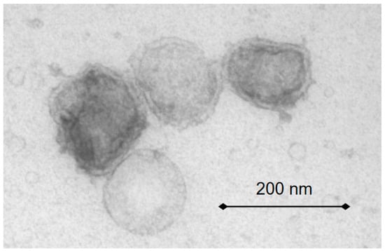
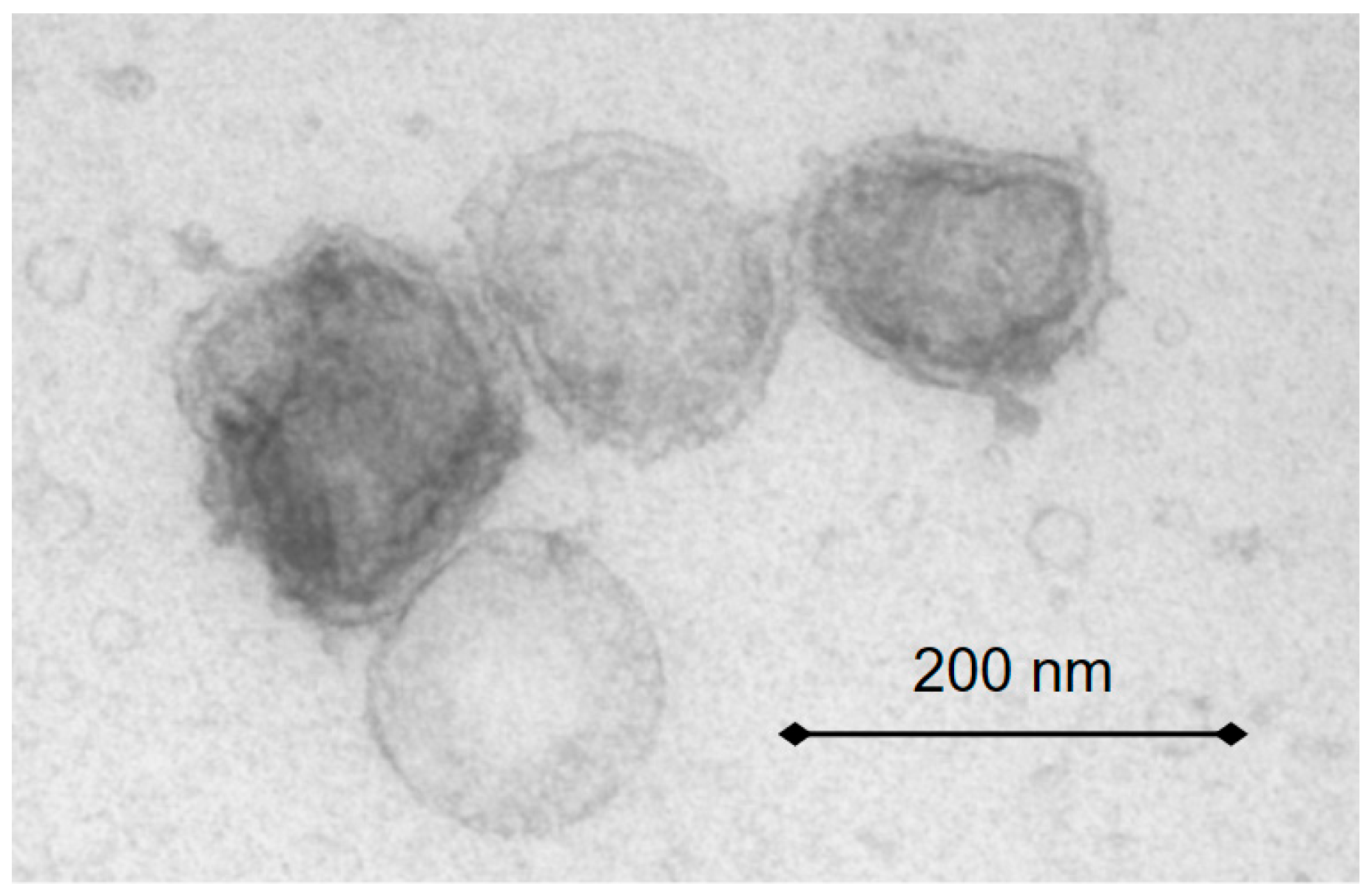
The physical characterization placed the final ingredient within the typical exosome size window. The NTA showed that the production concept could reproducibly generate nanovesicles. For the product-specific material studied here, the representative batches showed mean sizes of approximately 160 nm for the C. reinhardtii-derived vesicles, with standardized particle counts in the 1010 particles/mL range. The TEM imaging confirmed a vesicular, bilamellar morphology rather than amorphous particulate debris (Figure 1), compatible with exosome-inspired membrane organization.
Figure 1.
Characterization of EBVs. TEM images reveal population of spherical vesicles with distinct bilamellar morphology. Scale bar = 200 nm.
To complement the physical characterization, lipidomic and proteomic analyses were performed to define the molecular composition of the EBVs. A total of 109 membrane lipids across 23 classes and 1369 proteins corresponding to 1086 protein families were identified. These data support a complex vesicular system whose composition is far richer than that of a simple phosphatidylcholine-type liposome. Importantly, the identified lipids and proteins mapped onto the five exosome-inspired structural domains that underlie the biomimetic concept: the lipid bilayer membrane, membrane rafts, transmembrane proteins, exosome biogenesis-associated proteins, and functional cargo proteins.
Taken together (see Table 2), these data support a strict definition of structural biomimetism. The vesicles do not simply mimic exosomes visually; they reconstruct the molecular categories that allow exosomes to remain stable, interact with target cells, organize membrane signaling, and protect functional cargo. This forms the initial pillar of the product framework.
Table 2.
Summary of structural characterization of microalgae-derived vegan biomimetic vesicles.
3.1.2. Functional Biomimetism: Comparison with Human Fibroblast-Derived Exosomes in NHDFs
To evaluate the functional biomimetism, we tested whether the structural resemblance of EBVs translated into an exosome-mediated biological response. To test this, NHDFs were exposed for 24 h to the EBVs or to human fibroblast-derived exosomes at the same vesicle concentration, followed by 3′ mRNA sequencing. The sequencing quality was high across groups, with approximately 10 million final reads in most samples and Q20 values > 99%. The principal component analysis showed separation between the treated and control samples for both the EBVs and human fibroblast exosomes, indicating that both vesicle populations elicited measurable transcriptional responses.
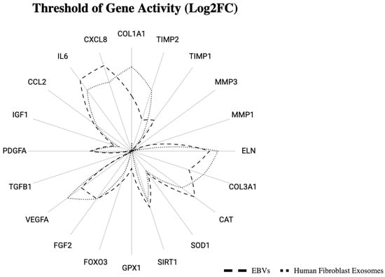
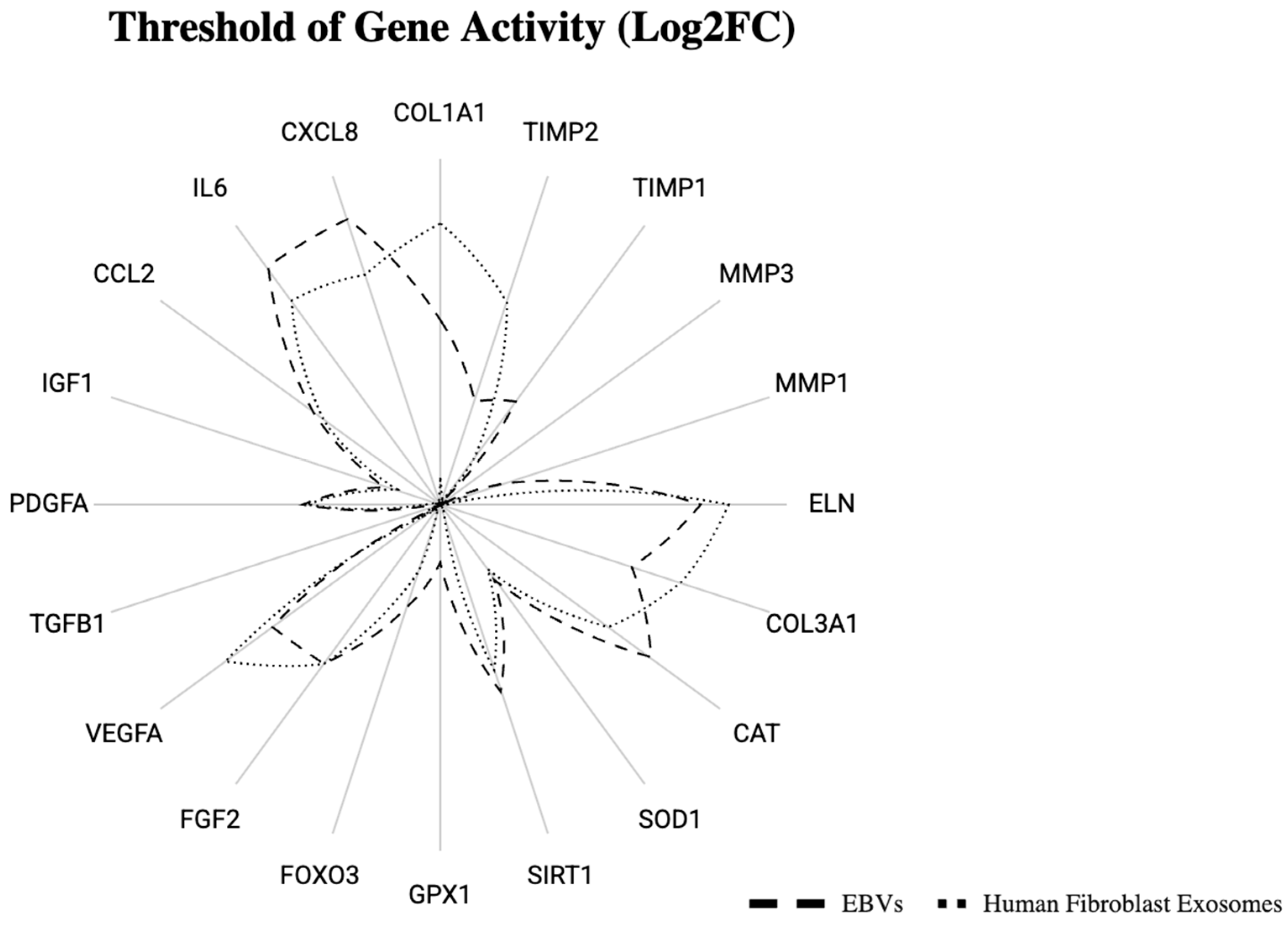
At the global level, the human fibroblast exosomes induced a stronger statistically resolved signal than the EBVs, with 27 genes significantly changed at |log2FC| < 1, whereas the EBV-induced changes were generally subtler. This difference is not unexpected: biomimetic systems are not intended to reproduce the full transcriptomic amplitude of native donor cell exosomes in every model. The central question is whether the EBVs reproduce the direction and biological logic of the response. On that point, the convergence was notable (Figure 2). In the key matrix-remodeling genes, both the EBVs and human fibroblast exosomes upregulated ELN, COL1A1, and COL3A1 while simultaneously reducing the expression of matrix-degrading metalloproteinases. Similarly, the clusters associated with anti-aging and growth factors showed coherent positive shifts involving CAT, SIRT1, VEGFA, and FGF2 in the EBV group, while the human exosome group displayed the same overall direction of change, albeit with a low statistical power for several individual genes.
Figure 2.
Transcriptomic impact of EBVs vs. human exosomes in NHDFs. Data are presented as a distribution of significantly regulated genes (log2FC) demonstrating the reciprocal expression patterns in fibroblast-specific functional axes.
It is interesting to mention that IL6 and CXCL8 were also upregulated after EBV exposure. While both genes are commonly regarded as inflammatory markers, their role in skin is context-dependent, and they also contribute to early wound healing and regenerative signaling [47,48,49]. The moderate and transient induction of IL-6 and CXCL8/IL-8 may therefore indicate a physiological repair response, particularly when it occurs without a broader inflammatory signature and in parallel with pro-regenerative outcomes. In this study, the increase in these markers was accompanied by favorable matrix-remodeling responses, enhanced fibroblast migration, and faster wound closure, supporting a repair-oriented interpretation. However, because the transcriptomic analysis was performed in an unstressed NHDF monoculture, these immunity-related markers should be interpreted cautiously and require tissue-level confirmation.
The NHDF model predictably exhibited low expression of barrier genes and certain inflammatory transcripts because fibroblasts are not optimal for keratinocyte-dominant or immune-dominant programs. It should be noted that skin renewal/barrier function and inflammatory cytokine readouts in this model were limited by the low transcript abundance in the fibroblasts. For that reason, the interpretation focuses on the fibroblast-relevant axes: ECM maintenance, anti-aging defense, and growth factor signaling. On these axes, the transcriptomic data support the idea that EBVs do not function as inert vesicles but as biomimetic communication platforms capable of reproducing the directional language of human exosomes.
From a product perspective, this result supports the hypothesis that the biomimetic vesicles can induce measurable fibroblast responses under the conditions tested. The biomimetic vesicles need not be identical to human exosomes in every analyte to be mechanistically persuasive; instead, they must preserve enough structural logic to induce the same beneficial direction of fibroblast changes in selected gene expression markers. The directional overlap observed here satisfies that criterion.
It is important to mention that the transcriptomic comparison should be interpreted as exploratory evidence of directional biological similarity rather than as definitive proof of functional equivalence between the exosome-like biomimetic vesicles and human fibroblast-derived exosomes. The most likely interpretation is based on the concordant direction of selected fibroblast-related pathways, particularly extracellular matrix remodeling, anti-aging defense, and growth factor-associated responses, rather than on broad transcriptomic equivalence.
3.1.3. Penetration Study: Comparison with Conventional Liposomes
Because exosome-inspired signaling is only useful when the vesicles or their encapsulated cargo reach the appropriate skin compartment, their penetration was compared directly against conventional liposomes in a full-thickness reconstructed human skin model. Both systems delivered detectable fluorescence to the epidermis and dermis after 24 h, but their compartmental distribution differed markedly.
The results are shown in Table 3. Fluorescence intensity is reported as arbitrary units (a.u.), representing relative signal intensity. The conventional liposomes showed stronger epidermal retention, with an epidermal fluorescence of 13,965.00 units and a dermal fluorescence of 1656.33 units, resulting in a D/E ratio of 0.12. In contrast, the EBVs produced lower epidermal accumulation (7194.67 units) but a substantially higher dermal signal (2320.67 units), yielding a D/E ratio of 0.32. Expressed differently, the biomimetic exosome system shifted delivery toward the dermis by 166.7% relative to the liposomal comparator. This result is highly consistent with the product hypothesis: a complex exosome-mimetic membrane is expected to interact with tissue barriers differently from a conventional liposomal vesicle and to exhibit improved navigation toward the biologically strategic dermal compartment.
Table 3.
Comparative dermal penetration performance of EBVs and conventional liposomes in reconstructed human skin.
Importantly, it should be noted that reconstructed full-thickness skin models do not fully reproduce the barrier strength of native human skin. Therefore, the fluorescence values reported here should not be interpreted as absolute predictors of in vivo skin penetration. Rather, this model was used to compare the relative compartmental distribution of EBVs and conventional liposomes under the same experimental conditions. In addition, the fluorescence quantification was not designed to determine the structural integrity of the vesicles after skin exposure or to resolve the precise transport mechanism. Ex vivo human skin studies, combined with vesicle integrity tracking, would further strengthen the characterization of EBVs penetration behavior.
Mechanistically, the data indicate that biomimetic vesicles are not merely efficient at carrying active ingredients, but are also more selective in where they deposit them. For anti-aging, support of skin appearance, and condition strategies centered on fibroblasts and dermal matrix biology, this feature is likely to be highly relevant.
3.1.4. Wound Healing Activity in Fibroblasts
The biological significance of dermal targeting was tested in a fibroblast scratch assay. As presented in Table 4, 1.0% EBVs accelerated closure versus the untreated control, confirming that the vesicles were not only able to reach the fibroblast-relevant compartments, but could also translate their delivery into a pro-regenerative cellular response. At 8 h, the untreated control cultures had reduced the wound width by 17.4% versus baseline, while 1.0% EBVs achieved 30.4% closure.
Table 4.
Fibroblast scratch assay: reduction in wound width versus baseline.
Statistically, the 1.0% treatment reached significance versus the control at 2 h (p = 0.034246), 4 h (p = 0.044286), and 8 h (p = 0.0131). Versus the corresponding baseline, the 1% active concentration also achieved significant closure at 8 h, with p = 1.04 × 10⁻⁵. These data indicate a rapid effect on fibroblast migration and wound gap resolution. In the context of exosome-inspired biology, this is consistent with an ingredient that enhances regenerative coordination rather than acting as a passive antioxidant alone.
3.2. Ex Vivo Results
3.2.1. UV-A-Challenged Skin Explants: Antioxidant, Pigmentary, Matrix, and Barrier Protection
The ex vivo skin explant study was designed to test whether the ingredient could protect living human skin tissue from a clinically relevant photoaging stress. This model moves beyond a simple cell culture by preserving the multicellular context and dermal–epidermal architecture.
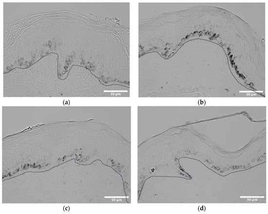
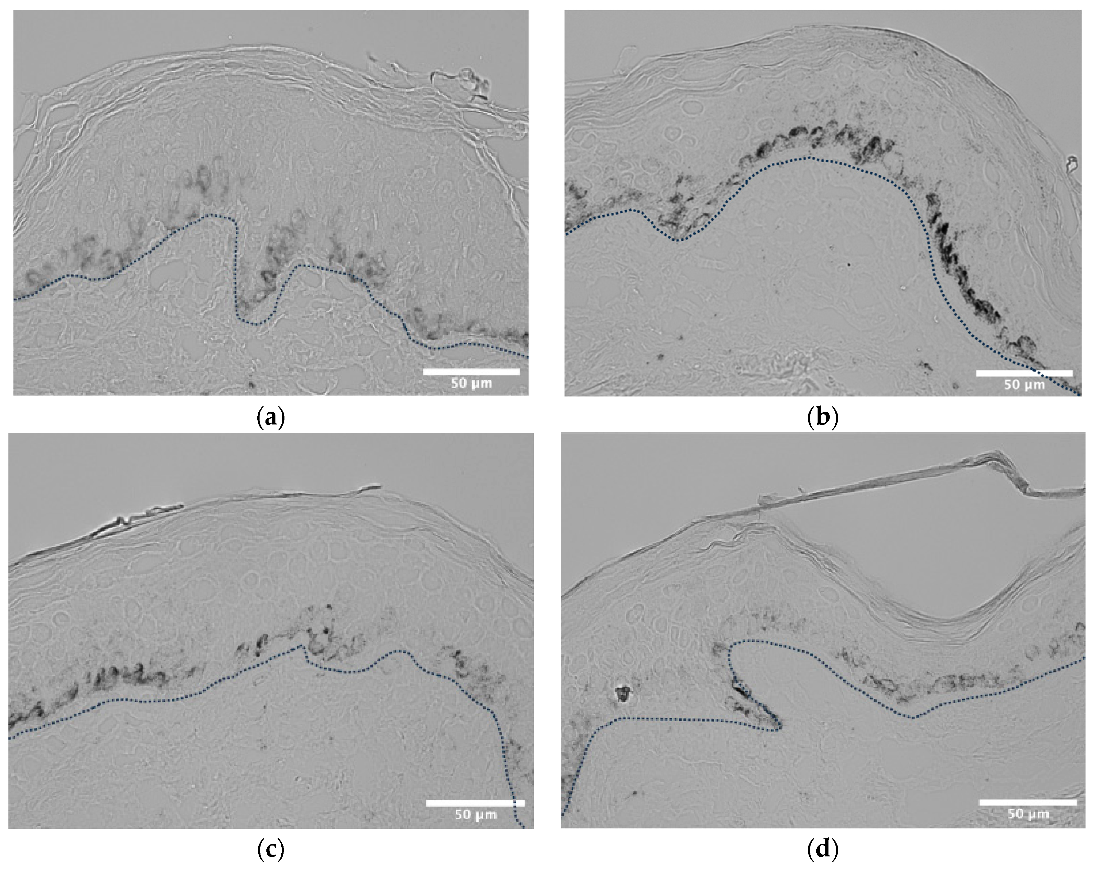
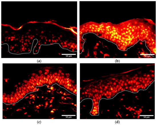
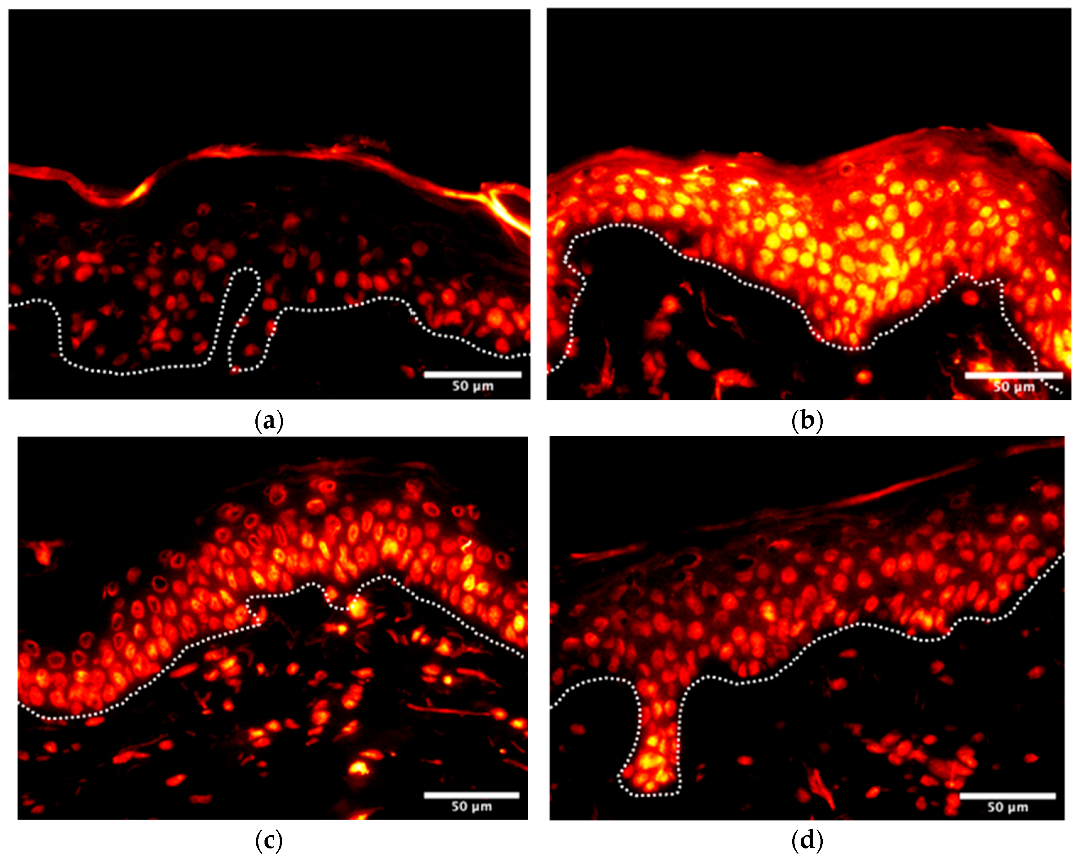
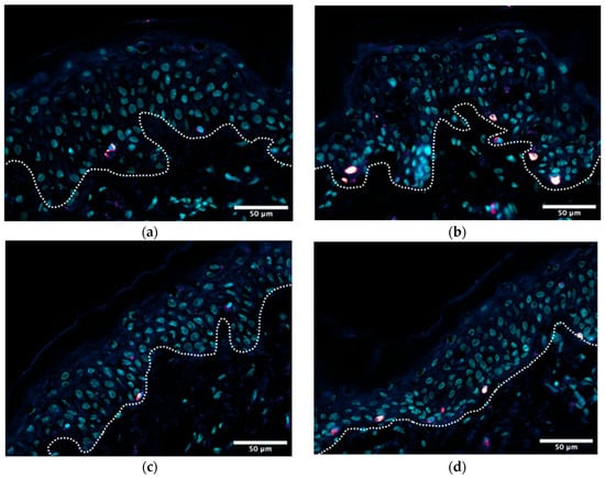
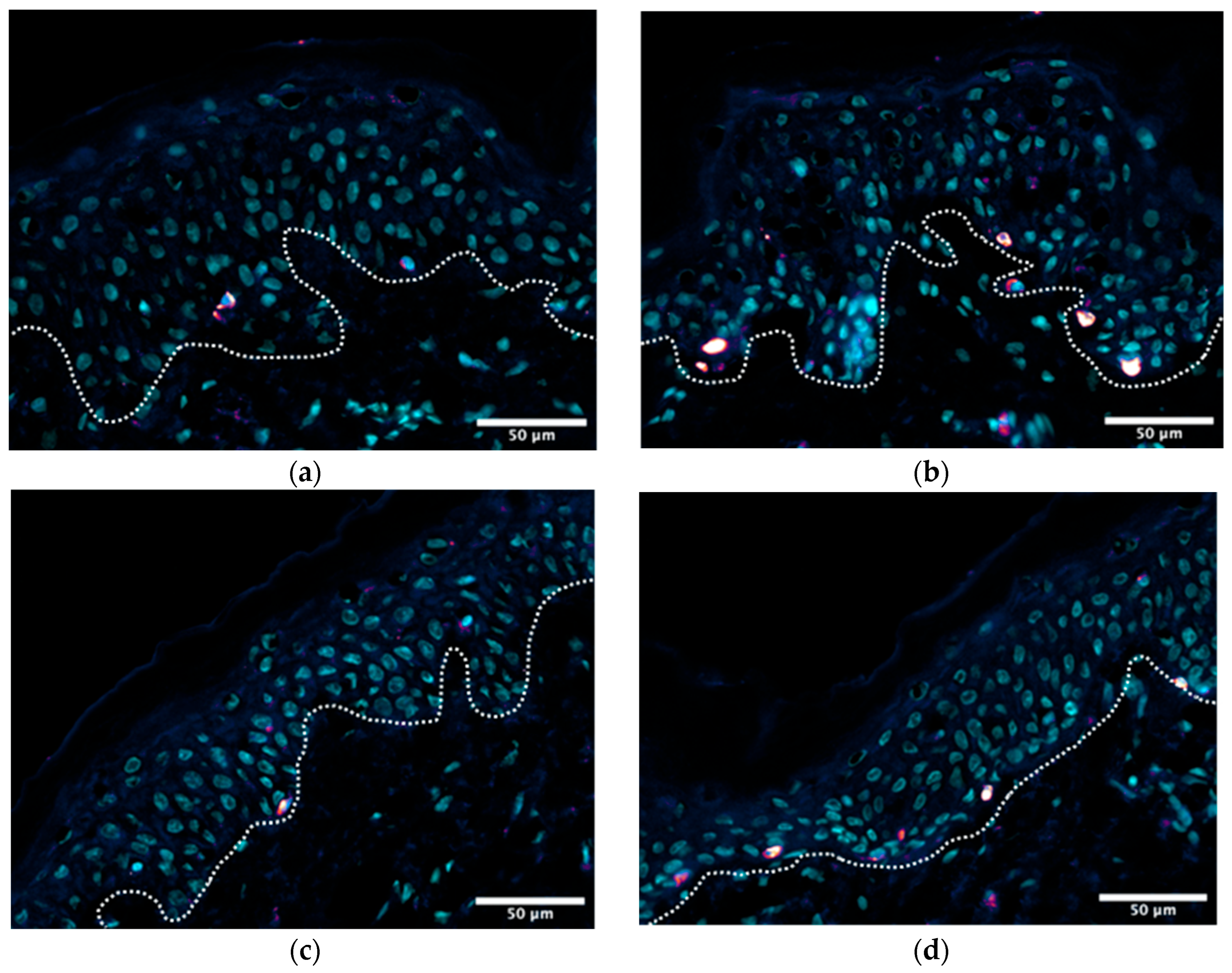
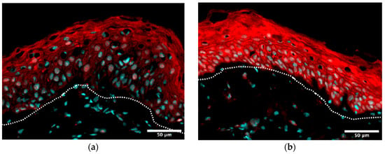
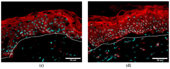
A synthesis of the key findings is presented in Table 5. UV-A exposure dramatically increased melanin, ROS, PMEL17, IL-18, and lipofuscin, thereby reproducing the core processes of photoaging: oxidative bursts, dysregulated pigmentation, inflammaging, matrix breakdown, and barrier weakening.
Table 5.
Ex vivo UV-A skin explant study: biomarker protection from EBVs, vitamin C, and their combination.
The EBVs showed broad-spectrum protection across all the biomarkers tested. Against UV-A-mediated hyperpigmentation, 2% EBVs reduced the melanin burden to 116.7% of control versus 298.0% in the stressed tissue, corresponding to a 92% protective efficacy. The ROS fell to 121.1% of control versus 196.5% in the stressed tissue (78% efficacy). PMEL17, a marker of melanosome maturation, was brought near the baseline (103.1% of control; 95% efficacy). IL-18, relevant to inflammatory stress signaling, was lowered to 120.2% of control (76% efficacy), and the lipofuscin accumulation was reduced with 52% efficacy.
The vitamin C comparator validated the model and provided a benchmark. For melanin and ROS, 5% vitamin C performed similarly to 2% EBVs, while the combination of 2.5% vitamin C and 1% EBVs achieved the highest protection, reaching 100% efficacy for melanin, 96% for ROS, 96% for PMEL17, 93% for IL-18, and 91% for lipofuscin. These findings suggest that the biomimetic exosomes are effective both as standalone protective systems and as synergistic partners in antioxidant formulations.
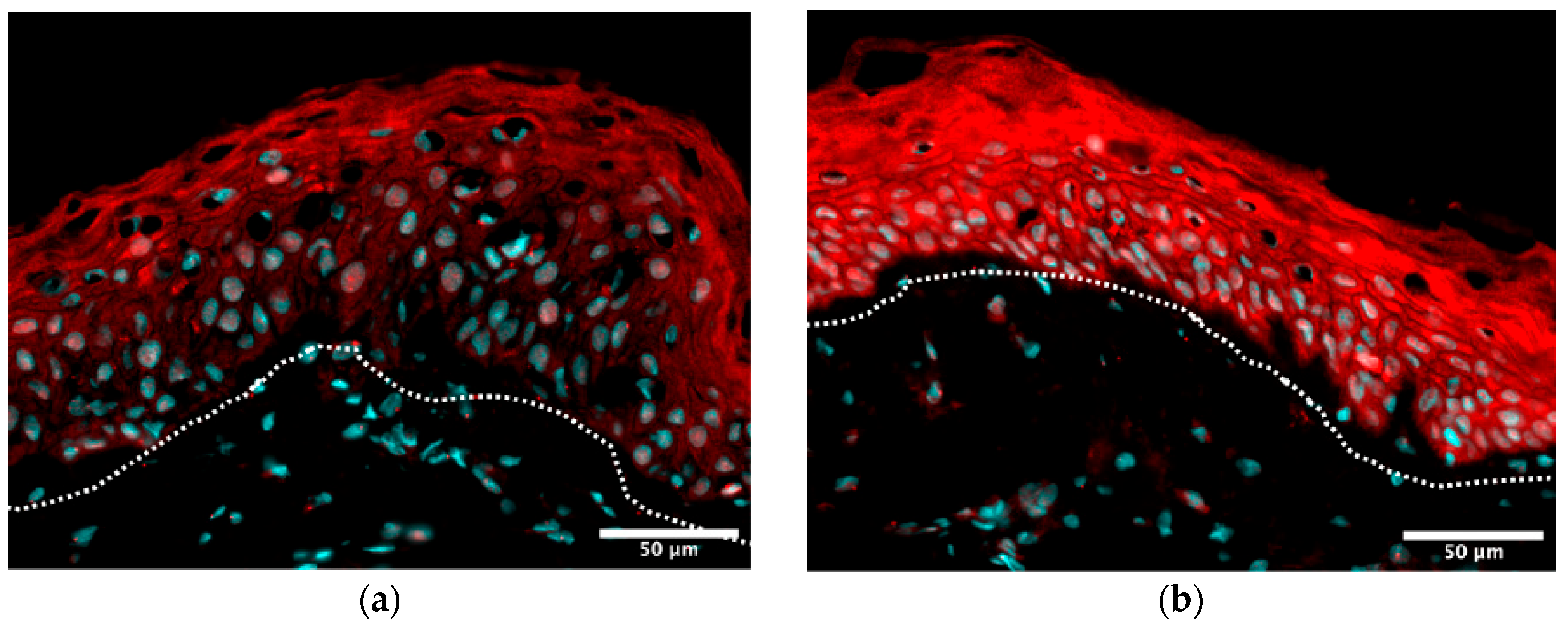
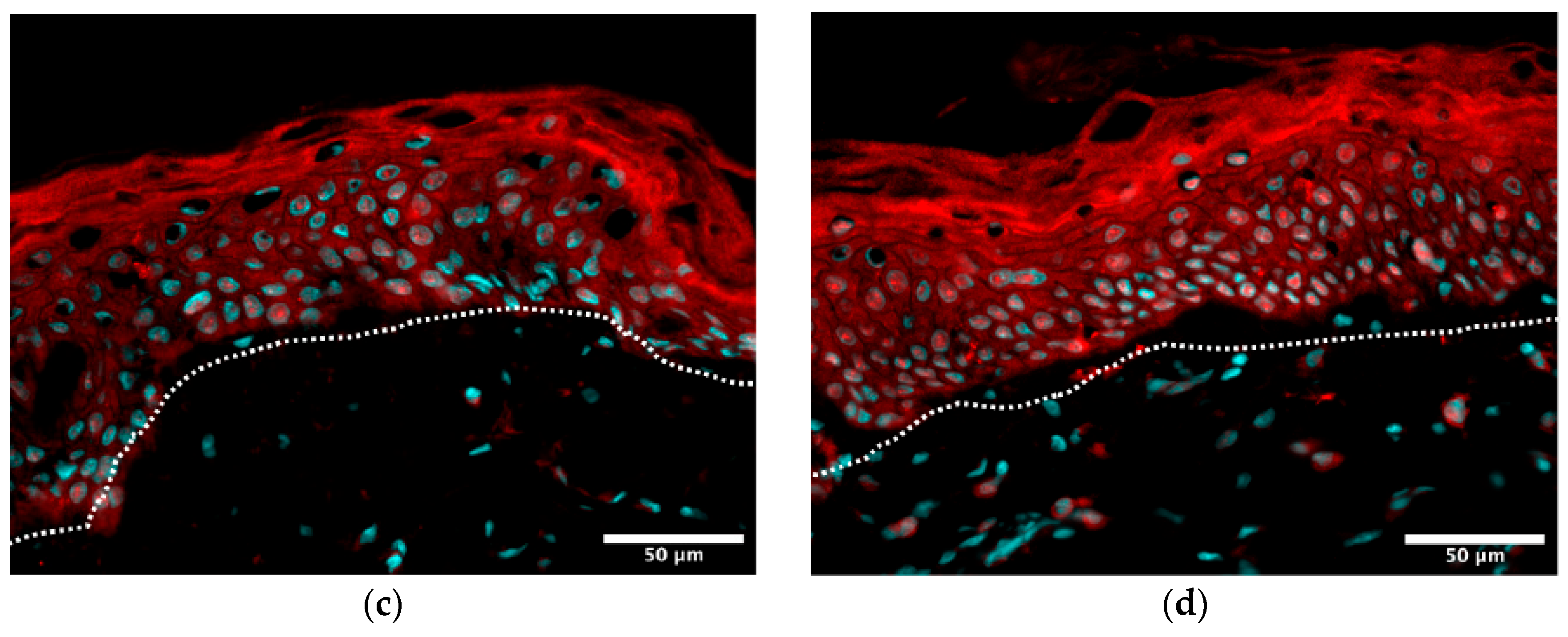
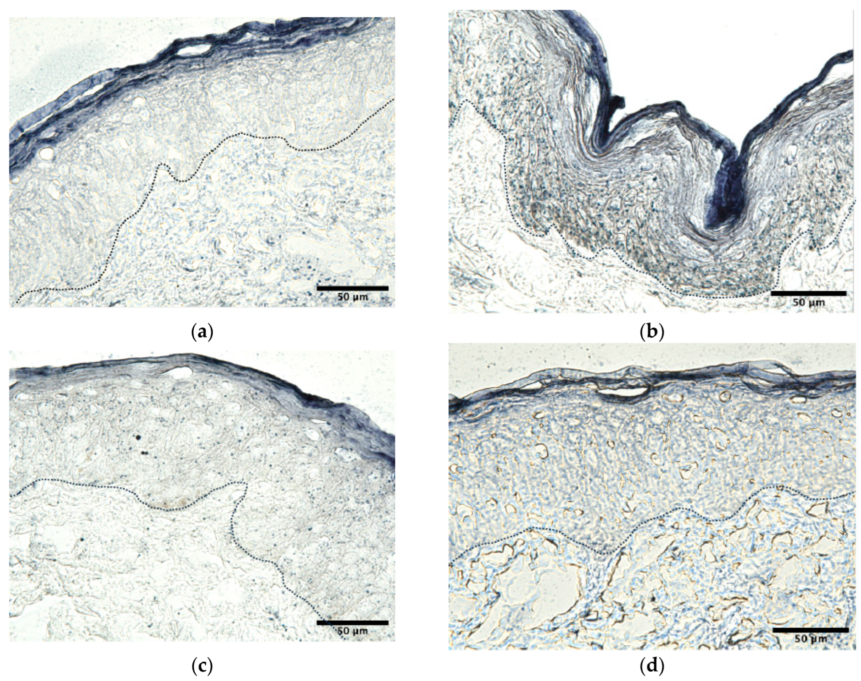
The representative fluorescence micrographs (Figure 3, Figure 4, Figure 5, Figure 6 and Figure 7) qualitatively corroborate the quantitative findings presented in Table 5, demonstrating a visible reduction in stress markers and the preservation of dermal–epidermal structural proteins following EBV treatment.From a mechanistic standpoint, these ex vivo results are especially strong because they connect multiple hallmarks of skin aging within the same model. The ingredient did not act along a single axis: it simultaneously reduced oxidative stress, normalized pigmentary signaling and limited inflammatory activation. This pattern is more compatible with an exosome-inspired biomimetic vesicle system than with a single-target active.
Figure 3.
In situ visualization of melanin content levels: (a) control; (b) stress (UV-A); (c) 2% EBVs + stress; and (d) 5% vitamin C + stress. Melanin content signal on skin explant sections was visualized using color range—low levels in white, high levels in dark. Epidermis and dermis are separated by dark dotted lines; 40× objective.
Figure 4.
In situ visualization of ROS levels: (a) control; (b) stress (UV-A); (c) 2% EBVs + stress; and (d) 5% vitamin C + stress. ROS content signal on skin explant sections is visualized using in color range (low levels in dark, high levels in bright colors). Epidermis and dermis are separated by white dotted lines; 40× objective.
Figure 5.
In situ visualization of PMEL17 levels: (a) control; (b) stress (UV-A); (c) 2% EBVs + stress; and (d) 5% vitamin C + stress. PMEL17 signal on skin explant sections is visualized using color range (low levels in dark, high levels in bright colors) and superposed to nuclear detection (DAPI, in cyan). Epidermis and dermis are separated by white dotted lines; 40× objective.
Figure 6.
In situ visualization of IL-18 levels: (a) control; (b) stress (UV-A); (c) 2% EBVs + stress; and (d) 5% vitamin C + stress. IL-18 signal on skin explant sections is visualized in red and superposed to nuclear detection (DAPI, in cyan). Epidermis and dermis are separated by white dotted lines; 40× objective.
Figure 7.
In situ visualization of lipofuscin levels: (a) control; (b) stress (UV-A); (c) 2% EBVs + stress; (d) 5% vitamin C + stress. Lipofuscin signal on skin explant sections is visualized in blue. Epidermis and dermis are separated by dark dotted lines; 40× objective.
3.2.2. Ex Vivo Human Hair Follicles: Safety and Anagen-Supportive Activity
The hair follicle studies extended the regenerative profile of EBVs beyond facial skin into an adnexal mini organ context. First, their safety was supported by the absence of overt LDH release over 7 days, indicating no detectable tissue or cell integrity damage in the cultured follicles. Second, the amputated follicle experiments showed faster shaft/tissue generation in the treated group than in the PBS controls. These growth differences did not reach significance in the preliminary donor-based experiments, but the direction of effect was consistent across studies and became more persuasive when only the follicles that remained in anagen were considered.
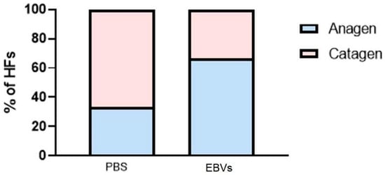
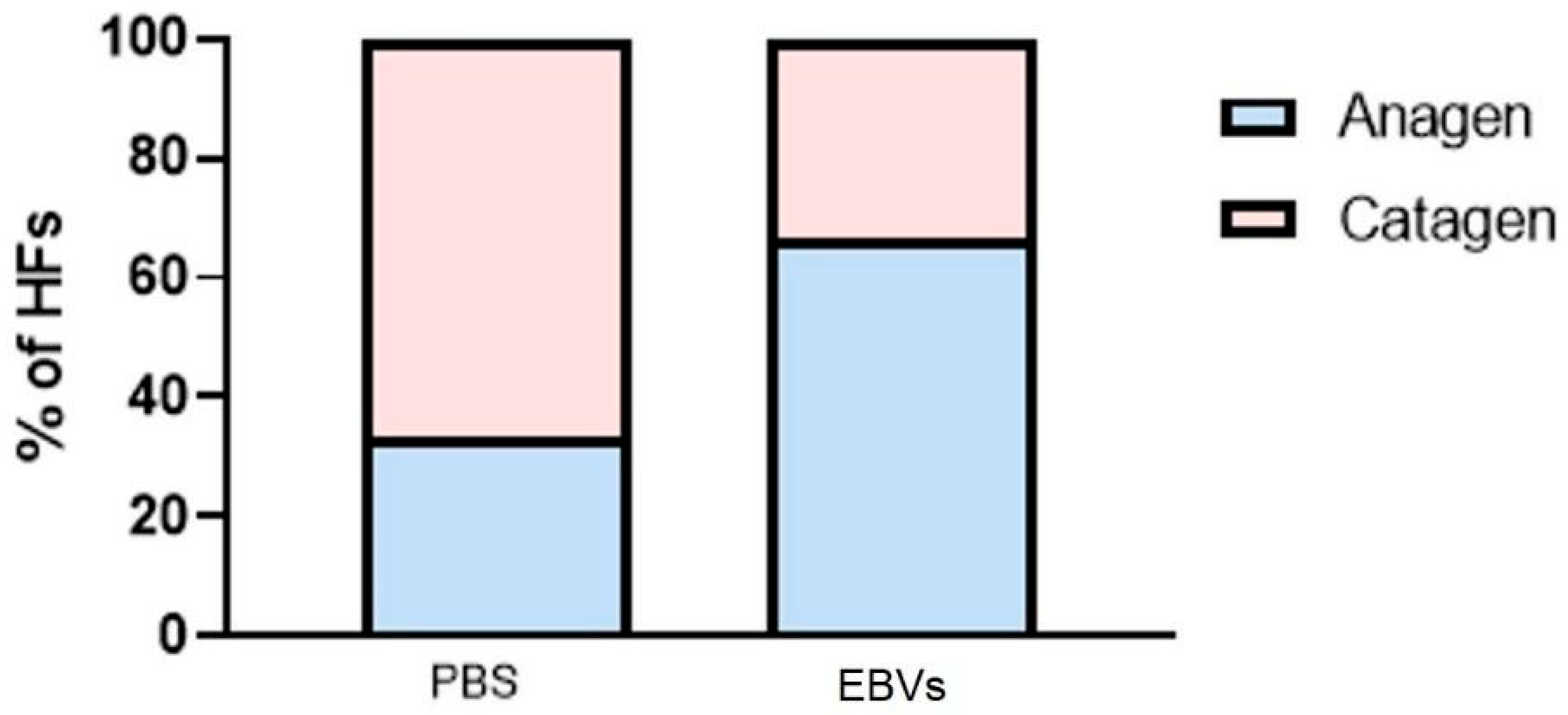
The most compelling evidence came from the full-length follicle model. The treated follicles more frequently remained in anagen at the end of culture than the controls (see Figure 8).
Figure 8.
Effect of EBV treatment on hair follicle (HF) cycle staging.
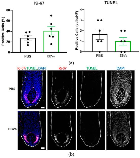
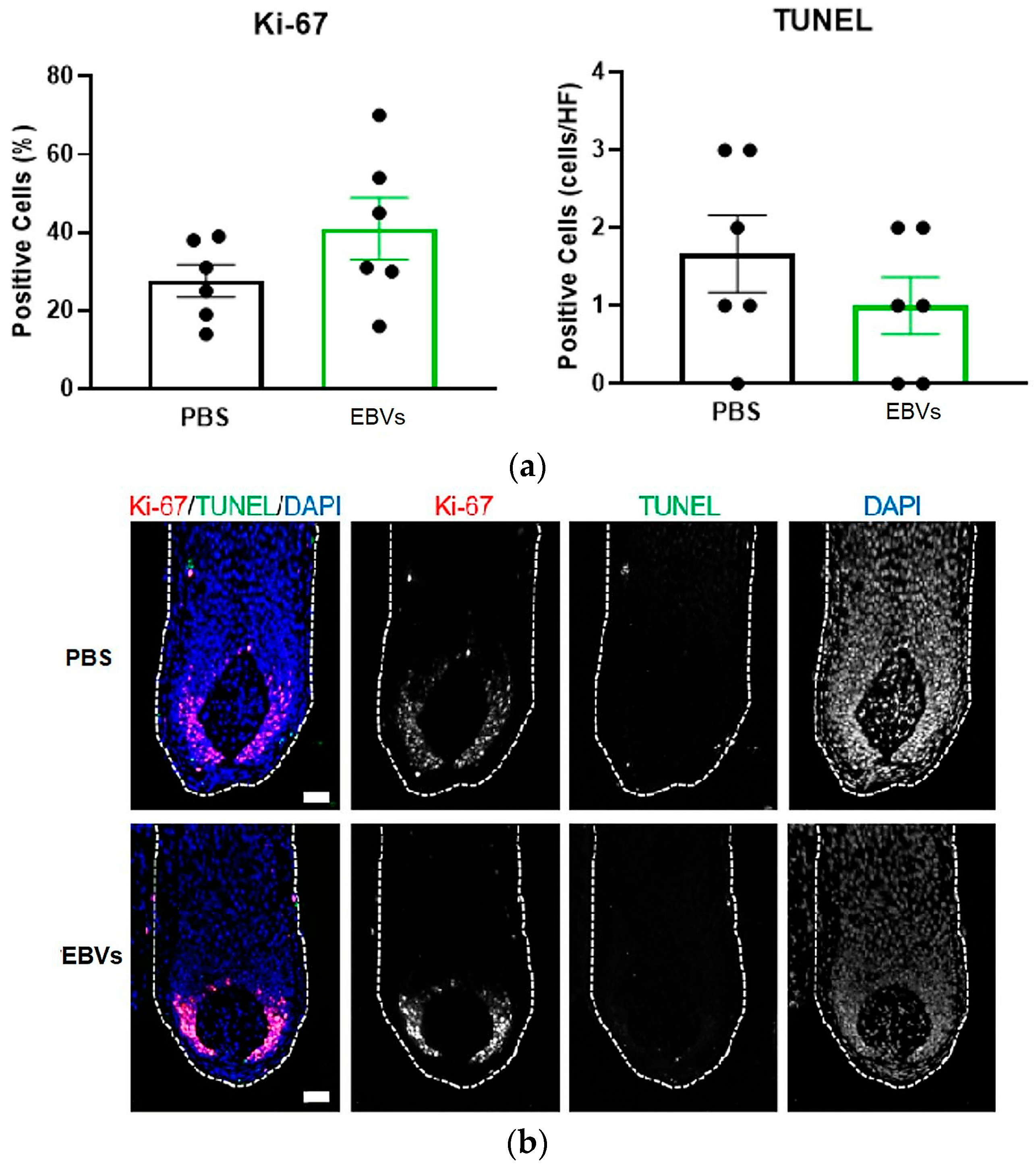
The Ki-67-positive germinative hair matrix cells increased from 27.7% in the PBS group to 41.0% with EBVs, supporting continued proliferative activity in the growth phase (Figure 9).
Figure 9.
Ki-67 immunofluorescence evaluation. (a) Mean ± SEM calculation of Ki-67+ cells and mean ± SEM number of TUNEL+ cells; (b) representative images of Ki-67/TUNEL double staining with DAPI counter-stain. White dotted lines demarcate hair bulbs. Scale bars: 50 µm.
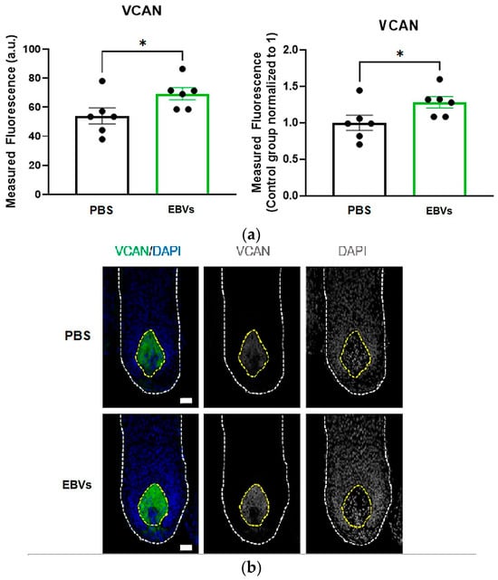
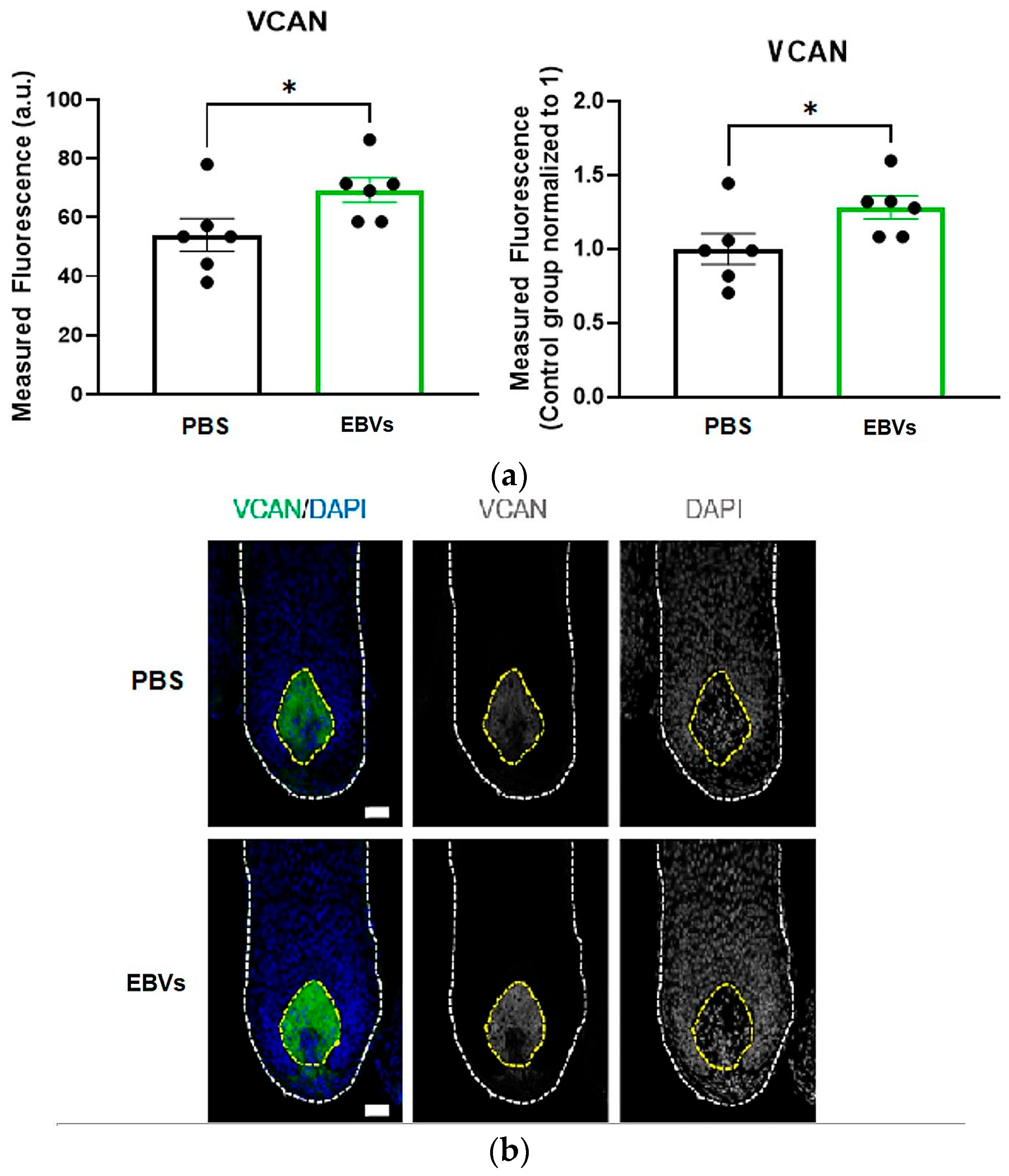
Versican, a classic dermal papilla anagen marker linked to follicle inductivity and Wnt/beta catenin-associated signaling [42,43,46], was significantly higher in the treated group (p = 0.0411) (Figure 10).
Figure 10.
Versican immunofluorescence evaluation. (a) Mean ± SEM measurement and normalization with mean value of PBS treatment group for VCAN expression in dermal papilla Mann-Whitney test was performed for paired comparisons (* p = 0.0411); (b) representative images of VCAN immuno-staining with DAPI counter-stain. White dotted lines demarcate hair bulbs. Scale bars: 50 µm.
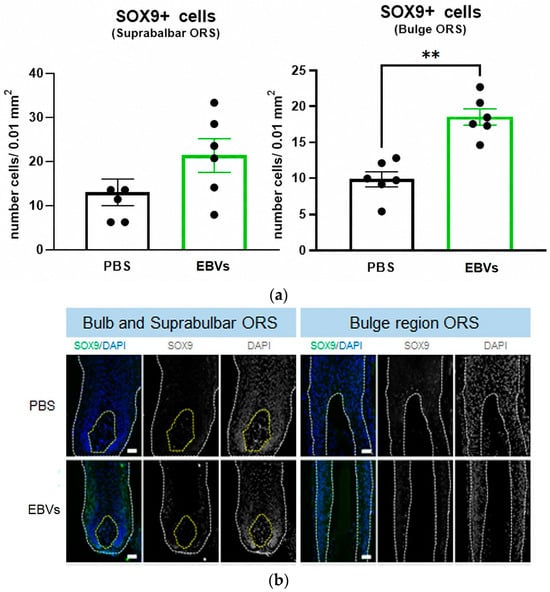
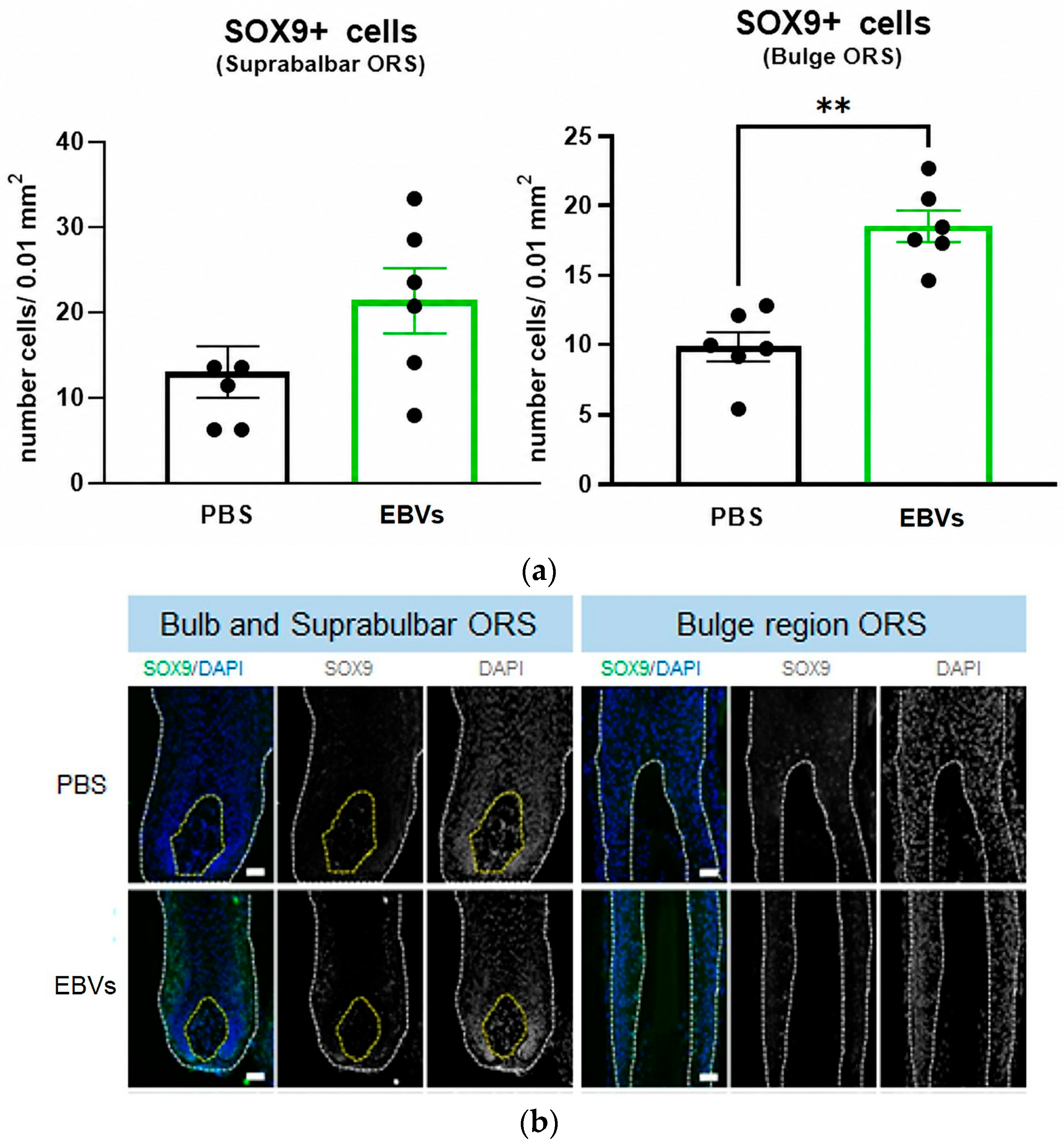
The SOX9-positive cells in the bulge outer root sheath, a progenitor-rich region relevant to follicle maintenance and regenerative competence [39,44,45], were also significantly increased (p = 0.0022) (Figure 11).
Figure 11.
SOX9+ immunofluorescence evaluation. (a) Mean ± SEM number of SOX9+ cells per 0.01 mm2 of suprabulbar ORS and bulge ORS Mann-Whitney test was performed for paired comparisons (** p = 0.0022); (b) representative images of SOX9 IF staining with DAPI counter-stain. White and yellow dotted lines demarcate follicular tissue and dermal papilla, respectively. Scale bars: 50 µm.
TUNEL-positive cells remained scarce, indicating that the anagen-supportive effect was not accompanied by overt damage.
The collective impact of EBV treatment on hair follicle viability and signaling is summarized in Table 6, which consolidates the observed improvements in growth phase retention and biomarker expression. Altogether, these data suggest that EBVs may help maintain follicles in a growth-supportive state while preserving progenitor markers and dermal papilla signaling. Although these ex vivo findings were generated in a limited donor set and should be interpreted as mechanistic support rather than as final clinical proof, they meaningfully extend the concept of biomimetic exosomal communication from skin rejuvenation to hair follicle vitality.
Table 6.
Key outcomes from the ex vivo human hair follicle studies.
3.3. In Vivo Results
Placebo-Controlled 2% Cream Clinical Study
To evaluate the in vivo efficacy of the 2% EBV formulation, a randomized, placebo-controlled clinical study was conducted over 56 days. The study utilized a hemi-face, intra-individual design, which provided a stringent internal control by using each of the 41 enrolled volunteers as their own comparator. This approach allowed for the assessment of both visual and instrumental parameters of facial aging under standardized topical application conditions. The primary outcomes relative to the placebo are summarized in Table 7.
Table 7.
Main outcomes of the 56-day placebo-controlled clinical study with a 2% EBV cream.
The 2% EBV formulation’s clinical performance was broad and progressive. Crow’s feet wrinkle depth decreased significantly from the baseline by 9.0% at day 14, 11.5% at day 28, and 12.2% at day 56, while the placebo showed minimal change. The arithmetic roughness parameter Sa, for which lower values indicate smoother skin, was reduced by 3.9%, 4.6%, and 7.1% at the same time points. The wrinkle depth shift also translated into a more youthful appearance according to the model derived from the instrumented periocular wrinkle–age correlations: the treated side corresponded to skin appearing 2.2 years younger at day 14, 2.8 years younger at day 28, and 3.0 years younger at day 56, versus only about 0.5–0.6 years with placebo.
The pigmentation outcomes were also robust. The whole skin ITA° increased by 4.5%, 8.0%, and 10.3% across the three time points, while the dark spot ITA° increased by 8.7%, 18.9%, and 22.8%, indicating progressive lightening of overall pigmentation and focal hyperpigmentation. The cutometer measurements showed increased elasticity (R2) of 2.0%, 3.7%, and 4.9% and reduced skin distensibility (R0) of 3.3%, 4.6%, and 7.4%, corresponding to improved firmness. The radiance increased rapidly, with the gloss values improving by 9.3% at day 14, 16.1% at day 28, and 20.0% at day 56. Facial contour reshaping was supported by a reduction in the measured jowl line distance of 0.160 mm at day 28 and 0.336 mm at day 56.
The clinically scored smoothness reinforced the instrumental results. At day 28, 58.5% of subjects were rated as improved, rising to 70.7% at day 56. Importantly, the report concluded that the results obtained with the EBVs were not only statistically significant versus the baseline, but globally significantly superior to the placebo side. This breadth of effect is noteworthy because it covers surface topography, chromatic homogeneity, biomechanical properties, radiance, contour, and clinical appearance in one integrated study.
4. Discussion
This study addresses a critical challenge in the exosome-inspired cosmetic field: whether a plant-derived, industrially scalable system can reproduce sufficient exosome biology to merit the term “biomimetic” in a rigorous scientific sense. The combined dataset suggests that this is achievable, provided biomimetism is evaluated at more than one level. Here, the argument does not rest on simple morphological similarities or on an isolated efficacy endpoint. It establishes a coherent chain of evidence linking composition, cell response, tissue distribution, tissue protection, and clinical outcome.
The first important point is that the EBV platform is not a conventional liposome. The compositional data show a membrane system of high molecular richness, including multiple lipid classes, membrane microdomain lipids, trafficking-related proteins, and cargo proteins relevant to oxidative defense and tissue regulation. This matters because exosome function is emergent: it depends on the relationship among membrane structure, surface interactions, and protected cargo. Simplified vesicles can encapsulate active ingredients, but they are not automatically exosomal in behavior. The EBV platform was specifically engineered to preserve this relationship while eliminating the sourcing problems associated with animal- and human-derived exosomes.
The second important point is that the structural biomimetism translates into functional biomimetism. The relevant result is that the EBVs reproduced the same directional pattern as human fibroblast exosomes in fibroblast-relevant pathways (see Figure 2), particularly extracellular matrix support, anti-aging defense, and growth factor signaling. In the context of high-impact research, this nuance is important. Biomimetic systems should not be judged by whether they are molecular clones of their biological inspirations, but by whether they preserve their operating logic. The directional convergence observed here supports precisely that interpretation. These data support the exosome-like biomimetic rationale of the vesicles, but they should not be considered evidence of full functional equivalence to natural human exosomes or of broad immune/barrier effects in skin.
The penetration data add a translational bridge that is often missing in exosome discussions. If a vesicle system claims to have exosome-like function in the skin, it must reach the biologically relevant compartments. The D/E ratio data in Table 3 show that the EBVs shift delivery away from the superficial epidermal trapping and toward the dermis more effectively than standard liposomes. This provides a plausible mechanistic explanation for the regenerative readouts obtained from the fibroblasts, ex vivo photoaging studies, and clinical anti-aging endpoints. In other words, the product does not merely carry a message; it appears better able to bring that message to the fibroblast-rich compartment where long-term skin architecture is regulated.
The wound healing assay and ex vivo photoaging model together demonstrate the breadth of the biological response. The accelerated scratch closure suggests fibroblast activation and improved collective migration, while the UV-A model shows simultaneous effects on oxidative stress, pigmentation, inflammatory signaling, extracellular matrix preservation, and hydration biology. Such multi-axis activity is highly congruent with exosome-inspired communication and less congruent with a narrow single-target mechanism. The comparison with vitamin C is particularly informative. Vitamin C performed strongly in several oxidative and pigmentary readouts, as expected; yet, the EBVs matched or closely approached it in melanin and ROS control and outperformed it in PMEL17 and IL-18 modulation (Figure 3, Figure 4, Figure 5, Figure 6 and Figure 7). Moreover, the vitamin C + EBV combination consistently yielded the strongest protection, as can be seen in Table 5, indicating that EBVs are compatible with classical antioxidant actives while contributing distinct biological value. The UV-A skin explant study was performed with a limited number of biological samples, and although the results were internally consistent across the measured biomarkers, they should be interpreted as supportive tissue-level evidence rather than definitive proof of clinical photoprotection.
The ex vivo hair follicle findings further support the concept that the vesicles interact with complex tissue systems in a regenerative manner. Hair follicles are mini organs with cyclical dynamics and multiple signaling compartments. The increase in Ki-67, VCAN, and SOX9 (shown in Figure 9, Figure 10 and Figure 11) suggests support for anagen maintenance, dermal papilla functionality, and progenitor-rich epithelial niches. These observations align with a mode of action centered on biological communication rather than on generic nutritive supplementation. At the same time, caution is warranted: the hair studies were preliminary and donor limited, so they are best interpreted as high-value mechanistic support rather than as definitive clinical hair growth proof.
The placebo-controlled clinical study is arguably the most compelling translational validation. The treated hemi-face improved across essentially every major axis relevant to visible facial aging: wrinkles, roughness, pigmentation, dark spots, elasticity, firmness, radiance, contour, and clinician-rated smoothness. The breadth of effect is important because it mirrors the breadth of the preceding non-clinical evidence. Clinical anti-aging claims often rely on one or two endpoints; here, the pattern is systemically coherent. A product that reduces melanin and PMEL17 ex vivo would be expected to reduce visible hyperpigmentation and dark spots. A product that improves dermal targeting and fibroblast wound repair would be expected to enhance smoothness and resilience over time. The clinical study supports this biological logic.
Several limitations should be acknowledged. First, the transcriptomic model used fibroblasts, which are appropriate for ECM-oriented biology but less informative for barrier genes and inflammation-dominant pathways. Future work could extend the functional biomimetism analysis to keratinocytes, co-cultures, or stress-challenged systems. Second, while the structural evidence is strong, a more exhaustive side-by-side omics comparison with purified mammalian exosomes would further sharpen the definition of biomimicry. Third, some of the ex vivo studies relied on limited donor material, especially the hair follicle work, and therefore require expansion for broader generalization. Finally, the clinical study tested a finished formulation containing 2% of ingredient, which is the correct translational format for cosmetics, but does not isolate every possible contribution of formulation context. In addition, the clinical statistical analysis relied on multiple pairwise comparisons; therefore, future confirmatory studies using repeated-measures ANOVA or mixed-effects models with multiplicity adjustment would further strengthen the statistical robustness of the clinical findings. None of these limitations invalidate the findings; rather, they help define the next research steps for strengthening the platform.
Overall, the different experimental levels provide a consistent evidence package supporting the further development of this exosome-inspired cosmetic ingredient. It supports the proposition that biomimetism is the scientifically appropriate route for vegan exosome innovation: more faithful than conventional liposomes, more scalable than naturally secreted vegan vesicles, and more practical and acceptable than human or animal exosomes. In a field crowded with imprecise terminology, the present data argue for a stricter but more useful standard: a biomimetic exosome should reproduce the exosome-relevant structure, directionally reproduce the exosome-relevant biological responses, and translate those properties into meaningful tissue and clinical benefits. By that standard, the microalgae-derived EBV platform performs convincingly.
5. Conclusions
This study presents a comprehensive scientific evaluation of a microalgae-derived vegan biomimetic exosome ingredient built from Chlamydomonas reinhardtii and developed for cosmetic skin renewal. The manuscript moves beyond a process description by connecting the manufacturing rationale, biomimetic theory, structural evidence, functional evidence, and translational efficacy.
The data support two central conclusions. First, the ingredient is structurally biomimetic: it forms nanosized bilamellar vesicles with high lipidomic and proteomic complexity that reproduce the principal building blocks of mammalian exosomes. Second, it is functionally biomimetic: it induces directionally convergent fibroblast transcriptional responses relative to human fibroblast exosomes; improves dermal targeting compared with liposomes; accelerates fibroblast repair; protects human skin explants from UV-A-driven oxidative, pigmentary, inflammatory, matrix, and barrier damage; supports anagen-associated features in ex vivo hair follicles; and delivers broad placebo-controlled clinical improvement in facial aging parameters.
Taken together, these findings show that biomimetism is not just a conceptual framing device but an experimentally supported strategy for exosome-inspired cosmetics. Microalgae-derived vegan exosome-like biomimetic vesicles offer a scalable and reproducible alternative to conventional exosome sourcing while preserving the multi-layered biological logic required for meaningful skin communication, rejuvenation, and measurable cosmetic benefit.
6. Patents
The work described in this manuscript has resulted in the following patent applications:
- PCT Application: International application number PCT/EP2026/050007, filed on 2 January 2026.
- Chinese National Phase: Application number 2026100000733, filed on 5 January 2026.
These applications relate to the production process and the cosmetic applications of the microalgae-derived vegan biomimetic exosomes (EBVs) described in this study.
Author Contributions
Conceptualization, J.A.; methodology, N.G.-D. and J.A.; validation, A.L. and J.A.; resources, N.G.-D. and J.A.; writing—original draft preparation, J.A.; writing—review and editing, N.G.-D.; visualization, N.G.-D. and J.A.; supervision, N.G.-D. and A.L.; project administration, J.A. All authors have read and agreed to the published version of the manuscript.
Funding
This study was funded and supported by ALGAKTIV, which provided the test ingredient and financial support for the studies described in the manuscript. No external grant funding was received, and no funding number is applicable.
Institutional Review Board Statement
All studies involving humans were conducted in compliance with the ethical principles for medical research (Ethical Principles for Medical Research Involving Human Subjects, adopted by the 18th WMA General Assembly, Helsinki, Finland, June 1964, and subsequent amendments). The UV-A-challenged skin explants study was conducted with informed consent and ethics approval (French Minister of Higher Education and Research, AC-2022-5147 for Oxiproteomics and AC-2021-4468 for BioToSkin). The ex vivo human hair follicle study was conducted with informed consent and ethics approval (Marmara University School of Medicine Clinical Studies Ethics Committee, 09.2023.830, on 14 July 2023). For the clinical trial, in Spain and the European Union, prospective registration in public clinical trial registries is governed by Royal Decree 1090/2015 and Regulation (EU) No 536/2014, which apply exclusively to clinical trials with medicinal products; neither the AEMPS nor the EU CTIS has jurisdiction over cosmetic efficacy studies, and these portals do not accept such submissions. Cosmetic products in the EU are regulated under Regulation (EC) No 1223/2009, which requires efficacy claims to be supported by adequate evidence documented in the product information file, but does not mandate prospective registration in a public clinical trial registry. Accordingly, and in line with common practice in the cosmetic sector, this study was not prospectively registered.
Informed Consent Statement
Informed consent for participation was obtained from all subjects involved in the study.
Data Availability Statement
The data presented in this study are available on request from the corresponding author due to privacy reasons.
Acknowledgments
ALGAKTIV, S.L. is kindly acknowledged for providing the Chlamydomonas reinhardtii EBVs. During the preparation of this manuscript, the authors used ChatGPT version 5.3 for the purposes of tone polishing and Gemini version 3.1 Pro for graphical abstract generation. The authors have reviewed and edited the output and take full responsibility for the content of this publication.
Conflicts of Interest
Authors N.G.-D., A.L., and J.A. were employed by ALGAKTIV, S.L. The authors declare that the research was conducted in the context of the company’s development of cosmetic ingredients. The authors declare that the research was conducted in the absence of any commercial or financial relationships that could be construed as a potential conflict of interest. The remaining authors declare that the research was conducted in the absence of any commercial or financial relationships that could be construed as a potential conflict of interest. Additionally, the authors declare that this study received funding from ALGAKTIV. The funder was not involved in the study design, collection, analysis, interpretation of data, the writing of this article, or the decision to submit it for publication. The funder did not affect the experimental process and results in the study. The funding relationship does not affect the scientific quality of the manuscript.
References
- He, R.; Sun, M.; Liu, T.; Geng, L.; Xing, C.; Xu, P.; Peng, Y.; Fang, Y. Hallmarks of the Aging Skin Microenvironment: Components and Mechanisms. J. Cell. Physiol. 2025, 240, e70111. [Google Scholar] [CrossRef] [Scilit] [PubMed]
- Nan, L.; Guo, P.; Hui, W.; Xia, F.; Yi, C. Recent advances in dermal fibroblast senescence and skin aging: Unraveling mechanisms and pioneering therapeutic strategies. Front. Pharmacol. 2025, 16, 1592596. [Google Scholar] [CrossRef] [Scilit]
- López-Otín, C.; Blasco, M.A.; Partridge, L.; Serrano, M.; Kroemer, G. Hallmarks of aging: An expanding universe. Cell 2023, 186, 243–278. [Google Scholar] [CrossRef] [Scilit] [PubMed]
- Shin, S.H.; Lee, Y.H.; Rho, N.-K.; Park, K.Y. Skin aging from mechanisms to interventions: Focusing on dermal aging. Front. Physiol. 2023, 14, 1195272. [Google Scholar] [CrossRef] [Scilit] [PubMed]
- Bollag, W.B.; Aitkens, L.; White, J.; Hyndman, K.A. Aquaporin-3 in the epidermis: More than skin deep. Am. J. Physiol. Cell Physiol. 2020, 318, C1144–C1153. [Google Scholar] [CrossRef] [Scilit]
- Hara-Chikuma, M.; Verkman, A.S. Roles of aquaporin-3 in the epidermis. J. Investig. Dermatol. 2008, 128, 2145–2151. [Google Scholar] [CrossRef] [Scilit] [PubMed]
- Hajialiasgary Najafabadi, A.; Soheilifar, M.H.; Masoudi-Khoram, N. Exosomes in skin photoaging: Biological functions and therapeutic opportunity. Cell Commun. Signal. 2024, 22, 32. [Google Scholar] [CrossRef] [Scilit]
- Tienda-Vázquez, M.A.; Hanel, J.M.; Márquez-Arteaga, E.M.; Salgado-Álvarez, A.P.; Scheckhuber, C.Q.; Alanis-Gómez, J.R.; Espinoza-Silva, J.I.; Ramos-Kuri, M.; Hernández-Rosas, F.; Melchor-Martínez, E.M.; et al. Exosomes: A promising strategy for repair, regeneration and treatment of skin disorders. Cells 2023, 12, 1625. [Google Scholar] [CrossRef] [Scilit]
- Li, M.; Li, S.; Du, C.; Zhang, Y.; Li, Y.; Chu, L.; Han, X.; Galons, H.; Zhang, Y.; Sun, H.; et al. Exosomes from different cells: Characteristics, modifications, and therapeutic applications. Eur. J. Med. Chem. 2020, 207, 112784. [Google Scholar] [CrossRef] [Scilit]
- Sreeraj, H.; AnuKiruthika, R.; Tamilselvi, K.; Subha, D. Exosomes for skin treatment: Therapeutic and cosmetic application. Nano TransMed 2024, 3, 100048. [Google Scholar] [CrossRef] [Scilit]
- Kalluri, R.; LeBleu, V.S. The biology, function, and biomedical applications of exosomes. Science 2020, 367, eaau6977. [Google Scholar] [CrossRef] [Scilit]
- Welsh, J.A.; Goberdhan, D.C.I.; O’DRiscoll, L.; Buzas, E.I.; Blenkiron, C.; Bussolati, B.; Cai, H.; Di Vizio, D.; Driedonks, T.A.P.; Erdbrügger, U.; et al. Minimal information for studies of extracellular vesicles (MISEV2023): From basic to advanced approaches. J. Extracell. Vesicles 2024, 13, e12404, Erratum in J. Extracell. Vesicles 2024, 13, e12451. [Google Scholar] [CrossRef] [Scilit]
- Mathieu, M.; Martin-Jaular, L.; Lavieu, G.; Théry, C. Specificities of secretion and uptake of exosomes and other extracellular vesicles for cell-to-cell communication. Nat. Cell Biol. 2019, 21, 9–17. [Google Scholar] [CrossRef] [Scilit]
- van Niel, G.; D’Angelo, G.; Raposo, G. Shedding light on the cell biology of extracellular vesicles. Nat. Rev. Mol. Cell Biol. 2018, 19, 213–228. [Google Scholar] [CrossRef] [Scilit]
- Pegtel, D.M.; Gould, S.J. Exosomes. Annu. Rev. Biochem. 2019, 88, 487–514. [Google Scholar] [CrossRef] [Scilit] [PubMed]
- Yáñez-Mó, M.; Siljander, P.R.-M.; Andreu, Z.; Bedina Zavec, A.; Borràs, F.E.; Buzas, E.I.; Buzas, K.; Casal, E.; Cappello, F.; Carvalho, J.; et al. Biological properties of extracellular vesicles and their physiological functions. J. Extracell. Vesicles 2015, 4, 27066. [Google Scholar] [CrossRef] [Scilit]
- Bin Dayel, S.; Hussein, R.S. Exosomes in Dermatology: Emerging Roles in Skin Health and Disease. Pharmaceutics 2025, 17, 600. [Google Scholar] [CrossRef] [Scilit] [PubMed]
- Domaszewska-Szostek, A.; Krzyżanowska, M.; Polak, A.; Puzianowska-Kuźnicka, M. Effectiveness of extracellular vesicle application in skin aging treatment and regeneration: Do we have enough evidence from clinical trials? Int. J. Mol. Sci. 2025, 26, 2354. [Google Scholar] [CrossRef] [Scilit] [PubMed]
- Vyas, K.S.; Kaufman, J.; Munavalli, G.S.; Robertson, K.; Behfar, A.; Wyles, S.P. Exosomes: The latest in regenerative aesthetics. Regen. Med. 2023, 18, 181–194. [Google Scholar] [CrossRef] [Scilit]
- Mehta, R.C.; Smith, S.R.; Grove, G.L. Fibroblast-derived growth factors and exosomes as cosmeceuticals. Dermatol. Rev. 2023, 4, 250–259. [Google Scholar] [CrossRef] [Scilit]
- Syromiatnikova, V.; Prokopeva, A.; Gomzikova, M. Methods of the large-scale production of extracellular vesicles. Int. J. Mol. Sci. 2022, 23, 10522. [Google Scholar] [CrossRef] [Scilit]
- Ahn, S.H.; Ryu, S.W.; Choi, H.; You, S.; Park, J.; Choi, C. Manufacturing therapeutic exosomes: From bench to industry. Mol. Cells 2022, 45, 284–290. [Google Scholar] [CrossRef] [Scilit]
- Busatto, S.; Vilanilam, G.; Ticer, T.; Lin, W.-L.; Dickson, D.W.; Shapiro, S.; Bergese, P.; Wolfram, J. Tangential flow filtration for highly efficient concentration of extracellular vesicles from large volumes of fluid. Cells 2018, 7, 273. [Google Scholar] [CrossRef] [Scilit]
- Dilsiz, N. A comprehensive review on recent advances in exosome isolation and characterization: Toward clinical applications. Transl. Oncol. 2024, 50, 102121. [Google Scholar] [CrossRef] [Scilit]
- Jin, Z.; Na, J.; Lin, X.; Jiao, R.; Liu, X.; Huang, Y. Plant-derived exosome-like nanovesicles: A novel nanotool for disease therapy. Heliyon 2024, 10, e30630. [Google Scholar] [CrossRef] [Scilit]
- Cao, M.; Diao, N.; Cai, X.; Chen, X.; Xiao, Y.; Guo, C.; Chen, D.; Zhang, X. Plant exosome nanovesicles: Green delivery platforms. Mater. Horiz. 2023, 10, 3879–3894. [Google Scholar] [CrossRef] [Scilit]
- Kameli, N.; Dragojlovic-Kerkache, A.; Savelkoul, P.; Stassen, F.R. Plant-derived extracellular vesicles: Current findings, challenges, and future applications. Membranes 2021, 11, 411. [Google Scholar] [CrossRef] [Scilit] [PubMed]
- Mu, N.; Li, J.; Zeng, L.; You, J.; Li, R.; Qin, A.; Liu, X.; Yan, F.; Zhou, Z. Plant-derived exosome-like nanovesicles: Current progress and prospects. Int. J. Nanomed. 2023, 18, 4987–5009. [Google Scholar] [CrossRef] [Scilit] [PubMed]
- Bayat, F.; Afshar, A.; Baghban, N. Algal cells-derived extracellular vesicles: A review with special emphasis on their antimicrobial effects. Front. Microbiol. 2021, 12, 785716. [Google Scholar] [CrossRef] [Scilit] [PubMed]
- Picciotto, S.; Santonicola, P.; Paterna, A.; Rao, E.; Raccosta, S.; Romancino, D.P.; Noto, R.; Touzet, N.; Manno, M.; Di Schiavi, E.; et al. Extracellular vesicles from microalgae: Uptake studies in human cells and caenorhabditis elegans. Front. Bioeng. Biotechnol. 2022, 10, 830189. [Google Scholar] [CrossRef] [Scilit]
- Adamo, G.; Fierli, D.; Romancino, D.P.; Picciotto, S.; Barone, M.E.; Aranyos, A.; Božič, D.; Morsbach, S.; Raccosta, S.; Stanly, C.; et al. Nanoalgosomes: Introducing extracellular vesicles produced by microalgae. J. Extracell. Vesicles 2021, 10, e12081. [Google Scholar] [CrossRef] [Scilit] [PubMed]
- Shen, H.; Li, S.; Lin, L.; Wu, Q.; Dong, Z.; Xu, W. Advancements in plant-derived exosome-like vesicles: Versatile bioactive carriers for targeted drug delivery systems. J. Pharm. Anal. 2025, 15, 101300. [Google Scholar] [CrossRef] [Scilit] [PubMed]
- Choi, D.-S.; Kim, D.-K.; Kim, Y.-K.; Gho, Y.S. Proteomics of extracellular vesicles: Exosomes and ectosomes. Mass Spectrom. Rev. 2020, 39, 34–53. [Google Scholar] [CrossRef] [Scilit]
- Palomar-Alonso, N.; Lee, M.; Kim, M. Exosomes: Membrane-associated proteins, challenges and perspectives. Biochem. Biophys. Rep. 2023, 37, 101599. [Google Scholar] [CrossRef] [Scilit]
- Moll, P.; Ante, M.; Seitz, A.; Reda, T. QuantSeq 3′ mRNA sequencing for RNA quantification. Nat. Methods 2014, 11, i–iii. [Google Scholar] [CrossRef] [Scilit]
- Dobin, A.; Davis, C.A.; Schlesinger, F.; Drenkow, J.; Zaleski, C.; Jha, S.; Batut, P.; Chaisson, M.; Gingeras, T.R. STAR: Ultrafast universal RNA-seq aligner. Bioinformatics 2013, 29, 15–21. [Google Scholar] [CrossRef] [Scilit] [PubMed]
- Love, M.I.; Huber, W.; Anders, S. Moderated estimation of fold change and dispersion for RNA-seq data with DESeq2. Genome Biol. 2014, 15, 550. [Google Scholar] [CrossRef] [Scilit]
- Schneider, C.A.; Rasband, W.S.; Eliceiri, K.W. NIH Image to ImageJ: 25 years of image analysis. Nat. Methods 2012, 9, 671–675. [Google Scholar] [CrossRef] [Scilit]
- Purba, T.S.; Haslam, I.S.; Poblet, E.; Jiménez, F.; Gandarillas, A.; Izeta, A.; Paus, R. Human epithelial hair follicle stem cells and their progeny: Current state of knowledge, the widening gap in translational research and future challenges. Bioessays 2014, 36, 513–525. [Google Scholar] [CrossRef] [Scilit]
- Langan, E.A.; Philpott, M.P.; Kloepper, J.E.; Paus, R. Human hair follicle organ culture: Theory, application and perspectives. Exp. Dermatol. 2015, 24, 903–911. [Google Scholar] [CrossRef] [Scilit]
- Yang, C.C.; Cotsarelis, G. Review of hair follicle dermal cells. J. Dermatol. Sci. 2010, 57, 2–11. [Google Scholar] [CrossRef] [Scilit]
- Soma, T.; Tajima, M.; Kishimoto, J. Hair cycle-specific expression of versican in human hair follicles. J. Dermatol. Sci. 2005, 39, 147–154. [Google Scholar] [CrossRef] [Scilit]
- du Cros, D.L.; LeBaron, R.G.; Couchman, J.R. Association of versican with dermal matrices and its potential role in hair follicle development and cycling. J. Investig. Dermatol. 1995, 105, 426–431. [Google Scholar] [CrossRef] [Scilit]
- Vidal, V.P.; Chaboissier, M.-C.; Lützkendorf, S.; Cotsarelis, G.; Mill, P.; Hui, C.-C.; Ortonne, N.; Ortonne, J.-P.; Schedl, A. Sox9 is essential for outer root sheath differentiation and the formation of the hair stem cell compartment. Curr. Biol. 2005, 15, 1340–1351. [Google Scholar] [CrossRef] [Scilit]
- Qiu, W.; Lei, M.; Li, J.; Wang, N.; Lian, X. Activated hair follicle stem cells and Wnt/β-catenin signaling involve in pathnogenesis of sebaceous neoplasms. Int. J. Med. Sci. 2014, 11, 1022–1028. [Google Scholar] [CrossRef] [Scilit] [PubMed]
- Kishimoto, J.; Burgeson, R.E.; Morgan, B.A. Wnt signaling maintains the hair-inducing activity of the dermal papilla. Genes Dev. 2000, 14, 1181–1185. [Google Scholar] [CrossRef] [Scilit] [PubMed]
- Johnson, B.Z.; Stevenson, A.W.; Prêle, C.M.; Fear, M.W.; Wood, F.M. The role of IL-6 in skin fibrosis and cutaneous wound healing. Biomedicines 2020, 8, 101. [Google Scholar] [CrossRef] [Scilit]
- Jiang, W.G.; Sanders, A.J.; Ruge, F.; Harding, K.G. Influence of interleukin-8 (IL-8) and IL-8 receptors on the migration of human keratinocytes, the role of PLC-γ and potential clinical implications. Exp. Ther. Med. 2012, 3, 231–236. [Google Scholar] [CrossRef] [Scilit]
- Apte, A.; Liechty, K.W.; Zgheib, C. Immunomodulatory biomaterials on chemokine signaling in wound healing. Front. Pharmacol. 2023, 14, 1084948. [Google Scholar] [CrossRef] [Scilit] [PubMed]

Disclaimer/Publisher’s Note: The statements, opinions and data contained in all publications are solely those of the individual author(s) and contributor(s) and not of MDPI and/or the editor(s). MDPI and/or the editor(s) disclaim responsibility for any injury to people or property resulting from any ideas, methods, instructions or products referred to in the content. |
© 2026 by the authors. Licensee MDPI, Basel, Switzerland. This article is an open access article distributed under the terms and conditions of the Creative Commons Attribution (CC BY) license.