


Abstract
Caviar extract is widely used in luxury cosmetics and is generally defined as a homogeneous ingredient derived exclusively from sturgeon eggs. However, its molecular composition remains inadequately characterised. In this study, proteomic analyses were performed on 14 commercial caviar samples from different sturgeon species and geographical origins, examined independently in two laboratories. Across three single-origin samples (Germany, Poland, China), 1437 protein groups and 3452 unique peptides were identified, with consistent overlaps with sturgeon ovarian fluid (≥25–38 proteins). Extending this analysis across multiple species confirmed the presence of ovarian fluid-associated proteins in all examined groups, including 69 in Acipenser baerii, 61 in A. gueldenstaedtii, 55 in A. schrenckii, and 49 in H. huso. The results showed that ovarian fluid is consistently co-extracted during standard roe harvesting processes due to its strong egg surface adhesion, making this co-extraction intrinsic rather than incidental. Proteomic profiling revealed a complex mixture of egg- and ovarian fluid-derived proteins, including zona pellucida glycoproteins, immunoglobulins, complement components, proteases, coagulation factors, and antioxidant enzymes. Many of these proteins influence skin biology, suggesting that the functional effects attributed to caviar extract may partially originate from ovarian fluid constituents rather than yolk-derived nutrients alone. At the same time, the presence of immune-active and enzymatic proteins raises important questions regarding safety assessment and regulatory oversight. Species-specific proteomic clustering also indicates considerable composition heterogeneity, challenging assumptions of ingredient standardisation. Together, these results highlight a discrepancy between current definitions and the molecular reality and underscore the need for improved molecular characterisation, updated regulatory definitions, and the consideration of alternative, cell-based production strategies for cosmetic applications.
1. Introduction
Caviar, which is derived from the processed eggs of fish, is a high-value product that has long held a unique status as a luxury food. It is associated with exclusivity, cultural heritage, and premium value [1]. The global caviar market was valued at USD 417.4 million in 2023, and it is projected to reach USD 709.0 million by 2030, growing to USD 794.9 million by 2033 [2,3]. From a broader regulatory and trade perspective, the Food and Agriculture Organization (FAO) specifies that roe from fish outside the family Acipenseridae are considered “caviar substitutes”, while the Convention on International Trade in Endangered Species of Wild Fauna and Flora (CITES) defines caviar more broadly to include processed roe from all Acipenseriformes, including sturgeons and paddlefish [4,5]. This differentiation combines both species conservation concerns and regulatory specificities and has implications for product labelling, market authenticity, and international trade.
In recent decades, caviar application has extended beyond gastronomy into the cosmetics and nutraceutical industries, where “caviar extract” is used in skin creams, serums, and health and other functional supplements. Its incorporation is based on claims of high nutritional density, bioactive peptides, and antioxidant properties, which are proposed to support skin vitality and cellular health [6,7]. Caviar extract, which is used in cosmetics and nutraceuticals, represents an even larger market segment than caviar as a luxury food, with a market volume of USD 2.1 billion in 2024 and an estimated increase to USD 4.2 billion by 2033 [8].
Despite this rapid expansion across sectors, a consistent scientific framework linking product definition, biochemical composition, and regulatory classification is lacking. Within the European CosIng database and the International Nomenclature of Cosmetic Ingredients (INCI), “caviar extract” is defined as “an extract of the eggs of sturgeon, of the family Acipenseridae” [9]. Similarly, regarding the manufacturing process for “sturgeon caviar extract”, it is also described that the eggs, after cleaning, defatting/desalting, and other steps, are “purified” and then enzymatically hydrolysed and filtered, with the resulting supernatant being further processed [10]. These statements indicate that caviar extract is intended to be obtained exclusively from mature egg cells, implying a defined starting material derived from roe.
However, the extent to which this assumption is reflected in the final biochemical composition of commercial extracts remains insufficiently verified and biologically questionable. During sturgeon reproduction, oocytes develop within ovarian sacs immersed in ovarian fluid, a complex mixture containing secreted proteins, enzymes, coagulation factors, and immune regulators that are essential for fertilisation, egg maturation, and pathogen defence [11,12,13]. During harvesting and processing, the complete separation of roe from ovarian fluid is difficult, raising the possibility that extracts marketed as “caviar extract” may contain proteins from multiple ovarian compartments. However, this assumption has not yet been verified using modern proteomic methods. An additional relevant aspect is that the inclusion of “caviar” or “caviar extract” in product labels does not necessarily reflect a functionally relevant or quantitatively significant ingredient. Components may be present only in trace amounts for labelling or marketing purposes, meaning that the actual biochemical contribution and biological relevance of the extract may be limited compared to its declarative use.
Traditionally, the biochemical characterisation of fish egg extracts has generally focused on lipid and amino acid content, without distinguishing between egg proteins and proteins from the surrounding ovarian tissue [13,14]. However, the biology of sturgeon reproduction suggests that such distinctions are important. Recent proteomics analyses have revealed the complexity of sturgeon ovary and roe proteins, with 617 proteins in the ovarian fluid of Acipenser baerii, including 339 proteins unique to the fluid and absent from eggs [13]. These encompass immune mediators (complement components, immunoglobulins), coagulation factors (fibrinogen chains), and reproductive proteins such as zona pellucida glycoproteins, which are critical for sperm–egg interaction. This work provides the first detailed molecular map of ovarian fluid in the sturgeon, showing it to be a biologically active proteome rather than a passive medium [13].
These findings directly challenge the current regulatory and industrial assumptions regarding the identity of “caviar extract”. If ovarian proteins are systematically present in caviar extracts used for cosmetics and nutraceuticals, then the current regulatory definition is inaccurate and potentially misleading. This has implications not only for product authenticity and labelling integrity but also for consumer safety (owing to the presence of immunoglobulins, coagulation proteins, and enzymes of unknown safety in topical or ingestible formulations). Despite the growing economic importance of caviar across the food, cosmetic, and nutraceutical sectors, the protein/proteome composition of commercial caviar, also used for cosmetics, has not been systematically characterised across geographic origins or species.
To address this gap, the present study integrated three interconnected objectives. First, we performed the systematic proteomic characterisation of commercial caviar samples from different geographic origins and sturgeon species using standardised protocols. Second, we evaluated the extent to which ovarian fluid-derived proteins were present in these materials, which represent the biological starting point for products marketed as containing “caviar extract”, thereby linking the biochemical composition to current regulatory definitions. Third, we explored a biotechnological perspective by introducing a cell-based production system derived from spontaneously immortalised Acipenser baerii larval stem cells as a defined and potentially contamination-free alternative to traditional roe-based extraction.
Accordingly, two independent laboratories analysed caviar samples: one examined commercial products from Germany, Poland, and China, while the other analysed caviar samples from multiple sturgeon species (Acipenser baerii, A. gueldenstaedtii, Acipenser schrenckiii × Huso dauricus, and H. huso) from different product regions. This dual-laboratory approach enabled the cross-validation of proteomic patterns and strengthens the robustness of the findings.
The aim of this study was therefore to systematically characterise the protein composition of caviar across species and origins, determine the prevalence of ovarian proteins in caviar products that are also marketed as containing “caviar extract”, and provide an evidence-based framework for evaluating biochemical identity, regulatory compliance, and implications for both food and cosmetic applications. In parallel, the study established a foundation for future biotechnological production systems based on spontaneously immortalised Acipenser baerii larval stem cell lines, which may offer a more defined and controllable model compared than the currently used heterogeneous roe–ovarian tissues [15]. This may offer an ethically and ecologically sustainable alternative to traditional caviar production.
2. Materials and Methods
2.1. Proteomic Analysis
2.1.1. Sample Source
Three commercially available caviar samples (30 g each) were obtained from independent retail supply chains in Germany, Poland, and China, originating from Acipenser ruthenus (Desietra GmbH; Fulda, Germany) and Acipenser baerii (Antonius Caviar; Olsztyn, Poland and Sichuan Runzhao Food Co.; Ya’an, China), and delivered to the University of Oxford’s Advanced Proteomics Facility in July 2025 under controlled cold-chain conditions. The countries of origin were selected to cover both established European aquaculture locations (Germany, Poland) and a major Asian market (China). This was to minimise potential bias from individual farm- or region-specific processing methods.
In parallel, eleven caviar samples were provided to Biognosys AG (Zurich, Switzerland) in July 2025 for a comprehensive multi-species analysis. These samples represented four sturgeon species relevant to global aquaculture and the caviar trade.
- Acipenser baerii (Siberian sturgeon): Seven samples from different batches/farms (6* Switzerland and 1* Bulgaria), reflecting the species’ dominance in European aquaculture and its extensive use in both food and cosmetic applications.
- Acipenser gueldenstaedtii (Russian sturgeon): Two samples from Bulgaria and China, representing a species prized for high-end caviar and increasingly farmed in Europe and Asia.
- Acipenser schrenckii × Huso dauricus (Amur × Kaluga hybrid): One sample from China, representing one of the most commercially valuable hybrid strains in Chinese aquaculture, producing large eggs marketed as “Kaluga Queen” caviar.
- Huso huso (Beluga sturgeon): One sample from Bulgaria, representing the iconic species long associated with premium Beluga caviar but increasingly produced in aquaculture under CITES regulation.
All samples were supplied as sealed tins of caviar and were handled as independent commercial products, documented by CITES traceability, and logged upon receipt. No information regarding specific aquaculture practices, farm-level identifiers, or downstream manufacturing and processing methods (including, but not limited to, manual removal of roe sac membranes, enzymatic treatments, or mechanical homogenisation procedures) was available on the product containers or accompanying documentation. Samples were transported on ice and received 48 h after shipment. Protein extraction for proteomic analysis was initiated immediately thereafter. Each sample was homogenised and processed individually to preserve batch-level integrity and to avoid cross-contamination.
The procurement strategy reflected the conditions of real supply chains from which materials enter cosmetic and nutraceutical value chains. By combining geographically diverse commercial samples with a multi-species cohort, the study assessed whether ovarian protein contamination is a common feature of caviar derived materials, regardless of geographic origin, aquaculture practices, or sturgeon species. Including both purebred species and caviar from hybrids enabled an evaluation of whether ovarian protein signals are species-specific or consistent across sturgeon lineages and origin.
To ensure clarity regarding terminology and sample relevance, it should be noted that the term “caviar extract” is used inconsistently across industrial and cosmetic contexts. In commercial practice, caviar-derived cosmetic ingredients are typically produced from sturgeon roe obtained through standard aquaculture and processing procedures, which serve as the primary raw material for subsequent extraction, purification, and formulation steps conducted by manufacturers. In this study, we analysed commercially available sturgeon caviar (roe) from established supply chains as the upstream biological starting material for such industrial processes, rather than finished cosmetic formulations or proprietary extract preparations. No additional industrial processing beyond standard food-grade handling (e.g., salting and preservation) was performed prior to proteomic analysis.
2.1.2. Proteomic Analysis of Geographical Cohort
- (1)
- Protein isolation
Proteins were extracted from 250 mg of 3 different sources of caviar (Germany, Poland, China) in a lysis buffer containing 1% SDS, 0.1 M ammonium bicarbonate, pH 8.0, and a protease inhibitor cocktail (Roche, Burgess Hill, UK). Samples were homogenised using 0.5 mm glass beads in a FastPrep-24 system (MP Biomedicals, Eschwege, Germany) 4 times for 30 s at 6.0 m/s. This was followed by sonication in a water bath, six times for 5 s each, with brief incubations on ice in between each cycle. The samples were subsequently incubated for one hour on ice to facilitate protein extraction. Following incubation, the samples were centrifuged at 14,000× g for 30 min at 4 °C. Then, 50 μg of protein was taken for SP3 clean-up [16] and protein digestion with endoproteinase LysC (Wako, Neuss Germany; 1:100, at 37 °C for 2 h) and trypsin (Promega, Southhampton, UK; 1:40, at 37 °C for 18 h). Digested peptides were desalted using C18 stage tips [17]. Eluted peptides were dried in a SpeedVac before injection.
- (2)
- LC-MS/MS analysis
Peptides were resuspended in 5% formic acid/5% acetonitrile and separated by nano-liquid chromatography (Thermo Scientific Ultimate RSLC 3000, Hemel Hempstead, UK) coupled in line with a Q Exactive mass spectrometer equipped with an Easy-Spray source (Thermo Fisher Scientific, Hemel Hempstead, UK). Peptides were trapped onto a C18 PepMac100 precolumn (300 μm i.d. × 5 mm, 100 Å, Thermo Fisher Scientific, Hemel Hempstead, UK) using solvent A (0.1% formic acid, HPLC-grade water). Peptides were further separated onto an Easy-Spray RSLC C18 column (75 μm i.d., 50 cm length, Thermo Fisher Scientific, Hemel Hempstead, UK) using a 120 min linear gradient (15% to 38% solvent B (0.1% formic acid in acetonitrile)) at a flow rate of 200 nL/min. Raw data were acquired on the mass spectrometer in data-dependent acquisition mode (DDA). Full-scan MS spectra were acquired in an Orbitrap (Thermo Fisher Scientific, Hemel Hempstead, UK) (scan range, 350–1500 m/z; resolution, 70,000; AGC target, 3 × 10⁶; maximum injection time, 50 ms). The 20 most intense peaks were selected for higher-energy collision dissociation (HCD) fragmentation at 30% of the normalised collision energy. HCD spectra were acquired in the Orbitrap at a resolution of 17,500, AGC target of 5 × 10⁴, and maximum injection time of 120 ms with a fixed mass at 180 m/z. Charge exclusion was selected for unassigned and 1+ ions. The dynamic exclusion value was set to 40 s.
- (3)
- Protein identification by database searching
Tandem mass (MS/MS) spectra were searched using Sequest HT in the Proteome Discoverer software version 1.4 against a protein sequence database containing 127,976 protein entries, including protein sequences from Acipenser (NCBI release from 24 July 2025), and common sample processing contaminants. During database searching, cysteines (C) were considered to be fully carbamidomethylated (+57.0215, statically added), methionine (M) to be fully oxidised (+15.9949, dynamically added), all N-terminal residues to be acetylated (+42.0106, dynamically added), and asparagine (N) and glutamine (Q) to be fully deamidated (+0.984, dynamically added). Two missed cleavages were permitted. Peptide mass tolerance was set at 20 ppm on the precursor and 0.6 Da on the fragment ions. Data were filtered at an FDR below 1% at the PSM level.
2.1.3. Proteomic Analysis of Multi-Species Cohort
- (1)
- Protein isolation
Multi-species caviar cohort samples were shipped at 4 °C to Biognosys AG (Schlieren, Zurich, Switzerland), a specialised proteomics research organisation. Biognosys’ SDS lysis buffer was added and samples were further processed on a KingFisher Flex (Thermo Scientific) according to Biognosys’ SOP, which includes reduction, alkylation, and overnight digestion to peptides at 37 °C, using trypsin (Promega, 1:100 protease to total protein ratio) and Lys-C (Fujifilm Wako Chemicals, Neuss, Germany; 1:200 protease to total protein ratio). Clean-up for mass spectrometry was carried out using an Oasis HLB µElution Plate (Wexford, Ireland) (30 µm waters), according to the manufacturer’s instructions. Peptides were dried down using a SpeedVac system and dissolved in LC solvent A (1% acetonitrile/0.1% formic acid (FA) in water) containing Biognosys’ iRT peptide mix for retention time calibration. Peptide concentrations in mass spectrometry-ready samples were measured using an mBCA assay (Pierce, Thermo Scientific, Langenselbold, Germany). For data-independent acquisition (DIA) and LC-MS/MS measurements, 800 ng of peptide per sample was injected into an IonOpticks AuroraSeries Ultimate CSI 75 µm × 250 mm C18 reversed-phase column (AUR3-25075C18-CSI, Melbourne, Australia) on a Thermo Scientific EASY-nLC 1200 nano-liquid chromatography system connected to a Bruker Daltonics timsTOF HT mass spectrometer equipped with a Captive Spray II ion source (Bruker Daltonics, Bremen, Germany). LC solvents were A—water with 0.1% FA; B—80% acetonitrile, 0.1% FA in water. The nonlinear LC gradient was 1–45% solvent B in 18 min, followed by a column washing step in 90% B for 2.5 min and a final equilibration step of 1% B for 3 min at 60 °C, with the flow rate set to a ramp between 600 and 400 nL/min (min 0: 600 nL/min, min 18: 400 nL/min, washing at 600 nL/min). The diaPASEF® method consisted of one full-range MS1 scan from 100 to 1700 m/z with an applied ion mobility range of 0.85–1.45 1/k0, with ramp and accumulation times set to 70 ms (100% duty cycle) and 12 PASEF ramps. During the operation of the instrument, every acquisition had in-batch calibration enabled, whereby the Tuning MIX ES-TOF CCS (Bruker Daltonics, Bremen, Germany) reference list was used as a calibration template.
- (2)
- Database search of LC-MS/MS data and spectral library generation
The DIA mass-spectrometric analysis was conducted using the Spectronaut software (version 19.9, Biognosys); the false discovery rate at the peptide and protein level was set to 1%. Data were searched against the following databases derived from NCBI (16 July 2025): Acipenser haerii, Acipenser ruthenus, Acipenser gueldenstaedtii, Acipenser schrenckii, Huso Huso). Two missed cleavages were allowed, and variable modifications (N-term acetylation, methionine oxidation, proline oxidation, and ammonia loss) were included in the search. The generated directDIA+ library was used for further analysis using the Spectronaut software (version 19.9, Biognosys) with the default settings, but Q-value filtering without background signal imputation was enabled and the directDIA+ spectral library generated in this study was used. Method evaluation setting was enabled on samples from different species. Default settings included peptide- and protein-level false discovery rate control at 1% at the global and run-wise levels, and cross-run normalisation using local regression normalisation was performed [18]. Both workflows enabled high proteome coverage, allowing the detection of ovarian fluid proteins alongside roe proteins. The combined dataset supported an evaluation of whether ovarian protein signals were species-specific or systematic across geographic origins and sturgeon lineages.
- (3)
- Data analysis
The distance in heat maps was calculated using the “Manhattan” method, with clustering using “ward.D” for both axes. To further evaluate the potential contribution of ovarian fluid proteins to the caviar extracts, a targeted intersection analysis was performed using the 338 ovarian fluid-specific proteins identified for A. baerii by Kodzik et al. (2024), as listed in Supplementary Materials Table S2 [13]. This stringent approach excluded general sturgeon proteins and focused solely on markers unique to ovarian fluid physiology.
2.2. Cell Culture Platform
To establish a contamination-free reference cell-based model for the production of sturgeon-derived ingredients, a parallel programme was undertaken in collaboration between Cellroe, Ltd. (London, UK) and the Research Institute for Farm Animal Biology (FBN; Dummerstorf, Germany). The project aimed to generate stem cell lines from Acipenser baerii larvae as a scalable source of ovarian-free caviar proteins.
2.2.1. Cell Line Establishment and Cultivation
Larvae were collected at the yolk sac stage (10 days post-fertilisation, 4 days post-hatch from the Royal Belgium Caviar Farm (Turnhout, Belgium), prior to exogenous feeding. Following euthanasia, single larvae were processed individually for intra-species comparisons [15]. Five independent larval cell lines (RBC-ABAlar7, 8, 9, 11, and 12) were established from a single A. baerii larva. Due to the low cell density observed during the first two weeks of cultivation, two additional cell lines (RBC-ABAlar1 and RBC-ABAlar4) were generated by pooling cells isolated from three larvae each, starting from passage 1. All seven cell lines were routinely propagated and cryopreserved under standardised conditions [15] and were maintained for more than nine months, up to passage 30. Throughout this period, monthly testing for bacterial, fungal, and mycoplasma contamination was performed using CASO-Bouillon n. EP+USP (146432, Merck KGaA, Darmstadt, Germany), Thioglycolate Medium n. EP (146220, Merck KGaA, Darmstadt, Germany), and the MycoStrip™ kit (rep-mysnc-50, InvivoGen Europe, Toulouse, France), in accordance with the manufacturers’ instructions. All cell lines tested negative for contamination throughout the study. Further details regarding cell line establishment, cultivation conditions, and the assessment of metabolic activity and gene expression are described in Di Leonardo et al. (2025) [15].
2.2.2. Gene Expression Analysis
The larval cell lines were characterised by gene expression profiling across multiple genes (Table 1). The protocol is described in detail in Di Leonardo et al. (2025) [15]. A transcriptional analysis was conducted with a quantitative real-time PCR using the Biomark HD system (Standard BioTools, South San Francisco, CA, USA) with GoTaq® qPCR master mix (Promega Corporation, Madison, WI, USA) containing BRYT green dye, following the Fluidigm 48.48 Dynamic Array IFC (Standard BioTools, Boston, MA, USA). Each gene and sample were run in duplicate at two different passages. Raw cycle threshold (CT) values were obtained using the real-time PCR analysis software (Fluidigm, Version 3.0.2, Standard BioTools, Boston, MA, USA). Gene expression was normalised to the reference genes eef1a1 and rpl6 using the DAG Expression software (version 1.0.5.6); see Ballester et al. (2013) [19].
Table 1.
Gene-specific primers used in this study.
3. Results
3.1. Proteome Profiling of Geographical Origin
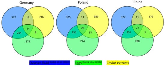
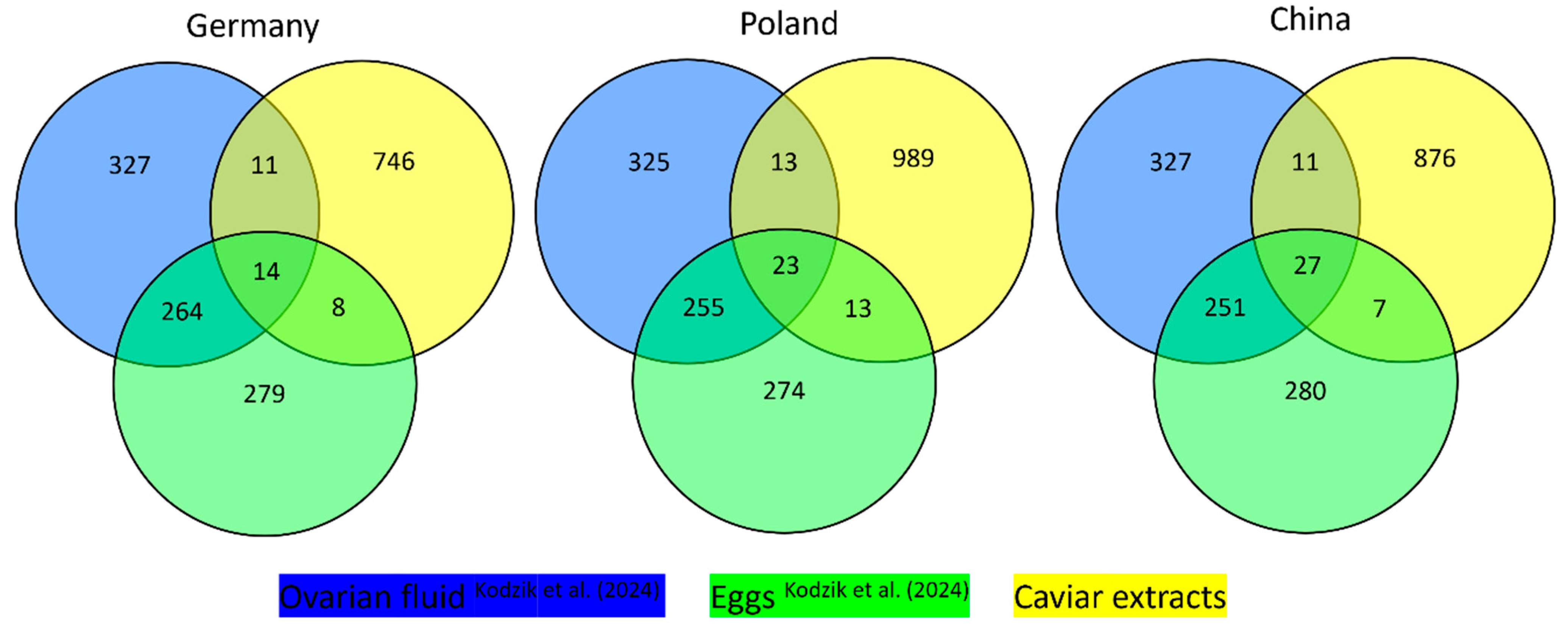
Three commercial caviar samples from Germany, Poland, and China were analysed, and a total of 1437 protein groups and 3452 unique peptides were identified across these three samples (Table 2). Comparison with the Acipenser baerii ovarian fluid proteome (617 proteins; [13]) showed overlaps in all samples (Germany: ≥25 proteins; Poland: ≥36 proteins; and China: ≥38 proteins); see Figure 1 and Supplemental Table S1. Proteins detected in all three samples included zona pellucida proteins (ZP3, ZPAX, ZP sperm-binding protein 3), fibrinogen gamma chain, catalase, superoxide dismutase, haptoglobin, and immunoglobulin chains (Supplemental Table S1). These results provide a dataset of identified proteins and ovarian fluid overlaps across the analysed caviar samples, which can be used for subsequent quantitative or comparative analyses.
Table 2.
Numbers of identified protein groups, peptides, and peptide ion variants according to the 3 samples and species groups.
Figure 1.
Venn diagrams comparing proteins identified in commercial caviar samples originating from Germany, Poland, and China (yellow) with proteins identified in ovarian fluid (blue) and egg/roe extracts (green) as reported by Kodzik et al. (2024) [13]. Caviar samples originating from Acipenser ruthenus (Germany) and Acipenser baerii (Poland and China).
3.2. Proteome Profiling of Multi-Species Origin
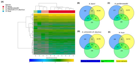
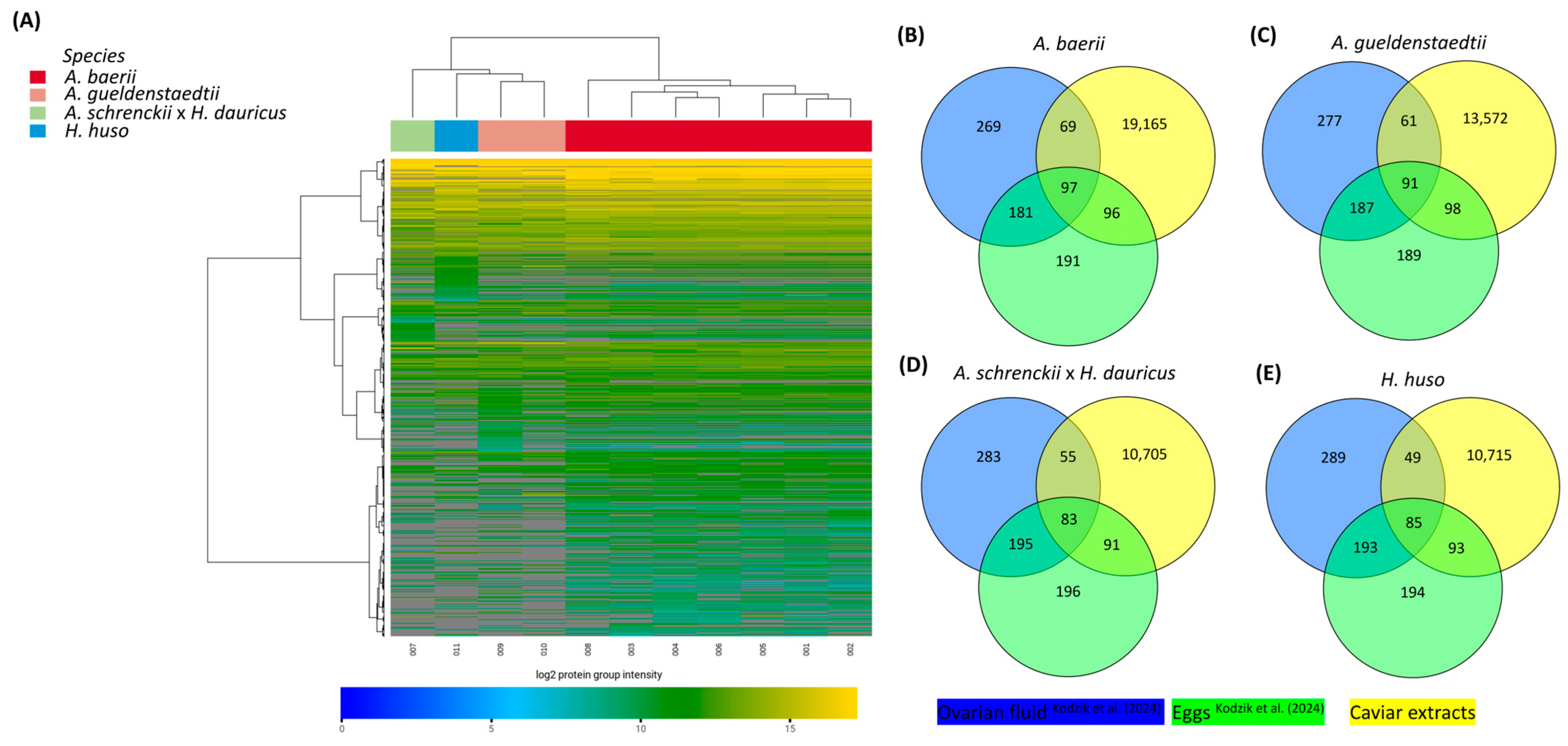
Using a multi-species approach, a total of 6244 protein groups, 42,917 peptides, and 48,182 peptide ion variants were quantified across the eleven analysed caviar samples (Table 2). On average, each sample yielded 4435 ± 0.6 protein groups, 27,432 ± 6.16 peptides, and 30,418 ± 6.90 peptide ion variants (Table 3, Figure 2A, Supplemental Table S2). Comparison with the 338 proteins previously reported from sturgeon ovarian fluid showed that 69 were detected in A. baerii, 61 in A. gueldenstaedtii, 55 in A. schrenckii, and 49 in H. huso samples analysed in the current study [13]; see Figure 2B. The heatmap revealed the highly consistent clustering of samples within each species, indicating low intra-species variability and the robust reproducibility of the measurements (Figure 2A). Clear separation between A. baerii, A. gueldenstaedtii, A. schrenckii, and H. huso was observed, confirming that the global proteomic signatures are strongly species-specific. Despite species-specific clustering and quantitative differences in protein abundance, all analysed samples shared a conserved core proteome dominated by egg- and ovarian fluid-associated proteins. Distinct protein clusters displayed differential abundance patterns across species, suggesting group-specific biological signatures, while a subset of proteins showed conserved high expression across all species. No evidence of batch or production origin effects was observed, as samples did not cluster according to the manufacturer (Figure 2A). It should be noted that several A. baerii samples originated from the same production source. Therefore, the dataset does not allow an assessment of farm-level variability and should not be interpreted as fully independent biological replication at the production level. Instead, the analysis was designed to capture species-level proteomic patterns and the consistency of ovarian fluid-associated protein detection across samples. Intersection analysis between proteins identified in the current dataset and those previously reported from sturgeon ovarian fluid or eggs revealed species-specific but consistently overlapping protein complements [13]. For the sampled A. baerii caviar, 166 proteins overlapped with ovarian fluid and 193 proteins with egg proteins, resulting in 262 of the 903 proteins being detected in total in the samples (Figure 2B). In A. gueldenstaedtii, 152 ovarian fluid proteins, 189 egg proteins, and 250 of the 903 total reference proteins were recovered (Figure 2C). Similarly, A. schrenckii showed an overlap of 138 proteins with ovarian fluid, 174 with eggs, and 229 of the 903 reference proteins in total (Figure 2D). For H. huso, 134 ovarian fluid proteins, 178 egg proteins, and 227 of the 903 proteins were identified (Figure 2E). Overall, these results indicate a substantial and consistent proportion of reproductive fluid-associated proteins across all analysed species.
Table 3.
Numbers of identified protein groups, peptides, and peptide ion variants according to the 11 samples and species groups.
Figure 2.
Protein analysis of caviar from A. baerii, A. gueldenstaedtii, A. schrenckii × H. dauricus, and H. huso reveals species-driven clustering and conserved overlaps with ovarian fluid proteomes. (A) Hierarchical clustering based on Manhattan distance was performed on log2-transformed protein group intensities across all samples. (B–E) Venn diagrams comparing proteins identified in commercial caviar samples (yellow) from A. baerii (B), A. gueldenstaedtii (C), A. schrencki (D), and H. huso (E) with proteins identified in ovarian fluid (blue) and the egg/roe proteomes (green) reported by Kodzik et al. (2024) [13].
A substantial difference in proteome depth was observed between the two analytical workflows. While the DDA-based analysis of geographically distinct samples identified 1437 protein groups, the DIA approach applied to the multi-species cohort yielded markedly higher proteome coverage. This difference is primarily attributable to methodological and instrumental factors, as DIA-based acquisition enables more comprehensive and reproducible peptide detection compared to DDA, rather than reflecting biological variation between sample sets.
3.3. Unique Ovarian Fluid Proteins
To assess the contribution of ovarian fluid proteins to the caviar extracts of different geographical and multi-species origin, a targeted intersection analysis was conducted using the 338 ovarian fluid-specific proteins reported for the Siberian sturgeon by Kodzik et al. (2024) [13]. By restricting the comparison to markers unique to ovarian fluid physiology, general sturgeon proteins were excluded. Despite this stringent filter, each species showed a substantial number of ovarian fluid proteins (A. baerii: 69; A. gueldenstaedtii: 61; A. schrenckii: 55; and H. huso: 49). The overlapping proteins span several functional categories, including zona pellucida glycoproteins, coagulation and complement factors, oxidative stress enzymes, and immunoglobulins—protein groups that are characteristic of ovarian fluid biology.
Overall, the detection of 49–69 ovarian fluid-specific proteins per species corresponds to approximately 14–20% of the ovarian fluid proteome in the sturgeon [13]; see Supplemental Table S2. This subgroup represents abundant and physiologically defining ovarian proteins, suggesting that standard procedures for the harvesting and extraction of roe routinely co-extract components of ovarian fluid in addition to proteins derived from the yolk.
Several important biological categories of ovarian fluid proteins were represented among these unique overlaps (Supplemental Tables S1 and S2).
- Zona pellucida proteins (ZPs): Multiple isoforms of zona pellucida glycoproteins (ZP3, ZPAX, ZP3-4) were consistently detected. ZPs form the extracellular egg envelope surrounding oocytes and are not present in mature roe alone. Their detection indicates the presence of ovarian wall or follicular material and aligns with their classification as abundant ovarian fluid proteins in Kodzik et al. (2024) [13].
- Fibrinogen gamma chain (FGG): FGG was identified in both datasets—the geographic and multi-species cohorts. This coagulation factor is abundant in ovarian fluid but absent from yolk-rich eggs, suggesting the co-extraction of ovarian fluid-associated plasma proteins during processing.
- Catalase and superoxide dismutase (SOD1, SOD3): These antioxidant enzymes were consistently observed. In ovarian physiology, they mitigate reactive oxygen species (ROS) produced during oocyte maturation. Their presence in extracts indicates the carryover of ovarian oxidative stress-related proteins.
- Haptoglobin: Haptoglobin, an acute-phase protein that binds free haemoglobin, was reproducibly detected. It is abundant in ovarian fluid and was observed across all sturgeon species analysed.
- Immunoglobulins (Ig chains): Both heavy and light chains of immunoglobulins were identified repeatedly. These maternal immune proteins are present in ovarian fluid and were detected in commercial extracts across species.
3.4. Cell Culture Alternative
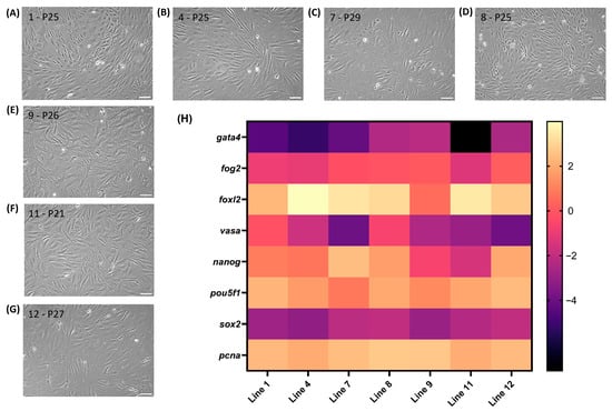
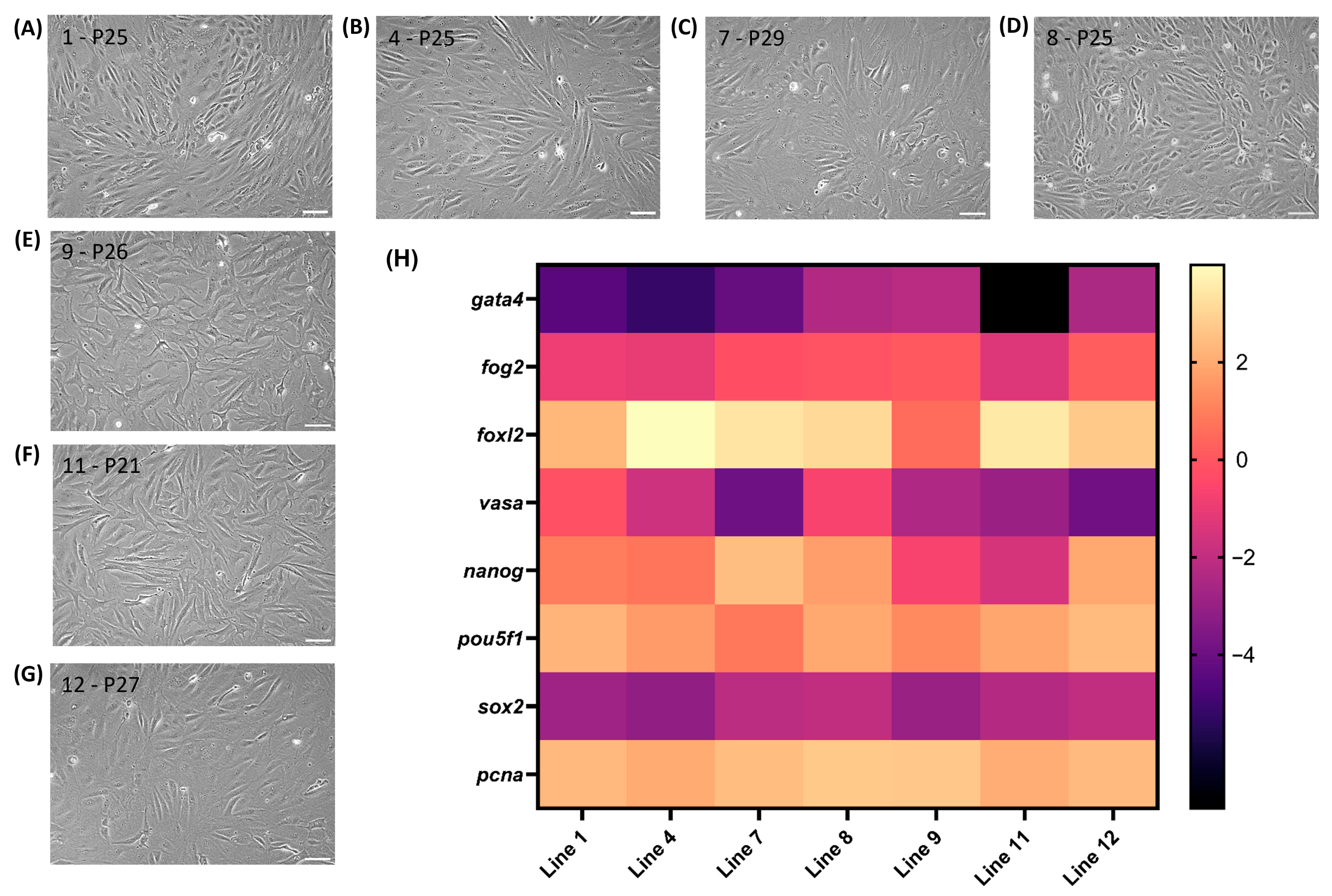
Seven independent cell lines were successfully isolated from yolk sac-stage A. baerii larvae [15]. Despite being derived from sibling animals following a standardised protocol, these lines displayed unique biological behaviours—including a variable morphology (Figure 3A–G) and metabolism—yet all exhibited adaptation to in vitro cultivation and maintained consistently high viability (>90%) and stable cell sizes (22–26 µm) across passages. The cell lines reached P24 to P36 within the first 9 months of cultivation, demonstrating long-term stability. Up to 100 cryovials per line were frozen, and cryopreservation experiments confirmed that the tested freezing media preserved post-thaw viability, enabling cell biobanking and transfer. The expression of genes associated with gonadal development, pluripotency, proliferation, and germline specification was detected in RBC-ABAlar cell lines (Figure 3H). The gene gata4, which is involved in somatic gonadal development, was expressed at low levels and was not detectable in cell line 11. In contrast, fog2 was expressed at intermediate levels in all cell lines. The female-associated gene foxl2, an early marker of ovarian differentiation, was consistently expressed across the cell lines, while the germline marker vasa was detected at intermediate levels and lower levels in cell line 7 and 12. Pluripotency-related genes pou5f1 (oct4) and nanog were expressed at higher levels in all RBC-ABAlar cells, indicating the retention of stem cell-associated features. The gene sox2, which plays a role in early embryonic development and also neural differentiation, showed intermediate expression levels in the RBC-ABAlar cells. The proliferation-associated gene pcna was uniformly highly expressed across the cell lines. Overall, the observed intermediate and low expression levels of several gonadal and germline-associated genes are likely attributable to the applied in vitro culture conditions. Nevertheless, the detectable expression of key proliferation, pluripotency, and germline-related markers indicates that RBC-ABAlar cells maintain fundamental molecular features consistent with potential for lineage commitment. These findings suggest that the targeted modulation of culture conditions may facilitate directed differentiation and could support the activation of gonadal development pathways in these cells.
Figure 3.
Analysis of cell lines. (A–G) Phase contrast pictures of RBC-ABAlar cell lines: line 1 at passage (P) 25 (A), line 4 at P25 (B), line 7 at P29 (C), line 8 at P25 (D), line 9 at P26 (E), line 11 at P21 (F), and line 12 at P27 (G). Scale bars: 100 µm. (H) Heatmap of normalised relative gene expression (reference genes: rlp6 and eef1a1) across RBC-ABAlar cell lines 1, 4, 7, 8, 9, 11, and 12. Colour intensity represents the log2-transformed normalised expression level, with light yellow indicating higher and black indicating lower expression in relation to the reference genes.
4. Discussion
The results of this study provide new molecular insights into the composition of commercial caviar and caviar extracts, and they challenge several assumptions that currently underpin their use in cosmetic formulations. “Caviar extract” is defined as an ingredient obtained from the eggs of sturgeons (Acipenseridae), implying that the material is biologically homogeneous, egg-derived, and free from additional reproductive tissues [9]. The present study analysed commercially available caviar (roe) as the upstream raw material from established supply chains, which represent the biological starting point for subsequent industrial extraction processes. An analysis conducted independently in two laboratories on caviar samples from different geographic origins and species showed that ovarian fluid is likely co-extracted during standard processing, as it adheres strongly to the surfaces of fish eggs [20,21,22]. The contamination is therefore not accidental or batch-specific but an intrinsic consequence of harvesting roe directly from the ovary without rigorous separation of fluid and tissue compartments. The presence of ovarian fluid, rich in enzymes, immune proteins, complement components, and reproductive glycoproteins [13,23,24], highlights potential impacts on both functionality and safety in cosmetic formulations and underscores the need for a molecular-based assessment of ingredients. A limitation of this study is that it does not allow the attribution of cosmetic effects to specific molecular fractions. It therefore remains unclear whether roe-derived or ovarian fluid-associated proteins drive the reported bioactivity, highlighting a key gap in the molecular and regulatory characterisation of “caviar extract”.
In cosmetics, the presence of such proteins in raw materials has important implications. First, the detected zona pellucida glycoproteins (ZPs), immunoglobulins, coagulation factors like fibrinogen γ-chain, and antioxidant enzymes parallel molecular categories known to modulate skin biology. Immune proteins applied topically can influence irritation potential or allergenicity or exert unintended immunomodulatory effects [25,26]. Proteases and complement factors may affect barrier integrity or inflammatory pathways [27]. Thus, the assumption that caviar extract consists primarily of nutritive lipoproteins and peptides derived from yolk is incomplete. We would even go so far as to say that any claim that cosmetics or nutraceuticals contain “pure roe extract” is scientifically untenable without proteomic validation and strict separation protocols. Cosmetic claims such as the notion that bioactive peptides from caviar improve skin vitality [28] may in fact rely on constituents originating from ovarian fluid rather than from yolk alone. While this may offer functional diversity, it also complicates safety assessment, as toxicological data for ovarian fluid proteins in topical applications are sparse. In addition, the high variability in ovarian fluid across different batches and species represents a significant challenge for standardised safety assessments, such as OECD 406 (Skin Sensitization). A traditional extract that is inadvertently or partially contaminated with ovarian fluid may therefore yield inconsistent toxicological results between batches, further limiting the reproducibility and regulatory comparability of such products.
Second, the consistent presence of ovarian fluid-specific proteins across multiple species and production origins contradicts the notion of a highly standardised ingredient. In other marine-derived cosmetic ingredients—such as salmon roe extract, cod collagen hydrolysates, and sea cucumber extracts—compositional variability has been identified as a major challenge for reproducibility and regulatory certainty [29,30]. Our data suggest that sturgeon caviar extract falls into the same category: a bioactive but heterogeneous material whose composition is shaped not only by species identity but also by its reproductive physiology. The species-specific proteomic clustering analysed here parallels the genetic and metabolomic differentiation previously documented in sturgeon aquaculture [31,32]. For cosmetics formulators, this means that products labelled simply as “caviar extract” may differ substantially depending on the species and biological origin, potentially leading to variable performance outcomes or instability in formulations.
Third, this study addresses a longstanding gap in cosmetic ingredient regulation. The INCI nomenclature, although widely used, often lags behind advances in analytical chemistry [33]. Ingredients historically defined by origin rather than chemical composition—such as placenta extract, elastin, or fish collagen—have been reformulated or reclassified following deeper molecular characterisation. Our findings place caviar extract in a similar regulatory position: current definitions, which state that caviar extract is “an extract of the eggs of sturgeon”, do not accurately reflect the molecular reality revealed by proteomics. This discrepancy has implications for labelling accuracy, claims substantiation, and cross-border regulatory compliance, especially given that caviar extract is used widely in high-value anti-aging products marketed in Europe, East Asia, and further emerging markets [8]. Moreover, the presence of bioactive immunoglobulins, complement factors, and enzymes may necessitate updated safety assessments in line with the EU Cosmetic Regulation [34], which mandates the evaluation of biological materials with immune or enzymatic activity.
The findings also highlight the potential value of cell culture-based caviar protein systems for the cosmetic industry. As discussed in the Introduction, the spontaneously immortalised A. baerii larval stem cell lines may represent a defined, controllable, and contaminant-free production system. Their gene expression signatures, which include a set of markers linked to ovarian differentiation pathways, suggest that these cultures can be adapted to generate caviar-relevant proteins without co-isolating follicular fluid or reproductive tissue contaminants. Molecular modifications happen before morphological differentiation [35]. In the Siberian sturgeon, migrating primordial germ cells have been detected as early as one day post-hatch, and, by 115 days post-hatch, gonadal types can already be distinguished [36]. The sex-specific molecular modifications of selected markers have been identified at three-to-four months post-hatch [37]. Accordingly, RBC-ABAlar cells were isolated at four days post-hatch, a developmental stage at which primordial germ cells are present but gonadal differentiation has not yet occurred.
Similar cell-based approaches are already being explored for marine collagen [38], the in vitro meat sector [39], and plant stem cell extracts used in cosmetics [40,41]. A cell-based caviar proteome could therefore offer multiple advantages: increased batch-to-batch consistency, the elimination of fluid-derived proteins of uncertain safety, alignment with sustainability and CITES conservation principles, and the potential for customised protein profiles tailored to cosmetic functionality. At the same time, important limitations must be considered. Although the established sturgeon larval cell lines demonstrate long-term stability and viability in vitro, translation into industrial applications requires further optimisation to achieve economically viable yields. In addition, cost structures remain uncertain and large-scale scalability has yet to be demonstrated. Thus, while cell-based systems represent a promising alternative for producing defined caviar-associated proteins, further development is required to assess their industrial feasibility. From a functional perspective, distinguishing between egg-derived and ovarian fluid-derived bioactives may also expand opportunities for targeted cosmetic innovation. Antioxidant enzymes such as catalase and superoxide dismutase, which are abundant in ovarian fluid, have established skin-protective potential but are typically unstable in topical formulations unless properly encapsulated or stabilised [42,43]. By providing molecular resolution of these categories, our findings support the development of more targeted, claim-relevant protein fractions for cosmetics—whether derived from natural roe, selectively purified ovarian proteins, or engineered cell-based alternatives.
5. Conclusions
Overall, this study provides a new biochemical understanding of caviar extract in a cosmetic science context. Rather than being a homogeneous ingredient derived from eggs, caviar extract is shown to be a mixed proteome derived from reproductive tissue, containing proteins relevant to immunity, oxidative stress, coagulation, and fertilisation. This complexity may contribute to the perceived efficacy of the ingredient but also raises regulatory and safety issues that have been largely overlooked until now. As the market for caviar-based cosmetics expands due to its positioning in the luxury segment and strong growth in Asia and Europe, ingredient transparency and molecular characterisation will become increasingly important to maintain consumer trust and ensure regulatory compliance. Future work should further resolve the functional roles of specific protein fractions and their contributions to bioactivity, while standardised analytical frameworks and cell-based production systems may enable more defined and reproducible ingredient profiles.
By integrating deep proteomics with insights from reproductive biology, cosmetic science, and emerging alternative cell culture platforms, this study provides a refined biochemical framework for understanding and regulating caviar extract. The molecular evidence presented here underscores the need for updated definitions, improved standardisation, and the consideration of novel biological production strategies as the cosmetic industry moves toward the more transparent, sustainable, and scientifically grounded use of marine-derived ingredients.
Supplementary Materials
The following supporting information can be downloaded at https://www.mdpi.com/article/10.3390/cosmetics13030105/s1. Table S1: Core Contamination Markers; Geographic origin: proteins identified in common between the caviar analysed in our study and ovarian fluid from Kodzik et al. (2024) [13]; Table S2: Core Contamination Markers; Multi-species origin: proteins identified in common between the caviar analysed in our study and ovarian fluid from Kodzik et al. (2024) [13].
Author Contributions
Conceptualisation: K.B. and B.G.; Formal Analysis: K.B., M.A.D., B.G. and V.D.L.; Visualisation: B.G. and K.B.; Supervision: M.A.D., B.G. and K.B.; Writing—Original Draft: B.G., M.P. and K.B.; Writing—Review and Editing: all authors; Resources: K.B. and B.G. All authors have read and agreed to the published version of the manuscript.
Funding
This research was funded by London Fine Foods and Caviar Biotech, Ltd., as well as the Research Institute for Farm Animal Biology (FBN) Dummerstorf. MP was supported by the Czech Science Foundation (project 26-22963S).
Institutional Review Board Statement
Not applicable.
Informed Consent Statement
Not applicable.
Data Availability Statement
All relevant data are provided within the manuscript.
Acknowledgments
The authors gratefully acknowledge the Advanced Proteomics Facility (APF) team at the University of Oxford for their expertise in sample preparation, high-resolution LC-MS/MS acquisition, and protein identification. In addition, we acknowledge Biognosys for their DIA-based proteomic profiling, spectral library generation, quantitative data analysis, and statistical interpretation, which were central to this work. We also thank Daniela Ohde from the FBN for assisting in the Fluidigm PCR. Special thanks go to Gert-Jan Goeminne from Royal Belgian Caviar for providing the Siberian sturgeon larvae for the cell culture work. During the preparation of this work, the authors used Chat-AI 5.3–mini (Academic Cloud) as a supporting tool in order to improve the language and readability.
Conflicts of Interest
The author Kenneth Benning declares a commercial affiliation with Caviar Biotec Limited and CellRoe Limited, which are engaged in related biotechnology research and intellectual property development. This affiliation did not influence the design, execution, analysis, or interpretation of the data. All other authors declare no competing interests. All research was conducted in accordance with institutional policies regarding conflicts of interest and intellectual property.
Abbreviations
The following abbreviations are used in this manuscript:
| AGC | Automatic gain control |
| C18 | Octadecyl carbon chain (reversed-phase material) |
| CASO | Casein soy peptone medium |
| CITES | Convention on International Trade in Endangered Species of Wild Fauna and Flora |
| CT | Cycle threshold |
| Da | Dalton |
| DDA | Data-dependent acquisition |
| DIA | Data-independent acquisition |
| DIA | Data-independent acquisition parallel accumulation–serial fragmentation |
| EP | European Pharmacopoeia |
| FA | Formic acid |
| FAO | Food and Agriculture Organization |
| FDR | False discovery rate |
| FGG | Fibrinogen gamma chain |
| fog2 | Friend of GATA 2 |
| foxl2 | Forkhead box L2 |
| gata4 | GATA binding protein 4 |
| HCD | Higher-energy collisional dissociation |
| HPLC | High-performance liquid chromatography |
| i.d. | Internal diameter |
| IFC | Integrated fluidic circuit |
| Ig | Immunoglobulin chains |
| iRT | Indexed retention time |
| LC | Liquid chromatography |
| LC-MS/MS | Liquid chromatography–tandem mass spectrometry |
| m/z | Mass-to-charge ratio |
| mBCA | Micro bicinchoninic acid assay |
| MS | Mass spectrometry |
| MS/MS | Tandem mass spectrometry |
| nanog | Nanog homeobox |
| NCBI | National Center for Biotechnology Information |
| P | Passage number (cell culture passage) |
| PASEF | Parallel accumulation–serial fragmentation |
| pcna | Proliferating cell nuclear antigen |
| pou5f1 (oct4) | POU class 5 homeobox 1 (octamer-binding transcription factor 4) |
| ppm | Parts per million |
| PSM | Peptide–spectrum match |
| qPCR | Quantitative polymerase chain reaction |
| RBC-ABAlar | Acipenser baerii larval cell line |
| ROS | Reactive oxygen species |
| SDS | Sodium dodecyl sulphate |
| SOD1/SOD3 | Superoxide dismutase 1 and 3 |
| sox2 | SRY-box transcription factor 2 |
| SOP | Standard operating procedure |
| SP3 | Single-pot solid-phase-enhanced sample preparation |
| USD | US dollar |
| USP | United States Pharmacopeia |
| vasa | Vasa germ cell marker (DDX4) |
| ZPs | Zona pellucida proteins |
References
- Stroe Dudu, A.; Georgescu, S.E. Exploring the multifaceted potential of endangered sturgeon: Caviar, meat and by-product benefits. Animals 2024, 14, 2425. [Google Scholar] [CrossRef] [Scilit]
- Global Growth Insights. Caviar Market Report. Available online: https://www.globalgrowthinsights.com/market-reports/caviar-market-107012 (accessed on 19 January 2026).
- Grand View Research. Caviar Market Size & Outlook, Global. Available online: https://www.grandviewresearch.com/horizon/outlook/caviar-market-size/global (accessed on 19 January 2026).
- FAO/WHO Codex Alimentarius Commission. Standard for Sturgeon Caviar (CXS 291-2010). Available online: https://www.fao.org/fao-who-codexalimentarius/sh-proxy/en/?lnk=1&url=https%3A%2F%2Fworkspace.fao.org%2Fsites%2Fcodex%2FStandards%2FCXS%2B291-2010%2FCXS_291e.pdf (accessed on 19 January 2026).
- CITES Secretariat. Resolution Conf. 12.7 (Rev. CoP17): Conservation of and Trade in Sturgeons and Paddlefish. 2017. Available online: https://cites.unia.es/file.php/1/handbook/eng/res/all/E-Res-12-07R16.pdf (accessed on 19 January 2026).
- Le, L.T.T.; Kim, B.K.; Chien, P.N.; Choi, K.W.; Kim, H.B.; Hwang, U.J.; Han, H.S.; Heo, C.Y. Investigating the anti-aging effects of caviar oil on human skin. In Vivo 2023, 37, 2078–2091. [Google Scholar] [CrossRef] [Scilit] [PubMed]
- Park, J.; Kim, D.; Lee, M.; Han, S.; Jun, W.; Jung, H.M.; Koo, Y.K.; Na, G.H.; Han, S.H.; Han, J.; et al. Enzyme-treated caviar prevents UVB irradiation-induced skin photoaging. Mar. Drugs 2022, 20, 685. [Google Scholar] [CrossRef] [Scilit]
- DataHorizzon Research. Caviar Extract Market (By Type: Black Caviar Extract, Red Caviar Extract, By Application: Cosmetic & Personal Care, Food & Beverages, By Distribution Channel)—Lobal Market Size, Share, Growth, Trends, Statistics Analysis Report, By Region, and Forecast 2025–2033. DataHorizzon Research 2025, 38171, 154. Available online: https://datahorizzonresearch.com/caviar-extract-market-38171 (accessed on 19 January 2026).
- SpecialChem. Caviar Extract (INCI). Available online: https://www.specialchem.com/cosmetics/inci-ingredients/caviar-extract (accessed on 19 January 2026).
- Yantai Sturgeon Biotechnology Co., Ltd. Preparation Method of Sturgeon Caviar Extract. Patent CN 111803434 A, 7 August 2020. Available online: https://patents.google.com/patent/CN111803434A/en (accessed on 19 January 2026).
- Siddique, M.A.M.; Cosson, J.; Psenicka, M.; Linhart, O. A review of the structure of sturgeon egg membranes and of the associated terminology. J. Appl. Ichthyol. 2014, 30, 1246–1255. [Google Scholar] [CrossRef] [Scilit]
- Kholodnyy, V.; Dzyuba, B.; Gadêlha, H.; Cosson, J.; Boryshpolets, S. Egg-sperm interaction in sturgeon: Role of ovarian fluid. Fish Physiol. Biochem. 2021, 47, 653–669. [Google Scholar] [CrossRef] [Scilit]
- Kodzik, N.; Ciereszko, A.; Szczepkowska, B.; Malinowska, A.; Dietrich, M.A. Comparative proteomic analysis of the ovarian fluid and eggs of Siberian sturgeon. BMC Genom. 2024, 25, 451. [Google Scholar] [CrossRef] [Scilit] [PubMed]
- Schmidt, C.V.; Raza, H.; Olsen, K.; Mouritsen, O.G. Proximate nutritional composition of roe from fish, crustaceans, mussels, echinoderms, and cephalopods. Int. J. Gastr. Food Sci. 2024, 36, 100944. [Google Scholar] [CrossRef] [Scilit]
- Di Leonardo, V.; Tönißen, K.; Brenmoehl, J.; Ohde, D.; Wanka, H.; Benning, K.; Grunow, B. Sibling-derived cell lines of whole larval siberian sturgeon as an in vitro model system for studying inter-individual differences within the same genomic heritage. Cells 2025, 14, 2004. [Google Scholar] [CrossRef] [Scilit]
- Hughes, C.S.; Moggridge, S.; Müller, T.; Sorensen, P.H.; Morin, G.B.; Krijgsveld, J. Single-pot, solid-phase-enhanced sample preparation for proteomics experiments. Nat. Protoc. 2019, 14, 68–85. [Google Scholar] [CrossRef] [Scilit] [PubMed]
- Rappsilber, J.; Ishihama, Y.; Mann, M. Stop and go extraction tips for matrix-assisted laser desorption/ionization, nanoelectrospray, and LC/MS sample pretreatment in proteomics. Anal. Chem. 2003, 75, 663–670. [Google Scholar] [CrossRef] [Scilit]
- Callister, S.J.; Barry, R.C.; Adkins, J.N.; Johnson, E.T.; Qian, W.J.; Webb-Robertson, B.J.; Smith, R.D.; Lipton, M.S. Normalization approaches for removing systematic biases associated with mass spectrometry and label-free proteomics. J. Proteome Res. 2006, 5, 277–286. [Google Scholar] [CrossRef] [Scilit]
- Ballester, M.; Cordón, R.; Folch, J.M. DAG expression: High-throughput gene expression analysis of real-time PCR data using standard curves for relative quantification. PLoS ONE 2013, 8, e80385. [Google Scholar] [CrossRef] [Scilit]
- Roufidou, C.; Borg, B. Ovarian fluid in the three-spined stickleback Gasterosteus aculeatus: Effects of egg overripening and sex steroid treatment. J. Fish Biol. 2019, 94, 446–457. [Google Scholar] [CrossRef] [Scilit] [PubMed]
- Chatakondi, N.G.; Torrans, E.L. The influence of ovarian fluid PH of stripped unfertilized channel catfish, Ictalurus punctatus, eggs on the hatching success of channel catfish ♀ x blue catfish, Ictalurus furcatus♂, hybrid catfish eggs. J. World Aquac. Soc. 2012, 43, 585–593. [Google Scholar] [CrossRef] [Scilit]
- Mansour, N.; Lahnsteiner, F.; Patzner, R.A. Ovarian fluid plays an essential role in attachment of Eurasian perch, Perca fluviatilis eggs. Theriogenology 2009, 71, 586–593. [Google Scholar] [CrossRef] [Scilit] [PubMed]
- Nynca, J.; Arnold, G.J.; Fröhlich, T.; Ciereszko, A. Shotgun proteomics of rainbow trout ovarian fluid. Reprod. Fertil. Dev. 2015, 27, 504–512. [Google Scholar] [CrossRef] [Scilit] [PubMed]
- Gueho, A.; Żarski, D.; Rime, H.; Guével, B.; Com, E.; Lavigne, R.; Nguyen, T.; Montfort, J.; Pineau, C.; Bobe, J. Evolutionarily conserved ovarian fluid proteins are responsible for extending egg viability in salmonid fish. Sci. Rep. 2024, 14, 9651. [Google Scholar] [CrossRef] [Scilit]
- Basketter, D.A.; Kimber, I. Enzymes and sensitization via skin exposure: A critical analysis. Regul. Toxicol. Pharmacol. 2022, 129, 105112. [Google Scholar] [CrossRef] [Scilit]
- Secchi, G. Role of protein in cosmetics. Clin. Dermatol. 2008, 26, 321–325. [Google Scholar] [CrossRef] [Scilit]
- Lee, S.E.; Jeong, S.K.; Lee, S.H. Protease and protease-activated receptor-2 signaling in the pathogenesis of atopic dermatitis. Yonsei Med. J. 2010, 51, 808–822. [Google Scholar] [CrossRef] [Scilit] [PubMed]
- Na, G.H.; Kim, S.; Jung, H.M.; Han, S.H.; Han, J.; Koo, Y.K. Skin anti-aging efficacy of enzyme-treated supercritical caviar extract: A randomized, double-blind, placebo-controlled clinical trial. Nutrients 2024, 16, 137. [Google Scholar] [CrossRef] [Scilit] [PubMed]
- Costa, J.P.; Custódio, L.; Reis, C.P. Exploring the potential of using marine-derived ingredients: From the extraction to cutting-edge cosmetics. Mar. Drugs 2023, 21, 620. [Google Scholar] [CrossRef] [Scilit] [PubMed]
- Senadheera, T.R.L.; Dave, D.; Shahidi, F. Sea cucumber derived type i collagen: A comprehensive review. Mar. Drugs 2020, 18, 471. [Google Scholar] [CrossRef] [Scilit]
- Zhang, X.; Wu, W.; Li, L.; Ma, X.; Chen, J. Genetic variation and relationships of seven sturgeon species and ten interspecific hybrids. Genet. Sel. Evol. 2013, 45, 21. [Google Scholar] [CrossRef] [Scilit] [PubMed]
- Liu, Q.; Naganuma, T. Metabolomics in sturgeon research: A mini-review. Fish Physiol. Biochem. 2024, 50, 1895–1910. [Google Scholar] [CrossRef] [Scilit]
- International Nomenclature of Cosmetic Ingredients (INCI). INCI Nomenclature Conventions. Personal Care Products Council. 2021. Available online: https://www.personalcarecouncil.org/wp-content/uploads/2021/02/Conventions2021_v1.pdf (accessed on 23 February 2026).
- EUR-Lex. Regulation (EC) No 1223/2009 of the European Parliament and of the Council of 30 November 2009 on Cosmetic Products (Recast) (Text with EEA Relevance). Off. J. Eur. Union 2009, EC 1223/2009. Available online: https://eur-lex.europa.eu/legal-content/EN/ALL/?uri=celex:32009R1223 (accessed on 24 February 2026).
- Ruan, R.; Li, Y.; Yue, H.; Ye, H.; Jin, J.; Wu, J.; Du, H.; Li, C. Transcriptome analyses reveal expression profiles of morphologically undifferentiated and differentiated gonads of yangtze sturgeon Acipenser dabryanus. Genes 2023, 14, 2058. [Google Scholar] [CrossRef] [Scilit]
- Rzepkowska, M.; Ostaszewska, T. Proliferating cell nuclear antigen and Vasa protein expression during gonadal development and sexual differentiation in cultured Siberian (Acipenser baerii Brandt, 1869) and Russian (Acipenser gueldenstaedtii Brandt & Ratzeburg, 1833) sturgeon. Rev. Aquacult. 2014, 6, 75–88. [Google Scholar] [CrossRef] [Scilit]
- Vizziano-Cantonnet, D.; Di Landro, S.; Lasalle, A.; Martínez, A.; Mazzoni, T.S.; Quagio-Grassiotto, I. Identification of the molecular sex-differentiation period in the Siberian sturgeon. Mol. Reprod. Dev. 2016, 83, 19–36. [Google Scholar] [CrossRef] [Scilit]
- Liu, C. Application of marine collagen for stem-cell-based therapy and tissue regeneration (review). Med. Int. 2021, 1, 6. [Google Scholar] [CrossRef] [Scilit]
- Kumar, P.; Sharma, N.; Sharma, S.; Mehta, N.; Verma, A.K.; Chemmalar, S.; Sazili, A.Q. In-vitro meat: A promising solution for sustainability of meat sector. J. Anim. Sci. Technol. 2021, 63, 693–724. [Google Scholar] [CrossRef] [Scilit]
- Gardiki, V.; Pavlou, P.; Siamidi, A.; Papageorgiou, S.; Papadopoulos, A.; Iakovou, K.; Varvaresou, A. Plant stem cells in cosmetic industry. Plants 2025, 14, 433. [Google Scholar] [CrossRef] [Scilit] [PubMed]
- Barbulova, A.; Apone, F.; Colucci, G. Plant cell cultures as source of cosmetic active ingredients. Cosmetics 2014, 1, 94–104. [Google Scholar] [CrossRef] [Scilit]
- Di Mambro, V.M.; Borin, M.F.; Fonseca, M.J.V. Topical formulations with superoxide dismutase: Influence of formulation composition on physical stability and enzymatic activity. J. Pharm. Biomed. Anal. 2003, 32, 97–105. [Google Scholar] [CrossRef] [Scilit]
- Alkhalaf, A.; Hooshiar, A.; Dargahi, J. Composite magnetorheological elastomers for tactile displays: Enhanced MR-effect through bi-layer composition. Compos. Part B Eng. 2020, 190, 107888. [Google Scholar] [CrossRef] [Scilit]

Disclaimer/Publisher’s Note: The statements, opinions and data contained in all publications are solely those of the individual author(s) and contributor(s) and not of MDPI and/or the editor(s). MDPI and/or the editor(s) disclaim responsibility for any injury to people or property resulting from any ideas, methods, instructions or products referred to in the content. |
© 2026 by the authors. Licensee MDPI, Basel, Switzerland. This article is an open access article distributed under the terms and conditions of the Creative Commons Attribution (CC BY) license.