


Abstract
Background: Retinoids are established as a cornerstone in the management of photoaging; however, their clinical utility is frequently limited by local irritation, leading to poor patient compliance. This study evaluates the efficacy and safety of a new retinol formulation designed to improve skin texture and wrinkles while minimizing adverse effects. Methods: A prospective, rater-blinded pilot clinical study was conducted involving 21 women with moderate-to-severe photoaging over a 180-day period. Participants followed a progressive application protocol using a specific retinol emulsion alternating with a replenishing cream. Efficacy was assessed using the validated Rao–Goldman scale and VISIA® image analysis, while safety was monitored through the reporting of adverse events (AEs) and dermatological evaluations. The primary endpoint was the percentage of patients achieving a Rao–Goldman score of 2 at day 180. Results: A statistically significant improvement on the Rao–Goldman scale was observed, with 76.2% of patients improving from their baseline classification to exhibit only fine wrinkles (Grade 2) (p = 0.028). Instrumental analysis was performed using the VISIA® system. Regarding safety, although 81% of participants initially reported adverse effects such as erythema and scaling, these incidents decreased significantly over time. Conclusions: The novel retinol formulation demonstrates significant clinical efficacy in ameliorating the signs of photoaging, while maintaining a favorable safety profile
1. Introduction
Cutaneous aging encompasses both physical and psychological transformations, with visible manifestations such as wrinkles and alterations in skin texture impacting social interactions and overall well-being. This process is influenced by intrinsic and extrinsic factors, with direct sun damage being a predominant contributor [1]. While skin aging is unavoidable, measures can be taken to mitigate its impact. Topical rejuvenation strategies remain at the lower end of the risk spectrum compared to invasive injectables or surgical approaches.
However, the implications of skin aging extend far beyond the aesthetic. Recent research underscores the profound psychosocial impact of facial dermatoses and aging, linking perceptible skin changes to decreased self-esteem, social anxiety, and even depressive symptoms in affected individuals. In a society where youthful appearance is often equated with health and vitality, the demand for effective interventions has surged [2]. The global market reflects this shift, with a marked increase in the consumption of anti-aging products and non-invasive cosmetic procedures [3]. According to recent statistics from the International Society of Aesthetic Plastic Surgery (ISAPS), non-surgical procedures such as chemical peels and photorejuvenation have seen exponential growth, driven by patients seeking visible results without the downtime associated with surgery [4]. Patients are also willing to protect their skin against pollution that accelerates the skin aging process [5]. Despite this shift towards minimally invasive techniques, topical treatments remain the foundation of any comprehensive anti-aging regimen.
Nevertheless, realistic expectations must be established regarding the overall impact on facial morphology, as even optimal topical skin rejuvenation programs cannot mitigate volumetric changes [6].
Consequently, as anti-aging treatments remain highly relevant, current therapeutic approaches are predominantly topical or minimally invasive, leading to rigorous testing of advanced formulations [7]. Retinoids have long been established as a cornerstone in the prevention and treatment of photoaging, as several, and particularly tretinoin, have demonstrated the ability to enhance skin texture, reduce rhytids and dyschromia, regulate keratinocyte growth and differentiation, induce epidermal hyperplasia, compact the stratum corneum, stimulate collagen synthesis, inhibit the activity of matrix metalloproteinases (MMPs) responsible for collagen degradation, and promote the deposition of fibrillin-rich microfibrils [1,8,9]. The mechanism of action involves the binding of retinoic acid to nuclear receptors (RARs and RXRs), which subsequently modulate gene expression to promote cellular turnover and extracellular matrix repair [10]. This balance between cell proliferation, differentiation, and desquamation is crucial for treating keratinization disorders such as psoriasis, lichen planus, and various ichthyoses, as well as ameliorating fine wrinkles [11]. Furthermore, several retinoid compounds have exhibited promise for the prevention and treatment of non-melanoma skin cancer in experimental settings. However, the use of retinoids is not without its challenges. Common adverse effects include erythema, peeling, dryness, and a sensation of burning or tightness [12]. These side effects can lead to decreased patient compliance and discontinuation of treatment.
Nonetheless, many of these compounds have not been studied in human subjects nor in clinical trials at this time [11].
While tretinoin is the most potent, its use is often limited by “retinoid dermatitis,” characterized by erythema, scaling, and burning [13]. Consequently, there is a continuous search for retinol formulations that can deliver comparable efficacy to prescription retinoids but with an improved tolerability profile. Thus, it is crucial to balance the therapeutic benefits of retinoids with their potential to cause irritation, especially in individuals with sensitive skin.
This study aims to evaluate a novel retinol formulation that employs a specialized delivery system to enhance stability and penetration, thereby hypothesizing a reduction in irritation while maintaining high clinical efficacy.
This study aims to evaluate a novel retinol formulation that employs a specialized delivery system designed to enhance stability and cutaneous penetration, thereby hypothesizing a reduction in irritation while maintaining high clinical efficacy. We conducted a pilot study involving 21 women to evaluate the efficacy and safety of this formulation. The primary objective was to assess the clinical improvement of photoaging signs, specifically the percentage of patients achieving a Rao–Goldman score of 2 at day 180, while secondarily monitoring the incidence and severity of adverse effects.
2. Materials and Methods
Study Design and Participants
A pilot prospective, rater-blinded, single-center clinical study was conducted involving 21 female subjects aged 35 to 65 years (mean age of 44.43 years [SD: 7.69 years]). Eleven of the patients were menopausal. The study duration was 180 days. The protocol adhered to the ethical guidelines of the Declaration of Helsinki. The sample size was determined based on standard exploratory cohorts for cosmetic efficacy studies in order to provide the effect size to adequately power future, larger-scale, randomized controlled trials.
All participants provided written informed consent prior to participation in this dermatological compatibility and efficacy study of cosmetic product under RD 1090/4 December 2015 and did not receive financial compensation, but all the topical products used were freely provided.
Inclusion and Exclusion Criteria: Participants were included if they presented with moderate-to-severe signs of photoaging, with a specific focus on wrinkles ranking as Grade 3 or higher on the Rao–Goldman scale.
Exclusion criteria were strictly defined to avoid confounding factors. Subjects were excluded if they had undergone any facial surgical or cosmetic procedures that could alter skin texture or wrinkling within the previous 6 months. Specific exclusionary procedures included rhytidectomy (facelift), blepharoplasty, ablative or non-ablative laser resurfacing, chemical peels (medium or deep depth), dermabrasion, or the injection of botulinum toxin or soft tissue fillers. Patients with suspicious premalignant or malignant lesions were strictly excluded. They were also excluded if they had used topical retinoids in the 30 days prior to enrollment or oral isotretinoin in the previous 6 months. Participants were excluded if they were pregnant, breastfeeding, planning to conceive in the next months, or had a known sensitivity to retinol.
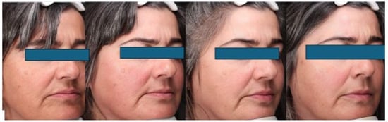
Treatment protocol. Figure 1. Participants were instructed to apply the novel retinol formulation (ASM® Retinol Texture Repair Emulsion) to the face three nights a week for the first 15 days, followed by application every other night (alternating with ASM® Replenish Cream) until the completion of the first month (Figure 1, Table 1). From day 30 to day 180, the retinol emulsion was applied nightly. From day 120 to day 180, patients were instructed to apply the products specifically to the eyelid and periocular areas on alternate days. Daily application of a broad-spectrum sunscreen (SPF 50) and a moisturizer was expected.
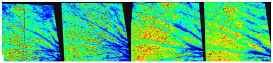
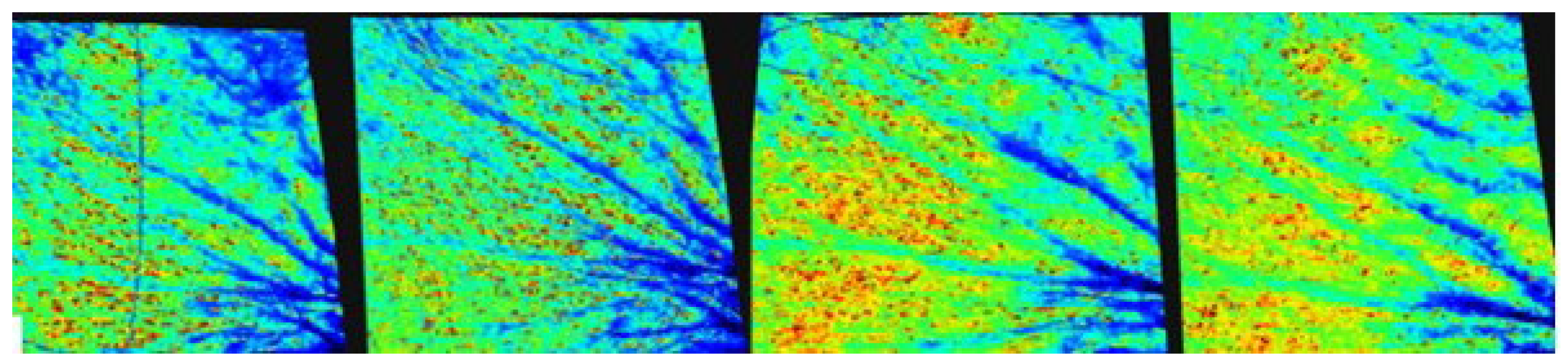
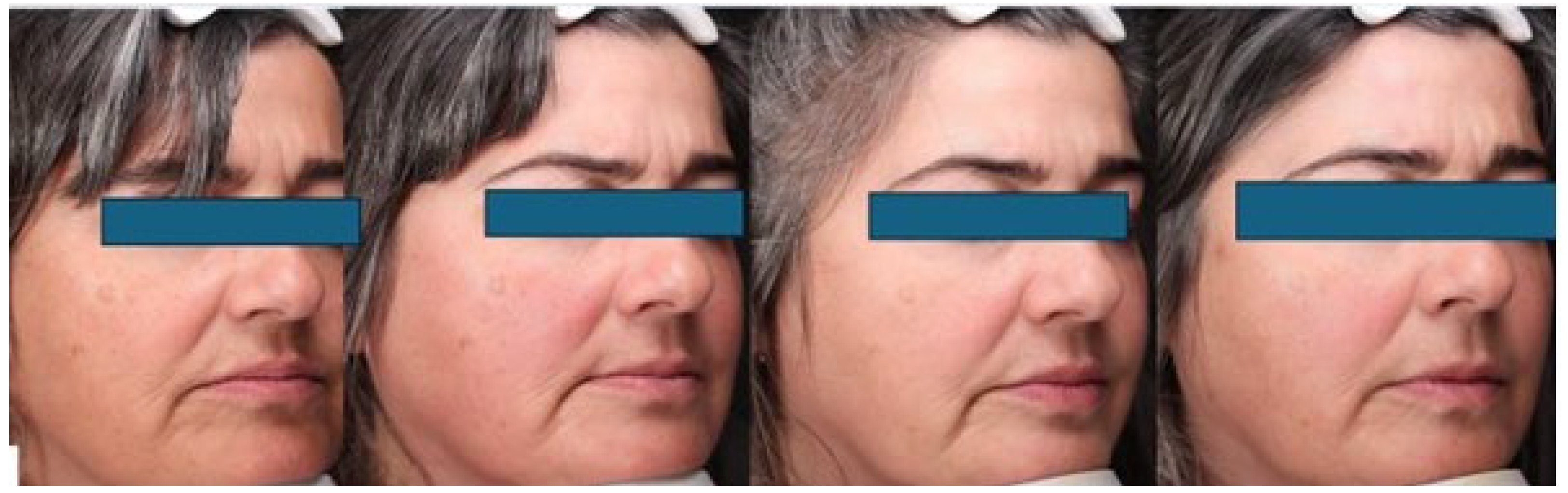
Figure 1.
VISIA picture’s example obtained from one of the patients. Depth and area of periorbicular wrinkles, as an example, can be addressed with VISIA 3D.
Table 1.
Protocol of topical treatment used in the study.
Clinical Assessments: Efficacy was assessed at baseline (D0), D30, D60, D90, and D180. We evaluated the same 21 females at D0, D30, D60, D90, and D180. No patients were lost for follow-up. To minimize inter-rater variability, a known source of bias in dermatological grading, all clinical assessments were performed by the same expert dermatologist throughout the study. While this introduces a potential for intra-rater bias, the consistency of a single evaluator is often preferred in pilot studies to ensure uniform application of grading criteria.
The following scales were employed:
Rao–Goldman Scale: A validated 5-point photonumeric scale used to assess wrinkles. The grades are defined as follows: Grade 1 (no wrinkles), Grade 2 (visible but fine wrinkles), Grade 3 (moderately deep wrinkles), Grade 4 (deep wrinkles with defined edges), and Grade 5 (very deep wrinkles with defined furrows).
Patient’s Global Assessment (PGA) and Investigator’s Global Assessment (IGA): These 5-point scales evaluated the overall severity of photodamage and improvement. The scoring system was as follows: 0 (Clear/None), 1 (Minimal), 2 (Mild), 3 (Moderate), and 4 (Severe).
Instrumental Analysis Objective changes in skin parameters were measured using the VISIA® Complexion Analysis System (Canfield Scientific, Parsippany, NJ, USA). This system utilizes multispectral imaging and standardized positioning to capture high-resolution facial images. The analysis included the evaluation of:
Wrinkles: Detected by their characteristic shadow patterns.
Texture: Analyzing skin smoothness and surface irregularities.
Spots and UV Spots: Using cross-polarized and UV lighting modes to visualize surface pigmentation and subsurface photodamage (accumulated melanin) not visible to the naked eye.
Porphyrins: UV fluorescence imaging to detect bacterial excretions in pores.
Baseline assessments, as previously stated, included a comprehensive evaluation of skin condition, including the severity of wrinkles and overall skin texture, as well as VISIA CR system ® photography and clinical parameters evaluated with VISIA ACR Generation 7, including texture, scars, erythema, wrinkles, depth of wrinkles, polarized and cross light for highlight hyperpigmentation, and irregularities in the skin tone.
The primary endpoint was to evaluate the percentage of patients evaluated as Rao–Goldman scale 2 at 180 days.
Follow-up visits were scheduled at 30, 90, and 180 days to monitor changes in skin condition and record any adverse effects, including digital photography, VISIA CR analysis, Rao–Goldman evaluation, patient global assessment (PGA), investigator global assessment (IGA), and adverse events assessment (erythema, burn, desquamation, and tightness). An additional telematic appointment for 120 days to assess adverse effects was done. All the evaluations were done for the same physician to reduce bias. The statistical evaluation was conducted in a blind way to prevent biases. The primary outcome measure was the percentage of improvement in the Rao–Goldman scale score (% of patients reaching a Rao–Goldman score of 2) while secondary outcomes included patient-reported satisfaction and the incidence of adverse effects.
To mitigate the inherent risk of intra-rater bias associated with this open-label design, an additional independent evaluation was conducted. Standardized high-resolution photographs taken with the VISIA system at each visit were evaluated by two independent dermatologists who were blinded to patient identity, treatment, and chronological time point of the photographs.
Statistical calculations were performed using SPSS software version 24.0. For ordinal efficacy variables, such as PGA, IGA, the Rao–Goldman scale, and the intensity of adverse effects at each measured time point, the non-parametric Wilcoxon test was used. For quantitative variables, the paired Student’s t-test or the Wilcoxon test will be employed, depending on the normality of the variables. A significant difference from baseline will be considered when the significance level is less than 5% (p ≤ 0.05).
3. Results
At the study’s inception, the baseline characteristics of the cohort reflected a population of 21 females with a mean age of 44.4 y (SD 7.69 y), with advanced signs of photoaging (Rao–Goldman 3 71.84% and Rao–Goldman 4 28.6%), presenting a significant therapeutic challenge. Physician assessment classified the vast majority of subjects (71.5%) as having moderately deep wrinkles, while a substantial subset (28.6%) presented with deep wrinkles. Only a marginal fraction—specifically, 4.8% of patients—rated their initial skin condition as mild.
The therapeutic intervention yielded rapid, perceptible results. By day 90, patient-reported outcomes indicated a favorable trajectory, with improvement acknowledged by 17 participants. Of these, 14.3% characterized the change as mild, while the remaining majority perceived a moderate to intense amelioration of their cutaneous condition (Figure 1).
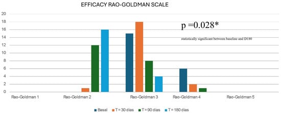
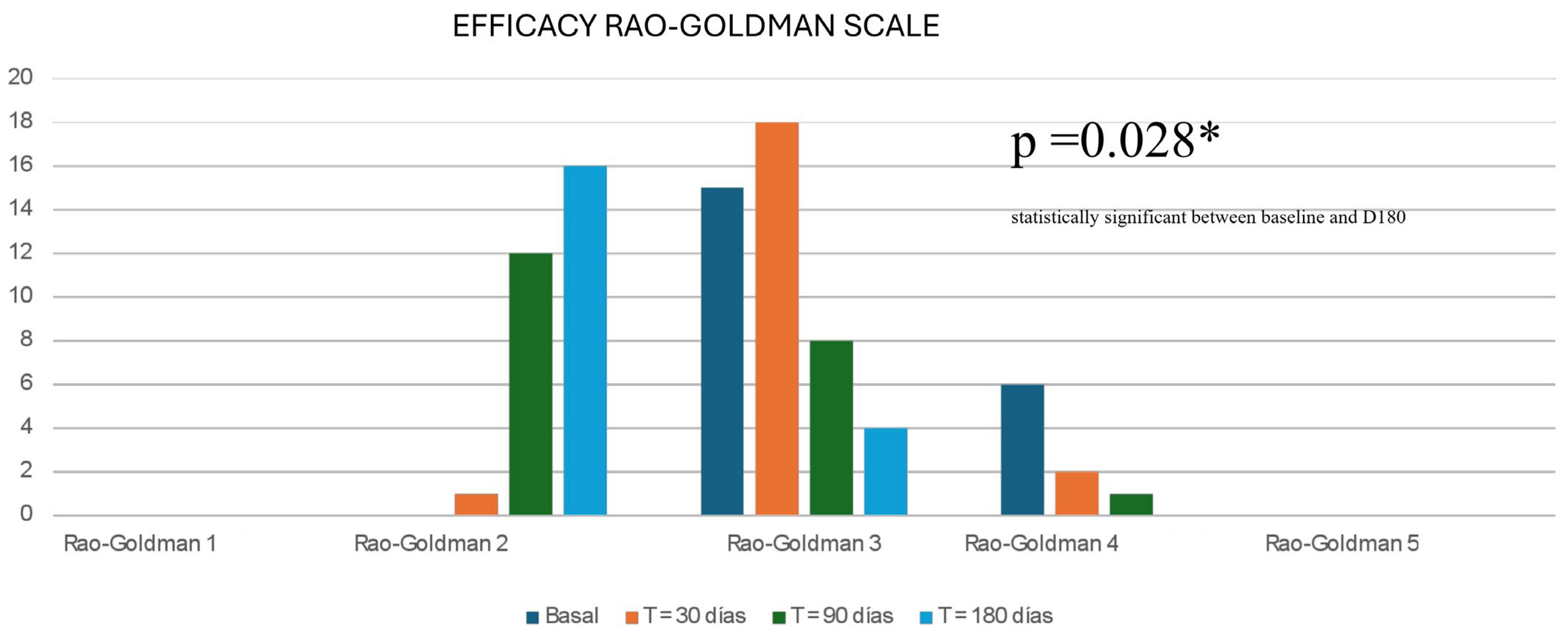
Longitudinal clinical evaluation revealed a progressive and statistically significant reversal of photoaging signs. Although all subjects presented with moderate-to-severe photodamage (Rao–Goldman score ≥ 3) at baseline, a remarkable shift in dermal quality was documented by day 180. The data demonstrated that 76.2% of patients transitioned from their baseline severity into the “fine wrinkle” (Grade 2) category. Consequently, the prevalence of moderately deep wrinkles persisted in only 19% of the cohort. This quantifiable reduction in rhytide depth was further substantiated by the Physician’s Global Assessment (PGA) scores, in which the investigator documented “significant” or “marked” global improvement in 85% of the study population (Figure 1, Figure 2 and Figure 3) (Table 2).
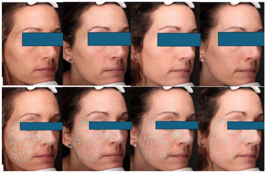
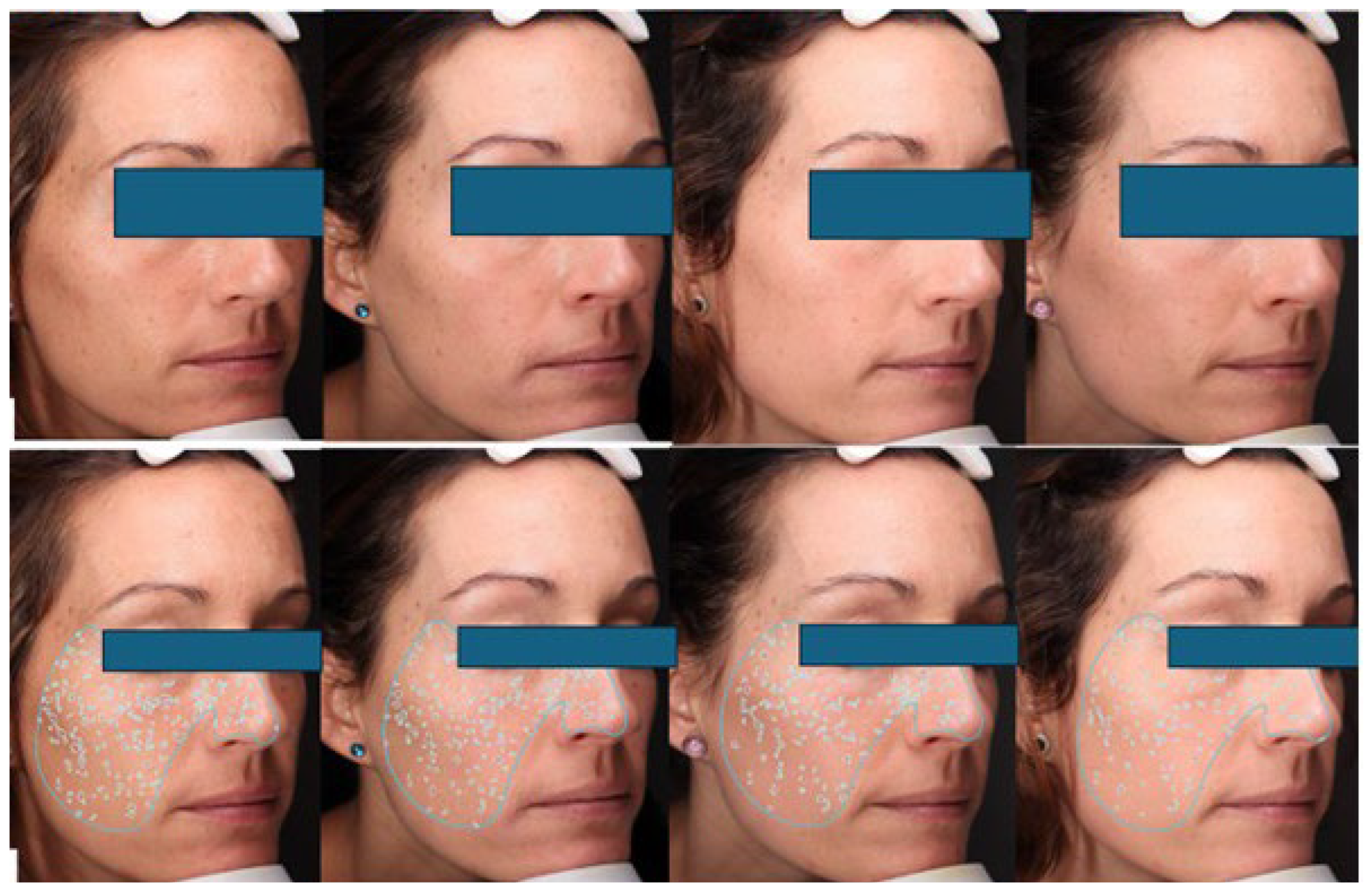
Figure 2.
VISIA pictures are standardized digital photographs. Upper row: The patient who shows improvement in texture and tone during the treatment, according to both blind raters. Botton row: Patient showing dark spot improvement according to both blind raters.
Figure 3.
VISIA pictures are standardized digital photographs. Improvement of tone, dark spots, and glow according to blind raters.
Table 2.
Expanded results of the Rao–Goldman scores in every visit.
At the conclusion of the treatment period, the improvement perceived by the investigator showed a statistically significant difference compared to baseline (T0) (p = 0.028), with 76% of women presenting mostly fine visible wrinkles (Figure 2). Thus, statistical analysis confirmed the robustness of these findings.
The alignment between the clinician’s objective grading and the patients’ subjective experience was notably strong, yielding a kappa correlation coefficient of 0.840 at day 180. Patient satisfaction was evident early in the treatment course; distinct from the investigator’s timeline, the improvement perceived by patients was significant after just the first month. At day 30, 80% of participants reported some degree of benefit, with over 60% classifying this early improvement as moderate.
Regarding instrumental analysis, repeated measures of the VISIA parameters did not yield statistically significant differences across the evaluated time points (Table 3). While this lack of statistical significance may be attributed to the limited sample size inherent in a pilot study, these instrumental data are reported strictly as non-significant trends. The automated analysis indicated a numerical reduction in absolute scores for the ‘Wrinkles’ and ‘Texture’ parameters. Other parameters, such as ‘spots,’ showed numerical increases. Furthermore, photographic evaluation by the two independent, fully blinded dermatologists identified a positive trend in the reduction in all wrinkle subtypes, as well as improvements in skin tone, dark spots, and radiance (Figure 2 and Figure 3). Therefore, although the instrumental analysis via VISIA® did not reach statistical significance to definitively corroborate the clinical observations, the observed quantitative trends parallel the clinical reports of skin surface smoothing and tone homogenization. These trends remain consistent with the known biological effects of retinol on epidermal turnover and collagen stimulation.
Table 3.
Table showing the VISIA mean data of spots, brown spots, texture, and wrinkles along the treatment.
Because all 21 patients attended every scheduled visit, strictly adhered to the treatment protocol, and experienced no loss to follow-up, the observed-case and intent-to-treat analyses were identical in this cohort. Although Fitzpatrick’s evaluation was not included in the material and methods, retrospective analysis showed a distribution equal to our general population (II in 4 patients; III in 11 patients; and IV in 6 patients).
Safety and Tolerability Profile
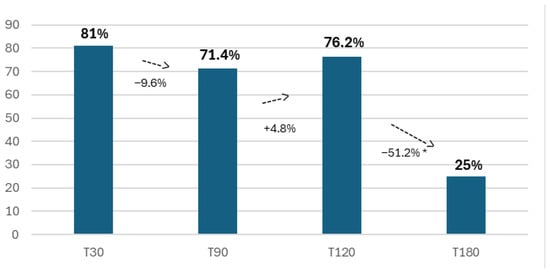
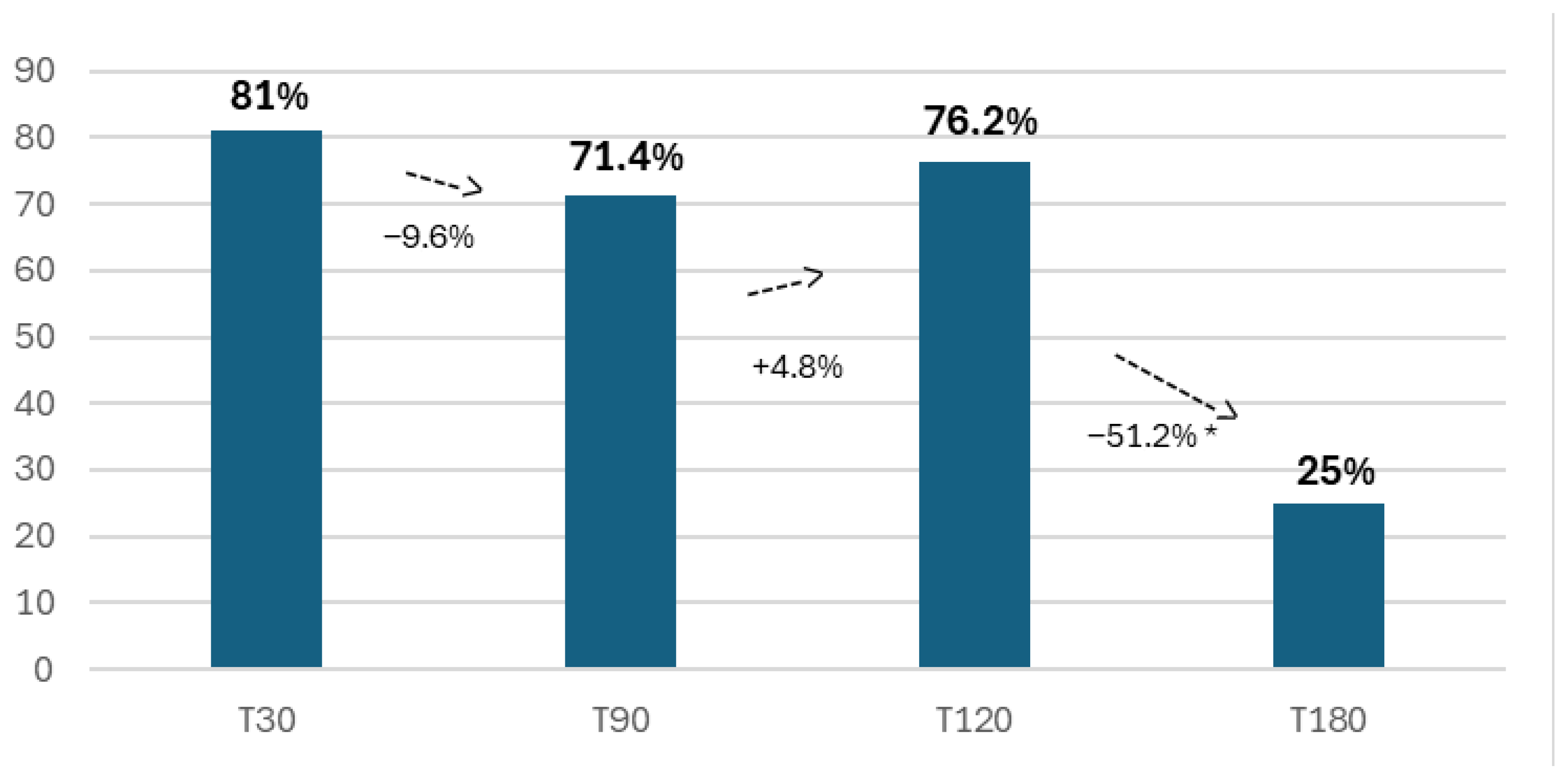
The safety profile observed in this study followed the characteristic “retinization” curve associated with high-potency retinoid therapy. Adverse events (AEs) were predominantly concentrated in the initial phase of the study. During the first four weeks, 81% of participants experienced at least one retinoid-associated effect, primarily manifesting as mild erythema, desquamation (peeling), and dryness. However, the implementation of a “step-up” dosing protocol proved highly effective in mitigating these effects over time. During the 120-day telephonic visit, we observed the accumulation of up to 76.2% of the total reported adverse events, indicating a decline in new incidents as the study progressed.
By the final visit (D180), the skin had successfully accommodated to the molecule; 76.2% of patients reported absolutely no adverse events (Figure 4 and Table 4) and only 5 patients reported any residual effects, and Rao–Goldman changes can be seen in Figure 5. This confirms the phenomenon of “retinization,” where tolerance is achieved after the initial barrier adaptation. Crucially, no severe adverse events necessitating study withdrawal were recorded, confirming the long-term tolerability of the formulation.
Figure 4.
Percentage of adverse effects reported during the treatment. * statistically significant.
Table 4.
Adverse events type and severity during every visit.
Figure 5.
Rao–Goldman scores of the entire cohort at different times of evaluation from basal time to the end of treatment (D 180). X2 score was done and was significant, p = 0.028 between baseline and 180 days is marked with * as it is statistically significant as written in the figure.
4. Discussion
Despite the increasing prevalence of anti-aging products containing retinoids, there remain significant knowledge gaps, and comparing studies is challenging due to variations in formulations, retinoid concentrations, types, study designs, and the lack of standardized objective scales [11,14,15,16,17]. (Table 5)
Table 5.
Comparison of articles testing different retinoids and their efficacy in improving photodamage-related problems.
It is important to use stabilized retinol, which is achieved with the use of specific carriers and vehicles. For this reason, the efficacy of retinol in the treatment of photodamage depends greatly on formulation and its mode of delivery to the target area. These two factors will be key to determining the efficacy and tolerance of the various retinoids present in the market. Our study addresses these issues by evaluating both patient and dermatologist perceptions, as well as incorporating objective measurements such as VISIA analysis and the validated Rao–Goldman aging scale.
Our study addresses these methodological inconsistencies by comprehensively evaluating both patient and dermatologist perceptions and by incorporating objective multimodal measurements—specifically, VISIA analysis and the validated Rao–Goldman aging scale. The accurate clinical assessment of photoaging requires to be standardized, reproducible instruments to mitigate the subjective variability inherent in human observation. In this context, the Rao–Goldman scale has emerged as a superior reference tool for quantifying rhytidosis and textural alterations compared to traditional global grading systems. Unlike purely descriptive classifications, the Rao–Goldman scale is a 5-point photonumeric instrument providing explicit visual references for each severity grade, ranging from the absence of wrinkles (Grade 1) to deep furrows with defined edges (Grade 5). This visual anchoring significantly reduces inter- and intra-rater bias by allowing the evaluator to match the patient’s clinical presentation against a standardized photographic baseline, rather than relying on memory. Historically, the Glogau scale has served as the “gold standard” for classifying actinic damage. However, its utility in modern clinical trials focused on specific cosmetic efficacy presents notable limitations. The Glogau system is a global assessment that aggregates multiple clinical signs—including dyschromia, keratosis, and the quality of makeup required to conceal defects—and distinguishes between wrinkles “in motion” versus “at rest.” Crucially, the Glogau scale stratifies patients by chronological age groups (e.g., Type I for 20–30 years; Type IV for >60 years), introducing a significant selection bias. This age dependence reduces precision when evaluating patients who exhibit severe photodamage at a younger age or, conversely, those with well-preserved skin in later life. By contrast, the Rao–Goldman scale isolates the specific variable of wrinkle morphology and depth from confounding factors such as pigmentation, keratosis, or chronological age. This specificity grants the scale superior sensitivity in detecting subtle yet statistically significant improvements in dermal quality following non-invasive interventions. While Glogau remains useful for broad surgical planning, Rao–Goldman provides the granular resolution necessary to monitor the progressive epidermal remodeling and collagen synthesis induced by topical retinoid therapy, making it the preferred instrument for longitudinal efficacy studies.
The VISIA® Complexion Analysis System represents a pivotal advancement in objective skin evaluation. Its multi-spectral approach allows for the comprehensive assessment of both surface pathology and subsurface photodamage that remains invisible to the naked eye. When compared to standard clinical photography—which often suffers from inconsistencies in ambient lighting and patient positioning—VISIA provides an automated, algorithm-driven scoring system within a controlled lighting environment. In our study, the numerical increase observed in ‘spots’ could be related to incidental sun exposure or the retinization process itself; however, these variations were not statistically significant, and patients were strictly instructed to use a daily SPF 50 per protocol. In clinical research, VISIA is instrumental for tracking longitudinal changes, enabling the precise monitoring of therapeutic outcomes from retinoids, laser resurfacing, or chemical peels by validating visual endpoints with quantitative percentile scores.
When compared to other measurement methods, VISIA offers distinct advantages, primarily regarding standardization and reproducibility. VISIA provides an automated algorithm-driven scoring, and some authors have studied the correlation between the Glogau Photoaging Scale to find that it correlates and indicates that aging appears to be an exponential process [18]. Furthermore, unlike standard clinical photography, which suffers from inconsistencies in ambient lighting and patient positioning, VISIA’s fixed head support and controlled lighting environment ensure that observed changes are due to treatment efficacy rather than environmental artifacts. Additionally, its ability to benchmark a patient’s results against a global database of age-matched peers enhances patient communication and compliance. Our numerical increase in spots could be related to incidental sun exposure or to the retinization process, although there were no statistically significant differences, and patients were supposed to use daily SPF50 per protocol.
However, the system is not without limitations. VISIA acts primarily as a 2D surface analysis tool [10], and it cannot assess functional biophysical properties such as elasticity or hydration, which require specific contact probes like the Cutometer® or Corneometer®, nor can it provide the cellular-level detail of confocal microscopy or invasive biopsies. Despite these limitations, its non-invasive nature, speed, and ability to visualize the “invisible” spectrum of photodamage make it a superior tool for holistic skin analysis in efficacy studies [19].
To rigorously validate the clinical impact of this specific delivery system and overcome the methodological heterogeneity prevalent in the literature, our study prioritized assessment tools that minimize subjective bias and ensure reproducibility. By integrating advanced digital imaging with a strict photonumeric grading system, we aimed to provide a granular evaluation of the retinoid’s biological activity [20]. This methodological approach allows for the precise quantification of epidermal remodeling and collagen synthesis, distinguishing true structural improvement from transient effects, as detailed in the following sections. Previous studies have demonstrated that combining several approaches yields a more comprehensive evaluation of skin aging, reinforcing the relevance of multimodal assessment in dermatological research [21].
Back in 1989, a randomized, double-blind, vehicle-controlled study with 0.05% tretinoin [all-trans-retinoic acid] cream showed a moderate improvement in 53% of patients treated with tretinoin and only 6% of patients without changes based on pre- and post-treatment photographs assessment, along with a statistically significant improvement of fine and coarse wrinkling, sallowness, looseness, and hyperpigmentation [14]. These early studies demonstrated the efficacy of tretinoin for topical skin rejuvenation. Later, the widespread inclusion of other cosmeceutical retinoid compounds has increased interest and research in the photorejuvenation field [22]. As in our series, where furrow depth was evaluated with VISIA, they were not able to find statistically significant differences using computerized analysis of the depth of the crow’s feet region wrinkles [14].
Although most retinoids used in topical applications must be converted to tretinoin after application, raising questions about their efficacy compared to tretinoin, there is substantial evidence that retinol can improve various cutaneous signs related to photoaging. Tretinoin is cost-effective but more irritating and photolabile than other retinoids [23].
Retinol is also better tolerated than tretinoin, a finding crucial for patient compliance and satisfaction [24].
Regarding differences between doses, a randomized, parallel control checking the efficacy of retinol 0.15 and 0.30 in 20 patients each showed an overall improvement of 30.2% and 31% and a facial wrinkle decrease of 29.1 and 32.2% [16]. Despite the fact that we have used different types of measures and a higher concentration of retinol, 0.5%, it is worthy to highlight a 76.2% improvement in our cohort. As most studies comparing topical therapies with tretinoin are of poor quality, retinoid precursors can be considered as a second-line option for anti-aging treatment [25] (Table 4).
The results of this study reinforce the efficacy of topical retinol as a potent anti-aging agent, comparable to the findings reported in larger clinical trials. Our observation of significant wrinkle reduction aligns with the mechanism proposed by Draelos et al., who demonstrated that novel retinol formulations can induce collagen synthesis and epidermal thickening to a degree like prescription tretinoin, but with a better safety profile [3,8,26].
The critical challenge in retinoid therapy is balancing efficacy with tolerability [27]. The initial irritation observed in our cohort is consistent with the pharmacological action of retinoids, which initially disrupts the stratum corneum barrier function before strengthening it. Truchuelo et al. similarly reported that side effects such as peeling and tightness are transient and peak during the first weeks of treatment, subsiding as the skin’s tolerance threshold increases [27]. Our data confirms this trajectory, validating the necessity of a progressive escalation protocol (the “retinization” phase) to ensure patient compliance.
Furthermore, the improvement in VISIA texture scores results is consistent with the formulation effectively stimulating keratinocyte proliferation, leading to a smoother skin surface. This “polishing” effect, combined with the dermal remodeling as suggested by the Rao–Goldman score improvements, addresses both the epidermal and dermal components of skin aging. Unlike invasive procedures that address volume or muscle activity, topical retinol is thought to improve the intrinsic quality of the skin envelope, making it an essential component of a holistic rejuvenation strategy, and this tested formulation’s effects align with previous knowledge on topical retinoids.
Finally, the use of combination therapy, by formulating at least two active compounds, has proven to be advantageous from the perspective of including a multimodal mechanism of action [8]. In photorejuvenation, there is a clinical study comparing AHA-ret formulation with retinol 1% regarding the improvement in fine lines and wrinkles, which was higher at 12 weeks (33.3%), and improvement of erythema, dyschromia, and pore size was 28.6, 27.6, and 25% [15]. Retinol 1% showed similar data, and tretinoin was almost 10% more effective than the other two products [15]. The results of our study indicate that the new retinol formulation is effective in reducing the signs of photoaging, with a significant improvement in the Rao–Goldman scale scores. Notably, as early as week 20, one out of three participants reported moderate to severe improvement in their skin condition, with a high degree of satisfaction. The incidence of adverse effects decreased over time, suggesting that the formulation is well-tolerated with continued use and with the retinization scheme [28].
The strong correlation between patient and investigator assessments in our study reinforces the reliability of the observed improvements. The statistical significance observed—despite the small sample size—may be attributed to the specific stability of the encapsulated retinol formulation and the strict adherence to the “step-up” application protocol, which minimized dropout rates, which were zero in our cohort. The reduction in adverse effects over time may be attributed to the process of retinization, where the skin gradually adapts to the retinol treatment [28]. This finding is consistent with previous studies that have shown a decrease in retinoid-associated irritation with prolonged use.
Vitamin A [retinol] is crucial for various cellular processes [8], and either oral or topical retinoids are contraindicated in pregnant women due to their potential to cause abnormal embryonic development [23,29]. While the systemic absorption of topically applied retinol is limited and epidemiological data have not definitively confirmed a causal link to birth defects, the theoretical risk necessitates a strict contraindication as a precautionary measure in clinical dermatology [30]. In our study, we excluded women who were pregnant, breastfeeding, or planning to conceive to ensure safety.
This absolute restriction creates a significant therapeutic void for patients seeking to maintain skin rejuvenation regimens during gestation, and although chemically distinct from the vitamin A family, bakuchiol functions as a functional retinoid analog [31] that regulates a gene expression signature similar to that of retinol, including the upregulation of type I and IV collagen and the stimulation of aquaporin-3. Crucially, bakuchiol achieves these anti-aging effects without binding to retinoic acid receptors (RARs), thereby circumventing the teratogenic potential and the classic irritative profile associated with retinoids [31]. Consequently, bakuchiol represents a promising, safety-conscious therapeutic option for managing photoaging in pregnant or lactating women who are otherwise precluded from using standard retinoid therapies. New formulations like the one used in the study are important, as optimizing retinoid drug delivery systems has been postulated as a means of maximizing efficacy, durability of response, and specificity [24,26].
The primary limitations of our study include a small cohort size and the absence of a placebo control group, although the open-label design is standard for pilot efficacy studies. Additionally, the single-center nature of the trial and the exclusive inclusion of female participants may restrict the generalizability of our results to broader populations. As the patients used a treatment protocol including mandatory daily SPF and a replenishing cream as well as the retinoid, the degree of improvement could be related to the entire treatment protocol adherence. Despite these constraints, the study methodology incorporated significant strengths to mitigate bias, including the blinded analysis of data and the consistent evaluation of all patients by a single investigator at each visit, ensuring homogeneity. Furthermore, we considered the observed improvements in texture and hyperpigmentation as valid surrogate markers for enhanced molecular pathways related to photoaging.
5. Conclusions
In conclusion, the new retinol formulation tested in this pilot study seems to show significant clinical efficacy in improving signs of photoaging, along with a favorable safety profile. The reduction in adverse effects over time and the high level of patient satisfaction suggest that this formulation could be a valuable topical option to prevent and treat photoaging. Further studies with larger sample sizes, histological analysis to directly quantify collagen neo-collagenesis, and longer follow-up periods are warranted to confirm these findings and explore the long-term benefits of this retinol formulation.
Author Contributions
Conceptualization, M.L.-V. and C.E.; methodology, M.L.-V. and C.E.; formal analysis, M.L.-V.; investigation, M.L.-V. and C.E.; resources, M.L.-V., M.V. and C.E.; data curation, M.L.-V. and C.E.; writing—original draft preparation, M.L.-V.; writing—review and editing, M.L.-V., M.V. and C.E.; funding acquisition, M.V. and C.E. All authors have read and agreed to the published version of the manuscript.
Funding
This was an investigator-initiated study where the product was supplied by the manufacturer, but clinical execution and data analysis were conducted independently. The Article Processing Charges (APC) for this publication were funded by Cantabria Labs.
Institutional Review Board Statement
The study was conducted in accordance with the principles of the Declaration of Helsinki. Formal Institutional Review Board (IRB) or Ethics Committee approval was waived because the intervention consisted solely of the topical application of a cosmetic product already commercialized in our country, falling outside the scope of clinical pharmacological trials. Written informed consent for study participation and photographic documentation was obtained from all subjects.
Informed Consent Statement
Informed consent was obtained from all subjects involved in the study. Written informed consent has been obtained from the patient(s) to publish this paper.
Data Availability Statement
Data may be shared upon request.
Conflicts of Interest
M.L.V. has worked in clinical trials or as a speaker with Amgen, Celgene, Abbvie, Almirall, Novartis, Leo, Lilly, UCB, Takeda, and Kyowa Kirin. Maria Vitale is a worker at Cantabria labs. The funders had no role in the design of the study; in the collection, analyses, or interpretation of data; in the writing of the manuscript; or in the decision to publish the results. All authors declare that the research was conducted in the absence of any commercial or financial relationships that could be construed as a potential conflict of interest.
References
- Morgado-Carrasco, D.; Gil-Lianes, J.; Jourdain, E.; Piquero-Casals, J. Oral Supplementation and Systemic Drugs for Skin Aging: A Narrative Review. Actas Dermo-Sifiliogr. 2023, 114, 114–124. [Google Scholar] [CrossRef] [Scilit] [PubMed]
- Fabi, S.G.; Firsowicz, M.; Kamrani, P.; Draelos, Z.D.; Dayan, S.H. Round Table Discussion: Aesthetic Treatment Considerations for the Perimenopausal & Menopausal Patient. J. Cosmet. Dermatol. 2026, 25, e70626. [Google Scholar]
- Draelos, Z.; Bogdanowicz, P.; Saurat, J.H. Top weapons in skin aging and actives to target the consequences of skin cell senescence. J. Eur. Acad. Dermatol. Venereol. 2024, 38, 15–22. [Google Scholar] [CrossRef] [Scilit]
- de Almeida Silva, L.; de Almeida, K.R.; Ortolani, C.L.F. Aesthetic and Psychosocial Impacts of Hyaluronic Acid Fillers in Patients with Retrognathic Profiles. Aesthetic Plast. Surg. 2025, 50, 762–773. [Google Scholar] [CrossRef] [Scilit] [PubMed]
- Perez Davo, A.; Truchuelo, M.T.; Vitale, M.; Gonzalez-Castro, J. Efficacy of an Antiaging Treatment Against Environmental Factors: Deschampsia antarctica Extract and High-tolerance Retinoids Combination. J. Clin. Aesthet. Dermatol. 2019, 12, E65–E70. [Google Scholar] [PubMed]
- Lee, C.; Chae, S.; Kwon, H.J.; Jeong, W.; Lee, K.K.; Chae, M. Non-Surgical Correction of Facial Asymmetry: A Narrative Review of Non-Surgical Modalities and Clinical Case Examples. J. Clin. Med. 2025, 14, 8828. [Google Scholar] [CrossRef] [Scilit]
- Rashid, Y.; Estela-Fernandez, C.; Sardar, A.; Deowan, D.; Safdar, O.M.; Issimdar, I.A.; Céspedes, S.C.T.; Dawood, N.; Ali, R.; Silva, A.B. Advances in Anti-aging Procedures: A Comprehensive Review of Surgical and Non-surgical Rejuvenation Techniques. Cureus 2025, 17, e92666. [Google Scholar] [CrossRef] [Scilit]
- Sorg, O.; Saurat, J.H. Topical retinoids in skin ageing: A focused update with reference to sun-induced epidermal vitamin A deficiency. Dermatology 2014, 228, 314–325. [Google Scholar] [CrossRef] [Scilit]
- Quan, T. Human Skin Aging and the Anti-Aging Properties of Retinol. Biomolecules 2023, 13, 1614. [Google Scholar] [CrossRef] [Scilit]
- Wang, K.; Yang, Z.; Ma, Y.; Liu, W.; Li, G.; Xu, X.; Li, Q. Retinoids in scarless skin regeneration: From molecular mechanisms to therapeutic strategies. Front. Cell. Dev. Biol. 2025, 13, 1683851. [Google Scholar] [CrossRef] [Scilit]
- Ramchatesingh, B.; Martinez Villarreal, A.; Arcuri, D.; Lagace, F.; Setah, S.A.; Touma, F.; Al-Badarin, F.; Litvinov, I.V. The Use of Retinoids for the Prevention and Treatment of Skin Cancers: An Updated Review. Int. J. Mol. Sci. 2022, 23, 12622. [Google Scholar] [CrossRef] [Scilit]
- Zhong, J.; Zhao, N.; Song, Q.; Du, Z.; Shu, P. Topical retinoids: Novel derivatives, nano lipid-based carriers, and combinations to improve chemical instability and skin irritation. J. Cosmet. Dermatol. 2024, 23, 3102–3115. [Google Scholar] [CrossRef] [Scilit]
- Baldwin, H.E.; Nighland, M.; Kendall, C.; Mays, D.A.; Grossman, R.; Newburger, J. 40 years of topical tretinoin use in review. J. Drugs Dermatol. 2013, 12, 638–642. [Google Scholar] [PubMed]
- Leyden, J.J.; Grove, G.L.; Grove, M.J.; Thorne, E.G.; Lufrano, L. Treatment of photodamaged facial skin with topical tretinoin. J. Am. Acad. Dermatol. 1989, 21, 638–644. [Google Scholar] [CrossRef] [Scilit]
- McDaniel, D.H.; Mazur, C.; Wortzman, M.S.; Nelson, D.B. Efficacy and tolerability of a double-conjugated retinoid cream vs 1.0% retinol cream or 0.025% tretinoin cream in subjects with mild to severe photoaging. J. Cosmet. Dermatol. 2017, 16, 542–548. [Google Scholar] [CrossRef] [Scilit] [PubMed]
- Zasada, M.; Budzisz, E. Randomized parallel control trial checking the efficacy and impact of two concentrations of retinol in the original formula on the aging skin condition: Pilot study. J. Cosmet. Dermatol. 2020, 19, 437–443. [Google Scholar] [CrossRef] [Scilit] [PubMed]
- Dhaliwal, S.; Rybak, I.; Ellis, S.R.; Notay, M.; Trivedi, M.; Burney, W.; Vaughn, A.R.; Nguyen, M.; Reiter, P.; Bosanac, S.; et al. Prospective, randomized, double-blind assessment of topical bakuchiol and retinol for facial photoageing. Br. J. Dermatol. 2019, 180, 289–296. [Google Scholar] [CrossRef] [Scilit]
- Oesch, S.; Vingan, N.R.; Li, X.; Hoopman, J.; Akgul, Y.; Kenkel, J.M. A correlation of the Glogau scale with VISIA-CR complexion analysis measurements in assessing facial photoaging for clinical research. Aesthetic Surg. J. 2022, 42, 1175–1184. [Google Scholar] [CrossRef] [Scilit]
- Korponai, J.; Remenyik, E.; Gellén, E. Revealing the Invisible: In Vivo Imaging for Photoaging Therapies-review of the literature. Photodiagn. Photodyn. Ther. 2025, 56, 105298. [Google Scholar] [CrossRef] [Scilit]
- Lee, C.M. Fifty years of research and development of cosmeceuticals: A contemporary review. J. Cosmet. Dermatol. 2016, 15, 527–539. [Google Scholar] [CrossRef] [Scilit]
- Matei, M.C.; Gracia-Cazaña, T.; García-Malinis, A.J.; Masferrer, L.B.; Almenara-Blasco, M.; Muelas Rives, I.; Gilaberte, Y. Correlation Between Objective and Subjective Methods in Skin Aging Assessment: A Comparative Approach. Photoimmunol. Photomed. 2026, 42, e70070. [Google Scholar] [CrossRef] [Scilit] [PubMed]
- Motamedi, M.; Chehade, A.; Sanghera, R.; Grewal, P. A Clinician’s Guide to Topical Retinoids. J. Cutan. Med. Surg. 2022, 26, 71–78. [Google Scholar] [CrossRef] [Scilit] [PubMed]
- Griffiths, T.W.; Watson, R.E.B.; Langton, A.K. Skin ageing and topical rejuvenation strategies. Br. J. Dermatol. 2023, 189, i17–i23. [Google Scholar] [CrossRef] [Scilit] [PubMed]
- Ferreira, R.; Napoli, J.; Enver, T.; Bernardino, L.; Ferreira, L. Advances and challenges in retinoid delivery systems in regenerative and therapeutic medicine. Nat. Commun. 2020, 11, 4265. [Google Scholar] [CrossRef] [Scilit]
- Siddiqui, Z.; Zufall, A.; Nash, M.; Rao, D.; Hirani, R.; Russo, M. Comparing Tretinoin to Other Topical Therapies in the Treatment of Skin Photoaging: A Systematic Review. Am. J. Clin. Dermatol. 2024, 25, 873–890. [Google Scholar] [CrossRef] [Scilit]
- Draelos, Z.; Tanghetti, E.; Guenin, E. Vehicle Formulation Impacts Tolerability and Patient Preference: Comparison of Tretinoin Branded Lotion and Generic Cream. J. Drugs Dermatol. 2022, 21, 875–880. [Google Scholar] [CrossRef] [Scilit]
- Truchuelo, M.T.; Pielasinski-Rodríguez, U.; Maria, V. Evaluation of Effectiveness and Tolerance of a Cosmeceutic Regimen Based on Topical Retinoids. J. Clin. Exp. Dermatol. Res. 2022, 13, 1000606. [Google Scholar]
- Draelos, Z.D.; Peterson, R.S. A Double-Blind, Comparative Clinical Study of Newly Formulated Retinol Serums vs Tretinoin Cream in Escalating Doses: A Method for Rapid Retinization With Minimized Irritation. J. Drugs Dermatol. 2020, 19, 625–631. [Google Scholar] [CrossRef] [Scilit]
- Brzezinski, P.; Zonda, G.I.; Hincu, M.A.; Vasilache, I.A.; Chiriac, A.; Ciuhodaru, M.I.; Borowska, B.; Borowska, L. A Multicenter Cohort Study Evaluating the Teratogenic Effects of Isotretinoin on Neonates. Children 2022, 9, 1612. [Google Scholar] [CrossRef] [Scilit]
- Tirmikcioglu, Z.; Tatliparmak, A. Face-to-face: Isotretinoin use and pregnancy outcome. Int. J. Dermatol. 2024, 63, 232–238. [Google Scholar] [CrossRef] [Scilit]
- Fanning, J.E.; McGee, S.A.; Ibrahim, O.I. Human Clinical Trials Using Topical Bakuchiol Formulations for the Treatment of Skin Disorders: A Systematic Review. J. Drugs Dermatol. 2024, 23, 239–243. [Google Scholar] [CrossRef] [Scilit] [PubMed]

Disclaimer/Publisher’s Note: The statements, opinions and data contained in all publications are solely those of the individual author(s) and contributor(s) and not of MDPI and/or the editor(s). MDPI and/or the editor(s) disclaim responsibility for any injury to people or property resulting from any ideas, methods, instructions or products referred to in the content. |
© 2026 by the authors. Licensee MDPI, Basel, Switzerland. This article is an open access article distributed under the terms and conditions of the Creative Commons Attribution (CC BY) license.