


Abstract
Ceramides are central to stratum corneum barrier organization and hydration. Beyond topical replenishment, ceramide-stimulating strategies increasingly aim to enhance endogenous ceramide biosynthesis, processing, and homeostatic remodeling in coordination with keratinocyte differentiation. In this review, we summarize the three major metabolic routes that shape epidermal ceramide output—de novo synthesis, salvage, and sphingomyelin hydrolysis—and organize representative bioactive ingredients by their primary molecular targets rather than by origin. Specifically, we map ingredients to tractable regulatory nodes, including transcriptional “liposensors” (PPAR/LXR), the induction of biosynthetic/elongation and processing enzymes (e.g., SPT, CerS3, ELOVL4), the provision of structural substrates and precursors (e.g., linoleate-rich lipids and glycosylceramides), salvage-pathway sphingoid bases that can reshape ceramide subclass output, and metabolic sensing/stress-response pathways centered on AMPK–mTOR–SIRT1/autophagy. Across these mechanisms, agents spanning botanical and fermented extracts, vitamins, sphingoid intermediates, lipid precursors, and pathway modulators (including autophagy-focused probes) have been reported to increase ceramide abundance and, in some contexts, favor barrier-relevant ultra-long-chain species and ω-O-acylceramides that support lamellar organization and the corneocyte lipid envelope. Translational and clinical studies in dry, sensitive, and aged skin generally associate such interventions with improved barrier function and reduced dryness. Aligning ingredient selection with defined biosynthetic and processing checkpoints—and verifying outcomes with lipidomics alongside clinical endpoints—may accelerate the development of evidence-based, ceramide-stimulating cosmetics.
1. Introduction
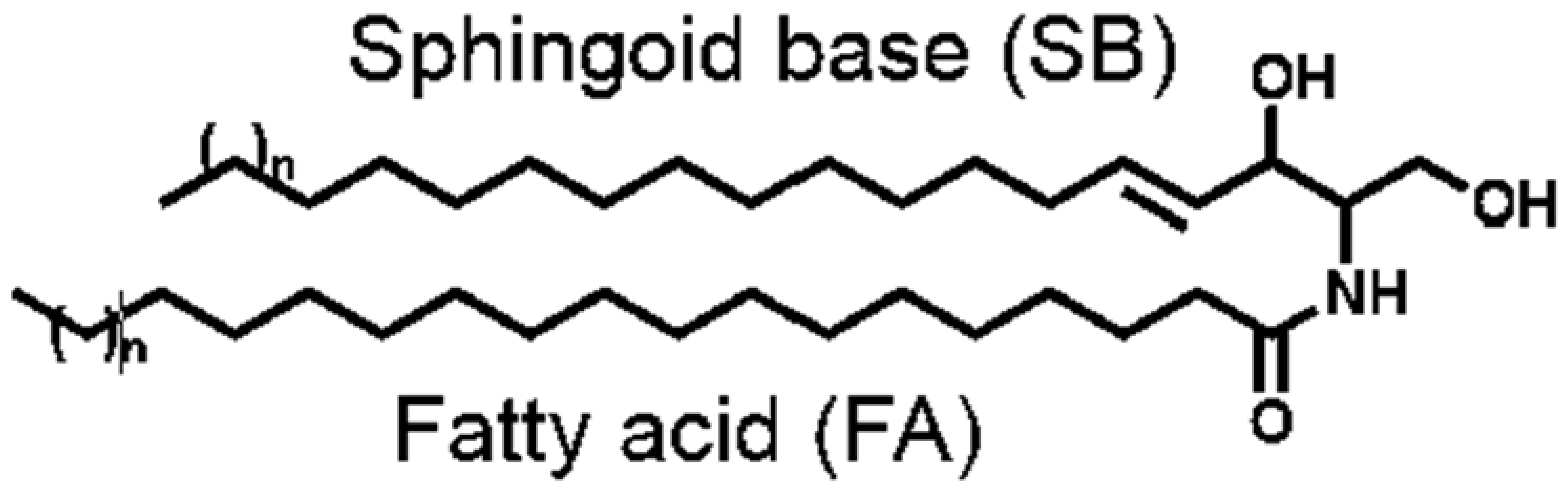
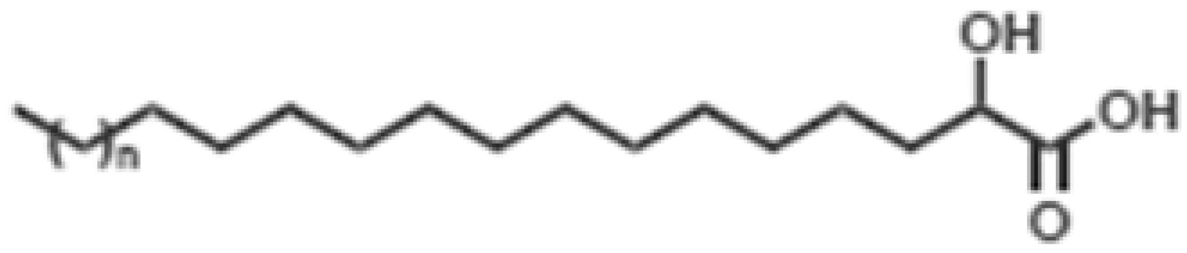
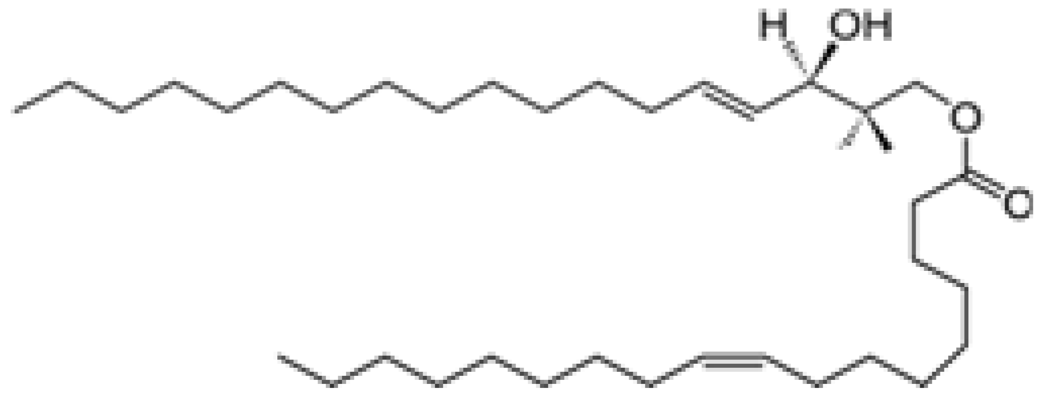
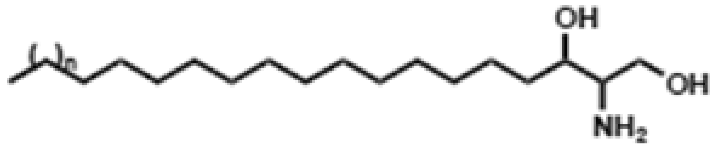
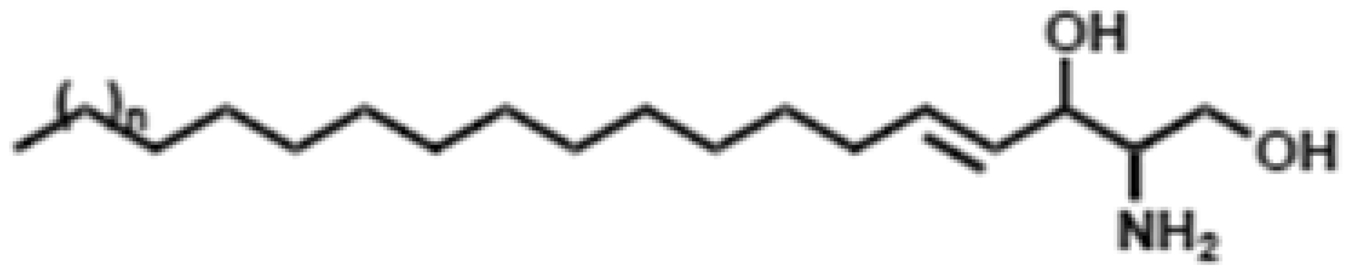
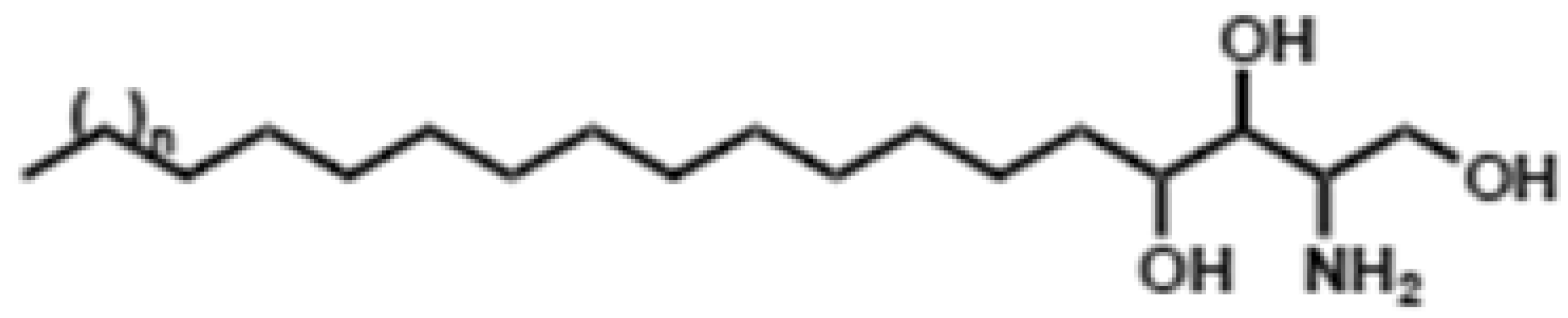
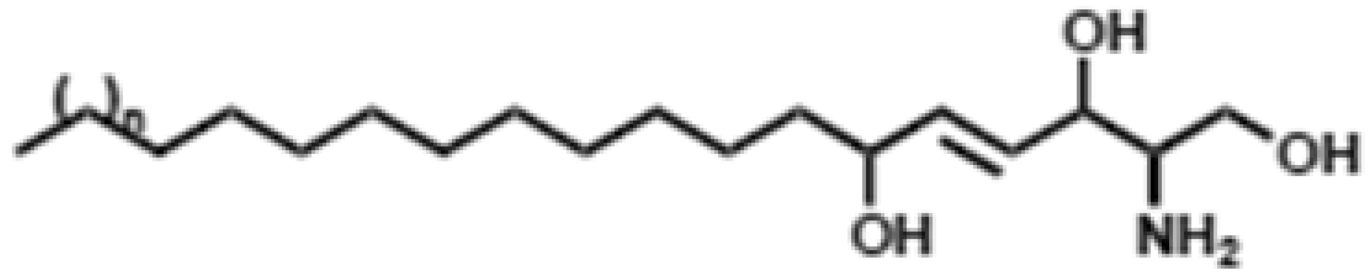
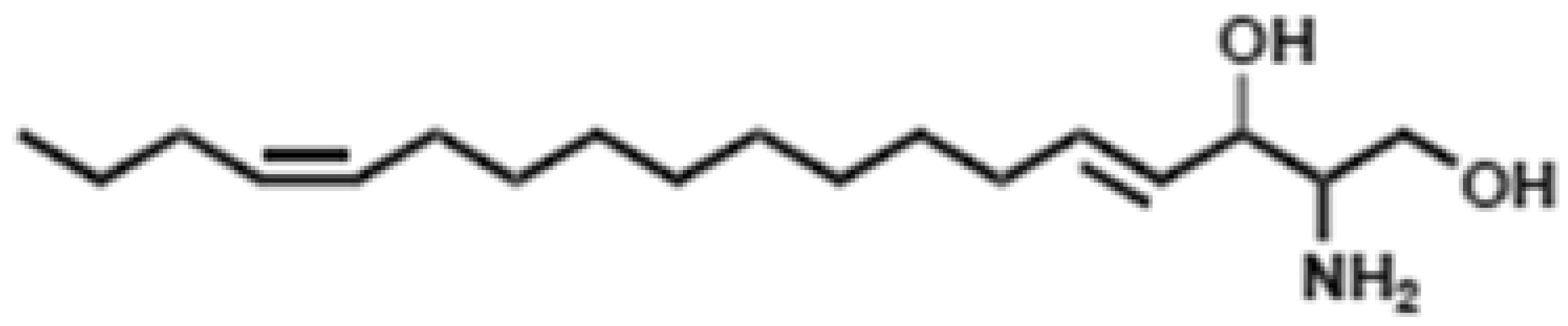
The skin barrier, primarily formed by the stratum corneum, is essential for protection against external insults and for limiting transepidermal water loss [1]. A compromised barrier manifests as common cosmetic concerns such as sensitivity, dryness, and dullness, undermining overall skin quality [2]. At the molecular core of maintaining this resilient barrier stand ceramides [1]. Ceramides, a class of sphingolipids, are recognized as central to epidermal permeability barrier function [1,3]. The molecular structure of ceramides is uniquely suited for forming the lamellar organization of the lipid matrix, which serves to prevent the loss of internal water and electrolytes and to block the ingress of harmful external substances [1]. Based on research demonstrating a significant correlation between the quantity and composition of stratum corneum ceramides and skin barrier function [4,5,6], there have been persistent efforts to develop ceramide-based moisturizers and topical agents [2,7]. However, the clinical performance of exogenous ceramides depends strongly on formulation factors—such as vehicle selection, solubilization, and lamellar organization—so the nominal ceramide content alone may not translate into consistent barrier improvement [8]. Beyond supplementation, strategies that enhance endogenous ceramide homeostasis are increasingly pursued as a more foundational approach to barrier repair. In this review, we synthesize evidence for diverse ceramide-supportive bioactives and organize them by their primary molecular targets and pathways—rather than by ingredient origin—spanning transcriptional liposensors (PPAR/LXR), key biosynthetic/elongation and processing enzymes (e.g., SPT, CerS3, ELOVL4), the provision of structural lipid substrates/precursors, salvage-pathway sphingoid intermediates, and metabolic sensing networks centered on AMPK–mTOR–SIRT1/autophagy. We integrate mechanistic, translational, and clinical findings to highlight effects on barrier-relevant ceramide subclasses (including ultra-long-chain and ω-O-acylceramides) and corresponding improvements in barrier function. Table 1 summarizes the major sphingoid bases and fatty acid moieties that constitute epidermal ceramides, along with the standard abbreviations used to classify ceramide subclasses in human skin [9,10,11]. This framework provides the structural context necessary for the mechanisms discussed in the following sections. To inform this narrative review, we performed a targeted literature search of PubMed/MEDLINE, Scopus, and Web of Science (supplemented by Google Scholar) for studies published through December 2025, using combinations of terms related to epidermal ceramide metabolism and barrier-relevant subclasses (e.g., ceramide, ω-O-acylceramide/acylceramide, CerS/CerS3, ELOVL4, SPT, glucosylceramide, sphingomyelinase, and corneocyte lipid envelope) and regulatory pathways (e.g., PPAR, LXR, AMPK, mTOR, autophagy, and SIRT1). We prioritized peer-reviewed primary studies and clinically relevant reports that assessed epidermal/stratum corneum ceramides (total abundance and/or subclass-resolved profiles) and barrier-related outcomes in human skin, relevant in vivo models, or keratinocyte-based systems, and additionally screened reference lists of key papers to identify further pertinent studies.
Table 1.
The structure and nomenclature of ceramides adapted from Refs. [9,10,11], licensed under CC BY 4.0. A ceramide is composed of a sphingoid base (SB) and a fatty acid (FA). The major ceramide classes in human epidermis are designated by combinations of SB and FA abbreviations. The numbers indicate their former numerical nomenclature.
2. Ceramide Homeostasis in Epidermis
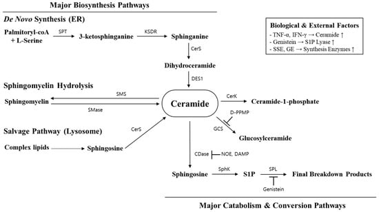
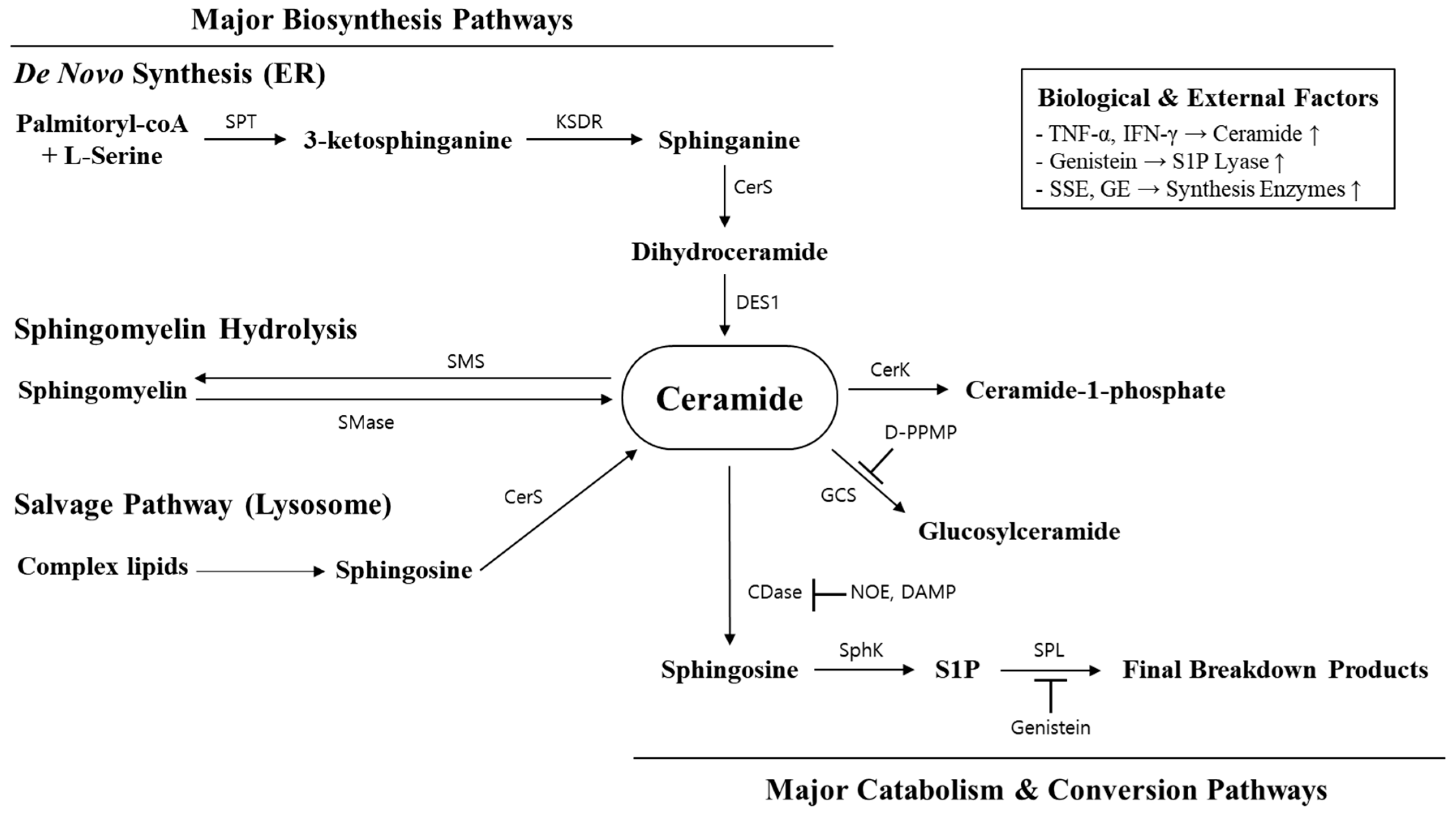
To contextualize the coordinated routes that sustain epidermal ceramide balance, Figure 1 provides an integrated schematic of ceramide homeostasis in the epidermis. The diagram summarizes the three principal entry routes—de novo synthesis in the endoplasmic reticulum, salvage recycling of sphingosine, and sphingomyelin hydrolysis—together with major interconversion and catabolic nodes that shape ceramide pools across relevant compartments. These interconnected reactions generate, remodel, and recycle ceramide species via key branch points, including conversion to ceramide-1-phosphate and glucosylceramide, as well as turnover through sphingosine/S1P metabolism. Collectively, this network supports the dynamic lipid demands of keratinocyte differentiation and stratum corneum barrier formation.
Figure 1.
Overview of Major Ceramide Metabolic Pathways and Regulation Mechanisms. This schematic illustrates ceramide as the central metabolic hub, outlining its primary biosynthesis (De novo, SM hydrolysis, salvage) and catabolism pathways, along with key enzymatic inhibitors and biological factors that modulate its homeostasis based on refs. [13,14]. ER, endoplasmic reticulum; SPT, Serine Palmitoyltransferase; KDSR, 3-Ketodihydrosphingosine Reductase; CerS, Ceramide Synthase; DES1, Dihydroceramide Desaturase 1; SMS, Sphingomyelin Synthase; SMase, Sphingomyelinase; CerK, Ceramide Kinase; CerS, Ceramide Synthase; GCS, Glucosylceramide Synthase; D-PPMP, D-threo-1-phenyl-2-decanoylamino-3-morpholino-1-propanol; CDase, Ceramidase; NOE, N-oleoylethanolamine; DMAP, 4-(Dimethylamino)-pyridine; SphK, Sphingosine Kinase; S1P, Sphingosine-1-phosphate; SPL: Sphingosine-1-phosphate Lyase; TNF-α, Tumor Necrosis Factor-alpha; IFN-γ, Interferon-gamma. Arrows indicate the direction of metabolic conversion; bar-headed lines indicate inhibition.
2.1. Biosynthesis Pathways
The biosynthetic pathways of ceramide can be broadly categorized into three main routes: de novo synthesis, the salvage pathway, and sphingomyelin hydrolysis. Each mechanism operates within distinct cellular compartments and is governed by unique regulatory controls [15,16,17] (Figure 1). The de novo pathway is initiated in the endoplasmic reticulum (ER) through the condensation of serine and palmitoyl-CoA by the enzyme serine palmitoyltransferase (SPT) [15]. In the salvage pathway, sphingosine, a metabolic product of complex sphingolipids, is reconverted to ceramide via ceramide synthase (CerS) [18]. The sphingomyelin hydrolysis pathway involves the enzymatic breakdown of sphingomyelin into ceramide by sphingomyelinase (SMase), counterbalanced by sphingomyelin synthase (SMS) [19].
2.1.1. De Novo Synthesis Pathway
The de novo synthesis of ceramide is a multi-step enzymatic process that occurs in the endoplasmic reticulum. It begins with the condensation of L-serine and palmitoyl-CoA by serine palmitoyltransferase (SPT) to form 3-keto-sphinganine (3-keto-SA) [15]. Subsequently, 3-keto-SA is reduced to sphinganine (also known as dihydrosphingosine) by 3-keto-SA reductase [16,20]. Sphinganine is then acylated by a ceramide synthase (CERS) via an amide bond with various fatty acids to produce dihydroceramide. Dihydroceramide can then be desaturated to yield ceramide [16,20]. The overall rate of the de novo pathway is determined by SPT activity, which is regulated not only by the concentrations of its substrates (serine and palmitoyl-CoA) but also by external factors [15,16].
2.1.2. Salvage Pathway and Sphingomyelin Hydrolysis
The salvage pathway is uniquely characterized by its ability to recycle sphingosine; a catabolic product derived from various complex sphingolipids [21]. Specifically, sphingosine generated from the breakdown of sphingolipids can follow one of two fates: it can be phosphorylated by sphingosine kinase to form sphingosine-1-phosphate, a potent signaling molecule, or it can be re-acylated by ceramide synthase to regenerate ceramide [18]. Among the complex sphingolipids, sphingomyelin—composed of a ceramide backbone linked to a phosphorylcholine headgroup—serves as a crucial reservoir for ceramide generation. Upon stimulation, sphingomyelin is hydrolyzed to release ceramide [19]. Notably, sphingomyelinase activity is not restricted to host enzymes; commensal Staphylococcus epidermidis can generate barrier-protective ceramides from sphingomyelin and thereby contribute to skin barrier homeostasis [22]. This ceramide can then be further metabolized by ceramidase into sphingosine and a free fatty acid or be re-utilized in other metabolic routes [23].
2.2. Catabolic Pathways
Ceramide turnover in epidermis is governed by a set of catabolic reactions that both remodel barrier lipids and generate signaling metabolites [20,21]. Complex sphingolipids internalized to lysosomes are sequentially hydrolyzed by ceramide, which is then deacylated by ceramidases to sphingosine; sphingosine can be recycled to ceramide (salvage pathway) or phosphorylated by sphingosine kinases (SphK1/2) to sphingosine-1-phosphate (S1P) [18,21,23]. S1P is irreversibly cleaved by ER-localized S1P lyase (SGPL1), representing the terminal exit from the sphingolipid pool [18,24]. This bidirectional flux establishes a ceramide–S1P “rheostat” with major consequences for cell fate and tissue homeostasis [18,24]. In addition, ceramide can be diverted into major branch pathways including ceramide-1-phosphate formation (CerK) and glucosylceramide synthesis (GCS) (Figure 1). Functionally, these catabolic arms shape both structure and signaling. Together, these interconnected reactions coordinate ceramide remodeling with the dynamic requirements of keratinocyte differentiation and barrier formation. However, epidermal barrier competence ultimately depends not only on intracellular ceramide flux but also on how ceramide precursors are extracellularly processed and structurally anchored within the stratum corneum—most notably through the formation of the corneocyte lipid envelope (CLE).
2.3. Extracellular Processing and Corneocyte Lipid Envelope (CLE) Formation
Beyond intracellular synthesis and turnover, a critical step for durable barrier assembly is the extracellular maturation and organization of ceramide-derived lipids in the stratum corneum [25]. During this process, ω-linoleoyloxyacylceramides undergo oxidative processing and subsequent cleavage of the linoleate moiety, which exposes ω-hydroxy ceramides as key substrates for corneocyte-envelope anchoring [26]. In particular, ω-hydroxy ceramides derived from acylceramide processing are covalently attached to corneocyte envelope proteins (including involucrin) through TG1-dependent pathways, thereby forming the corneocyte lipid envelope (CLE) [26,27]. This protein–lipid scaffold provides a hydrophobic surface that supports the alignment and stability of extracellular lamellar membranes, coupling ceramide metabolism to barrier architecture [25]. Accordingly, strategies that increase ceramide pools should be interpreted not only by changes in total abundance but also by the availability of ω-hydroxy ceramide substrates and the efficiency of CLE maturation [25,26,27,28].
3. Key Signaling Hubs for Ceramide Regulation
3.1. Transcriptional Regulation: PPARs and LXRs
PPARs (α/β[δ]/γ) are lipid-sensing nuclear receptors that coordinate keratinocyte differentiation with barrier-lipid synthesis, trafficking, and permeability barrier repair [29,30]. Activation of all three isoforms increases canonical differentiation markers—including involucrin, filaggrin, loricrin, and transglutaminase-1—in vitro and in vivo, and receptor-deficient models confirm their on-target relevance [29,31]. At the metabolic level, topical PPAR agonists enhance key transcriptional nodes that support ceramide and allied barrier lipid production, together with lipid trafficking and processing required for lamellar membrane assembly [29,32]. Isoform- and ligand-dependent differences have been reported; however, the unifying concept is that PPAR signaling couples differentiation to barrier lipid metabolism [29,33]. PPAR signaling also promotes lamellar body (LB) biogenesis and secretion: PPAR activation supports lamellar-body–associated lipid transport and secretion, facilitating the delivery of sphingolipid precursors for extracellular barrier maturation [29,34]. PPAR activation also enhances post-secretory lipid processing, increasing β-glucocerebrosidase activity and supporting stratum corneum acidification—key steps for the efficient conversion of glucosylceramides into ceramides and for lamellar-membrane organization [20,29]. These combined mechanisms accelerate permeability barrier recovery following acute disruption [33]. Several natural ingredients act through PPAR pathways. For example, oat lipid extract promotes keratinocyte differentiation and ceramide synthesis via PPAR signaling [35], and caffeic acid enhances differentiation through PPAR-α activation [36]. While PPARs can influence inflammatory tone, this review emphasizes their primary roles as transcriptional “liposensors” controlling differentiation-linked barrier lipid programs [21,29].
LXRs (LXR-α/β) are oxysterol-sensing nuclear receptors expressed across the epidermis [30]. As RXR heterodimers, LXRs regulate epidermal lipid metabolism and differentiation. Upon activation by oxysterols or synthetic agonists (e.g., 22(R)-hydroxycholesterol, TO901317), LXRs induce differentiation markers, restrain proliferation, and activate transcriptional programs that support cholesterol and sphingolipid handling relevant to barrier homeostasis [29]. LXR activation further enhances epidermal maturation and lipid-handling pathways, positioning LXR as an additional transcriptional lever within the multi-target framework for ceramide homeostasis [29,32]. Although most epidermal LXR research emphasizes cholesterol metabolism, LXR signaling intersects sphingolipid pathways at multiple nodes, underscoring its role as a central regulatory hub for ceramide homeostasis [29].
3.2. Cellular Sensing: Autophagy, mTOR, and AMPK
Autophagy is a lysosome-dependent quality control process that degrades damaged proteins and organelles, recycling substrates essential for homeostasis under stress [37]. mTORC1 suppresses autophagy and favors proliferative programs, whereas its restraint is permissive for differentiation-associated lipid handling [38]. Barrier disruption reduces ATP availability and increases oxidative stress, activating AMPK as a metabolic sensor [39]. Activated AMPK phosphorylates mTORC1 regulators (e.g., TSC2 and Raptor) and suppresses mTORC1 activity, shifting keratinocytes from proliferation toward a differentiation-permissive, lipid-anabolic state [39]. Sustained mTORC1 signaling can therefore be framed as a risk factor for impaired differentiation and disordered lipid assembly, while AMPK activation promotes barrier-oriented programs [39]. mTORC1 suppression permits ULK1-complex activation and initiates autophagic flux, supporting the intracellular trafficking required for lipid handling during repair [37,39]. In keratinocytes, autophagy modulation has been linked to lamellar body formation and lipidomic remodeling, connecting autophagic flux to barrier lipid delivery and maturation [40]. This framework supports a functional role for autophagy as a “buffer” that helps align lipid delivery and processing with the demands of differentiation and barrier formation [37,40]. In addition, autophagy—particularly lipophagy—can mobilize lipid stores by turning over lipid droplets, thereby supplying fatty acid substrates that may support ceramide synthesis and barrier lipid remodeling during repair [41]. Consistent with this axis, pharmacologic mTOR inhibition (e.g., rapamycin) activates autophagy and has been reported to increase epidermal ceramide levels in experimental settings [42]. Collectively, the AMPK-dependent restraint of mTORC1 and downstream autophagy provide a coherent axis linking energy status to lipid remodeling during barrier repair [37,39,40,41,42].
3.3. Keratinocyte Differentiation Signaling
Keratinocyte differentiation is governed by intersecting signaling networks that coordinate cell cycle exit with differentiation-linked barrier lipid assembly [43,44]. A major regulatory axis involves the Notch–p63 switch: Notch activation suppresses ΔNp63 to promote stratification and terminal differentiation, whereas impaired Notch signaling leads to hyperplasia and defective differentiation [43]. This transcriptional transition supports barrier assembly by up-regulating lipid-handling programs required for ceramide synthesis, lamellar body (LB) loading/transport, and extracellular lipid processing [12,15]. Calcium signaling constitutes a second major regulatory layer via the epidermal Ca2+ gradient and Ca2+-dependent signaling pathways that rise toward the granular layer, engaging ER Ca2+ handling and downstream transcriptional modules (including MAPK/AP-1) to drive differentiation [44]. Consistent with this framework, calcium-induced MEK/ERK→AP-1 activation enhances the expression of differentiation markers (e.g., involucrin, filaggrin, loricrin) and selected downstream effectors such as Sema3A, while supporting vesicular trafficking processes required for LB maturation and secretion [44,45]. Protein kinase C (PKC) isoforms serve as additional regulators: PKCδ promotes growth arrest and KLF4-mediated involucrin transcription, whereas PKCη—activated by cholesterol sulfate—induces G1 arrest and differentiation [44]. Through these pathways, calcium and PKC signaling shape both structural differentiation (cornified-envelope formation, desmosomal remodeling) and lipid outcomes, enhancing the synthesis of barrier ceramides and optimizing post-secretory processing [15,20,44]. Differentiation signaling also intersects functionally with the metabolic hubs described above (PPAR/LXR and AMPK–mTOR–autophagy), thereby aligning transcriptional commitment with the lipid-synthetic and trafficking capacity needed for barrier repair [15,20]. This convergence amplifies lamellar body biogenesis and enhances the β-glucocerebrosidase-mediated conversion of glucosylceramides into ceramides. The result is a synchronized progression—from cell-cycle exit to transcriptional commitment, vesicle maturation, and extracellular lipid remodeling—that ensures the orderly accumulation of barrier-specific ceramides and durable permeability barrier function [15,20,39,44].
To integrate the pathways discussed in Section 3.1, Section 3.2 and Section 3.3, Table 2 summarizes the major signaling nodes that collectively regulate epidermal ceramide homeostasis and barrier function. This consolidated overview also provides a mechanistic framework for interpreting the bioactive ingredient actions described in Section 4.
Table 2.
Key signaling hubs regulating ceramide homeostasis in the epidermis.
4. Bioactive Ingredients Modulating Ceramide Metabolism
Recent studies have reported diverse bioactive ingredients that can modulate epidermal ceramide homeostasis and thereby support skin barrier function [8,18,47,48,49,50]. Rather than categorizing these agents by origin, they can be organized by their primary molecular targets, including (i) transcriptional “liposensors” regulating differentiation-linked lipid programs (e.g., PPAR/LXR), (ii) the induction of key biosynthetic/elongation enzymes (e.g., SPT, CerS, ELOVL4), (iii) the provision of structural substrates and precursors, (iv) salvage-pathway sphingoid intermediates that remodel ceramide subclass output, and (v) metabolic sensing and stress response pathways centered on AMPK–mTOR–SIRT1 [21,25,30,33,34,37,38,39,51,52]. Collectively, these mechanisms can enhance not only ceramide abundance but also the differentiation-coupled processing and extracellular organization of barrier lipids required for effective barrier recovery [5,21,26,27]. Table 3 summarizes representative ingredients by primary target and simplified mechanism, while indicating whether evidence is demonstrated or suggested to maintain mechanistic consistency across Section 4.
Table 3.
Bioactive ingredients regulating epidermal ceramide homeostasis by primary molecular target.
While the present framework prioritizes a mechanism-first organization, the source of a bioactive (e.g., plant-derived vs. fermentation-derived) can still influence translational interpretation, including compositional reproducibility, standardization, and the availability of in vivo evidence. Plant-derived extracts frequently comprise chemically diverse mixtures, for which batch-to-batch variability and marker-based standardization become key determinants of reproducibility, whereas fermentation-derived materials (including postbiotic- or metabolite-enriched preparations) may offer improved compositional consistency but still require the clear identification of active components and dose–response characterization. Importantly, the overall strength of in vivo/clinical support is shaped more by study design, endpoint selection (including subclass-resolved ceramide profiling), and product context than by ingredient origin alone; therefore, we retain the mechanism-first structure while noting origin-related practical considerations where relevant.
4.1. Transcriptional Liposensors (PPAR/LXR Axis)
PPARs (α/β[δ]/γ) and LXRs (α/β) function as epidermal transcriptional “liposensors” that couple keratinocyte differentiation to coordinated programs of barrier lipid synthesis, trafficking, and post-secretory processing [16,30,33]. Activation of these nuclear receptors upregulates differentiation-associated markers and lipid-handling pathways, thereby supporting lamellar body (LB) biogenesis/secretion and the maturation of extracellular lamellar membranes required for permeability-barrier repair [16,30,33]. Consistent with this framework, PPAR/LXR activation has been linked to the enhanced expression of lipid transport machinery such as ABCA12, which facilitates the delivery of glucosylceramide-rich cargo to LBs for subsequent extracellular processing [16,33,34]. Within this axis, oat (Avena sativa) lipids have been reported to engage PPAR-linked differentiation and lipid programs, conceptually supporting the coordinated synthesis/handling of barrier lipids and downstream processing; however, the mechanistic claims should be anchored to ingredient-specific experimental/clinical evidence [53]. Similarly, caffeic acid has been reported to associate with PPARα-linked differentiation signaling and may support differentiation-coupled lipid handling, but ingredient-specific data should be cited to substantiate directionality and endpoints (e.g., ceramide subclass output, LB markers, or barrier readouts) [54]. Because LXRs are primarily characterized as oxysterol-sensing nuclear receptors, LXR agonism is included here as a conceptual mechanistic node rather than a specific “ingredient” class [16,30,33]. In keratinocyte models, LXR activation engages transcriptional programs supporting cholesterol/fatty-acid/sphingolipid handling relevant to barrier homeostasis, and has been linked to lipogenic responses in HaCaT cells, highlighting pathway relevance at the cellular level [16,30,33,46]. Together, the PPAR/LXR axis provides a mechanistic framework to interpret bioactive ingredients that act upstream at the transcriptional level to restore barrier lipid programs and support ceramide homeostasis [16,30,33].
4.2. Enzyme Induction/Cofactors (Biosyn-Thetic & Processing Enzymes)
Beyond transcriptional liposensors, several bioactives have been reported to support ceramide homeostasis by acting on biosynthetic and processing enzyme nodes and/or differentiation-permissive conditions that indirectly favor lipid anabolism. Niacinamide increases SPT expression/activity and elevates ceramides together with other major stratum-corneum lipids (FFA and cholesterol), consistent with barrier-supportive clinical/biophysical outcomes [18,55]. Gentiana lutea extract upregulates CERS3 and ELOVL4 under stress/differentiation contexts, conceptually supporting VLC/ULC ceramide production [47], while strawberry seed extract (tiliroside) has been reported to increase SPT/CerS expression and promote ceramide biosynthesis in epidermal equivalents [56]. Lactic acid (and its isomers) has also been linked to increased keratinocyte ceramide synthesis and improved barrier-related parameters, although outcomes can be formulation- and pH-dependent [50]. For Eucalyptus leaf extract, enzyme node support has been proposed (e.g., the induction of ceramide-synthesis/processing-related transcripts in keratinocytes has been described in the literature), but mechanistic attribution can vary by preparation and formulation; therefore, overly specific single-enzyme claims should be avoided unless directly supported [57]. In this context, hesperidin is better framed as a barrier/differentiation-supportive flavonoid: topical hesperidin improved permeability barrier function and epidermal differentiation in murine skin (reported), which may secondarily support lipid organization rather than serving as a direct “enzyme activator” [58]. Finally, fermented citrus peel extract provides a closer match to an “early-program/enzyme-node” entry: fermented dried Citrus unshiu peel extracts induced filaggrin and SPT expression in HaCaT keratinocytes and increased hyaluronic acid production (reported), consistent with a differentiation-permissive, lipid-supportive program relevant to barrier moisturization [59,60]. In addition, ursolic acid, a plant-derived triterpenoid, has been reported to increase the ceramide content in human skin, including hydroxylated ceramide forms. Although the underlying molecular target(s) were not fully delineated in that study, this finding supports the broader concept that certain botanically derived small molecules may modulate ceramide homeostasis in vivo and should be interpreted alongside formulation context and endpoint specificity [61].
4.3. Precursors & Structural Substrates (Sub-Strate Supply/Acylceramide-CLE Axis Support)
In addition to transcriptional “liposensors” and enzyme induction strategies, substrate-level interventions can support epidermal ceramide homeostasis by supplying structural building blocks for ceramide synthesis and extracellular barrier assembly. Such approaches are particularly relevant for barrier-specific ceramides and their downstream architecture, where lipid availability and proper extracellular processing jointly determine lamellar organization and permeability barrier competence [8,21]. Oral plant-derived glucosylceramides (GlcCer) from dietary sources (e.g., rice, wheat, konjac) are commonly positioned as precursor-support actives. Conceptually, dietary GlcCer are hydrolyzed and remodeled during digestion/absorption, providing sphingoid precursors that can be delivered systemically and subsequently reacylated in the epidermis, thereby contributing to ceramide pools used for lamellar body cargo and stratum corneum lipid assembly [12,20]. While multiple human studies have reported improvements in hydration and/or reductions in TEWL after oral GlcCer supplementation, ingredient- and study-specific trial citations should be provided to substantiate clinical claims in this section [62]. Complementary to oral approaches, topical plant GlcCer and topical ceramide-based formulations provide direct structural supplementation that can improve lamellar organization and lipid order in the stratum corneum, with outcomes that depend strongly on formulation variables (vehicle composition, delivery system, and the balance of lipid classes/subclasses) [2,8]. In barrier-impaired skin, ceramide-dominant topical regimens have demonstrated clinically meaningful improvements in permeability barrier function, supporting the translational relevance of structural lipid replacement when appropriately formulated [63]. Within this substrate-focused framework, evening primrose oil (EPO)—as a linoleate/γ-linolenic acid source—can be discussed primarily on biochemical grounds: essential fatty acid availability is integral to the chemistry and processing of ω-linoleoyloxyacylceramides and the downstream steps that enable corneocyte lipid envelope (CLE) maturation [5,21,26]. However, if the manuscript intends to claim that EPO directly increases ω-O-acylceramide levels or improves CLE maturation in vivo, this should be supported by ingredient-specific biochemical/clinical evidence; otherwise, EPO should be framed as a substrate rationale rather than a demonstrated ω-O-acylceramide enhancer [64].
4.4. Salvage Substrates/Sphingoid Intermediates (Ceramide Subclass Remodeling)
Beyond transcriptional and enzyme induction routes, the substrate-level supplementation of sphingoid intermediates provides a practical strategy to modulate epidermal ceramide pools through the salvage pathway. In differentiating keratinocytes, exogenous sphinganine, sphingosine, and phytosphingosine can be reacylated by ceramide synthases and thereby remodel the distribution of ceramide subclasses rather than uniformly increasing total ceramide abundance, with outcomes depending on the supplied long-chain base species and differentiation context [20,24]. Mechanistically, these intermediates enter a recycling node that can shift the balance among dihydroceramides, ceramides, and phytoceramides and can secondarily influence downstream processing steps relevant to barrier assembly [20,24]. Importantly, available data suggest species- and pathway-selective effects. For example, phytosphingosine has been reported to preferentially enhance phytoceramide-related outputs, including the induction of enzymes linked to phytoceramide production (e.g., DES2), indicating that salvage substrates may bias ceramide subclass composition rather than acting as a generic “ceramide booster” [65]. Given that barrier competence is tightly associated with qualitative features (subclass distribution/chain-length patterns) as well as quantity, salvage-substrate approaches should be interpreted using subclass-resolved lipidomics when possible, and claims should remain limited to the specific ceramide shifts supported by the cited experiments [20,24,65]. Exogenous ceramide itself has also been reported to function as a precursor/modulator for endogenous ceramide synthesis and keratinocyte differentiation programs, suggesting a potential “feed-in” route to endogenous remodeling under controlled conditions [49]. However, because topical ceramide delivery is highly vehicle- and penetration-dependent, this concept is best framed as a context-dependent modulator rather than a uniform enhancer of endogenous biosynthesis across formulations [49].
4.5. Metabolic Sensing and Autophagy Modulators (AMPK–mTOR–SIRT1)
A complementary “multi-target” axis focuses on metabolic sensing and intracellular quality control, where autophagy and nutrient-sensing pathways intersect lipid handling during differentiation and barrier repair. In keratinocytes, modulation of autophagic flux has been linked to changes in lipid handling and barrier-relevant remodeling, supporting the concept that autophagy can act as a buffer aligning lipid processing and delivery with the demands of terminal differentiation [37,41]. Within this framework, mTORC1 inhibition represents a mechanistically clear experimental lever. Rapamycin, as a canonical mTOR inhibitor, increases autophagic flux and has been reported to promote ceramide synthesis in skin models, including work implicating downstream signaling (e.g., TGF-β/Smad) in the observed ceramide-related changes [42]. Because mTOR activity also governs epidermal homeostasis and repair programs at the tissue level, rapamycin is best discussed as an experimental proof-of-concept for an mTOR-dependent shift toward autophagy and lipid remodeling rather than a simple “cosmetic ingredient” analogue [38,42]. Among polyphenols, resveratrol provides an example where sphingolipid signaling intersects barrier defense: it has been shown to stimulate sphingosine-1-phosphate (S1P) signaling linked to cathelicidin production in human skin, indicating a route by which sphingolipid metabolism can connect to innate immune function [66]. While resveratrol is often discussed in broader AMPK/SIRT1/autophagy contexts, mechanistic statements in this review should remain anchored to the specific cited endpoints (e.g., S1P–cathelicidin signaling) unless additional ingredient-specific autophagy/ceramide data are cited [66]. Finally, SIRT1-linked autophagy activation has been reported to support barrier formation through ceramide-synthase-dependent mechanisms. Activation of SIRT1 enhances epidermal permeability barrier formation via CerS2/3-dependent pathways and is associated with increases in ultra-long-chain ceramide outputs relevant to barrier integrity [51]. Consistent with this mechanistic direction, a topical “autophagy activator” clinical study reported functional skin outcomes compatible with improved oxidative/aging-related parameters, supporting the translational plausibility of SIRT1/autophagy-linked strategies [67]. Collectively, these examples position metabolic sensing and autophagy as a distinct intervention layer that may influence ceramide quality, trafficking, and barrier maturation, complementing the transcriptional and enzyme-induction strategies described above [37,41,51,67].
5. Clinical Implications and Future Directions
5.1. Applications in Dry, Sensitive, and Aged Skin
Ceramides are central determinants of epidermal barrier competence, and barrier performance reflects not only total ceramide abundance but also chain length distribution and subclass composition [5,14,48]. Altered ceramide profiles—such as the depletion of very-long-chain species and disturbances in acylceramide-related fractions—have been reported in xerotic, atopic, and aged skin, aligning lipid quality with clinical barrier fragility. In barrier-compromised dermatoses (e.g., atopic dermatitis/eczema), ceramide-containing regimens have shown consistent improvements in symptoms and barrier-related endpoints in systematic reviews and randomized trials, providing translational support for ceramide-dominant barrier care strategies in cosmetically relevant dry or sensitive skin settings [63,68,69]. In parallel, “ceramide-boosting” actives such as niacinamide can augment de novo lipid synthesis (including ceramides together with cholesterol and free fatty acids), supporting a multi-target approach that combines topical lipid replacement with the restoration of endogenous biosynthetic capacity [18,55]. In aged or post-menopausal skin, where ceramide quantity and chain length profiles may decline, combining multi-chain ceramide supplementation with mechanism-based bioactives offers a rational framework to improve persistent dryness and barrier resilience [2,13,70]
5.2. Importance of Lipidomics for Mechanistic and Clinical Validation
Advances in lipidomics enable the high-resolution profiling of stratum corneum ceramide species from minimally invasive tape-stripped samples, supporting mechanistic interpretation beyond total lipid levels. Lipidomic shifts in ultra-long-chain ω-O-acylceramides—and, where feasible, protein-bound ceramide/CLE-related readouts—can track with clinically meaningful changes in TEWL and hydration, linking molecular remodeling to barrier architecture. Accordingly, integrating lipidomics with biophysical (TEWL, capacitance) and standardized clinical endpoints strengthens the causal plausibility for active ingredients that claim ceramide modulation and barrier recovery [5,6,13,71].
5.3. Toward Evidence-Based Development of Ceramide-Targeting Cosmetics
The future development of ceramide-targeting cosmetics should align efficacy claims with evidence standards that emphasize reproducibility, appropriate controls, and validated endpoints. In the EU context, claims should satisfy the common criteria for the justification and substantiation of cosmetic claims, favoring well-designed human studies and transparent reporting. For products positioned around barrier repair, best-practice study features include pre-specified primary outcomes (e.g., TEWL), randomization/blinding where feasible, and multi-modal readouts that combine biophysical measures with molecular endpoints (e.g., ceramide subclass lipidomics). Finally, multi-target formulations that pair direct ceramide replenishment with bioactives modulating transcriptional liposensors (PPAR/LXR-linked programs) or metabolic sensing (AMPK–mTOR–SIRT1/autophagy networks) provide a mechanistic rationale for durable barrier support, provided ingredient-specific evidence matches the stated mechanism. From a formulation standpoint, the practical translation of ceramide-stimulating actives requires attention to chemical stability and compatibility with the vehicle. Lipid precursors and unsaturated components may be susceptible to oxidation, while certain small molecules and peptides can exhibit limited solubility or pH-dependent stability, and complex botanical/fermentation-derived mixtures may introduce variability in polarity, odor/color, and preservative demand. Accordingly, vehicle selection (e.g., emulsion type, solvent system, pH window), stabilization approaches (antioxidant systems, encapsulation, or controlled-release carriers), and packaging should be aligned with the physicochemical properties of the active(s) to maintain potency throughout shelf life. These considerations should be incorporated alongside efficacy endpoints to support robust product development [2,8,50,63].
6. Conclusions
Ceramide metabolism is a pivotal determinant of epidermal barrier integrity, governed not only by total abundance but also by the distribution of barrier-critical subclasses, including ultra-long-chain (ULC) and ω-O-acylceramides. Ceramide homeostasis is sustained through principal routes—de novo synthesis, salvage recycling, and sphingomyelin hydrolysis—together with post-secretory processing that supports acylceramide maturation and corneocyte lipid envelope (CLE) formation. These pathways are coordinated by transcriptional “liposensors” (PPARs/LXRs) and cellular sensing modules (AMPK–mTOR–autophagy), thereby linking keratinocyte differentiation to lipid synthesis, trafficking, and extracellular assembly. Accordingly, next-generation cosmetic development should prioritize multi-pathway strategies that combine balanced lipid replenishment with the mechanism-guided activation of endogenous programs, and should substantiate efficacy using lipidomics (subclass/chain-length shifts) alongside validated biophysical endpoints.
Author Contributions
Conceptualization, J.M., S.J., H.K. and G.N.; Methodology, J.M., S.J. and G.N.; Validation, S.J., H.K. and G.N.; Formal analysis, J.M.; Investigation, J.M. and S.J.; Resources, G.N.; Data curation, J.M. and S.J.; Writing—original draft preparation, J.M.; Writing—review and editing, J.M. and G.N.; Visualization, J.M.; Supervision, G.N.; Project administration, G.N. All authors have read and agreed to the published version of the manuscript.
Funding
This work was supported by the Regional Innovation Leading Enterprise Development (R&D) Program (S3460045) funded by the Ministry of SMEs and Startups (MSS, Korea).
Data Availability Statement
No new data were created or analyzed in this study. Data sharing is not applicable to this article.
Acknowledgments
During the preparation of this manuscript, the author used ChatGPT (OpenAI; GPT-5.1) for the purposes of language polishing and clarity improvement. The authors have reviewed and edited the output and take full responsibility for the content of this publication.
Conflicts of Interest
Author Sekyoo Jeong was employed by the Incospharm Corp. The author declares that the research was conducted in the absence of any commercial or financial relationships that could be construed as a potential conflict of interest. The remaining authors declare that the research was conducted in the absence of any commercial or financial relationships that could be construed as a potential conflict of interest.
References
- Moore, D.J.; Rawlings, A.V. The chemistry, function and (patho)physiology of stratum corneum barrier ceramides. Int. J. Cosmet. Sci. 2017, 39, 366–372. [Google Scholar] [CrossRef] [Scilit]
- Kahraman, E.; Kaykın, M.; Bektay, H.Ş.; Güngör, S. Recent Advances on Topical Application of Ceramides to Restore Barrier Function of Skin. Cosmetics 2019, 6, 52. [Google Scholar] [CrossRef] [Scilit]
- Green, C.D.; Maceyka, M.; Cowart, L.A.; Spiegel, S. Sphingolipids in metabolic disease: The good, the bad, and the unknown. Cell Metab. 2021, 33, 1293–1306. [Google Scholar] [CrossRef] [Scilit]
- Novotny, J.; Hrabalek, A.; Vavrova, K. Synthesis and structure-activity relationships of skin ceramides. Curr. Med. Chem. 2010, 17, 2301–2324. [Google Scholar] [CrossRef] [Scilit]
- Akiyama, M. Acylceramide is a key player in skin barrier function: Insight into the molecular mechanisms of skin barrier formation and ichthyosis pathogenesis. FEBS J. 2020, 288, 2119–2130. [Google Scholar] [CrossRef] [Scilit]
- Akiyama, F.; Takahashi, N.; Ueda, Y.; Tada, S.; Takeuchi, N.; Ohno, Y.; Kihara, A. Correlations between Skin Condition Parameters and Ceramide Profiles in the Stratum Corneum of Healthy Individuals. Int. J. Mol. Sci. 2024, 25, 8291. [Google Scholar] [CrossRef] [Scilit]
- Uchida, Y.; Park, K. Ceramides in Skin Health and Disease: An Update. Am. J. Clin. Dermatol. 2021, 22, 853–866. [Google Scholar] [CrossRef] [Scilit] [PubMed]
- Schild, J.; Kalvodova, A.; Zbytovska, J.; Farwick, M.; Pyko, C. The role of ceramides in skin barrier function and the importance of their correct formulation for skincare applications. Int. J. Cosmet. Sci. 2024, 46, 526–543. [Google Scholar] [CrossRef] [Scilit]
- Fujii, M. The Pathogenic and Therapeutic Implications of Ceramide Abnormalities in Atopic Dermatitis. Cells 2021, 10, 2386. [Google Scholar] [CrossRef] [Scilit] [PubMed]
- Kang, B.G.; Choi, H.K.; Liu, K.H.; Hong, S.K.; Kim, J.W.; Lee, E.O.; Park, C.S. Identification of Phytosphingosine-Based 1-O-Acylceramide in Human Stratum Corneum and Investigation of Its Role in Skin Barrier. Cosmetics 2025, 12, 47. [Google Scholar] [CrossRef] [Scilit]
- Suzuki, M.; Ohno, Y.; Kihara, A. Whole picture of human stratum corneum ceramides, including the chain-length diversity of long-chain bases. J. Lipid Res. 2022, 63, 100235. [Google Scholar] [CrossRef] [Scilit]
- Rabionet, M.; Bayerle, A.; Marsching, C.; Jennemann, R.; Gröne, H.J.; Yildiz, Y.; Wachten, D.; Shaw, W.; Shayman, J.A.; Sandhoff, R. 1-O-acylceramides are natural components of human and mouse epidermis. J. Lipid Res. 2013, 54, 3312–3321. [Google Scholar] [CrossRef] [Scilit]
- Huang, W.; Liu, J.; Zhao, L.; He, H. Function of ceramides in the skin and its relationship with skin disease. J. Steroid Biochem. Mol. Biol. 2025, 254, 106842. [Google Scholar] [CrossRef] [Scilit] [PubMed]
- Tan-Chen, S.; Guitton, J.; Bourron, O.; Le Stunff, H.; Hajduch, E. Sphingolipid Metabolism and Signaling in Skeletal Muscle: From Physiology to Physiopathology. Front. Endocrinol. 2020, 11, 491. [Google Scholar] [CrossRef] [Scilit] [PubMed]
- Menaldino, D.S.; Bushnev, A.; Sun, A.; Liotta, D.C.; Symolon, H.; Desai, K.; Dillehay, D.L.; Peng, Q.; Wang, E.; Allegood, J.; et al. Sphingoid bases and de novo ceramide synthesis: Enzymes involved, pharmacology and mechanisms of action. Pharmacol. Res. 2003, 47, 373–381. [Google Scholar] [CrossRef] [Scilit]
- Man, M.Q.; Choi, E.H.; Schmuth, M.; Crumrine, D.; Uchida, Y.; Elias, P.M.; Holleran, W.M.; Feingold, K.R. Basis for improved permeability barrier homeostasis induced by PPAR and LXR activators: Liposensors stimulate lipid synthesis, lamellar body secretion, and post-secretory lipid processing. J. Investig. Dermatol. 2006, 126, 386–392. [Google Scholar] [CrossRef] [Scilit] [PubMed]
- Ishikawa, J.; Narita, H.; Kondo, N.; Hotta, M.; Takagi, Y.; Masukawa, Y.; Kitahara, T.; Takema, Y.; Koyano, S.; Yamazaki, S.; et al. Changes in the ceramide profile of atopic dermatitis patients. J. Investig. Dermatol. 2010, 130, 2511–2514. [Google Scholar] [CrossRef] [Scilit]
- Tanno, O.; Ota, Y.; Kitamura, N.; Katsube, T.; Inoue, S. Nicotinamide increases biosynthesis of ceramides as well as other stratum corneum lipids to improve the epidermal permeability barrier. Br. J. Dermatol. 2000, 143, 524–531. [Google Scholar] [CrossRef]
- Quadri, Z.; Bieberich, E. Staying sane in the membrane: Neutral sphingomyelinase 2 as a master regulator of plasma membrane ceramide. J. Lipid Res. 2025, 66, 100737. [Google Scholar] [CrossRef] [Scilit]
- Mikami, D.; Sakai, S.; Sasaki, S.; Igarashi, Y. Effects of Asterias amurensis-derived Sphingoid Bases on the de novo Ceramide Synthesis in Cultured Normal Human Epidermal Keratinocytes. J. Oleo Sci. 2016, 65, 671–680. [Google Scholar] [CrossRef] [Scilit]
- Kihara, A. Synthesis and degradation pathways, functions, and pathology of ceramides and epidermal acylceramides. Prog. Lipid Res. 2016, 63, 50–69. [Google Scholar] [CrossRef] [Scilit] [PubMed]
- Zheng, Y.; Hunt, R.L.; Villaruz, A.E.; Fisher, E.L.; Liu, R.; Liu, Q.; Cheung, G.Y.C.; Li, M.; Otto, M. Commensal Staphylococcus epidermidis contributes to skin barrier homeostasis by generating protective ceramides. Cell Host Microbe 2022, 30, 301–313e309. [Google Scholar] [CrossRef] [Scilit]
- Katsuyama, Y.; Taira, N.; Tsuboi, T.; Yoshioka, M.; Masaki, H.; Muraoka, O. 3-O-Laurylglyceryl ascorbate reinforces skin barrier function through not only the reduction of oxidative stress but also the activation of ceramide synthesis. Int. J. Cosmet. Sci. 2017, 39, 49–55. [Google Scholar] [CrossRef] [Scilit]
- Sigruener, A.; Tarabin, V.; Paragh, G.; Liebisch, G.; Koehler, T.; Farwick, M.; Schmitz, G. Effects of sphingoid bases on the sphingolipidome in early keratinocyte differentiation. Exp. Dermatol. 2013, 22, 677–679. [Google Scholar] [CrossRef] [Scilit]
- Berdyshev, E. Skin Lipid Barrier: Structure, Function and Metabolism. Allergy Asthma Immunol. Res. 2024, 16, 445–461. [Google Scholar] [CrossRef] [Scilit] [PubMed]
- Rawlings, A.V.; Lane, M.E.; Voegeli, R. The importance of stratum corneum ω-linoleoyloxyacylceramides in human skin barrier health: Their biochemistry, processing enzymes and metabolites involved in corneocyte lipid envelope maturation. Int. J. Cosmet. Sci. 2024, 46, 623–642. [Google Scholar] [CrossRef] [Scilit]
- Ge, F.; Sun, K.; Hu, Z.; Dong, X. Role of Omega-Hydroxy Ceramides in Epidermis: Biosynthesis, Barrier Integrity and Analyzing Method. Int. J. Mol. Sci. 2023, 24, 5035. [Google Scholar] [CrossRef] [Scilit]
- Ohno, Y.; Nakamura, T.; Iwasaki, T.; Katsuyama, A.; Ichikawa, S.; Kihara, A. Determining the structure of protein-bound ceramides, essential lipids for skin barrier function. iScience 2023, 26, 108248. [Google Scholar] [CrossRef] [Scilit] [PubMed]
- Mizutani, Y.; Mitsutake, S.; Tsuji, K.; Kihara, A.; Igarashi, Y. Ceramide biosynthesis in keratinocyte and its role in skin function. Biochimie 2009, 91, 784–790. [Google Scholar] [CrossRef] [Scilit]
- Yin, X.; Yan, Y.; Li, J.; Cao, Z.; Shen, S.; Chang, Q.; Zhao, Y.; Wang, X.; Wang, P. Nuclear receptors for epidermal lipid barrier: Advances in mechanisms and applications. Exp. Dermatol. 2024, 33, e15107. [Google Scholar] [CrossRef] [Scilit]
- Man, M.Q.; Barish, G.D.; Schmuth, M.; Crumrine, D.; Barak, Y.; Chang, S.; Jiang, Y.; Evans, R.M.; Elias, P.M.; Feingold, K.R. Deficiency of PPARβ/δ in the epidermis results in defective cutaneous permeability barrier homeostasis and increased inflammation. J. Investig. Dermatol. 2008, 128, 370–377. [Google Scholar] [CrossRef] [Scilit]
- Fluhr, J.W.; Man, M.Q.; Hachem, J.P.; Crumrine, D.; Mauro, T.M.; Elias, P.M.; Feingold, K.R. Topical peroxisome proliferator activated receptor activators accelerate postnatal stratum corneum acidification. J. Investig. Dermatol. 2009, 129, 365–374. [Google Scholar] [CrossRef] [Scilit]
- Schmuth, M.; Jiang, Y.J.; Dubrac, S.; Elias, P.M.; Feingold, K.R. Thematic review series: Skin lipids. Peroxisome proliferator-activated receptors and liver X receptors in epidermal biology. J. Lipid Res. 2008, 49, 499–509. [Google Scholar] [CrossRef] [Scilit]
- Jiang, Y.J.; Lu, B.; Kim, P.; Paragh, G.; Schmitz, G.; Elias, P.M.; Feingold, K.R. PPAR and LXR activators regulate ABCA12 expression in human keratinocytes. J. Investig. Dermatol. 2008, 128, 104–109. [Google Scholar] [CrossRef] [Scilit]
- Boiten, W.; Absalah, S.; Vreeken, R.; Bouwstra, J.; van Smeden, J. Quantitative analysis of ceramides using a novel lipidomics approach with three dimensional response modelling. Biochim. Biophys. Acta 2016, 1861, 1652–1661. [Google Scholar] [CrossRef] [Scilit]
- Murphy, B.; Grimshaw, S.; Hoptroff, M.; Paterson, S.; Arnold, D.; Cawley, A.; Adams, S.E.; Falciani, F.; Dadd, T.; Eccles, R.; et al. Alteration of barrier properties, stratum corneum ceramides and microbiome composition in response to lotion application on cosmetic dry skin. Sci. Rep. 2022, 12, 5223. [Google Scholar] [CrossRef] [Scilit]
- Eckhart, L.; Gruber, F.; Sukseree, S. Autophagy-Mediated Cellular Remodeling during Terminal Differentiation of Keratinocytes in the Epidermis and Skin Appendages. Cells 2024, 13, 1675. [Google Scholar] [CrossRef] [Scilit]
- Wang, J.; Cui, B.; Chen, Z.; Ding, X. The regulation of skin homeostasis, repair and the pathogenesis of skin diseases by spatiotemporal activation of epidermal mTOR signaling. Front. Cell Dev. Biol. 2022, 10, 950973. [Google Scholar] [CrossRef] [Scilit]
- Crane, E.D.; Wong, W.; Zhang, H.; O’Neil, G.; Crane, J.D. AMPK Inhibits mTOR-Driven Keratinocyte Proliferation after Skin Damage and Stress. J. Investig. Dermatol. 2021, 141, 2170–2177e2173. [Google Scholar] [CrossRef] [Scilit]
- Geng, Q.; Wei, G.; Hu, Y.; Xu, J.; Song, X. Alterations of Autophagy Modify Lipids in Epidermal Keratinocytes. Clin. Cosmet. Investig. Dermatol. 2023, 16, 1569–1581. [Google Scholar] [CrossRef] [Scilit]
- Jarocki, M.; Turek, K.; Saczko, J.; Tarek, M.; Kulbacka, J. Lipids associated with autophagy: Mechanisms and therapeutic targets. Cell Death Discov. 2024, 10, 460. [Google Scholar] [CrossRef] [Scilit]
- Yamane, T.; Muramatsu, A.; Yoshino, S.; Matsui, S.; Shimura, M.; Tsujii, Y.; Iwatsuki, K.; Kobayashi-Hattori, K.; Oishi, Y. mTOR inhibition by rapamycin increases ceramide synthesis by promoting transforming growth factor-β1/Smad signaling in the skin. FEBS Open Bio 2016, 6, 317–325. [Google Scholar] [CrossRef] [Scilit]
- Di Girolamo, D.; Di Iorio, E.; Missero, C. Molecular and Cellular Function of p63 in Skin Development and Genetic Diseases. J. Investig. Dermatol. 2025, 145, 766–779. [Google Scholar] [CrossRef] [Scilit]
- Lee, S.E.; Lee, S.H. Skin Barrier and Calcium. Ann. Dermatol. 2018, 30, 265–275. [Google Scholar] [CrossRef] [Scilit]
- Kamata, Y.; Tominaga, M.; Umehara, Y.; Honda, K.; Kamo, A.; Moniaga, C.S.; Komiya, E.; Toyama, S.; Suga, Y.; Ogawa, H.; et al. Calcium-Inducible MAPK/AP-1 Signaling Drives Semaphorin 3A Expression in Normal Human Epidermal Keratinocytes. J. Investig. Dermatol. 2020, 140, 1346–1354.e1345. [Google Scholar] [CrossRef] [Scilit]
- Hong, I.; Rho, H.S.; Kim, D.H.; Lee, M.O. Activation of LXRα induces lipogenesis in HaCaT cells. Arch. Pharmacal Res. 2010, 33, 1443–1449. [Google Scholar] [CrossRef] [Scilit]
- Gendrisch, F.; Nováčková, A.; Sochorová, M.; Haarhaus, B.; Vávrová, K.; Schempp, C.M.; Wölfle, U. Gentiana lutea Extract Modulates Ceramide Synthesis in Primary and Psoriasis-Like Keratinocytes. Molecules 2020, 25, 1832. [Google Scholar] [CrossRef] [Scilit]
- Lee, S.H.; Seo, H.S.; Seo, S.J.; Kim, C.D.; Hong, S.P. Screening of Plant-Derived Natural Extracts to Identify a Candidate Extract Capable of Enhancing Lipid Synthesis in Keratinocytes. Ann. Dermatol. 2022, 34, 331–339. [Google Scholar] [CrossRef] [Scilit] [PubMed]
- Shin, K.O.; Mihara, H.; Ishida, K.; Uchida, Y.; Park, K. Exogenous Ceramide Serves as a Precursor to Endogenous Ceramide Synthesis and as a Modulator of Keratinocyte Differentiation. Cells 2022, 11, 1742. [Google Scholar] [CrossRef] [Scilit]
- Rawlings, A.V.; Davies, A.; Carlomusto, M.; Pillai, S.; Zhang, K.; Kosturko, R.; Verdejo, P.; Feinberg, C.; Nguyen, L.; Chandar, P. Effect of lactic acid isomers on keratinocyte ceramide synthesis, stratum corneum lipid levels and stratum corneum barrier function. Arch. Dermatol. Res. 1996, 288, 383–390. [Google Scholar] [CrossRef]
- Shin, K.O.; Lim, C.J.; Park, H.Y.; Kim, S.; Kim, B.; Lee, Y.; Chung, H.; Jeong, S.K.; Park, K.; Park, K. Activation of SIRT1 Enhances Epidermal Permeability Barrier Formation through Ceramide Synthase 2–and 3–Dependent Mechanisms. J. Investig. Dermatol. 2020, 140, 1435–1438 e1435. [Google Scholar] [CrossRef] [Scilit]
- Kitatani, K.; Idkowiak-Baldys, J.; Hannun, Y.A. The sphingolipid salvage pathway in ceramide metabolism and signaling. Cell. Signal. 2008, 20, 1010–1018. [Google Scholar] [CrossRef] [Scilit]
- Chon, S.H.; Tannahill, R.; Yao, X.; Southall, M.D.; Pappas, A. Keratinocyte differentiation and upregulation of ceramide synthesis induced by an oat lipid extract via the activation of PPAR pathways. Exp. Dermatol. 2015, 24, 290–295. [Google Scholar] [CrossRef] [Scilit]
- Jang, S.; Ohn, J.; Kim, J.W.; Kang, S.M.; Jeon, D.; Heo, C.Y.; Lee, Y.S.; Kwon, O.; Kim, K.H. Caffeoyl-Pro-His amide relieve DNCB-Induced Atopic Dermatitis-Like phenotypes in BALB/c mice. Sci. Rep. 2020, 10, 8417. [Google Scholar] [CrossRef] [Scilit]
- Mohammed, D.; Crowther, J.M.; Matts, P.J.; Hadgraft, J.; Lane, M.E. Influence of niacinamide containing formulations on the molecular and biophysical properties of the stratum corneum. Int. J. Pharm. 2013, 441, 192–201. [Google Scholar] [CrossRef] [Scilit]
- Takeda, S.; Shimoda, H.; Takarada, T.; Imokawa, G. Strawberry seed extract and its major component, tiliroside, promote ceramide synthesis in the stratum corneum of human epidermal equivalents. PLoS ONE 2018, 13, e0205061. [Google Scholar] [CrossRef] [Scilit]
- Takagi, Y. Efficacy of Topical Application of a Skin Moisturizer Containing Pseudo-Ceramide and a Eucalyptus Leaf Extract on Atopic Dermatitis: A Review. J. Clin. Med. 2024, 13, 1749. [Google Scholar] [CrossRef] [Scilit]
- Hou, M.; Man, M.; Man, W.; Zhu, W.; Hupe, M.; Park, K.; Crumrine, D.; Elias, P.M.; Man, M.Q. Topical hesperidin improves epidermal permeability barrier function and epidermal differentiation in normal murine skin. Exp. Dermatol. 2012, 21, 337–340. [Google Scholar] [CrossRef] [Scilit]
- Rodrigues, C.V.; Pintado, M. Hesperidin from Orange Peel as a Promising Skincare Bioactive: An Overview. Int. J. Mol. Sci. 2024, 25, 1890. [Google Scholar] [CrossRef] [Scilit]
- Kim, C.; Ji, J.; Baek, S.H.; Lee, J.H.; Ha, I.J.; Lim, S.S.; Yoon, H.J.; Nam, Y.J.; Ahn, K.S. Fermented dried Citrus unshiu peel extracts exert anti-inflammatory activities in LPS-induced RAW264.7 macrophages and improve skin moisturizing efficacy in immortalized human HaCaT keratinocytes. Pharm. Biol. 2019, 57, 392–402. [Google Scholar] [CrossRef] [Scilit]
- Both, D.M.; Goodtzova, K.; Yarosh, D.B.; Brown, D.A. Liposome-encapsulated ursolic acid increases ceramides and collagen in human skin cells. Arch. Dermatol. Res. 2002, 293, 569–575. [Google Scholar] [CrossRef] [Scilit]
- Bizot, V.; Cestone, E.; Michelotti, A.; Nobile, V. Improving Skin Hydration and Age-related Symptoms by Oral Administration of Wheat Glucosylceramides and Digalactosyl Diglycerides: A Human Clinical Study. Cosmetics 2017, 4, 37. [Google Scholar] [CrossRef] [Scilit]
- Spada, F.; Harrison, I.P.; Barnes, T.M.; Greive, K.A.; Daniels, D.; Townley, J.P.; Mostafa, N.; Fong, A.T.; Tong, P.L.; Shumack, S. A daily regimen of a ceramide-dominant moisturizing cream and cleanser restores the skin permeability barrier in adults with moderate eczema: A randomized trial. Dermatol. Ther. 2021, 34, e14970. [Google Scholar] [CrossRef] [Scilit]
- Muggli, R. Systemic evening primrose oil improves the biophysical skin parameters of healthy adults. Int. J. Cosmet. Sci. 2005, 27, 243–249. [Google Scholar] [CrossRef] [Scilit]
- Choi, H.K.; Kim, H.J.; Liu, K.H.; Park, C.S. Phytosphingosine Increases Biosynthesis of Phytoceramide by Uniquely Stimulating the Expression of Dihydroceramide C4-desaturase (DES2) in Cultured Human Keratinocytes. Lipids 2018, 53, 909–918. [Google Scholar] [CrossRef] [Scilit]
- Park, K.; Elias, P.M.; Hupe, M.; Borkowski, A.W.; Gallo, R.L.; Shin, K.O.; Lee, Y.M.; Holleran, W.M.; Uchida, Y. Resveratrol stimulates sphingosine-1-phosphate signaling of cathelicidin production. J. Investig. Dermatol. 2013, 133, 1942–1949. [Google Scholar] [CrossRef] [Scilit]
- Lim, J.; Lim, C.J.; Kim, S.; Nam, G.; Chang, M.; Park, K.; Park, K.; Kim, H.J.; Ahn, J.K.; Jeong, S. Antiaging and antioxidant effects of topical autophagy activator: A randomized, placebo-controlled, double-blinded study. J. Cosmet. Dermatol. 2019, 18, 197–203. [Google Scholar] [CrossRef] [Scilit]
- Nugroho, W.T.; Sawitri, S.; Astindari, A.; Utomo, B.; Listiawan, M.Y.; Ervianti, E.; Astari, L. The Efficacy of Moisturisers Containing Ceramide Compared with Other Moisturisers in the Management of Atopic Dermatitis: A Systematic Literature Review and Meta-Analysis. Indian. J. Dermatol. 2023, 68, 53–58. [Google Scholar] [CrossRef] [Scilit]
- Koppes, S.A.; Charles, F.; Lammers, L.; Frings-Dresen, M.; Kezic, S.; Rustemeyer, T. Efficacy of a Cream Containing Ceramides and Magnesium in the Treatment of Mild to Moderate Atopic Dermatitis: A Randomized, Double-blind, Emollient- and Hydrocortisone-controlled Trial. Acta Derm. Venereol. 2016, 96, 948–953. [Google Scholar] [CrossRef] [Scilit]
- Yong, T.L.; Zaman, R.; Rehman, N.; Tan, C.K. Ceramides and Skin Health: New Insights. Exp. Dermatol. 2025, 34, e70042. [Google Scholar] [CrossRef] [Scilit]
- Cha, H.J.; He, C.; Zhao, H.; Dong, Y.; An, I.S.; An, S. Intercellular and intracellular functions of ceramides and their metabolites in skin (Review). Int. J. Mol. Med. 2016, 38, 16–22. [Google Scholar] [CrossRef] [Scilit] [PubMed]




Disclaimer/Publisher’s Note: The statements, opinions and data contained in all publications are solely those of the individual author(s) and contributor(s) and not of MDPI and/or the editor(s). MDPI and/or the editor(s) disclaim responsibility for any injury to people or property resulting from any ideas, methods, instructions or products referred to in the content. |
© 2026 by the authors. Licensee MDPI, Basel, Switzerland. This article is an open access article distributed under the terms and conditions of the Creative Commons Attribution (CC BY) license.