


Abstract
Fungal growth and insufficient thermal comfort degrade building durability and indoor quality, especially in humid and high-radiation regions. Zinc oxide (ZnO) stands out for its strong antifungal activity and radiative cooling potential. In this study, a commercial coating was modified with ZnO nanoparticles synthesized via a green chemistry route using Cymbopogon citratus (lemongrass) leaf extract as a reducing agent. Structural and morphological characterization by XRD, SEM, and EDS confirmed the formation of hexagonal wurtzite-phase nanoparticles with hemispherical and ellipsoidal morphologies, presenting average sizes of 50.27 ± 19.84 nm and 128.25 ± 33.43 nm, respectively, and an average crystallite size of 20.32 nm. Antifungal activity, evaluated using the poisoned food technique against Aspergillus niger and Penicillium spp., showed significant growth inhibition, reaching up to 94.63% for A. niger and 72.64% for Penicillium at a concentration of 3 mg/mL after 120 h of incubation. Thermal comfort performance was assessed to direct sunlight, in which coatings modified with 5% w/w ZnO nanoparticles achieved an average internal temperature reduction of 0.6 °C and a maximum reduction of 2.4 °C compared to uncoated surfaces. These results demonstrate that ZnO nanoparticles synthesized through environmentally friendly methods can effectively enhance both antifungal resistance and passive cooling performance.
1. Introduction
In recent decades, nanotechnology has been one of the most studied and advanced fields, yielding results in areas such as technology, medicine, electricity, construction, materials modification, and many more [1,2,3]. Nanotechnology has enabled the fabrication of advanced nanostructures with enhanced properties capable of overcoming the limitations of conventional materials. In particular, the implementation of nanomaterials for modifying paint-type coatings has opened up a wide range of new application possibilities, creating functional surfaces capable of repelling unwanted agents, maintaining temperature stability, and contributing to the development of more sustainable and environmentally friendly alternatives [4,5,6].
Recent studies have also explored advanced coating strategies based on metal–organic frameworks (MOFs) and hybrid nanocomposite systems, which have demonstrated promising improvements in the functional performance and stability of coating materials [7,8].
Among nanomaterials, metal oxides stand out due to their stability, versatility, ease of production, and a very broad range of functional properties. Materials such as Titanium dioxide (TiO2), silver oxide (Ag2O), copper oxide (CuO), iron oxide (Fe2O3), and zinc oxide (ZnO) have proven functional in a wide range of applications, including catalytic, optical, sensor, antifungal, and antimicrobial uses [9,10,11,12]. However, despite their synthesis, conventional procedures often involve reagents with inherent toxicity and harsh experimental conditions, raising concerns about environmental sustainability, occupational safety, and the economic viability of the process [13]. Consequently, nowadays, interest has intensified in applying green chemistry principles to nanomaterial synthesis as a more sustainable and environmentally friendly alternative. This approach employs plant extracts as reducing and/or stabilizing agents, offering a cleaner and safer synthetic route [12,14,15,16]. Beyond mitigating the environmental impact associated with conventional methods, these strategies have demonstrated significant improvements in the morphology, distribution, and homogeneity of the resulting nanomaterials, thereby optimizing their structural and functional properties.
ZnO has established itself as far more than just a stable oxide; it is a dynamic defender against fungal growth. This effectiveness stems from a strategic effect on microbial structures, where the generation of reactive oxygen species (ROS) and the release of Zn ions work in tandem to halt pathogens in their tracks [17]. Beyond its protective role, ZnO offers a thermal advantage. By reflecting near-infrared (NIR) radiation, these particles act as a functional shield in coatings, effectively keeping buildings cooler and improving indoor comfort [18].
These characteristics, along with their biocompatibility and low cost, have positioned them as an ideal material for applications in sensors, paints, textiles, cosmetics, and photocatalysis [12,17,18]. This work presents the synthesis and characterization of ZnO nanoparticles obtained through green chemistry using lemongrass (Cymbopogon citratus) leaf extract. The aim is to evaluate the antifungal and thermal comfort properties of the nanomaterial added to a coating. The nanoparticles were analyzed using characterization techniques such as XRD, SEM, and EDS to determine their crystalline phase, morphology, and elemental composition. The nanoparticles were then incorporated into a coating to assess their impact on its antifungal and thermal comfort properties, for potential applications in civil engineering. This synergy of biological defense and heat regulation positions ZnO as a cornerstone for multifunctional, sustainable materials.
2. Materials and Methods
2.1. Materials
Lemongrass (Cymbopogon citratus) leaves were collected in the Gambote district, municipality of Arjona, Colombia. Zinc chloride (ZnCl2, 99%) and absolute ethanol (C2H6O, 99%) were purchased from Merck KGaA (Darmstadt, Germany). Sodium hydroxide (NaOH, 99%) was supplied by Sigma-Aldrich (Burlington, MA, USA). All reagents were analytical grade without further purification. PARAGUAS® Multipurpose CORONA paint (Bogotá, Colombia) was used as a base coating for part of the experimental phase, and strains of fungi commonly found [19,20,21] on wall surfaces, specifically Aspergillus niger and Penicillium spp., were used for the antifungal activity tests.
2.2. Green Synthesis of ZnO Nanoparticles
The plant extract was obtained from lemongrass (Cymbopogon citratus) leaves. The leaves were sorted, using only the green portion, which was cut and washed with distilled water to remove surface impurities that could affect the extract’s composition. They were then dried in an oven at 80 °C for 4 h to remove moisture and facilitate grinding, yielding a biomass of homogeneous size that favors the extraction of phytocomponents [22,23].
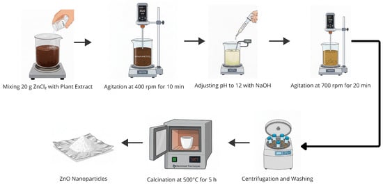
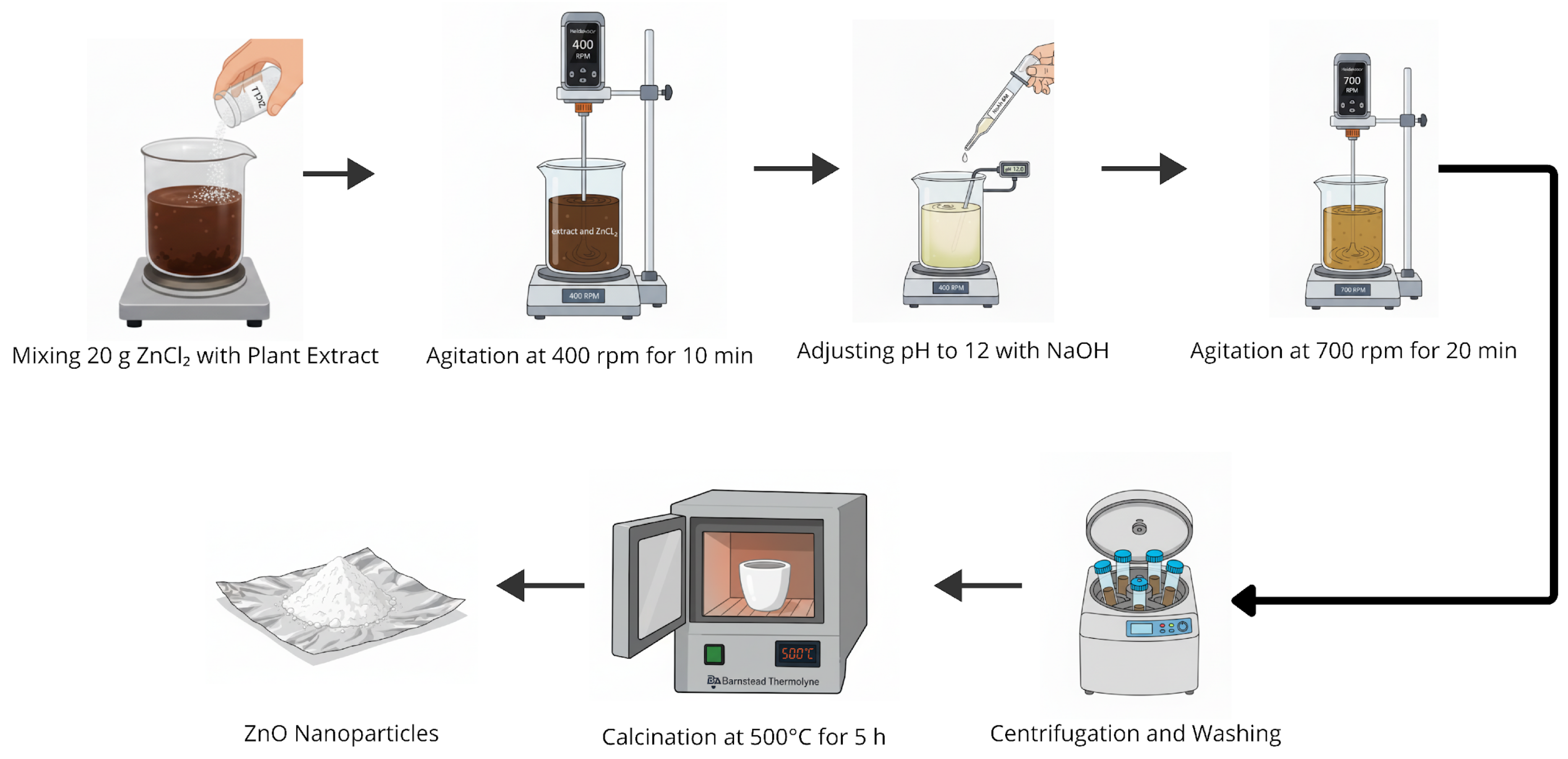
The extract was prepared by aqueous infusion, using a biomass-to-water ratio of 1:8. For this, 75 g of biomass were added to 600 mL of distilled water, and the mixture was gently stirred while boiling for 30 min. It was then filtered to remove solid residues, and the resulting extract was stored at 4 °C until use [23]. Figure 1 illustrates the experimental methodology applied to synthesize the ZnO nanoparticles. In a typical procedure, 20 g of ZnCl2 was dissolved in 200 mL of the plant extract, and the mixture was stirred mechanically at 400 rpm for 10 min. Subsequently, 6 M NaOH was gradually added until the pH was adjusted to 12; once stabilized, the stirring speed was increased to 700 rpm for an additional 20 min. The product was separated by centrifugation at 6000 rpm and washed alternately with distilled water (four times) and absolute ethanol (99%) (twice) to remove residual impurities. Finally, the washed material was calcined in a muffle furnace at 500 °C for 5 h, yielding ZnO nanoparticles with the characteristic white color of zinc oxide [22,24,25].
Figure 1.
Schematic model of the green synthesis of ZnO nanoparticles.
2.3. Characterizations
To identify the crystalline phase present in the ZnO nanoparticles, they were subjected to X-ray diffraction (XRD) analysis using a Malvern-PANalytical Empyrean 2012 X-ray diffractometer (Malvern Panalytical, Malvern, UK) with a 3D pixel detector and a copper source (λ = 1.541) at 45 kV and 40 mA. Measurements were taken with a 0.05° step size and a 50 s step time. A 2θ range of 10°–80° was covered, providing details of the nanoparticles’ crystalline structure and crystal size [25,26]. The morphology of the nanoparticles was evaluated by field-emission scanning electron microscopy (FE-SEM) using a Thermo Fisher Scientific Scios 2 LoVac instrument (ThermoFisher Scientific, Waltham, MA, USA), with a resolution of 0.7 nm at 30 kV (STEM mode) and 1.4 nm at 1 kV (FE-SEM mode). Additionally, elemental analysis was performed by energy-dispersive X-ray spectroscopy (EDS) using an UltraDry (Waltham, MA, USA) 129 eV, 30 mm2 detector (model SDBX-30PM-B) to determine the elemental composition of the synthesized nanoparticles [27].
2.4. Antifungal Efficacy Analysis
This property was evaluated using the poisoned-food technique with ZnO nanoparticles, potato dextrose agar (PDA), and Aspergillus niger and Penicillium fungi, which are characterized by growing on cell walls due to humidity [28]. For the modification of the culture medium, the nanoparticles were dispersed in deionized water using ultrasound for 20 min. For this purpose, 10 mL of water and the required amounts of nanoparticles were used to obtain two samples with final concentrations in the agar of 1 mg/mL and 3 mg/mL. Subsequently, each sample was mixed with 50 mL of PDA and placed in an autoclave at 121 °C for 1 h. After this, the mixture was added to Petri dishes (90 mm × 15 mm), each dish containing 25 mL of the modified culture medium [29,30]. The fungi were inoculated using the puncture technique, with the inoculum placed at three different points on the surface of the culture medium, arranged in a triangular pattern. Growth was evaluated for 48 and 120 h at room temperature. Additionally, a control trial without nanoparticles was conducted for each fungus. All tests were performed under the same environmental conditions. Furthermore, the study was conducted by directly incorporating the nanoparticles into the culture medium without using any coating material, to prevent coating properties from affecting the results.
2.5. Evaluation of Thermal Comfort Properties
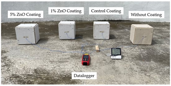
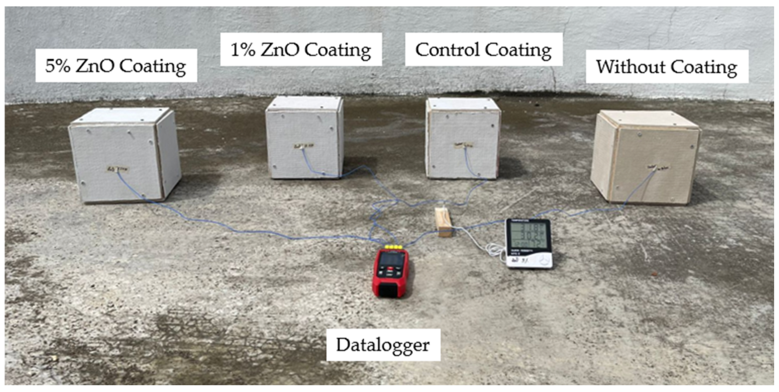
Four boxes (20 cm × 20 cm × 20 cm) were constructed with a wooden base and 6 mm thick fiber cement faces (Figure 2). The tests were performed under the same external environmental factors such as temperature, radiation, and humidity, since these factors influence data collection [31]. Before applying the coatings, a layer of Acrilcor 50 primer was applied to seal the fiber cement’s pores. Two boxes were coated with PARAGUAS® Multipurpose (CORONA) commercial paint modified with different concentrations of ZnO nanoparticles (1% w/w and 5% w/w). A third box was coated with the unmodified paint, and a fourth box remained uncoated (see Figure 2). Both primer and coatings were applied by brush [32,33]. The internal temperature of the boxes was recorded using a THE-373 K/J/T THERMOMETER digital datalogger with four type K temperature sensors, located in the center of each box. Ambient temperature and humidity were recorded using a Ker Germanv HTC-2 digital thermo-hygrometer (Jiangsu, Shaanxi, Fujian, China). The boxes were simultaneously exposed to sunlight between 10:00 and 14:00 h, the period of highest solar radiation. Each measurement was taken at 1-min intervals during the exposure period.
Figure 2.
Experimental setup for evaluating the thermal comfort property of ZnO nanoparticles.
3. Results and Discussions
3.1. SEM—EDS Analysis
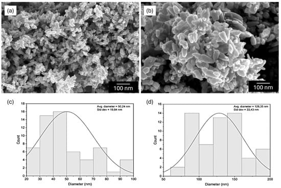
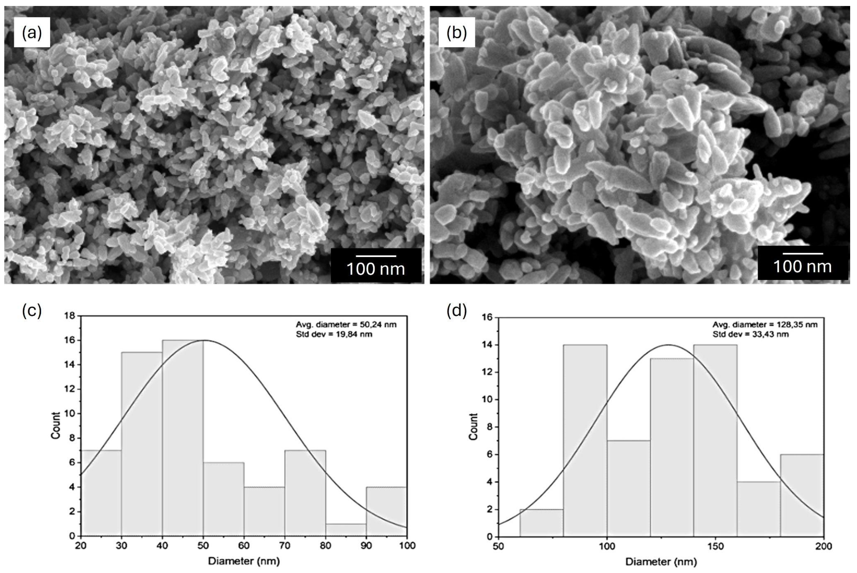
The morphology of the zinc oxide nanoparticles was characterized via scanning electron microscopy (SEM), the analysis revealed that green synthesis using lemongrass extract produced nanoparticles of semi-spherical and ellipsoidal shapes, as shown in Figure 3a,b, with an average particle size of 50.2 ± 19.8 nm (Figure 3c) and 128.3 ± 33.4 nm (Figure 3d), respectively. Although two morphologies were observed in the SEM images, they belong to the same ZnO sample; therefore, all physicochemical analyses were performed on the bulk powder.
Figure 3.
(a,b) SEM images of the ZnO nanoparticles. (c) Size distribution of semi-spherical ZnO nanoparticles. (d) Size distribution of ellipsoidal ZnO nanoparticles.
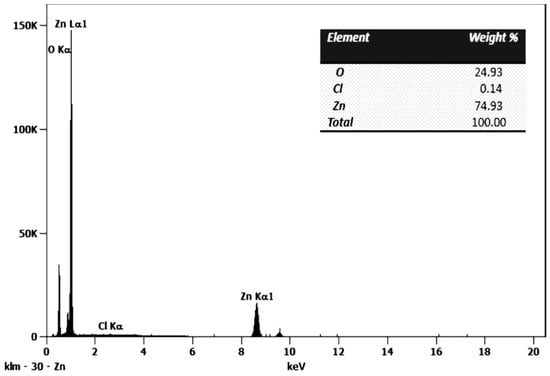
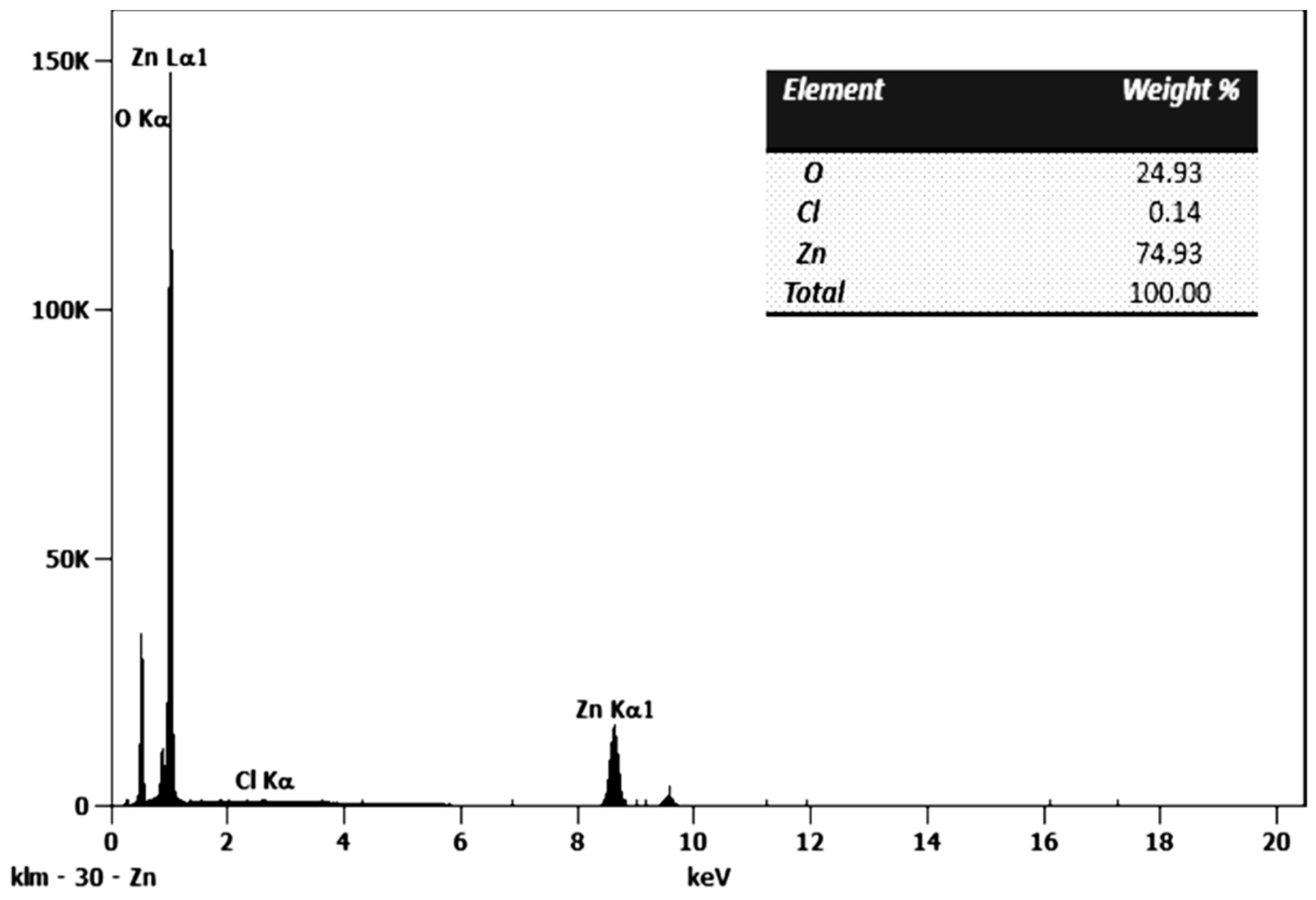
Additionally, elemental analysis of the nanoparticles was performed using Energy Dispersive Spectroscopy (EDS). The results, shown in Figure 4, indicate that the nanoparticles are composed of 74.93 wt% zinc and 24.93 wt% oxygen, in addition to a small percentage of chlorine (0.14 wt%), which possibly remained from the zinc chloride (ZnCl2), the inorganic salt precursor used in the synthesis of these nanoparticles. These results are similar to those reported by Solano et al. [22], in which ZnO nanoparticles were synthesized using an ultrasound-assisted method in the presence of the Cymbopogon citratus extract. In contrast to the findings of Solano et al. [22], the ZnO nanoparticles synthesized in this work exhibit higher compositional purity and a more controlled morphology (spherical and ellipsoidal). These structural advantages enhance surface reactivity and dispersion, potentially leading to superior performance in multifunctional antimicrobial and thermal applications.
Figure 4.
Energy-dispersive X-ray spectroscopy (EDS) of ZnO nanoparticles.
3.2. X-Ray Diffraction Analysis
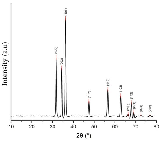
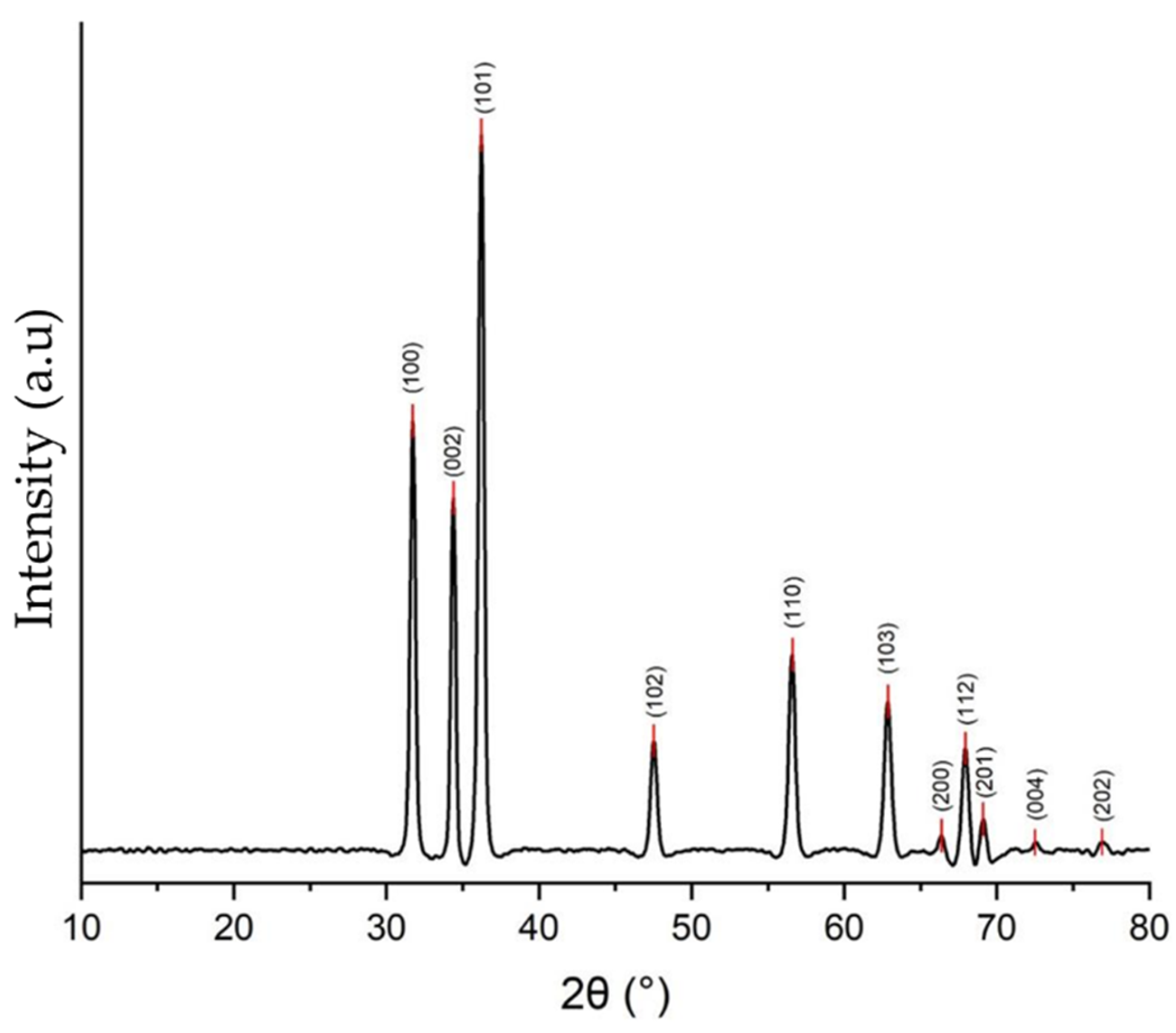
Figure 5 shows the X-ray diffraction pattern for the synthesized ZnO nanoparticles. In this, peaks can be observed located at 31.71°, 34.38°, 36.22°, 47.51°, 56.58°, 62.85°, 66.36°, 67.94°, 69.10°, 72.51°, and 76.93°, which correspond to the (100), (002), (101), (102), (110), (103), (200), (112), (201), (004), and (202) planes of the Miller index (hkl), respectively, characteristic planes of the hexagonal (Wurtzite) phase of ZnO. These results coincide with chart No. 79-2205 of the Joint Committee on Powder Diffraction Standards (JCPDS) [34]. To determine the average crystal size, the Debye-Scherrer equation (Equation (1)) was used:
where is the crystal size in nm, k is the form factor (0.89), λ is the wavelength of the X-ray source (Cu source (λ = 0.1541 nm)), β is the full width at half height (FWHM) of the diffraction peak in radians, and θ is the diffraction angle. The average crystal size was calculated from the three highest-intensity peaks corresponding to the (100), (002), and (101) planes, yielding an average size of 20.32 nm [35].
Figure 5.
XRD pattern of the ZnO nanoparticles.
3.3. Evaluation of the Antifungal Property
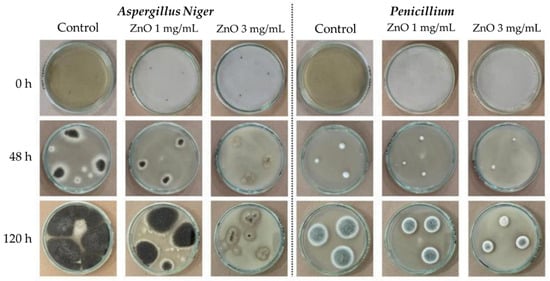
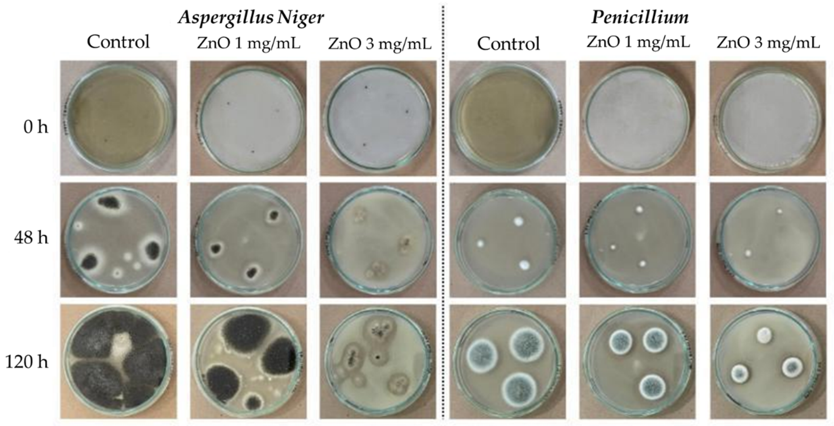
Figure 6 shows the inhibitory effect of ZnO nanoparticles on the growth of Aspergillus niger and Penicillium at different incubation times (0, 48, and 120 h) and concentrations (1 and 3 mg/mL). Visually, a progressive reduction in the growth area is observed with increasing ZnO concentration. Quantitative data are detailed in Table 1, where it can be seen that at 3 mg/mL the growth inhibition was most pronounced for both fungi, especially in Aspergillus niger after 120 h of incubation.
Figure 6.
Experimental results of the evaluation of the antifungal properties of ZnO nanoparticles.
Table 1.
Average mycelial growth area (mm2) of Aspergillus niger and Penicillium fungi.
According to Das et al. [36], in the culture-poisoning technique, the percentage of mycelial growth inhibition can be evaluated by comparing the diameter of the fungal colony in the poisoned culture medium with the diameter of the colony in the unpoisoned medium [35,37]. Considering that the growth of Aspergillus niger is irregular, the colony area was chosen as the comparative measure, using Equation (2), and the results obtained are presented in Table 2.
Table 2.
Results of percentage of mycelial growth inhibition.
As shown in Table 2, ZnO nanoparticles exhibited a marked capacity to inhibit the mycelial growth of Aspergillus niger and Penicillium. The results indicate that, even at a concentration of 1 mg/mL, inhibition percentages exceeding 50% were achieved after 120 h of incubation, demonstrating significant antifungal activity at moderate concentrations.
At higher concentrations (3 mg/mL), inhibition was considerably greater, with values of 94.63% for Aspergillus niger and 72.64% for Penicillium 120 h after inoculation. These results demonstrate that the antifungal efficacy of ZnO nanoparticles depends on both the agent concentration and exposure time, with a direct relationship observed between increasing concentration and the percentage of inhibition. This is consistent with previous studies on this property of this nanoparticle type [28,29].
Furthermore, Aspergillus niger showed greater sensitivity to ZnO treatment compared to Penicillium, which could be attributed to structural differences in the cell wall or fungal membrane composition that affect interactions with the nanoparticles. These findings are consistent with previous studies reporting ZnO’s high capacity to generate reactive oxygen species (ROS) and alter fungal cell integrity [38] as well as the Zn interaction with the membrane disruption and electrostatic interactions [39].
3.4. Thermal Performance and Comfort Evaluation
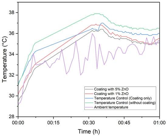
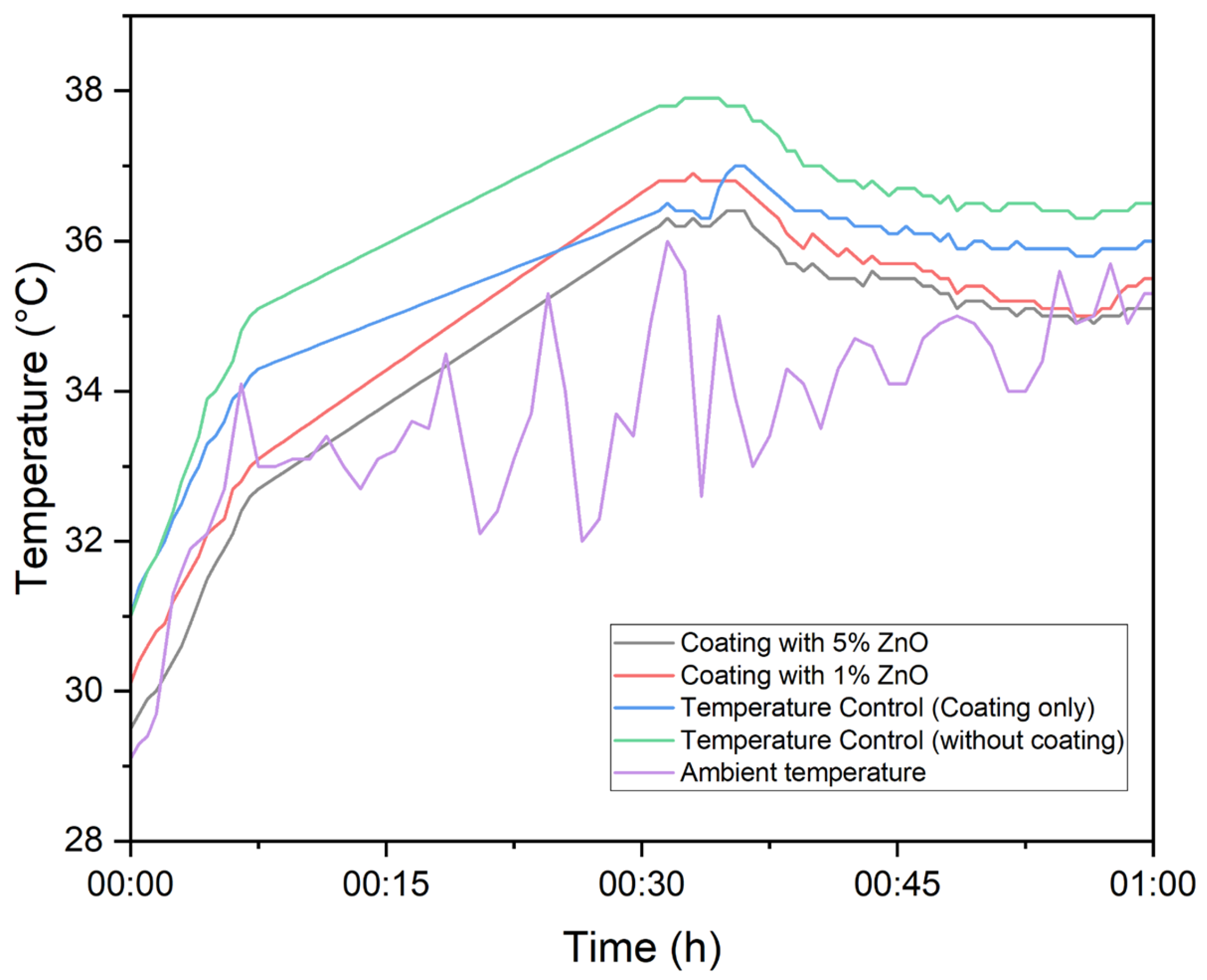
By modifying the paint-type coating with the addition of ZnO nanoparticles, the aim was to impart radiative cooling properties. This passive cooling mechanism, according to Mishra et al. [40], relies on the ability of these materials to effectively reflect incident solar radiation and release thermal radiation in the long-wave infrared (LWIR) spectrum. This allows radiation to penetrate Earth’s atmosphere through the so-called “atmospheric transparency window,” which encompasses the wavelength range of 8 to 13 µm. Together, these two properties enable surfaces coated with this type of compound to cool. Studies conducted on ZnO nanoparticles have achieved solar radiation reflectance values of up to 90% and infrared emissivity values of up to 95% [41]. First, an experiment was conducted to continuously record the internal temperature of the boxes and the ambient temperature for one hour, from 10:00 to 11:00 h, in order to analyze the thermal behavior of the modified coating compared to the base coating and the control, as shown in Figure 7. It was observed that the ambient temperature exhibited fluctuations with maximum and minimum peaks, attributable to the sensor’s direct exposure to high solar radiation and wind. This situation can generate unstable or unrepresentative readings of the actual ambient temperature, as noted by Mishra et al. [40]. To minimize this effect, sensors should be protected from direct solar radiation and wind. Consequently, the evaluation of thermal behavior was performed by considering only the temperature measurements inside the boxes painted with the modified coating, the base coating, and the uncoated boxes.
Figure 7.
Temperature behavior of the coating modified with ZnO nanoparticles and ambient temperature measurement.
Subsequently, experiments were conducted to evaluate the thermal comfort properties of coatings modified with ZnO nanoparticles at concentrations of 1% w/w and 5% w/w, as shown in Figure 2.
Figure 7 shows the results of the test performed for the thermal comfort property evaluation, demonstrating the great performance of the box with the coating modified with 5% ZnO nanoparticles. It achieved a maximum temperature difference of 1.9 °C compared to the base coating and 2.4 °C compared to the uncoated box. On the other hand, the box with the coating modified with 1% w/w achieved the greatest reductions of 1.3 °C and 2 °C compared to the base-coated box and the uncoated box, respectively.
In general, the sample box with 5% of ZnO nanoparticles/coating achieved an average temperature reduction of 0.6 °C compared to the box with the base coating, whereas the box with the 1% ZnO modified coating only achieved an average reduction of 0.2 °C.
These results reflect a positive performance in the ability of the ZnO-modified coatings to reduce internal temperature, although their effect may have been attenuated by the environmental conditions during the test. In the city of Cartagena, Colombia, the average annual relative humidity exceeds 80% [42], and, according to Mishra et al. [40], an increase in humidity decreases the transparency of the atmospheric window, affecting the emission of thermal radiation in the long-wave infrared (LWIR) range. This phenomenon can reduce the radiative efficiency of the coatings, partially limiting their ability to dissipate heat and, consequently, the magnitude of the observed temperature difference.
4. Conclusions
The synthesis of ZnO nanoparticles using green chemistry with Cymbopogon citratus leaf extract proved to be an efficient, sustainable, and environmentally friendly route for obtaining nanomaterials with functional properties. Structural analyses confirmed the formation of a wurtzite-type crystalline phase with hemispherical and ellipsoidal morphology and average particle sizes in the nanometer range.
Antifungal activity tests demonstrated significant inhibition of the growth of Aspergillus niger and Penicillium spp., reaching percentages of up to 94.6% and 72.6%, respectively, at concentrations of 3 mg/mL, highlighting the efficacy of ZnO as an antifungal agent even at low concentrations. Regarding thermal comfort, coatings modified with ZnO nanoparticles showed an average reduction in internal temperature of up to 0.6 °C compared to the base coating, with a maximum difference of 2.4 °C compared to the uncoated surface. Although high humidity may have limited the material’s radiative performance, the results confirm the potential of ZnO nanoparticles to enhance passive surfaces cooling.
The results obtained demonstrate the feasibility of incorporating ZnO nanoparticles synthesized using environmentally friendly methods into architectural coatings, simultaneously providing antifungal and thermal comfort benefits. This type of development promotes sustainable alternatives for improving the functional performance of building materials, with potential applications in environments with high humidity and solar radiation.
Author Contributions
Conceptualization, A.H. and M.S.; methodology, M.C., M.W. and D.M.-B.; validation, A.H. and M.S.; formal analysis, M.C., M.W., D.M.-B. and A.H.; investigation, M.C., M.W., D.M.-B. and A.H.; resources, A.H. and M.S.; data curation, M.C. and D.M.-B.; writing—original draft preparation, M.C., M.W. and D.M.-B.; writing—review and editing, A.H. and M.S.; supervision, A.H. and M.S.; project administration, A.H. and M.S.; funding acquisition, A.H. and M.S. All authors have read and agreed to the published version of the manuscript.
Funding
The authors are grateful for funding from the University of Cartagena through the projects “Desarrollo de filtros con propiedades antimicrobiales a partir de la impresión 3D de estructuras poliméricas modificadas con nanopartículas ecoamigables de lignina, celulosa y óxido de zinc. Acta No. 103-2021” and “Evaluación de las propiedades antimicóticas, de autolimpieza y confort térmico de un recubrimiento encapsulante para cubiertas de asbesto-cemento modificado con nanoaditivos de óxidos metálicos procesados con métodos eco-amigables. Acta No. 030-2022”. Furthermore, this article forms part of the outcomes of the project “Formulation of a Comprehensive Strategy to Reduce the Impact of Asbestos on Public and Environmental Health in the Department of Bolívar”, funded by the Sistema General de Regalías de Colombia, under project code BPIN 2020000100366. The project was implemented by the University of Cartagena in partnership with the Asbestos-Free Colombia Foundation (FundClas).
Institutional Review Board Statement
Not applicable.
Informed Consent Statement
Not applicable.
Data Availability Statement
The original contributions presented in this study are included in the article. Further inquiries can be directed to the corresponding author.
Conflicts of Interest
The authors declare no conflicts of interest.
References
- Amos, Y.; Rubaka, C.; Sauli, E.; Swai, H.; Tumbo, A. Nanotechnology-based delivery strategies for drugs and vaccines targeting blood stage malaria: A systematic review. Nanomed. Nanotechnol. Biol. Med. 2025, 70, 102869. [Google Scholar] [CrossRef] [Scilit] [PubMed]
- Boruah, S.; Jose, S. Nanotechnology in textiles: Environmental safety and sustainable practices. Environ. Nanotechnol. Monit. Manag. 2025, 23, 101062. [Google Scholar] [CrossRef] [Scilit]
- Adikesavan, R.; Kasivelu, G.; Kamalakannan, M.; Saravanan, N.; Malaichamy, K. Advancing sericulture through nanotechnology: Sustainable innovations for the future. Sustain. Mater. Technol. 2025, 46, e01726. [Google Scholar] [CrossRef] [Scilit]
- Abdullah, M.; Obayedullah, M.; Shuvo, S.I.; Khair, A.; Hossain, D.; Islam, N. A review on multifunctional applications of nanoparticles: Analyzing their multi-physical properties. Results Surf. Interfaces 2025, 21, 100635. [Google Scholar] [CrossRef] [Scilit]
- Agbogo, V.U.; Sadiku, E.R.; Mavhungu, L.; Kupolati, W.K.; Injor, O.M. Nanotechnology coatings in the defense and aerospace industry. Next Nanotechnol. 2025, 7, 100197. [Google Scholar] [CrossRef] [Scilit]
- Zhu, Q.; Chua, M.H.; Ong, P.J.; Lee, J.J.C.; Chin, K.L.O.; Wang, S.; Kai, D.; Ji, R.; Kong, J.; Dong, Z.; et al. Recent advances in nanotechnology-based functional coatings for the built environment. Mater. Today Adv. 2022, 15, 100270. [Google Scholar] [CrossRef] [Scilit]
- Hayati, D.; Ghasemi, S.; Lakouraj, M.M.; Mousavi, F. A smart epoxy coating based on Zn-imidazole metal-organic framework/graphene oxide nanocomposite for corrosion protection. Compos. Commun. 2025, 59, 102556. [Google Scholar] [CrossRef] [Scilit]
- Wang, Y.; Cheng, F.; Huang, Y.; Cai, C.; Fu, Y. Vertically-oriented growth of MgMOF layer via heteroepitaxial guidance for highly stable magnesium-metal anode. Energy Storage Mater. 2023, 61, 102911. [Google Scholar] [CrossRef] [Scilit]
- Raj, L.A.; Annushrie, A.; Namasivayam, S.K.R. Anti bacterial efficacy of photo catalytic active titanium di oxide (TiO2) nanoparticles synthesized via green science principles against food spoilage pathogenic bacteria. Microbe 2025, 7, 100331. [Google Scholar] [CrossRef] [Scilit]
- Khaled, I.M.; Umair, M.; Pecoraro, C.M.; Kheniche, A.; Bellardita, M.; Soltani, S. Green synthesis of ZnO and CuO nanoparticles and ZnO-CuO nanocomposites: Characterization and photocatalytic dyes removal efficiency’s determination. Colloids Surf. A Physicochem. Eng. Asp. 2026, 728, 138544. [Google Scholar] [CrossRef] [Scilit]
- Masho, T.J.; Ayane, M.M.; Endale, G.N.; Adula, S.H.; Negasa, Y.D.; Likasa, B.W.; Mudda, T.S.; Negari, G.K.; Amante, H.C.; Jida, S.M. Biosynthesis, characterization of Ag2O nanoparticles for enhancement of antioxidant and photo degradation activities. Results Chem. 2025, 13, 101919. [Google Scholar] [CrossRef] [Scilit]
- Lithi, I.J.; Nakib, K.I.A.; Chowdhury, A.M.S.; Hossain, S. A review on the green synthesis of metal (Ag, Cu, and Au) and metal oxide (ZnO, MgO, Co3O4, and TiO2) nanoparticles using plant extracts for developing antimicrobial properties. Nanoscale Adv. 2025, 7, 2446–2473. [Google Scholar] [CrossRef] [Scilit]
- Radulescu, D.-M.; Surdu, V.-A.; Ficai, A.; Ficai, D.; Grumezescu, A.-M.; Andronescu, E. Green Synthesis of Metal and Metal Oxide Nanoparticles: A Review of the Principles and Biomedical Applications. Int. J. Mol. Sci. 2023, 24, 15397. [Google Scholar] [CrossRef] [Scilit]
- Abdelbaky, A.S.; Mohamed, A.M.H.A.; Sharaky, M.; Mohamed, N.A.; Diab, Y.M. Green approach for the synthesis of ZnO nanoparticles using Cymbopogon citratus aqueous leaf extract: Characterization and evaluation of their biological activities. Chem. Biol. Technol. Agric. 2023, 10, 1–23. [Google Scholar] [CrossRef] [Scilit]
- Vijayalakshmi, K.; Swaramanjari, T.; Shanmugavel, M.; Gnanamani, A. Green-synthesized metal and metal oxide nanoparticles as emerging antifungal agents: Current advances, mechanisms, and future perspectives. Discov. Biotechnol. 2025, 2, 1–23. [Google Scholar] [CrossRef] [Scilit]
- Sajini, T.; Joseph, J. Microwave-assisted synthesis of nanomaterials: A green chemistry perspective and sustainability assessment. RSC Sustain. 2025, 3, 4911–4935. [Google Scholar] [CrossRef] [Scilit]
- Zhang, X.; Zhang, M.; Han, H.; Chen, Z.; Huang, A. Green synthesis of zinc oxide nanoparticles using Cnidium monnieri fruit extract: Prescription optimization, Characterization and antifungal activity. Microb. Pathog. 2025, 200, 107256. [Google Scholar] [CrossRef] [Scilit] [PubMed]
- Fedel, M.; Rosati, A.; Bertasini, M.; Rossi, S. Cu doped YInO3-ZnO green colored NIR reflective pigments: Synthesis and application in PMMA based cool-roof coatings. Prog. Org. Coat. 2023, 182, 107708. [Google Scholar] [CrossRef] [Scilit]
- Konovalova, V.S.; Rumyantseva, V.E.; Strokin, K.B.; Galtsev, A.A.; Novikov, D.G.; Monastyrev, P.V. Degradation of Concrete Cement Stone Under the Influence of Aspergillus niger Fungi. Corros. Mater. Degrad. 2024, 5, 476–489. [Google Scholar] [CrossRef] [Scilit]
- Wirth, A.; Pacheco, F.; Toma, N.; Valiati, V.; Tutikian, V.; Gomes, L. Analysis of Fungal Growth on Different Coatings Applied to Lightweight Systems. Rev. Ing. Constr. 2019, 34, 5–14. [Google Scholar] [CrossRef] [Scilit]
- Subramaniam, M.; Sunar, N.M.; Latif, A.; Parjo, U.K.; Er, C.M.; Ab Razak, A.R. The Growth of Aspergillus niger on a Wood Based Material with 4 Types of Wall Finishing. MATEC Web Conf. 2016, 47, 05007. [Google Scholar] [CrossRef] [Scilit]
- Solano, R.; Patiño-Ruiz, D.; Herrera, A. Preparation of modified paints with nano-structured additives and its potential applications. Nanomater. Nanotechnol. 2020, 10, 184798042090918. [Google Scholar] [CrossRef] [Scilit]
- Solano Pizarro, R.A.; Herrera Barros, A.P. Cypermethrin elimination using Fe-TiO2 nanoparticles supported on coconut palm spathe in a solar flat plate photoreactor. Adv. Compos. Lett. 2020, 29, 1–13. [Google Scholar] [CrossRef] [Scilit]
- Mohammadi-Aloucheh, R.; Habibi-Yangjeh, A.; Bayrami, A.; Latifi-Navid, S.; Asadi, A. Enhanced anti-bacterial activities of ZnO nanoparticles and ZnO/CuO nanocomposites synthesized using Vaccinium arctostaphylos L. fruit extract. Artif. Cells Nanomed. Biotechnol. 2018, 46, 1200–1209. [Google Scholar] [CrossRef] [Scilit]
- Thiam, M.; Bernardo, A.d.J.; Pellegrini, V.d.O.A.; Possatto, J.F.; Dlamini, Z.W.; Mahule, T.S.; Ngom, B.D.; Mosepele, B.Q.; Thema, F.T.; Mamba, B.B.; et al. Green Synthesis of ZnO Nanoparticles Using Licania tomentosa Benth (Oiti) Leaf Extract: Characterization and Applications for the Photocatalytic Degradation of Crystal Violet Dye. Processes 2025, 13, 880. [Google Scholar] [CrossRef] [Scilit]
- Múnera-Gómez, L.F.; Muñoz-Acevedo, J.C.; Pabón-Gelves, E. Synthesis and evaluation of the antibacterial activity of Cu(II) and Ni(II) complexes with mixed ligands based on glycine and dicarboxylic acids. DYNA 2024, 91, 16–23. [Google Scholar] [CrossRef] [Scilit]
- Sierra, L.A.Q.; Escobar, D.M. Characterization and bioactivity behavior of sol–gel derived bioactive vitroceramic from non-conventional precursors. Bol. La Soc. Esp. Ceram. Vidr. 2019, 58, 85–92. [Google Scholar] [CrossRef] [Scilit]
- Kumawat, G.; Rajpurohit, D.; Vyas, D.; Bhojiya, A.A.; Upadhyay, S.K.; Jain, D. Characterization of green-synthesized zinc oxide nanoparticles and its influence on post-harvest shelf-life of garlic against black mold disease caused by Aspergillus niger. Front. Microbiol. 2025, 16, 1532593. [Google Scholar] [CrossRef] [Scilit]
- Elshafie, H.S.; Osman, A.; El-Saber, M.M.; Camele, I.; Abbas, E. Antifungal Activity of Green and Chemically Synthesized ZnO Nanoparticles against Alternaria citri, the Causal Agent Citrus Black Rot. Plant Pathol. J. 2023, 39, 265–274. [Google Scholar] [CrossRef] [Scilit]
- González Merino, A.M. Actividad antifúngica de nanopartículas de óxido de zinc en el patosistema Fusarium oxysporum–Solanum lycopersicum bajo condiciones controladas. Tesis de Maestría, Universidad Autónoma Agraria Antonio Narro, Saltillo, Coahuila, México, 2020. Available online: http://repositorio.uaaan.mx:8080/xmlui/handle/123456789/47050 (accessed on 5 March 2026).
- Qin, M.; Xiong, F.; Aftab, W.; Shi, J.; Han, H.; Zou, R. Phase-change materials reinforced intelligent paint for efficient daytime radiative cooling. iScience 2022, 25, 104584. [Google Scholar] [CrossRef] [Scilit]
- Mishra, B.R.; Sundaram, S.; Sasihithlu, K. Cooling Performance of TiO2-Based Radiative Cooling Coating in Tropical Conditions. ACS Omega 2024, 9, 49494–49502. [Google Scholar] [CrossRef] [Scilit] [PubMed]
- Guo, W.; Song, L.; Wang, H.; Liu, J.; Li, N.; Zhao, Y.; Xiong, J. Efficient radiative cooling and super-hydrophobic ZnO/P(VDF-HFP)-PDMS coated fabric. Appl. Therm. Eng. 2025, 268, 125852. [Google Scholar] [CrossRef] [Scilit]
- Vijayakumar, S.; Mahadevan, S.; Arulmozhi, P.; Sriram, S.; Praseetha, P.K. Green synthesis of zinc oxide nanoparticles using Atalantia monophylla leaf extracts: Characterization and antimicrobial analysis. Mater. Sci. Semicond. Process. 2018, 82, 39–45. [Google Scholar] [CrossRef] [Scilit]
- Hernández, R.V.; Lemus, M.A.A.; García, S.D.l.R.; González, R.L.; Quintana, P.; Zaleta, D.G.; Vázquez, V.V.; Cornelio, S.G. Antifungal Activity of ZnO Nanoparticles Synthesized from Eichhornia crassipes Extract for Construction Applications. Nanomaterials 2024, 14, 1007. [Google Scholar] [CrossRef] [Scilit]
- Das, K.; Tiwari, R.K.S.; Shrivastava, D.K. Techniques for evaluation of medicinal plant products as antimicrobial agent: Current methods and future trends. J. Med. Plants Res. 2010, 4, 104–111. Available online: https://www.researchgate.net/publication/330556242_Techniques_for_evaluation_of_medicinal_plant_products_as_antimicrobial_agent_Current_methods_and_future_trends (accessed on 5 March 2026).
- Shenashen, M.; Derbalah, A.; Hamza, A.; Mohamed, A.; El Safty, S. Antifungal activity of fabricated mesoporous alumina nanoparticles against root rot disease of tomato caused by Fusarium oxysporium. Pest Manag. Sci. 2017, 73, 1121–1126. [Google Scholar] [CrossRef] [Scilit]
- Janani, M.; Gomathi, T.; Babujanarthanam, R.; Kaviyarasu, K. An evaluation of the biological activity of zinc oxide nanoparticles fabricated from aqueous bark extracts of Acacia nilotica. J. King Saud Univ. Sci. 2023, 35, 102753. [Google Scholar] [CrossRef] [Scilit]
- Reda, A.T.; Kwon, S.; Choi, S.S.; Cho, C.; Park, Y.T. Stirring-Assisted Layered Coating of Polyurethane Foam for Antimicrobial Activity and Flame Retardant. Appl. Surf. Sci. 2025, 714, 164407. [Google Scholar] [CrossRef] [Scilit]
- Mishra, B.R.; Sundaram, S.; Sasihithlu, K. Design of Radiative Cooling Paint Coating and Insights into its Sub-Ambient Cooling Behaviour. arXiv 2024. [Google Scholar] [CrossRef] [Scilit]
- Kang, H.; Qiao, Y.; Li, Y.; Qin, W.; Wu, X. Keep Cool: Polyhedral ZnO@ZIF-8 Polymer Coatings for Daytime Radiative Cooling. Ind. Eng. Chem. Res. 2020, 59, 15226–15232. [Google Scholar] [CrossRef] [Scilit]
- Secretaría del Medio Ambiente de Cartagena de Indias. Humedad Relativa Promedio—Observatorio Ambiental de Cartagena. Available online: https://observatorio.epacartagena.gov.co/gestion-ambiental/calidad-ambiental/sistema-urbano/humedad-relativa-promedio/ (accessed on 5 March 2026).

Disclaimer/Publisher’s Note: The statements, opinions and data contained in all publications are solely those of the individual author(s) and contributor(s) and not of MDPI and/or the editor(s). MDPI and/or the editor(s) disclaim responsibility for any injury to people or property resulting from any ideas, methods, instructions or products referred to in the content. |
© 2026 by the authors. Licensee MDPI, Basel, Switzerland. This article is an open access article distributed under the terms and conditions of the Creative Commons Attribution (CC BY) license.