Next Issue
Volume 12, February
Previous Issue
Volume 11, December


Biosensors, Volume 12, Issue 1 (January 2022) – 46 articles

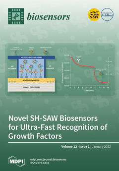
Cover Story (view full-size image): Shear horizontal surface acoustic wave (SH-SAW) biosensors were used to recognize growth factors (GF) in real time in a label-free and selective mode. This type of biosensor is a very promising method for ultra-fast recognition of these biomolecules due to its great advantages, such as portability, simplicity of use, reusability, low cost, and detection within a relatively short period of time. SH-SAW sensors operated in dynamic mode with a microfluidic system detect Nano grams in minutes. This kind of biosensor technology is a hopeful analysis tool for applications in clinical tests, and it can compete with the already well-established but much more complex techniques, such as surface plasmon resonance (SPR) and enzyme-linked immunosorbent assay (ELISA). View this paper
  • Issues are regarded as officially published after their release is announced to the table of contents alert mailing list.
  • You may sign up for e-mail alerts to receive table of contents of newly released issues.
  • PDF is the official format for papers published in both, html and pdf forms. To view the papers in pdf format, click on the "PDF Full-text" link, and use the free Adobe Reader to open them.
Order results
Result details
Section
Select all
Export citation of selected articles as:
16 pages, 4354 KiB  
Review
NIR-II Aggregation-Induced Emission Luminogens for Tumor Phototheranostics
by Yonghong Tan, Peiying Liu, Danxia Li, Dong Wang and Ben Zhong Tang
Biosensors 2022, 12(1), 46; https://doi.org/10.3390/bios12010046 - 17 Jan 2022
Cited by 17 | Viewed by 5217
Abstract
As an emerging and powerful material, aggregation-induced emission luminogens (AIEgens), which could simultaneously provide a precise diagnosis and efficient therapeutics, have exhibited significant superiorities in the field of phototheranostics. Of particular interest is phototheranostics based on AIEgens with the emission in the range [...] Read more.
As an emerging and powerful material, aggregation-induced emission luminogens (AIEgens), which could simultaneously provide a precise diagnosis and efficient therapeutics, have exhibited significant superiorities in the field of phototheranostics. Of particular interest is phototheranostics based on AIEgens with the emission in the range of second near-infrared (NIR-II) range (1000–1700 nm), which has promoted the feasibility of their clinical applications by virtue of numerous preponderances benefiting from the extremely long wavelength. In this minireview, we summarize the latest advances in the field of phototheranostics based on NIR-II AIEgens during the past 3 years, including the strategies of constructing NIR-II AIEgens and their applications in different theranostic modalities (FLI-guided PTT, PAI-guided PTT, and multimodal imaging-guided PDT–PTT synergistic therapy); in addition, a brief conclusion of perspectives and challenges in the field of phototheranostics is given at the end. Full article
(This article belongs to the Special Issue Nanoprobes for Tumor Theranostics)
Show Figures

Figure 1

12 pages, 2264 KiB  
Article
Butyl Benzyl Phthalate in Urban Sewage by Magnetic-Based Immunoassay: Environmental Levels and Risk Assessment
by Xia Hong, Yin Cui, Ming Li, Yifan Xia, Daolin Du and Chengwu Yi
Biosensors 2022, 12(1), 45; https://doi.org/10.3390/bios12010045 - 15 Jan 2022
Cited by 2 | Viewed by 3146
Abstract
A magnetic-based immunoassay (MBI) combined with biotin-streptavidin amplification was proposed for butyl benzyl phthalate (BBP) investigation and risk assessment. The values of LOD (limit of detection, IC10) and IC50 were 0.57 ng/mL and 119.61 ng/mL, with a detection range of [...] Read more.
A magnetic-based immunoassay (MBI) combined with biotin-streptavidin amplification was proposed for butyl benzyl phthalate (BBP) investigation and risk assessment. The values of LOD (limit of detection, IC10) and IC50 were 0.57 ng/mL and 119.61 ng/mL, with a detection range of 0.57–24,977.71 ng/mL for MBI. The specificity, accuracy and precision are well demonstrated. A total of 36 environmental water samples of urban sewage from Zhenjiang, China, were collected and assessed for BBP contamination. The results show that BBP-positive levels ranged from 2.47 to 89.21 ng/mL, with a positive rate of 77.8%. The health effects of BBP in the urban sewage were within a controllable range, and the ambient severity for health (ASI) was below 1.49. The highest value of AS for ecology (ASII) was 7.43, which indicates a potential harm to ecology. The entropy value of risk quotient was below 100, the highest being 59.47, which poses a low risk to the environment and ecology, indicating that there is a need to strengthen BBP controls. The non-carcinogenic risk of BBP exposure from drinking water was higher for females than that for males, and the non-carcinogenic risk from drinking-water and bathing pathways was negligible. This study could provide an alternative method for detecting BBP and essential information for controlling BBP contamination. Full article
Show Figures

Figure 1

38 pages, 5209 KiB  
Review
Artificial Biomimetic Electrochemical Assemblies
by Tanja Zidarič, Matjaž Finšgar, Uroš Maver and Tina Maver
Biosensors 2022, 12(1), 44; https://doi.org/10.3390/bios12010044 - 15 Jan 2022
Cited by 12 | Viewed by 4523
Abstract
Rapid, selective, and cost-effective detection and determination of clinically relevant biomolecule analytes for a better understanding of biological and physiological functions are becoming increasingly prominent. In this regard, biosensors represent a powerful tool to meet these requirements. Recent decades have seen biosensors gaining [...] Read more.
Rapid, selective, and cost-effective detection and determination of clinically relevant biomolecule analytes for a better understanding of biological and physiological functions are becoming increasingly prominent. In this regard, biosensors represent a powerful tool to meet these requirements. Recent decades have seen biosensors gaining popularity due to their ability to design sensor platforms that are selective to determine target analytes. Naturally generated receptor units have a high affinity for their targets, which provides the selectivity of a device. However, such receptors are subject to instability under harsh environmental conditions and have consequently low durability. By applying principles of supramolecular chemistry, molecularly imprinted polymers (MIPs) can successfully replace natural receptors to circumvent these shortcomings. This review summarizes the recent achievements and analytical applications of electrosynthesized MIPs, in particular, for the detection of protein-based biomarkers. The scope of this review also includes the background behind electrochemical readouts and the origin of the gate effect in MIP-based biosensors. Full article
(This article belongs to the Special Issue Bioinspired and Biobased Materials for Biosensor Applications)
Show Figures

Figure 1

16 pages, 3825 KiB  
Article
Towards Multi-Analyte Detection with Field-Effect Capacitors Modified with Tobacco Mosaic Virus Bioparticles as Enzyme Nanocarriers
by Melanie Welden, Arshak Poghossian, Farnoosh Vahidpour, Tim Wendlandt, Michael Keusgen, Christina Wege and Michael J. Schöning
Biosensors 2022, 12(1), 43; https://doi.org/10.3390/bios12010043 - 14 Jan 2022
Cited by 14 | Viewed by 3387
Abstract
Utilizing an appropriate enzyme immobilization strategy is crucial for designing enzyme-based biosensors. Plant virus-like particles represent ideal nanoscaffolds for an extremely dense and precise immobilization of enzymes, due to their regular shape, high surface-to-volume ratio and high density of surface binding sites. In [...] Read more.
Utilizing an appropriate enzyme immobilization strategy is crucial for designing enzyme-based biosensors. Plant virus-like particles represent ideal nanoscaffolds for an extremely dense and precise immobilization of enzymes, due to their regular shape, high surface-to-volume ratio and high density of surface binding sites. In the present work, tobacco mosaic virus (TMV) particles were applied for the co-immobilization of penicillinase and urease onto the gate surface of a field-effect electrolyte-insulator-semiconductor capacitor (EISCAP) with a p-Si-SiO2-Ta2O5 layer structure for the sequential detection of penicillin and urea. The TMV-assisted bi-enzyme EISCAP biosensor exhibited a high urea and penicillin sensitivity of 54 and 85 mV/dec, respectively, in the concentration range of 0.1–3 mM. For comparison, the characteristics of single-enzyme EISCAP biosensors modified with TMV particles immobilized with either penicillinase or urease were also investigated. The surface morphology of the TMV-modified Ta2O5-gate was analyzed by scanning electron microscopy. Additionally, the bi-enzyme EISCAP was applied to mimic an XOR (Exclusive OR) enzyme logic gate. Full article
(This article belongs to the Special Issue Biosensors: 10th Anniversary Feature Papers)
Show Figures

Figure 1

28 pages, 17364 KiB  
Review
Optical Fiber, Nanomaterial, and THz-Metasurface-Mediated Nano-Biosensors: A Review
by B. M. Azizur Rahman, Charusluk Viphavakit, Ratchapak Chitaree, Souvik Ghosh, Akhilesh Kumar Pathak, Sneha Verma and Natsima Sakda
Biosensors 2022, 12(1), 42; https://doi.org/10.3390/bios12010042 - 14 Jan 2022
Cited by 51 | Viewed by 8767
Abstract
The increasing use of nanomaterials and scalable, high-yield nanofabrication process are revolutionizing the development of novel biosensors. Over the past decades, researches on nanotechnology-mediated biosensing have been on the forefront due to their potential application in healthcare, pharmaceutical, cell diagnosis, drug delivery, and [...] Read more.
The increasing use of nanomaterials and scalable, high-yield nanofabrication process are revolutionizing the development of novel biosensors. Over the past decades, researches on nanotechnology-mediated biosensing have been on the forefront due to their potential application in healthcare, pharmaceutical, cell diagnosis, drug delivery, and water and air quality monitoring. The advancement of nanoscale science relies on a better understanding of theory, manufacturing and fabrication practices, and the application specific methods. The topology and tunable properties of nanoparticles, a part of nanoscale science, can be changed by different manufacturing processes, which separate them from their bulk counterparts. In the recent past, different nanostructures, such as nanosphere, nanorods, nanofiber, core–shell nanoparticles, nanotubes, and thin films, have been exploited to enhance the detectability of labelled or label-free biological molecules with a high accuracy. Furthermore, these engineered-materials-associated transducing devices, e.g., optical waveguides and metasurface-based scattering media, widened the horizon of biosensors over a broad wavelength range from deep-ultraviolet to far-infrared. This review provides a comprehensive overview of the major scientific achievements in nano-biosensors based on optical fiber, nanomaterials and terahertz-domain metasurface-based refractometric, labelled and label-free nano-biosensors. Full article
(This article belongs to the Special Issue Optical Imaging and Biophotonic Sensors (OIBS))
Show Figures

Figure 1

10 pages, 4271 KiB  
Communication
Fluorescent Carbon Dots for Sensitive and Rapid Monitoring of Intracellular Ferrous Ion
by Le Minh Tu Phan, Thi Xoan Hoang and Sungbo Cho
Biosensors 2022, 12(1), 41; https://doi.org/10.3390/bios12010041 - 14 Jan 2022
Cited by 13 | Viewed by 3308
Abstract
Although iron is an essential constituent for almost all living organisms, iron dyshomeostasis at a cellular level may trigger oxidative stress and neuronal damage. Hence, there are numerous reported carbon dots (CDs) that have been synthesized and applied to determine intracellular iron ions. [...] Read more.
Although iron is an essential constituent for almost all living organisms, iron dyshomeostasis at a cellular level may trigger oxidative stress and neuronal damage. Hence, there are numerous reported carbon dots (CDs) that have been synthesized and applied to determine intracellular iron ions. However, among reported CDs focused to detect Fe3+ ions, only a few CDs have been designed to specifically determine Fe2+ ions over Fe3+ ions for monitoring of intracellular Fe2+ ions. We have developed the nitrogen-doped CDs (NCDs) for fluorescence turn-off detection of Fe2+ at cellular level. The as-synthesized NCDs exhibit a strong blue fluorescence and low cytotoxicity, acting as fluorescence probes to detect Fe2+ as low as 0.702 µM in aqueous solution within 2 min and visualize intracellular Fe2+ in the concentration range from 0 to 500 µM within 20 min. The as-prepared NCDs possess some advantages such as high biocompatibility, strong fluorescence properties, selectivity, and rapidity for intracellular Fe2+ monitoring, making NCDs an excellent nanoprobe for biosensing of intracellular ferrous ions. Full article
(This article belongs to the Special Issue Intracellular Optical Bio-sensing in Biomedicine)
Show Figures

Figure 1

9 pages, 13512 KiB  
Article
Optical Hydrogel Detector for pH Measurements
by Yousef Alqurashi, Mohamed Elsherif, Asail Hendi, Khamis Essa and Haider Butt
Biosensors 2022, 12(1), 40; https://doi.org/10.3390/bios12010040 - 13 Jan 2022
Cited by 11 | Viewed by 3951
Abstract
Measuring pH has become a major key for determining health conditions, and food safety. The traditional pH assessment approaches are costly and offer low sensitivity. Here, a novel pH sensor based on a pH-responsive hydrogel has been developed. A Fresnel lens pattern was [...] Read more.
Measuring pH has become a major key for determining health conditions, and food safety. The traditional pH assessment approaches are costly and offer low sensitivity. Here, a novel pH sensor based on a pH-responsive hydrogel has been developed. A Fresnel lens pattern was replicated on the surface of the pH-responsive hydrogel using the replica mould method. The pH sensors were tested in a pH range of 4–7. Introducing various pH solutions to the pH sensor led to volumetric shifts as the hydrogel swelled with pH. Consequently, the dimensions of the replicated Fresnel lens changed, modifying the focal length and the focus efficiency of the optical sensor. As a result, the measured optical power at a fixed distance from the sensor changed with pH. The optical sensor showed the best performance in the acidic region when pH changed from 4.5 to 5.5, in which the recorded power increased by 13%. The sensor exhibited high sensitivity to pH changes with a short respond time in a reversible manner. The developed pH optical sensor may have applications in medical point-of-care diagnostics and wearable continuous pH detection devices. Full article
(This article belongs to the Section Biosensor and Bioelectronic Devices)
Show Figures

Figure 1

14 pages, 3820 KiB  
Article
Electrochemical Detection Platform Based on RGO Functionalized with Diazonium Salt for DNA Hybridization
by Elena A. Chiticaru, Luisa Pilan and Mariana Ioniţă
Biosensors 2022, 12(1), 39; https://doi.org/10.3390/bios12010039 - 13 Jan 2022
Cited by 7 | Viewed by 3001
Abstract
In this paper, we propose an improved electrochemical platform based on graphene for the detection of DNA hybridization. Commercial screen-printed carbon electrodes (SPCEs) were used for this purpose due to their ease of functionalization and miniaturization opportunities. SPCEs were modified with reduced graphene [...] Read more.
In this paper, we propose an improved electrochemical platform based on graphene for the detection of DNA hybridization. Commercial screen-printed carbon electrodes (SPCEs) were used for this purpose due to their ease of functionalization and miniaturization opportunities. SPCEs were modified with reduced graphene oxide (RGO), offering a suitable surface for further functionalization. Therefore, aryl-carboxyl groups were integrated onto RGO-modified electrodes by electrochemical reduction of the corresponding diazonium salt to provide enough reaction sites for the covalent immobilization of amino-modified DNA probes. Our final goal was to determine the optimum conditions needed to fabricate a simple, label-free RGO-based electrochemical platform to detect the hybridization between two complementary single-stranded DNA molecules. Each modification step in the fabrication process was monitored by cyclic voltammetry (CV) and electrochemical impedance spectroscopy (EIS) using [Fe(CN)6]3−/4− as a redox reporter. Although, the diazonium electrografted layer displayed the expected blocking effect of the charge transfer, the next steps in the modification procedure resulted in enhanced electron transfer properties of the electrode interface. We suggest that the improvement in the charge transfer after the DNA hybridization process could be exploited as a prospective sensing feature. The morphological and structural characterization of the modified electrodes performed by scanning electron microscopy (SEM) and Raman spectroscopy, respectively, were used to validate different modification steps in the platform fabrication process. Full article
(This article belongs to the Special Issue Biosensors for Diagnosis and Monitoring)
Show Figures

Figure 1

21 pages, 3095 KiB  
Review
Design of Magnetic Nanoplatforms for Cancer Theranostics
by Wangbo Jiao, Tingbin Zhang, Mingli Peng, Jiabao Yi, Yuan He and Haiming Fan
Biosensors 2022, 12(1), 38; https://doi.org/10.3390/bios12010038 - 12 Jan 2022
Cited by 23 | Viewed by 4389
Abstract
Cancer is the top cause of death globally. Developing smart nanomedicines that are capable of diagnosis and therapy (theranostics) in one–nanoparticle systems are highly desirable for improving cancer treatment outcomes. The magnetic nanoplatforms are the ideal system for cancer theranostics, because of their [...] Read more.
Cancer is the top cause of death globally. Developing smart nanomedicines that are capable of diagnosis and therapy (theranostics) in one–nanoparticle systems are highly desirable for improving cancer treatment outcomes. The magnetic nanoplatforms are the ideal system for cancer theranostics, because of their diverse physiochemical properties and biological effects. In particular, a biocompatible iron oxide nanoparticle based magnetic nanoplatform can exhibit multiple magnetic–responsive behaviors under an external magnetic field and realize the integration of diagnosis (magnetic resonance imaging, ultrasonic imaging, photoacoustic imaging, etc.) and therapy (magnetic hyperthermia, photothermal therapy, controlled drug delivery and release, etc.) in vivo. Furthermore, due to considerable variation among tumors and individual patients, it is a requirement to design iron oxide nanoplatforms by the coordination of diverse functionalities for efficient and individualized theranostics. In this article, we will present an up–to–date overview on iron oxide nanoplatforms, including both iron oxide nanomaterials and those that can respond to an externally applied magnetic field, with an emphasis on their applications in cancer theranostics. Full article
(This article belongs to the Special Issue Nanoprobes for Tumor Theranostics)
Show Figures

Figure 1

12 pages, 1455 KiB  
Article
SPR Analysis of SUMO-Murine Rap1-Interacting Factor 1 C-Terminal Domain Interaction with G4
by Sana Alavi, Hamed Ghadiri, Bahareh Dabirmanesh and Khosro Khajeh
Biosensors 2022, 12(1), 37; https://doi.org/10.3390/bios12010037 - 12 Jan 2022
Cited by 3 | Viewed by 2189
Abstract
One of the advantages of surface plasmon resonance is its sensitivity and real-time analyses performed by this method. These characteristics allow us to further investigate the interactions of challenging proteins like Rap1-interacting factor 1 (Rif1). Rif1 is a crucial protein responsible for regulating [...] Read more.
One of the advantages of surface plasmon resonance is its sensitivity and real-time analyses performed by this method. These characteristics allow us to further investigate the interactions of challenging proteins like Rap1-interacting factor 1 (Rif1). Rif1 is a crucial protein responsible for regulating different cellular processes including DNA replication, repair, and transcription. Mammalian Rif1 is yet to be fully characterized, partly because it is predicted to be intrinsically disordered for a large portion of its polypeptide. This protein has recently been the target of research as a potential biomarker in many cancers. Therefore, finding its most potent interacting partner is of utmost importance. Previous studies showed Rif1’s affinity towards structured DNAs and amongst them, T6G24 was superior. Recent studies have shown mouse Rif1 (muRif1) C-terminal domain’s (CTD) role in binding to G-quadruplexes (G4). There were many concerns in investigating the Rif1 and G4 interaction, which can be minimized using SPR. Therefore, for the first time, we have assessed its binding with G4 at nano-molar concentrations with SPR which seems to be crucial for its binding analyses. Our results indicate that muRif1-CTD has a high affinity for this G4 sequence as it shows a very low KD (6 ± 1 nM). Full article
Show Figures

Figure 1

16 pages, 3919 KiB  
Article
Electrochemical Detection of Waterborne Bacteria Using Bi-Functional Magnetic Nanoparticle Conjugates
by Dharanivasan Gunasekaran, Yoram Gerchman and Sefi Vernick
Biosensors 2022, 12(1), 36; https://doi.org/10.3390/bios12010036 - 12 Jan 2022
Cited by 18 | Viewed by 3276
Abstract
Detection of microbial contamination in water is imperative to ensure water quality. We have developed an electrochemical method for the detection of E. coli using bi-functional magnetic nanoparticle (MNP) conjugates. The bi-functional MNP conjugates were prepared by terminal-specific conjugation of anti-E. coli [...] Read more.
Detection of microbial contamination in water is imperative to ensure water quality. We have developed an electrochemical method for the detection of E. coli using bi-functional magnetic nanoparticle (MNP) conjugates. The bi-functional MNP conjugates were prepared by terminal-specific conjugation of anti-E. coli IgG antibody and the electroactive marker ferrocene. The bi-functional MNP conjugate possesses both E. coli-specific binding and electroactive properties, which were studied in detail. The conjugation efficiency of ferrocene and IgG antibodies with amine-functionalized MNPs was investigated. Square-wave voltammetry enabled the detection of E. coli concentrations ranging from 101–107 cells/mL in a dose-dependent manner, as ferrocene-specific current signals were inversely dependent on E. coli concentrations, completely suppressed at concentrations higher than 107 cells/mL. The developed electrochemical method is highly sensitive (10 cells/mL) and, coupled to magnetic separation, provides specific signals within 1h. Overall, the bi-functional conjugates serve as ideal candidates for electrochemical detection of waterborne bacteria. This approach can be applied for the detection of other bacteria and viruses. Full article
(This article belongs to the Section Environmental Biosensors and Biosensing)
Show Figures

Graphical abstract

12 pages, 2559 KiB  
Article
Disposable Stainless-Steel Wire-Based Electrochemical Microsensor for In Vivo Continuous Monitoring of Hydrogen Peroxide in Vein of Tomato Leaf
by Doudou Huo, Daodong Li, Songzhi Xu, Yujie Tang, Xueqian Xie, Dayong Li, Fengming Song, Yali Zhang, Aixue Li and Lijun Sun
Biosensors 2022, 12(1), 35; https://doi.org/10.3390/bios12010035 - 12 Jan 2022
Cited by 14 | Viewed by 2475
Abstract
As one of the pivotal signal molecules, hydrogen peroxide (H2O2) has been demonstrated to play important roles in many physiological processes of plants. Continuous monitoring of H2O2 in vivo could help understand its regulation mechanism more [...] Read more.
As one of the pivotal signal molecules, hydrogen peroxide (H2O2) has been demonstrated to play important roles in many physiological processes of plants. Continuous monitoring of H2O2 in vivo could help understand its regulation mechanism more clearly. In this study, a disposable electrochemical microsensor for H2O2 was developed. This microsensor consists of three parts: low-cost stainless-steel wire with a diameter of 0.1 mm modified by gold nanoparticles (disposable working electrode), an untreated platinum wire with a diameter of 0.1 mm (counter electrode), and an Ag/AgCl wire with a diameter of 0.1 mm (reference electrode), respectively. The microsensor could detect H2O2 in levels from 10 to 1000 µM and exhibited excellent selectivity. On this basis, the dynamic change in H2O2 in the vein of tomato leaf under high salinity was continuously monitored in vivo. The results showed that the production of H2O2 could be induced by high salinity within two hours. This study suggests that the disposable electrochemical microsensor not only suits continuously detecting H2O2 in microscopic plant tissue in vivo but also reduces the damage to plants. Overall, our strategy will help to pave the foundation for further investigation of the generation, transportation, and elimination mechanism of H2O2 in plants. Full article
(This article belongs to the Section Biosensor and Bioelectronic Devices)
Show Figures

Figure 1

11 pages, 4508 KiB  
Communication
Digital E. coli Counter: A Microfluidics and Computer Vision-Based DNAzyme Method for the Isolation and Specific Detection of E. coli from Water Samples
by Sakandar Rauf, Nouran Tashkandi, José Ilton de Oliveira Filho, Claudia Iluhí Oviedo-Osornio, Muhammad S. Danish, Pei-Ying Hong and Khaled N. Salama
Biosensors 2022, 12(1), 34; https://doi.org/10.3390/bios12010034 - 10 Jan 2022
Cited by 7 | Viewed by 3779
Abstract
Biological water contamination detection-based assays are essential to test water quality; however, these assays are prone to false-positive results and inaccuracies, are time-consuming, and use complicated procedures to test large water samples. Herein, we show a simple detection and counting method for E. [...] Read more.
Biological water contamination detection-based assays are essential to test water quality; however, these assays are prone to false-positive results and inaccuracies, are time-consuming, and use complicated procedures to test large water samples. Herein, we show a simple detection and counting method for E. coli in the water samples involving a combination of DNAzyme sensor, microfluidics, and computer vision strategies. We first isolated E. coli into individual droplets containing a DNAzyme mixture using droplet microfluidics. Upon bacterial cell lysis by heating, the DNAzyme mixture reacted with a particular substrate present in the crude intracellular material (CIM) of E. coli. This event triggers the dissociation of the fluorophore-quencher pair present in the DNAzyme mixture leading to a fluorescence signal, indicating the presence of E. coli in the droplets. We developed an algorithm using computer vision to analyze the fluorescent droplets containing E. coli in the presence of non-fluorescent droplets. The algorithm can detect and count fluorescent droplets representing the number of E. coli present in the sample. Finally, we show that the developed method is highly specific to detect and count E. coli in the presence of other bacteria present in the water sample. Full article
(This article belongs to the Special Issue Biosensors for Bacterial and Viral Detection)
Show Figures

Figure 1

18 pages, 3363 KiB  
Article
Subdividing Stress Groups into Eustress and Distress Groups Using Laterality Index Calculated from Brain Hemodynamic Response
by SuJin Bak, Jaeyoung Shin and Jichai Jeong
Biosensors 2022, 12(1), 33; https://doi.org/10.3390/bios12010033 - 9 Jan 2022
Cited by 10 | Viewed by 5657
Abstract
A stress group should be subdivided into eustress (low-stress) and distress (high-stress) groups to better evaluate personal cognitive abilities and mental/physical health. However, it is challenging because of the inconsistent pattern in brain activation. We aimed to ascertain the necessity of subdividing the [...] Read more.
A stress group should be subdivided into eustress (low-stress) and distress (high-stress) groups to better evaluate personal cognitive abilities and mental/physical health. However, it is challenging because of the inconsistent pattern in brain activation. We aimed to ascertain the necessity of subdividing the stress groups. The stress group was screened by salivary alpha-amylase (sAA) and then, the brain’s hemodynamic reactions were measured by functional near-infrared spectroscopy (fNIRS) based on the near-infrared biosensor. We compared the two stress subgroups categorized by sAA using a newly designed emotional stimulus-response paradigm with an international affective picture system (IAPS) to enhance hemodynamic signals induced by the target effect. We calculated the laterality index for stress (LIS) from the measured signals to identify the dominantly activated cortex in both the subgroups. Both the stress groups exhibited brain activity in the right frontal cortex. Specifically, the eustress group exhibited the largest brain activity, whereas the distress group exhibited recessive brain activity, regardless of positive or negative stimuli. LIS values were larger in the order of the eustress, control, and distress groups; this indicates that the stress group can be divided into eustress and distress groups. We built a foundation for subdividing stress groups into eustress and distress groups using fNIRS. Full article
(This article belongs to the Special Issue Electrical/Optical Biosensing and Regulating Technology)
Show Figures

Graphical abstract

11 pages, 1819 KiB  
Article
Detection of Hypertension-Induced Changes in Erythrocytes by SERS Nanosensors
by Evelina I. Nikelshparg, Adil A. Baizhumanov, Zhanna V. Bochkova, Sergey M. Novikov, Dmitry I. Yakubovsky, Aleksey V. Arsenin, Valentyn S. Volkov, Eugene A. Goodilin, Anna A. Semenova, Olga Sosnovtseva, Georgy V. Maksimov and Nadezda A. Brazhe
Biosensors 2022, 12(1), 32; https://doi.org/10.3390/bios12010032 - 8 Jan 2022
Cited by 10 | Viewed by 3510
Abstract
Surface-enhanced Raman spectroscopy (SERS) is a promising tool that can be used in the detection of molecular changes triggered by disease development. Cardiovascular diseases (CVDs) are caused by multiple pathologies originating at the cellular level. The identification of these deteriorations can provide a [...] Read more.
Surface-enhanced Raman spectroscopy (SERS) is a promising tool that can be used in the detection of molecular changes triggered by disease development. Cardiovascular diseases (CVDs) are caused by multiple pathologies originating at the cellular level. The identification of these deteriorations can provide a better understanding of CVD mechanisms, and the monitoring of the identified molecular changes can be employed in the development of novel biosensor tools for early diagnostics. We applied plasmonic SERS nanosensors to assess changes in the properties of erythrocytes under normotensive and hypertensive conditions in the animal model. We found that spontaneous hypertension in rats leads (i) to a decrease in the erythrocyte plasma membrane fluidity and (ii) to a decrease in the mobility of the heme of the membrane-bound hemoglobin. We identified SERS parameters that can be used to detect pathological changes in the plasma membrane and submembrane region of erythrocytes. Full article
(This article belongs to the Section Biosensors and Healthcare)
Show Figures

Figure 1

8 pages, 1632 KiB  
Communication
Sensing of C-Reactive Protein Using an Extended-Gate Field-Effect Transistor with a Tungsten Disulfide-Doped Peptide-Imprinted Conductive Polymer Coating
by Kai-Hsi Liu, Hung-Yin Lin, James L. Thomas, Chen-Yuan Chen, Yen-Ting Chen, Chuen-Yau Chen, Chien-Hsin Yang and Mei-Hwa Lee
Biosensors 2022, 12(1), 31; https://doi.org/10.3390/bios12010031 - 7 Jan 2022
Cited by 5 | Viewed by 2426
Abstract
C-reactive protein (CRP) is a non-specific biomarker of inflammation and may be associated with cardiovascular disease. In recent studies, systemic inflammatory responses have also been observed in cases of coronavirus disease 2019 (COVID-19). Molecularly imprinted polymers (MIPs) have been developed to replace natural [...] Read more.
C-reactive protein (CRP) is a non-specific biomarker of inflammation and may be associated with cardiovascular disease. In recent studies, systemic inflammatory responses have also been observed in cases of coronavirus disease 2019 (COVID-19). Molecularly imprinted polymers (MIPs) have been developed to replace natural antibodies with polymeric materials that have low cost and high stability and could thus be suitable for use in a home-care system. In this work, a MIP-based electrochemical sensing system for measuring CRP was developed. Such a system can be integrated with microfluidics and electronics for lab-on-a-chip technology. MIP composition was optimized using various imprinting template (CRP peptide) concentrations. Tungsten disulfide (WS2) was doped into the MIPs. Doping not only enhances the electrochemical response accompanying the recognition of the template molecules but also raises the top of the sensing range from 1.0 pg/mL to 1.0 ng/mL of the imprinted peptide. The calibration curve of the WS2-doped peptide-imprinted polymer-coated electrodes in the extended-gate field-effect transistor platform was obtained and used for the measurement of CRP concentration in real human serum. Full article
(This article belongs to the Special Issue Lab-on-a-Chip Devices and Biosensors to Model Biological Barriers)
Show Figures

Figure 1

22 pages, 3692 KiB  
Review
Advances in the Development of Phage-Based Probes for Detection of Bio-Species
by Kameshpandian Paramasivam, Yuanzhao Shen, Jiasheng Yuan, Ibtesam Waheed, Chuanbin Mao and Xin Zhou
Biosensors 2022, 12(1), 30; https://doi.org/10.3390/bios12010030 - 7 Jan 2022
Cited by 19 | Viewed by 5404
Abstract
Bacteriophages, abbreviated as “phages”, have been developed as emerging nanoprobes for the detection of a wide variety of biological species, such as biomarker molecules and pathogens. Nanosized phages can display a certain length of exogenous peptides of arbitrary sequence or single-chain variable fragments [...] Read more.
Bacteriophages, abbreviated as “phages”, have been developed as emerging nanoprobes for the detection of a wide variety of biological species, such as biomarker molecules and pathogens. Nanosized phages can display a certain length of exogenous peptides of arbitrary sequence or single-chain variable fragments (scFv) of antibodies that specifically bind to the targets of interest, such as animal cells, bacteria, viruses, and protein molecules. Metal nanoparticles generally have unique plasmon resonance effects. Metal nanoparticles such as gold, silver, and magnetism are widely used in the field of visual detection. A phage can be assembled with metal nanoparticles to form an organic–inorganic hybrid probe due to its nanometer-scale size and excellent modifiability. Due to the unique plasmon resonance effect of this composite probe, this technology can be used to visually detect objects of interest under a dark-field microscope. In summary, this review summarizes the recent advances in the development of phage-based probes for ultra-sensitive detection of various bio-species, outlining the advantages and limitations of detection technology of phage-based assays, and highlighting the commonly used editing technologies of phage genomes such as homologous recombination and clustered regularly interspaced palindromic repeats/CRISPR-associated proteins system (CRISPR-Cas). Finally, we discuss the possible scenarios for clinical application of phage-probe-based detection methods. Full article
(This article belongs to the Section Biosensor Materials)
Show Figures

Figure 1

18 pages, 5481 KiB  
Article
Tween 80 Improves the Acid-Fast Bacilli Quantification in the Magnetic Nanoparticle-Based Colorimetric Biosensing Assay (NCBA)
by Cristina Gordillo-Marroquín, Héctor J. Sánchez-Pérez, Anaximandro Gómez-Velasco, Miguel Martín, Karina Guillén-Navarro, Janeth Vázquez-Marcelín, Adriana Gómez-Bustamante, Letisia Jonapá-Gómez and Evangelyn C. Alocilja
Biosensors 2022, 12(1), 29; https://doi.org/10.3390/bios12010029 - 7 Jan 2022
Cited by 2 | Viewed by 2862
Abstract
Despite its reduced sensitivity, sputum smear microscopy (SSM) remains the main diagnostic test for detecting tuberculosis in many parts of the world. A new diagnostic technique, the magnetic nanoparticle-based colorimetric biosensing assay (NCBA) was optimized by evaluating different concentrations of glycan-functionalized magnetic nanoparticles [...] Read more.
Despite its reduced sensitivity, sputum smear microscopy (SSM) remains the main diagnostic test for detecting tuberculosis in many parts of the world. A new diagnostic technique, the magnetic nanoparticle-based colorimetric biosensing assay (NCBA) was optimized by evaluating different concentrations of glycan-functionalized magnetic nanoparticles (GMNP) and Tween 80 to improve the acid-fast bacilli (AFB) count. Comparative analysis was performed on 225 sputum smears: 30 with SSM, 107 with NCBA at different GMNP concentrations, and 88 with NCBA-Tween 80 at various concentrations and incubation times. AFB quantification was performed by adding the total number of AFB in all fields per smear and classified according to standard guidelines (scanty, 1+, 2+ and 3+). Smears by NCBA with low GMNP concentrations (≤1.5 mg/mL) showed higher AFB quantification compared to SSM. Cell enrichment of sputum samples by combining NCBA-GMNP, incubated with Tween 80 (5%) for three minutes, improved capture efficiency and increased AFB detection up to 445% over SSM. NCBA with Tween 80 offers the opportunity to improve TB diagnostics, mainly in paucibacillary cases. As this method provides biosafety with a simple and inexpensive methodology that obtains results in a short time, it might be considered as a point-of-care TB diagnostic method in regions where resources are limited. Full article
(This article belongs to the Section Biosensors and Healthcare)
Show Figures

Figure 1

21 pages, 3845 KiB  
Review
Hyaluronic Acid Allows Enzyme Immobilization for Applications in Biomedicine
by Jackie Arnold, Jordan Chapman, Myra Arnold and Cerasela Zoica Dinu
Biosensors 2022, 12(1), 28; https://doi.org/10.3390/bios12010028 - 7 Jan 2022
Cited by 7 | Viewed by 3825
Abstract
Enzymes are proteins that control the efficiency and effectiveness of biological reactions and systems, as well as of engineered biomimetic processes. This review highlights current applications of a diverse range of enzymes for biofuel production, plastics, and chemical waste management, as well as [...] Read more.
Enzymes are proteins that control the efficiency and effectiveness of biological reactions and systems, as well as of engineered biomimetic processes. This review highlights current applications of a diverse range of enzymes for biofuel production, plastics, and chemical waste management, as well as for detergent, textile, and food production and preservation industries respectively. Challenges regarding the transposition of enzymes from their natural purpose and environment into synthetic practice are discussed. For example, temperature and pH-induced enzyme fragilities, short shelf life, low-cost efficiency, poor user-controllability, and subsequently insufficient catalytic activity were shown to decrease pertinence and profitability in large-scale production considerations. Enzyme immobilization was shown to improve and expand upon enzyme usage within a profit and impact-oriented commercial world and through enzyme-material and interfaces integration. With particular focus on the growing biomedical market, examples of enzyme immobilization within or onto hyaluronic acid (HA)-based complexes are discussed as a definable way to improve upon and/or make possible the next generation of medical undertakings. As a polysaccharide formed in every living organism, HA has proven beneficial in biomedicine for its high biocompatibility and controllable biodegradability, viscoelasticity, and hydrophilicity. Complexes developed with this molecule have been utilized to selectively deliver drugs to a desired location and at a desired rate, improve the efficiency of tissue regeneration, and serve as a viable platform for biologically accepted sensors. In similar realms of enzyme immobilization, HA’s ease in crosslinking allows the molecule to user-controllably enhance the design of a given platform in terms of both chemical and physical characteristics to thus best support successful and sustained enzyme usage. Such examples do not only demonstrate the potential of enzyme-based applications but further, emphasize future market trends and accountability. Full article
(This article belongs to the Special Issue Bio–Nano-Interfaces for Engineering and Biomedical Applications)
Show Figures

Figure 1

11 pages, 2306 KiB  
Communication
High-Sensitivity PtSe2 Surface Plasmon Resonance Biosensor Based on Metal-Si-Metal Waveguide Structure
by Zhitao Lin, Yiqing Shu, Weicheng Chen, Yang Zhao and Jianqing Li
Biosensors 2022, 12(1), 27; https://doi.org/10.3390/bios12010027 - 6 Jan 2022
Cited by 8 | Viewed by 2424
Abstract
PtSe2 as a novel TMDCs material is used to modify the traditional SPR biosensors to improve the performance. On this basis, this research proposes a metal-Si-metal waveguide structure to further improve the performance of the biosensor. In this study, we not only [...] Read more.
PtSe2 as a novel TMDCs material is used to modify the traditional SPR biosensors to improve the performance. On this basis, this research proposes a metal-Si-metal waveguide structure to further improve the performance of the biosensor. In this study, we not only studied the effects of waveguide structures containing different metals on the performance of biosensor, but also discussed the performance change of the biosensor with the change of PtSe2 thickness. After the final optimization, a BK7-Au-Si-Au-PtSe2 (2 nm) biosensor structure achieved the highest sensitivity of 193.8°/RIU. This work provides a new development idea for the study of SPR biosensors with waveguide structures in the future. Full article
(This article belongs to the Special Issue Optical Biosensor with 2D Materials and Metamaterials)
Show Figures

Figure 1

21 pages, 10868 KiB  
Review
Electroanalysis of Fentanyl and Its New Analogs: A Review
by Marta Katarzyna Choińska, Ivana Šestáková, Vojtěch Hrdlička, Jana Skopalová, Jan Langmaier, Vítězslav Maier and Tomáš Navrátil
Biosensors 2022, 12(1), 26; https://doi.org/10.3390/bios12010026 - 5 Jan 2022
Cited by 10 | Viewed by 5462
Abstract
The review describes fentanyl and its analogs as new synthetic opioids and the possibilities of their identification and determination using electrochemical methods (e.g., voltammetry, potentiometry, electrochemiluminescence) and electrochemical methods combined with various separation methods. The review also covers the analysis of new synthetic [...] Read more.
The review describes fentanyl and its analogs as new synthetic opioids and the possibilities of their identification and determination using electrochemical methods (e.g., voltammetry, potentiometry, electrochemiluminescence) and electrochemical methods combined with various separation methods. The review also covers the analysis of new synthetic opioids, their parent compounds, and corresponding metabolites in body fluids, such as urine, blood, serum, and plasma, necessary for a fast and accurate diagnosis of intoxication. Identifying and quantifying these addictive and illicit substances and their metabolites is necessary for clinical, toxicological, and forensic purposes. As a reaction to the growing number of new synthetic opioid intoxications and increasing fatalities observed over the past ten years, we provide thorough background for developing new biosensors, screen-printed electrodes, or other point-of-care devices. Full article
Show Figures

Graphical abstract

11 pages, 1187 KiB  
Communication
Substituent Effects Impact Surface Charge and Aggregation of Thiophenol-Labeled Gold Nanoparticles for SERS Biosensors
by Nolan File, Joseph Carmicheal, Alexey V. Krasnoslobodtsev, Nicole C. Japp, Joshua J. Souchek, Sudesna Chakravarty, Michael A. Hollingsworth, Aaron A. Sasson, Gopalakrishnan Natarajan, Prakash G. Kshirsagar, Maneesh Jain, Chihiro Hayashi, Wade M. Junker, Sukhwinder Kaur and Surinder K. Batra
Biosensors 2022, 12(1), 25; https://doi.org/10.3390/bios12010025 - 5 Jan 2022
Cited by 3 | Viewed by 2270
Abstract
SERS immunoassay biosensors hold immense potential for clinical diagnostics due to their high sensitivity and growing interest in multi-marker panels. However, their development has been hindered by difficulties in designing compatible extrinsic Raman labels. Prior studies have largely focused on spectroscopic characteristics in [...] Read more.
SERS immunoassay biosensors hold immense potential for clinical diagnostics due to their high sensitivity and growing interest in multi-marker panels. However, their development has been hindered by difficulties in designing compatible extrinsic Raman labels. Prior studies have largely focused on spectroscopic characteristics in selecting Raman reporter molecules (RRMs) for multiplexing since the presence of well-differentiated spectra is essential for simultaneous detection. However, these candidates often induce aggregation of the gold nanoparticles used as SERS nanotags despite their similarity to other effective RRMs. Thus, an improved understanding of factors affecting the aggregation of RRM-coated gold nanoparticles is needed. Substituent electronic effects on particle stability were investigated using various para-substituted thiophenols. The inductive and resonant effects of functional group modifications were strongly correlated with nanoparticle surface charge and hence their stability. Treatment with thiophenols diminished the negative surface charge of citrate-stabilized gold nanoparticles, but electron-withdrawing substituents limited the magnitude of this diminishment. It is proposed that this phenomenon arises by affecting the interplay of competing sulfur binding modes. This has wide-reaching implications for the design of biosensors using thiol-modified gold surfaces. A proof-of-concept multiplexed SERS biosensor was designed according to these findings using the two thiophenol compounds with the most electron-withdrawing substitutions: NO2 and CN. Full article
(This article belongs to the Section Biosensor Materials)
Show Figures

Graphical abstract

10 pages, 4229 KiB  
Article
An Ultrasensitive Silicon-Based Electrolyte-Gated Transistor for the Detection of Peanut Allergens
by Donghoon Kim, Bo Jin, Sol-A Kim, Wonyeong Choi, Seonghwan Shin, Jiwon Park, Won-Bo Shim, Kihyun Kim and Jeong-Soo Lee
Biosensors 2022, 12(1), 24; https://doi.org/10.3390/bios12010024 - 4 Jan 2022
Cited by 5 | Viewed by 2359
Abstract
The highly sensitive detection of peanut allergens (PAs) using silicon-based electrolyte-gated transistors (Si-EGTs) was demonstrated. The Si-EGT was made using a top-down technique. The fabricated Si-EGT showed excellent intrinsic electrical characteristics, including a low threshold voltage of 0.7 V, low subthreshold swing of [...] Read more.
The highly sensitive detection of peanut allergens (PAs) using silicon-based electrolyte-gated transistors (Si-EGTs) was demonstrated. The Si-EGT was made using a top-down technique. The fabricated Si-EGT showed excellent intrinsic electrical characteristics, including a low threshold voltage of 0.7 V, low subthreshold swing of <70 mV/dec, and low gate leakage of <10 pA. Surface functionalization and immobilization of antibodies were performed for the selective detection of PAs. The voltage-related sensitivity (SV) showed a constant behavior from the subthreshold regime to the linear regime. The current-related sensitivity (SI) was high in the subthreshold regime and then significantly decreased as the drain current increased. The limit of detection (LOD) was calculated to be as low as 25 pg/mL based on SI characteristics, which is the lowest value reported to date in the literature for various sensor methodologies. The Si-EGT showed selective detection of PA through a non-specific control test. These results confirm that Si-EGT is a high-sensitivity and low-power biosensor for PA detection. Full article
(This article belongs to the Section Biosensor and Bioelectronic Devices)
Show Figures

Figure 1

20 pages, 3835 KiB  
Article
AuNP Aptasensor for Hodgkin Lymphoma Monitoring
by Maria Slyusarenko, Sergey Shalaev, Alina Valitova, Lidia Zabegina, Nadezhda Nikiforova, Inga Nazarova, Polina Rudakovskaya, Maxim Vorobiev, Alexey Lezov, Larisa Filatova, Natalia Yevlampieva, Dmitry Gorin, Pavel Krzhivitsky and Anastasia Malek
Biosensors 2022, 12(1), 23; https://doi.org/10.3390/bios12010023 - 4 Jan 2022
Cited by 13 | Viewed by 2937
Abstract
A liquid biopsy based on circulating small extracellular vesicles (SEVs) has not yet been used in routine clinical practice due to the lack of reliable analytic technologies. Recent studies have demonstrated the great diagnostic potential of nanozyme-based systems for the detection of SEV [...] Read more.
A liquid biopsy based on circulating small extracellular vesicles (SEVs) has not yet been used in routine clinical practice due to the lack of reliable analytic technologies. Recent studies have demonstrated the great diagnostic potential of nanozyme-based systems for the detection of SEV markers. Here, we hypothesize that CD30-positive Hodgkin and Reed–Sternberg (HRS) cells secrete CD30 + SEVs; therefore, the relative amount of circulating CD30 + SEVs might reflect classical forms of Hodgkin lymphoma (cHL) activity and can be measured by using a nanozyme-based technique. A AuNP aptasensor analytics system was created using aurum nanoparticles (AuNPs) with peroxidase activity. Sensing was mediated by competing properties of DNA aptamers to attach onto surface of AuNPs inhibiting their enzymatic activity and to bind specific markers on SEVs surface. An enzymatic activity of AuNPs was evaluated through the color reaction. The study included characterization of the components of the analytic system and its functionality using transmission and scanning electron microscopy, nanoparticle tracking analysis (NTA), dynamic light scattering (DLS), and spectrophotometry. AuNP aptasensor analytics were optimized to quantify plasma CD30 + SEVs. The developed method allowed us to differentiate healthy donors and cHL patients. The results of the CD30 + SEV quantification in the plasma of cHL patients were compared with the results of disease activity assessment by positron emission tomography/computed tomography (PET-CT) scanning, revealing a strong positive correlation. Moreover, two cycles of chemotherapy resulted in a statistically significant decrease in CD30 + SEVs in the plasma of cHL patients. The proposed AuNP aptasensor system presents a promising new approach for monitoring cHL patients and can be modified for the diagnostic testing of other diseases. Full article
(This article belongs to the Section Biosensors and Healthcare)
Show Figures

Figure 1

21 pages, 3937 KiB  
Article
A Multibranch of Convolutional Neural Network Models for Electroencephalogram-Based Motor Imagery Classification
by Ghadir Ali Altuwaijri and Ghulam Muhammad
Biosensors 2022, 12(1), 22; https://doi.org/10.3390/bios12010022 - 3 Jan 2022
Cited by 32 | Viewed by 4316
Abstract
Automatic high-level feature extraction has become a possibility with the advancement of deep learning, and it has been used to optimize efficiency. Recently, classification methods for Convolutional Neural Network (CNN)-based electroencephalography (EEG) motor imagery have been proposed, and have achieved reasonably high classification [...] Read more.
Automatic high-level feature extraction has become a possibility with the advancement of deep learning, and it has been used to optimize efficiency. Recently, classification methods for Convolutional Neural Network (CNN)-based electroencephalography (EEG) motor imagery have been proposed, and have achieved reasonably high classification accuracy. These approaches, however, use the CNN single convolution scale, whereas the best convolution scale varies from subject to subject. This limits the precision of classification. This paper proposes multibranch CNN models to address this issue by effectively extracting the spatial and temporal features from raw EEG data, where the branches correspond to different filter kernel sizes. The proposed method’s promising performance is demonstrated by experimental results on two public datasets, the BCI Competition IV 2a dataset and the High Gamma Dataset (HGD). The results of the technique show a 9.61% improvement in the classification accuracy of multibranch EEGNet (MBEEGNet) from the fixed one-branch EEGNet model, and 2.95% from the variable EEGNet model. In addition, the multibranch ShallowConvNet (MBShallowConvNet) improved the accuracy of a single-scale network by 6.84%. The proposed models outperformed other state-of-the-art EEG motor imagery classification methods. Full article
(This article belongs to the Section Intelligent Biosensors and Bio-Signal Processing)
Show Figures

Figure 1

13 pages, 764 KiB  
Article
A Comparison of Various Chips Used for the Manufacture of Biosensors Applied in Non-Fluidic Array SPRi, Based on the Example of Determination of Cathepsin D
by Pawel Falkowski, Piotr Mrozek, Piotr Miluski, Zenon Lukaszewski and Ewa Gorodkiewicz
Biosensors 2022, 12(1), 21; https://doi.org/10.3390/bios12010021 - 31 Dec 2021
Cited by 5 | Viewed by 1844
Abstract
Non-fluidic array SPR imaging (SPRi) with appropriate biosensors is a new tool for the determination of various biomarkers in body fluids. Numerous biomarkers can be determined without signal enhancement or preliminarily preconcentration. The introduction of a new material solution of the chip may [...] Read more.
Non-fluidic array SPR imaging (SPRi) with appropriate biosensors is a new tool for the determination of various biomarkers in body fluids. Numerous biomarkers can be determined without signal enhancement or preliminarily preconcentration. The introduction of a new material solution of the chip may increase the scope of the application of this technique. Solutions with adhesive separating foil and an Ag/Au chip were compared with the previously used two-paint separating polymer and pure gold chip. These solutions were tested using the example of a biosensor for cathepsin D (Cath D), which consisted of pepstatin A (a Cath D inhibitor) immobilized via a cysteamine linker using the NHS/EDC protocol. Four material versions of the Cath D biosensor proved adequate in terms of range of linearity, LOQ, precision and recovery. All four versions of the biosensor were used for the determination of Cath D in the blood serum patients with glioblastoma and control samples, producing very similar results and showing an elevated biomarker concentration in the case of cancer. Therefore, the problem of determining the correct level of Cath D in the serum of healthy individuals has been resolved, correcting literature data which ranged over three orders of magnitude. Full article
(This article belongs to the Special Issue Liquid Biopsy with Surface Plasmon Resonance Biosensors)
Show Figures

Figure 1

29 pages, 6588 KiB  
Review
Microfluidic-Based Droplets for Advanced Regenerative Medicine: Current Challenges and Future Trends
by Hojjatollah Nazari, Asieh Heirani-Tabasi, Sadegh Ghorbani, Hossein Eyni, Sajad Razavi Bazaz, Maryam Khayati, Fatemeh Gheidari, Keyvan Moradpour, Mousa Kehtari, Seyed Mohsen Ahmadi Tafti, Seyed Hossein Ahmadi Tafti and Majid Ebrahimi Warkiani
Biosensors 2022, 12(1), 20; https://doi.org/10.3390/bios12010020 - 31 Dec 2021
Cited by 18 | Viewed by 6148
Abstract
Microfluidics is a promising approach for the facile and large-scale fabrication of monodispersed droplets for various applications in biomedicine. This technology has demonstrated great potential to address the limitations of regenerative medicine. Microfluidics provides safe, accurate, reliable, and cost-effective methods for encapsulating different [...] Read more.
Microfluidics is a promising approach for the facile and large-scale fabrication of monodispersed droplets for various applications in biomedicine. This technology has demonstrated great potential to address the limitations of regenerative medicine. Microfluidics provides safe, accurate, reliable, and cost-effective methods for encapsulating different stem cells, gametes, biomaterials, biomolecules, reagents, genes, and nanoparticles inside picoliter-sized droplets or droplet-derived microgels for different applications. Moreover, microenvironments made using such droplets can mimic niches of stem cells for cell therapy purposes, simulate native extracellular matrix (ECM) for tissue engineering applications, and remove challenges in cell encapsulation and three-dimensional (3D) culture methods. The fabrication of droplets using microfluidics also provides controllable microenvironments for manipulating gametes, fertilization, and embryo cultures for reproductive medicine. This review focuses on the relevant studies, and the latest progress in applying droplets in stem cell therapy, tissue engineering, reproductive biology, and gene therapy are separately evaluated. In the end, we discuss the challenges ahead in the field of microfluidics-based droplets for advanced regenerative medicine. Full article
(This article belongs to the Special Issue Microfluidics for Biomedical Applications)
Show Figures

Figure 1

13 pages, 3913 KiB  
Communication
SERS Platform Based on Hollow-Core Microstructured Optical Fiber: Technology of UV-Mediated Gold Nanoparticle Growth
by Anastasiia A. Merdalimova, Polina G. Rudakovskaya, Timur I. Ermatov, Alexander S. Smirnov, Sergey S. Kosolobov, Julia S. Skibina, Polina A. Demina, Boris N. Khlebtsov, Alexey M. Yashchenok and Dmitry A. Gorin
Biosensors 2022, 12(1), 19; https://doi.org/10.3390/bios12010019 - 31 Dec 2021
Cited by 4 | Viewed by 2931
Abstract
Surface-enhanced Raman spectroscopy (SERS) is a powerful technique for biosensing. However, SERS analysis has several concerns: the signal is limited by a number of molecules and the area of the plasmonic substrate in the laser hotspot, and quantitative analysis in a low-volume droplet [...] Read more.
Surface-enhanced Raman spectroscopy (SERS) is a powerful technique for biosensing. However, SERS analysis has several concerns: the signal is limited by a number of molecules and the area of the plasmonic substrate in the laser hotspot, and quantitative analysis in a low-volume droplet is confusing due to the change of concentration during quick drying. The usage of hollow-core microstructured optical fibers (HC-MOFs) is thought to be an effective way to improve SERS sensitivity and limit of detection through the effective irradiation of a small sample volume filling the fiber capillaries. In this paper, we used layer-by-layer assembly as a simple method for the functionalization of fiber capillaries by gold nanoparticles (seeds) with a mean diameter of 8 nm followed by UV-induced chloroauric acid reduction. We also demonstrated a simple and quick technique used for the analysis of the SERS platform formation at every stage through the detection of spectral shifts in the optical transmission of HC-MOFs. The enhancement of the Raman signal of a model analyte Rhodamine 6G was obtained using such type of SERS platform. Thus, a combination of nanostructured gold coating as a SERS-active surface and a hollow-core fiber as a microfluidic channel and a waveguide is perspective for point-of-care medical diagnosis based on liquid biopsy and exhaled air analysis. Full article
(This article belongs to the Special Issue Highly Sensitive Biosensors Based on Two-Dimensional Materials)
Show Figures

Figure 1

13 pages, 18007 KiB  
Article
On-Line Monitoring of Biofilm Accumulation on Graphite-Polypropylene Electrode Material Using a Heat Transfer Sensor
by Andreas Netsch, Harald Horn and Michael Wagner
Biosensors 2022, 12(1), 18; https://doi.org/10.3390/bios12010018 - 30 Dec 2021
Cited by 5 | Viewed by 2381
Abstract
Biofilms growing on electrodes are the heart piece of bioelectrochemical systems (BES). Moreover, the biofilm morphology is key for the efficient performance of BES and must be monitored and controlled for a stable operation. For the industrial use of BES (i.e., microbial fuel [...] Read more.
Biofilms growing on electrodes are the heart piece of bioelectrochemical systems (BES). Moreover, the biofilm morphology is key for the efficient performance of BES and must be monitored and controlled for a stable operation. For the industrial use of BES (i.e., microbial fuel cells for energy production), monitoring of the biofilm accumulation directly on the electrodes during operation is desirable. In this study a commercially available on-line heat transfer biofilm sensor is applied to a graphite-polypropylene (C-PP) pipe and compared to its standard version where the sensor is applied to a stainless-steel pipe. The aim was to investigate the transferability of the sensor to a carbonaceous material (C-PP), that are preferably used as electrode materials for bioelectrochemical systems, thereby enabling biofilm monitoring directly on the electrode surface. The sensor signal was correlated to the gravimetrically determined biofilm thickness in order to identify the sensitivity of the sensor for the detection and quantification of biofilm on both materials. Results confirmed the transferability of the sensor to the C-PP material, despite the sensor sensitivity being decreased by a factor of approx. 5 compared to the default biofilm sensor applied to a stainless-steel pipe. Full article
Show Figures

Figure 1

9 pages, 2591 KiB  
Article
Novel SH-SAW Biosensors for Ultra-Fast Recognition of Growth Factors
by Daniel Matatagui, Ágatha Bastida and M. Carmen Horrillo
Biosensors 2022, 12(1), 17; https://doi.org/10.3390/bios12010017 - 30 Dec 2021
Cited by 6 | Viewed by 2808
Abstract
In this study, we investigated a label-free time efficient biosensor to recognize growth factors (GF) in real time, which are of gran interesting in the regulation of cell division and tissue proliferation. The sensor is based on a system of shear horizontal surface [...] Read more.
In this study, we investigated a label-free time efficient biosensor to recognize growth factors (GF) in real time, which are of gran interesting in the regulation of cell division and tissue proliferation. The sensor is based on a system of shear horizontal surface acoustic wave (SH-SAW) immunosensor combined with a microfluidic chip, which detects GF samples in a dynamic mode. In order to prove this method, to our knowledge not previously used for this type of compounds, two different GFs were tested by two immunoreactions: neurotrophin-3 and fibroblast growth factor-2 using its polyclonal antibodies. GF detection was conducted via an enhanced sequential workflow to improve total test time of the immunoassay, which shows that this type of biosensor is a very promising method for ultra-fast recognition of these biomolecules due to its great advantages: portability, simplicity of use, reusability, low cost, and detection within a relatively short period of time. Finally, the biosensor is able to detect FGF-2 growth factor in a concentration wide range, from 1–25 µg/mL, for a total test time of ~15 min with a LOD of 130 ng/mL. Full article
Show Figures

Figure 1

Previous Issue
Next Issue
Back to TopTop