Next Issue
Volume 16, April
Previous Issue
Volume 16, February


J. Funct. Biomater., Volume 16, Issue 3 (March 2025) – 42 articles

Cover Story (view full-size image): 
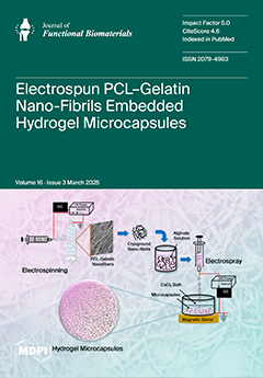
3D hydrogel constructs are widely used in drug delivery, cell therapy, tissue engineering, and regenerative medicine. They mimic the native ECM, offering controlled microenvironments and shielding cells from shear stress and immune responses. In this research, electrospun PCL–gelatin composite nanofibers were fabricated and converted into nanofibrils using a cryogrinding method. These nanofibrils were then dispersed in an alginate solution. Next, electrostatic encapsulation was used to form microcapsules with embedded nanofibrils in a gelling bath. We conclude that embedding nanofibrils in alginate hydrogel microcapsules enhances their physicochemical properties and demonstrates strong biological potential for promoting cell growth, proliferation, and differentiation. View this paper
  • Issues are regarded as officially published after their release is announced to the table of contents alert mailing list.
  • You may sign up for e-mail alerts to receive table of contents of newly released issues.
  • PDF is the official format for papers published in both, html and pdf forms. To view the papers in pdf format, click on the "PDF Full-text" link, and use the free Adobe Reader to open them.
Order results
Result details
Section
Select all
Export citation of selected articles as:
22 pages, 3094 KiB  
Article
Assessing the Effects of Surface-Stabilized Zero-Valent Iron Nanoparticles on Diverse Bacteria Species Using Complementary Statistical Models
by Brittany J. Carnathan, Dinny Stevens, Swarna Shikha, Carson Slater, Nathen Byford, Rodney X. Sturdivant, Kuzy Zarzosa, W. Evan Braswell and Christie M. Sayes
J. Funct. Biomater. 2025, 16(3), 113; https://doi.org/10.3390/jfb16030113 - 20 Mar 2025
Viewed by 675
Abstract
Nanoparticles are proposed as alternatives to traditional antimicrobial agents. By manipulating a nanoparticle’s core and surface coating, antimicrobial effects against various microbial populations can be customized, known as the “designer effect”. However, the antimicrobial properties of nanoparticle core–coating combinations are understudied; little research [...] Read more.
Nanoparticles are proposed as alternatives to traditional antimicrobial agents. By manipulating a nanoparticle’s core and surface coating, antimicrobial effects against various microbial populations can be customized, known as the “designer effect”. However, the antimicrobial properties of nanoparticle core–coating combinations are understudied; little research exists on their effects on diverse bacteria. The antimicrobial effects of surface-stabilized zero-valent iron nanoparticles (FeNPs) are particularly interesting due to their stability in water and ferromagnetic properties. This study explores the impact of FeNPs coated with three surface coatings on six diverse bacterial species. The FeNPs were synthesized and capped with L-ascorbic acid (AA), cetyltrimethylammonium bromide (CTAB), or polyvinylpyrrolidone (PVP) using a bottom-up approach. Zone of inhibition (ZOI) values, assessed through the disc diffusion assay, indicated that AA-FeNPs and CTAB-FeNPs displayed the most potent antibacterial activity. Bacteria inhibition results ranked from most sensitive to least sensitive are the following: Bacillus nealsonii > Escherichia coli > Staphylococcus aureus > Delftia acidovorans > Chryseobacterium sp. > Sphingobacterium multivorum. Comparisons using ordinal regression and generalized linear mixed models revealed significant differences in bacterial responses to the different coatings and nanoparticle concentrations. The statistical model results are in agreement, thus increasing confidence in these conclusions. This study supports the feasibility of the “designer nanoparticle” concept and offers a framework for future research. Full article
(This article belongs to the Section Antibacterial Biomaterials)
Show Figures

Graphical abstract

19 pages, 6443 KiB  
Article
Biocompatible and Antibacterial Chemical Coatings on TiZr Dental Implants
by Vlad Gabriel Vasilescu, Toma Lucian Ciocan, Andreea Mihaela Custura, Florin Miculescu, Miruna Stan, Ionela Cristina Voinea, Dumitru Dima, Florentina Ionela Bucur, Andreea Veronica Dediu-Botezatu, Marian Iulian Neacșu, Elisabeta Vasilescu and Marina Imre
J. Funct. Biomater. 2025, 16(3), 112; https://doi.org/10.3390/jfb16030112 - 20 Mar 2025
Viewed by 580
Abstract
This research aims to study the antibacterial coatings of invasive surgical medical devices, including dental implants, to reduce superficial and deep local infections over the long term. To obtain the coating without altering the initial properties of the substrate (dental implant made of [...] Read more.
This research aims to study the antibacterial coatings of invasive surgical medical devices, including dental implants, to reduce superficial and deep local infections over the long term. To obtain the coating without altering the initial properties of the substrate (dental implant made of TiZr bioalloy), simple, cost-effective, and efficient methods were employed, such as chemical deposition of silver (Ag). The deposition characteristics were analyzed using scanning electron microscopy (SEM), EDX analysis, and FT-IR infrared analysis. The in vitro testing of antimicrobial activity was conducted using the diffusion method by cultivating the bacterial strains Escherichia coli (E. coli) ATCC25922 and Staphylococcus aureus (S. aureus) ATCC25923 and measuring the diameter of the bacterial inhibition zone. Investigations and biocompatibility evaluations were performed on both uncoated and silver-coated (Ag) samples by analyzing cell viability and morphology in the presence of human fetal osteoblasts (hFOB cell line) and human gingival fibroblasts (HFIB-G cells) after 8 days of incubation. The research results confirm the biocompatibility of the coating, demonstrated by the lack of significant differences in cell density between the Ag-coated samples and the control group, as well as by the fact that the silver-coated surface effectively supports actin cytoskeleton organization, adhesion, and migration of both human osteoblasts and gingival fibroblasts. The results regarding the antibacterial efficiency of the silver implant coating indicated that the E. coli bacterial strain is more resistant than S. aureus. The resistance difference between the two bacterial strains was attributed to differences in the structure of their cell envelopes. Full article
(This article belongs to the Special Issue The Development and Future of Dental Implants)
Show Figures

Figure 1

12 pages, 870 KiB  
Review
Class-I and Class-II Restorations with the Application of a Flowable Composite as an Intermediate Layer—A Narrative Review of Clinical Trials
by Anh Duc Nguyen, Kerstin Bitter and Christian Ralf Gernhardt
J. Funct. Biomater. 2025, 16(3), 111; https://doi.org/10.3390/jfb16030111 - 20 Mar 2025
Viewed by 546
Abstract
The objective of this review is to investigate the effect of an additional layer of flowable composite for cavity lining on the clinical outcome of direct posterior composite restorations. The PICO question (patient, intervention, comparison, and outcome) was stated as follows: Does the [...] Read more.
The objective of this review is to investigate the effect of an additional layer of flowable composite for cavity lining on the clinical outcome of direct posterior composite restorations. The PICO question (patient, intervention, comparison, and outcome) was stated as follows: Does the additional application of a flowable composite as a cavity liner improve the clinical outcome of Class-I and Class-II restorations? The electronic databases MEDLINE, Web of Science, LILAS, and BBO were assessed for identifying relevant clinical studies. After removal of duplicate records, 309 records could be identified and, after a screening of the title and abstract, 20 articles were selected for full-text analysis. Finally, six studies met the eligibility criteria and were included in this review for further investigation. Four of the included studies have a follow-up period of two years, while the other two studies had an observation period of three and seven years, respectively. No significant differences in annual failure rates were observed between restorations with and without a flowable composite liner. Consequently, the additional usage of flowable composites as a cavity liner seems to have no effect on the clinical longevity of direct composite restorations in Class-I and Class-II cavities. Therefore, the application of a flowable composite is a possible option in everyday dental clinical practice. Full article
Show Figures

Figure 1

25 pages, 2427 KiB  
Review
Dentistry Insights: Single-Walled and Multi-Walled Carbon Nanotubes, Carbon Dots, and the Rise of Hybrid Materials
by Roxana-Ionela Vasluianu, Ana Maria Dima, Livia Bobu, Alice Murariu, Ovidiu Stamatin, Elena-Raluca Baciu and Elena-Odette Luca
J. Funct. Biomater. 2025, 16(3), 110; https://doi.org/10.3390/jfb16030110 - 20 Mar 2025
Viewed by 553
Abstract
We are committed to writing this narrative review given that carbon-based nanomaterials are revolutionizing dental medicine. Since the groundbreaking discovery of carbon nanotubes in 1991, their dental applications have skyrocketed. The numbers speak for themselves: in 2024, the global carbon nanotubes market hit [...] Read more.
We are committed to writing this narrative review given that carbon-based nanomaterials are revolutionizing dental medicine. Since the groundbreaking discovery of carbon nanotubes in 1991, their dental applications have skyrocketed. The numbers speak for themselves: in 2024, the global carbon nanotubes market hit USD 1.3 billion and is set to double to USD 2.6 billion by 2029. Over the past few decades, various forms of carbon nanomaterials have been integrated into dental practices, elevating the quality and effectiveness of dental treatments. They represent a transformative advancement in dentistry, offering numerous benefits such as augmented mechanical properties, antimicrobial activity, and potential for regenerative applications. Both carbon nanotubes (CNTs) and carbon dots (CDs) are derived from carbon and integral to nanotechnology, showcasing the versatility of carbon nanostructures and delivering cutting-edge solutions across diverse domains, such as electronics, materials science, and biomedicine. CNTs are ambitiously examined for their capability to reinforce dental materials, develop biosensors for detecting oral diseases, and even deliver therapeutic agents directly to affected tissues. This review synthesizes their current applications, underscores their interdisciplinary value in bridging nanotechnology and dentistry, identifies key barriers to clinical adoption, and discusses hybrid strategies warranting further research to advance implementation. Full article
(This article belongs to the Section Dental Biomaterials)
Show Figures

Figure 1

20 pages, 15356 KiB  
Article
A Carbon-Based Nanomaterial with Dichotomous Effects: Antineoplastic on Oral Cancer Cells and Osteoinductive/Chondroinductive on Dental Pulp Stem Cells
by Milica Jaksic Karisik, Nataša Jović Orsini, Jelena Carkic, Milos Lazarevic, Dijana Mitić, Bojan Jokanovic, Vukoman Jokanović and Jelena Milasin
J. Funct. Biomater. 2025, 16(3), 109; https://doi.org/10.3390/jfb16030109 - 19 Mar 2025
Viewed by 421
Abstract
Background: Oral cancer is an aggressive malignancy with modest survival rates. It also causes disfigurement following surgical removal of the tumor, thus highlighting the need for new cancer treatment and tissue repair modalities. Carbon-based nanomaterials have emerged as promising tools in both anticancer [...] Read more.
Background: Oral cancer is an aggressive malignancy with modest survival rates. It also causes disfigurement following surgical removal of the tumor, thus highlighting the need for new cancer treatment and tissue repair modalities. Carbon-based nanomaterials have emerged as promising tools in both anticancer and regenerative therapies. Objectives: We aimed to synthesize a new carbon-based nanomaterial (CBN) and test its antineoplastic effects, as well as its potential regenerative capacity. Materials and Methods: A carbon nanomaterial, obtained by ball milling graphite flakes, was functionalized with polyvinylpyrrolidone (CBN/PVP). Its physicochemical properties were explored with X-ray diffraction (XRD), attenuated total reflection–Fourier transform infrared spectroscopy (ATR-FTIR), micro-Raman spectroscopy, fluorescent and scanning electron microscopy, and wettability analysis. For the antineoplastic effects investigation, oral cancer cells were treated with CBN/PVP and examined with MTT and migration assays, as well as cell-cycle and ROS production analyses. Gene expression was determined by qPCR. To examine the pro-regenerative capacity of CBN/PVP, dental pulp stem cell cultures (DPSCs) were treated with the nanomaterial and subjected to osteo- and chondro-induction. Results: Lower concentrations of CBN/PVP (50, 100 μg/mL) applied on cancer cells exerted remarkable cytotoxic effects, induced G1 cell-cycle arrest, and reduced cancer cell invasion potential by different mechanisms, including downregulation of the PI3K/AKT/mTOR pathway. In contrast, the addition of 50 µg/mL of CBN/PVP to DPSCs stimulated their survival and proliferation. CBN/PVP significantly enhanced both the osteogenic (p < 0.05) and chondrogenic (p < 0.01) induction of DPSCs. Conclusions: The novel carbon-based nanomaterial displays unique characteristics, making it suitable in anticancer and regenerative therapies concomitantly. Full article
(This article belongs to the Special Issue Recent Studies on Biomaterials for Tissue Repair and Regeneration)
Show Figures

Graphical abstract

29 pages, 5047 KiB  
Article
ZnO Nanoparticle-Infused Vaterite Coatings: A Novel Approach for Antimicrobial Titanium Implant Surfaces
by Atiđa Selmani, Scarlett Zeiringer, Ankica Šarić, Anamarija Stanković, Aleksander Učakar, Janja Vidmar, Anže Abram, Branka Njegić Džakula, Jasminka Kontrec, Anamarija Zore, Klemen Bohinc, Eva Roblegg and Nives Matijaković Mlinarić
J. Funct. Biomater. 2025, 16(3), 108; https://doi.org/10.3390/jfb16030108 - 19 Mar 2025
Viewed by 496
Abstract
Loss of implant function is a common complication in orthopaedic and dental surgery. Among the primary causes of implant failure are peri-implant infections which often result in implant removal. This study demonstrates the development of a new antimicrobial titanium coating with ZnO nanoparticles [...] Read more.
Loss of implant function is a common complication in orthopaedic and dental surgery. Among the primary causes of implant failure are peri-implant infections which often result in implant removal. This study demonstrates the development of a new antimicrobial titanium coating with ZnO nanoparticles of various sizes and morphologies immobilised in poly(allylamine hydrochloride) and alginate multilayers, combined with epitaxially grown vaterite crystals. The coated samples were characterised with various methods (FTIR, XRD, SEM) and surface properties were evaluated via water contact angle and surface charge measurements. Zinc ion release was quantified using ICP-MS. The antimicrobial efficacy of the coatings was tested against Staphylococcus aureus, Staphylococcus epidermidis, and Candida albicans while the biocompatibility was tested with preosteoblast cells (MC3T3-E1). Results demonstrated the successful preparation of a calcium carbonate/ZnO composite coating with epitaxially grown vaterite on titanium surfaces. The Zn ions released from ZnO nanoparticles dramatically influenced the morphology of vaterite where a new flower-like morphology was observed. The coated titanium surfaces exhibited robust antimicrobial activity, achieving over 90% microbial viability reduction for Staphylococcus aureus, Staphylococcus epidermidis, and Candida albicans. Importantly, the released Zn2+ concentrations remained below the cytotoxicity limit for MC3T3-E1 cells, showing potential for safe and effective implant applications. Full article
(This article belongs to the Section Antibacterial Biomaterials)
Show Figures

Graphical abstract

15 pages, 5359 KiB  
Article
Biomechanical Evaluation of a Novel Non-Engaging Abutment and Screw in Internal Implant Systems: Comparative Fatigue and Load Testing
by Su-Min Cho, Soo-Hwan Byun, So-Yee Ahn, Hyun-Sook Han, Sung-Woon On, Sang-Yoon Park, Sang-Min Yi, In-Young Park, Byoung-Eun Yang and Lee-Kyoung Kim
J. Funct. Biomater. 2025, 16(3), 107; https://doi.org/10.3390/jfb16030107 - 19 Mar 2025
Viewed by 697
Abstract
Dental implants rely on precise prosthetic design and biomechanical stability to ensure long-term success. This study evaluates the mechanical performance of non-engaging abutments in multi-unit combined screw- and cement-retained prostheses (CSCRP) using two internal implant systems: the BlueDiamond (BD) and AnyOne (AO) systems. [...] Read more.
Dental implants rely on precise prosthetic design and biomechanical stability to ensure long-term success. This study evaluates the mechanical performance of non-engaging abutments in multi-unit combined screw- and cement-retained prostheses (CSCRP) using two internal implant systems: the BlueDiamond (BD) and AnyOne (AO) systems. Unlike conventional implant systems that utilize the same type of screw for both engaging and non-engaging abutments, the BD system employs a distinct screw design for non-engaging abutments. A total of 80 implants were tested, with 40 in each group. Mechanical testing included static compressive load and fatigue tests following ISO 14801 standards. The BD system demonstrated significantly higher compressive strength (326.32 kgf vs. 231.82 kgf, p < 0.001) and 23.4% greater fatigue strength compared to the AO system. Precision fit analysis confirmed no significant deformation, microcracks, or fractures after 5 million loading cycles. These findings suggest that the BD system’s unique screw design for non-engaging abutments contributes to improved mechanical performance and durability. Further clinical studies are needed to assess the long-term implications of this design on prosthetic stability and implant longevity. Full article
(This article belongs to the Special Issue Advanced Biomaterials and Oral Implantology—3rd Edition)
Show Figures

Figure 1

15 pages, 5200 KiB  
Article
Designing Superhydrophilic 3D Porous Surfaces on Polyetherketoneketone Surfaces to Promote Biocompatibility
by Hui-Ching Lin, Chiang-Sang Chen, Kai-Yi Lin, Ya-Lin Huang, Hao-Hsiang Hsu, Yu-Lin Kuo, Wei-Cheng Chen and Her-Hsiung Huang
J. Funct. Biomater. 2025, 16(3), 106; https://doi.org/10.3390/jfb16030106 - 14 Mar 2025
Viewed by 684
Abstract
Polyetherketoneketone (PEKK) exhibits satisfactory mechanical properties and biocompatibility, with an elastic modulus closely resembling that of natural bone. This property reduces the stress-shielding effect associated with bone implants. However, the biological inertness of the PEKK surface remains a significant limitation for its application [...] Read more.
Polyetherketoneketone (PEKK) exhibits satisfactory mechanical properties and biocompatibility, with an elastic modulus closely resembling that of natural bone. This property reduces the stress-shielding effect associated with bone implants. However, the biological inertness of the PEKK surface remains a significant limitation for its application in bone tissue engineering. The objective of this study was to create a superhydrophilic 3D porous structure on the surface of PEKK to enhance biocompatibility, in terms of vascularization and bone remodeling. A combination of mechanical, chemical, and physical surface treatments was employed to modify the PEKK surface. Initially, mechanical sandblasting was used to create a rough surface to promote mechanical interlocking with bone tissue. Subsequently, chemical acid etching and physical low-temperature atmospheric plasma cleaning were applied to develop a superhydrophilic 3D porous surface. The modified surfaces were characterized for morphology, roughness, hydrophilicity, and functional groups. Cellular responses, including vascularization and bone remodeling, were evaluated to assess the potential for improved biocompatibility. The combination of acid etching and low-temperature atmospheric plasma cleaning, with or without prior sandblasting, successfully created a superhydrophilic 3D porous structure on the PEKK surface. This modified surface enhanced the tube formation in human umbilical vein endothelial cells. It also promoted the adhesion and mineralization of human bone marrow mesenchymal stem cells and slightly reduced tartrate-resistant acid phosphatase expression and F-actin ring size in mouse macrophage cells. This study introduces an innovative and effective surface modification strategy for PEKK surface, combining mechanical, chemical, and physical treatments to enhance biocompatibility. The modified PEKK surface promotes angiogenic and osteogenic responses while slightly inhibiting osteoclastic activity, making it a potential alternative for dental and orthopedic PEKK implant applications. Full article
(This article belongs to the Collection Feature Papers in Bone Biomaterials)
Show Figures

Figure 1

36 pages, 3511 KiB  
Review
Three-Dimensional Bioprinting for Intervertebral Disc Regeneration
by Md Amit Hasan Tanvir, Md Abdul Khaleque, Junhee Lee, Jong-Beom Park, Ga-Hyun Kim, Hwan-Hee Lee and Young-Yul Kim
J. Funct. Biomater. 2025, 16(3), 105; https://doi.org/10.3390/jfb16030105 - 14 Mar 2025
Viewed by 1033
Abstract
The rising demand for organ transplants and the need for precise tissue models have positioned the in vitro biomanufacturing of tissues and organs as a pivotal area in regenerative treatment. Considerable development has been achieved in growing tissue-engineered intervertebral disc (IVD) scaffolds, designed [...] Read more.
The rising demand for organ transplants and the need for precise tissue models have positioned the in vitro biomanufacturing of tissues and organs as a pivotal area in regenerative treatment. Considerable development has been achieved in growing tissue-engineered intervertebral disc (IVD) scaffolds, designed to meet stringent mechanical and biological compatibility criteria. Among the cutting-edge approaches, 3D bioprinting stands out due to its unparalleled capacity to organize biomaterials, bioactive molecules, and living cells with high precision. Despite these advancements, polymer-based scaffolds still encounter limitations in replicating the extracellular matrix (ECM)-like environment, which is fundamental for optimal cellular activities. To overcome these challenges, integrating polymers with hydrogels has been recommended as a promising solution. This combination enables the advancement of porous scaffolds that nurture cell adhesion, proliferation, as well as differentiation. Additionally, bioinks derived from the decellularized extracellular matrix (dECM) have exhibited potential in replicating biologically relevant microenvironments, enhancing cell viability, differentiation, and motility. Hydrogels, whether derived from natural sources involving collagen and alginate or synthesized chemically, are highly valued for their ECM-like properties and superior biocompatibility. This review will explore recent advancements in techniques and technologies for IVD regeneration. Emphasis will be placed on identifying research gaps and proposing strategies to bridge them, with the goal of accelerating the translation of IVDs into clinical applications. Full article
(This article belongs to the Special Issue Three-Dimensional-Printable Biomaterials for Bone Regeneration)
Show Figures

Figure 1

43 pages, 1876 KiB  
Review
Evolution of Dental Resin Adhesives—A Comprehensive Review
by Waad Khalid Alomran, Mohammed Zahedul Islam Nizami, Hockin H. K. Xu and Jirun Sun
J. Funct. Biomater. 2025, 16(3), 104; https://doi.org/10.3390/jfb16030104 - 14 Mar 2025
Viewed by 936
Abstract
This comprehensive review of dental resin adhesives explores their historical development, key components, recent innovations, and potential future directions, highlighting a dynamic and continually advancing field. From Buonocore’s breakthrough acid-etching technique and Bowen’s pioneering dental resin invention, successive generations of clinicians and scientists [...] Read more.
This comprehensive review of dental resin adhesives explores their historical development, key components, recent innovations, and potential future directions, highlighting a dynamic and continually advancing field. From Buonocore’s breakthrough acid-etching technique and Bowen’s pioneering dental resin invention, successive generations of clinicians and scientists have pushed forward the technological and materials development for secure bonding, while preserving dental tissues. The review discusses the substantial advances in improving adhesive reliability, enabling more conservative treatment approaches. It also delves into enhancing fundamental adhesive components and their synergistic combinations. Recent innovations, including biostable and functional resins, nanotechnology, and bioactive components, address persistent challenges such as durability, antimicrobial efficacy, and therapeutic functionality. Emerging technologies, such as digital dentistry, artificial intelligence, and bioinspired adhesives, portend an exciting and promising future for dental adhesives. This review underscores the critical role of ongoing research in developing biocompatible, multifunctional, and durable adhesives. It aims to support dental professionals and researchers by providing a comprehensive understanding of the dynamic progression of dental adhesives, inspiring continued innovation and excellence in restorative dentistry. Full article
(This article belongs to the Special Issue Recent Advances in Dental Resin Composites)
Show Figures

Figure 1

22 pages, 5628 KiB  
Article
Effect of Pixel Offset Adjustments for XY Plane Dimensional Compensation in Digital Light Processing 3D Printing on the Surface Trueness and Fit of Zirconia Crowns
by KeunBaDa Son, Ji-Min Lee, Kyoung-Jun Jang, Sang-Kyu Lee, Jun Ho Hwang, Jong Hoon Lee, Hyun Deok Kim, So-Yeun Kim and Kyu-Bok Lee
J. Funct. Biomater. 2025, 16(3), 103; https://doi.org/10.3390/jfb16030103 - 14 Mar 2025
Viewed by 579
Abstract
This study aimed to evaluate the effect of pixel offset adjustments in digital light processing (DLP) three-dimensional (3D) printing on the marginal and internal fit and surface trueness of zirconia crowns. Zirconia crowns were designed using dental computer-aided design software (Dentbird; Imagoworks) and [...] Read more.
This study aimed to evaluate the effect of pixel offset adjustments in digital light processing (DLP) three-dimensional (3D) printing on the marginal and internal fit and surface trueness of zirconia crowns. Zirconia crowns were designed using dental computer-aided design software (Dentbird; Imagoworks) and fabricated with a vat photopolymerization DLP 3D printer (TD6+; 3D Controls) under three pixel offset conditions (−1, 0, and 1). Pixel offset refers to the controlled modification of the outermost pixels in the XY plane during printing to compensate for potential dimensional inaccuracies. The marginal and internal fit was assessed using a triple-scan protocol and quantified using root mean square (RMS) values. Surface trueness was evaluated by measuring RMS, positive and negative errors between the designed and fabricated crowns. Statistical analyses included one-way ANOVA and Pearson correlation analysis (α = 0.05). The Pixel offset had a significant effect on fit accuracy and surface trueness (p < 0.05). Higher pixel offsets increased marginal discrepancies (p = 0.004), with the marginal gap exceeding 120 µm at a pixel offset of 1 (114.5 ± 14.6 µm), while a pixel offset of −1 (85.5 ± 18.6 µm) remained within acceptable limits (p = 0.003). Surface trueness worsened with increasing pixel offset, showing greater positive errors (p < 0.001). Optimizing pixel offset in DLP 3D printing is crucial to ensuring clinically acceptable zirconia crowns. Improper settings may increase marginal discrepancies and surface errors, compromising restoration accuracy. Full article
(This article belongs to the Special Issue Advanced 3D Printing Biomaterials)
Show Figures

Figure 1

10 pages, 3362 KiB  
Communication
Preclinical Performance of a Novel Dental Implant Design Reducing Mechanical Stress in Cortical Bone
by Carolin Erbel, Matthias W. Laschke, Tanja Grobecker-Karl and Matthias Karl
J. Funct. Biomater. 2025, 16(3), 102; https://doi.org/10.3390/jfb16030102 - 14 Mar 2025
Viewed by 1777
Abstract
This animal study compared the healing performance of a novel implant design characterized by a shift in thread geometry and core diameter with two different surfaces with that of an apically tapered implant. Test Bioactive (n = 9), Test Porous (n = 7) [...] Read more.
This animal study compared the healing performance of a novel implant design characterized by a shift in thread geometry and core diameter with two different surfaces with that of an apically tapered implant. Test Bioactive (n = 9), Test Porous (n = 7) and Control (n = 8) implants were placed in the mandibles of minipigs. Following healing, bone samples were harvested for determining bone-to-implant contact (BIC) and marginal bone loss (MBL). Comparative statistics were based on Levene’s test, Shapiro–Wilk tests, the Kruskal–Wallis test and Wilcoxon tests with Holm correction (α = 0.05). The mean undersizing of the osteotomy was 0.15 mm for Control, while in the test groups 0.33 mm and 0.34 mm were calculated. Insertion torques ranged from 61.5 Ncm (Control) to 76.1 Ncm (Test Bioactive). Maximum BIC was seen in Test Porous with 55.83%, while Test Bioactive showed only 48.11%. MBL was 4.1 mm in Test Bioactive, while Test Porous and Control exhibited 2.8 mm. No significant differences between the implant groups were observed (p > 0.05). Despite greater undersizing, the novel implant type performed comparably to the established Control implants. The rougher surface of the bioactive implants increased the insertion torque and led to more MBL. Full article
(This article belongs to the Section Dental Biomaterials)
Show Figures

Figure 1

14 pages, 3056 KiB  
Article
Spatial Platform for Periodontal Ligament Angulation and Regeneration: In Vivo Pilot Study
by Min Guk Kim, Do-Yeon Kim, Hyoung-Gon Ko, Jin-Seok Byun, Joong-Hyun Kim and Chan Ho Park
J. Funct. Biomater. 2025, 16(3), 99; https://doi.org/10.3390/jfb16030099 - 13 Mar 2025
Viewed by 570
Abstract
The periodontal ligament (PDL) is a fibrous connective tissue that anchors the tooth-root surface to the alveolar bone with specific orientations. It plays a crucial role in functional restoration, optimal position stabilities, biomechanical stress transmission, and appropriate tissue remodeling in response to masticatory [...] Read more.
The periodontal ligament (PDL) is a fibrous connective tissue that anchors the tooth-root surface to the alveolar bone with specific orientations. It plays a crucial role in functional restoration, optimal position stabilities, biomechanical stress transmission, and appropriate tissue remodeling in response to masticatory loading conditions. This pilot study explored spatial microarchitectures to promote PDL orientations while limiting mineralized tissue formation. A computer-designed perio-complex scaffold was developed with two parts: (1) PDL-guiding architectures with defined surface topography and (2) a bone region with open structures. After SEM analysis of micropatterned topographies on PDL-guiding architectures, perio-complex scaffolds were transplanted into two-wall periodontal defects in the canine mandible. Despite the limited bone formation at the 4-week timepoint, bone parameters in micro-CT quantifications showed statistically significant differences between the no-scaffold and perio-complex scaffold transplantation groups. Histological analyses demonstrated that the PDL-guiding architecture regulated fiber orientations and facilitated the functional restoration of PDL bundles in immunohistochemistry with periostin and decorin (DCN). The perio-complex scaffold exhibited predictable and controlled fibrous tissue alignment with specific angulations, ensuring spatial compartmentalization for PDL tissues and bone regenerations. These findings highlighted that the perio-complex scaffold could serve as an advanced therapeutic approach to contribute periodontal tissue regeneration and functional restoration in tooth-supporting structures. Full article
(This article belongs to the Special Issue Advanced Biomaterials for Periodontal Regeneration)
Show Figures

Figure 1

15 pages, 5530 KiB  
Article
Formulating Mechanically Robust Composite Restorative Materials for High Performance
by Austyn Salazar, Natalie Anderson and Jeffrey Stansbury
J. Funct. Biomater. 2025, 16(3), 101; https://doi.org/10.3390/jfb16030101 - 13 Mar 2025
Viewed by 672
Abstract
Although dental resin composite restoratives offer a widely used direct-placement treatment option aimed at replacing the form and function of a natural tooth, there are several clinically relevant performance aspects of these materials that can be improved. The formulation of the resin matrix [...] Read more.
Although dental resin composite restoratives offer a widely used direct-placement treatment option aimed at replacing the form and function of a natural tooth, there are several clinically relevant performance aspects of these materials that can be improved. The formulation of the resin matrix phase of dental composites for high-efficiency photopolymerization leading to polymers with excellent mechanical properties has always been a challenge that is addressed here through the use of structurally new and more reactive monomers as well as the formation of polymer networks that incorporate non-covalent reinforcing interactions. The purpose of this study was to validate that a set of tetraurethane diacrylates (TUDAs) with a novel configuration of their urethane linkages in coordination with acidic comonomers could be devised to obtain highly robust new composite materials. Due to the novel molecular design, this exploratory approach was conducted using reaction kinetics and three-point bend testing to assess the performance. Conversion and mechanical properties were measured to refine these formulations prior to the addition of filler. The initial formulations demonstrated outstanding dry mechanical test results that subsequently showed a major intolerance to water storage, which led to a model study using urethane diacrylate (UDA) followed by the addition of hydrophobic TUDA monomers. Once the resin formulations were optimized, silane-treated particulate filler was added to determine the effectiveness as composite materials. The final formulation used a hydrophobic, aromatic TUDA along with 4-methacryloxyethyl trimellitic anhydride (4-META) as a latent acidic comonomer and a mixture of acrylic acid (AA) and methacrylic acid (MAA). This formulation achieves a very high level of both reactivity and mechanical properties relative to current dental composite restoratives. Full article
(This article belongs to the Special Issue State-of-the-Art Dental Adhesives and Restorative Composites)
Show Figures

Figure 1

22 pages, 4851 KiB  
Article
Porous Hydrogels Prepared by Two-Step Gelation Method for Bone Regeneration
by Yongzhi Li, Jiangshan Liu, Jiawei Wei, Li Yuan, Jiaxin Hu, Siluo Dai, Yubao Li and Jidong Li
J. Funct. Biomater. 2025, 16(3), 100; https://doi.org/10.3390/jfb16030100 - 13 Mar 2025
Viewed by 621
Abstract
Hierarchical porous hydrogels possess advantageous characteristics that facilitate cell adhesion, promote tissue growth, and enhance angiogenesis and osteogenesis. In this study, porous composite hydrogels were successfully prepared by a two-step gelation method with sodium alginate (SA), gelatin (GEL), and calcium hydrogen phosphate (DCP) [...] Read more.
Hierarchical porous hydrogels possess advantageous characteristics that facilitate cell adhesion, promote tissue growth, and enhance angiogenesis and osteogenesis. In this study, porous composite hydrogels were successfully prepared by a two-step gelation method with sodium alginate (SA), gelatin (GEL), and calcium hydrogen phosphate (DCP) as the main components. The fabricated porous hydrogels initially featured small pores (approximately 60 μm), and gradually evolved to large pores (exceeding 250 μm) during the gradual degradation in the cellular microenvironment. In vitro cell culture experiments indicated that these hydrogels could enhance the proliferation and osteogenic differentiation of bone marrow mesenchymal stem cells due to the hierarchical porous structure and the incorporation of DCP. Subcutaneous implantation and cranial defect repair experiments in Sprague−Dawley rats further confirmed that the small initial pore size of hydrogel scaffolds can provide more sites for cell adhesion. Additionally, the gradual degradation to form large pores was conducive to cell/tissue growth and blood vessel formation, ultimately being beneficial for vascularized bone regeneration. In summary, this study proposes an innovative strategy for developing porous hydrogels with gradual degradation for functional bone regeneration. Full article
(This article belongs to the Section Bone Biomaterials)
Show Figures

Figure 1

15 pages, 3413 KiB  
Article
Hybridization Chain Reaction-Enhanced Ultrasensitive Electrochemical Analysis of miRNAs with a Silver Nano-Reporter on a Gold Nanostructured Electrode Array
by Bin Wang, Huiqiang Ma, Mingxing Zhou, Xian Huang, Ying Gan and Hong Yang
J. Funct. Biomater. 2025, 16(3), 98; https://doi.org/10.3390/jfb16030098 - 12 Mar 2025
Viewed by 540
Abstract
Abnormal expression of miRNAs is associated with the occurrence and progression of cancer and other diseases, making miRNAs essential biomarkers for disease diagnosis and prognosis. However, the intrinsic properties of miRNAs, such as short length, low abundance, and high sequence homology, represent great [...] Read more.
Abnormal expression of miRNAs is associated with the occurrence and progression of cancer and other diseases, making miRNAs essential biomarkers for disease diagnosis and prognosis. However, the intrinsic properties of miRNAs, such as short length, low abundance, and high sequence homology, represent great challenges for fast and accurate miRNA detection in clinics. Herein, we developed a novel hybridization chain reaction (HCR)-based electrochemical miRNAs chip (e-miRchip), featured with gold nanostructured electrodes (GNEs) and silver nanoparticle reporters (AgNRs), for sensitive and multiplexed miRNA detection. AgNRs were synthesized and applied on the e-miRchip to generate strong redox signals in the presence of miRNA. The stem–loop capture probe was covalently immobilized on the GNEs, and was opened upon miRNA hybridization to consequently trigger the HCR for signal amplification. The multiple long-repeated DNA helix generated by HCR provides the binding sites for the AgNRs, contributing to the amplification of the electrochemical signals of miRNA hybridization. To optimize the detection sensitivity, GNEs with three distinct structures were electroplated, in which flower-like GNEs were found to be the best electrode morphology for miRNAs analysis. Under optimal conditions, the HCR-based e-miRchip showed an excellent detection performance with an LOD of 0.9 fM and a linear detection range from 1 fM to 10 pM. Moreover, this HCR-based e-miRchip platform was able to effectively distinguish miRNAs from the one- or two-base mismatches. This HCR-based e-miRchip holds great potential as a highly efficient and promising miRNA detection platform for the diagnosis and prognosis of cancer and other diseases in the future. Full article
(This article belongs to the Special Issue Women’s Special Issue Series: Functional Biomaterials (2nd Edition))
Show Figures

Figure 1

14 pages, 1860 KiB  
Review
Optimizing Flexor Digitorum Profundus Tendon Repair: A Narrative Review
by Rishith R. Mereddy, Emily E. Zona, Camille J. LaLiberte and Aaron M. Dingle
J. Funct. Biomater. 2025, 16(3), 97; https://doi.org/10.3390/jfb16030097 - 11 Mar 2025
Viewed by 1157
Abstract
Zone II flexor digitorum profundus (FDP) tendon injuries are complex, and present significant challenges in hand surgery, due to the need to balance strength and flexibility during repair. Traditional suture techniques often lead to complications such as adhesions or tendon rupture, prompting the [...] Read more.
Zone II flexor digitorum profundus (FDP) tendon injuries are complex, and present significant challenges in hand surgery, due to the need to balance strength and flexibility during repair. Traditional suture techniques often lead to complications such as adhesions or tendon rupture, prompting the exploration of novel strategies to improve outcomes. This review investigates the use of flexor digitorum superficialis (FDS) tendon autografts to reinforce FDP repairs, alongside the integration of biomaterials to enhance mechanical strength without sacrificing FDS tissue. Key biomaterials, including collagen–polycaprolactone (PCL) composites, are evaluated for their biocompatibility, mechanical integrity, and controlled degradation properties. Collagen-PCL emerges as a leading candidate, offering the potential to reduce adhesions and promote tendon healing. Although nanomaterials such as nanofibers and nanoparticles show promise in preventing adhesions and supporting cellular proliferation, their application remains limited by manufacturing challenges. By combining advanced repair techniques with biomaterials like collagen-PCL, this approach aims to improve surgical outcomes and minimize complications. Future research will focus on validating these findings in biological models, assessing tendon healing through imaging, and comparing the cost-effectiveness of biomaterial-enhanced repairs with traditional methods. This review underscores the potential for biomaterial-based approaches to transform FDP tendon repair. Full article
Show Figures

Figure 1

20 pages, 6685 KiB  
Article
Changes in the Properties of Different Zones in Multilayered Translucent Zirconia Used in Monolithic Restorations During Aging Process
by Phil-Joon Koo, Jong-Hyuk Lee, Seung-Ryong Ha, Deog-Gyu Seo, Jin-Soo Ahn and Yu-Sung Choi
J. Funct. Biomater. 2025, 16(3), 96; https://doi.org/10.3390/jfb16030096 - 10 Mar 2025
Viewed by 650
Abstract
This study assessed the changes in the mechanical and surface properties of the transition zone in multilayered translucent monolithic zirconia subjected to long-term hydrothermal aging. A total of 360 disk-shaped specimens (diameter: 15.0 mm; thickness: 1.2 mm) were prepared using conventional (3Y-TZP in [...] Read more.
This study assessed the changes in the mechanical and surface properties of the transition zone in multilayered translucent monolithic zirconia subjected to long-term hydrothermal aging. A total of 360 disk-shaped specimens (diameter: 15.0 mm; thickness: 1.2 mm) were prepared using conventional (3Y-TZP in LT; ZL, 4Y-TZP in MT; ZM) and multilayered translucent zirconia (5Y-TZP in MT Multi; ZT, 3Y/5Y-TZP in Prime; ZP) among IPS e.max ZirCAD blocks. Specimens were divided into three groups (n = 30) and aged in the autoclave at 134 °C under 0.2 MPa for 0 h (control group), 5 h (first aged group), and 10 h (second aged group). The mechanical and surface properties of the transition zone in the multilayered translucent zirconia were investigated, followed by statistical analysis (α = 0.05). Before and after aging, ZL (1102.64 ± 41.37 MPa) and ZP (1014.71 ± 139.86 MPa) showed the highest biaxial flexural strength (BFS); ZL showed the highest Weibull modulus (31.46) and characteristic strength (1121.63 MPa); and ZT exhibited the highest nanoindentation hardness (20.40 ± 1.80 GPa) and Young’s modulus (284.90 ± 20.07 GPa). After aging, ZL (116.75 ± 9.80 nm) exhibited the highest surface roughness (Ra); the monoclinic phase contents in ZL and ZP increased; and surface uplifts, microcracks, and irregular defects caused by phase transformation appeared on ZL and ZP surfaces. The 3Y/5Y-TZP transition zone exhibited flexural strength, Vickers hardness, phase distribution changes, and surface microstructure changes similar to those of 3Y-TZP before and after aging; however, the surface roughness was lower than that of 3Y-TZP and higher than those of 4Y-TZP and 5Y-TZP after aging. The mechanical and surface characteristics, excluding BFS and Vickers hardness, were influenced by the yttrium oxide content in each zone and the aging process. Full article
Show Figures

Graphical abstract

17 pages, 1156 KiB  
Article
Guided Tissue Regeneration of Periodontal Infrabony Defects with Frozen Radiation-Sterilized Allogenic Bone Graft Versus Deproteinized Bovine Bone Mineral: 5-Year Outcomes of RCT
by Bartłomiej Górski, Aniela Brodzikowska, Kacper Nijakowski and Mariano Sanz
J. Funct. Biomater. 2025, 16(3), 95; https://doi.org/10.3390/jfb16030095 - 10 Mar 2025
Viewed by 689
Abstract
The aim of this study was to compare the efficacy of the guided tissue regeneration (GTR) of periodontal infrabony defects using the frozen radiation-sterilized allogenic bone graft (FRSABG) versus deproteinized bovine bone mineral (DBBM) 5 years after treatment. The association between patients’ compliance [...] Read more.
The aim of this study was to compare the efficacy of the guided tissue regeneration (GTR) of periodontal infrabony defects using the frozen radiation-sterilized allogenic bone graft (FRSABG) versus deproteinized bovine bone mineral (DBBM) 5 years after treatment. The association between patients’ compliance and periodontitis recurrence with 5-year outcomes was also evaluated. Thirty infrabony defects in 15 stage III/IV periodontitis patients were randomly allocated to the FRSBAG group (tests) or the DBBM group (controls). Between 1 and 5 years, one patient was lost to follow-up and one tooth was extracted due to root fracture. No tooth was extracted for periodontal reasons. Consequently, 13 teeth in test sites and 14 teeth in control sites were available for the 5-year analysis. The clinical attachment level gain (CAL-G, primary outcome), probing pocket depth (PPD), radiographic defect depth (DD), and linear defect fill (LDF) were examined at baseline and 5 years post-surgically. Both groups showed statistically significant improvements in all evaluated clinical and radiographic parameters at 5 years, with insignificant intergroup differences. CAL-Gs were 4.46 ± 2.07 mm in the FRSBAG group, and 3.86 ± 1.88 mm in the DBBM group (p = 0.5442). In six (43%) patients, we observed periodontitis recurrence, among whom two (33.33%) participated regularly in supportive periodontal care (SPC) and the other four (66.7%) did not take part in SPC. A regression analysis revealed that periodontitis recurrence was a significant predictor of CAL loss and DD increase. FRSBAG and DBBM were both equally effective 5 years after the GTR of infrabony defects. Within the limitations of the present study, its outcomes advocate that both grafts may be considered as a viable option based on patient preferences and clinical considerations. Full article
(This article belongs to the Special Issue Advanced Biomaterials for Periodontal Regeneration)
Show Figures

Figure 1

13 pages, 16595 KiB  
Article
Effects of ECM Components on Periodontal Ligament Stem Cell Differentiation Under Conditions of Disruption of Wnt and TGF-β Signaling Pathways
by Alla V. Kuznetsova, Olga P. Popova, Tamara I. Danilova, Andrey V. Latyshev, Oleg O. Yanushevich and Alexey A. Ivanov
J. Funct. Biomater. 2025, 16(3), 94; https://doi.org/10.3390/jfb16030094 - 9 Mar 2025
Viewed by 681
Abstract
Periodontitis is accompanied by inflammation that causes dysregulation of the Wnt/β-catenin and TGF-β signaling pathways. This leads to a violation of the homeostasis of periodontal tissues. Components of the extracellular matrix (ECM) are an important part of biomaterials used for the repair of [...] Read more.
Periodontitis is accompanied by inflammation that causes dysregulation of the Wnt/β-catenin and TGF-β signaling pathways. This leads to a violation of the homeostasis of periodontal tissues. Components of the extracellular matrix (ECM) are an important part of biomaterials used for the repair of periodontal tissue. The purpose of this study was to evaluate the components of the effect of ECM (hyaluronic acid (HA), fibronectin (Fn), and laminin (Lam)) on the osteogenic and odontogenic differentiation of periodontal ligament stem cells (PDLSCs) in the collagen I hydrogel under conditions of disruption of the Wnt/β-catenin and TGF-β signaling pathways. The study showed that the addition of components of the ECM restored the expression of odontogenic markers in PDLSCs, which was absent during inhibition of the canonical Wnt signaling pathway, and their multidirectional effect on the secretion of transforming growth factor-β1 (TGF-β1) and bone morphogenetic protein 2 (BMP-2). Fn and Lam suppressed the expression of odontogenic markers in PDLSCs against the background of inhibition of the TGF-β signaling pathway. The addition of HA under the conditions of the TGF-β signaling pathway improved BMP-2 secretion, preserving odontogenic differentiation. Thus, our results demonstrated that disruption of the Wnt/β-catenin and TGF-β signaling pathways causes disorders in the differentiation of PDLSCs, preventing the regeneration of periodontal tissues. This should be taken into account when developing multicomponent scaffolds that recapitulate the ECM microenvironment at endogenic regeneration of the periodontium. Inclusion of hyaluronic acid as one of these components may enhance the therapeutic effect of such biomaterials. Full article
(This article belongs to the Special Issue Advances in Multifunctional Hydrogels for Biomedical Application)
Show Figures

Figure 1

19 pages, 6726 KiB  
Article
Intraoperative Profiling of the Supracrestal Implant Complex Minimizes Peri-Implant Crestal Bone Remodeling: The Guided Bone Profiling Concept
by Milan Stoilov, Joerg Winterhoff, Lea Stoilov, Anastasia Timoschenko, Helmut Stark, Florian Heuser, Michael Marder, Dominik Kraus and Norbert Enkling
J. Funct. Biomater. 2025, 16(3), 93; https://doi.org/10.3390/jfb16030093 - 8 Mar 2025
Viewed by 843
Abstract
(1) Background: Early-stage bone resorption following implant placement can significantly impact the long-term success of implants. This study evaluates whether a fully digitally planned implant position based on the E-point concept, along with guided profiling of the supracrestal complex, contributes to improved stability [...] Read more.
(1) Background: Early-stage bone resorption following implant placement can significantly impact the long-term success of implants. This study evaluates whether a fully digitally planned implant position based on the E-point concept, along with guided profiling of the supracrestal complex, contributes to improved stability of peri-implant bone levels. (2) Methods: 29 implants were placed in 27 patients utilizing both immediate (Group 1; n = 19) and delayed placement (Group 2; n = 10) protocols. Implant position and emergence profile were preoperatively determined and consistently executed through guided surgery and CAD/CAM-fabricated restorations. Due to the subcrestal positioning of the implant, a corresponding bone profiler with a guide pin was used to shape the emergence profile and prevent the provisional restoration from impinging on the proximal bone. Provisional restorations were immediately placed to support the emergence profile. Bone level changes were documented radiographically over a two-year period. The first Bone-to-Implant Contact Level (∆ fBIC), change in highest approximal Bone Level (∆ haBL), and formation of an emergence profile width (WEP) were measured. (3) Results: All implants and restorations survived after two years, no significant change in first Bone-to-Implant Contact Level (∆ fBIC = 0 ± 0.02 mm), no change in highest approximal Bone Level (∆ haBL) of −0.23 mm ± 0.71 mm, and formation of an emergence profile width (WEP) averaging 0.18 ± 0.19 mm. (4) Conclusions: Despite the initial stress on the bone caused by bone profiling, guided implant placement and bone shaping, supported by an immediate provisional, have a positive effect on peri-implant bone stability. Full article
(This article belongs to the Special Issue Advanced Biomaterials and Oral Implantology—3rd Edition)
Show Figures

Figure 1

13 pages, 2206 KiB  
Article
Fabrication and Characterization of Immature Porcine Cartilage-Derived Cell Biomembranes
by Phuong-Vy Bui, Vang Pham Thi, Trung-Nhan Vo, Viet-Trinh Nguyen, Thai-Duong Tran, Vy-Khanh Vo, Phuong Le Thi, Dieu Linh Tran and Minh-Dung Truong
J. Funct. Biomater. 2025, 16(3), 92; https://doi.org/10.3390/jfb16030092 - 5 Mar 2025
Viewed by 673
Abstract
(1) Background: Knee cartilage injury is at the top of the rising concerns among bone and joint disorder patients. Autologous chondrocyte implantation (ACI) is widely used to approach knee cartilage deterioration. Integrating autologous chondrocytes and periosteal patches aids in forming new cartilage-like tissue [...] Read more.
(1) Background: Knee cartilage injury is at the top of the rising concerns among bone and joint disorder patients. Autologous chondrocyte implantation (ACI) is widely used to approach knee cartilage deterioration. Integrating autologous chondrocytes and periosteal patches aids in forming new cartilage-like tissue at the lesion area. This study uses a novel cell source from one-day-old porcine cartilage to fabricate a biomembrane as a substitute for periosteal membranes in cell implantation techniques for treating knee cartilage injuries. (2) Methods: Cells isolated from one-day-old porcine cartilage tissue were identified and assessed for their proliferation capability, differentiation ability, and membrane formation potential. The protein component of the biomembrane was also defined by proteomics. The cartilage repair ability was also confirmed using an in vitro transplantation model. (3) Results: Negative results for porcine infectious diseases are pivotal in selecting suitable piglets to provide cartilage tissue. The cells successfully obtained from one-day-old porcine cartilage exhibited stem-cell-like characteristics (CD34-, CD45-, CD90+, CD105+), including a high proliferation to 20 passages (doubling time: 1–2 days) and a capacity to differentiate into various cell types (osteogenesis, adipogenesis, and chondrogenesis). The stem cells were successfully applied in the fabrication of the biomembranes. The protein components of the biomembrane included an extracellular matrix and growth factors. The in vitro transplantation model showed that the biomembrane induced the repair ability of cartilage defects. (4) Conclusions: This study is the first to successfully harvest stem cells from one-day-old porcine cartilage for biomembrane fabrication for a knee cartilage injury therapeutic application. Full article
Show Figures

Figure 1

25 pages, 10472 KiB  
Article
Nanoparticle-Enhanced Collagen Hydrogels for Chronic Wound Management
by Alexandra Cătălina Bîrcă, Mihai Alexandru Minculescu, Adelina-Gabriela Niculescu, Ariana Hudiță, Alina Maria Holban, Adina Alberts and Alexandru Mihai Grumezescu
J. Funct. Biomater. 2025, 16(3), 91; https://doi.org/10.3390/jfb16030091 - 5 Mar 2025
Cited by 5 | Viewed by 1499
Abstract
Chronic wound infections present a persistent medical challenge; however, advancements in wound dressings and antimicrobial nanomaterials offer promising solutions for improving healing outcomes. This study introduces a hydrothermal synthesis approach for producing zinc oxide (ZnO) and copper oxide (CuO) nanoparticles, subsequently incorporated into [...] Read more.
Chronic wound infections present a persistent medical challenge; however, advancements in wound dressings and antimicrobial nanomaterials offer promising solutions for improving healing outcomes. This study introduces a hydrothermal synthesis approach for producing zinc oxide (ZnO) and copper oxide (CuO) nanoparticles, subsequently incorporated into PLGA microspheres and embedded within collagen hydrogels. The nanoparticles’ physicochemical properties were characterized using X-ray diffraction (XRD) to confirm crystalline structure, scanning electron microscopy (SEM) for surface morphology, and Fourier-transform infrared spectroscopy (FT-IR) to verify functional groups and successful hydrogel integration. The hydrogels were tested for antimicrobial activity against Staphylococcus aureus, Pseudomonas aeruginosa, and Candida albicans, which are key pathogens in chronic wounds. Biocompatibility was assessed using the human HaCat keratinocyte cell line. Both ZnO- and CuO-loaded hydrogels exhibited broad-spectrum antimicrobial efficacy. Cytocompatibility tests demonstrated that both ZnO- and CuO-loaded hydrogels sustain cell viability and proliferation, highlighting their biocompatibility and suitability for chronic wound healing applications, with superior biological performance of ZnO-loaded hydrogels. Furthermore, the distinct antimicrobial profiles of ZnO and CuO hydrogels suggest their tailored use based on wound microbial composition, with CuO hydrogels excelling in antibacterial applications and ZnO hydrogels showing potential for antifungal treatments. These results underscore the potential of nanoparticle-based collagen hydrogels as innovative therapeutic tools for managing chronic wounds. Full article
(This article belongs to the Special Issue Active Biomedical Materials and Their Applications, 2nd Edition)
Show Figures

Figure 1

22 pages, 3744 KiB  
Review
Application of Antimicrobial Peptides (AMPs) in Treatment of Osteomyelitis in Human and Veterinary Orthopedics
by Dominika Nguyen Ngoc, Michał Latalski, Anna Danielewicz, Tomasz Szponder, Joanna Wessely-Szponder and Ewa Mazur
J. Funct. Biomater. 2025, 16(3), 90; https://doi.org/10.3390/jfb16030090 - 5 Mar 2025
Viewed by 817
Abstract
Osteomyelitis, a severe bone infection, poses a significant therapeutic challenge in both human and veterinary medicine, especially due to the increasing prevalence of antibiotic-resistant pathogens like methicillin-resistant Staphylococcus aureus (MRSA). Conventional treatments, including surgical debridement and systemic antibiotics, often prove inadequate due to [...] Read more.
Osteomyelitis, a severe bone infection, poses a significant therapeutic challenge in both human and veterinary medicine, especially due to the increasing prevalence of antibiotic-resistant pathogens like methicillin-resistant Staphylococcus aureus (MRSA). Conventional treatments, including surgical debridement and systemic antibiotics, often prove inadequate due to the ability of bacteria to form biofilms and evade host immune responses. Antimicrobial peptides (AMPs), such as LL-37 and β-defensins, have emerged as a promising alternative therapeutic strategy. AMPs exhibit broad-spectrum antimicrobial activity, including efficacy against resistant strains, and possess immunomodulatory properties that can promote bone regeneration. This article comprehensively reviews AMP applications in treating osteomyelitis across both human and veterinary medicine. We discuss diverse therapeutic approaches, including free AMPs, their conjugation with biomaterials such as collagen and chitosan to enhance delivery and stability, and the development of AMP-based nanoparticles. Furthermore, we analyze preclinical and clinical findings, highlighting the efficacy and safety of AMPs in combating osteomyelitis in both human and animal patients. Finally, we explore future perspectives and challenges, such as optimizing delivery, stability, and efficacy, while minimizing cytotoxicity, and in translating AMP-based therapies into clinical practice to effectively manage this debilitating disease. Full article
(This article belongs to the Special Issue Antimicrobial Peptides and Their Potential Clinical Applications)
Show Figures

Graphical abstract

15 pages, 81411 KiB  
Article
Long-Term Safety Evaluation of Fluorescent Gold Nanoclusters Conjugated with α-Lipoic Acid: Insights from a Six-Month In Vivo Study
by Yu-Wei Lai, Yi-Nan Lee, Hung-I Yeh, Yih-Jer Wu, Wen-Hsiung Chan, Shih-Wei Wang, Chao-Feng Lin, Chun-Hsuan Lin, Yun-Fang Chen and Ching-Hu Chung
J. Funct. Biomater. 2025, 16(3), 89; https://doi.org/10.3390/jfb16030089 - 5 Mar 2025
Viewed by 578
Abstract
Background: Fluorescent gold nanoclusters conjugated with α-lipoic acid (FANCs) have shown great promise for drug development. In a previous study, FANCs did not show any acute or subacute toxicity under 0.6–20 μM/100 μL/25 g body weight in male and female ICR mice. However, [...] Read more.
Background: Fluorescent gold nanoclusters conjugated with α-lipoic acid (FANCs) have shown great promise for drug development. In a previous study, FANCs did not show any acute or subacute toxicity under 0.6–20 μM/100 μL/25 g body weight in male and female ICR mice. However, the chronic toxicity of FANCs has not been studied. Aim of study: This study used oral administration of FANCs to determine the long-term safety profile and adverse effects in ICR mice. Methods: In vivo chronic toxicity was examined via oral administration of FANCs to male and female ICR mice. The daily food consumption, body weight, hematological profile, serum biochemical profile, organ coefficient, histopathological changes, and survival rate of the mice were calculated. Results: FANCs did not result in mortality due to chronic toxicity in both male and female mice. The animal behavior, body weight, hematological profile, serum biochemical profile, and organ coefficient showed no treatment-related malignant changes. This indicates that FANCs do not cause liver, renal, or other organ damage. Conclusions: These results indicate that the no-observed-adverse-effect level (NOAEL) is 20 μM/100 μL/25 g for 6 months of treatment in male and female ICR mice. Full article
Show Figures

Figure 1

18 pages, 7394 KiB  
Article
Preparation of Zinc-Doped Hydroxyapatite Ceramics and Evaluation of Biocompatibility and Antibacterial Activity
by José R. Guerra-López, Ana E. Bianchi, Marcelo A. Ramos, Mauro Ubertino, Verónica Ferraresi-Curotto, Jorge A. Güida, Katia Barbaro, Anna A. Zhukova, Victoria Yu. Grigorieva, Julietta V. Rau and Gustavo A. Echeverría
J. Funct. Biomater. 2025, 16(3), 88; https://doi.org/10.3390/jfb16030088 - 4 Mar 2025
Viewed by 1003
Abstract
Bacterial resistance and the demand for novel antibacterial strategies represent major challenges in contemporary medicine. In this study, zinc-doped hydroxyapatite (Zn-HA) samples with 3, 5, and 10 wt% Zn(II) were synthesized using wet precipitation synthesis and sintered at 700 and 800 °C. The [...] Read more.
Bacterial resistance and the demand for novel antibacterial strategies represent major challenges in contemporary medicine. In this study, zinc-doped hydroxyapatite (Zn-HA) samples with 3, 5, and 10 wt% Zn(II) were synthesized using wet precipitation synthesis and sintered at 700 and 800 °C. The samples were characterized by X-ray Diffraction, Fourier Transform Infrared Spectroscopy, Raman Spectroscopy, and Scanning Electron Microscopy. The antimicrobial properties of the Zn-HA were tested against four bacterial strains—Staphylococcus aureus, Enterococcus faecalis, Salmonella typhimurium, Escherichia coli—and the fungus Candida albicans. Both 5 wt% and 10 wt% Zn-HA effectively inhibited the growth of all microorganisms. Notably, 10wt% Zn-HA exhibited the best results, with inhibition rates of 50.2% against S. aureus, 36.5% against E. faecalis, 47.5% against P. aeruginosa, 31.8% against E. coli, and 24.7% against C. albicans. There were no significant differences in the growth of adipose mesenchymal stem cells between the prepared samples and the control. For osteogenic differentiation, dye uptake was 1.2 times higher for HA and 5 wt% Zn-HA, and 1.3 times higher for 3 wt% Zn-HA compared to the control. These results suggest that developed ceramics may be effective in regenerative medicine, paving the way for innovative treatments. Full article
Show Figures

Figure 1

16 pages, 3453 KiB  
Article
Multiple Recessions Coverage Using the Modified Tunnel Technique and Connective Tissue Graft with or Without Cross-Linked Hyaluronic Acid: 2-Year Outcomes of RCT
by Bartłomiej Górski, Izabela Maria Skierska, Andrea Gelemanović, Marija Roguljić and Darko Bozic
J. Funct. Biomater. 2025, 16(3), 87; https://doi.org/10.3390/jfb16030087 - 4 Mar 2025
Viewed by 2375
Abstract
There is continuing interest in using biologics in root coverage procedures. The aim of the present study was to explore the 2-year outcomes following multiple gingival recessions (GRs) coverage using the application of cross-linked hyaluronic acid (HA) in combination with modified coronally advanced [...] Read more.
There is continuing interest in using biologics in root coverage procedures. The aim of the present study was to explore the 2-year outcomes following multiple gingival recessions (GRs) coverage using the application of cross-linked hyaluronic acid (HA) in combination with modified coronally advanced tunnel (MCAT) together with subepithelial connective tissue graft (SCTG). Adopting a split-mouth design, 266 GRs were randomly allocated to either a test (MCAT + SCTG + HA) or control group (MCAT + SCTG). The main outcome variable was the stability of the obtained mean root coverage from 6 months to 24 months. Twenty-four patients were evaluated at the 2-year follow-up. Comparisons between test and control sides at the same time points were evaluated using the t-test for independent variables. The changes in time were compared by one-way analysis of variance with the Tukey post hoc test separately for the test and control groups. The study protocol was registered at ClinicalTrials.gov (NCT05045586). At 2 years, around ninety percent of recessions showed complete root coverage (87.02% of the test group and 91.90% of the control group). Mean root coverage did not differ between the two sides, with 81.37 ± 37.17% (test) and 84.63 ± 35.33% (control), respectively. Significant improvements in the reduction of gingival recession height, clinical attachment level gain, gingival thickness increase, and the root esthetic score were found in both groups after 2 years, but no statistically significant difference was observed between the groups. The adjunctive application of HA significantly improved soft tissue texture (STT, 0.94 ± 0.23 for the test group vs. 0.71 ± 0.46 for the control group). Treatment of multiple gingival recessions with MCAT + SCTG with or without HA yielded marked and comparable 2-year clinical outcomes, which could be maintained over a period of 24 months. The clinical relevance of the demonstrated significant difference in STT between groups may be minimal. Full article
(This article belongs to the Special Issue Role of Dental Biomaterials in Promoting Oral Health)
Show Figures

Figure 1

23 pages, 5431 KiB  
Article
Emulsion-Based Encapsulation of Fibrinogen with Calcium Carbonate for Hemorrhage Control
by Henry T. Peng, Tristan Bonnici, Yanyu Chen, Christian Kastrup and Andrew Beckett
J. Funct. Biomater. 2025, 16(3), 86; https://doi.org/10.3390/jfb16030086 - 3 Mar 2025
Viewed by 614
Abstract
Hemorrhage, particularly non-compressible torso bleeding, remains the leading cause of preventable death in trauma. Self-propelling hemostats composed of thrombin-calcium carbonate (CaCO3) particles and protonated tranexamic acid (TXA+) have been shown to reduce blood loss and mortality in severe bleeding [...] Read more.
Hemorrhage, particularly non-compressible torso bleeding, remains the leading cause of preventable death in trauma. Self-propelling hemostats composed of thrombin-calcium carbonate (CaCO3) particles and protonated tranexamic acid (TXA+) have been shown to reduce blood loss and mortality in severe bleeding animal models. To further enhance both hemostatic and self-propelling properties, this study was to investigate fibrinogen-CaCO3 particles prepared via a water-oil-water (W/O/W) emulsion method. The particles were characterized using light and fluorescence microscopy, gel electrophoresis, rotational thromboelastometry (ROTEM), and video motion tracking. The method produced spherical micrometer-sized particles with various yields and fibrinogen content, depending on the preparation conditions. The highest yield was achieved with sodium carbonate (SC), followed by ammonium carbonate (AC) and sodium bicarbonate (SBC). AC and paraffin generated smaller particles compared to SC and heptane, which were used as the carbonate source and oil phase, respectively. Fibrinogen incorporation led to an increase in particle size, indicating a correlation between fibrinogen content and particle size. Fluorescence microscopy confirmed successful fibrinogen encapsulation, with various amounts and hemostatic effects as assessed by gel electrophoresis and ROTEM. Combining fibrinogen-CaCO3 particles with TXA+ and thrombin-CaCO3 particles showed synergistic hemostatic effects. All fibrinogen-encapsulated particles exhibited self-propulsion when mixed with TXA+ and exposed to water, regardless of fibrinogen content. This study advances current hemostatic particle technology by demonstrating enhanced self-propulsion and fibrinogen incorporation via the W/O/W emulsion method. Further optimization of the encapsulation method could enhance the effectiveness of fibrinogen-CaCO3 particles for hemorrhage control. Full article
(This article belongs to the Collection Feature Papers in Biomaterials for Healthcare Applications)
Show Figures

Figure 1

23 pages, 9992 KiB  
Article
Electrospun Polycaprolactone–Gelatin Fibrils Enabled 3D Hydrogel Microcapsules for Biomedical Applications
by Felix Tettey-Engmann, Thakur Sapkota, Sita Shrestha, Narayan Bhattarai and Salil Desai
J. Funct. Biomater. 2025, 16(3), 85; https://doi.org/10.3390/jfb16030085 - 2 Mar 2025
Viewed by 713
Abstract
Microcapsules provide a microenvironment by improving the protection and delivery of cells and drugs to specific tissue areas, promoting cell integration and tissue regeneration. Effective microcapsules must not only be permeable for micronutrient diffusion but mechanically stable. Alginate hydrogel is one of the [...] Read more.
Microcapsules provide a microenvironment by improving the protection and delivery of cells and drugs to specific tissue areas, promoting cell integration and tissue regeneration. Effective microcapsules must not only be permeable for micronutrient diffusion but mechanically stable. Alginate hydrogel is one of the commonly used biomaterials for fabricating microcapsules due to its gel-forming ability and low toxicity. However, its mechanical instability, inertness, and excessive porosity have impeded its use. Embedding nanofibrils in the alginate hydrogel microcapsules improves their biological and mechanical properties. In this research, electrospun composite nanofibers of PCL–gelatin (PG) were first fabricated, characterized, and cryoground. The filtered and cryoground powder solution was mixed with the alginate solution and through electrospray, fabricated into microcapsules. Parameters such as flow rate, voltage, and hydrogel composition, which are critical in the electrostatic encapsulation process, were optimized. The microcapsules were further immersed in different solvent environments (DI water, complete media, and PBS), which were observed and compared for their morphology, size distribution, and mechanical stability properties. The average diameters of the PG nanofibers ranged between 0.2 and 2 μm, with an average porosity between 58 and 73%. The average size of the microcapsules varied between 300 and 900 μm, depending on the solvent environment. Overall, results showed an improved alginate 3D hydrogel network suitable for biomedical applications. Full article
Show Figures

Figure 1

11 pages, 664 KiB  
Systematic Review
Treatment of Knee Osteoarthritis and Chondral Injury with Umbilical Cord/Wharton’s Jelly-Derived Mesenchymal Stem Cells: A Systematic Review of Safety and Efficacy
by Mohd Ishak-Samrin, Isa Naina-Mohamed, Mohamed S. Zulfarina, S. Fadilah Abdul Wahid, Ahmad Farihan Mohd Don, Norlelawati Mohamad, Muhamad Karbela Reza Ramlan and Akmal Hisham Md Yusoff Badrul
J. Funct. Biomater. 2025, 16(3), 84; https://doi.org/10.3390/jfb16030084 - 1 Mar 2025
Viewed by 1061
Abstract
Knee osteoarthritis (KOA) is a major cause of pain and disability worldwide, with no current treatment modality able to prevent the progressive destruction of articular cartilage. Mesenchymal stem cells (MSCs) have attracted interest in treating KOA and cartilage injury due to their self-renewal [...] Read more.
Knee osteoarthritis (KOA) is a major cause of pain and disability worldwide, with no current treatment modality able to prevent the progressive destruction of articular cartilage. Mesenchymal stem cells (MSCs) have attracted interest in treating KOA and cartilage injury due to their self-renewal and multidirectional differentiation potential, as well as local bioactive factors with immunomodulatory and regenerative potential. This study aimed to evaluate the currently available studies using the intra-articular injection of Wharton’s jelly MSCs for KOA and cartilage injury. We analyzed all clinical trials published from inception to 31 December 2023. Six studies met the inclusion criteria, for a total of 97 patients and 134 knees. The follow-up period ranged from 3 to 48 months. There were no serious adverse effects noted. There was significant improvement in functional outcomes in the form of VAS, WOMAC, KOOS, and IKDC parameters, though radiological outcomes demonstrated mixed results. In conclusion, patients with KOA treated with intra-articular injections of Wharton’s jelly MSCs showed improvements in functional outcomes with no severe adverse effects. Multiple injections of Wharton’s jelly MSCs showed better outcomes compared to single-injection techniques. Wharton’s jelly MSCs may have potential as a cornerstone in the future treatment of KOA. Full article
Show Figures

Figure 1

Previous Issue
Next Issue
Back to TopTop