Article

Assessing the Effects of Surface-Stabilized Zero-Valent Iron Nanoparticles on Diverse Bacteria Species Using Complementary Statistical Models

1
Department of Biology, Baylor University, Waco, TX 76798, USA
2
Department of Environmental Science, Baylor University, Waco, TX 76798, USA
3
Department of Statistical Science, Baylor University, Waco, TX 76798, USA
4
Insect Management and Molecular Diagnostics Laboratory, United States Department of Agriculture, Animal and Plant Health Inspection Service, Plant Protection and Quarantine, Science and Technology, Edinburg, TX 78541, USA
*
Author to whom correspondence should be addressed.
J. Funct. Biomater. 2025, 16(3), 113; https://doi.org/10.3390/jfb16030113
Submission received: 20 February 2025 / Revised: 11 March 2025 / Accepted: 14 March 2025 / Published: 20 March 2025
(This article belongs to the Section Antibacterial Biomaterials)

Abstract

:
Nanoparticles are proposed as alternatives to traditional antimicrobial agents. By manipulating a nanoparticle’s core and surface coating, antimicrobial effects against various microbial populations can be customized, known as the “designer effect”. However, the antimicrobial properties of nanoparticle core–coating combinations are understudied; little research exists on their effects on diverse bacteria. The antimicrobial effects of surface-stabilized zero-valent iron nanoparticles (FeNPs) are particularly interesting due to their stability in water and ferromagnetic properties. This study explores the impact of FeNPs coated with three surface coatings on six diverse bacterial species. The FeNPs were synthesized and capped with L-ascorbic acid (AA), cetyltrimethylammonium bromide (CTAB), or polyvinylpyrrolidone (PVP) using a bottom-up approach. Zone of inhibition (ZOI) values, assessed through the disc diffusion assay, indicated that AA-FeNPs and CTAB-FeNPs displayed the most potent antibacterial activity. Bacteria inhibition results ranked from most sensitive to least sensitive are the following: Bacillus nealsonii > Escherichia coli > Staphylococcus aureus > Delftia acidovorans > Chryseobacterium sp. > Sphingobacterium multivorum. Comparisons using ordinal regression and generalized linear mixed models revealed significant differences in bacterial responses to the different coatings and nanoparticle concentrations. The statistical model results are in agreement, thus increasing confidence in these conclusions. This study supports the feasibility of the “designer nanoparticle” concept and offers a framework for future research.

Graphical Abstract

1. Introduction

Surface-stabilized nanoparticles made of metal cores are postulated to offer numerous advantages over conventional antimicrobial agents. These benefits include fewer adverse effects on mammalian cells, effective antimicrobial action at low concentrations, and stability under a broad range of physiologically and environmentally relevant conditions [1,2,3,4,5]. Large relative surface area makes nanoparticles ideal for targeted drug delivery as therapeutics, functioning either as the carrier or the active agent [6]. Nanoparticles have a variety of tailorable properties contributing to antimicrobial propensities, such as core composition, size, surface coating, and surface charge [7,8,9,10]. Some nanoparticles induce multiple antibacterial modes of action, including destroying the bacterial cellular structure, producing reactive oxygen species (ROS), inhibiting biofilm formation, and disrupting enzymatic processes [11,12,13,14,15]. These modes of toxicity show promise for slowing the evolution of antimicrobial resistance, which causes about 700,000 deaths per year and billions of U.S. dollars in annual economic losses [12,16,17,18].
To combat resistance using nanotechnology, systematically testing designer nanoparticles against diverse bacteria species is critical. “Designer nanoparticle” refers to the ability to design next-generation drugs and drug delivery devices that selectively interfere with cells of interest [19,20,21]. The designer nanoparticle concept for antibacterial applications is based on the phenomenon that bacteria-specific taxonomic characteristics are responsive to engineered nanoparticle characteristics. However, no systematic studies have investigated this idea; few studies have used multiple taxonomically diverse bacteria species in a single study, and none have analyzed the results relative to bacteria features versus nanoparticle properties. Before the scientific community can develop designer nanoparticles for antibacterial applications, systematic evaluations of gradual variations in nanoparticle properties against diverse bacterial species must be performed [22,23,24].
Despite their antibacterial potential, designer nanoparticles’ practical application is still limited. Studies investigating bacterial inhibition across diverse microbial species using surface-stabilized nanoparticles must be significantly expanded. The crucial role of surface coatings in maintaining the stability of zero-valent metal nanoparticles in aqueous suspensions cannot be overstated. Furthermore, exploring the vast array of available coating materials is paramount to realizing the full potential of nanoparticle-based antibacterial agents.
The antimicrobial propensities of zinc, copper, and silver nanoparticles have been studied extensively [25,26,27,28,29,30,31,32,33,34,35]. However, only a handful of reports have been published on the antibacterial activity of zero-valent iron nanoparticles (FeNPs, also known as nZVI) [36,37,38,39,40,41,42,43,44,45]. Furthermore, even less is known about the role of specific nanoparticle surface coatings. While it has been shown that iron can induce various cytotoxic effects, including disruption of the bacterial cell wall, interruption in electron transfer, and radical species formation through Fenton’s reaction, less is known about the differential antibacterial effects of FeNPs stabilized with different surface coatings [38,40,42,46]. It is unknown if an iron nanoparticle’s stabilizing agent affects the treatment’s antimicrobial potency.
The bacteria used in this study are phylogenetically diverse and occupy various environments (Table 1). Niches range from human pathogens to agriculturally relevant plant symbionts, including taxa ubiquitous across environments. Understanding the effect of surface-coated nanoparticles on these diverse bacterial strains will aid the development of iron nanoparticles as antibacterial agents and provide insights into the environmental impact of nanoparticle exposure on commensal bacteria communities.
This paper describes the experimental approach and resultant data probing the effects of surface-stabilized zero-valent iron nanoparticles on diverse bacteria species using complementary statistical models. This study investigates the antibacterial effects of FeNPs capped with three different surface coatings (i.e., L-ascorbic acid (AA), cetyltrimethylammonium bromide (CTAB), or polyvinylpyrrolidone (PVP)). Increasing FeNP exposure doses (1, 2, and 4 μM) were tested on six bacterial taxa (i.e., Bacillus nealsonii, Chryseobacterium sp., Delftia acidovorans, Escherichia coli, Sphingobacterium multivorum, and Staphylococcus aureus) using the Kirby–Bauer disc diffusion assay. Disc diffusion assays assess antibacterial effects by measuring the zone of inhibition (ZOI) around FeNP-impregnated discs. Disc diffusion assays assess antibacterial effects by measuring the zone of inhibition (ZOI) around FeNP-impregnated discs. Often, this assay generates data that does not conform to normality, which is a central assumption of many statistical tests such as ANOVA. Instead, the data generated are often zero-inflated, necessitating more complex modeling for analysis.
Here, we applied ordinal regression and generalized linear mixed models (GLMM) to assess the impact of nanoparticle coating, bacterial species, and nanoparticle dose on antibacterial efficacy. Comparing species, dosage, and coatings can help direct nano-enabled antibacterial research and development efforts.

2. Materials and Methods

2.1. Reagents

Polyvinylpyrrolidone (PVP, CAS #9003-39-8, MW 40,000) and iron (III) nitrate nonahydrate (Fe(NO3)3·9H2O, CAS #7782-61-8) were purchased from Alfa Aesar (Haverhill, MA, USA). Cetyltrimethylammonium bromide (CTAB, CAS #57-09-0) was purchased from Sigma-Aldrich (St. Louis, MO, USA). L-ascorbic acid (C6H8O6, CAS #50-81-7), sodium borohydride (NaBH4, CAS #16940-66-2), and hydrazine hydrate (N2H4·H2O, CAS #10217-52-4) were purchased from Acros Organics (Thermo Fisher Scientific, Waltham, MA, USA). Nutrient broth and 0.5 McFarland standard were purchased from Remel (Thermo Fisher Scientific). Nutrient agar was purchased from Oxoid (Thermo Fisher Scientific).

2.2. Nanoparticle Synthesis

Zero-valent iron nanoparticles (FeNPs) were synthesized using a chemical reduction method. They were surface-stabilized with an acid (L-ascorbic acid (AA)), a surfactant (cetyltrimethylammonium bromide (CTAB)), or a polymer (polyvinylpyrrolidone (PVP)). Synthesis techniques for each capped FeNP utilized water as the solvent to promote green chemistry. Ascorbic acid-coated iron nanoparticles (AA-FeNPs) were synthesized by adding 25 mL of 1 M AA dropwise to 25 mL of 10 mM Fe(NO3)3·9H2O heated to 80 °C and stirred vigorously for 17 h. A color change to red-brown was observed at reaction completion [63].
Cetyltrimethylammonium bromide-coated iron nanoparticles (CTAB-FeNPs) were synthesized in ultrapure water with 25 mL of freshly made 0.01 M CTAB and mixed with 25 mL of 1 mM Fe(NO3)3·9H2O. Next, 50 mL of a 50% v/v mixture of 0.01 M CTAB and 0.08 M hydrazine hydrate (NH2NH2 (H2O)) solution was added. Pale yellow coloring was observed after 3 h of vigorous magnetic stirring [64].
Polyvinylpyrrolidone-coated iron nanoparticles (PVP-FeNPs) were synthesized by adding 40 mL of 0.033 M Fe(NO3)3·9H2O dropwise to 40 mL of 3.5% PVP under vigorous magnetic stirring. The solution was reduced by adding 20 mL of freshly prepared 0.03 M NaBH4 dropwise under vigorous stirring. The reaction occurred immediately, and black coloration was observed after bubble formation ceased [65].

2.3. Nanoparticle Characterization

Spectroscopic techniques characterized the size, shape, concentration, agglomeration state, and refractive index. Absorption profiles were obtained using a BioTek Epoch 2 spectrophotometer (Winooski, VT, USA). Dynamic light scattering (DLS) was performed to obtain hydrodynamic diameter, dispersity, and zeta potential measurements (ZEN 3690 Nanoseries Zetasizer; Malvern, Worcestershire, UK). Zeta potential indicated the surface charge of the coated nanoparticle when suspended in water. Values are the charge of the nanoparticle across the interface of the electrical double layer, which forms between the nanoparticle and the solvent.

2.4. Bacterial Cultures

Six bacterial lineages were used for the study. E. coli 25922 and S. aureus 6538 were purchased from ATCC (St. Cloud, MN, USA). B. nealsonii, Chryseobacterium sp., D. acidovorans, and S. multivorum were a generous gift from Dr. Don Vacek, USDA, PPQ, APHIS, S&T. The characteristics of each bacterium are described in Table 1.

2.5. Antibacterial Disc Assay

The agar disc diffusion assay, or Kirby–Bauer test, was performed to determine the antibacterial efficacy of each coated FeNP on the six bacterial species. Each bacteria colony was transferred into a tube with 10 mL nutrient broth, incubated overnight at 37 °C, and shaken at 100 rpm [8]. Nutrient agar and broth were prepared according to manufacturer instructions. Bacteria were plated at 1 × 10⁸ CFU/mL using the 0.5 McFarland standard [66]. Three dosing concentrations were prepared (1, 2, and 4 mM) for each FeNP type. The nanoparticle-loaded discs were prepared by impregnating 8 mm plain discs with each nanoparticle suspension on cultured Petri plates and tested in triplicate using three discs per plate. The diameter of the disc and zone of inhibition (ZOI) were measured for each replicate. The difference between the ZOI and disc diameter was used to determine antibacterial efficacy. Environmental contamination was controlled by placing FeNP-impregnated discs on agar plates without bacteria and incubating them alongside cultured Petri plates.

2.6. Statistical Methods Overview

Traditional tests assessing significance between treatments include assumptions that may not be met when comparing nanoparticle treatments that elicit different antibacterial responses. In this study, the response variable (i.e., bacterial growth inhibition) has a right-skew distribution for all three FeNPs (Figure A1). This deviation from normality egregiously violated assumptions for traditional statistical methods (e.g., ANOVA) and revealed the need for alternative model development.
First, an initial logistic regression was performed, comparing the probabilities of all three FeNP types to induce an antibacterial response [67]. The results of this test confirmed that PVP-FeNP did not cause significant antibacterial activity; thus, it was not included in the subsequent models as it would have decreased the statistical accuracy.
Second, two distinct statistical models were developed: a parametric generalized linear mixed model (GLMM) and a semi-parametric ordinal regression model. Each analysis revealed significant differences between treatment groups (i.e., bacteria species [species], nanoparticle dose [dose], and particle surface coating [coating]).

2.7. Generalized Linear Mixed Model Development

The GLMM model was chosen for its ability to describe zero-inflated distributions [68]. It is considered an advanced and more parsimonious two-way ANOVA that extends the traditional generalized linear regression model by adding mixed effects to the fixed effects to capture variation within groups. By using a link function, the model can specify conditional distributions that are not normal. The choice of link function determines the interpretation of the coefficients. These data’s best distribution and link function choices were the hurdle gamma distribution and the log link function. The hurdle-gamma distribution can be considered a two-component distribution with a probability assigned to observing a zero and a scaled likelihood of observing a non-zero value [69]. This can be shown as:
f y = 1 π                                               if   y = 0 , π × g a m m a y λ , ν   if   y > 0 .
The variables included in the model are shown in Table 2.

2.8. Ordinal Regression Model Development

Ordinal regression was chosen as a complementary approach to the GLMM analysis, which lacks distributional assumptions [70] and offers confirmation of the GLMM output. For the ordinal assumptions in this dataset, see Appendix A (Figure A2). If the GLMM and ordinal models agree, we can confidently use the GLMM for inference. Although the two models have different interpretations, they validate each other if they insinuate the same conclusions.
The ordinal regression model estimates the log-odds inhibition ratio of higher versus lower values for a specific treatment relative to the baseline treatment. The baseline treatment information for this model remained the same, i.e., Coating = AA, Species = S. multivorum, and Dose = 1 µM. If Y represents the bacterial zone of inhibition with j treatments, then logit ( P ( Y j ) ) = log P ( Y j ) P ( Y > j ) represents the log odds of Y being less than or equal to a specific treatment j . The model can be written as follows:
logit P ( Y j j ) = α j 0 + β 1 x 1 + + β 8 x 8 + β 9 x 1 x 7 + β 10 x 1 x 8
where logit p i = log p i 1 p i , the log odds ratio of a given treatment induced a greater inhibition than the base treatment. The variables correspond to the same ones in Table 2, except for the no error ( ε ) term since the response is an odds ratio describing the probability of an increase or decrease response to each treatment.

3. Results and Discussion

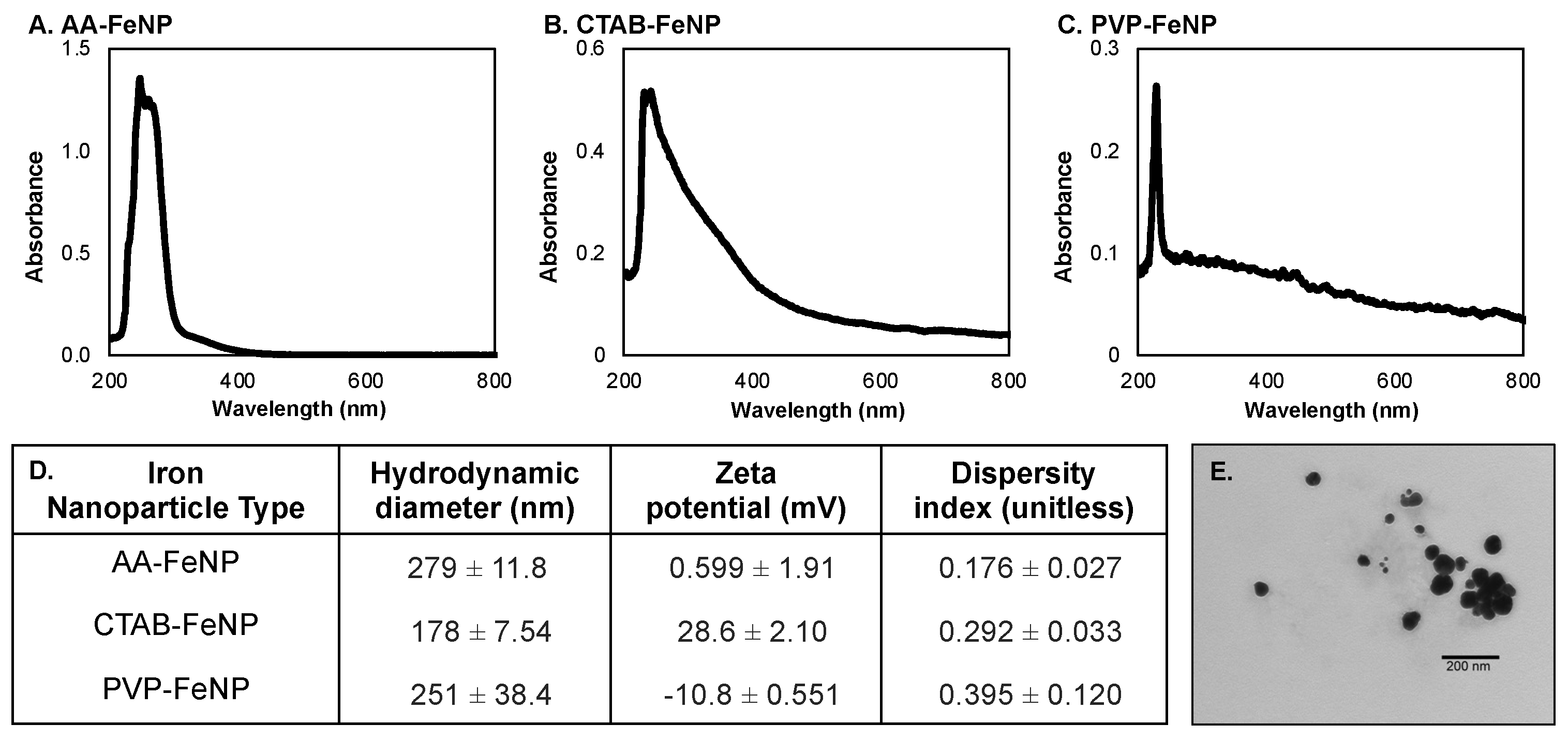
3.1. Iron Nanoparticle Characterization

Iron nanoparticles were characterized using UV/Vis spectroscopy and dynamic light scattering (DLS) (Figure 1). AA-FeNP and CTAB-FeNP exhibited absorption maxima at 245 nm, while the absorption maxima for PVP-FeNPs was 230 nm. These results are consistent with previously reported zero-valent FeNP characterization properties [71,72,73]. The hydrodynamic diameter and zeta potential of the FeNP were determined using DLS. The hydrodynamic diameter of AA-FeNPs was 279 ± 12 nm; that of CTAB-FeNPs was 178 ± 8 nm, and that of PVP-FeNPs was 251 ± 38 nm. The surface charge of the nanoparticles was assessed from zeta potential. AA-FeNP was considered neutral with a zeta potential of 0.6 ± 1.9 mV, CTAB was cationic with a zeta potential of +28.6 ± 2.1 mV, and PVP-FeNP was slightly anionic with a charge of −10.8 ± 0.6 mV. Dispersity index values of < 0.400 indicate that particle size was relatively uniform across all FeNP types [74]. The increasing shoulder of the CTAB-FeNP and PVP-FeNP spectra peaks in comparison with AA-FeNP could be attributed to an increase in particle dispersity, which is known to shift absorption of nanoparticle solutions [75].
Previous studies have revealed each capping agent to be effective in forming and stabilizing metallic nanoparticles. Ascorbic acid is a reducing agent during particle core formation and a capping agent to maintain particle stability [76,77]. CTAB is a positively charged surfactant with a hydrophilic head group and hydrocarbon tail, which coats particles and hinders aggregation [78,79]. PVP causes steric hindrance between particles, preventing agglomeration, and can function as a reducing agent during particle formation [80,81].

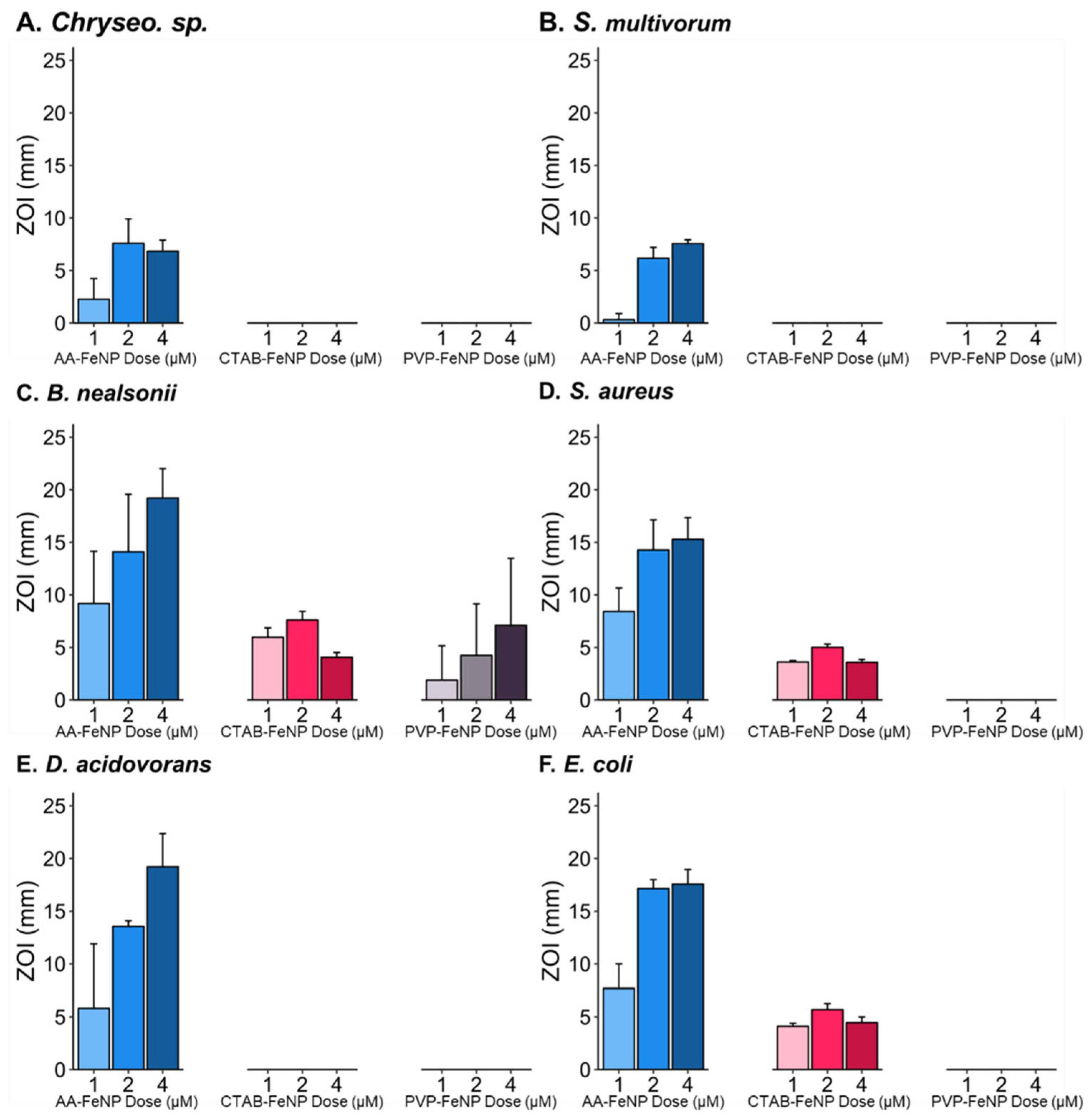
3.2. Coating and Dose Effect on Bacterial Inhibition

The antibacterial effects of each FeNP particle type were assessed by the zone of inhibition (ZOI) diameter (Figure 2). AA-FeNPs exhibited the strongest antibacterial effect of the three coatings tested, with a higher collective ZOI diameter than CTAB and PVP coatings. CTAB-FeNPs showed a larger collective ZOI than PVP-FeNPs, which showed little antibacterial activity. Logistical regression output shows a significant increase in the probability of AA-FeNP and CTAB-FeNP inducing an antibacterial effect compared to PVP-FeNP (Table 3).
The diverse antimicrobial propensities of each coating may be due to electrostatic or non-bonding (i.e., steric) interactions. The negative charge of PVP may repel particles from the negatively charged bacteria membrane, leading to the low antimicrobial propensity seen for PVP-FeNP in this study [8]. While CTAB confers a positive charge, the decreased antimicrobial propensity in comparison with the neutrally charged AA could be caused by van der Waals forces or steric interactions; AA has multiple polar groups, which could interact with the polar groups on the bacteria surface to a greater degree than CTAB or PVP [82]. Further, the steric hindrance of the pyrrolidone ring in the PVP polymer and the long hydrocarbon tails of CTAB may hinder interaction with the bacteria membrane.

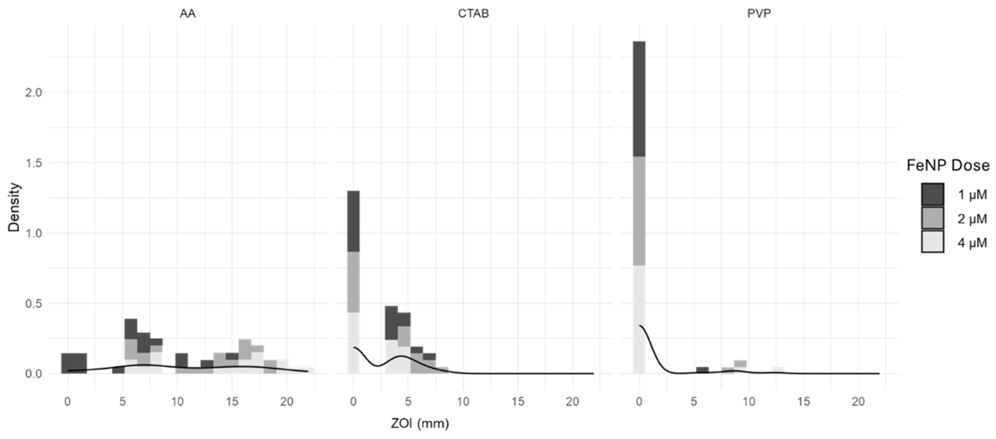
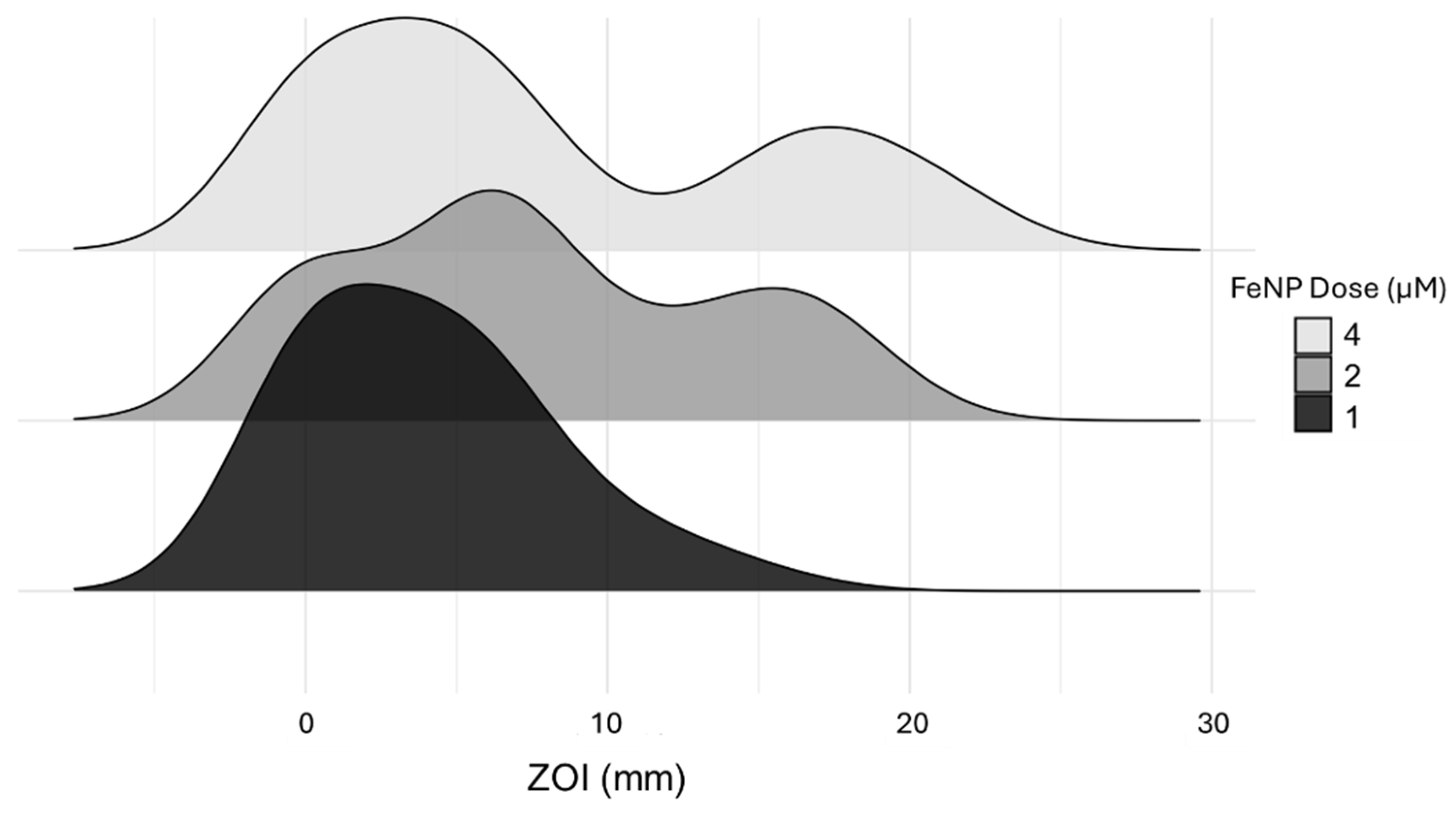
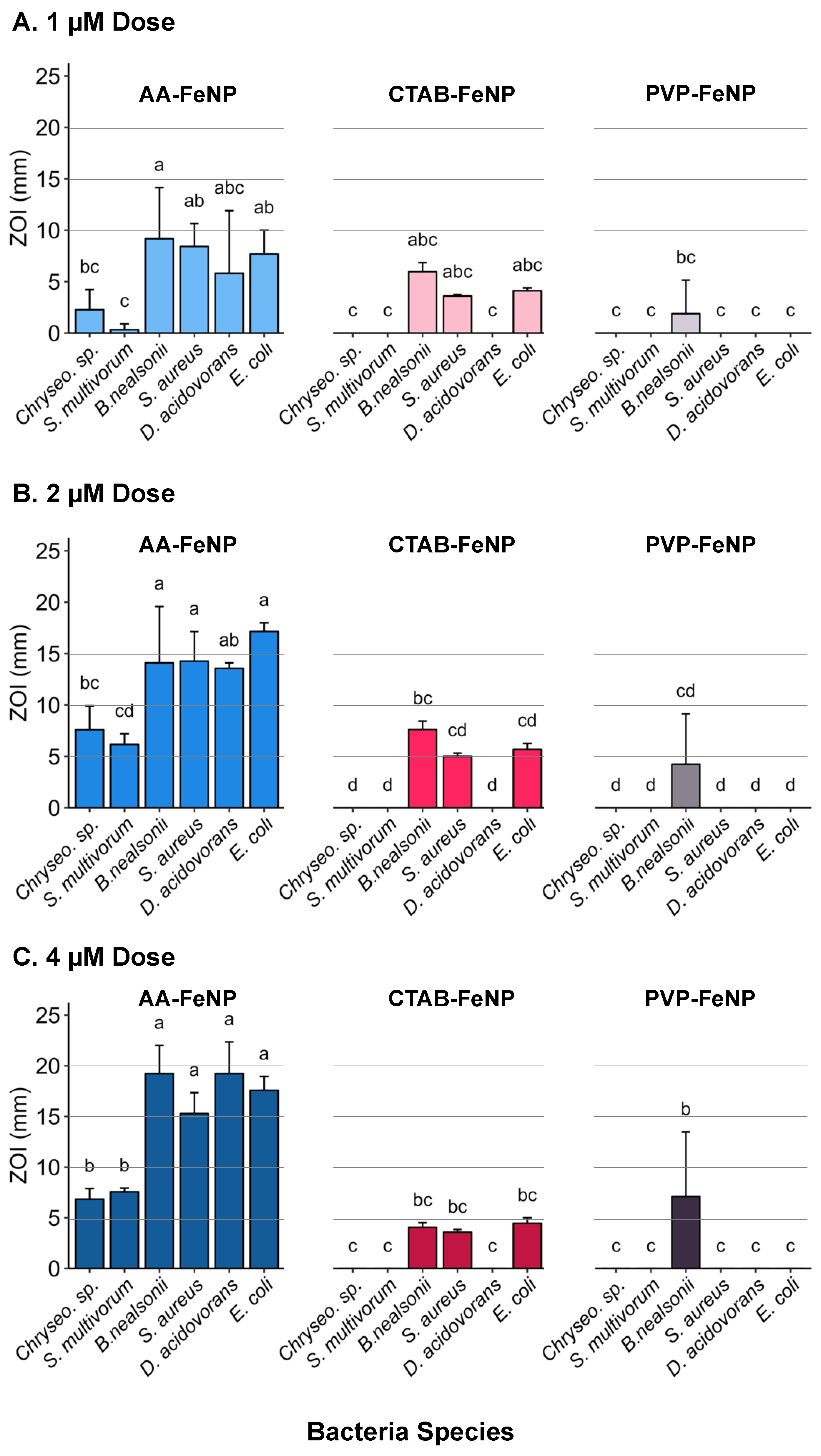
3.3. Dose Effects on the ZOI Varied Based on FeNP Surface Coating

Graphical analysis of inhibition measurements grouped by dose and coating across species shows that the antibacterial effects of AA-FeNPs were dose-dependent. In contrast, the inhibition response to CTAB-FeNPs was not dose-dependent. The effects of the dose on bacterial growth inhibition for PVP-FeNP were unclear in the collective analysis of this coating. Density plots of compiled AA-FeNP and CTAB-FeNP data show a marked change in distribution between 1 and 2 µM doses, but the distribution remained similar between 2 and 4 µM doses (Figure 3). The absence of a ZOI increase with increasing dose may be due to nanoparticle agglomeration, which could affect particle interaction with the bacteria [83]. The low effective dose of AA-FeNPs and CTAB-FeNPs further demonstrates their usefulness as antibacterial agents.
The antibacterial effects of each surface coating alone have been tested previously, although studies investigating the antibacterial impact of coated FeNPs are limited. This is the first study testing the antibacterial effects of AA-FeNPs, PVP-FeNPs, or CTAB-FeNPs. Previous studies investigating the antibacterial effects of bare (uncapped) zero-valent FeNPs revealed oxidative stress and membrane alteration as dominant modes of action [38,40,42,84]. AA is an antioxidant with antibacterial properties [85,86]. Here, we demonstrate that AA-FeNP is a highly effective antibacterial agent, with ZOI values surpassing 20 mm. The surfactant CTAB is also known to have antibacterial properties apart from a metallic core. However, greater antibacterial efficacy was associated with a metal core, such as silver and copper nanoparticles [8]. PVP is not known to have antibacterial effects apart from its association with nanoparticle metal cores. The variation in efficacy resulting from chemical coatings shows promise for multiple nano-bio applications, including developing effective, safe, and economical solutions to antibacterial resistance and other areas of biomedicine. More studies investigating the relationship between ROS production and the antioxidant capacity of the AA-FeNPs should be designed and reported.

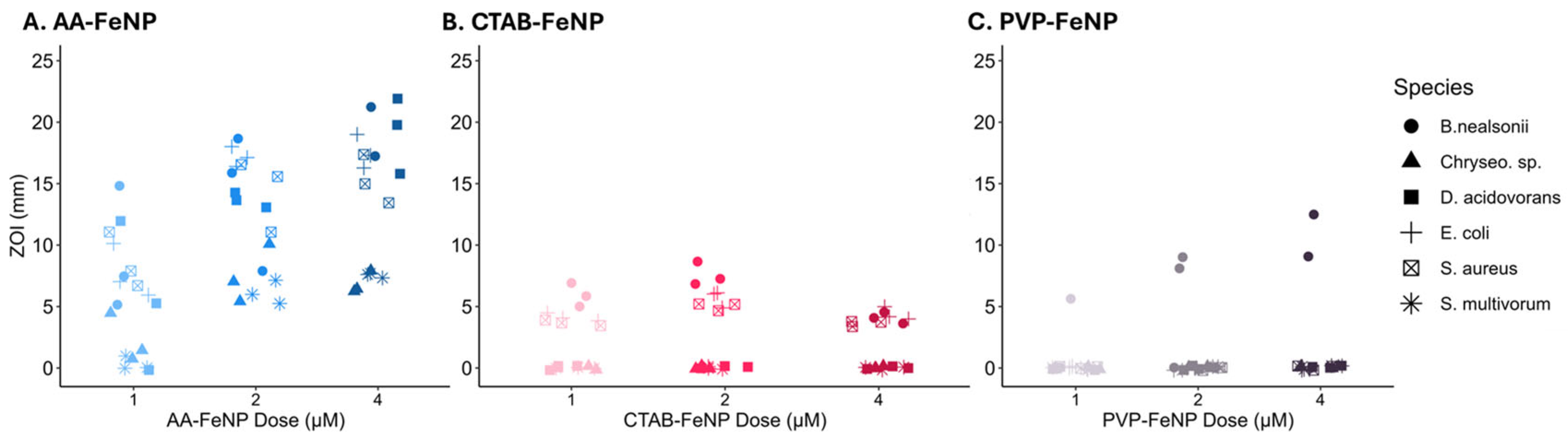
3.4. Intra-Species Analysis Revealed Differences in Response to FeNP Coating

Increasing AA-FeNP dose revealed a diminishing effect across all taxa; little increase in efficacy was observed in the intra-species comparison between the 2 and 4 µM doses (Figure 4). Three bacteria exhibited an effect after exposure to CTAB-FeNP: B. nealsonii, S. aureus, and E. coli. B. nealsonii was the only species inhibited by PVP-FeNPs.

3.5. Species Susceptibility to Coated Iron Nanoparticles

The degree of susceptibility to FeNP treatments varied by bacterial species (Figure 5). Species-specific analysis of efficacy data grouped across nanoparticle coating and concentration revealed that B. nealsonii and E. coli were most susceptible to collective treatments. The two taxa from the phylum Bacteroidota (Chryseobacterium sp. and S. multivorum) were the least susceptible. S. aureus and D. acidovorans showed intermediate responses.
Characteristic traits of each taxon may determine the species’ response to nanoparticle treatments. Bacillota and Pseudomonadota showed similar responses to FeNP treatments. The two most susceptible species of these phyla were B. nealsonii and E. coli. Previously, B. nealsonii was shown to be resistant to oxidative stress [87] but was affected by bare zero-valent iron nanoparticles more than other taxa, indicating that another mode of action other than oxidative stress may be at play [37]. E. coli is known to have metal-resistant genes and may be able to tolerate higher concentrations of intracellular ions from other metals [54]. However, previous studies have shown that bare zero-valent iron disrupts the E. coli membrane, producing ROS [41]. Gammaproteobacteria (the respective phylogenetic class of E. coli) abundance decreased in soil samples treated with bare zero-valent iron nanoparticles, supporting this study’s findings [37]. The mean order of susceptibility based on the disc assay is B. nealsonii > E. coli > S. aureus > D. acidovorans > Chryseobacterium sp. > S. multivorum.
Inter-species comparison across coatings revealed differential responses between species and surface coatings (Figure 6). The two species with the lowest response to the AA-FeNP treatment, Chryseobacterium sp. and S. multivorum, did not show responses to either CTAB-FeNPs or PVP-FeNPs, indicating that taxa-specific responses enabling relative resistance to one nanoparticle type are consistent across capping agents.
Bacteria most susceptible to AA-FeNPs were also susceptible to CTAB-FeNPs. An exception was D. acidovorans, which had a relatively large ZOI in response to the AA-FeNP treatment but did not show a response to either the CTAB-FeNP or PVP-FeNP treatment. Interestingly, D. acidovorans is known to have porin proteins that strongly select against cations due to positively charged amino acids surrounding the porin’s constriction site [88,89]. This supports the thought that ascorbic acid adds antimicrobial properties to the core. At the same time, D. acidovorans can select against cation entry; ascorbic acid is sufficient to cause a large ZOI in AA-FeNP treatments. The difference in effects between AA-FeNPs and CTAB-FeNPs in D. acidovorans may be due to the different modes of action between coatings. Studies are needed to elucidate the role of acid-based and surfactant-containing capping agents in conjunction with and separate from metallic cores. The coating-specific differences observed across taxa support the idea of a “designer nanoparticle” framework, indicating that nanoparticles could be designed to target taxa of interest.
An antibacterial potency threshold can also be proposed irrespective of dose for each FeNP type. In this scenario, the surface coating modifies the potency of the FeNP core, with less potent FeNP types affecting only the most sensitive species. This idea is seen most clearly in Panel A (1 µM dose), where the same three species with the highest responses to the AA-FeNP treatment likewise responded to the CTAB-FeNP treatment (i.e., B. nealsonii, S. aureus, and E.coli). The species with the highest response to CTAB-FeNP is also the only species with a response to PVP-FeNP (i.e., B. nealsonii). Here, the three most susceptible species with ZOI values above the proposed potency “threshold” for AA-FeNP were also above the “threshold” for the CTAB-FeNP, although less inhibition was observed. Nonetheless, B. nealsonii, the most sensitive species to all FeNP treatments, was the only bacteria to respond to PVP-FeNP, indicating that its sensitivity was above the proposed threshold for all coatings. Additional studies should be designed to investigate the thresholds related to the species, dose, and coating.

3.6. Statistical Data Analysis

The antibacterial nanotherapeutic field lacks a statistical framework for comparing efficacy across diverse treatments. Often, datasets generated to compare diverse treatments are non-normal, making it difficult to assess efficacy differences due to experimental factors without violating assumptions for standard analytical tests. The statistical framework used in this study enabled comparison across multiple variables and provided a rich analysis of the effects of FeNP coating, dose, and species.
The coefficient estimates for the GLMM model are shown in Table 4. Our analysis found a significant interaction between the dose and the AA coating, suggesting these factors are correlated in inhibiting bacterial growth. Since all the covariates are indicator variables for treatments, the coefficient estimates are interpreted as an estimated average multiplicative increase in inhibition relative to the base treatment. For example, since the estimated average inhibition for the base treatment (Coating = CTAB, Species = S. multivorum, and Dose = 1 µM) is a ZOI of 1.61 mm, the model suggests that the estimated average inhibition for Coating = AA, Species = S. aureus, and Dose = 2 mM would be 1.61 × 1.69 × 2.49 × 1.34 × 1.58 = 14.34 mm. The various expected inhibitions for different treatments can be explored using this framework.
Table 5 shows the results of the ordinal regression model. This analysis also found a significant interaction between the dose and the AA coating. One key difference between this model and the GLMM is that the coefficients represent the change in the odds ratio for each treatment. For example, on average, the AA-FeNPs are 160 times more likely to induce a greater inhibition than CTAB-FeNP, holding all other treatments fixed. Results from this model were consistent with GLMM output.

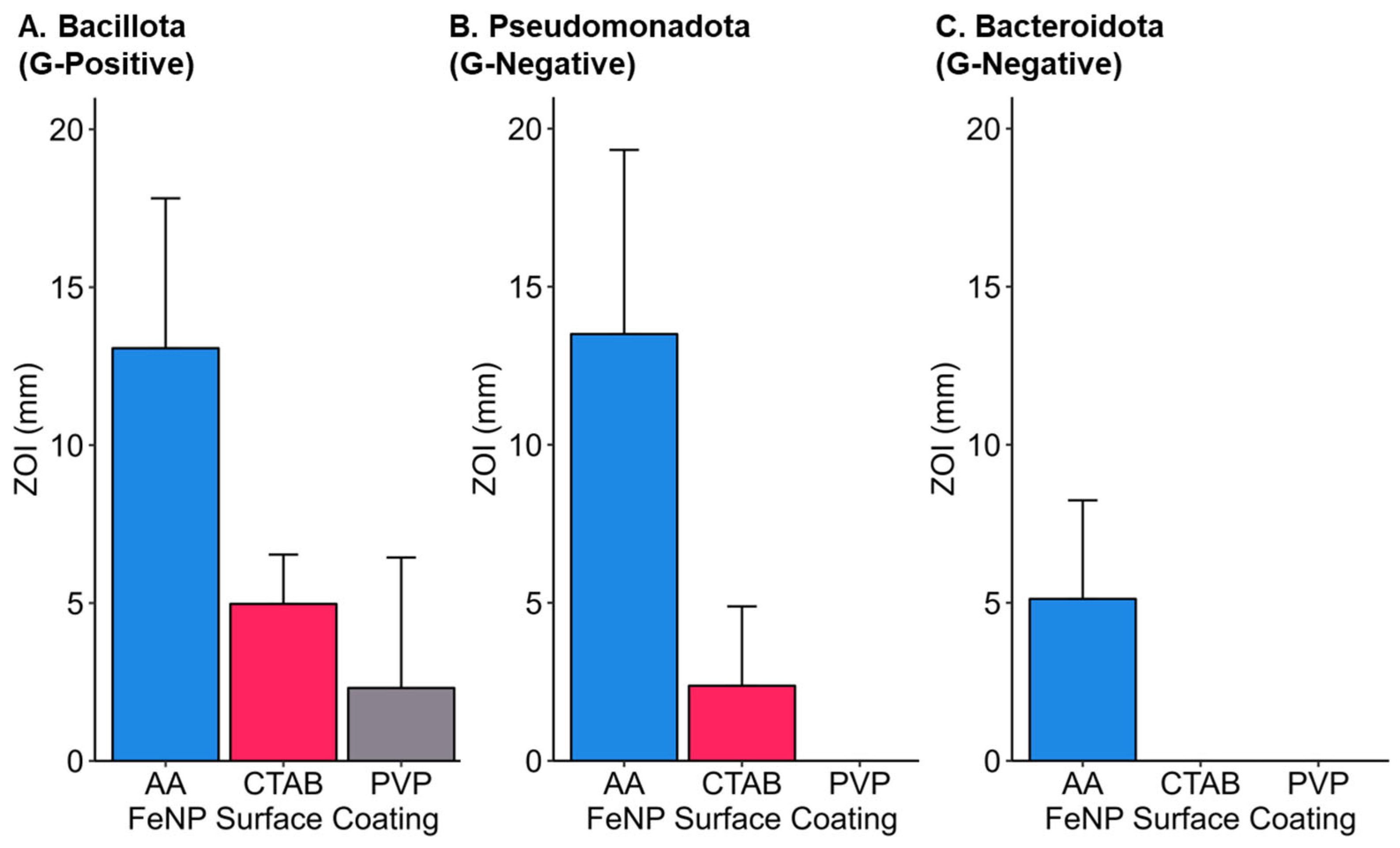
3.7. Phylogenetic Considerations for Iron Nanoparticle Efficacy

Phylogenetic characteristics may play a role in determining nanoparticle antibacterial effects. Grouping all inhibition data by phyla shows differences in means between bacteria phyla (Figure 7). Collective Bacteroidota ZOI was lower than both Bacillota and Pseudomonadota. Bacillota, the Gram-positive phyla in the study, had the largest mean ZOI. Grouping all data by cell wall structure revealed differences between Gram-positive and Gram-negative responses. Various studies have shown Gram-positive bacteria to be more susceptible to nanoparticle treatments than Gram-negative [90,91,92], although this is not always observed [11,93,94]. However, as the complete mechanism of antibacterial nanoparticle action is yet to be discovered, it is unclear how different nanoparticle cores and coatings affect different taxonomic groupings of bacteria, especially for zero-valent FeNPs, for which there has been little research. A previous study testing zero-valent iron clusters showed a preferential effect against Gram-positive bacteria over Gram-negative, similar to our findings of capped, zero-valent FeNP [95]. While some research has suggested that the porosity in the peptidoglycan cell wall allows for the translocation of nanoparticles, recent research indicates that the same size threshold that begins to cause antibacterial effects is the same for Gram-positive as Gram-negative bacteria, suggesting a different mechanism [96]. Another reason for the differential effects between Gram-positive and Gram-negative bacteria stems from the charge of the bacteria membrane. Although bacteria membranes are most commonly negatively charged, some species have different charge magnitudes due to membrane composition, which may be associated with taxonomic groups of bacteria [90,97,98]. Differences in bacteria membrane composition influence electrostatic and non-polar interactions, causing varied susceptibility of bacteria groups to nanoparticle treatments [8,82,99]. Multiple biochemical and physicochemical mechanisms may be at work to cause differential antibacterial effects across diverse bacteria.

3.8. Implications for Environmental and Biomedical Applications

In addition to their potential utilization as antimicrobial agents, nanoparticles with metal cores have been proposed for diverse environmental applications, including treatments for plant disease, targeted/slow-release fertilizers, and general-use pesticides [100,101,102]. The ability of FeNPs to adsorb toxic pollutants, such as heavy metals, has led to their application for remediation efforts [103,104,105,106,107]. However, the collateral effects of FeNPs on the environment are understudied, including their influence on microbial communities [108]. Metal nanoparticles may persist in the environment for long periods and could degrade soil quality by harming beneficial microbial communities and reducing crop growth and production [109,110]. Previous research has shown microbial community shifts in response to FeNP treatment, highlighting this concern [37,111]. It is unknown if trace metal ions leached from particle surfaces are used as micronutrients, thus increasing microbial enzyme function and growth, or if the exposure reduces collective metabolic function due to toxic effects.
Iron nanoparticles can effectively kill bacteria while also scavenging contaminants in soils. Therefore, the dosage of iron nanoparticles must be optimized to unlock their full potential in agricultural applications. In this study, our findings indicate that some bacteria found in the soil are affected by low concentrations of FeNP treatments, including taxa known to have plant-benefiting traits. Optimization of FeNP treatment concentration has the potential to balance practical remediation benefits against adverse collateral effects [112]. Our work also shows that the antibacterial effects of FeNPs can be moderated through a capping agent, as PVP-FeNP treatments yielded little antibacterial response. Future studies should explore the role of nanoparticle coatings in environmental applications to minimize collateral damage to non-target organisms. FeNPs may offer several remediation benefits, but further research is necessary before applying them in large-scale environments.
FeNPs have many potential applications in biomedical research, but clinical use is limited due to a lack of understanding of FeNP mechanisms [113]. Although ion leaching and resulting cellular toxicity were found during previous investigations of the in vitro use of FeNPs [114,115,116], the potential of using a capping agent to reduce toxicity while retaining ferromagnetic capability should be investigated [117,118,119]. Results from this study show many potential biomedical applications for capped zero-valent FeNPs. Our work demonstrates that FeNPs capped with an antioxidant, such as L-ascorbic acid, induce significant antibacterial effects, which should be investigated further for applications in vivo. PVP-FeNPs were shown to have minimal antibacterial action. This suggests the capping agent may prevent ion leaching or harmful interaction with bacterial membranes. Based on these results, this capping agent may also prevent harmful effects when applied in vivo in clinical settings. Capping agents may be tailorable to maintain chemical properties of interest (ferromagnetism or antibacterial properties) while preventing cellular damage to human cells.
Our work shows that capped FeNPs have antibacterial effects against bacterial taxa known for developing antibacterial resistance. Capped nanoparticles show promise as a solution to antibacterial resistance, as their multi-mechanistic mode of action and ability to be tailored allows them to bypass many mechanisms of bacterial resistance [15]. The differential effects of capped FeNP against diverse species indicate that nanoparticles could be developed to target specific bacterial taxa of interest. However, published studies lack tests on diverse bacteria, creating difficulty in understanding species-specific mechanisms. This research clarifies avenues for further research, highlighting the benefits of capped FeNPs and their effects on diverse bacterial species.

4. Conclusions

Our research shows that bacteria FeNPs have significant antibacterial effects against diverse bacterial taxa. Through the statistical framework developed in this study, we determined that the capping agent significantly influences nanoparticle antibacterial efficacy. Significant interactions between dose and coating differentially affect bacteria species, indicating that taxa-specific characteristics may govern the antibacterial effectiveness of nanoparticle core–coating combinations. This supports the “designer nanoparticle” ideology, suggesting that core and capping agents can be modified to increase targeting ability. More research is needed to elucidate mechanisms influencing bacterial susceptibility to better design nano-antibiotics that are safe for the environment and effective against bacteria strains of interest. Implementing robust statistical models is necessary to understand complex treatments’ effects, interactions, and collective influence on antibacterial response. The antibacterial effects of FeNPs on agriculturally relevant taxa should be considered before implementing environmental applications. This research strongly supports the feasibility of designing nano-antibiotics for targeting bacterial populations of interest, which will significantly increase the efficacy of FeNP applications in environmental and biomedical fields.

Author Contributions

Conceptualization, C.M.S.; data curation, C.M.S. and B.J.C.; formal analysis, D.S., C.S., N.B., C.M.S. and B.J.C.; investigation, S.S., C.S., K.Z., C.M.S. and B.J.C.; methodology, D.S., S.S., C.S., N.B., R.X.S., K.Z., W.E.B., C.M.S. and B.J.C.; resources, R.X.S., W.E.B. and C.M.S.; software, N.B., C.M.S. and B.J.C.; validation, C.M.S. and B.J.C.; writing—original draft, N.B., C.M.S. and B.J.C.; writing—review and editing, D.S., R.X.S., W.E.B. and C.M.S. All authors have read and agreed to the published version of the manuscript.

Funding

This material was made possible, in part, by a Cooperative Agreement from the United States Department of Agriculture’s Animal and Plant Health Inspection Service (APHIS) AP23PPQS&T00C071. It may not necessarily express APHIS’ views.

Institutional Review Board Statement

Not applicable.

Informed Consent Statement

Not applicable.

Data Availability Statement

The original contributions presented in the study are included in the article/Appendix A, further inquiries can be directed to the corresponding author.

Acknowledgments

The authors would like to thank Don Vacek, USDA, PPQ, APHIS, S&T, who provided bacteria cultures used in this study. The authors thank Amanda Charlton-Sevcik (Baylor University) for helpful insights into experimental procedures and manuscript proofreading services. The authors thank the Center for Microscopy and Imaging (CMI) and Center for Molecular Biosciences (MBC) at Baylor University for the use of equipment and preparation of samples. Mention of trade names or commercial products in this publication is solely for the purpose of providing specific information and does not imply recommendation or endorsement by the U.S. Department of Agriculture, an equal opportunity employer.

Conflicts of Interest

The authors declare no conflicts of interest.

Appendix A.

Appendix A.1. Iron Nanoparticle Transmission Electron Microscopy

Transmission electron microscopy (TEM) was performed for morphological characterization (JEOL, JEM-1010, TEM, Tokyo, Japan). A drop of the prepared nanoparticle suspension was added to the surface of carbon-coated copper grids (Electron Microscopy Sciences; Hartfield, PA, USA) and dried in a hot air oven at 160 °C. The dried TEM grids were then viewed on the TEM at an accelerating voltage of between 10 and 100 kV.

Appendix A.2. Response Variable Distribution

Analysis of response variable distributions by FeNP type revealed that the distributions of each nanoparticle coating are highly zero-inflated (Figure A1) and highly skewed right. No transformations were found to be stabilizing. Therefore, more than traditional statistical methods are required to conduct robust analysis, and alternative models are needed.
Figure A1. Histograms for coatings and dose. For two-way ANOVA to be viable, the distribution shape would need to resemble a traditional bell curve, and the sample size would need to be far greater than the size n = 162.
Jfb 16 00113 g0a1

Appendix A.3. Ordinal Model Assumptions

One way to validate ordinal model assumptions is to check if the odds ratio for each treatment level is proportional to one another (Figure A2). Within this experiment, the number of observations within each treatment group was too small to validate this assumption fully; however, proportional odds ratios were validated across factor levels. It has been determined that ordinal regression models are still very robust to this assumption, which makes it one of the safest models for drawing inferences on the zone of inhibition given different treatments [120]. All the empirical cumulative density functions (eCDF) under a logistic link for each group look reasonably proportional (roughly parallel), validating our assumption.
Figure A2. Graphics illustrating the proportional odds for each factor and its levels. The y-axis is the empirical cumulative density function with a logistic link function. Lines that are relatively parallel in the center of the distribution are considered to have proportional odds.
Jfb 16 00113 g0a2

References

  1. Zhang, L.; Jiang, Y.; Ding, Y.; Daskalakis, N.; Jeuken, L.; Povey, M.; O’neill, A.J.; York, D.W. Mechanistic investigation into antibacterial behaviour of suspensions of ZnO nanoparticles against E. coli. J. Nanopart. Res. 2010, 12, 1625–1636. [Google Scholar] [CrossRef]
  2. Dizaj, S.M.; Lotfipour, F.; Barzegar-Jalali, M.; Zarrintan, M.H.; Adibkia, K. Antimicrobial activity of the metals and metal oxide nanoparticles. Mater. Sci. Eng. C 2014, 44, 278–284. [Google Scholar] [CrossRef]
  3. Prabhu, S.; Poulose, E.K. Silver nanoparticles: Mechanism of antimicrobial action, synthesis, medical applications, and toxicity effects. Int. Nano Lett. 2012, 2, 32. [Google Scholar] [CrossRef]
  4. Palza, H. Antimicrobial polymers with metal nanoparticles. Int. J. Mol. Sci. 2015, 16, 2099–2116. [Google Scholar] [CrossRef]
  5. Slavin, Y.N.; Asnis, J.; Hńfeli, U.O.; Bach, H. Metal nanoparticles: Understanding the mechanisms behind antibacterial activity. J. Nanobiotechnol. 2017, 15, 65. [Google Scholar] [CrossRef]
  6. Chandrakala, V.; Aruna, V.; Angajala, G. Review on metal nanoparticles as nanocarriers: Current challenges and perspectives in drug delivery systems. Emergent Mater. 2022, 5, 1593–1615. [Google Scholar] [CrossRef] [PubMed]
  7. Allahverdiyev, A.M.; Kon, K.V.; Abamor, E.S.; Bagirova, M.; Rafailovich, M. Coping with antibiotic resistance: Combining nanoparticles with antibiotics and other antimicrobial agents. Expert Rev. Anti-Infect. Ther. 2011, 9, 1035–1052. [Google Scholar] [CrossRef]
  8. Ameh, T.; Zarzosa, K.; Dickinson, J.; Braswell, W.E.; Sayes, C.M. Nanoparticle surface stabilizing agents influence antibacterial action. Front. Microbiol. 2023, 14, 1119550. [Google Scholar] [CrossRef]
  9. Applerot, G.; Lipovsky, A.; Dror, R.; Perkas, N.; Nitzan, Y.; Lubart, R.; Gedanken, A. Enhanced antibacterial activity of nanocrystalline ZnO due to increased ROS-mediated cell injury. Adv. Funct. Mater. 2009, 19, 842–852. [Google Scholar] [CrossRef]
  10. Bharti, S. Harnessing the potential of bimetallic nanoparticles: Exploring a novel approach to address antimicrobial resistance. World J. Microbiol. Biotechnol. 2024, 40, 89. [Google Scholar] [CrossRef]
  11. Aazam, E.S.; Zaheer, Z. Growth of Ag-nanoparticles in an aqueous solution and their antimicrobial activities against Gram positive, Gram negative bacterial strains and Candida fungus. Bioprocess Biosyst. Eng. 2016, 39, 575–584. [Google Scholar] [CrossRef] [PubMed]
  12. Luo, L.; Huang, W.; Zhang, J.; Yu, Y.; Sun, T. Metal-Based Nanoparticles as Antimicrobial Agents: A Review. ACS Appl. Nano Mater. 2024, 7, 2529–2545. [Google Scholar] [CrossRef]
  13. McQuillan, J.S.; Groenaga Infante, H.; Stokes, E.; Shaw, A.M. Silver nanoparticle enhanced silver ion stress response in Escherichia coli K12. Nanotoxicology 2012, 6, 857–866. [Google Scholar] [CrossRef]
  14. Neihaya, H.; Zaman, H. Investigating the effect of biosynthesized silver nanoparticles as antibiofilm on bacterial clinical isolates. Microb. Pathog. 2018, 116, 200–208. [Google Scholar] [CrossRef]
  15. Modi, S.K.; Gaur, S.; Sengupta, M.; Singh, M.S. Mechanistic insights into nanoparticle surface-bacterial membrane interactions in overcoming antibiotic resistance. Front. Microbiol. 2023, 14, 1135579. [Google Scholar] [CrossRef]
  16. Dadgostar, P. Antimicrobial resistance: Implications and costs. Infect. Drug Resist. 2019, 12, 3903–3910. [Google Scholar] [CrossRef] [PubMed]
  17. Li, B.; Webster, T.J. Bacteria antibiotic resistance: New challenges and opportunities for implant-associated orthopedic infections. J. Orthop. Res. 2018, 36, 22–32. [Google Scholar] [CrossRef]
  18. Willyard, C. Drug-resistant bacteria ranked. Nature 2017, 543, 15. [Google Scholar] [CrossRef]
  19. Thimiri Govinda Raj, D.B.; Khan, N.A. Designer nanoparticle: Nanobiotechnology tool for cell biology. Nano Converg. 2016, 3, 22. [Google Scholar] [CrossRef]
  20. Mahon, E.; Salvati, A.; Bombelli, F.B.; Lynch, I.; Dawson, K.A. Designing the nanoparticle–biomolecule interface for “targeting and therapeutic delivery”. J. Control. Release 2012, 161, 164–174. [Google Scholar] [CrossRef]
  21. Caldorera-Moore, M.; Guimard, N.; Shi, L.; Roy, K. Designer nanoparticles: Incorporating size, shape and triggered release into nanoscale drug carriers. Expert Opin. Drug Deliv. 2010, 7, 479–495. [Google Scholar] [CrossRef] [PubMed]
  22. Love, S.A.; Maurer-Jones, M.A.; Thompson, J.W.; Lin, Y.-S.; Haynes, C.L. Assessing nanoparticle toxicity. Annu. Rev. Anal. Chem. 2012, 5, 181–205. [Google Scholar] [CrossRef] [PubMed]
  23. Chen, P.-C.; Liu, X.; Hedrick, J.L.; Xie, Z.; Wang, S.; Lin, Q.-Y.; Hersam, M.C.; Dravid, V.P.; Mirkin, C.A. Polyelemental nanoparticle libraries. Science 2016, 352, 1565–1569. [Google Scholar] [CrossRef]
  24. Wilhelm, S.; Tavares, A.J.; Dai, Q.; Ohta, S.; Audet, J.; Dvorak, H.F.; Chan, W.C. Analysis of nanoparticle delivery to tumours. Nat. Rev. Mater. 2016, 1, 16014. [Google Scholar] [CrossRef]
  25. Yougbaré, S.; Mutalik, C.; Okoro, G.; Lin, I.-H.; Krisnawati, D.I.; Jazidie, A.; Nuh, M.; Chang, C.-C.; Kuo, T.-R. Emerging trends in nanomaterials for antibacterial applications. Int. J. Nanomed. 2021, 16, 5831–5867. [Google Scholar] [CrossRef]
  26. Van Hengel, I.; Tierolf, M.; Valerio, V.; Minneboo, M.; Fluit, A.; Fratila-Apachitei, L.; Apachitei, I.; Zadpoor, A. Self-defending additively manufactured bone implants bearing silver and copper nanoparticles. J. Mater. Chem. B 2020, 8, 1589–1602. [Google Scholar] [CrossRef] [PubMed]
  27. Liu, Q.; Liu, E.; Li, J.; Qiu, Y.; Chen, R. Rapid ultrasonic-microwave assisted synthesis of spindle-like Ag/ZnO nanostructures and their enhanced visible-light photocatalytic and antibacterial activities. Catal. Today 2020, 339, 391–402. [Google Scholar] [CrossRef]
  28. Paterson, T.E.; Bari, A.; Bullock, A.J.; Turner, R.; Montalbano, G.; Fiorilli, S.; Vitale-Brovarone, C.; MacNeil, S.; Shepherd, J. Multifunctional copper-containing mesoporous glass nanoparticles as antibacterial and proangiogenic agents for chronic wounds. Front. Bioeng. Biotechnol. 2020, 8, 246. [Google Scholar] [CrossRef]
  29. Kumar, A.; Behl, T.; Chadha, S. Synthesis of physically crosslinked PVA/Chitosan loaded silver nanoparticles hydrogels with tunable mechanical properties and antibacterial effects. Int. J. Biol. Macromol. 2020, 149, 1262–1274. [Google Scholar] [CrossRef]
  30. Li, R.; Xu, Z.; Jiang, Q.; Zheng, Y.; Chen, Z.; Chen, X. Characterization and biological evaluation of a novel silver nanoparticle-loaded collagen-chitosan dressing. Regen. Biomater. 2020, 7, 371–380. [Google Scholar] [CrossRef]
  31. Kim, J.-H.; Cho, H.; Ryu, S.-E.; Choi, M.-U. Effects of metal ions on the activity of protein tyrosine phosphatase VHR: Highly potent and reversible oxidative inactivation by Cu2+ ion. Arch. Biochem. Biophys. 2000, 382, 72–80. [Google Scholar] [CrossRef] [PubMed]
  32. Ding, H.; Han, D.; Han, Y.; Liang, Y.; Liu, X.; Li, Z.; Zhu, S.; Wu, S. Visible light responsive CuS/protonated g-C3N4 heterostructure for rapid sterilization. J. Hazard. Mater. 2020, 393, 122423. [Google Scholar] [CrossRef] [PubMed]
  33. Brayner, R.; Ferrari-Iliou, R.; Brivois, N.; Djediat, S.; Benedetti, M.F.; Fiévet, F. Toxicological impact studies based on Escherichia coli bacteria in ultrafine ZnO nanoparticles colloidal medium. Nano Lett. 2006, 6, 866–870. [Google Scholar] [CrossRef]
  34. Zhang, L.; Jiang, Y.; Ding, Y.; Povey, M.; York, D. Investigation into the antibacterial behaviour of suspensions of ZnO nanoparticles (ZnO nanofluids). J. Nanopart. Res. 2007, 9, 479–489. [Google Scholar] [CrossRef]
  35. Stevens, D.; Charlton-Sevcik, A.K.; Braswell, W.E.; Sayes, C.M. Evaluating the Antibacterial Potential of Distinct Size Populations of Stabilized Zinc Nanoparticles. ACS Appl. Mater. Interfaces 2024, 17, 322–332. [Google Scholar] [CrossRef]
  36. Diao, M.; Yao, M. Use of zero-valent iron nanoparticles in inactivating microbes. Water Res. 2009, 43, 5243–5251. [Google Scholar] [CrossRef] [PubMed]
  37. Fajardo, C.; Ortíz, L.; Rodríguez-Membibre, M.; Nande, M.; Lobo, M.; Martin, M. Assessing the impact of zero-valent iron (ZVI) nanotechnology on soil microbial structure and functionality: A molecular approach. Chemosphere 2012, 86, 802–808. [Google Scholar] [CrossRef]
  38. Hsueh, Y.-H.; Tsai, P.-H.; Lin, K.-S.; Ke, W.-J.; Chiang, C.-L. Antimicrobial effects of zero-valent iron nanoparticles on gram-positive Bacillus strains and gram-negative Escherichia coli strains. J. Nanobiotechnol. 2017, 15, 77. [Google Scholar] [CrossRef]
  39. Auffan, M.; Achouak, W.; Rose, J.; Roncato, M.-A.; Chanéac, C.; Waite, D.T.; Masion, A.; Woicik, J.C.; Wiesner, M.R.; Bottero, J.-Y. Relation between the Redox State of Iron-Based Nanoparticles and Their Cytotoxicity toward Escherichia coli. Environ. Sci. Technol. 2008, 42, 6730–6735. [Google Scholar] [CrossRef]
  40. Kotchaplai, P.; Khan, E.; Vangnai, A.S. Membrane Alterations in Pseudomonas putida F1 Exposed to Nanoscale Zerovalent Iron: Effects of Short-Term and Repetitive nZVI Exposure. Environ. Sci. Technol. 2017, 51, 7804–7813. [Google Scholar] [CrossRef]
  41. Wang, J.; Liu, C.; Sun, H.; Wang, S.; Liao, X.; Zhang, L. Membrane disruption boosts iron overload and endogenous oxidative stress to inactivate Escherichia coli by nanoscale zero-valent iron. J. Hazard. Mater. 2022, 435, 128951. [Google Scholar] [CrossRef] [PubMed]
  42. Sacca, M.L.; Fajardo, C.; Martinez-Gomariz, M.; Costa, G.; Nande, M.; Martin, M. Molecular stress responses to nano-sized zero-valent iron (nZVI) particles in the soil bacterium Pseudomonas stutzeri. PLoS ONE 2014, 9, e89677. [Google Scholar] [CrossRef]
  43. Lee, C.; Kim, J.Y.; Lee, W.I.; Nelson, K.L.; Yoon, J.; Sedlak, D.L. Bactericidal Effect of Zero-Valent Iron Nanoparticles on Escherichia coli. Environ. Sci. Technol. 2008, 42, 4927–4933. [Google Scholar] [CrossRef]
  44. Amin, R.M.; Mahmoud, R.K.; Gadelhak, Y.; Abo El-Ela, F.I. Gamma irradiated green synthesized zero valent iron nanoparticles as promising antibacterial agents and heavy metal nano-adsorbents. Environ. Nanotechnol. Monit. Manag. 2021, 16, 100461. [Google Scholar] [CrossRef]
  45. Chau, T.P.; Brindhadevi, K.; Krishnan, R.; Alyousef, M.A.; Almoallim, H.S.; Whangchai, N.; Pikulkaew, S. A novel synthesis, analysis and evaluation of Musa coccinea based zero valent iron nanoparticles for antimicrobial and antioxidant. Environ. Res. 2022, 209, 112770. [Google Scholar] [CrossRef] [PubMed]
  46. Lemire, J.A.; Harrison, J.J.; Turner, R.J. Antimicrobial activity of metals: Mechanisms, molecular targets and applications. Nat. Rev. Microbiol. 2013, 11, 371–384. [Google Scholar] [CrossRef]
  47. Phulpoto, I.A.; Yu, Z.; Hu, B.; Wang, Y.; Ndayisenga, F.; Li, J.; Liang, H.; Qazi, M.A. Production and characterization of surfactin-like biosurfactant produced by novel strain Bacillus nealsonii S2MT and it’s potential for oil contaminated soil remediation. Microb. Cell Factories 2020, 19, 1–12. [Google Scholar] [CrossRef]
  48. Chen, F.-L.; Wang, G.-C.; Teng, S.-O.; Ou, T.-Y.; Yu, F.-L.; Lee, W.-S. Clinical and epidemiological features of Chryseobacterium indologenes infections: Analysis of 215 cases. J. Microbiol. Immunol. Infect. 2013, 46, 425–432. [Google Scholar] [CrossRef]
  49. Kämpfer, P.; McInroy, J.A.; Glaeser, S.P. Chryseobacterium zeae sp. nov., Chryseobacterium arachidis sp. nov., and Chryseobacterium geocarposphaerae sp. nov. isolated from the rhizosphere environment. Antonie Van Leeuwenhoek 2014, 105, 491–500. [Google Scholar] [CrossRef]
  50. Montero-Calasanz, M.d.C.; Göker, M.; Rohde, M.; Spröer, C.; Schumann, P.; Busse, H.-J.; Schmid, M.; Tindall, B.J.; Klenk, H.-P.; Camacho, M. Chryseobacterium hispalense sp. nov., a plant-growth-promoting bacterium isolated from a rainwater pond in an olive plant nursery, and emended descriptions of Chryseobacterium defluvii, Chryseobacterium indologenes, Chryseobacterium wanjuense and Chryseobacterium gregarium. Int. J. Syst. Evol. Microbiol. 2013, 63, 4386–4395. [Google Scholar]
  51. Bhat, S.V.; Maughan, H.; Cameron, A.D.; Yost, C.K. Phylogenomic analysis of the genus Delftia reveals distinct major lineages with ecological specializations. Microb. Genom. 2022, 8, 000864. [Google Scholar] [CrossRef]
  52. Højgaard, S.M.M.; Rezahosseini, O.; Knudsen, J.D.; Fuglebjerg, N.J.U.; Skov, M.; Nielsen, S.D.; Harboe, Z.B. Characteristics and outcomes of patients with Delftia acidovorans infections: A retrospective cohort study. Microbiol. Spectr. 2022, 10, e00326-22. [Google Scholar] [CrossRef] [PubMed]
  53. Suchan, D.M.; Bergsveinson, J.; Manzon, L.; Pierce, A.; Kryachko, Y.; Korber, D.; Tan, Y.; Tambalo, D.D.; Khan, N.H.; Whiting, M. Transcriptomics reveal core activities of the plant growth-promoting bacterium Delftia acidovorans RAY209 during interaction with canola and soybean roots. Microb. Genom. 2020, 6, e000462. [Google Scholar] [CrossRef] [PubMed]
  54. Tan, Y.; Zhao, K.; Yang, S.; Chen, S.; Li, C.; Han, X.; Li, J.; Hu, K.; Liu, S.; Ma, M. Insights into antibiotic and heavy metal resistance interactions in Escherichia coli isolated from livestock manure and fertilized soil. J. Environ. Manag. 2024, 351, 119935. [Google Scholar] [CrossRef] [PubMed]
  55. Verma, S.K.; Chen, Q.; White, J.F. Evaluation of colonization and mutualistic endophytic symbiosis of Escherichia coli with tomato and Bermuda grass seedlings. PeerJ 2022, 10, e13879. [Google Scholar] [CrossRef]
  56. Wight, J.; Byrne, A.S.; Tahlan, K.; Lang, A.S. Anthropogenic contamination sources drive differences in antimicrobial-resistant Escherichia coli in three urban lakes. Appl. Environ. Microbiol. 2024, 90, e01809-23. [Google Scholar] [CrossRef]
  57. An, X.; Tian, C.; Xu, J.; Dong, F.; Liu, X.; Wu, X.; Zheng, Y. Characterization of hexaconazole-degrading strain Sphingobacterium multivorum and analysis of transcriptome for biodegradation mechanism. Sci. Total Environ. 2020, 722, 137171. [Google Scholar] [CrossRef]
  58. Barahona, F.; Slim, J. Sphingobacterium multivorum: Case report and literature review. New Microbes New Infect. 2015, 7, 33–36. [Google Scholar] [CrossRef]
  59. Hassan, I.A.; Mohamedelhassan, E.E.; Yanful, E.K.; Yuan, Z.-C. Mitigation of soil contaminated with diesel fuel using bioelectrokinetics. J. Environ. Sci. Health Part A 2019, 54, 416–426. [Google Scholar] [CrossRef]
  60. Tien, C.J.; Huang, H.J.; Chen, C.S. Accessing the carbofuran degradation ability of cultures from natural river biofilms in different environments. CLEAN–Soil Air Water 2017, 45, 1600380. [Google Scholar] [CrossRef]
  61. Howden, B.P.; Giulieri, S.G.; Wong Fok Lung, T.; Baines, S.L.; Sharkey, L.K.; Lee, J.Y.; Hachani, A.; Monk, I.R.; Stinear, T.P. Staphylococcus aureus host interactions and adaptation. Nat. Rev. Microbiol. 2023, 21, 380–395. [Google Scholar] [CrossRef] [PubMed]
  62. Richardson, E.J.; Bacigalupe, R.; Harrison, E.M.; Weinert, L.A.; Lycett, S.; Vrieling, M.; Robb, K.; Hoskisson, P.A.; Holden, M.T.; Feil, E.J. Gene exchange drives the ecological success of a multi-host bacterial pathogen. Nat. Ecol. Evol. 2018, 2, 1468–1478. [Google Scholar] [CrossRef]
  63. Xiong, J.; Wang, Y.; Xue, Q.; Wu, X. Synthesis of highly stable dispersions of nanosized copper particles using l-ascorbic acid. Green Chem. 2011, 13, 900–904. [Google Scholar] [CrossRef]
  64. Wu, S.-H.; Chen, D.-H. Synthesis of high-concentration Cu nanoparticles in aqueous CTAB solutions. J. Colloid Interface Sci. 2004, 273, 165–169. [Google Scholar] [CrossRef] [PubMed]
  65. Huang, K.-C.; Ehrman, S.H. Synthesis of Iron Nanoparticles via Chemical Reduction with Palladium Ion Seeds. Langmuir 2007, 23, 1419–1426. [Google Scholar] [CrossRef] [PubMed]
  66. Wiegand, I.; Hilpert, K.; Hancock, R.E. Agar and broth dilution methods to determine the minimal inhibitory concentration (MIC) of antimicrobial substances. Nat. Protoc. 2008, 3, 163–175. [Google Scholar] [CrossRef]
  67. Hosmer Jr, D.W.; Lemeshow, S.; Sturdivant, R.X. Applied Logistic Regression; John Wiley & Sons: Hoboken, NJ, USA, 2013. [Google Scholar]
  68. Skrondal, A.; Rabe-Hesketh, S. Generalized Latent Variable Modeling: Multilevel, Longitudinal, and Structural Equation Models; Chapman and Hall/CRC: London, UK, 2004. [Google Scholar]
  69. Feng, C.X. A comparison of zero-inflated and hurdle models for modeling zero-inflated count data. J. Stat. Distrib. Appl. 2021, 8, 8. [Google Scholar] [CrossRef]
  70. Harrell, F.E. Regression Modeling Strategies: With Applications to Linear Models, Logistic and Ordinal Regression, and Survival Analysis; Springer International Publishing: Berlin/Heidelberg, Germany, 2015. [Google Scholar]
  71. Abdelfatah, A.M.; Fawzy, M.; Eltaweil, A.S.; El-Khouly, M.E. Green synthesis of nano-zero-valent iron using ricinus communis seeds extract: Characterization and application in the treatment of methylene blue-polluted water. ACS Omega 2021, 6, 25397–25411. [Google Scholar] [CrossRef]
  72. Morgada, M.E.; Levy, I.K.; Salomone, V.; Farías, S.S.; Lopez, G.; Litter, M.I. Arsenic (V) removal with nanoparticulate zerovalent iron: Effect of UV light and humic acids. Catal. Today 2009, 143, 261–268. [Google Scholar] [CrossRef]
  73. Singh, A.; Hou, W.-C.; Lin, T.-F. Combined impact of silver nanoparticles and chlorine on the cell integrity and toxin release of Microcystis aeruginosa. Chemosphere 2021, 272, 129825. [Google Scholar] [CrossRef]
  74. Bhattacharjee, S. DLS and zeta potential–what they are and what they are not? J. Control. Release 2016, 235, 337–351. [Google Scholar] [CrossRef]
  75. Bhatia, P. Comparison study of iron and iron-oxide nanoparticles for thermoplasmonic applications. Mater. Today Commun. 2023, 35, 106008. [Google Scholar] [CrossRef]
  76. Hussain, M.H.; Abu Bakar, N.F.; Mustapa, A.N.; Low, K.-F.; Othman, N.H.; Adam, F. Synthesis of various size gold nanoparticles by chemical reduction method with different solvent polarity. Nanoscale Res. Lett. 2020, 15, 1–10. [Google Scholar] [CrossRef]
  77. Sood, A.; Arora, V.; Shah, J.; Kotnala, R.; Jain, T.K. Ascorbic acid-mediated synthesis and characterisation of iron oxide/gold core–shell nanoparticles. J. Exp. Nanosci. 2016, 11, 370–382. [Google Scholar] [CrossRef]
  78. Elfeky, S.A.; Mahmoud, S.E.; Youssef, A.F. Applications of CTAB modified magnetic nanoparticles for removal of chromium (VI) from contaminated water. J. Adv. Res. 2017, 8, 435–443. [Google Scholar] [CrossRef]
  79. Sui, Z.; Chen, X.; Wang, L.; Xu, L.; Zhuang, W.; Chai, Y.; Yang, C. Capping effect of CTAB on positively charged Ag nanoparticles. Phys. E Low-Dimens. Syst. Nanostruct. 2006, 33, 308–314. [Google Scholar] [CrossRef]
  80. Koczkur, K.M.; Mourdikoudis, S.; Polavarapu, L.; Skrabalak, S.E. Polyvinylpyrrolidone (PVP) in nanoparticle synthesis. Dalton Trans. 2015, 44, 17883–17905. [Google Scholar] [CrossRef] [PubMed]
  81. Rónavári, A.; Bélteky, P.; Boka, E.; Zakupszky, D.; Igaz, N.; Szerencsés, B.; Pfeiffer, I.; Kónya, Z.; Kiricsi, M. Polyvinyl-pyrrolidone-coated silver nanoparticles—The colloidal, chemical, and biological consequences of steric stabilization under biorelevant conditions. Int. J. Mol. Sci. 2021, 22, 8673. [Google Scholar] [CrossRef]
  82. Esfandiari, N.; Khosrovan, S.; Mirjalili, M. A study on disinfection and adhesion behaviour between bacteria and photocatalytic nanostructures by extended DLVO. Environ. Technol. 2024, 45, 6106–6120. [Google Scholar] [CrossRef]
  83. Ammendolia, M.G.; De Berardis, B. Nanoparticle impact on the bacterial adaptation: Focus on nano-titania. Nanomaterials 2022, 12, 3616. [Google Scholar] [CrossRef]
  84. Gaminda, K.A.P.; Thomas, I.B.K.; Lakmauri, P.; Abeysinghe, T.; Jayasinghe, C.; Senthilnithy, R. Green synthesis of iron nanoparticles using Syzygium aromaticum extracts and their applications: Nitrate removal, malachite green degradation and antibacterial activity. Environ. Nanotechnol. Monit. Manag. 2024, 21, 100925. [Google Scholar] [CrossRef]
  85. Abdelraheem, W.M.; Refaie, M.M.; Yousef, R.K.M.; Abd El Fatah, A.S.; Mousa, Y.M.; Rashwan, R. Assessment of antibacterial and anti-biofilm effects of vitamin C against Pseudomonas aeruginosa clinical isolates. Front. Microbiol. 2022, 13, 847449. [Google Scholar] [CrossRef] [PubMed]
  86. Hassuna, N.A.; Rabie, E.; Mahd, W.; Refaie, M.M.; Yousef, R.K.M.; Abdelraheem, W.M. Antibacterial effect of vitamin C against uropathogenic E. coli in vitro and in vivo. BMC Microbiol. 2023, 23, 112. [Google Scholar] [CrossRef]
  87. Venkateswaran, K.; Kempf, M.; Chen, F.; Satomi, M.; Nicholson, W.; Kern, R. Bacillus nealsonii sp. nov., isolated from a spacecraft-assembly facility, whose spores are γ-radiation resistant. Int. J. Syst. Evol. Microbiol. 2003, 53, 165–172. [Google Scholar] [CrossRef]
  88. Mathes, A.; Engelhardt, H. Nonlinear and asymmetric open channel characteristics of an ion-selective porin in planar membranes. Biophys. J. 1998, 75, 1255–1262. [Google Scholar] [CrossRef]
  89. Zachariae, U.; Kluhspies, T.; De, S.; Engelhardt, H.; Zeth, K. High resolution crystal structures and molecular dynamics studies reveal substrate binding in the porin Omp32. J. Biol. Chem. 2006, 281, 7413–7420. [Google Scholar] [CrossRef] [PubMed]
  90. Pajerski, W.; Ochonska, D.; Brzychczy-Wloch, M.; Indyka, P.; Jarosz, M.; Golda-Cepa, M.; Sojka, Z.; Kotarba, A. Attachment efficiency of gold nanoparticles by Gram-positive and Gram-negative bacterial strains governed by surface charges. J. Nanopart. Res. 2019, 21, 1–12. [Google Scholar] [CrossRef]
  91. Urnukhsaikhan, E.; Bold, B.-E.; Gunbileg, A.; Sukhbaatar, N.; Mishig-Ochir, T. Antibacterial activity and characteristics of silver nanoparticles biosynthesized from Carduus crispus. Sci. Rep. 2021, 11, 21047. [Google Scholar] [CrossRef]
  92. Wenck, C.; Leopoldt, D.; Habib, M.; Hegermann, J.; Stiesch, M.; Doll-Nikutta, K.; Heisterkamp, A.; Torres-Mapa, M.L. Colorimetric detection of oral bacteria using functionalized gold nanoparticles as a plasmonic biosensor array. Nanoscale Adv. 2024, 6, 1447–1459. [Google Scholar] [CrossRef]
  93. Bankier, C.; Matharu, R.; Cheong, Y.; Ren, G.; Cloutman-Green, E.; Ciric, L. Synergistic antibacterial effects of metallic nanoparticle combinations. Sci. Rep. 2019, 9, 16074. [Google Scholar] [CrossRef]
  94. Sinha, R.; Karan, R.; Sinha, A.; Khare, S. Interaction and nanotoxic effect of ZnO and Ag nanoparticles on mesophilic and halophilic bacterial cells. Bioresour. Technol. 2011, 102, 1516–1520. [Google Scholar] [CrossRef]
  95. Liu, J.; Lu, J.; Li, Z.; Fan, Y.; Liu, S. An ultra-small fluorescence zero-valent iron nanoclusters selectively kill gram-positive bacteria by promoting reactive oxygen species generation. Colloids Surf. B Biointerfaces 2023, 227, 113343. [Google Scholar] [CrossRef] [PubMed]
  96. Jiang, Y.; Zheng, W.; Tran, K.; Kamilar, E.; Bariwal, J.; Ma, H.; Liang, H. Hydrophilic nanoparticles that kill bacteria while sparing mammalian cells reveal the antibiotic role of nanostructures. Nat. Commun. 2022, 13, 197. [Google Scholar] [CrossRef] [PubMed]
  97. Arakha, M.; Saleem, M.; Mallick, B.C.; Jha, S. The effects of interfacial potential on antimicrobial propensity of ZnO nanoparticle. Sci. Rep. 2015, 5, 9578. [Google Scholar] [CrossRef]
  98. Halder, S.; Yadav, K.K.; Sarkar, R.; Mukherjee, S.; Saha, P.; Haldar, S.; Karmakar, S.; Sen, T. Alteration of Zeta potential and membrane permeability in bacteria: A study with cationic agents. SpringerPlus 2015, 4, 1–14. [Google Scholar] [CrossRef] [PubMed]
  99. Wiese, A.; Münstermann, M.; Gutsmann, T.; Lindner, B.; Kawahara, K.; Zähringer, U.; Seydel, U. Molecular Mechanisms of Polymyxin B-Membrane Interactions: Direct Correlation Between Surface Charge Density and Self-Promoted Transport. J. Membr. Biol. 1998, 162, 127–138. [Google Scholar] [CrossRef]
  100. Ndaba, B.; Roopnarain, A.; Haripriya, R.; Maaza, M. Biosynthesized metallic nanoparticles as fertilizers: An emerging precision agriculture strategy. J. Integr. Agric. 2022, 21, 1225–1242. [Google Scholar] [CrossRef]
  101. Li, X.-Q.; Elliott, D.W.; Zhang, W.-X. Zero-valent iron nanoparticles for abatement of environmental pollutants: Materials and engineering aspects. In Particulate Systems in Nano and Biotechnologies; CRC Press: Boca Raton, FL, USA, 2008; pp. 309–330. [Google Scholar]
  102. Basak, S.; Bhattacharyya, P.; Pudake, R.N.; Lokhande, P.E.; Rednam, U.; Chakrabarti, S. Metal-organic framework as nanocarriers for agricultural applications: A review. Front. Nanotechnol. 2024, 6, 1385981. [Google Scholar] [CrossRef]
  103. Marco-Brown, J.L.; Valiente, R.; Ramos, C.P.; Fernandez, M.A.; Candal, R. Stable nZVI-based nanocomposites for adsorption and reduction processes: The case of U (VI) removal. Environ. Nanotechnol. Monit. Manag. 2021, 16, 100563. [Google Scholar] [CrossRef]
  104. Mohammed, L.; Gomaa, H.G.; Ragab, D.; Zhu, J. Magnetic nanoparticles for environmental and biomedical applications: A review. Particuology 2017, 30, 1–14. [Google Scholar] [CrossRef]
  105. Zhang, W.-X. Nanoscale Iron Particles for Environmental Remediation: An Overview. J. Nanoparticle Res. 2003, 5, 323–332. [Google Scholar] [CrossRef]
  106. Stephano-Hornedo, J.L.; Torres-Gutiérrez, O.; Toledano-Magaña, Y.; Gradilla-Martínez, I.; Pestryakov, A.; Sánchez-González, A.; García-Ramos, J.C.; Bogdanchikova, N. Argovit™ silver nanoparticles to fight Huanglongbing disease in Mexican limes (Citrus aurantifolia Swingle). RSC Adv. 2020, 10, 6146–6155. [Google Scholar] [CrossRef] [PubMed]
  107. Li, X.-Q.; Elliott, D.W.; Zhang, W.-x. Zero-Valent Iron Nanoparticles for Abatement of Environmental Pollutants: Materials and Engineering Aspects. Crit. Rev. Solid State Mater. Sci. 2006, 31, 111–122. [Google Scholar] [CrossRef]
  108. Li, K.; Li, J.; Qin, F.; Dong, H.; Wang, W.; Luo, H.; Qin, D.; Zhang, C.; Tan, H. Nano zero valent iron in the 21st century: A data-driven visualization and analysis of research topics and trends. J. Clean. Prod. 2023, 415, 137812. [Google Scholar] [CrossRef]
  109. Khanna, K.; Kohli, S.K.; Handa, N.; Kaur, H.; Ohri, P.; Bhardwaj, R.; Yousaf, B.; Rinklebe, J.; Ahmad, P. Enthralling the impact of engineered nanoparticles on soil microbiome: A concentric approach towards environmental risks and cogitation. Ecotoxicol. Environ. Saf. 2021, 222, 112459. [Google Scholar] [CrossRef] [PubMed]
  110. Pérez-Hernández, H.; Fernández-Luqueño, F.; Huerta-Lwanga, E.; Mendoza-Vega, J.; Álvarez-Solís José, D. Effect of engineered nanoparticles on soil biota: Do they improve the soil quality and crop production or jeopardize them? Land Degrad. Dev. 2020, 31, 2213–2230. [Google Scholar] [CrossRef]
  111. Suman, J.; Rakshit, A.; Ogireddy, S.D.; Singh, S.; Gupta, C.; Chandrakala, J. Microbiome as a key player in sustainable agriculture and human health. Front. Soil Sci. 2022, 2, 821589. [Google Scholar] [CrossRef]
  112. Yang, Y.-F.; Chen, P.-J.; Liao, V.H.-C. Nanoscale zerovalent iron (nZVI) at environmentally relevant concentrations induced multigenerational reproductive toxicity in Caenorhabditis elegans. Chemosphere 2016, 150, 615–623. [Google Scholar] [CrossRef]
  113. Meng, Y.Q.; Shi, Y.N.; Zhu, Y.P.; Liu, Y.Q.; Gu, L.W.; Liu, D.D.; Ma, A.; Xia, F.; Guo, Q.Y.; Xu, C.C. Recent trends in preparation and biomedical applications of iron oxide nanoparticles. J. Nanobiotechnol. 2024, 22, 24. [Google Scholar] [CrossRef]
  114. Laffon, B.; Fernández-Bertólez, N.; Costa, C.; Brandão, F.; Teixeira, J.P.; Pásaro, E.; Valdiglesias, V. Cellular and molecular toxicity of iron oxide nanoparticles. Cell. Mol. Toxicol. Nanopart. 2018, 1048, 199–213. [Google Scholar]
  115. Mahmoudi, M.; Hofmann, H.; Rothen-Rutishauser, B.; Petri-Fink, A. Assessing the in vitro and in vivo toxicity of superparamagnetic iron oxide nanoparticles. Chem. Rev. 2012, 112, 2323–2338. [Google Scholar] [CrossRef] [PubMed]
  116. Joris, F.; Valdepérez, D.; Pelaz, B.; Soenen, S.J.; Manshian, B.B.; Parak, W.J.; De Smedt, S.C.; Raemdonck, K. The impact of species and cell type on the nanosafety profile of iron oxide nanoparticles in neural cells. J. Nanobiotechnol. 2016, 14, 1–13. [Google Scholar] [CrossRef] [PubMed]
  117. Malhotra, N.; Lee, J.-S.; Liman, R.A.D.; Ruallo, J.M.S.; Villaflores, O.B.; Ger, T.-R.; Hsiao, C.-D. Potential Toxicity of Iron Oxide Magnetic Nanoparticles: A Review. Molecules 2020, 25, 3159. [Google Scholar] [CrossRef] [PubMed]
  118. Patil, U.S.; Adireddy, S.; Jaiswal, A.; Mandava, S.; Lee, B.R.; Chrisey, D.B. In Vitro/In Vivo Toxicity Evaluation and Quantification of Iron Oxide Nanoparticles. Int. J. Mol. Sci. 2015, 16, 24417–24450. [Google Scholar] [CrossRef]
  119. Feng, Q.; Liu, Y.; Huang, J.; Chen, K.; Huang, J.; Xiao, K. Uptake, distribution, clearance, and toxicity of iron oxide nanoparticles with different sizes and coatings. Sci. Rep. 2018, 8, 2082. [Google Scholar] [CrossRef]
  120. Harrell, F. Violation of Proportional Odds Is Not Fatal; Creative Commons: Mountain View, CA, USA, 2020; Volume 2024. [Google Scholar]
Figure 1. Summary of iron nanoparticle (FeNP) characterization. Panels (AC) show the UV–vis spectra for (A) AA-FeNP, (B) CTAB-FeNP, and (C) PVP-FeNP. Table (D) summarizes the hydrodynamic diameter, zeta potential, and dispersity index measurements. Panel (E) shows a representative TEM image of AA-FeNP (experimental details are included in the Appendix A).
Jfb 16 00113 g001
Figure 2. Antimicrobial response to each treatment comparing nanoparticle surface coatings and nanoparticle dose. Zone of inhibition (ZOI) values are grouped by coating and dose across species for (A) AA-FeNP, (B) CTAB-FeNP, and (C) PVP-FeNP exposures. Graphs show the influence of dose and coating on antimicrobial effect across species. Results show that ascorbic acid nanoparticles were most effective at inhibiting bacteria growth.
Jfb 16 00113 g002
Figure 3. AA-FeNP and CTAB-FeNP zone of inhibition (ZOI) probability distribution grouped by dose. The top, middle, and bottom panels represent joint density results for 4, 2, and 1 µM doses. As stated previously, PVP significantly differs from the other two FeNP types. Results show similar ZOI distributions between 2 and 4 µM treatments.
Jfb 16 00113 g003
Figure 4. Analysis of nanoparticle efficacy and dose–response relationship for each species. Mean zone of inhibition (ZOI) values are compared across coated iron nanoparticle (FeNP) exposure dose for species (A) Chryseobacterium sp., (B) S. multivorum, (C) B. nealsonii, (D) S. aureus, (E) D. acidovorans, and (F) E. coli. Intra-species responses showed similarity between 2 and 4 µM doses. See Table 4 and Table 5 for statistical analyses of these data.
Jfb 16 00113 g004
Figure 5. Analysis of the antimicrobial response of each nanoparticle treatment grouped by bacteria species. Zone of inhibition (ZOI) values show differences in response across species. Results show that Chryseobacterium sp. and S. multivorum are less susceptible than other species in the study. See Table 4 and Table 5 for statistical analyses of these data.
Jfb 16 00113 g005
Figure 6. Analysis of bacteria species, surface coating, and dose. The comparison of mean zone of inhibition (ZOI) values among species after iron nanoparticle (FeNP) inoculation for (A) 1 µM exposure dose, (B) 2 µM exposure dose, and (C) 4 µM exposure dose. Results show Chryseobacterium sp. and S. multivorum had consistently lower responses across coatings than other species, while the response of D. acidovorans varied by coating and dose. See Table 4 and Table 5 for statistical analyses of these data. Data for different letters varied significantly.
Jfb 16 00113 g006
Figure 7. Inhibition grouped by phyla and nanoparticle coating. Each panel shows the zone of inhibition (ZOI) measurement value versus iron nanoparticle (FeNP) coating for two species from phylum (A) Bacillota, (B) Pseudomonadota, and (C) Bacteroidota. Phylum Bacillota is a Gram-positive bacterium, while Pseudomonadota and Bacteroidota are Gram-negative.
Jfb 16 00113 g007
Table 1. The six bacterial taxa used in this study. This table includes information on cell wall type and morphology, the environmental compartment for which it is commonly found, and references related to biological structure and function.
BacteriaPhylumClassCell Wall and MorphologyEnvironmental CompartmentReferences
Bacillus nealsoniiBacillotaBacilliGram-positive rodsSediment[47]
Chryseobacterium sp.BacteroidotaFlavobacteriiaGram-negative rodsHuman tissue, Soil, Water, Plants[48,49,50]
Delftia acidovoransPseudomonadotaBetaproteobacteriaGram-negative rodsHuman tissue, Soil, Water, Plants[51,52,53]
Escherichia coliPseudomonadotaGammaproteobacteriaGram-negative rodsHuman tissue, Soil, Water, Plants[54,55,56]
Sphingobacterium multivorumBacteroidotaSphingobacteriiaGram-negative rodsHuman tissue, Soil, Water[57,58,59,60]
Staphylococcus aureusBacillotaBacilliGram-positive coccusHuman tissue, Livestock[61,62]
Table 2. Variables and descriptions used in the GLMM analysis of antibacterial action.
SymbolVariable Explanation
α Intercept term; Estimated logged mean inhibition for Coating = CTAB on Species = S. multivorum using Dose = 1 µM
β i Coefficient for each covariate; Estimated multiplicative factor for log mean inhibition
x 1 Indicator variable for Coating = AA, x 1 0 , 1
x 2 Indicator variable for Species = Chryseobacterium sp., x 2 0 , 1
x 3 Indicator variable for Species = S. aureus, x 3 0 , 1
x 4 Indicator variable for Species = D. acidovorans, x 4 0 , 1
x 5 Indicator variable for Species = E. coli, x 5 0 , 1
x 6 Indicator variable for Species = B. nealsonii, x 6 0 , 1
x 7 Indicator variable for Dose = 2 µM, x 7 0 , 1
x 8 Indicator variable for Dose = 4 µM, x 8 0 , 1
Table 3. Results of logistical regression showing the relative probability of a treatment inducing an antibacterial effect compared to the PVP-FeNP treatment. Odds ratios reveal that CTAB-FeNP and AA-FeNP are estimated to be 10 times and 167 times more likely to have a non-zero inhibition than PVP-FeNP, respectively. The p-value indicates statistical significance. NA stands for not applicable.
CoatingOdds Ratiop-Value
PVP-FeNP1NA
CTAB-FeNP10.0<0.001
AA-FeNP167<0.001
Table 4. Exponentiated coefficients for GLMM using S. multivorum exposed to CTAB-FeNP at a dose of 1 µM as the base treatment. p-values less than 0.05 are considered significant. NA stands for not applicable.
ModelCoefficientEstimatep-Value
GammaIntercept1.60790.0015
Coating = AA1.6944<0.001
Species = Chryseo. sp.1.06070.668
Species = S. aureus2.4879<0.001
Species = D. acidovorans2.7729<0.001
Species = E. coli2.7901<0.001
Species = B. nealsonii3.1417<0.001
Dose = 21.34220.0208
Dose = 40.89690.3933
Coating = AA × Dose = 21.57560.0044
Coating = AA × Dose = 42.7055<0.001
ZIIntercept0.3896<0.001
DispersionIntercept13.7187NA
Table 5. Ordinal model output for statistical comparison using S. multivorum exposed to CTAB-FeNP at a dose of 1 µM as the base treatment. p-values represent significance in the probability that the treatment differs from the baseline. The estimate gives the degree of increase and decrease in antibacterial response, with estimate values less than one indicating a decreased response and values greater than one indicating an increased response. The p-values <0.05 are considered significant.
VariableEstimate (Exponentiated)p-Value
Coating = AA160.4317<0.0001
Species = Chryseo. sp.1.8263<0.0001
Species = S. aureus883.8004<0.0001
Species = D. acidovorans81.94980.1204
Species = E. coli3051.80020.0007
Species = B. nealsonii7912.0398<0.0001
Dose = 27.60410.0064
Dose = 40.54710.3816
Coating = AA × Dose = 243.20760.0002
Coating = AA × Dose = 42628.5285<0.0001
Disclaimer/Publisher’s Note: The statements, opinions and data contained in all publications are solely those of the individual author(s) and contributor(s) and not of MDPI and/or the editor(s). MDPI and/or the editor(s) disclaim responsibility for any injury to people or property resulting from any ideas, methods, instructions or products referred to in the content.

