


J. Funct. Biomater., Volume 15, Issue 6 (June 2024) – 33 articles
Cover Story (view full-size image):
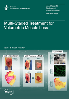
Several factors can necessitate delaying regenerative treatments for severe muscle trauma, including infection, management of comorbidities, and the time required for biomanufacturing of the therapeutics themselves. This study investigated using a polyvinyl alcohol-based, in situ-forming muscle void filler (MVF) to provide initial stabilization following volumetric muscle loss injury. These MVFs were removed after four weeks and replaced with minced muscle grafts to promote the regeneration of functional skeletal muscle within the defect. While this multi-stage approach effectively maintained the defect volume and minimized complications, the delayed application of MMGs did not demonstrate significant regenerative benefits. View this paper
- Issues are regarded as officially published after their release is announced to the table of contents alert mailing list.
- You may sign up for e-mail alerts to receive table of contents of newly released issues.
- PDF is the official format for papers published in both, html and pdf forms. To view the papers in pdf format, click on the "PDF Full-text" link, and use the free Adobe Reader to open them.
Previous Issue
Next Issue