Next Issue
Volume 12, December
Previous Issue
Volume 12, June


J. Funct. Biomater., Volume 12, Issue 3 (September 2021) – 12 articles

Cover Story (view full-size image): 
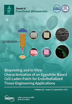
The image presents egg-white-based cell-laden patches fabricated using the 3D bioprinting technique. Egg white was utilized for enhanced biological properties, while a small amount of sodium alginate was used to improve bioink printability. The fabricated patch possessed suitable mechanical and biological properties for soft tissue engineering applications. A highly porous structure, tissue-like elastic modulus, and a high survival rate of printed vascular endothelial cells (loaded into the bioink) were some of these desirable characteristics. View this paper
  • Issues are regarded as officially published after their release is announced to the table of contents alert mailing list.
  • You may sign up for e-mail alerts to receive table of contents of newly released issues.
  • PDF is the official format for papers published in both, html and pdf forms. To view the papers in pdf format, click on the "PDF Full-text" link, and use the free Adobe Reader to open them.
Order results
Result details
Section
Select all
Export citation of selected articles as:
12 pages, 3215 KiB  
Article
In Vitro Corrosion of SiC-Coated Anodized Ti Nano-Tubular Surfaces
by Shu-Min Hsu, Chaker Fares, Xinyi Xia, Md Abu Jafar Rasel, Jacob Ketter, Samira Esteves Afonso Camargo, Md Amanul Haque, Fan Ren and Josephine F. Esquivel-Upshaw
J. Funct. Biomater. 2021, 12(3), 52; https://doi.org/10.3390/jfb12030052 - 16 Sep 2021
Cited by 2 | Viewed by 3244
Abstract
Peri-implantitis leads to implant failure and decreases long-term survival and success rates of implant-supported prostheses. The pathogenesis of this disease is complex but implant corrosion is believed to be one of the many factors which contributes to progression of this disease. A nanostructured [...] Read more.
Peri-implantitis leads to implant failure and decreases long-term survival and success rates of implant-supported prostheses. The pathogenesis of this disease is complex but implant corrosion is believed to be one of the many factors which contributes to progression of this disease. A nanostructured titanium dioxide layer was introduced using anodization to improve the functionality of dental implants. In the present study, we evaluated the corrosion performance of silicon carbide (SiC) on anodized titanium dioxide nanotubes (ATO) using plasma-enhanced chemical vapor deposition (PECVD). This was investigated through a potentiodynamic polarization test and bacterial incubation for 30 days. Scanning electron microscopy (SEM) and transmission electron microscopy (TEM) were used to analyze surface morphologies of non-coated and SiC-coated nanotubes. Energy dispersive X-ray (EDX) was used to analyze the surface composition. In conclusion, SiC-coated ATO exhibited improved corrosion resistance and holds promise as an implant coating material. Full article
(This article belongs to the Special Issue Nanoengineered Materials for Biomedical Applications)
Show Figures

Figure 1

13 pages, 2866 KiB  
Article
A Two-Year Comparative Evaluation of Clinical Performance of a Nanohybrid Composite Resin to a Flowable Composite Resin
by Carelle Badr, Gianrico Spagnuolo, Francesco Amenta, Carlos Khairallah, Syed Sarosh Mahdi, Elie Daher, Gopi Battineni, Nadim Z. Baba, Tatiana Zogheib, Syed Saad B. Qasim, Tony Daher, Nalini Chintalapudi and Carina Mehanna Zogheib
J. Funct. Biomater. 2021, 12(3), 51; https://doi.org/10.3390/jfb12030051 - 09 Sep 2021
Cited by 7 | Viewed by 5091
Abstract
Objective: This prospective in vivo study aimed to compare the clinical behavior of a flowable composite resin (Genial Universal Flo, GC) and a nanohybrid universal composite resin (Tetric Evo Ceram, Ivoclar Vivadent) used in Class I and II direct esthetic restorations in posterior [...] Read more.
Objective: This prospective in vivo study aimed to compare the clinical behavior of a flowable composite resin (Genial Universal Flo, GC) and a nanohybrid universal composite resin (Tetric Evo Ceram, Ivoclar Vivadent) used in Class I and II direct esthetic restorations in posterior teeth. Methods: A total of 108 Class I and II direct restorations were performed in patients aged between 20 and 60 years. The originality of this study lies in the fact that both materials were placed in pairs, in the same clinical environment (i.e., the same patient and the same type of tooth). The evaluations were performed now of restoration and after 2-weeks, 6-, 12-, and 24-months intervals using clinical examination, clinical photographs, and radiological examination, according to modified USPHS criteria. Statistical analysis was performed using the Fisher exact test and chi-square analysis. Results: At baseline, the universal composite resin showed better esthetic properties such as surface luster, surface staining marginal staining. Both materials regressed significantly over time with no significant difference between groups. Conclusions: Both flowable and nanohybrid composite resins exhibit acceptable clinical performance. The present 24 months of evaluation of different composites showed that the G-ænial Universal Flo could be an effective esthetic material for posterior restoration. No significant difference between both materials over time concerning surface luster, surface staining, and marginal staining. Full article
Show Figures

Figure 1

22 pages, 35476 KiB  
Article
Proposal for a Novel Abrasive Machining Method for Preparing the Surface of Periarticular Tissue during Orthopedic Surgery on Hip Joints
by Paweł Zawadzki
J. Funct. Biomater. 2021, 12(3), 50; https://doi.org/10.3390/jfb12030050 - 08 Sep 2021
Cited by 2 | Viewed by 2045
Abstract
Drilling, cutting, and milling are the most common methods used in orthopedic surgery. However, popular machining methods do not obtain the complex shape of the periarticular tissue surfaces, increasing operation time and patient recovery. This paper reports an attempt to research a novel [...] Read more.
Drilling, cutting, and milling are the most common methods used in orthopedic surgery. However, popular machining methods do not obtain the complex shape of the periarticular tissue surfaces, increasing operation time and patient recovery. This paper reports an attempt to research a novel design of a machining process for surgical procedures. A device using abrasion machining based on mechanical erosion was proposed. Machining uses an undefined geometry of the cutting grains to cut tissue in any direction during oscillatory tool movement. This new concept is based on a cylindrical abrasive device made of brown fused alumina and silicon carbide grains deposited with an epoxy resin binder on the surface of a polyamide shaft. The best results in terms of machining efficiency were obtained for grains of the BFA80 type. Cutting experiments with different values in terms of cutting speed, granulation of the abrasive grains, pressure forces, and machining scope showed that the proposed concept, by developing the shape of the device, allows for penetration of the tissue structure. The research shows the possibility of using the proposed method during periarticular tissue machining. Full article
Show Figures

Figure 1

11 pages, 2500 KiB  
Article
Prediction of Bone Mineral Density (BMD) Adaptation in Pelvis–Femur Model with Hip Arthroplasties
by Abdul Halim Abdullah and Mitsugu Todo
J. Funct. Biomater. 2021, 12(3), 49; https://doi.org/10.3390/jfb12030049 - 03 Sep 2021
Cited by 2 | Viewed by 3809
Abstract
The prediction of bone remodeling behaviour is a challenging factor in encouraging the long-term stability of hip arthroplasties. The presence of femoral components modifies the biomechanical environment of the bone and alters the bone growth process. Issues of bone loss and gait instability [...] Read more.
The prediction of bone remodeling behaviour is a challenging factor in encouraging the long-term stability of hip arthroplasties. The presence of femoral components modifies the biomechanical environment of the bone and alters the bone growth process. Issues of bone loss and gait instability on both limbs are associated with the remodeling process. In this study, finite element analysis with an adaptive bone remodeling algorithm was used to predict the changes in bone mineral density following total hip and resurfacing hip arthroplasty. A three-dimensional model of the pelvis–femur was constructed from computed tomography (CT-based) images of a 79-year-old female patient with hip osteoarthritis. The prosthesis stem of the total hip arthroplasty was modelled with a titanium alloy material, while the femoral head had alumina properties. Meanwhile, resurfacing of the hip implant was completed with a cobalt-chromium material. Contact between the components and bone was designed to be perfectly bonded at the interface. Results indicate that the bone mineral density was modified over five years on all models, including hip osteoarthritis. The changes of BMD were predicted as being high between year zero and year one, especially in the proximal region. Changes were observed to be minimal in the following years. The bone remodeling process was also predicted for the non-operated femur. However, the adaptation was lower compared to the operated limbs. The reduction in bone mineral density suggested the bone loss phenomenon after a few years. Full article
Show Figures

Figure 1

16 pages, 1176 KiB  
Review
Effect of Resin Infiltration on Enamel: A Systematic Review and Meta-Analysis
by Madalena Soveral, Vanessa Machado, João Botelho, José João Mendes and Cristina Manso
J. Funct. Biomater. 2021, 12(3), 48; https://doi.org/10.3390/jfb12030048 - 16 Aug 2021
Cited by 20 | Viewed by 5386
Abstract
Subsurface enamel demineralization beneath an intact surface layer or white spots lesions (WSL) can and should be treated with non-invasive procedures to impede the development of a cavitated lesion. We aim to analyze if infiltrative resin improves enamel roughness, microhardness, shear bond strength, [...] Read more.
Subsurface enamel demineralization beneath an intact surface layer or white spots lesions (WSL) can and should be treated with non-invasive procedures to impede the development of a cavitated lesion. We aim to analyze if infiltrative resin improves enamel roughness, microhardness, shear bond strength, and penetration depth. MEDLINE [via Pubmed], Cochrane Central Register of Controlled Trials, Embase, Web of Science, Scholar, and LILACS were searched until May 2021. Methodological quality was assessed using the Joanna Briggs Institute Clinical Appraisal Checklist for Experimental Studies. Pairwise ratio of means (ROM) meta-analyses were carried out to compare the enamel properties after treatment with infiltrative resin on sound enamel and WSLs. From a total of 1604 articles, 48 studies were included. Enamel surface roughness decreased 35% in sound enamel (95%CI: 0.49–0.85, I2 = 98.2%) and 54% in WSLs (95%CI: 0.29–0.74, I2 = 98.5%). Microhardness reduced 24% in sound enamel (95%CI: 0.73–0.80, I2 = 99.1%) and increased by 68% in WSLs (95%CI: 1.51; 1.86, I2 = 99.8%). Shear bond strength reduced of 25% in sound enamel (95%CI: 0.60; 0.95, I2 = 96.9%) and increased by 89% in WSLs (95%CI: 1.28–2.79, I2 = 99.8%). Penetration depth was 65.39% of the WSLs (95%CI: 56.11–74.66, I2 = 100%). Infiltrative resins effectively promote evident changes in enamel properties in sound and WSLs. Future studies with long-term follow-ups are necessary to corroborate these results from experimental studies. Full article
(This article belongs to the Special Issue Recent Advances in Dental Implants and Biomaterials)
Show Figures

Figure 1

12 pages, 8091 KiB  
Article
Improvement of Mechanical Strength of Tissue Engineering Scaffold Due to the Temperature Control of Polymer Blend Solution
by Azizah Intan Pangesty and Mitsugu Todo
J. Funct. Biomater. 2021, 12(3), 47; https://doi.org/10.3390/jfb12030047 - 14 Aug 2021
Cited by 8 | Viewed by 3239
Abstract
Polymeric scaffolds made of PCL/PLCL (ratio 1:3, respectively) blends have been developed by using the Thermally Induced Phase Separation (TIPS) process. A new additional technique has been introduced in this study by applying pre-heat treatment to the blend solution before the TIPS process. [...] Read more.
Polymeric scaffolds made of PCL/PLCL (ratio 1:3, respectively) blends have been developed by using the Thermally Induced Phase Separation (TIPS) process. A new additional technique has been introduced in this study by applying pre-heat treatment to the blend solution before the TIPS process. The main objective of this study is to evaluate the influence of the pre-heat treatment on mechanical properties. The mechanical evaluation showed that the mechanical strength of the scaffolds (including tensile strength, elastic modulus, and strain) improved as the temperature of the polymer blend solution increased. The effects on the microstructure features were also observed, such as increasing strut size and differences in phase separation morphology. Those microstructure changes due to temperature control contributed to the increasing of mechanical strength. The in vitro cell study showed that the PCL/PLCL blend scaffold exhibited better cytocompatibility than the neat PCL scaffold, indicated by a higher proliferation at 4 and 7 days in culture. This study highlighted that the improvement of the mechanical strength of polymer blends scaffolds can be achieved using a very versatile way by controlling the temperature of the polymer blend solution before the TIPS process. Full article
Show Figures

Figure 1

16 pages, 3248 KiB  
Article
Formulation and Characterization of a New Injectable Bone Substitute Composed PVA/Borax/CaCO3 and Demineralized Bone Matrix
by Daniela Medrano-David, Aura María Lopera, Martha Elena Londoño and Pedronel Araque-Marín
J. Funct. Biomater. 2021, 12(3), 46; https://doi.org/10.3390/jfb12030046 - 11 Aug 2021
Cited by 7 | Viewed by 3293
Abstract
The occurrence of bone-related disorders and diseases has dramatically increased in recent years around the world. Demineralized bone matrix (DBM) has been widely used as a bone implant due to its osteoinduction and bioactivity. However, the use of DBM is limited because it [...] Read more.
The occurrence of bone-related disorders and diseases has dramatically increased in recent years around the world. Demineralized bone matrix (DBM) has been widely used as a bone implant due to its osteoinduction and bioactivity. However, the use of DBM is limited because it is a particulate material, which makes it difficult to manipulate and implant with precision. In addition, these particles are susceptible to migration to other sites. To address this situation, DBM is commonly incorporated into a variety of carriers. An injectable scaffold has advantages over bone grafts or preformed scaffolds, such as the ability to flow and fill a bone defect. The aim of this research was to develop a DBM carrier with such viscoelastic properties in order to obtain an injectable bone substitute (IBS). The developed DBM carrier consisted of a PVA/glycerol network cross-linked with borax and reinforced with CaCO3 as a pH neutralizer, porosity generator, and source of Ca. The physicochemical properties were determined by an injectability test, FTIR, SEM, and TGA. Porosity, degradation, bioactivity, possible cytotoxic effect, and proliferation in osteoblasts were also determined. The results showed that the developed material has great potential to be used in bone tissue regeneration. Full article
Show Figures

Graphical abstract

16 pages, 4792 KiB  
Article
Bioprinting and In Vitro Characterization of an Eggwhite-Based Cell-Laden Patch for Endothelialized Tissue Engineering Applications
by Yasaman Delkash, Maxence Gouin, Tanguy Rimbeault, Fatemeh Mohabatpour, Petros Papagerakis, Sean Maw and Xiongbiao Chen
J. Funct. Biomater. 2021, 12(3), 45; https://doi.org/10.3390/jfb12030045 - 11 Aug 2021
Cited by 18 | Viewed by 4568
Abstract
Three-dimensional (3D) bioprinting is an emerging fabrication technique to create 3D constructs with living cells. Notably, bioprinting bioinks are limited due to the mechanical weakness of natural biomaterials and the low bioactivity of synthetic peers. This paper presents the development of a natural [...] Read more.
Three-dimensional (3D) bioprinting is an emerging fabrication technique to create 3D constructs with living cells. Notably, bioprinting bioinks are limited due to the mechanical weakness of natural biomaterials and the low bioactivity of synthetic peers. This paper presents the development of a natural bioink from chicken eggwhite and sodium alginate for bioprinting cell-laden patches to be used in endothelialized tissue engineering applications. Eggwhite was utilized for enhanced biological properties, while sodium alginate was used to improve bioink printability. The rheological properties of bioinks with varying amounts of sodium alginate were examined with the results illustrating that 2.0–3.0% (w/v) sodium alginate was suitable for printing patch constructs. The printed patches were then characterized mechanically and biologically, and the results showed that the printed patches exhibited elastic moduli close to that of natural heart tissue (20–27 kPa) and more than 94% of the vascular endothelial cells survived in the examination period of one week post 3D bioprinting. Our research also illustrated the printed patches appropriate water uptake ability (>1800%). Full article
Show Figures

Figure 1

20 pages, 3102 KiB  
Article
Comparative Evaluation of Two Glass Polyalkenoate Cements: An In Vivo Pilot Study Using a Sheep Model
by Leyla Hasandoost, Daniella Marx, Paul Zalzal, Oleg Safir, Mark Hurtig, Cina Mehrvar, Stephen D. Waldman, Marcello Papini and Mark R. Towler
J. Funct. Biomater. 2021, 12(3), 44; https://doi.org/10.3390/jfb12030044 - 05 Aug 2021
Cited by 1 | Viewed by 2699
Abstract
Poly(methyl methacrylate) (PMMA) is used to manage bone loss in revision total knee arthroplasty (rTKA). However, the application of PMMA has been associated with complications such as volumetric shrinkage, necrosis, wear debris, and loosening. Glass polyalkenoate cements (GPCs) have potential bone cementation applications. [...] Read more.
Poly(methyl methacrylate) (PMMA) is used to manage bone loss in revision total knee arthroplasty (rTKA). However, the application of PMMA has been associated with complications such as volumetric shrinkage, necrosis, wear debris, and loosening. Glass polyalkenoate cements (GPCs) have potential bone cementation applications. Unlike PMMA, GPC does not undergo volumetric shrinkage, adheres chemically to bone, and does not undergo an exothermic setting reaction. In this study, two different compositions of GPCs (GPCA and GPCB), based on the patented glass system SiO2-CaO-SrO-P2O5-Ta2O5, were investigated. Working and setting times, pH, ion release, compressive strength, and cytotoxicity of each composition were assessed, and based on the results of these tests, three sets of samples from GPCA were implanted into the distal femur and proximal tibia of three sheep (alongside PMMA as control). Clinical CT scans and micro-CT images obtained at 0, 6, and 12 weeks revealed the varied radiological responses of sheep bone to GPCA. One GPCA sample (implanted in the sheep for 12 weeks) was characterized with no bone resorption. Furthermore, a continuous bone–cement interface was observed in the CT images of this sample. The other implanted GPCA showed a thin radiolucent border at six weeks, indicating some bone resorption occurred. The third sample showed extensive bone resorption at both six and 12 weeks. Possible speculative factors that might be involved in the varied response can be: excessive Zn2+ ion release, low pH, mixing variability, and difficulty in inserting the samples into different parts of the sheep bone. Full article
Show Figures

Figure 1

12 pages, 1839 KiB  
Review
Finite Element Method for the Evaluation of the Human Spine: A Literature Overview
by Symeon Naoum, Angelo V. Vasiliadis, Christos Koutserimpas, Nikolaos Mylonakis, Michail Kotsapas and Konstantinos Katakalos
J. Funct. Biomater. 2021, 12(3), 43; https://doi.org/10.3390/jfb12030043 - 31 Jul 2021
Cited by 23 | Viewed by 5178
Abstract
The finite element method (FEM) represents a computer simulation method, originally used in civil engineering, which dates back to the early 1940s. Applications of FEM have also been used in numerous medical areas and in orthopedic surgery. Computing technology has improved over the [...] Read more.
The finite element method (FEM) represents a computer simulation method, originally used in civil engineering, which dates back to the early 1940s. Applications of FEM have also been used in numerous medical areas and in orthopedic surgery. Computing technology has improved over the years and as a result, more complex problems, such as those involving the spine, can be analyzed. The spine is a complex anatomical structure that maintains the erect posture and supports considerable loads. Applications of FEM in the spine have contributed to the understanding of bone biomechanics, both in healthy and abnormal conditions, such as scoliosis, fractures (trauma), degenerative disc disease and osteoporosis. However, since FEM is only a digital simulation of the real condition, it will never exactly simulate in vivo results. In particular, when it concerns biomechanics, there are many features that are difficult to represent in a FEM. More FEM studies and spine research are required in order to examine interpersonal spine stiffness, young spine biomechanics and model accuracy. In the future, patient-specific models will be used for better patient evaluations as well as for better pre- and inter-operative planning. Full article
Show Figures

Figure 1

15 pages, 3624 KiB  
Article
Rheological Properties, Surface Microhardness, and Dentin Shear Bond Strength of Resin-Modified Glass Ionomer Cements Containing Methacrylate-Functionalized Polyacids and Spherical Pre-Reacted Glass Fillers
by Whithipa Thepveera, Wisitsin Potiprapanpong, Arnit Toneluck, Somruethai Channasanon, Chutikarn Khamsuk, Naruporn Monmaturapoj, Siriporn Tanodekaew and Piyaphong Panpisut
J. Funct. Biomater. 2021, 12(3), 42; https://doi.org/10.3390/jfb12030042 - 14 Jul 2021
Cited by 11 | Viewed by 2961
Abstract
The aim of this study was to prepare experimental resin-modified glass ionomer cements (RMGICs) containing low levels of hydroxyethyl methacrylate (HEMA) for pulp protection. Liquid and powder phases of the experimental RMGICs were polyacid functionalized with methacrylate groups and spherical pre-reacted glass fillers [...] Read more.
The aim of this study was to prepare experimental resin-modified glass ionomer cements (RMGICs) containing low levels of hydroxyethyl methacrylate (HEMA) for pulp protection. Liquid and powder phases of the experimental RMGICs were polyacid functionalized with methacrylate groups and spherical pre-reacted glass fillers (SPG). Two types of liquid phase containing 0 wt. % HEMA (CM liquid) or 5 wt. % HEMA (CMH liquid) were formulated. The experimental RMGICs were prepared by mixing SPG fillers with CM liquid (F1) or CMH liquid (F2). Rheological properties were examined using a strain-controlled rheometer (n = 5). The Vickers microhardness (n = 5) and dentin shear bond strength (SBS) (n = 10) of the materials were tested. Commercial pulp protection materials (Vitrebond and TheraCal LC) were used as comparisons. The viscosity and surface microhardness of F1 (22 m Pa·s, 18 VHN) and F2 (18 m Pa·s, 16 VHN) were significantly higher than those of Vitrebond (6 mPa·s, 6 VHN) and TheraCal (0.1 mPa·s, 7 VHN). The SBS of F1 (10.7 MPa) and F2 (11.9 MPa) was comparable to that of Vitrebond (15.4 MPa) but higher than that of TheraCal LC (5.6 MPa). The addition of 5 wt. % HEMA showed no significant effect on viscosity, surface microhardness, or SBS of the experimental RMGICs. The experimental materials showed higher viscosity and microhardness but similar SBS when compared with the commercial RMGIC. Full article
Show Figures

Graphical abstract

15 pages, 2700 KiB  
Review
Biomaterials for Cell-Surface Engineering and Their Efficacy
by Seoyoung Jang, Jin Gil Jeong, Tong In Oh and EunAh Lee
J. Funct. Biomater. 2021, 12(3), 41; https://doi.org/10.3390/jfb12030041 - 13 Jul 2021
Cited by 3 | Viewed by 3008
Abstract
Literature in the field of stem cell therapy indicates that, when stem cells in a state of single-cell suspension are injected systemically, they show poor in vivo survival, while such cells show robust cell survival and regeneration activity when transplanted in the state [...] Read more.
Literature in the field of stem cell therapy indicates that, when stem cells in a state of single-cell suspension are injected systemically, they show poor in vivo survival, while such cells show robust cell survival and regeneration activity when transplanted in the state of being attached on a biomaterial surface. Although an attachment-deprived state induces anoikis, when cell-surface engineering technology was adopted for stem cells in a single-cell suspension state, cell survival and regenerative activity dramatically improved. The biochemical signal coming from ECM (extracellular matrix) molecules activates the cell survival signal transduction pathway and prevents anoikis. According to the target disease, various therapeutic cells can be engineered to improve their survival and regenerative activity, and there are several types of biomaterials available for cell-surface engineering. In this review, biomaterial types and application strategies for cell-surface engineering are presented along with their expected efficacy. Full article
Show Figures

Figure 1

Previous Issue
Next Issue
Back to TopTop