Next Issue
Volume 11, December
Previous Issue
Volume 11, June


J. Funct. Biomater., Volume 11, Issue 3 (September 2020) – 22 articles

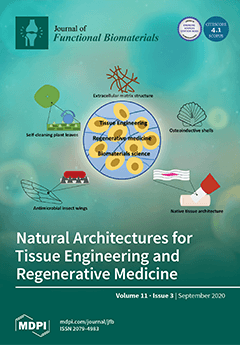
Cover Story (view full-size image): Over millions of years, nature has produced a wide variety of biological materials optimised for distinct functions. Many of these systems found in biology possess unique surface properties recognised to regulate cell behaviour. Integration of such natural surface properties in biomaterials can bring about novel cell responses and provide greater insights into the processes occurring at the cell–biomaterial interface. Using natural surfaces as templates for bioinspired design can stimulate progress in the field of regenerative medicine, tissue engineering and biomaterials science. This literature review aims to combine the state-of-the-art knowledge in natural and nature-inspired surfaces. View this paper
  • Issues are regarded as officially published after their release is announced to the table of contents alert mailing list.
  • You may sign up for e-mail alerts to receive table of contents of newly released issues.
  • PDF is the official format for papers published in both, html and pdf forms. To view the papers in pdf format, click on the "PDF Full-text" link, and use the free Adobe Reader to open them.
Order results
Result details
Section
Select all
Export citation of selected articles as:
36 pages, 9300 KiB  
Review
Bio-Based Electrospun Fibers for Wound Healing
by Bahareh Azimi, Homa Maleki, Lorenzo Zavagna, Jose Gustavo De la Ossa, Stefano Linari, Andrea Lazzeri and Serena Danti
J. Funct. Biomater. 2020, 11(3), 67; https://doi.org/10.3390/jfb11030067 - 22 Sep 2020
Cited by 127 | Viewed by 9961
Abstract
Being designated to protect other tissues, skin is the first and largest human body organ to be injured and for this reason, it is accredited with a high capacity for self-repairing. However, in the case of profound lesions or large surface loss, the [...] Read more.
Being designated to protect other tissues, skin is the first and largest human body organ to be injured and for this reason, it is accredited with a high capacity for self-repairing. However, in the case of profound lesions or large surface loss, the natural wound healing process may be ineffective or insufficient, leading to detrimental and painful conditions that require repair adjuvants and tissue substitutes. In addition to the conventional wound care options, biodegradable polymers, both synthetic and biologic origin, are gaining increased importance for their high biocompatibility, biodegradation, and bioactive properties, such as antimicrobial, immunomodulatory, cell proliferative, and angiogenic. To create a microenvironment suitable for the healing process, a key property is the ability of a polymer to be spun into submicrometric fibers (e.g., via electrospinning), since they mimic the fibrous extracellular matrix and can support neo- tissue growth. A number of biodegradable polymers used in the biomedical sector comply with the definition of bio-based polymers (known also as biopolymers), which are recently being used in other industrial sectors for reducing the material and energy impact on the environment, as they are derived from renewable biological resources. In this review, after a description of the fundamental concepts of wound healing, with emphasis on advanced wound dressings, the recent developments of bio-based natural and synthetic electrospun structures for efficient wound healing applications are highlighted and discussed. This review aims to improve awareness on the use of bio-based polymers in medical devices. Full article
Show Figures

Graphical abstract

20 pages, 2900 KiB  
Article
Biosynthesis, Characterization, and Biological Activities of Procyanidin Capped Silver Nanoparticles
by Umar M. Badeggi, Jelili A. Badmus, Subelia S. Botha, Enas Ismail, Jeanine L. Marnewick, Charlene W. J. Africa and Ahmed A. Hussein
J. Funct. Biomater. 2020, 11(3), 66; https://doi.org/10.3390/jfb11030066 - 19 Sep 2020
Cited by 14 | Viewed by 2877
Abstract
In this study, procyanidin dimers and Leucosidea sericea total extract (LSTE) were employed in the synthesis of silver nanoparticles (AgNPs) and characterized by ultraviolet-visible (UV-Visible) spectroscopy, high-resolution transmission electron microscopy (HRTEM), selected area electron diffraction (SAED), X-ray diffraction (XRD), and dynamic light scattering [...] Read more.
In this study, procyanidin dimers and Leucosidea sericea total extract (LSTE) were employed in the synthesis of silver nanoparticles (AgNPs) and characterized by ultraviolet-visible (UV-Visible) spectroscopy, high-resolution transmission electron microscopy (HRTEM), selected area electron diffraction (SAED), X-ray diffraction (XRD), and dynamic light scattering (DLS) techniques. AgNPs of about 2–7 nm were obtained. DLS and stability evaluations confirmed that the AgNPs/procyanidins conjugates were stable. The formed nanoparticles exhibited good inhibitory activities against the two enzymes studied. The IC50 values against the amylase enzyme were 14.92 ± 1.0, 13.24 ± 0.2, and 19.13 ± 0.8 µg/mL for AgNPs coordinated with LSTE, F1, and F2, respectively. The corresponding values for the glucosidase enzyme were 21.48 ± 0.9, 18.76 ± 1.0, and 8.75 ± 0.7 µg/mL. The antioxidant activities were comparable to those of the intact fractions. The AgNPs also demonstrated bacterial inhibitory activities against six bacterial species. While the minimum inhibitory concentrations (MIC) of F1-AgNPs against Pseudomonas aeruginosa and Staphylococcus aureus were 31.25 and 15.63 µg/mL respectively, those of LSTE-AgNPs and F2-AgNPs against these organisms were both 62.50 µg/mL. The F1-AgNPs demonstrated a better bactericidal effect and may be useful in food packaging. This research also showed the involvement of the procyanidins as reducing and capping agents in the formation of stable AgNPs with potential biological applications. Full article
(This article belongs to the Special Issue Bioactive Properties of Advanced Nanomaterials)
Show Figures

Graphical abstract

13 pages, 5363 KiB  
Article
Biocompatibility of α-Al2O3 Ceramic Substrates with Human Neural Precursor Cells
by Akrivi Asimakopoulou, Ioannis Gkekas, Georgia Kastrinaki, Alessandro Prigione, Vasileios T. Zaspalis and Spyros Petrakis
J. Funct. Biomater. 2020, 11(3), 65; https://doi.org/10.3390/jfb11030065 - 16 Sep 2020
Cited by 6 | Viewed by 3309
Abstract
Background: Biocompatible materials-topography could be used for the construction of scaffolds allowing the three-dimensional (3D) organization of human stem cells into functional tissue-like structures with a defined architecture. Methods: Structural characterization of an alumina-based substrate was performed through XRD, Brunauer–Emmett–Teller (BET) analysis, scanning [...] Read more.
Background: Biocompatible materials-topography could be used for the construction of scaffolds allowing the three-dimensional (3D) organization of human stem cells into functional tissue-like structures with a defined architecture. Methods: Structural characterization of an alumina-based substrate was performed through XRD, Brunauer–Emmett–Teller (BET) analysis, scanning electron microscopy (SEM), and wettability measurements. Biocompatibility of the substrate was assessed by measuring the proliferation and differentiation of human neural precursor stem cells (NPCs). Results: α-Al2O3 is a ceramic material with crystallite size of 40 nm; its surface consists of aggregates in the range of 8–22 μm which forms a rough surface in the microscale with 1–8 μm cavities. The non-calcined material has a surface area of 5.5 m2/gr and pore size distribution of 20 nm, which is eliminated in the calcined structure. Thus, the pore network on the surface and the body of the ceramic becomes more water proof, as indicated by wettability measurements. The alumina-based substrate supported the proliferation of human NPCs and their differentiation into functional neurons. Conclusions: Our work indicates the potential use of alumina for the construction of 3D engineered biosystems utilizing human neurons. Such systems may be useful for diagnostic purposes, drug testing, or biotechnological applications. Full article
(This article belongs to the Special Issue Advanced Bioceramics)
Show Figures

Figure 1

12 pages, 14759 KiB  
Article
Development of Bionanocomposites Based on Poly(3-Hydroxybutyrate-co-3-Hydroxyvalerate)/PolylActide Blends Reinforced with Cloisite 30B
by Clément Lacoste, Benjamin Gallard, José-Marie Lopez-Cuesta, Ozlem Ipek Kalaoglu-Altan and Karen De Clerck
J. Funct. Biomater. 2020, 11(3), 64; https://doi.org/10.3390/jfb11030064 - 16 Sep 2020
Cited by 8 | Viewed by 2196
Abstract
In the present study, poly(3-hydroxybuturate-co-3-hydroxyvalerate) (PHBV) and plasticized polylactide acid (PLA) blends were processed by melt extrusion with different weight ratio (up to 20 wt.% of PHBV). Bionanocomposites were obtained through the incorporation of an organomodified montmorillonite (C30B) at 3 wt.%. The main [...] Read more.
In the present study, poly(3-hydroxybuturate-co-3-hydroxyvalerate) (PHBV) and plasticized polylactide acid (PLA) blends were processed by melt extrusion with different weight ratio (up to 20 wt.% of PHBV). Bionanocomposites were obtained through the incorporation of an organomodified montmorillonite (C30B) at 3 wt.%. The main features of the processing and physico-chemical characterization of films and injected samples were assessed and the influence of the components on the chemical, thermal and mechanical properties of the bionanocomposites was investigated. The results indicated that plasticized PLA/PHBV/C30B bionanocomposites present optimal mechanical properties for sanitary applications. Moreover, plasticized PLA/PHBV could lead to finely tuned biomaterials able to form electrospun nanofibers. Full article
Show Figures

Figure 1

30 pages, 10960 KiB  
Article
Carbonate Apatite and Hydroxyapatite Formulated with Minimal Ingredients to Deliver SiRNA into Breast Cancer Cells In Vitro and In Vivo
by Rowshan Ara Islam, Hamed Al-Busaidi, Rahela Zaman, Syafiq Asnawi Zainal Abidin, Iekhsan Othman and Ezharul Hoque Chowdhury
J. Funct. Biomater. 2020, 11(3), 63; https://doi.org/10.3390/jfb11030063 - 10 Sep 2020
Cited by 14 | Viewed by 3476
Abstract
Introduction: Cancer is one of the top-ranked noncommunicable diseases causing deaths to nine million people and affecting almost double worldwide in 2018. Tremendous advancement in surgery, chemotherapy, radiation and targeted immunotherapy have improved the rate of cure and disease-free survival. As genetic mutations [...] Read more.
Introduction: Cancer is one of the top-ranked noncommunicable diseases causing deaths to nine million people and affecting almost double worldwide in 2018. Tremendous advancement in surgery, chemotherapy, radiation and targeted immunotherapy have improved the rate of cure and disease-free survival. As genetic mutations vary in different cancers, potential of customized treatment to silence the problem gene/s at the translational level is being explored too. Yet delivering therapeutics at the required dosage only to the affected cells without affecting the healthy ones, is a big hurdle to be overcome. Scientists worldwide have been working to invent a smart drug delivery system for targeted delivery of therapeutics to tumor tissues only. As part of such an effort, few organic nanocarriers went to clinical trials, while inorganic nanoparticles (NPs) are still in development stage despite their many customizable properties. Carbonate apatite (CA), a pH sensitive nanocarrier has emerged as an efficient delivery system for drugs, plasmids and siRNAs in preclinical models of breast and colon cancers. Like hydroxyapatite (HA) which serves as a classical tool for delivery of genetic materials such as siRNA and plasmid, CA is an apatite-based synthetic carrier. We developed simplified methods of formulating CA-in-DMEM and a DMEM-mimicking buffer and HA in a HEPES-buffered solution and characterized them in terms of size, stability, protein corona (PC) composition, cytotoxicity, siRNA delivery efficiency in breast cancer cells and siRNA biodistribution profile in a mouse model of breast cancer. Methods: Particle growth was analyzed via spectrophotometry and light microscopy, size was measured via dynamic light scattering and scanning electron microscopy and confirmation of functional groups in apatite structures was made by FT-IR. siRNA-binding was analyzed via spectrophotometry. Stability of the formulation solutions/buffers was tested over various time points and at different temperatures to determine their compatibility in the context of practical usage. Cellular uptake was studied via fluorescence microscopy. MTT assay was performed to measure the cytotoxicity of the NPs. Liquid chromatography—mass spectrometry was carried out to analyze the PC formed around all three different NPs in serum-containing media. To explore biodistribution of all the formulations, fluorescence-labeled siRNA-loaded NPs were administered intravenously prior to analysis of fluorescence intensity in the collected organs and tumors of the treated mice. Results: The size of NPs in 10% serum-containing media was dramatically different where CA-in-DMB and HA were much larger than CA-in-DMEM. Effect of media was notable on the PC composition of all three NPs. All three NPs bound albumin and some common protease inhibitors involved in bone metabolism due to their compositional similarity to our bone materials. Moreover, CA also bound heme-binding proteins and opsonins. Unlike CA, HA bound different kinds of keratins. Difference in PC constitution was likely to influence accumulation of NPs in various organs including those of reticuloendothelial system, such as liver and spleen and the tumor. We found 10 times more tumor accumulation of CA-in-DMB than CA-in-DMEM, which could be due to more stable siRNA-binding and distinct PC composition of the former. Conclusion: As a nanocarrier CA is more efficient than HA for siRNA delivery to the tumor. CA prepared in a buffer containing only the mere constituents was potentially more efficient than classical CA prepared in DMEM, owing to the exclusion of interference attributed by the inorganic ions and organic molecules present in DMEM. Full article
(This article belongs to the Special Issue Nanotechnology and Biomaterials in Drug Delivery System)
Show Figures

Figure 1

17 pages, 11862 KiB  
Article
Electrosprayed Chitin Nanofibril/Electrospun Polyhydroxyalkanoate Fiber Mesh as Functional Nonwoven for Skin Application
by Bahareh Azimi, Lily Thomas, Alessandra Fusco, Ozlem Ipek Kalaoglu-Altan, Pooja Basnett, Patrizia Cinelli, Karen De Clerck, Ipsita Roy, Giovanna Donnarumma, Maria-Beatrice Coltelli, Serena Danti and Andrea Lazzeri
J. Funct. Biomater. 2020, 11(3), 62; https://doi.org/10.3390/jfb11030062 - 03 Sep 2020
Cited by 46 | Viewed by 5199
Abstract
Polyhydroxyalkanoates (PHAs) are a family of bio-based polyesters that have found different biomedical applications. Chitin and lignin, byproducts of fishery and plant biomass, show antimicrobial and anti-inflammatory activity on the nanoscale. Due to their polarities, chitin nanofibril (CN) and nanolignin (NL) can be [...] Read more.
Polyhydroxyalkanoates (PHAs) are a family of bio-based polyesters that have found different biomedical applications. Chitin and lignin, byproducts of fishery and plant biomass, show antimicrobial and anti-inflammatory activity on the nanoscale. Due to their polarities, chitin nanofibril (CN) and nanolignin (NL) can be assembled into micro-complexes, which can be loaded with bioactive factors, such as the glycyrrhetinic acid (GA) and CN-NL/GA (CLA) complexes, and can be used to decorate polymer surfaces. This study aims to develop completely bio-based and bioactive meshes intended for wound healing. Poly(3-hydroxybutyrate)/Poly(3-hydroxyoctanoate-co-3-hydroxydecanoate), P(3HB)/P(3HO-co-3HD) was used to produce films and fiber meshes, to be surface-modified via electrospraying of CN or CLA to reach a uniform distribution. P(3HB)/P(3HO-co-3HD) fibers with desirable size and morphology were successfully prepared and functionalized with CN and CLA using electrospinning and tested in vitro with human keratinocytes. The presence of CN and CLA improved the indirect antimicrobial and anti-inflammatory activity of the electrospun fiber meshes by downregulating the expression of the most important pro-inflammatory cytokines and upregulating human defensin 2 expression. This natural and eco-sustainable mesh is promising in wound healing applications. Full article
Show Figures

Graphical abstract

9 pages, 3171 KiB  
Article
Proton Conduction via Water Bridges Hydrated in the Collagen Film
by Hiroshi Matsui and Yasumitsu Matsuo
J. Funct. Biomater. 2020, 11(3), 61; https://doi.org/10.3390/jfb11030061 - 02 Sep 2020
Cited by 13 | Viewed by 3003
Abstract
Collagen films with proton conduction are a candidate of next generation of fuel-cell electrolyte. To clarify a relation between proton conductivity and formation of water networks in the collagen film originating from a tilapia’s scale, we systematically measured the ac conductivity, infrared absorption [...] Read more.
Collagen films with proton conduction are a candidate of next generation of fuel-cell electrolyte. To clarify a relation between proton conductivity and formation of water networks in the collagen film originating from a tilapia’s scale, we systematically measured the ac conductivity, infrared absorption spectrum, and weight change as a function of relative humidity (RH) at room temperature. The integrated absorbance concerning an O–H stretching mode of water molecules increases above 60% RH in accordance with the weight change. The dc conductivity varies in the vicinity of 60 and 83% RH. From those results, we have determined the dc conductivity vs. hydration number (N) per unit (Gly-X-Y). The proton conduction is negligible in the collagen molecule itself, but dominated by the hydration shell, the development of which is characterized with three regions. For 0 < N < 2, the conductivity is extremely small, because the water molecule in the primary hydration shell has a little hydrogen bonded with each other. For 2 < N < 4, a quasi-one-dimensional proton conduction occurs through intra-water bridges in the helix. For 4 < N, the water molecule fills the helix, and inter-water bridges are formed in between the adjacent helices, so that a proton-conducting network is extended three dimensional. Full article
(This article belongs to the Special Issue Ionics in Functional Biomaterials)
Show Figures

Figure 1

20 pages, 3361 KiB  
Review
Biodegradable Polymeric Micro/Nano-Structures with Intrinsic Antifouling/Antimicrobial Properties: Relevance in Damaged Skin and Other Biomedical Applications
by Mario Milazzo, Giuseppe Gallone, Elena Marcello, Maria Donatella Mariniello, Luca Bruschini, Ipsita Roy and Serena Danti
J. Funct. Biomater. 2020, 11(3), 60; https://doi.org/10.3390/jfb11030060 - 19 Aug 2020
Cited by 30 | Viewed by 6101
Abstract
Bacterial colonization of implanted biomedical devices is the main cause of healthcare-associated infections, estimated to be 8.8 million per year in Europe. Many infections originate from damaged skin, which lets microorganisms exploit injuries and surgical accesses as passageways to reach the implant site [...] Read more.
Bacterial colonization of implanted biomedical devices is the main cause of healthcare-associated infections, estimated to be 8.8 million per year in Europe. Many infections originate from damaged skin, which lets microorganisms exploit injuries and surgical accesses as passageways to reach the implant site and inner organs. Therefore, an effective treatment of skin damage is highly desirable for the success of many biomaterial-related surgical procedures. Due to gained resistance to antibiotics, new antibacterial treatments are becoming vital to control nosocomial infections arising as surgical and post-surgical complications. Surface coatings can avoid biofouling and bacterial colonization thanks to biomaterial inherent properties (e.g., super hydrophobicity), specifically without using drugs, which may cause bacterial resistance. The focus of this review is to highlight the emerging role of degradable polymeric micro- and nano-structures that show intrinsic antifouling and antimicrobial properties, with a special outlook towards biomedical applications dealing with skin and skin damage. The intrinsic properties owned by the biomaterials encompass three main categories: (1) physical–mechanical, (2) chemical, and (3) electrostatic. Clinical relevance in ear prostheses and breast implants is reported. Collecting and discussing the updated outcomes in this field would help the development of better performing biomaterial-based antimicrobial strategies, which are useful to prevent infections. Full article
Show Figures

Graphical abstract

13 pages, 1604 KiB  
Article
Antimicrobial Properties of the Polyaniline Composites against Pseudomonas aeruginosa and Klebsiella pneumoniae
by Moorthy Maruthapandi, Arumugam Saravanan, John H. T. Luong and Aharon Gedanken
J. Funct. Biomater. 2020, 11(3), 59; https://doi.org/10.3390/jfb11030059 - 19 Aug 2020
Cited by 14 | Viewed by 3183
Abstract
CuO, TiO2, or SiO2 was decorated on polyaniline (PANI) by a sonochemical method, and their antimicrobial properties were investigated for two common Gram-negative pathogens: Pseudomonas aeruginosa (PA) and Klebsiella pneumoniae (KP). Without PANI, CuO, TiO2, or SiO2 with [...] Read more.
CuO, TiO2, or SiO2 was decorated on polyaniline (PANI) by a sonochemical method, and their antimicrobial properties were investigated for two common Gram-negative pathogens: Pseudomonas aeruginosa (PA) and Klebsiella pneumoniae (KP). Without PANI, CuO, TiO2, or SiO2 with a concentration of 220 µg/mL exhibited no antimicrobial activities. In contrast, PANI-CuO and PANI-TiO2 (1 mg/mL, each) completely suppressed the PA growth after 6 h of exposure, compared to 12 h for the PANI-SiO2 at the same concentration. The damage caused by PANI-SiO2 to KP was less effective, compared to that of PANI-TiO2 with the eradication time of 12 h versus 6 h, respectively. This bacterium was not affected by PANI-CuO. All the composites bind tightly to the negative groups of bacteria cell walls to compromise their regular activities, leading to the damage of the cell wall envelope and eventual cell lysis. Full article
Show Figures

Figure 1

17 pages, 4203 KiB  
Article
Electrospun Scaffolds Containing Silver-Doped Hydroxyapatite with Antimicrobial Properties for Applications in Orthopedic and Dental Bone Surgery
by Thomas E. Paterson, Rui Shi, Jingjing Tian, Caroline J. Harrison, Mailys De Sousa Mendes, Paul V. Hatton, Zhou Li and Ilida Ortega
J. Funct. Biomater. 2020, 11(3), 58; https://doi.org/10.3390/jfb11030058 - 14 Aug 2020
Cited by 27 | Viewed by 3871
Abstract
Preventing the development of osteomyelitis while enhancing bone regeneration is challenging, with relatively little progress to date in translating promising technologies to the clinic. Nanoscale hydroxyapatite (nHA) has been employed as a bone graft substitute, and recent work has shown that it may [...] Read more.
Preventing the development of osteomyelitis while enhancing bone regeneration is challenging, with relatively little progress to date in translating promising technologies to the clinic. Nanoscale hydroxyapatite (nHA) has been employed as a bone graft substitute, and recent work has shown that it may be modified with silver to introduce antimicrobial activity against known pathogens. The aim of this study was to incorporate silver-doped nHA into electrospun scaffolds for applications in bone repair. Silver-doped nHA was produced using a modified, rapid mixing, wet precipitation method at 2, 5, 10 mol.% silver. The silver-doped nHA was added at 20 wt.% to a polycaprolactone solution for electrospinning. Bacteria studies demonstrated reduced bacterial presence, with Escherichia coli and Staphylococcus aureus undetectable after 96 h of exposure. Mesenchymal stem cells (MSCs) were used to study both toxicity and osteogenicity of the scaffolds using PrestoBlue® and alkaline phosphatase (ALP) assays. Innovative silver nHA scaffolds significantly reduced E. coli and S. aureus bacterial populations while maintaining cytocompatibility with mammalian cells and enhancing the differentiation of MSCs into osteoblasts. It was concluded that silver-doped nHA containing scaffolds have the potential to act as an antimicrobial device while supporting bone tissue healing for applications in orthopedic and dental bone surgery. Full article
(This article belongs to the Special Issue Bacterial Interactions with Dental and Medical Materials)
Show Figures

Figure 1

11 pages, 701 KiB  
Article
The Accumulated Effect of the Number of Ethylene Oxide Units and/or Carbon Chain Length in Surfactants Structure on the Nano-Micellar Extraction of Flavonoids
by Karolina Śliwa and Paweł Śliwa
J. Funct. Biomater. 2020, 11(3), 57; https://doi.org/10.3390/jfb11030057 - 07 Aug 2020
Cited by 9 | Viewed by 2739
Abstract
Classical extraction methods used for isolation of active substances from plant material are expensive, complicated and often environmentally unfriendly. The ultrasonic assistance micelle-mediated extraction method (UAMME), based on green chemistry principles, seems to be an interesting alternative. This work aimed to find a [...] Read more.
Classical extraction methods used for isolation of active substances from plant material are expensive, complicated and often environmentally unfriendly. The ultrasonic assistance micelle-mediated extraction method (UAMME), based on green chemistry principles, seems to be an interesting alternative. This work aimed to find a connection between the chemical structure of non-ionic surfactants and the efficiency of the extraction process. The effect of hydrophobic chain length and number of ethoxy groups on the quality of Bidens tripartite extracts was investigated. Several ethoxylated fatty alcohols were used: Ceteareth-20, Steareth-20, Oleth-20, Oleth-10, Oleth-5, C12-C13 Pareth-12, C12-C15 Pareth-12 and Ceteareth-12. The bioflavonoid compositions with the HPLC method was determined. The hydrophilic lipophilic balance (HLB) of studied surfactants, as well as the surface tension of surfactant solutions, were compared, to determine the explanation for the obtained differences in bioflavonoids concentration. The structural changes influenced by polyphenol extraction were monitored using Dynamic Light Scattering (DLS) measurements. In this work, probably for the first time, the connection between the chemical structure of non-ionic surfactants and the efficiency of the extraction process was found. The experimental and theoretical approach rationalized the choice of an appropriate eluent. We propose some structurally dependent factors, whose optimal value gave a high efficiency to the UAMME. Full article
Show Figures

Graphical abstract

17 pages, 5424 KiB  
Article
Novel CaF2 Nanocomposites with Antibacterial Function and Fluoride and Calcium Ion Release to Inhibit Oral Biofilm and Protect Teeth
by Heba Mitwalli, Abdulrahman A. Balhaddad, Rashed AlSahafi, Thomas W. Oates, Mary Anne S. Melo, Hockin H. K. Xu and Michael D. Weir
J. Funct. Biomater. 2020, 11(3), 56; https://doi.org/10.3390/jfb11030056 - 01 Aug 2020
Cited by 38 | Viewed by 4129
Abstract
(1) Background: The objective of this study was to develop a novel dental nanocomposite containing dimethylaminohexadecyl methacrylate (DMAHDM), 2-methacryloyloxyethyl phosphorylcholine (MPC), and nanoparticles of calcium fluoride (nCaF2) for preventing recurrent caries via antibacterial, protein repellent and fluoride releasing capabilities. (2) Methods: [...] Read more.
(1) Background: The objective of this study was to develop a novel dental nanocomposite containing dimethylaminohexadecyl methacrylate (DMAHDM), 2-methacryloyloxyethyl phosphorylcholine (MPC), and nanoparticles of calcium fluoride (nCaF2) for preventing recurrent caries via antibacterial, protein repellent and fluoride releasing capabilities. (2) Methods: Composites were made by adding 3% MPC, 3% DMAHDM and 15% nCaF2 into bisphenol A glycidyl dimethacrylate (Bis-GMA) and triethylene glycol dimethacrylate (TEGDMA) (denoted BT). Calcium and fluoride ion releases were evaluated. Biofilms of human saliva were assessed. (3) Results: nCaF2+DMAHDM+MPC composite had the lowest biofilm colony forming units (CFU) and the greatest ion release; however, its mechanical properties were lower than commercial control composite (p < 0.05). nCaF2+DMAHDM composite had similarly potent biofilm reduction, with mechanical properties matching commercial control composite (p > 0.05). Fluoride and calcium ion releases from nCaF2+DMAHDM were much more than commercial composite. Biofilm CFU on composite was reduced by 4 logs (n = 9, p < 0.05). Biofilm metabolic activity and lactic acid were also substantially reduced by nCaF2+DMAHDM, compared to commercial control composite (p < 0.05). (4) Conclusions: The novel nanocomposite nCaF2+DMAHDM achieved strong antibacterial and ion release capabilities, without compromising the mechanical properties. This bioactive nanocomposite is promising to reduce biofilm acid production, inhibit recurrent caries, and increase restoration longevity. Full article
(This article belongs to the Special Issue Bacterial Interactions with Dental and Medical Materials)
Show Figures

Figure 1

13 pages, 3382 KiB  
Article
Effect of Novel Antibacterial Composites on Bacterial Biofilms
by Rayan B. Yaghmoor, Wendy Xia, Paul Ashley, Elaine Allan and Anne M. Young
J. Funct. Biomater. 2020, 11(3), 55; https://doi.org/10.3390/jfb11030055 - 01 Aug 2020
Cited by 18 | Viewed by 3516
Abstract
Continuing cariogenic bacterial growth demineralizing dentine beneath a composite filling is the most common cause of tooth restoration failure. Novel composites with antibacterial polylysine (PLS) (0, 4, 6, or 8 wt%) in its filler phase were therefore produced. Remineralising monocalcium phosphate was also [...] Read more.
Continuing cariogenic bacterial growth demineralizing dentine beneath a composite filling is the most common cause of tooth restoration failure. Novel composites with antibacterial polylysine (PLS) (0, 4, 6, or 8 wt%) in its filler phase were therefore produced. Remineralising monocalcium phosphate was also included at double the PLS weight. Antibacterial studies involved set composite disc placement in 1% sucrose-supplemented broth containing Streptococcus mutans (UA159). Relative surface bacterial biofilm mass (n = 4) after 24 h was determined by crystal violet-binding. Live/dead bacteria and biofilm thickness (n = 3) were assessed using confocal laser scanning microscopy (CLSM). To understand results and model possible in vivo benefits, cumulative PLS release from discs into water (n = 3) was determined by a ninhydrin assay. Results showed biofilm mass and thickness decreased linearly by 28% and 33%, respectively, upon increasing PLS from 0% to 8%. With 4, 6, and 8 wt% PLS, respectively, biofilm dead bacterial percentages and PLS release at 24 h were 20%, 60%, and 80% and 85, 163, and 241 μg/disc. Furthermore, initial PLS release was proportional to the square root of time and levelled after 1, 2, and 3 months at 13%, 28%, and 42%. This suggested diffusion controlled release from water-exposed composite surface layers of 65, 140, and 210 μm thickness, respectively. In conclusion, increasing PLS release initially in any gaps under the restoration to kill residual bacteria or longer-term following composite/tooth interface damage might help prevent recurrent caries. Full article
(This article belongs to the Special Issue Bacterial Interactions with Dental and Medical Materials)
Show Figures

Figure 1

13 pages, 1209 KiB  
Review
Use of Protein Repellents to Enhance the Antimicrobial Functionality of Quaternary Ammonium Containing Dental Materials
by Leopoldo Torres Jr and Diane R. Bienek
J. Funct. Biomater. 2020, 11(3), 54; https://doi.org/10.3390/jfb11030054 - 01 Aug 2020
Cited by 5 | Viewed by 4807
Abstract
An advancement in preventing secondary caries has been the incorporation of quaternary ammonium containing (QAC) compounds into a composite resin mixture. The permanent positive charge on the monomers allows for electrostatic-based killing of bacteria. Spontaneous adsorption of salivary proteins onto restorations dampens the [...] Read more.
An advancement in preventing secondary caries has been the incorporation of quaternary ammonium containing (QAC) compounds into a composite resin mixture. The permanent positive charge on the monomers allows for electrostatic-based killing of bacteria. Spontaneous adsorption of salivary proteins onto restorations dampens the antimicrobial capabilities of QAC compounds. Protein-repellent monomers can work with QAC restorations to achieve the technology’s full potential. We discuss the theory behind macromolecular adsorption, direct and indirect characterization methods, and advances of protein repellent dental materials. The translation of protein adsorption to microbial colonization is covered, and the concerns and fallbacks of the state-of-the-art protein-resistant monomers are addressed. Last, we present new and exciting avenues for protein repellent monomer design that have yet to be explored in dental materials. Full article
(This article belongs to the Special Issue Bacterial Interactions with Dental and Medical Materials)
Show Figures

Graphical abstract

10 pages, 2089 KiB  
Article
Early Polylysine Release from Dental Composites and Its Effects on Planktonic Streptococcus mutans Growth
by Nikos N. Lygidakis, Elaine Allan, Wendy Xia, Paul F. Ashley and Anne M. Young
J. Funct. Biomater. 2020, 11(3), 53; https://doi.org/10.3390/jfb11030053 - 27 Jul 2020
Cited by 8 | Viewed by 2646
Abstract
The study aim was to assess the effect of incorporating polylysine (PLS) filler at different mass fractions (0.5, 1 and 2 wt%) on PLS release and Streptococcus mutans planktonic growth. Composite containing PLS mass and volume change and PLS release upon water immersion [...] Read more.
The study aim was to assess the effect of incorporating polylysine (PLS) filler at different mass fractions (0.5, 1 and 2 wt%) on PLS release and Streptococcus mutans planktonic growth. Composite containing PLS mass and volume change and PLS release upon water immersion were assessed gravimetrically and via high-performance liquid chromatography (HPLC), respectively. Disc effects on bacterial counts in broth initially containing 8 × 10⁵ versus 8 × 10⁶ CFU/mL Streptococcus mutans UA159 were determined after 24 h. Survival of sedimented bacteria after 72 h was determined following LIVE/DEAD staining of composite surfaces using confocal microscopy. Water sorption-induced mass change at two months increased from 0.7 to 1.7% with increasing PLS concentration. Average volume increases were 2.3% at two months whilst polylysine release levelled at 4% at 3 weeks irrespective of composite PLS level. Early percentage PLS release, however, was faster with higher composite content. With 0.5, 1 and 2% polylysine initially in the composite filler phase, 24-h PLS release into 1 mL of water yielded 8, 25 and 93 ppm respectively. With initial bacterial counts of 8 × 10⁵ CFU/mL, this PLS release reduced 24-h bacterial counts from 109 down to 108, 107 and 102 CFU/mL respectively. With a high initial inoculum, 24-h bacterial counts were 109 with 0, 0.5 or 1% PLS and 107 with 2% PLS. As the PLS composite content was raised, the ratio of dead to live sedimented bacteria increased. The antibacterial action of the experimental composites could reduce residual bacteria remaining following minimally invasive tooth restorations. Full article
(This article belongs to the Special Issue Bacterial Interactions with Dental and Medical Materials)
Show Figures

Figure 1

11 pages, 1043 KiB  
Article
Titanium Nickelide in Midface Fractures Treatment
by Liudmila Shamanaeva, Ekaterina Diachkova, Pavel Petruk, Kirill Polyakov, Igor Cherkesov and Sergei Ivanov
J. Funct. Biomater. 2020, 11(3), 52; https://doi.org/10.3390/jfb11030052 - 27 Jul 2020
Cited by 1 | Viewed by 2456
Abstract
Background: The question of reconstruction of human tissues and organs with the use of medical materials is still open, because of the accurate requirements for their biological and physical features. The aim of this study was to prove the efficiency of titanium nickelide [...] Read more.
Background: The question of reconstruction of human tissues and organs with the use of medical materials is still open, because of the accurate requirements for their biological and physical features. The aim of this study was to prove the efficiency of titanium nickelide constructors in treatment of isolated orbital floor fractures or combination with zygomatico-orbital complex fractures. Methods: Patients with a fracture of zygomatico-orbital complex and/or low orbital floor (n = 44) carried out different treatments: in the first group, osteosynthesis and endoprosthesis with titanium nickelide structures; in the second group, titan mini-plates osteosynthesis; in the third group (‘blow-out’), endoprosthesis with a titanium nickelide mesh; and in the fourth group (‘blow-out’), conservative treatment and monitoring (archive data) (p > 0.05). The paraesthesia, diplopia, enophthalmos and exophthalmos degree were measured in points. Results: In one year, the first and second groups had no differences in level of paraesthesia (p > 0.05). The absence of exophthalmos and differences between first and second groups, and between the third and the fourth groups with positive dynamics inside the groups were proved (p < 0.05). In the first and third groups, enophthalmos was absent, and it increased in the second and fourth groups (p < 0.01, p < 0.11). Diplopia in the first and third groups was absent, and it increased in the second and fourth groups (p < 0.05, p < 0.01). Conclusion: The elasticity and biocompatibility of titanium nickelide make the implant insertion and restoration of the lower orbital wall anatomy easier, with good postoperative clinical results. Full article
Show Figures

Figure 1

9 pages, 774 KiB  
Communication
Investigating the Effect of Biomaterials Such as Poly-(l-Lactic Acid) Particles on Collagen Synthesis In Vitro: Method Is Matter
by Subarna Ray and Hang T. Ta
J. Funct. Biomater. 2020, 11(3), 51; https://doi.org/10.3390/jfb11030051 - 24 Jul 2020
Cited by 15 | Viewed by 6747
Abstract
Poly-l-lactic acid (PLLA), a synthetic, biocompatible, biodegradable polymer, has been safely used in several clinical applications in recent decades. Typically, SculptraTM, the commercially injectable PLLA in the form of microparticles, has been used as facial volumizer in the treatment [...] Read more.
Poly-l-lactic acid (PLLA), a synthetic, biocompatible, biodegradable polymer, has been safely used in several clinical applications in recent decades. Typically, SculptraTM, the commercially injectable PLLA in the form of microparticles, has been used as facial volumizer in the treatment of lipoatrophy in HIV patients. It also has various applications in tissue engineering by improving cell proliferation and adhesion. Sculptra™ can be categorised as a stimulatory filler as it stimulates the synthesis and deposition of fibrous tissue and collagen. Collagen is one of the most significant components of the extracellular matrix and beneficial for the normal physiology. It is also the structural component of a human body. In most of the studies, the effect of Sculptra on collagen synthesis was investigated in vivo and the majority of the data were from clinical and histological reports. There is only one study reporting this effect in vitro using fibroblasts. Here, we investigated whether PLLA in the form of nanoparticles can provide the same effect on collagen synthesis in fibroblasts as Sculptra. We surprisingly found that there was no stimulation of collagen in fibroblasts alone, whereas the co-cultures of fibroblast and macrophage had shown collagen stimulation by PLLA nanoparticles. It is also confirmed that collagen synthesis was caused by fibroblasts but not macrophages. Although further study needs to be conducted to evaluate its mechanism, our findings showed that choosing an appropriate method is essential for investigating the effect of PLLA or other biomaterials on collagen synthesis by fibroblasts in vitro. Full article
Show Figures

Figure 1

21 pages, 31867 KiB  
Article
Interactions of Osteoprogenitor Cells with a Novel Zirconia Implant Surface
by Thomas Munro, Catherine M. Miller, Elsa Antunes and Dileep Sharma
J. Funct. Biomater. 2020, 11(3), 50; https://doi.org/10.3390/jfb11030050 - 16 Jul 2020
Cited by 15 | Viewed by 3357
Abstract
Background: This study compared the in vitro response of a mouse pre-osteoblast cell line on a novel sandblasted zirconia surface with that of titanium. Material and Methods: The MC3T3-E1 subclone 4 osteoblast precursor cell line was cultured on either sandblasted titanium (SBCpTi) or [...] Read more.
Background: This study compared the in vitro response of a mouse pre-osteoblast cell line on a novel sandblasted zirconia surface with that of titanium. Material and Methods: The MC3T3-E1 subclone 4 osteoblast precursor cell line was cultured on either sandblasted titanium (SBCpTi) or sandblasted zirconia (SBY-TZP). The surface topography was analysed by three-dimensional laser microscopy and scanning electron microscope. The wettability of the discs was also assessed. The cellular response was quantified by assessing the morphology (day 1), proliferation (day 1, 3, 5, 7, 9), viability (day 1, 9), and migration (0, 6, 24 h) assays. Results: The sandblasting surface treatment in both titanium and zirconia increased the surface roughness by rendering a defined surface topography with titanium showing more apparent nano-topography. The wettability of the two surfaces showed no significant difference. The zirconia surface resulted in improved cellular spreading and a significantly increased rate of migration compared to titanium. However, the cellular proliferation and viability noted in our experiments were not significantly different on the zirconia and titanium surfaces. Conclusions: The novel, roughened zirconia surface elicited cellular responses comparable to, or exceeding that, of titanium. Therefore, this novel zirconia surface may be an acceptable substitute for titanium as a dental implant material. Full article
Show Figures

Figure 1

13 pages, 2529 KiB  
Article
Anti-Metastatic Effects of Plant Sap-Derived Extracellular Vesicles in a 3D Microfluidic Cancer Metastasis Model
by Kimin Kim, Jik-Han Jung, Hye Ju Yoo, Jae-Kyung Hyun, Ji-Ho Park, Dokyun Na and Ju Hun Yeon
J. Funct. Biomater. 2020, 11(3), 49; https://doi.org/10.3390/jfb11030049 - 08 Jul 2020
Cited by 22 | Viewed by 3978
Abstract
Natural medicinal plants have attracted considerable research attention for their potential as effective drugs. The roots, leaves and stems of the plant, Dendropanax morbifera, which is endemic to southern regions of Asia, have long been used as a folk medicine to treat [...] Read more.
Natural medicinal plants have attracted considerable research attention for their potential as effective drugs. The roots, leaves and stems of the plant, Dendropanax morbifera, which is endemic to southern regions of Asia, have long been used as a folk medicine to treat variety of diseases. However, the sap of this plant has not been widely studied and its bioactive properties have yet to be clearly elucidated. Here, we isolated extracellular vesicles from D. morbifera sap with the goal of improving the intracellular delivery efficiency and clinical effectiveness of bioactive compounds in D. morbifera sap. We further investigated the anti-metastatic effects of D. morbifera sap-derived extracellular vesicles (DMS-EVs) using a cancer metastasis model based on 3D microfluidic system that closely mimics the in vivo tumor environment. We found that DMS-EVs exerted a concentration-dependent suppressive effect on cancer-associated fibroblasts (CAFs), which are important mediators of cancer metastasis. DMS-EVs also altered expression level of genes, especially growth factor and extracellular matrix (ECM)-related genes, including integrin and collagen. Our findings suggest that DMS-EVs can act as anti-CAF agents to reduce CAFs in the tumor microenvironment. They further indicate the utility of our 3D microfluidic model for various drug-screening assays as a potential alternative to animal testing for use in validating therapeutic effects on cancer metastasis. Full article
(This article belongs to the Special Issue Application of Biomechanical Model on Tissue Engineering)
Show Figures

Graphical abstract

12 pages, 2724 KiB  
Article
Controlled Release of 5-FU from Chi–DHA Nanoparticles Synthetized with Ionic Gelation Technique: Evaluation of Release Profile Kinetics and Cytotoxicity Effect
by Mariarosa Ruffo, Ortensia Ilaria Parisi, Francesco Patitucci, Marco Dattilo, Rocco Malivindi, Fabio Amone, Catia Morelli, Alessandra Nigro, Diego Sisci and Francesco Puoci
J. Funct. Biomater. 2020, 11(3), 48; https://doi.org/10.3390/jfb11030048 - 08 Jul 2020
Cited by 3 | Viewed by 2741
Abstract
The ionic gelation technique allows us to obtain nanoparticles able to function as carriers for hydrophobic anticancer drugs, such as 5-fluoruracil (5-FU). In this study, reticulated chitosan– docosahexaenoic acid (Chi–DHAr) nanoparticles were synthesized by using a chemical reaction between amine groups of chitosan [...] Read more.
The ionic gelation technique allows us to obtain nanoparticles able to function as carriers for hydrophobic anticancer drugs, such as 5-fluoruracil (5-FU). In this study, reticulated chitosan– docosahexaenoic acid (Chi–DHAr) nanoparticles were synthesized by using a chemical reaction between amine groups of chitosan (Chi) and carboxylic acids of docosahexaenoic acid (DHA) and the presence of a link between Chi and DHA was confirmed by FT-IR, while the size and morphology of the obtained Chi-DHAr nanoparticles was evaluated with dynamic light scattering (DLS) and scanning electron microscopy (SEM), respectively. Drug-loading content (DLC) and drug-loading efficiency (DLE) of 5-FU in Chi-DHAr nanoparticles were 33.74 ± 0.19% and 7.9 ± 0.26%, respectively, while in the non-functionalized nanoparticles (Chir + 5FU), DLC, and DLE were in the ranges of 23.73 ± 0.14%, 5.62%, and 0.23%, respectively. The in vitro release profile, performed in phosphate buffer saline (PBS, pH 7.4) at 37 °C, indicated that the synthetized Chi–DHAr nanoparticles provided a sustained release of 5-FU. Based on the obtained regression coefficient value (R2), the first order kinetic model provided the best fit for both Chir and Chi-DHAr nanoparticles. Finally, cytotoxicity studies of chitosan, 5-FU, Chir, Chir + 5-FU, Chi-DHAr, and Chi-DHAr + 5-FU nanoparticles were conducted. Overall, Chi-DHAr nanoparticles proved to be much more biocompatible than Chir nanoparticles while retaining the ability to release the drug with high efficiency, especially towards specific types of cancerous cells. Full article
(This article belongs to the Special Issue Functional Biomaterials in Drug Delivery Applications)
Show Figures

Graphical abstract

24 pages, 8934 KiB  
Review
Natural Architectures for Tissue Engineering and Regenerative Medicine
by Floris Honig, Steven Vermeulen, Amir A. Zadpoor, Jan de Boer and Lidy E. Fratila-Apachitei
J. Funct. Biomater. 2020, 11(3), 47; https://doi.org/10.3390/jfb11030047 - 07 Jul 2020
Cited by 10 | Viewed by 5188
Abstract
The ability to control the interactions between functional biomaterials and biological systems is of great importance for tissue engineering and regenerative medicine. However, the underlying mechanisms defining the interplay between biomaterial properties and the human body are complex. Therefore, a key challenge is [...] Read more.
The ability to control the interactions between functional biomaterials and biological systems is of great importance for tissue engineering and regenerative medicine. However, the underlying mechanisms defining the interplay between biomaterial properties and the human body are complex. Therefore, a key challenge is to design biomaterials that mimic the in vivo microenvironment. Over millions of years, nature has produced a wide variety of biological materials optimised for distinct functions, ranging from the extracellular matrix (ECM) for structural and biochemical support of cells to the holy lotus with special wettability for self-cleaning effects. Many of these systems found in biology possess unique surface properties recognised to regulate cell behaviour. Integration of such natural surface properties in biomaterials can bring about novel cell responses in vitro and provide greater insights into the processes occurring at the cell-biomaterial interface. Using natural surfaces as templates for bioinspired design can stimulate progress in the field of regenerative medicine, tissue engineering and biomaterials science. This literature review aims to combine the state-of-the-art knowledge in natural and nature-inspired surfaces, with an emphasis on material properties known to affect cell behaviour. Full article
Show Figures

Figure 1

14 pages, 2136 KiB  
Article
Feedback-Controlled Release of Alendronate from Composite Microparticles
by Sofia S. H. Matrali and Anita K. Ghag
J. Funct. Biomater. 2020, 11(3), 46; https://doi.org/10.3390/jfb11030046 - 01 Jul 2020
Cited by 5 | Viewed by 2760
Abstract
Extended bone fractures or fractures coexisting with bone disorders can lead to non-unions where surgical intervention is required. Composite drug delivery systems are being used increasingly more in order to treat such defects locally. Alendronate (ALD), a bisphosphonate extensively used in clinical practice [...] Read more.
Extended bone fractures or fractures coexisting with bone disorders can lead to non-unions where surgical intervention is required. Composite drug delivery systems are being used increasingly more in order to treat such defects locally. Alendronate (ALD), a bisphosphonate extensively used in clinical practice to treat conditions, such as osteoporosis, has been shown to assist bone fracture healing through its antiresorptive capacity. This study reports the development of a polymeric composite system for the in situ delivery of ALD, which possesses enhanced encapsulation efficiency (EE%) and demonstrates controlled release over a 70-day period. ALD and calcium phosphate (CaP) were incorporated within poly (lactic-co-glycolic acid) (PLGA) microspheres, giving rise to a 70% increase in EE% compared to a control system. Finally, a preliminary toxicological evaluation demonstrated a positive effect of the system on pre-osteoblastic cells over 72 h. Full article
Show Figures

Figure 1

Previous Issue
Next Issue
Back to TopTop