


Abstract
The spatial relationship between the incisive canal (IC) and maxillary central incisors (U1) is a critical anatomical consideration during orthodontic tooth movement, particularly in patients with maxillary dental midline deviation. This study aimed to evaluate the proximity between the IC and U1 roots in relation to maxillary dental midline deviation using cone-beam computed tomography (CBCT) images. Sixty-four patients with skeletal Class I malocclusion were divided into two groups according to the degree of the maxillary dental midline deviation. Group 1 (n = 32; mean age, 23.95 ± 5.40 years) exhibited < 2 mm deviation (0.28 ± 0.39 mm), whereas Group 2 (n = 32; mean age, 27.75 ± 6.21 years) showed > 2 mm deviation (2.45 ± 0.57 mm). CBCT images were analyzed to measure U1 root length and inclination, IC width, inter-root distance, and U1-IC proximity. In Group 2, the anteroposterior U1-IC distance on the deviated side was significantly shorter than on the contralateral side (p < 0.05), while the shortest U1–IC distances did not differ significantly between sides (p > 0.05). Moreover, a significant negative correlation was observed between differences in U1 inclination and root proximity at most vertical levels, indicating that a more proclined U1 on the deviated side tended to be closer to the IC. These findings suggest that maxillary dental midline deviation may be associated with asymmetric positioning of the U1 relative to the IC and underscore the importance of careful three-dimensional evaluation and individualized biomechanical control when planning orthodontic treatment in patients with midline asymmetry.
1. Introduction
The position of the maxillary incisor is a major concern for both patients and orthodontists, as it plays a crucial role not only in aesthetics but also in essential functions such as mastication and speech [1,2]. Consequently, a comprehensive three-dimensional evaluation of maxillary incisor position is indispensable when planning orthodontic treatment.
The movement of maxillary incisors should remain within the anatomical limits of the maxillary cortical plates—often referred to as the orthodontic wall—to minimize complications such as root resorption [3]. On the palatal side, the incisive canal (IC), encased in a thick layer of cortical bone, extends between the roots of the maxillary central incisors along the median plane [4,5]. When the maxillary incisors approximate the cortical bone surrounding the IC, root resorption may be induced, similar to the risk observed when they approach the maxillary cortical plate [6,7,8,9].
The facial midline is typically determined using soft tissue landmarks such as the philtrum, glabella, and soft tissue menton [10]. Among these, the philtrum—particularly its center or tip—has been cited as a reliable anatomical reference for identifying the facial midline [11,12]. Careful coordination of the dental and facial midlines is essential for achieving optimal orthodontic outcomes as it promotes maximum intercuspation, functional stability, and enhances both facial and dental esthetics while reducing the risk of temporomandibular joint (TMJ) dysfunction [13]. Maxillary dental midline deviations greater than 2 mm are readily noticeable and often regarded as esthetically displeasing by both orthodontists and patients [11,14,15].
Several studies have investigated the distance between the maxillary incisors and the IC using cone-beam computed tomography (CBCT) [6,7,8,16]. Compared with conventional two-dimensional imaging, CBCT provides more accurate three-dimensional (3D) visualization, making it an indispensable tool for evaluating the spatial relationship between the maxillary central incisors and the IC. This assessment is particularly critical when planning extensive incisor movements, such as retraction [7,8,9,17].
Although the relationship between U1-IC has been investigated in numerous studies, few have specifically examined their proximity in cases of dental midline deviation. The present study therefore aimed to evaluate the spatial relationship between U1 roots and the IC in relation to maxillary dental midline deviation using CBCT images. The null hypothesis was that this relationship would not differ between patients with and without maxillary dental midline deviation.
2. Materials and Methods
2.1. Study Design and Samples
This cross-sectional study included adults (≥18 years) with skeletal Class I malocclusion (0° < ANB < 4°). The study sample consisted of Korean adults recruited from a single university hospital in the Republic of Korea. Ethical approval was obtained by the Institutional Review Board of The Catholic University of Korea (IRB approval number: KC24RISI0382). The required sample size was calculated using G*Power (version 3.1.9.7; University of Düsseldorf, Düsseldorf, Germany) based on data from a pilot study of 30 participants. With an effect size of 0.54, a significance level (α) of 0.05, and a statistical power of 80%, the minimum sample size was determined to be 29 participants per group. To enhance study robustness and account for potential data loss or missing values, the sample size was increased by 10%, resulting in a final sample of 32 participants per group.
Patients were selected according to the following criteria. Inclusion criteria were (1) availability of pretreatment orthodontic diagnostic records, including clinical photographs, lateral cephalograms, and CBCT images; and (2) no prior history of orthodontic treatment. Exclusion criteria were (1) history of craniofacial trauma; (2) craniofacial deformities such as cleft lip or palate; and (3) presence of diastema in the maxillary anterior teeth. Subjects were divided into two groups based on the extent of the maxillary dental midline deviation relative to facial midline. Given the limited sample size and the primary focus on maxillary dental midline deviation, vertical growth pattern and sex were not included as study variables. The facial midline was defined based on the philtrum as described in previous studies [11,12]. On frontal clinical photographs, the facial midline was defined as a vertical line passing through the midpoint of the philtrum. This philtrum-based facial midline was used solely for clinical grouping according to the degree of maxillary dental midline deviation. Group 1 comprised subjects with < 2 mm of maxillary dental midline deviation, whereas Group 2 comprised subjects with > 2 mm deviation. The demographic and clinical characteristics of the subjects are summarized in Table 1.
Table 1.
Intergroup comparison of gender distribution, ages, and ANB.
2.2. CBCT Analysis
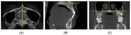
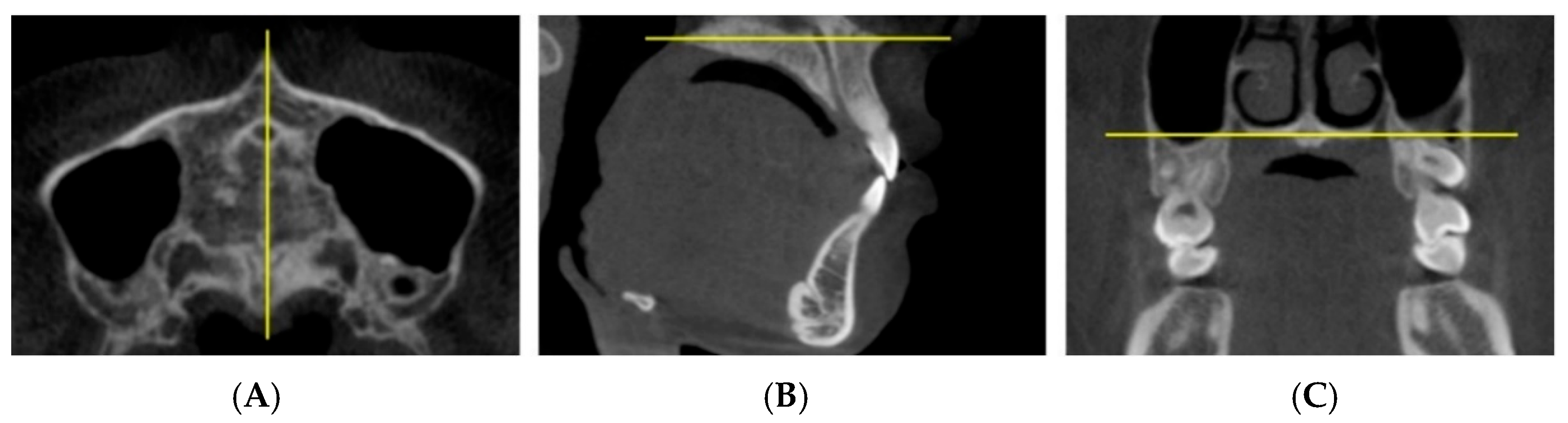
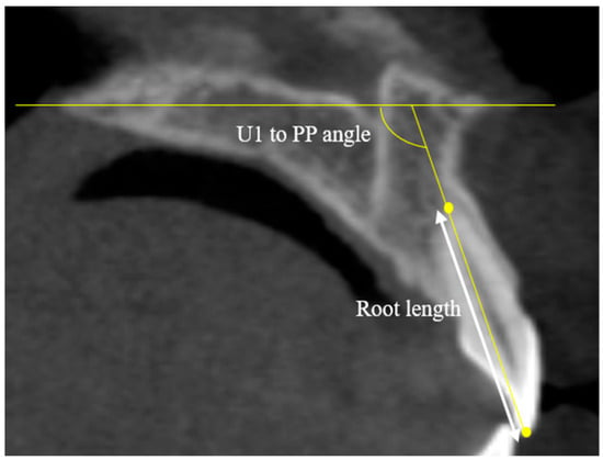
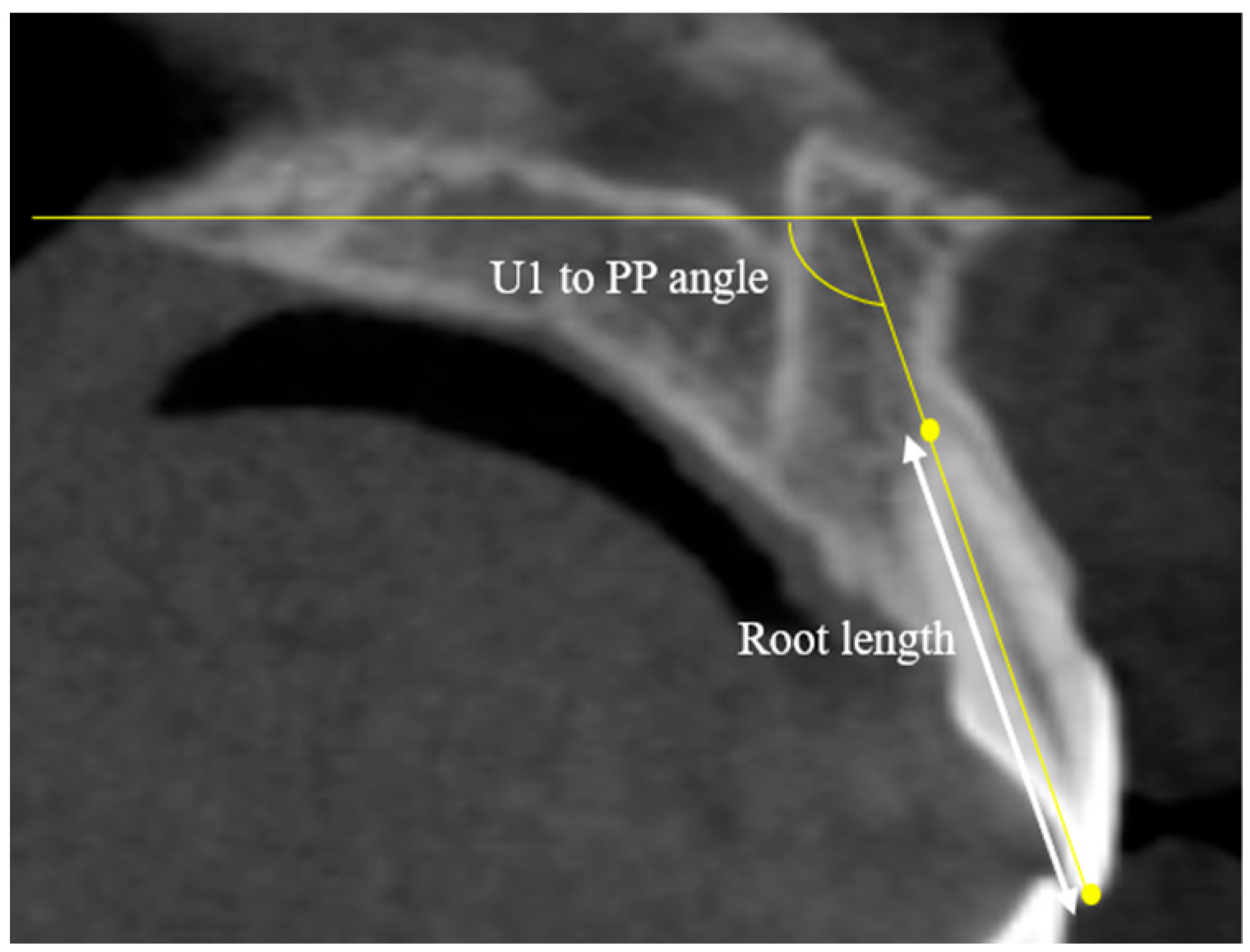
CBCT data were acquired using a Q Face scanner (HDX WILL, Seoul, Korea) with a slice thickness of 0.5 mm and a field of view (FOV) of 160 × 210 mm, operating at 60–90 kV and 4–10 mA. Images were reconstructed and analyzed in 3D using Invivo Anatomage version 7 (Anatomage, San Jose, CA, USA). Prior to measurements, the sagittal, horizontal, and coronal reference planes were reoriented for standardization (Figure 1). Root length and inclination of the U1 were then assessed on sagittal sections (Figure 2).
Figure 1.
Reorientation plane of cone-beam computed tomography images. (A) Axial planes: the plane passing through the mid-palatal suture, including ANS-PNS; (B) sagittal plane: the palatal plane, including ANS-PNS; (C) coronal plane: the most inferior plane of the nasal floor, passing through the right and left greater palatine foramina. ANS, anterior nasal spine; and PNS, posterior nasal spine.
Figure 2.
Root length and inclination of the maxillary central incisors (U1s). Root length, the distance from the U1 crown tip to the apex; and U1 to PP angle, the inclination of the U1 relative to the palatal plane (PP).
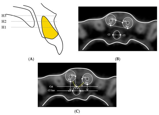
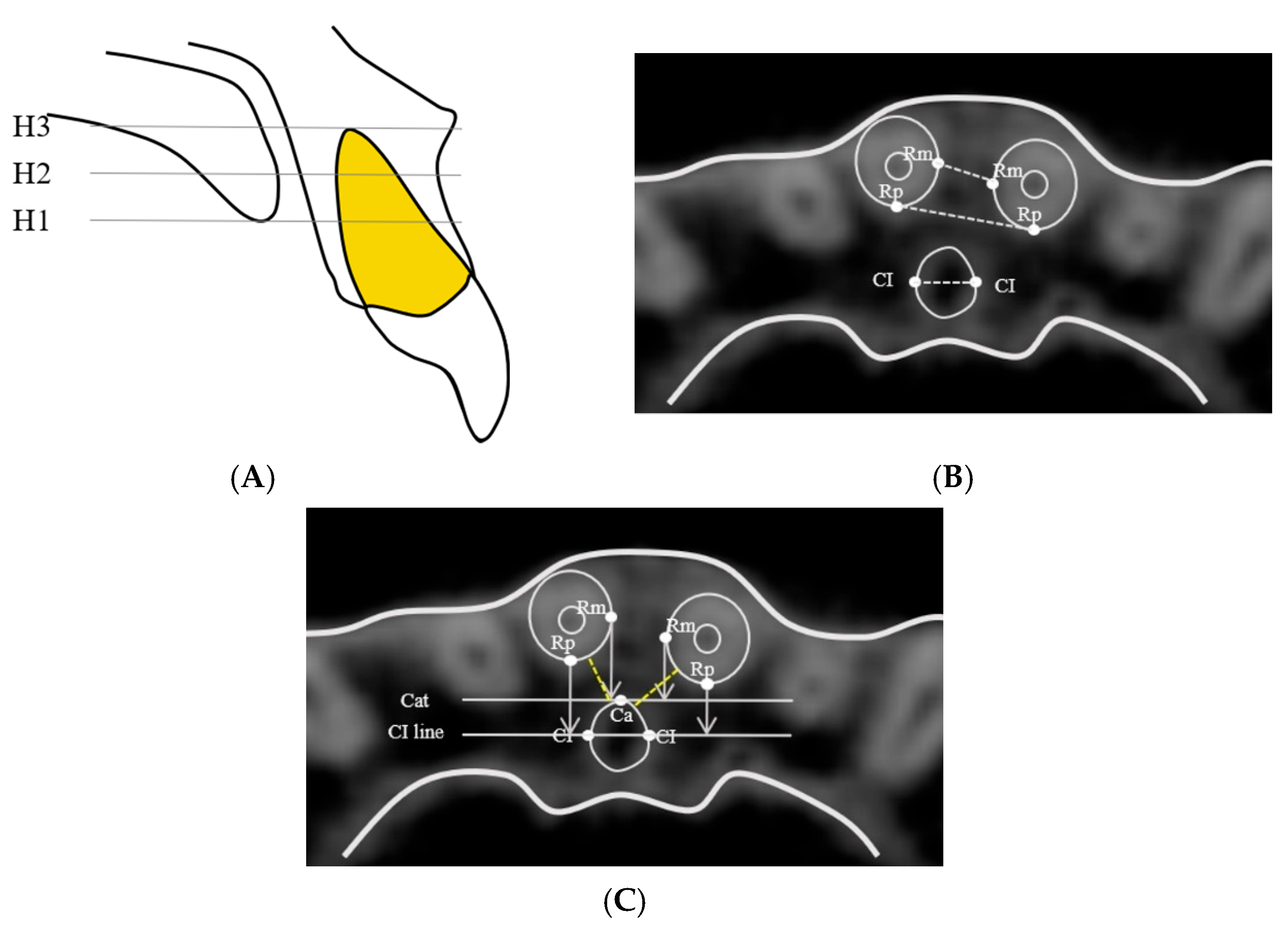
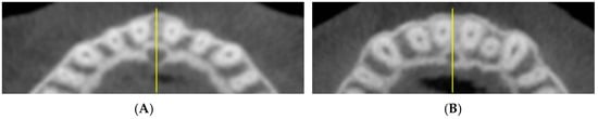
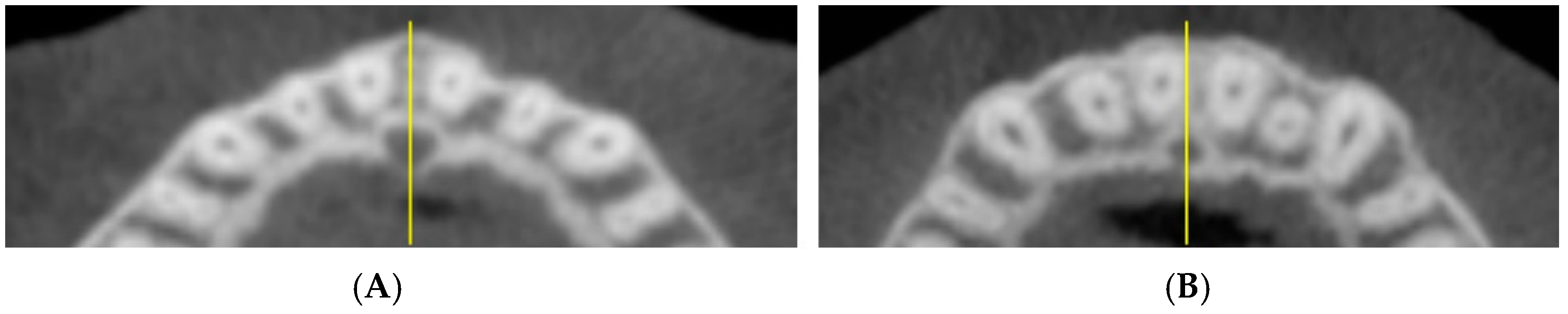
Linear measurements were performed from axial cross-sectional images at three vertical levels defined in the sagittal plane: (1) the palatal opening of the IC (palatal opening level, H1); (2) the mid-level between the palatal opening and the root apex of the U1 (mid-level, H2); and (3) the root apex of the U1 (apex level, H3). At each level, IC width and inter-root distance between the maxillary incisors were measured. For each incisor, both the anteroposterior distance to the IC and the shortest IC distance were also assessed. Detailed landmarks and measurement protocols are illustrated in Figure 3, with most measurements performed on the axial plane (Figure 4).
Figure 3.
Landmarks and linear measurements. (A) Three vertical levels of the incisive canal (IC): H1, palatal opening level; H2, mid-level; H3, root apex level. (B) Landmarks for inter-root distance and IC width: Rm, the most medial point of the maxillary central incisor roots; Rp, the most posterior point of the maxillary central incisor roots; CI, the most lateral point of the IC; Rm-Rm, inter-root distance; Rp-Rp, posterior inter-root distance; and width of IC, CI to CI distance. (C) Landmarks to assess the proximity between the IC and U1s: Ca, the most anterior point of the IC; Cat, the tangent line through Ca; CI line, the line through CIs; Rm-Ca, the distance from Rm to Cat; CI-Rp, the distance from Rp to the CI line; and IC to Root, the shortest distance from the IC to the maxillary central incisors root (dashed line).
Figure 4.
Axial plane images at the palatal opening level. (A) Group 1, group with a non-deviated dental midline. (B) Group 2, group with a deviated dental midline.
2.3. Statistical Analysis
Statistical analyses were conducted using SPSS 30.0.0 (IBM Corp., Armonk, NY, USA). Normality was assessed with the Shapiro–Wilk test. Intergroup comparisons were performed using either the independent t-test or Mann–Whitney U test, depending on data distribution. One-way analysis of variance (ANOVA) was applied to evaluate differences among continuous variables. Variables that satisfied the assumption of normality were analyzed using one-way ANOVA, whereas non-parametric tests were applied when the assumption of normality was not met. Within-group comparisons across the three vertical levels were analyzed using the Friedman test, followed by the Wilcoxon signed-rank test with Bonferroni correction. Categorical variables were assessed using the chi-square test. Spearman correlation analysis was employed to examine relationships among root proximity, midline deviation, and incisor inclination. Measurement reliability was assessed using the intraclass correlation coefficient (ICC) based on repeated measurements, with ICC values exceeding 0.90 considered to indicate excellent intra-examiner reliability. All measurements were performed twice by a single examiner (S.-H.M.), with a 2-week interval between measurements. Statistical significance was set at p < 0.05.
3. Results
3.1. Distribution of Maxillary Dental Midline Deviation and Characteristics of U1
In Group 2, maxillary dental midline deviation was directed to the right in 13 patients (40.6%) and to the left in 19 patients (59.4%). Mean deviations were 2.52 ± 0.62 and 2.30 ± 0.54 mm, respectively, with no significant difference between directions (p > 0.05) (Table 2). Root lengths of U1 and U1-palatal plane angles showed no significant differences between sides or between groups (p > 0.05) (Table 3).
Table 2.
Maxillary dental midline deviation distribution in Group 2.
Table 3.
Intergroup comparison of the characteristics of the maxillary central incisors (U1s).
3.2. Width of IC and Inter-Root Distance of U1
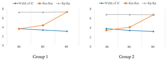
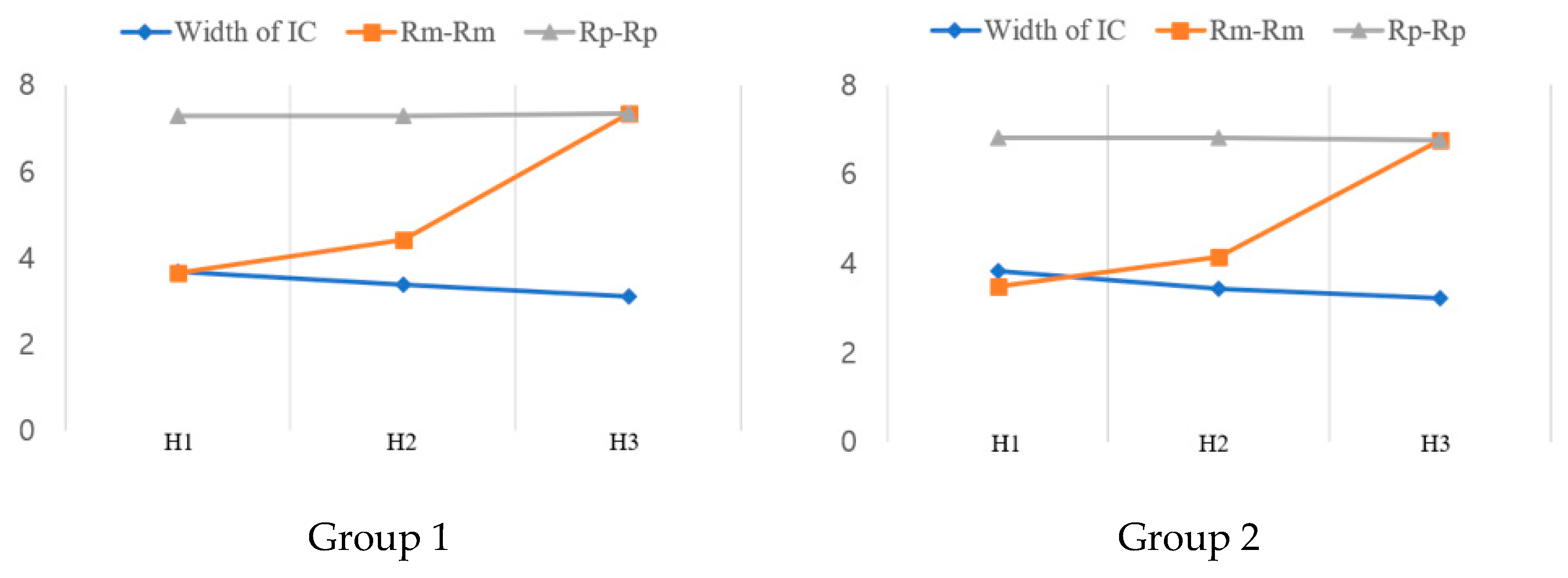
The width of the IC (CI-CI) did not differ significantly between groups at any vertical level (p > 0.05). In both groups, CI-CI decreased progressively from H1 to H3 (p < 0.01). The inter-root distance (Rm–Rm) increased significantly from H1 to H3 in both groups (p < 0.001), whereas the posterior inter-root distance (Rp–Rp) showed no significant variation across vertical levels. No significant differences in inter-root distance (Rm-Rm and Rp-Rp) were observed between the groups at any of the three vertical levels (Table 4). Mean values of each measurement across the three levels are presented in Figure 5. In Group 1, 53.13 % of cases at the H1 level and 15.63 % at the H2 level showed a greater incisive canal width (CI-CI) than the inter-root distance (Rm-Rm). In Group 2, this proportion increased to 65.63 % at H1 and 31.25 % at H2. Additionally, both groups exhibited a decreasing trend in the proportion of cases with CI-CI greater than Rm-Rm from H1 to H2. There were no significant differences between groups (Table 5).
Table 4.
The width of the incisive canal (IC) and inter-root distance at three vertical levels.
Figure 5.
Changes in the width of the incisive canal (IC) and inter-root distance of the maxillary central incisors (U1s) at three vertical levels in Group 1 and Group 2.
Table 5.
Percentage of subjects with incisive canal (IC) width greater than U1 inter-root distance.
3.3. Proximity of IC to U1
The proximity between U1 and the IC was evaluated at three vertical levels. In Group 1, anteroposterior measurements (Rm-Ca, CI-Rp) showed no significant side to side differences, and the shortest distance (IC to Root) also demonstrated no significant differences at the H1 and H3 levels. However, at the H2 level, the left side was significantly closer to the IC than the right side (p < 0.05). In Group 2, both Rm-Ca and CI-Rp distances were significantly shorter on the deviated side than the contralateral side across all three vertical levels (p < 0.05). The IC-Root distance was also slightly shorter on the deviated side, although these differences were not statistically significant (p > 0.05) (Table 6).
Table 6.
Proximity of the incisive canal to the maxillary incisors (U1s) at three vertical levels.
Correlations were evaluated between root proximity (Rm-Ca, CI-Rp) differences and two parameters: (1) the magnitude of maxillary dental midline deviation and (2) the difference in U1 inclination (U1 to PP angle). These analyses were performed using the signed values, calculated as the measurement on the deviated side minus that on the contralateral side. No significant correlations were found between the amount of maxillary dental midline deviation and the root proximity differences across any vertical level. In contrast, differences in U1 inclination between sides demonstrated significant negative correlations with root proximity at most levels (Table 7).
Table 7.
Correlation of root proximity differences with the maxillary dental midline deviation and maxillary central incisor (U1) inclination in Group 2.
4. Discussion
The anatomical proximity between the IC and the U1 is an important consideration in orthodontic treatment, particularly when asymmetric anterior tooth movement is required in patients with maxillary dental midline deviation. During orthodontic tooth movement, IC-U1 root contact may increase the risk of root resorption or compromise neurovascular structures [7,8,16,17]. In implant placement, inadvertent injury to the neurovascular structures within the canal can impair anterior sensory function [16,17]. Although previous studies have examined this relationship in the context of skeletal and vertical facial patterns [18,19,20], few have investigated the influence of maxillary dental midline deviation on U1 position relative to the IC. Therefore, the present study aimed to evaluate the proximity between the IC and each U1 according to the presence or absence of maxillary dental midline deviation.
The results showed that in both groups, the IC exhibited progressive apical narrowing from the palatal opening (H1) to the apical level (H3), whereas the inter-root distance of the U1 increased across the same vertical levels. Consequently, the proportion of cases in which the IC width exceeded the inter-root distance declined from H1 to H2 in both groups. This anatomical pattern aligns with previous studies reporting apical narrowing of the IC and a progressive divergence of U1 roots [6,18,20].
A previous study reported that, in patients with maxillary dental midline shift, the U1 on the side closer to the IC exhibited significantly greater apical root resorption than the contralateral side. However, the direction of deviation was not explicitly defined, and the findings were not directly related to clinical midline asymmetry [7]. In contrast, the present study prospectively included patients with clinically evident maxillary dental midline deviation, defined relative to the facial midline, providing greater clinical relevance. The results demonstrated that, in patients with maxillary dental midline deviation, the anteroposterior U1-IC distance was significantly shorter on the deviated side across all three vertical levels than the non-deviated side.
No significant correlation was found between the magnitude of maxillary dental midline deviation and differences in root proximity, which may be attributed to the limited variability of midline deviation within the sample. In contrast, inter-side differences in U1 inclination showed significant negative correlations with root proximity at most vertical levels, indicating that a more proclined U1 on the deviated side tended to be closer to the IC. Similar findings have been reported previously, suggesting that U1 inclination plays an important role in determining spatial relationships with the IC [21]. From a biomechanical perspective, these findings underscore the importance of careful torque control and force modulation during midline correction, particularly when asymmetric anterior retraction is planned. Excessive labiolingual tipping or uncontrolled torque on the deviated side may further reduce the U1–IC distance, potentially increasing the risk of root proximity to the canal. Therefore, individualized biomechanics incorporating controlled torque expression and staged or asymmetric force application may be advisable in patients with maxillary dental midline deviation.
Previous studies evaluating the shortest distance between U1 roots and the IC have reported increasing distances with vertical level and reductions following maxillary anterior retraction, although associated increases in root resorption were not statistically significant [22,23]. Most of these investigations assessed proximity using averaged values from both sides. In contrast, the present study focused on inter-side differences in U1–IC proximity. Although the shortest U1–IC distance did not differ significantly between sides, a consistent directional trend toward reduced proximity on the deviated side was observed. From a clinical perspective, such trends may still be meaningful, particularly during extensive or cumulative anterior tooth movement, where even small reductions in available anatomical clearance may become clinically relevant. Accordingly, careful evaluation of surrounding anatomical structures is warranted when planning substantial anterior tooth movement in patients with midline asymmetry.
This study was restricted to patients with skeletal Class I malocclusion to minimize the potential influence of sagittal skeletal discrepancies on the U1–IC relationship. Previous studies have demonstrated that the diameter of the incisive foramen, the length of the IC, and the distance between U1 and IC vary with age [24,25]. To reduce age-related effects, subjects in the growth period as well as those in older age groups were excluded from the present study. Furthermore, as this study included a relatively homogeneous Korean population from a single institution, the generalizability of the findings to other ethnic populations may be limited.
Nevertheless, several studies have suggested that vertical growth patterns influence the morphology and proximity of the IC [18,20], a factor not considered in the present analysis. In addition, sex-related differences in IC volume, canal length, and U1–IC proximity have been reported [18,19,20,24,26], but these factors were not assessed in the present study because further stratification by growth pattern or sex would have substantially reduced the number of subjects per subgroup, potentially limiting statistical power and complicating interpretation of midline-related effects. Future investigations with larger, well-balanced samples are warranted to obtain a more comprehensive understanding of the anatomical factors influencing IC–U1 proximity.
5. Conclusions
This study evaluated the spatial relationship between the incisive canal and maxillary central incisor roots in patients with and without maxillary dental midline deviation using CBCT images. Patients with maxillary dental midline deviation showed a tendency toward closer U1–IC proximity on the deviated side, although the shortest distance did not differ significantly between sides. In addition, differences in U1 inclination were significantly associated with root proximity to the IC, indicating that tooth inclination plays an important role in determining this anatomical relationship. These findings suggest that maxillary dental midline deviation and incisor inclination should be carefully considered during orthodontic or prosthetic treatment planning. CBCT-based evaluation of IC proximity may be particularly valuable when asymmetric anterior tooth movement is anticipated.
Author Contributions
Conceptualization, methodology, validation, investigation, H.S., Y.K. and S.-H.H.; software, formal analysis, resources, data curation, H.S., S.L. and H.J.; writing—original draft preparation, H.S. and S.-H.H.; and writing—review and editing, project administration, H.S., Y.K., J.-M.C. and S.-H.H.; supervision, J.-M.C. and S.-H.H. All authors have read and agreed to the published version of the manuscript.
Funding
This research was supported by an intramural research grant from The Catholic University of Korea (Grant No. 5-2025-B0001-00064).
Institutional Review Board Statement
This study was conducted in accordance with the Declaration of Helsinki and was approved by the Institutional Review Board (IRB) of The Catholic University of Korea (IRB approval number: KC24RISI0382; date of approval: 14 June 2024). The IRB determined that this project was exempt under 45 CFR §46.104(d)(4) due to the use of de-identified human data.
Informed Consent Statement
Informed consent was waived as this study used preexisting CBCT data obtained from routine orthodontic screenings, and no identifiable personal information was used.
Data Availability Statement
The original contributions presented in the study are included in the article, further inquiries can be directed to the corresponding authors.
Acknowledgments
This paper was supported by The Korean Orthodontic Research Institute Inc. and by Wonkwang University in 2025.
Conflicts of Interest
The authors declare no conflict of interest.
References
- Riedel, R.A. Esthetics and its relation to orthodontic therapy. Angle Orthod. 1950, 20, 168–178. [Google Scholar]
- Arnett, G.W.; Bergman, R.T. Facial keys to orthodontic diagnosis and treatment planning. Part I. Am. J. Orthod. Dentofacial Orthop. 1993, 103, 299–312. [Google Scholar] [CrossRef] [Scilit]
- Handelman, C.S. The anterior alveolus: Its importance in limiting orthodontic treatment and its influence on the occurrence of iatrogenic sequelae. Angle Orthod. 1996, 66, 95–109; discussion 109–110. [Google Scholar]
- Song, W.C.; Jo, D.I.; Lee, J.Y.; Kim, J.N.; Hur, M.S.; Hu, K.S.; Kim, H.J.; Shin, C.; Koh, K.S. Microanatomy of the incisive canal using three-dimensional reconstruction of microCT images: An ex vivo study. Oral Surg. Oral Med. Oral Pathol. Oral Radiol. Endod. 2009, 108, 583–590. [Google Scholar] [CrossRef] [Scilit]
- Mraiwa, N.; Jacobs, R.; Van Cleynenbreugel, J.; Sanderink, G.; Schutyser, F.; Suetens, P.; van Steenberghe, D.; Quirynen, M. The nasopalatine canal revisited using 2D and 3D CT imaging. Dentomaxillofac. Radiol. 2004, 33, 396–402. [Google Scholar] [CrossRef] [Scilit]
- Cho, E.A.; Kim, S.J.; Choi, Y.J.; Kim, K.H.; Chung, C.J. Morphologic evaluation of the incisive canal and its proximity to the maxillary central incisors using computed tomography images. Angle Orthod. 2016, 86, 571–576. [Google Scholar] [CrossRef] [Scilit]
- Nakada, T.; Motoyoshi, M.; Horinuki, E.; Shimizu, N. Cone-beam computed tomography evaluation of the association of cortical plate proximity and apical root resorption after orthodontic treatment. J. Oral Sci. 2016, 58, 231–236. [Google Scholar] [CrossRef] [Scilit]
- Yu, J.H.; Nguyen, T.; Kim, Y.I.; Hwang, S.; Kim, K.H.; Chung, C.J. Morphologic changes of the incisive canal and its proximity to maxillary incisor roots after anterior tooth movement. Am. J. Orthod. Dentofacial Orthop. 2022, 161, 396–403.e391. [Google Scholar] [CrossRef] [Scilit]
- Chung, C.J.; Choi, Y.J.; Kim, K.H. Approximation and contact of the maxillary central incisor roots with the incisive canal after maximum retraction with temporary anchorage devices: Report of 2 patients. Am. J. Orthod. Dentofacial Orthop. 2015, 148, 493–502. [Google Scholar] [CrossRef] [Scilit]
- Arnett, G.W.; Bergman, R.T. Facial keys to orthodontic diagnosis and treatment planning--Part II. Am. J. Orthod. Dentofacial Orthop. 1993, 103, 395–411. [Google Scholar] [CrossRef] [Scilit]
- Cardash, H.S.; Ormanier, Z.; Laufer, B.Z. Observable deviation of the facial and anterior tooth midlines. J. Prosthet. Dent. 2003, 89, 282–285. [Google Scholar] [CrossRef] [Scilit]
- Miller, E.L.; Bodden, W.R., Jr.; Jamison, H.C. A study of the relationship of the dental midline to the facial median line. J. Prosthet. Dent. 1979, 41, 657–660. [Google Scholar] [CrossRef] [Scilit]
- Jerrold, L.; Lowenstein, L.J. The midline: Diagnosis and treatment. Am. J. Orthod. Dentofacial Orthop. 1990, 97, 453–462. [Google Scholar] [CrossRef] [Scilit]
- Williams, R.P.; Rinchuse, D.J.; Zullo, T.G. Perceptions of midline deviations among different facial types. Am. J. Orthod. Dentofacial Orthop. 2014, 145, 249–255. [Google Scholar] [CrossRef] [Scilit]
- Beyer, J.W.; Lindauer, S.J. Evaluation of dental midline position. Semin. Orthod. 1998, 4, 146–152. [Google Scholar] [CrossRef] [Scilit]
- Al-Amery, S.M.; Nambiar, P.; Jamaludin, M.; John, J.; Ngeow, W.C. Cone Beam Computed Tomography Assessment of the Maxillary Incisive Canal and Foramen: Considerations of Anatomical Variations When Placing Immediate Implants. PLoS ONE 2015, 10, e0117251. Available online: https://journals.plos.org/plosone/article?id=10.1371/journal.pone.0117251 (accessed on 1 January 2026). [CrossRef] [Scilit]
- Pan, Y.; Chen, S. Contact of the incisive canal and upper central incisors causing root resorption after retraction with orthodontic mini-implants: A CBCT study. Angle Orthod. 2019, 89, 200–205. [Google Scholar] [CrossRef] [Scilit]
- Al-Rokhami, R.K.; Sakran, K.A.; Alhammadi, M.S.; Al-Tayar, B.; Al-Gumaei, W.S.; Al-Yafrusee, E.S.; Al-Shoaibi, L.H.; Cao, B. Tridimensional Analysis of Incisive Canal and Upper Central Incisor Approximation. Int. Dent. J. 2023, 73, 410–416. [Google Scholar] [CrossRef] [Scilit]
- Costa, E.D.; de Oliveira Reis, L.; Gaeta-Araujo, H.; Martins, L.A.C.; Oliveira-Santos, C.; Freitas, D.Q. Comparison of distance of upper central incisor root and incisive canal in different sagittal and vertical skeletal patterns and sex: A retrospective CBCT study. Int. Orthod. 2021, 19, 462–470. [Google Scholar] [CrossRef] [Scilit]
- Al-Rokhami, R.K.; Sakran, K.A.; Alhammadi, M.S.; Mashrah, M.A.; Cao, B.C.; Alsomairi, M.A.A.; Al-Worafi, N.A. Proximity of upper central incisors to incisive canal among subjects with maxillary dentoalveolar protrusion in various facial growth patterns. Angle Orthod. 2022, 92, 529–536. [Google Scholar] [CrossRef] [Scilit]
- Gupta, S.; Kothari, B.; Garg, K.; Shah, S.; Singh, N.; Chhaparwal, T. Comparative evaluation of the position of incisive canal and its proximity to the maxillary central incisor roots with different incisor inclinations–A CBCT study. APOS Trends Orthod. 2024, 15, 238–243. [Google Scholar] [CrossRef] [Scilit]
- Jang, M.; Yu, S.K.; Lim, S.H.; Jih, M.K.; Jeong, S. Radiologic study of the distance between the maxillary central incisor root and the incisive canal according to skeletal malocclusion classification. Sci. Rep. 2024, 14, 16875. [Google Scholar] [CrossRef] [Scilit]
- Ongprakobkul, N.; Ishida, Y.; Petdachai, S.; Ishizaki, A.; Shimizu, C.; Techalertpaisarn, P.; Ono, T. Morphometric and volumetric analysis of the proximity between the incisive canal and maxillary central incisors during anterior retraction: A retrospective cone-beam computed tomography study. Angle Orthod. 2023, 93, 159–167. [Google Scholar] [CrossRef] [Scilit]
- Kuc, A.E.; Kotula, J.; Nawrocki, J.; Szelag, E.; Kawala, B.; Lis, J.; Sarul, M. Morphological Evaluation of the Incisive Canal in the Aspect of the Diagnosis and Planning of Orthodontic Treatment-CBCT Study. Appl. Sci. 2023, 13, 12010. [Google Scholar] [CrossRef] [Scilit]
- Chatzipetros, E.; Tsiklakis, K.; Donta, C.; Damaskos, S.; Angelopoulos, C. Morphological Assessment of Nasopalatine Canal Using Cone Beam Computed Tomography: A Retrospective Study of 124 Consecutive Patients. Diagnostics 2023, 13, 1787. [Google Scholar] [CrossRef] [Scilit]
- Matsumura, T.; Ishida, Y.; Kawabe, A.; Ono, T. Quantitative analysis of the relationship between maxillary incisors and the incisive canal by cone-beam computed tomography in an adult Japanese population. Prog. Orthod. 2017, 18, 24. [Google Scholar] [CrossRef] [Scilit]

Disclaimer/Publisher’s Note: The statements, opinions and data contained in all publications are solely those of the individual author(s) and contributor(s) and not of MDPI and/or the editor(s). MDPI and/or the editor(s) disclaim responsibility for any injury to people or property resulting from any ideas, methods, instructions or products referred to in the content. |
© 2026 by the authors. Licensee MDPI, Basel, Switzerland. This article is an open access article distributed under the terms and conditions of the Creative Commons Attribution (CC BY) license.