


Abstract
Plastic pollution poses a severe environmental challenge in marine ecosystems, which could be addressed by plastic-degrading microorganisms. In previous studies, five fungal strains of the genus Cladosporium, isolated from the marine copepod Acartia tonsa, showed potential plastic polymer degradation activity. This study provides genomic and functional evidence of the metabolic activities of copepod-associated Cladosporium strains involved in polymer biodegradation. Five Cladosporium strains were tested for their ability to degrade poly(butylene succinate-co-adipate) (PBSA), poly(1,4-butylene adipate-co-terephthalate) (PBAT), and low-density polyethylene (LDPE) emulsions. These polymers are among the most used worldwide and contribute to plastic pollution. Whole-genome sequencing and functional annotation were conducted to elucidate the mechanisms of polymer degradation. Fungal activity was polymer dependent: the average sizes of zones of clearance were 40.6, 28.5, and 11.4 cm2 for PBSA, PBAT, and LDPE, respectively. Up to 35 different genes putatively associated with polymer degradation were found in all fungal strains. Most of them are shared among all tested strains, with few genes unique to each strain. Predicted gene content and putative fungal activity varied among strains and showed patterns coinciding with taxonomic grouping. Results provide additional evidence of the effectiveness of Cladosporium strains for plastic degradation. This aspect holds significance for the implementation of effective bioremediation strategies to counter marine plastic pollution.
1. Introduction
Plastic pollution represents a severe environmental challenge in marine ecosystems [1]. In these ecosystems, plastic [2] degradation is extremely slow, resulting in significant accumulation over decades [3]. During this period, plastics are subjected to physical fragmentation, partial hydrolysis and incomplete microbial degradation, resulting in the production of micro- and nano-plastics that accumulate in the marine food web. Consequently, plastic pollution causes severe impacts on both marine biota and human health [4]. Petro-plastics are the most used worldwide and include extremely heterogeneous polymers [5]. Polyethylene is one of the most used petro-plastics with several industrial applications. Low-density polyethylene (LDPE) alone accounts for approximately 20% of global plastic waste [6]. This polymer is widely used in packaging industries, and to produce mulching films. LDPE is the top CO2 contributor in the U.S., having more than a million metric tons of production volume [7]. LDPE possesses an extensive inert backbone formed by C-C single bonds without functional groups, which renders it highly resistant to enzymatic degradation [8].
Biodegradable plastics are crucial to lessening the environmental impact of the conventional petro-plastics [9]. They are designed to degrade via microbial action after their lifecycle, lessening the environmental impact of plastics waste. Among the biodegradable plastics derived from petroleum, polyesters such as poly(butylene succinate) (PBS) and its copolymer poly(butylene succinate-co-adipate) (PBSA), and poly(1,4-butylene adipate-co-terephthalate) (PBAT) are largely utilized in the food packaging, agricultural, and textile industries [10,11]. While most studies on PBSA and PBAT degradation are related to soil or compost environments [12,13], fewer investigations have been conducted in marine ecosystems [14,15]. The marine environment might represent a source of microorganisms harbouring cold-adapted enzymes. Recently, microorganisms isolated from marine environments have demonstrated interesting biodegradation activity against petro- and biodegradable plastics [14,15]. While most research has focused on the free-living and particle-attached microbial fraction [16], host-associated microbiomes remain underexplored and are mainly focused on vertebrate hosts [17].
Functional studies of the interactions between marine invertebrates and their associated microorganisms represent an emerging research frontier [18]. In particular, copepods, which constitute the most abundant component of marine zooplankton and a central element of marine food webs, function as mobile microbial hotspots [19]. Their external chitinous surfaces and digestive compartments host dense and metabolically active microbial communities that contribute to marine biogeochemical cycling and to the transformation of complex, high-molecular-weight organic matter [19,20]. This metabolic versatility suggests that copepod-associated microorganisms may possess broad extracellular hydrolytic and oxidative functions potentially relevant to recalcitrant polymers. The ecological connection with plastic exposure is further supported by the ingestion of microplastics suspended in the water column. Particles within a size range overlapping that of natural food items can be ingested and brought into close contact with copepod-associated microorganisms, with potential adverse effects on copepod growth and reproduction [21]. Although such exposure does not itself demonstrate microbial adaptation to, or degradation of, plastics, it provides a clear ecological rationale for investigating the copepod-associated microbiome as an underexplored source of plastic-interacting and plastic-transforming functions.
Consistent with this hypothesis, the bacterial fraction of copepod-associated microbiomes has already shown promising activity against PBSA granules [22]. Fungi represent another fundamental candidate for plastic degradation; however, their ecological and functional roles in marine environment remain underexplored. Marine yeasts possess genes involved in PE degradation [23], whereas other ascomycetes, e.g., Alternaria alternata, hosted hydrolases involved in the PBAT degradation [24]. Within this framework, Niccolini et al. [25] isolated nine fungal morphotypes from the carcasses of A. tonsa, belonging to the genera Fusarium, Stemphylium, and Cladosporium. Among these, the genus Cladosporium was the most represented, and Cladosporium psychrotolerans MUT6786 exhibited intense carboxyl-ester hydrolase activity against PBSA granules. Plastic degradation was already described for Cladosporium strains isolated from marine environments. For instance, C. sphaerospermum isolated from the plastisphere promoted a 15% weight loss of LDPE films, inducing changes in the functional groups and an increase in the carbonyl index of the plastic [26]. Furthermore, C. halotolerans isolated from deep sea sediments in the north of the Pacific Ocean showed the ability to completely degrade impranil polyurethane (PU) after 3 days of incubation at 28 °C, using the plastic as the sole carbon source [27]. Previous studies also reported an active role of marine Cladosporium in the carbon cycle and in the degradation of complex high-molecular-weight macromolecules such as lignocellulose and diatom polysaccharides [28,29]. Cladosporium activity on complex macromolecules might relate to plastic degradation. However, the functional traits involved in this process remain underexplored. Molecular tools such as genome sequencing are thought to better define this aspect by highlighting the presence of genes putatively involved in the process.
This study aimed to provide initial genomic and functional data on the interaction of copepod-associated Cladosporium strains with different environmentally relevant plastic polymers. These data will enrich the underexplored field of polymer degradation by promising copepod-associated marine fungi and provide useful insights to counter plastic pollution via biological means. The functional capacity of five Cladosporium strains, isolated from carcasses of A. tonsa, was evaluated against PBSA, PBAT, and LDPE. The three polymers were selected to represent an environmentally relevant gradient of chemical structure and expected biodegradability. Specifically, PBSA is an aliphatic biodegradable polyester containing hydrolysable ester bonds and served as a positive control for method validation. PBAT also contains hydrolysable ester bonds, but its aromatic terephthalate units confer greater structural recalcitrance than PBSA. LDPE, by contrast, is a persistent fossil-based polyolefin characterized by a hydrophobic carbon–carbon backbone and the absence of readily hydrolysable functional groups. Its inclusion therefore provided a stringent non-biodegradable benchmark to determine whether the activity of copepod-associated Cladosporium strains was restricted to ester-containing polyesters or could extend to a highly recalcitrant conventional plastic. Moreover, because PBSA, PBAT, and LDPE are widely used in packaging and/or agricultural applications and may enter marine environments, their joint assessment enables comparison of fungal responses across polymers of shared environmental relevance but markedly different susceptibility to biological attack.
Putative Fungal Degradation Activities (PFDAs) were investigated. Whole-genome sequencing followed by gene annotations were performed to identify genes encoding putative polymer-degrading enzymes, which suggested potential mechanisms of the plastic degradative activities.
2. Materials and Methods
2.1. Chemicals and Polymers
All chemicals were purchased from Sigma Aldrich (St. Louis, MO, USA). Poly(butylene succinate-co-adipate) (PBSA), commercially available as BioPBS™ FD92PM (granule, density = 1.24 g cm−3; melting point of 84 °C), was purchased from MCCP Germany GmbH (Mitsubishi Chemical Co., Tokyo, Japan). Poly(butylene adipate-co-terephthalate) (PBAT), commercially available as Ecoflex® F Blend C1200 (granule, density = 1.25 ÷ 1.27 g cm−3; Mw= 210,000 g mol−1; polydispersity = 1.6), was purchased from BASF (Mississauga, ON, Canada). Low-density polyethylene (LDPE) (Mw = 4000 g mol−1; Mn = 1700 g mol−1) was purchased from Millipore Sigma (Millipore Sigma Canada Ltd., Oakville, ON, USA). The chemical structure of PBSA, PBAT, and LDPE is reported in Figure 1.
Figure 1.
Chemical structure of the three polymers used in this study.
2.2. Fungal Isolation and Identification
The fungal strains considered in this study were isolated from the carcasses obtained from the marine calanoid copepod Acartia tonsa. Copepods were cultured as described in [30]. Fungal strains were isolated and identified as reported in Niccolini et al. [25]. Details on the copepod culture, fungal isolation and identification are reported in Supplementary Materials. The isolated fungal strains were routinely grown in 10 mL of minimal medium (MM), modified from Zeng et al. [31] (per L: 0.4 g KH2PO4, 0.6 g Na2HPO4, 1.0 g NH4NO3, 27.0 g NaCl, 0.1469 g MgSO4, 0.0151 g CaCl2, 0.0055 g FeSO4 in H2O), with 1.0 g L−1 yeast extract (MYE) and aerated by shaking at 20 ± 1 °C on a shaker at 150 rpm in the dark. The species Cladosporium velox MUT6781, Cladosporium pseudocladosporioides MUT6783, Cladosporium austrohemisphaericum MUT6784, Cladosporium cladosporioides MUT6785, and Cladosporium psychrotolerans MUT6786 were considered in this study for the polymer degradation experiments and genome sequencing analyses.
2.3. PBSA, PBAT and LDPE Degradation on Agar Plates
Conidial suspensions were used as initial inoculum for the degradation tests. To obtain the conidial suspensions, all the Cladosporium species were grown for 10 days in MYE-Agar plates (15.0 g L−1 Agar, bacteriological grade)at 20 ± 1 °C. After this time interval, 1 mL of MM was added on the top of 10-day fungal cultures on MYE plates, and a conidial suspension was collected. A measurement of 10 µL of conidial suspension was loaded in the hemacytometer (Bright-Line™ Hemacytometer, Portsmouth, NH, USA) and the number of conidia per mL of suspension was determined with Nikon Inverted Optical Microscope (Nikon Ti-U, Instrument Inc., Melville, NY, USA) with 40× magnification. Conidial suspensions were serially diluted with MM to a final density of 104 conidia mL−1, to get the same fungal inoculum for each fungal strain to set up the polymer degradation assay. Conidial germination tests were conducted by plating 100 µL of each conidial suspension (104 conidia mL−1) in water agar plates (per L: 15.0 g Agar). Conidial germination tests provided comparable germination rates for all strains.
A total of 10 µL of these conidial suspensions were inoculated onto MM agar plates, prepared with 2% w/v Sodium Dodecyl Sulphate (SDS) as surfactant, supplemented with 5% v/v of PBSA or PBAT or LDPE emulsions as the sole carbon sources. The adopted concentration of polymer emulsions equals to a final polymer load of 1 g L−1. Plates were prepared in triplicates following Urbanek et al. [32]. Emulsion concentration was initially selected following the cited protocol and preliminary growth tests confirmed their applicability to this study. Furthermore, preliminary tests were conducted on the fungal strains cultured on MM or MYE medium supplemented with anionic surfactant (Sarcosyl, Sodium Dodecyl Sulphate—SDS) and non-ionic surfactant (Tween80, Triton X-100 and Ramnolipids). Anionic surfactants were already used from other authors to produce polyester polymer emulsions [33]; SDS showed a lower toxicity than the other surfactants and formed a more stable PBSA, PBAT and LDPE emulsions than the others (Table S1). For this reason, SDS was selected to produce polymer emulsions. The in-depth protocol for the preparation of polymer emulsion plates, as well as the results of surfactant preliminary tests, are reported in the Supplementary Materials. Plates were incubated 35 days at 20 ± 1 °C in the dark. A negative control was performed preparing MM agar plates with SDS without the polymer. Abiotic controls without fungal inoculation were performed and did not develop a clarification halo after 35 days of incubation.
Images of the plates were acquired weekly and analyzed with the tool ImageJ (version 1.54s, http://imagej.org), monitoring both the area of fungal growth and the zone of clearance in each plate. The total area of the zones of clearance identifies the ‘Putative Fungal Degradation Activity’ (PFDA). A linear mixed effects model of the squared root of the area of PFDA overtime was performed with R v4.4.1, using the R package lmerTest 3.2.0. The significance of fixed effects was assessed using analysis of variance (ANOVA) paired with the post hoc Tukey HSD after 35 days of incubation. Statistical differences in PFDA between polymers after 35 days of growth were assessed with pairwise t-test. Raw PFDA data, ANOVA and post hoc results are reported in the Supplementary Materials. The time-point at the inflexion of the curve for the area of PFDA (mid-log time) identifying the growth rate, was evaluated with the SSlogis function in the R base package stats v.4.4.1. Heatmaps and graphs were obtained using the R packages pheatmap v1.0.13 and ggplot2 v3.5.2. Pearson correlations between predicted genomic features and PFDA were calculated with the r package smplot2 v0.2.6. ANOVA paired with post hoc t-test was conducted to assess the differences in predicted genomic features and PFDA dividing the tested strains between the C. cladosporioides complex and C. sphaerospermum complex.
2.4. DNA Extraction, Library Preparation and Genome Sequencing
For the genome sequencing analyses, Cladosporium species were cultured in 250 mL sterile Erlenmeyer flasks filled with 100 mL of MYE liquid medium, adding 100 mg L−1 Spectinomycin to limit bacterial growth and avoid DNA contamination. Flasks were incubated on a rotatory shaker at 100 rpm at 20 ± 1 °C in the dark. After 5 days of incubation, mycelia were collected and stored at −80 °C until further use. Mycelia were removed from −80 °C and rapidly grinded with sterile mortars and pestles, also collected from −80 °C. Total gDNA was extracted from the grinded mycelia via modified CTAB method [34,35]. A detailed protocol is reported in the Supplementary Materials. DNA was eluted in TE buffer (10 mM Tris-HCl, pH = 8.0, 0.1 mM EDTA). gDNA quantification was conducted with Qubit® 4 with the dsDNA Assay Kit (Thermo Fisher, Waltham, MA, USA), and gDNA integrity was evaluated using a TapeStation 4150 with Genomic DNA Screen Tapes and reagents (Agilent Technologies, Santa Clara, CA, USA). Subsequently, gDNA was physically sheared with g-Tubes (Covaris, Burlington, MA, USA) by centrifuging at 1700× g for 120 s, followed by a second centrifuge step at 3200× g for 60 s. Subsequently, g-Tubes were inverted, and the centrifuge steps were repeated a second time. Samples were then concentrated to 30 µL with a SpeedVac (Thermo Fisher, Waltham, MA, USA), and size selection of DNA molecules was performed with BluePippin (Sage Science, Beverly, MA, USA) to retain fragments between 10,000 and 30,000 bp. Library preparation for PacBio HiFi Circular Consensus Sequencing was performed following the manufacturer’s specifications. Genome sequencing was performed by the Canadian Grain Commission (Grain Research Laboratory, Government of Canada, Richardson Centre for Food Technology and Research 196 Innovation Dr, Winnipeg, MB, Canada) via PacBio Sequel IIe (PacBio, Menlo Park, CA, USA).
2.5. Genome Assembly and Annotation
Genome assembly was performed with Hifiasm v0.16.0 [36]. The quality of the assemblies was determined using Quast v5.3 [37], while their completeness was evaluated using BUSCO v6.0.0 [38], trained with the dataset capnodiales_odb10 (https://www.orthodb.org/). The absence of bacterial contamination was confirmed using BUSCO v6.0.0 trained with the dataset bacteria_odb10. This analysis reported <20% of BUSCO genes in all genomes. The absence of bacterial contamination was further confirmed by the Foreign Contamination Screen tool provided from NCBI during the genome submission process.
Gene prediction for the resulting genome assemblies was performed using Braker3 v3.0.8 [39] with default parameters. The RNAseq data from C. halotolerans TM138S3 (SRA accession number: SRR12633901, SRR12633902) were used as external evidence, i.e., additional information used from the bioinformatic tool to better predict gene structures. RNAseq data were not used to provide insight on gene expression patterns or actual involvement of predicted genes in polymer degradation. Results of gene prediction were summarized through Another Gtf/Gff Analysis Toolkit (AGAT) v1.6.1 (https://github.com/NBISweden/AGAT/blob/master/README.md, accessed on 3 November 2025). The annotation of predicted proteins was performed using Diamond v2.0.15 [40] with default parameters. Specifically, predicted protein were aligned against Trembl, Swissprot (version 2025_04, https://www.uniprot.org/uniprotkb, accessed on 10 November 2025) and NCBI nr (version 2024_02 https://ftp.ncbi.nlm.nih.gov/blast/db/FASTA/, accessed on 10 November 2025) databases. For each gene, the first 20 alignments per database were retained, following the recommendations of Werner et al. [41]. Alignment results were concatenated and uploaded to the UniProt Retrive/ID mapping tool (https://www.uniprot.org/id-mapping, accessed on 11 November 2025). This tool provided univocal protein names to each alignment result, as well as the Gene Ontology (GO) terms linked to it. Subsequently, each gene was paired with the most frequent protein name associated with its alignment results, so that only one annotation was linked to a gene. To account for differences in the total amount of GOs in each genome, GO frequencies were normalized as ‘GO per million’ using the following equation:
Furthermore, dbcan3 v.5.1.2 [42] was used with default parameters to annotate putative CAZymes (carbohydrate-active enzymes). Only concordant results between dbCAN_hmm and Diamond were considered for CAZyme annotation. Predicted proteins were also aligned against an online database with the sequences of microbial enzymes involved into plastic degradation, Plastic DB (329 annotated proteins from 875 microorganism, last update 4 November 2025, https://plasticdb.org/, [43]). The alignment was conducted with the PlasticDB blastP online tool with the following parameters: BlastP allignment predicted signal peptides with SignalP v 5.0 for Eukaryotes, 10−6 e-values cut-off and 30% as minimal percentage of identity. Results of GO per milion, BUSCO, Cazymes and PlasticDB were plotted using the R packages ggplot2 v3.5.2. Venn Diagrams were obtained with the online tool Evenn (https://www.bic.ac.cn/EVenn/#/, [44], accessed on 12 November 2025).
2.6. Data and Code Availability
Genome sequences for the 5 Cladosporium strains tested in this study are available on NCBI with BioProject PRJNA1370476. All bioinformatic pipelines are reported in Di Gregorio’s lab GitHub page (https://github.com/Di-Gregorio-s-Lab/Genome-Sequencing-of-Plastic-Degrading-Cladosporium-Strains, accessed on 10 September 2026).
3. Results
3.1. Putative Fungal Degradation Activity
Fungal growth over time and statistical analyses of PFDA were determined for the five Cladosporium species on MM agar plates supplemented with polymer emulsions (Figure 2). Images acquired after 35 days of incubation are shown in Figure S1. Interestingly, at the end of the incubation period (35 days) all fungi showed a wider PFDA zone in PBSA and PBAT than LDPE plates, with an overall average area of 40.6, 28.5, and 11.4 cm2, respectively (Figure 2A,B). Pairwise t-test confirmed the statistical significance of these differences (Supplementary Materials). The mid-log time-point, i.e., the time required for the PFDA zone to reach mid-log phase is shown in Figure 2C. A low mid-log time indicates a faster PFDA development and vice versa. All fungi retrieved the highest mid-log time in the case of PBSA plates and the lowest mid-log time in the case of PBAT plates (Figure 2C).
Figure 2.
Area of Putative Fungal Degradation Activities (PFDAs) of Cladosporium species on MM agar plates supplemented with PBSA, PBAT, or LDPE emulsions, over time. (A) Local regressions of the squared root of the PFDA zone overtime (cm2 day−1); shaded ribbons refer to the mean confidence interval (α = 0.05) of each curve. (B) Heatmap of the PFDA zone after 35 days of growth. The colour scale refers to the area (cm2), and letters represent statistical difference between different Cladosporium strains within each polymer emulsion. Statistical significance was assessed via ANOVA followed by post hoc Tukeys HSD test (α = 0.05). (C) Heatmap of the mid-log time (days) of the PFDA curves shown in panel (A). Lower values represent a faster development of the PFDA zone.
Specifically, on PBSA plates, C. cladosporioides and C. pseudocladosporioides (blue and orange curve, respectively) reached the widest PFDA zone (Figure 2A,B). Furthermore, C. cladosporioides had the lowest mid-log time (Figure 2C), indicating the highest development rate of PFDA zone. On PBAT plates, C. cladosporioides and C. velox (blue and red curve, respectively) reached the widest PFDA zone (Figure 2A,B). Interestingly, all the species showed a similar mid-log time value and reached a plateau after 20 days of incubation (Figure 2A,C). Finally, in the case of LDPE plates, C. velox and C. austrohemisphaericum (red and violet curve, respectively) reached the widest PFDA zone (Figure 2A,B) and C. austrohemisphaericum showed the lowest mid-log time value (Figure 2C). LDPE plates highlighted different mid-log time values for each fungal strain. No fungal strain was able to grow on SDS plates. Aside from SDS, polymer emulsions were the only carbon source available. And even though these observations do not demonstrate polymer assimilation as a carbon source, fungal growth and clearance-zone formation were observed only on polymer-containing plates (Figure 2A).
Strains belonging to the C. cladosporioides complex developed wider halos on PBSA than strains belonging to the C. sphaerospermum complex. This difference was statistically significant, contrary to results on PBAT and LDPE. Differences on LDPE are visible but not statistically significant, probably due to the high variability of biological replicates.
3.2. Genome Sequencing and Bioinformatics of Cladosporium Species
The genomes of the Cladosporium species were sequenced and assembled. Results of the sequencing run, quality and the gene prediction analyses are reported in Table 1. The genome size ranges from 33 to 41.4 Mbp and the genome coverage from 125× to 163×. The number of contigs ranges from 156 to 281. N50 is 1.9 Mbp on average and the GC content is around 49%. From the gene prediction results, the number of predicted genes range from 9886 to 12,291 with an average length ranging from 1671 to 2789 bp. Fungal genomes were retrieved from 28,996 to 39,686 exons and from 17,038 to 29,693 introns. The percentage of the genome covered by genes ranged from 47.6% in the case of C. cladosporioides to 67.3% for C. velox (Table 1).
Table 1.
General features associated with the genomes of the five Cladosporium species.
Assembly metrics differed moderately among strains. C. velox MUT6781 had the largest genome (41.4 Mb), whereas C. austrohemisphaericum MUT6784 had the smallest genome (33.0 Mb), the highest sequencing coverage (163×), and the lowest number of contigs (156). The highest N50 was obtained for C. pseudocladosporioides MUT6783 (2.13 Mb), followed by C. cladosporioides MUT6785 (2.04 Mb), while the other assemblies showed N50 values ranging from 1.73 to 1.91 Mb (Table 1). Despite these differences, all assemblies showed high and broadly comparable quality. BUSCO analysis identified 95.2–95.5% complete BUSCOs across the five assemblies, of which 94.3–94.9% were present as single copies; less than 4.2% were missing and less than 0.8% were fragmented (Figure 3A). Overall, these results indicate that assembly quality was sufficiently high and comparable across strains to support the subsequent between-strain comparisons of GO categories, CAZyme repertoires, and genes encoding enzymes putatively associated with polymer degradation.
Figure 3.
Genome annotation of the 5 Cladosporium strains. (A) BUSCO reports displaying the completeness of obtained genome assemblies. (B) Distribution of most represented gene ontology (GO) terms (expressed as GO per million GOs). (C) Venn diagram of the number of shared CAZymes among Cladosporium strains (expressed as # of shared CAZymes) and the total number of the CAZymes retrieved from each strain (detailed as # of CAZymes for each strain). (D) Number of CAZymes (expressed as # of CAZymes) per family, i.e., auxiliary activity (AA), glycosyl transferase (GT), polysaccharide lyase (PL), carbohydrate esterase (CE), carbohydrate-binding module (CBM), and glycoside hydrolase (GH).
3.3. Fungal Genome Annotation for Plastic Degradation Activity
To evaluate the potential plastic biodegradation activity of Cladosporium species, predicted proteins were aligned (blastp) against PlasticDB. This database includes bacterial and fungal proteins that were purified, identified and functionally characterized for their involvement in plastic degradation processes (https://plasticdb.org/). Genes encoding enzymes putatively associated with polymer degradation, which retrieved the highest number of hits with fungal predicted proteins, were considered for the annotation. Functions directly involved in LDPE degradation were not retrieved from the annotation of the Cladosporium sps against the PlasticDB database. Nonetheless, the annotation on PlasticDB retrieved some functions that were described for the degradation of other polymers formed by ethylene monomeric units, specifically enzymes involved in the degradation of polyethylene terephthalate (PET) and polyethylene glycol (PEG). Interestingly, PEG dehydrogenase and peroxidases (first described in the PET degradation) were reported as involved in the dissociation of the LDPE backbone [45,46]. For this reason, PET and PEG degradative enzymes were considered to potentially overcome the absence of functions directly annotated in the LDPE degradation. Furthermore, PBSA, PBS and PBAT were selected for the annotation, including enzymes directly involved in the degradation of the polymers tested with the fungi on agar plates.
The number of hits with PBAT-, PBS-, PBSA-, PET- and PEG-degrading enzymes is reported in Figure 4. Interestingly, all the Cladosporium species showed a higher number of hits with PET- and PEG-degrading enzymes than the others. In detail, C. cladosporioides and C. pseudocladosporioides showed a higher number of hits for PET- and PEG-degrading enzymes than the other species. C. psychrotolerans showed the highest proportion of hits for PBSA- and PBAT-degrading features (Figure 4A). Moreover, almost 80% of the genes encoding enzymes putatively associated with polymer degradation are shared among all the Cladosporium species (Figure 4B). However, strains belonging to the C. cladosporioides complex showcased an overall higher number of predicted putative polymer degradation genes when compared to strains belonging to the C. sphaerospermum complex (p < 0.05, Supplementary Materials).
Figure 4.
Annotation of genes encoding enzymes putatively associated with polymer degradation of the 5 Cladosporium strains. Selected genes derive from the database PlasicDB, filtered for the plastic PBSA, PBAT, PBS, PEG and PET. (A) Barplots represent the number of genes encoding enzymes putatively associated with polymer degradation for each polymer (expressed as # of plastic degrading genes), divided by fungal strain. (B) Venn diagram of the number of shared genes encoding enzymes putatively associated with polymer degradation among strains (expressed as # of shared plastic degrading genes) and the total number of genes encoding enzymes putatively associated with polymer degradation retrieved from each strain (# plastic degrading genes for each strain).
Details on the annotation of the genes encoding enzymes putatively associated with polymer degradation from the predicted proteins of Cladosporium are highlighted in Figure 5. PET-degrading enzymes represented the most differentiated group, including cutinase, MHETase, nitrobenzylesterase, PETase, esterase and lipase enzymes. Most of the enzymes were represented from PET hydrolase (PETase), nitrobenzylesterase and cutinase. In the case of PBAT-degrading enzymes, they were represented from carboxylesterase. Differently, PBSA-degrading activities were more evenly distributed among chitinase, cutinase, esterase and hydrolase enzymes. PBS- and PEG-degrading enzymes were represented from cutinase and PEG aldehyde-dehydrogenase and PEG dehydrogenase, respectively (Figure 5). Heatmap colours within the cells from the second to the sixth column represent the Z-score of the frequency of each function within each fungal strain. Z-score was calculated by autoscaling the counts of each gene encoding putative fungal degradative enzyme (PFDE) for each polymer against the average of the counts of gene encoding PFDE between each fungal strain.
Figure 5.
Heatmap of the number of genes encoding enzymes putatively associated with polymer degradation for each type of plastic and each fungal strain, based on results from PlasticDB. Different colours identify functions related to a specific plastic. The gene counts encoding enzymes putatively associated with polymer degradation are reported in each cell. The heatmap’s colours identify the standard score (Z-score) scaled within each fungal strain.
Pearson correlations between PFDA and key genomic features were calculated (Figure 6 and Figure S2). Multiple genomic traits correlated positively with PFDA on PBSA emulsions (Supplementary Materials). Specifically, total gene counts, as well as PBAT-, PBS- and PET-related genes, have a positive correlation with PFDA (R > 0.75, p < 0.05). The same traits negatively correlate with PFDA on LDPE emulsions. No strong correlations were found for PBAT. These correlations seem connected with the differences in gene count and PFDA efficiency between the C. cladosporioides and C. sphaerospermum complexes.
Figure 6.
Pearson correlation between area of Putative Fungal Degradation Activity and relevant genomic features. Shapes identify different Cladosporium species. Colours represent the C. cladosporioides complex and C. sphaerospermum complex. Squares highlighted in red identify strong correlations (R > 0.75, p < 0.05).
4. Discussion
The Cladosporium strains tested in this study were able to grow on Petri dishes containing PBSA, PBAT or LDPE emulsions as the only carbon source. However, well defined zones of clearance were observed only for PBSA plates, while the zones of clearance for PBAT and LDPE were less clearly defined. The process of emulsification of plastic polymers changes the crystalline/amorphous ratio of the polymer regions, increasing the amorphous part, where the polymer structure is easily accessible to the enzymatic degradative activities [47,48]. Furthermore, SDS is required to produce polymer emulsions; therefore, it is not possible to exclude that fungal activity against untreated polymers might be facilitated by surfactant-mediated emulsification. For this reason, caution in talking about polymer degradation by the fungal strains should be adopted. Nonetheless, no fungal strain was able to grow on Petri dishes containing only SDS, used as a negative control. The absence of growth on SDS-only plates indicates that SDS alone did not support visible fungal growth. Aside from SDS, polymer emulsions were the only carbon source available. Thus, fungal growth seems supported by the metabolization of polymer emulsions. In line with this, the term ‘Putative Fungal Degradation Activity’ (PFDA) was considered to describe the overall area of the Petri dish interested by the zone of clearance. Differences in PFDA and mid-log time, i.e., the time required to PFDA zone to reach the mid-log phase, were highly determined by the polymers, and differences among strains were mostly evident among polymers.
PBSA degradation is thought to involve carboxyl-ester hydrolase activities which are associated with the serine hydrolase site. The latter contains three aminoacidic residues (catalytic triad) of Ser/Asp-Glu-His that act on the ester bond in the carbon chain [48]. These enzymes were frequently described for the promiscuity and versatility among several ester substrates, promoting a random chain scission and acting on a wide group of polyesters [49,50], including PBAT. The similarities in the PFDA for PBSA and PBAT in each strain might be linked to the use of the same carboxyl-ester hydrolase activities. Dissimilarities between the two polymers, on the other hand, might derive from a different efficiency of the carboxyl-ester hydrolase activities for the functional groups of PBSA and PBAT. Fungal activity against PBAT suddenly ceased around 20 days of growth, while in PBSA it progressively increased even after 35 days of growth. At the same time, the mid-log time of PBAT is lower, indicating a faster activity rate.
Two main hypotheses might explain this result. Fungal activity might start from the amorphous phase of the polymer and pass to the crystalline phase later. This tendency was reported in multiple studies [51,52], and it was observed also for C. psychrotolerans MUT6786 [25]. The butylene adipate groups of PBSA reduce the degree of crystallinity of the polymer [53]. Thus, the fungal strains might have performed more efficiently in PBSA plates due to the higher availability of amorphous parts. On the other hand, Saadi et al. [52] suggested that fungal degradation of PBAT might start from the adipate units and later on attacking the terephthalate units. The terephthalate portion of the polymer might not be readily biodegradable by the fungal strains at 20 °C. In both cases, the Putative Fungal Degradation Activity of PBAT might have occurred only on a more available portion of the polymer, showing a rapid growth and activity of the strains, and stopped when the more recalcitrant part became too predominant.
LDPE is deemed as non-biodegradable. It represents a challenge to microorganisms due to its hydrophobicity and lack of reactive functional groups that could guide the first attack to the polymer [54]. For these reasons, it is not surprising that the tested Cladosporium strains resulted in being less effective in LDPE degradation. Nonetheless, a certain degree of PFDA was also observed for this polymer. Interestingly, the genus Cladosporium was reported in other studies as involved in LDPE degradation [26,55]. Additional tests need to be conducted on LDPE films to confirm the observed fungal activity on this polymer.
Observed PFDAs are noteworthy, especially due to the relatively low growth temperatures in which they occurred. In fact, polymer biodegradation is tightly related to growth temperatures, and high temperatures favour the process [52,56]. A growth temperature of 20 °C is significantly lower than the one often adopted in biodegradation assays and must be considered to better frame the efficiency of tested Cladosporium strains.
Tested fungi belonged to two different clades, namely the C. cladosporioides complex (C. cladosporioides and C. pseudocladosporioides) and the C. sphaerospermum complex (C. velox, C. austhroemisphaericum and C. psychrotolerans) [57]. C. cladosporioides MUT6785 was the most efficient in both PBSA and PBAT plates, followed by C. pseudocladosporioides MUT6783 for PBSA. The latter also showed remarkable activity in PBAT plates. Both strains performed poorly in LDPE plates. Interestingly, these two strains belong to the C. cladosporioides complex, hinting at a possible connection between taxonomy and functional profiles. This might also hold true for the other strains, all belonging to the C. sphaerospermum complex, with some exceptions. In fact, they all performed poorly in PBSA plates, and only C. velox MUT6781 showed remarkable PFDA in PBAT plates. Furthermore, C. velox MUT6781 and C. austrohemisphericum MUT6784 were the best performers in LDPE plates.
Correlations between taxonomy and functions in the genus Cladosporium were already reported [58,59]. C. cladosporioides and C. pseudocladosporioides already proved to degrade polyester emulsions embedded on agar plates such as polycaprolactone (PCL) [60], polyurethane (PU) [61], and LDPE [62]. However, these reports are not in line with the findings of this study. In fact, C. cladosporioides MUT6785 and C. pseudocladosporioides MUT6783 proved effective on PBSA and PBAT, but not LDPE. To the authors’ knowledge, this is the first report of the functional activity of C. cladosporioides against PBSA and PBAT and of C. pseudocladosporioides against PBSA. C. velox is reported for the first time to be active on both PBAT and LDPE. Furthermore, C. austrohemisphaericum is reported for the first time to be active on LDPE. The obtained results require further analyses on polymer films to confirm fungal activity. Furthermore, future studies on LDPE emulsions will highly benefit from increasing the number of biological replicates.
To better define the mechanisms employed for polymer degradation, the genome of all tested Cladosporium strains was sequenced and functionally annotated. Assembled fungal genomes are of high quality and completeness. Both the average genome size and number of putative genes are in line with the expected parameters for the genus Cladosporium [59]. Interestingly, more than half of the genomes on average are covered by predicted genes. Furthermore, the high N50 and complete single-copy BUSCO genes consistently higher than 95% prove that HiFi-PacBio sequencing is an optimal tool for genome sequencing of Cladosporium sps. strains. GO annotation evaluates the involvement of predicted genes in biological processes, molecular functions and cellular components for each fungal strain. Overall, a significant proportion of the genes in all fungal genomes encodes for fundamental cellular activities, like metabolic and cellular processes, as well as spatial arrangements within the cell. This holds true for most of the genomes ever annotated [63].
Carbohydrate-active enzymes (CAZymes) were also annotated. The most represented CAZymes families were glycoside hydrolases (GHs), glycosyl transferase (GTs) and auxiliary activity (AA). A higher proportion of GH- and GT-related genes might be related to the ability of Cladosporium strains to interact with biological macromolecules. In fact, they include pectinase, cellulase and xylanase activities, which seem related with the interaction with plant hosts [54], as well as phytoplankton [59]. Furthermore, these families include activities involved in chitin catabolism. This might act on copepod exuviae [60]. The high proportion of GTs annotated from the fungal predicted proteins possibly includes enzymes directly involved in the glycosylation for the secretion of enzymes such as cutinases, esterase or monoxygenase involved in PBSA, PBAT and LDPE degradation which act extracellularly. The AA enzyme family contains numerous catabolic functions of interest, including multi-copper oxidize (laccase) activities (AA1). Laccases, among others, are renowned to act on recalcitrant compounds [61]. The ability to interact with complex natural macromolecules might also be transferred to polymer degradation [62,63], including LDPE, PBSA or PBAT.
Overall, fungi represent fundamental candidates for plastic degradation due to their diverse enzymatic degradation capabilities against recalcitrant organic substances. Laccases and peroxidases derived from the genome sequencing and annotation of marine yeast showed some potential in polyethylene (PE) degradation [64], whereas esterases, such as cutinases and lipases, are effective in degrading PBAT [65], polyurethane, polycaprolactone [60], and PBS [66]. The host-associated microbial community showed a plethora of plastic biodegradation activities. In fact, specialized symbiotic microbial communities associated with the digestive systems of marine invertebrates (Polychaeta, Anellida, Echinodermata and Mollusca) showed promising degradative activities against polyethylene and polystyrene [67].
Within this ecological context, copepods act as mobile microbial hotspots, hosting dense and metabolically active communities on their external surfaces and within their digestive compartments [19,20]. During feeding and throughout their life cycle, copepods continually ingest and process microalgal biomass, exposing their associated microorganisms to a diverse pool of algal polysaccharides, lipids, and proteins, together with chitin-rich material originating from exuviae and carcasses [28,68,69]. This nutrient-rich and chemically heterogeneous niche may support microorganisms possessing broad extracellular hydrolytic and oxidative repertoires. Moreover, microplastic particles within a size range overlapping that of natural food items can be ingested by copepods, bringing synthetic polymers into close contact with copepod-associated microbial communities [21]. This recurrent contact does not demonstrate evolutionary adaptation to, or actual degradation of, plastics; however, it provides an ecologically grounded rationale for investigating copepod-associated Cladosporium strains as a potential source of plastic-interacting and plastic-transforming functions. In the present study, the hydrolase- and oxidoreductase-related functions identified through genome annotation, together with the polymer-dependent PFDA patterns, are consistent with this hypothesis and help prioritize candidate mechanisms and strains for targeted experimental validation.
Zheng et al. [59] observed that Cladosporium strains belonging to the C. cladosporioides complex encoded for a higher number of genes related to carbohydrate-active enzymes (CAZymes), plant cell wall degradation and secreted peptidases when compared to other species complexes. Interestingly, in this study no major differences in the number of CAZymes were identified between strains. Nonetheless, the strains belonging to the C. cladosporioides complex are characterized by a higher number of GO per million for cytoplasm and membrane-related functions, as well as functions such as ‘hydrolase activity’, ‘oxidoreductase activity’, ‘proteolysis’ and ‘extracellular region’. This aspect might be linked to the results observed in this study. Specifically, the Cladosporium strains belonging to the C. cladosporioides complex showcased a higher amount of genes encoding enzymes putatively associated with polymer degradation and higher efficiency in PBSA degradation. In fact, most of the known plastic-degrading enzymes are hydrolases [70]. There is also evidence of intra- and extracellular oxidoreductases and proteinases acting on different plastic polymers [71,72,73].
To further characterize the extracellular hydrolytic activities of Cladosporium sps. focusing on the plastic biodegradation potential, plastic-related genes were annotated against the curated database PlasticDB (https://plasticdb.org/). Overall, all tested strains carry multiple features putatively involved in plastic degradation. Features involved in the degradation of PBSA, PBS, PBAT, PET and PEG were recovered as the most represented from the alignment results. The main features in all strains are PETases and Cutinases, both linked to PET degradation, followed by carboxylesterase, linked to PBAT degradation. Cutinases are also linked to PBS and PBSA degradation, but fewer features were observed for these polymers. This aspect might derive from the presence of cutinases specifically acting on PET or from an underrepresentation of PBS- and PBSA-degrading cutinases in the PlasticDB database. In line with the most represented features, all the Cladosporium sps. strains showed a higher proportion of features related to PET and PEG metabolism than the other plastics, followed by PBAT, PBSA and PBS.
PETase is the most represented feature among all Cladosoprium strains. This enzyme, a member of the α/β-hydrolase superfamily, was isolated for the first time from the bacterium Ideonella sakaiensis and found several applications in biochemical engineering and industrial processes [74,75]. Interestingly, PETase is not the only PET-related feature identified in the tested strains. In fact, PETase, MHETase, nytrobenzylesterase and cutinase were observed. The first two enzymes are highly specialized in the transformation of PET and its degradation by-products [76]. PEG-related features are also highly represented features. C. velox MUT6781 and C. austroaemisphericum MUT6784 showed the highest relative abundance for PEG dehydrogenase. Interestingly, PEG dehydrogenase proved effective also on LDPE [45]. This might explain the higher PFDA on LDPE observed in these fungal strains. Esterase and hydrolase functions are shared between PBS, PBSA and PBAT. This is due to the presence of ester bonds on all three polymers and to the fact that carboxyl-ester hydrolases play a crucial role in their degradation [48]. Nonetheless, polymer-specific functions were also highlighted. Chitinases and cutinases are linked to PBS and PBSA, but not PBAT. On the other hand, carboxylesterases and oxydoreductases are linked only to PBAT. Some oxydoreductases were observed to act on recalcitrant polymers such as polyethylene (PE) [23]. These results might be related to the presence of the aromatic ring in the molecular structure of PBAT that might stop the access of enzymatic activities and render PBAT more recalcitrant to fungal biodegradation than PBSA. This is in line with the observations of PFDA in this study.
The Cladosporium sps. predicted features did not match with any enzyme directly involved in LDPE degradation on PlasticDB; nonetheless, the presence of PEG dehydrogenase and oxidoreductases does not exclude a potential involvement of the fungal strains in LDPE degradation. In fact, peroxidases (oxidoreductase) were described by other authors as involved in the dissociation of the LDPE backbone by the dissociation of the superoxide anion radicals [64,71]. The higher PFDA on LDPE plates, observed in the case of the C. sphaerospermum complex, than the C. cladosporioides complex might derive from the oxydoreductase annotated as PBAT-degrading enzymes from PlasticDB.
Most of the predicted functions are shared among all tested strains. Interestingly, both strains belonging to the C. cladosporioides complex possess a slightly higher number of predicted functions, two of which are shared only among them. These are a PLA-dependent protease and a PET-dependent lipase. These functions are not directly related to PBSA and PBAT; nonetheless, their effects on these polymers should be investigated. Furthermore, few functions are unique for each strain. No significant pattern was identified for these unique functions. However, strong positive (PBSA) and negative (LDPE) correlations were observed between multiple genomic features and PFDA. These correlations seem driven more by taxonomy than by genomic features alone. Strains belonging to the C. cladosporioides complex constantly showcased a higher number of plastic-related genes. This correlated with their higher PFDA on PBSA and lower PFDA on LDPE. It is worth noting that observed differences in the total number of plastic-related genes among the C. cladosporides complex and C. sphaerospermum complex might also be related to differences in the number of predicted genes after the genome assembly. This might provide additional context for the observed correlations. Additional Cladosporium strains belonging to the two complexes should be tested to strengthen and generalize the obtained results. If confirmed, the obtained data might suggest that strains belonging to the C. cladosporioides complex better fit bioremediation strategies, thanks to their higher number of plastic-related genes. Nonetheless, expression patterns and enzyme efficiencies might play a pivotal role in degradation profiles. This sets the basis for future transcriptomic and enzymatic assays, guided by the data obtained in this study.
This study makes an important contribution by integrating functional screening and high-quality whole-genome analysis to investigate an underexplored source of plastic-active microorganisms: fungi associated with a marine copepod. Rather than reporting fungal growth alone, the study links polymer-dependent activity with a broad genomic repertoire (up to 35 predicted plastic-related genes per strain) and with taxonomic patterns across five Cladosporium isolates. The mean clarification areas obtained for PBSA (40.6 cm2), PBAT (28.5 cm2), and LDPE (11.4 cm2) establish a clear substrate-response hierarchy and identify specific strains for targeted validation. To the best of the authors’ knowledge, the results include the first reports of functional activity of C. cladosporioides against PBSA and PBAT, C. pseudocladosporioides against PBSA, C. velox against PBAT and LDPE, and C. austrohemisphaericum against LDPE. This expands both the known functional diversity of the genus and the range of marine host-associated fungi potentially relevant to plastic transformation. The environmental relevance of the study is reinforced by the inclusion of biodegradable polyesters widely used in packaging and agriculture (PBSA and PBAT) [10,11] and the persistent, high-volume fossil-based polymer LDPE [6], as well as by the detection of activity at 20 °C in a saline medium, conditions closer to marine environments than those commonly adopted in laboratory biodegradation assays. These findings are particularly valuable because they convert largely unexplored marine fungal biodiversity into a genome-informed shortlist of strains and enzyme systems for further development: C. cladosporioides MUT6785 and C. pseudocladosporioides MUT6783 emerge as priority candidates for polyester degradation, whereas C. velox MUT6781 and C. austrohemisphaericum MUT6784 deserve focused investigation for LDPE transformation. At the same time, clarification halos on polymer emulsions do not yet demonstrate degradation of solid or weathered plastics, polymer-chain scission, or mineralization, and the predicted genes require expression and biochemical validation. The limited strain set also means that differences between the C. cladosporioides and C. sphaerospermum complexes should be regarded as strong working hypotheses rather than genus-wide patterns. Importantly, the next step is therefore not an exploratory screening from scratch, but targeted validation of the most promising strain–polymer and enzyme–polymer combinations identified here. Finally, this study highlights the need for in-depth exploration of other copepod species and their associated microbiome, which might serve as a biotechnological tool to degrade recalcitrant macropolymers. Future studies should test aged films or particles in natural or synthetic seawater and combine mass-loss measurements with Fourier-transform infrared spectroscopy (FTIR), nuclear magnetic resonance (NMR), gas chromatography–mass spectrometry (GC-MS), scanning electron microscopy (SEM), molecular-weight analysis, mineralization assays, and identification of transformation products. Comparative genomics, transcriptomics, and proteomics, together with enzyme isolation and functional testing, will validate the predicted pathways. From a practical perspective, the strain and gene prioritization achieved in this study can guide the development of contained ex situ treatments for recovered plastic residues or saline effluents and of purified or immobilized enzyme preparations. Although technological scale-up and environmental safety still need to be demonstrated, this work provides both a novel biological resource and a rational experimental roadmap for moving from marine fungal discovery toward environmentally relevant plastic biotransformation and bioremediation technologies.
5. Conclusions
This study provides functional and genomic support for the polymer-degrading potential of Cladosporium strains associated with the marine copepod Acartia tonsa. All five strains showed polymer-dependent putative degradation activity, with the strongest response observed for PBSA, intermediate activity for PBAT, and limited activity for LDPE. These results confirm that copepod-associated marine fungi can exploit synthetic polymers, although efficiency varies according to polymer chemistry.
The polymer set is central to the significance of these findings. By combining an aliphatic biodegradable polyester (PBSA), an aliphatic-aromatic biodegradable copolyester (PBAT), and a persistent conventional polyolefin (LDPE), the study established a comparative framework spanning increasing chemical recalcitrance. The progressive reduction in PFDA from PBSA to PBAT and LDPE, together with strain-specific differences, shows that the observed responses were strongly conditioned by polymer chemistry. LDPE therefore served as a stringent benchmark for assessing whether fungal potential extended beyond ester-containing polyesters and helped prioritize candidate oxidative functions and strains for subsequent validation. By linking polymer-specific functional responses with genomic evidence, this comparative framework enhances the environmental relevance of the study and provides a practical basis for prioritizing promising strain–polymer combinations and candidate enzymatic functions for targeted validation.
Genome sequencing and annotation further revealed a broad repertoire of genes encoding enzymes putatively associated with polymer degradation, including hydrolases, cutinases, esterases, carboxylesterases, and oxidoreductase-related functions. Most of these genes were shared across strains, while only a few appeared strain specific. Multiple correlations between plastic-related genes and functional results were observed. These correlations seem largely driven by taxonomy and might highlight the applicability of the C. cladosporioides complex to bioremediation strategies.
Overall, our findings identify copepod-associated Cladosporium as a promising and still underexplored source of marine fungal functions relevant to plastic biodegradation. This work strengthens the emerging view that host-associated marine microbiomes may represent valuable reservoirs for bioremediation and provides a genomic framework for future studies aimed at validating the enzymes and pathways involved under environmentally relevant conditions.
Supplementary Materials
The following supporting information can be downloaded at: https://www.mdpi.com/article/10.3390/environments13100540/s1, Figure S1: Images of Cladosporium sps growth on MM agar plates supplemented with PBSA, PBAT and LDPE and the negative control (SDS) after 35 days of incubation at 20 ± 1 °C in the dark.; Figure S2: Pearson correlation between area of Putative Fungal Degradation Activity and relevant genomic features. Shapes identify different Cladosporium species. Colours represent the C. cladosporioides complex and C. sphaerospermum complex; Table S1: Monitoring the fungal growth in liquid MM and MYE supplemented with anionic and non-ionic surfactants. Incubation conditions: 7 days at 20 °C in the dark. “x” = growth “-” = no growth; Table S2: raw Putative Fungal Degradation Activities measures; Table S3: MM recipe, modified from Zeng et al. [31]. References [77,78,79] are cited in the supplementary materials.
Author Contributions
L.N.: Writing—review and editing, Writing—original draft, Visualization, Validation, Methodology, Investigation, Formal analysis, Data curation, Conceptualization; G.D.S.: Writing—review and editing, Writing—original draft, Visualization, Validation, Methodology, Investigation, Formal analysis, Data curation, Conceptualization; J.B.: Writing—review and editing, Visualization, Validation, Supervision, Software, Methodology, Investigation, Formal analysis, Data curation, Conceptualization; S.W.: Writing—review and editing, Visualization, Validation, Supervision, Software, Methodology, Investigation, Formal analysis, Data curation, Conceptualization; D.L.: Writing—review and editing, Visualization, Validation, Supervision, Methodology, Investigation, Funding acquisition, Formal analysis, Data curation, Conceptualization; S.D.G.: Writing—review and editing, Visualization, Validation, Supervision, Methodology, Investigation, Funding acquisition, Formal analysis, Data curation, Conceptualization; I.B.: Writing—review and editing, Visualization, Validation, Supervision, Methodology, Investigation, Funding acquisition, Formal analysis, Data curation, Conceptualization. All authors have read and agreed to the published version of the manuscript.
Funding
This research was partially funded by the European Regional Development Fund through the Interreg Italia–Francia Marittimo programme, AQuaBioS project, grant number CUP I43C23000180005, to IB and ISPRA.
Institutional Review Board Statement
Not applicable.
Informed Consent Statement
Not applicable.
Data Availability Statement
Data will be available on request.
Acknowledgments
Bioinformatic analyses were possible thanks to the Digital Research Alliance of Canada (Digital Research Alliance of Canada) and the University of Manitoba, which provided access to the high-performance computing system Grex (University of Manitoba high-performance computing system 527 Grex|Information Services and Technology|University of Manitoba).
Conflicts of Interest
The authors declare no conflicts of interest.
References
- Yu, R.-S.; Yang, Y.-F.; Singh, S. Global analysis of marine plastics and implications of control measure strategies. Front. Mar. Sci. 2023, 10, 1305091. [Google Scholar] [CrossRef] [Scilit]
- Dokl, M.; Copot, A.; Krajnc, D.; Fan, Y.V.; Vujanović, A.; Aviso, K.B.; Tan, R.R.; Kravanja, Z.; Čuček, L. Global projections of plastic use, end-of-life fate and potential changes in consumption, reduction, recycling and replacement with bioplastics to 2050. Sustain. Prod. Consum. 2024, 51, 498–518. [Google Scholar] [CrossRef] [Scilit]
- MacLeod, M.; Arp, H.P.H.; Tekman, M.B.; Jahnke, A. The global threat from plastic pollution. Science 2021, 373, 61–65. [Google Scholar] [CrossRef] [Scilit] [PubMed]
- Thushari, G.G.N.; Senevirathna, J.D.M. Plastic pollution in the marine environment. Heliyon 2020, 6, e04709. [Google Scholar] [CrossRef] [Scilit] [PubMed]
- Lear, G.; Kingsbury, J.M.; Franchini, S.; Gambarini, V.; Maday, S.D.M.; Wallbank, J.A.; Weaver, L.; Pantos, O. Plastics and the microbiome: Impacts and solutions. Environ. Microbiome 2021, 16, 2. [Google Scholar] [CrossRef] [Scilit] [PubMed]
- Geyer, R.; Jambeck, J.R.; Law, K.L. Production, use, and fate of all plastics ever made. Sci. Adv. 2017, 3, e1700782. [Google Scholar] [CrossRef] [Scilit] [PubMed]
- Li, Z. A Comparative Analysis of the Environmental Impacts of LDPE Plastics and Various Widely Used Plastics in the United States. Appl. Comput. Eng. 2025, 129, 131–138. [Google Scholar] [CrossRef] [Scilit]
- Ballerstedt, H.; Tiso, T.; Wierckx, N.; Wei, R.; Averous, L.; Bornscheuer, U.; O’Connor, K.; Floehr, T.; Jupke, A.; Klankermayer, J.; et al. MIXed plastics biodegradation and UPcycling using microbial communities: EU Horizon 2020 project MIX-UP started January 2020. Environ. Sci. Eur. 2021, 33, 99. [Google Scholar] [CrossRef] [Scilit] [PubMed]
- Shen, M.; Song, B.; Zeng, G.; Zhang, Y.; Huang, W.; Wen, X.; Tang, W. Are biodegradable plastics a promising solution to solve the global plastic pollution? Environ. Pollut. 2020, 263, 114469. [Google Scholar] [CrossRef] [Scilit] [PubMed]
- Aliotta, L.; Seggiani, M.; Lazzeri, A.; Gigante, V.; Cinelli, P. A Brief Review of Poly (Butylene Succinate) (PBS) and Its Main Copolymers: Synthesis, Blends, Composites, Biodegradability, and Applications. Polymers 2022, 14, 844. [Google Scholar] [CrossRef] [Scilit] [PubMed]
- Roy, S.; Ghosh, T.; Zhang, W.; Rhim, J.-W. Recent progress in PBAT-based films and food packaging applications: A mini-review. Food Chem. 2024, 437, 137822. [Google Scholar] [CrossRef] [Scilit] [PubMed]
- Jia, X.; Zhao, K.; Zhao, J.; Lin, C.; Zhang, H.; Chen, L.; Chen, J.; Fang, Y. Degradation of poly(butylene adipate-co-terephthalate) films by Thermobifida fusca FXJ-1 isolated from compost. J. Hazard. Mater. 2023, 441, 129958. [Google Scholar] [CrossRef] [Scilit] [PubMed]
- Tseng, W.-S.; Lee, M.-J.; Wu, J.-A.; Kuo, S.-L.; Chang, S.-L.; Huang, S.-J.; Liu, C.-T. Poly(butylene adipate-co-terephthalate) biodegradation by Purpureocillium lilacinum strain BA1S. Appl. Microbiol. Biotechnol. 2023, 107, 6057–6070. [Google Scholar] [CrossRef] [Scilit] [PubMed]
- Liu, B.; Guan, T.; Wu, G.; Fu, Y.; Weng, Y. Biodegradation Behavior of Degradable Mulch with Poly (Butylene Adipate-co-Terephthalate) (PBAT) and Poly (Butylene Succinate) (PBS) in Simulation Marine Environment. Polymers 2022, 14, 1515. [Google Scholar] [CrossRef] [Scilit] [PubMed]
- Viel, T.; Liotta, I.; Avolio, R.; Errico, M.E.; Manfra, L.; Libralato, G.; Zupo, V.; Costantini, M.; Cocca, M. The fate of biodegradable polyesters in the marine environment. Polym. Degrad. Stab. 2025, 241, 111539. [Google Scholar] [CrossRef] [Scilit]
- Vila-Costa, M.; Lundin, D.; Fernández-Pinos, M.-C.; Iriarte, J.; Irigoien, X.; Piña, B.; Dachs, J. Responses to organic pollutants in the tropical Pacific and subtropical Atlantic Oceans by pelagic marine bacteria. Front. Environ. Sci. 2023, 11, 1110169. [Google Scholar] [CrossRef] [Scilit]
- Sehnal, L.; Brammer-Robbins, E.; Wormington, A.M.; Blaha, L.; Bisesi, J.; Larkin, I.; Martyniuk, C.J.; Simonin, M.; Adamovsky, O. Microbiome Composition and Function in Aquatic Vertebrates: Small Organisms Making Big Impacts on Aquatic Animal Health. Front. Microbiol. 2021, 12, 567408. [Google Scholar] [CrossRef] [Scilit] [PubMed]
- Carrillo, M.P.; Berrojalbiz, N.; Sánz, C.; Iriarte, J.; Trilla-Prieto, N.; Calbet, A.; Saiz, E.; Barata, C.; Dachs, J.; Vila-Costa, M. Copepod-associated microbiome responses to organophosphate ester plasticizers and other bioaccumulative organic pollutants in the ocean. Water Res. 2026, 296, 125640. [Google Scholar] [CrossRef] [Scilit] [PubMed]
- Feng, J.; Mazzei, M.; Di Gregorio, S.; Niccolini, L.; Vitiello, V.; Ye, Y.; Guo, B.; Yan, X.; Buttino, I. Marine Copepods as a Microbiome Hotspot: Revealing Their Interactions and Biotechnological Applications. Water 2023, 15, 4203. [Google Scholar] [CrossRef] [Scilit]
- Shoemaker, K.M.; Duhamel, S.; Moisander, P.H. Copepods Promote Bacterial Community Changes in Surrounding Seawater through Farming and Nutrient Enrichment. Environ. Microbiol. 2019, 21, 3737–3750. [Google Scholar] [CrossRef] [Scilit] [PubMed]
- Cheng, Y.; Wang, J.; Yi, X.; Li, L.; Liu, X.; Ru, S. Low microalgae availability increases the ingestion rates and potential effects of microplastics on marine copepod Pseudodiaptomus annandalei. Mar. Pollut. Bull. 2020, 152, 110919. [Google Scholar] [CrossRef] [Scilit] [PubMed]
- Di Gregorio, S.; Niccolini, L.; Seggiani, M.; Strangis, G.; Barbani, N.; Vitiello, V.; Becarelli, S.; Petroni, G.; Yan, X.; Buttino, I. Marine copepod culture as a potential source of bioplastic-degrading microbiome: The case of poly(butylene succinate-co-adipate). Chemosphere 2024, 362, 142603. [Google Scholar] [CrossRef] [Scilit] [PubMed]
- Alami, N.H.; Puteri, Z.R.; Esaki, T.; Sugai, Y.; Pratama, F.; Purwasena, I.A.; Aditiawati, P. Functional and genomic characterization of polyethylene degrading yeast Meyerozyma carpophila M6.0.2 isolated from marine plastic debris in East Java Indonesia. Sci. Rep. 2025, 15, 40437. [Google Scholar] [CrossRef] [Scilit] [PubMed]
- Fei, F.; Su, Z.; Liu, R.; Gao, R.; Sun, C. Efficient biodegradation of poly(butylene adipate-co-terephthalate) in mild temperature by cutinases derived from a marine fungus. J. Hazard. Mater. 2024, 480, 136008. [Google Scholar] [CrossRef] [Scilit] [PubMed]
- Niccolini, L.; De Simone, G.; Seggiani, M.; Barbani, N.; Rossi, D.; Cappello, M.; Levin, D.; Spina, F.; Yan, X.; Petroni, G.; et al. Nature-inspired biodegradation of poly(butylene succinate-co-adipate): The potential of Cladosporium psychrotolerans isolated from marine copepods. Environ. Sci. Pollut. Res. 2025, 32, 29789–29802. [Google Scholar] [CrossRef] [Scilit] [PubMed]
- Sathiyabama, M.; Boomija, R.V.; Sathiyamoorthy, T.; Mathivanan, N.; Balaji, R. Mycodegradation of low-density polyethylene by Cladosporium sphaerospermum, isolated from platisphere. Sci. Rep. 2024, 14, 8351. [Google Scholar] [CrossRef] [Scilit] [PubMed]
- Zhang, K.; Hu, J.; Yang, S.; Xu, W.; Wang, Z.; Zhuang, P.; Grossart, H.-P.; Luo, Z. Biodegradation of polyester polyurethane by the marine fungus Cladosporium halotolerans 6UPA1. J. Hazard. Mater. 2022, 437, 129406. [Google Scholar] [CrossRef] [Scilit] [PubMed]
- Cunliffe, M.; Hollingsworth, A.; Bain, C.; Sharma, V.; Taylor, J.D. Algal polysaccharide utilisation by saprotrophic planktonic marine fungi. Fungal Ecol. 2017, 30, 135–138. [Google Scholar] [CrossRef] [Scilit]
- Sen, K.; Sen, B.; Wang, G. Diversity, Abundance, and Ecological Roles of Planktonic Fungi in Marine Environments. J. Fungi 2022, 8, 491. [Google Scholar] [CrossRef] [Scilit] [PubMed]
- Zhang, J.; Wu, C.; Pellegrini, D.; Romano, G.; Esposito, F.; Ianora, A.; Buttino, I. Effects of different monoalgal diets on egg production, hatching success and apoptosis induction in a Mediterranean population of the calanoid copepod Acartia tonsa (Dana). Aquaculture 2013, 400–401, 65–72. [Google Scholar] [CrossRef] [Scilit]
- Zeng, J.; Lin, X.; Zhang, J.; Li, X. Isolation of polycyclic aromatic hydrocarbons (PAHs)-degrading Mycobacterium spp. and the degradation in soil. J. Hazard. Mater. 2010, 183, 718–723. [Google Scholar] [CrossRef] [Scilit] [PubMed]
- Urbanek, A.K.; Rymowicz, W.; Strzelecki, M.C.; Kociuba, W.; Franczak, Ł.; Mirończuk, A.M. Isolation and characterization of Arctic microorganisms decomposing bioplastics. AMB Express 2017, 7, 148. [Google Scholar] [CrossRef] [Scilit] [PubMed]
- Kim, S.H.; Cho, J.Y.; Cho, D.H.; Jung, H.J.; Kim, B.C.; Bhatia, S.K.; Park, S.-H.; Park, K.; Yang, Y.-H. Acceleration of Polybutylene Succinate Biodegradation by Terribacillus sp. JY49 Isolated from a Marine Environment. Polymers 2022, 14, 3978. [Google Scholar] [CrossRef] [Scilit] [PubMed]
- Möller, E.M.; Bahnweg, G.; Sandermann, H.; Geiger, H.H. A simple and efficient protocol for isolation of high molecular weight DNA from filamentous fungi, fruit bodies, and infected plant tissues. Nucleic Acids Res. 1992, 20, 6115–6116. [Google Scholar] [CrossRef] [Scilit] [PubMed]
- Murray, M.G.; Thompson, W.F. Rapid isolation of high molecular weight plant DNA. Nucleic Acids Res. 1980, 8, 4321–4326. [Google Scholar] [CrossRef] [Scilit] [PubMed]
- Cheng, H.; Concepcion, G.T.; Feng, X.; Zhang, H.; Li, H. Haplotype-resolved de novo assembly using phased assembly graphs with hifiasm. Nat. Methods 2021, 18, 170–175. [Google Scholar] [CrossRef] [Scilit] [PubMed]
- Gurevich, A.; Saveliev, V.; Vyahhi, N.; Tesler, G. QUAST: Quality assessment tool for genome assemblies. Bioinformatics 2013, 29, 1072–1075. [Google Scholar] [CrossRef] [Scilit] [PubMed]
- Simão, F.A.; Waterhouse, R.M.; Ioannidis, P.; Kriventseva, E.V.; Zdobnov, E.M. BUSCO: Assessing genome assembly and annotation completeness with single-copy orthologs. Bioinformatics 2015, 31, 3210–3212. [Google Scholar] [CrossRef] [Scilit] [PubMed]
- Gabriel, L.; Brůna, T.; Hoff, K.J.; Ebel, M.; Lomsadze, A.; Borodovsky, M.; Stanke, M. BRAKER3: Fully automated genome annotation using RNA-seq and protein evidence with GeneMark-ETP, AUGUSTUS, and TSEBRA. Genome Res. 2024, 34, 769–777. [Google Scholar] [CrossRef] [Scilit] [PubMed]
- Buchfink, B.; Xie, C.; Huson, D.H. Fast and sensitive protein alignment using DIAMOND. Nat. Methods 2015, 12, 59–60. [Google Scholar] [CrossRef] [Scilit] [PubMed]
- Werner, J.; Géron, A.; Kerssemakers, J.; Matallana-Surget, S. mPies: A novel metaproteomics tool for the creation of relevant protein databases and automatized protein annotation. Biol. Direct 2019, 14, 21. [Google Scholar] [CrossRef] [Scilit] [PubMed]
- Zheng, J.; Ge, Q.; Yan, Y.; Zhang, X.; Huang, L.; Yin, Y. dbCAN3: Automated carbohydrate-active enzyme and substrate annotation. Nucleic Acids Res. 2023, 51, W115–W121. [Google Scholar] [CrossRef] [Scilit] [PubMed]
- Gambarini, V.; Pantos, O.; Kingsbury, J.M.; Weaver, L.; Handley, K.M.; Lear, G. PlasticDB: A database of microorganisms and proteins linked to plastic biodegradation. Database 2022, 2022, baac008. [Google Scholar] [CrossRef] [Scilit] [PubMed]
- Yang, M.; Chen, T.; Liu, Y.-X.; Huang, L. Visualizing set relationships: EVenn’s comprehensive approach to Venn diagrams. iMeta 2024, 3, e184. [Google Scholar] [CrossRef] [Scilit] [PubMed]
- Rong, Z.; Xu, X.-W.; Wu, Y.-H. Biodegradation of low-density polyethylene film by two bacteria isolated from plastic debris in coastal beach. Ecotoxicol. Environ. Saf. 2024, 278, 116445. [Google Scholar] [CrossRef] [Scilit] [PubMed]
- Urbanek, A.K.; Arroyo, M.; de la Mata, I.; Mirończuk, A.M. Identification of novel extracellular putative chitinase and hydrolase from Geomyces sp. B10I with the biodegradation activity towards polyesters. AMB Express 2022, 12, 12. [Google Scholar] [CrossRef] [Scilit] [PubMed]
- Durairaju, P.; Bouarab, L.; Cottaz, A.; Planchon, S.; Oulahal, N.; Joly, C. Method for assessing the biodegradation of poly(lactic acid) in vitro (on agar plates): Application using PLA oligomers and Bacillus licheniformis vegetative cells or spores. Polym. Test. 2024, 132, 108345. [Google Scholar] [CrossRef] [Scilit]
- Shah, A.A.; Kato, S.; Shintani, N.; Kamini, N.R.; Nakajima-Kambe, T. Microbial degradation of aliphatic and aliphatic-aromatic co-polyesters. Appl. Microbiol. Biotechnol. 2014, 98, 3437–3447. [Google Scholar] [CrossRef] [Scilit] [PubMed]
- Gricajeva, A.; Nadda, A.K.; Gudiukaite, R. Insights into polyester plastic biodegradation by carboxyl ester hydrolases. J. Chem. Technol. Biotechnol. 2022, 97, 359–380. [Google Scholar] [CrossRef] [Scilit]
- Kulkarni, A.; Reiche, J.; Kratz, K.; Kamusewitz, H.; Sokolov, I.M.; Lendlein, A. Enzymatic Chain Scission Kinetics of Poly(ε-caprolactone) Monolayers. Langmuir 2007, 23, 12202–12207. [Google Scholar] [CrossRef] [Scilit] [PubMed]
- El-Hadi, A.; Schnabel, R.; Straube, E.; Müller, G.; Henning, S. Correlation between degree of crystallinity, morphology, glass temperature, mechanical properties and biodegradation of poly (3-hydroxyalkanoate) PHAs and their blends. Polym. Test. 2002, 21, 665–674. [Google Scholar] [CrossRef] [Scilit]
- Saadi, Z.; Cesar, G.; Bewa, H.; Benguigui, L. Fungal Degradation of Poly(Butylene Adipate-Co-Terephthalate) in Soil and in Compost. J. Polym. Environ. 2013, 21, 893–901. [Google Scholar] [CrossRef] [Scilit]
- Charlon, S.; Delbreilh, L.; Dargent, E.; Follain, N.; Soulestin, J.; Marais, S. Influence of crystallinity on the dielectric relaxations of poly(butylene succinate) and poly[(butylene succinate)-co-(butylene adipate)]. Eur. Polym. J. 2016, 84, 366–376. [Google Scholar] [CrossRef] [Scilit]
- Cowan, A.R.; Costanzo, C.M.; Benham, R.; Loveridge, E.J.; Moody, S.C. Fungal bioremediation of polyethylene: Challenges and perspectives. J. Appl. Microbiol. 2022, 132, 78–89. [Google Scholar] [CrossRef] [Scilit] [PubMed]
- Gong, Z.; Jin, L.; Yu, X.; Wang, B.; Hu, S.; Ruan, H.; Sung, Y.-J.; Lee, H.-G.; Jin, F. Biodegradation of Low Density Polyethylene by the Fungus Cladosporium sp. Recovered from a Landfill Site. J. Fungi 2023, 9, 605. [Google Scholar] [CrossRef] [Scilit] [PubMed]
- Zhang, W.; Cao, Y.; Ma, M.; Li, Z.; Jiang, X.; Wang, D.; Cao, D.; Rensing, C.; Liu, H.; Wei, X. Plant growth promoting fungus Cladosporium ‘BF-F’ promotes cadmium accumulation in Sesuvium portulacastrum by regulating plant metal ion transport. Ecotoxicol. Environ. Saf. 2025, 300, 118450. [Google Scholar] [CrossRef] [Scilit] [PubMed]
- Bensch, K.; Braun, U.; Groenewald, J.Z.; Crous, P.W. The genus Cladosporium. Stud. Mycol. 2012, 72, 1–401. [Google Scholar] [CrossRef] [Scilit] [PubMed]
- Ekanayaka, A.H.; Silva, N.T.D.; Tarafder, E.; Chen, X.-M.; Dai, D.-Q.; Stephenson, S.L.; Asad, S.; Tibpromma, S.; Karunarathana, S.C. Linking the Metabolic Activity of Plastic-Degrading Fungi to Their Taxonomy and Evolution. J. Fungi 2025, 11, 378. [Google Scholar] [CrossRef] [Scilit] [PubMed]
- Zheng, F.; Li, L.; Xiao, X.; Chen, Y.; Tang, J.; Li, C.; Huang, J.; Zhang, C.; Yang, T.; Xu, J.; et al. Species diversity of Cladosporium in Citrus and the genetic mechanisms for C. cladosporioides complex to adapt broad host plants. Fungal Divers. 2025, 133, 1–22. [Google Scholar] [CrossRef] [Scilit]
- Kim, S.H.; Lee, J.W.; Kim, J.S.; Lee, W.; Park, M.S.; Lim, Y.W. Plastic-inhabiting fungi in marine environments and PCL degradation activity. Antonie Van Leeuwenhoek Int. J. Gen. Mol. Microbiol. 2022, 115, 1379–1392. [Google Scholar] [CrossRef] [Scilit] [PubMed]
- Brunner, I.; Fischer, M.; Rüthi, J.; Stierli, B.; Frey, B. Ability of fungi isolated from plastic debris floating in the shoreline of a lake to degrade plastics. PLoS ONE 2018, 13, e0202047. [Google Scholar] [CrossRef] [Scilit] [PubMed]
- Puliga, F.; Zuffi, V.; Baldo, D.; Cavatorta, D.; Zambonelli, A.; Francioso, O.; Sanchez-Cortes, S. Cladosporium cladosporioides (strain Clc/1): A candidate for low-density polyethylene degradation. Chem. Biol. Technol. Agric. 2023, 10, 50. [Google Scholar] [CrossRef] [Scilit]
- Chaffey, N. Alberts, B., Johnson, A., Lewis, J., Raff, M., Roberts, K. and Walter, P. Molecular biology of the cell. 4th edn. Ann. Bot. 2003, 91, 401. [Google Scholar] [CrossRef] [Scilit]
- Guo, K.; Zhao, Z.; Breyer, E.; Baltar, F. Organic matter degradation by oceanic fungi differs between polar and non-polar waters. Nat. Commun. 2025, 16, 7589. [Google Scholar] [CrossRef] [Scilit] [PubMed]
- Paudel, L.; Pardhe, B.D.; Han, S.-R.; Lee, J.H.; Oh, T.-J. CAZyme analysis and functional characterization of a new GH18 chitin hydrolase from Gelidibacter salicanalis PAMC21136. Carbohydr. Polym. Technol. Appl. 2025, 10, 100773. [Google Scholar] [CrossRef] [Scilit]
- Lang, F.; Fei, F.; Sun, C.; Wu, S. Highly efficient degradation of polybutylene succinate (PBS) and polycaprolactone (PCL) by a recombinant marine fungal cutinase. Appl. Environ. Microbiol. 2025, 91, e00833-25. [Google Scholar] [CrossRef] [Scilit] [PubMed]
- Istomina, A.; Chelomin, V.; Mazur, A.; Zhukovskaya, A.; Karpenko, A.; Mazur, M. Biodegradation of polyethylene in digestive gland homogenates of marine invertebrates. PeerJ 2024, 12, e17041. [Google Scholar] [CrossRef] [Scilit] [PubMed]
- Jónasdóttir, S.H. Fatty Acid Profiles and Production in Marine Phytoplankton. Mar. Drugs 2019, 17, 151. [Google Scholar] [CrossRef] [Scilit] [PubMed]
- Souza, C.P.; Almeida, B.C.; Colwell, R.R.; Rivera, I.N.G. The Importance of Chitin in the Marine Environment. Mar. Biotechnol. 2011, 13, 823–830. [Google Scholar] [CrossRef] [Scilit] [PubMed]
- Kaushal, J.; Khatri, M.; Arya, S.K. Recent insight into enzymatic degradation of plastics prevalent in the environment: A mini-review. Clean. Eng. Technol. 2021, 2, 100083. [Google Scholar] [CrossRef] [Scilit]
- Han, Y.; Wang, R.; Wang, D.; Luan, Y. Enzymatic degradation of synthetic plastics by hydrolases/oxidoreductases. Int. Biodeterior. Biodegrad. 2024, 189, 105746. [Google Scholar] [CrossRef] [Scilit]
- Li, S.; Zhang, W. Computational identification of plastic-degrading enzymes in ocean microbiomes. Sci. Rep. 2025, 15, 15332. [Google Scholar] [CrossRef] [Scilit] [PubMed]
- Lim, H.-A.; Raku, T.; Tokiwa, Y. Hydrolysis of polyesters by serine proteases. Biotechnol. Lett. 2005, 27, 459–464. [Google Scholar] [CrossRef] [Scilit] [PubMed]
- Burgin, T.; Pollard, B.C.; Knott, B.C.; Mayes, H.B.; Crowley, M.F.; McGeehan, J.E.; Beckham, G.T.; Woodcock, H.L. The reaction mechanism of the Ideonella sakaiensis PETase enzyme. Commun. Chem. 2024, 7, 65. [Google Scholar] [CrossRef] [Scilit] [PubMed]
- Ermis, H. A mini-review on the role of PETase in polyethylene terephthalate degradation. Rev. Environ. Sci. Biotechnol. 2025, 24, 545–555. [Google Scholar] [CrossRef] [Scilit]
- Zhang, J.; Wang, H.; Luo, Z.; Yang, Z.; Zhang, Z.; Wang, P.; Li, M.; Zhang, Y.; Feng, Y.; Lu, D.; et al. Computational design of highly efficient thermostable MHET hydrolases and dual enzyme system for PET recycling. Commun. Biol. 2023, 6, 1135. [Google Scholar] [CrossRef] [Scilit] [PubMed]
- Bovio, E.; Gnavi, G.; Prigione, V.; Spina, F.; Denaro, R.; Yakimov, M.; Calogero, R.; Crisafi, F.; Varese, G.C. The culturable mycobiota of a Mediterranean marine site after an oil spill: Isolation, identification and potential application in bioremediation. Sci. Total Environ. 2017, 576, 310–318. [Google Scholar] [CrossRef] [Scilit] [PubMed]
- Nalim, F.A.; Elmer, W.H.; McGovern, R.J.; Geiser, D.M. Multilocus phylogenetic diversity of Fusarium avenaceum pathogenic on Lisianthus. Phytopathology 2009, 99, 462–468. [Google Scholar] [CrossRef] [Scilit] [PubMed]
- Woudenberg, J.H.C.; Hanse, B.; Van Leeuwen, G.C.M.; Groenewald, J.Z.; Crous, P.W. Stemphylium revisited. Stud. Mycol. 2017, 87, 77–103. [Google Scholar] [CrossRef] [Scilit] [PubMed]
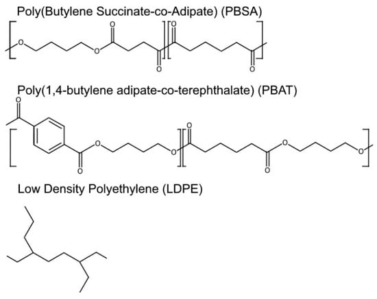
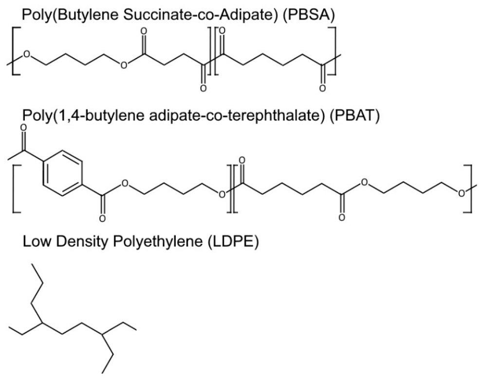
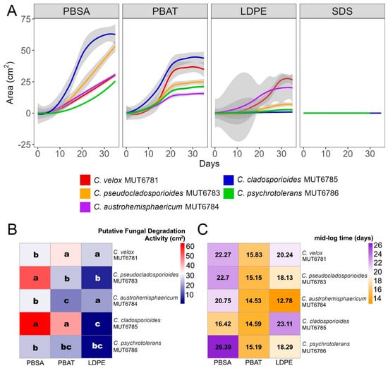
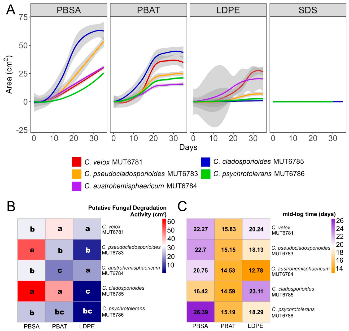
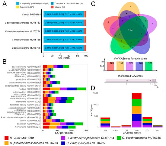
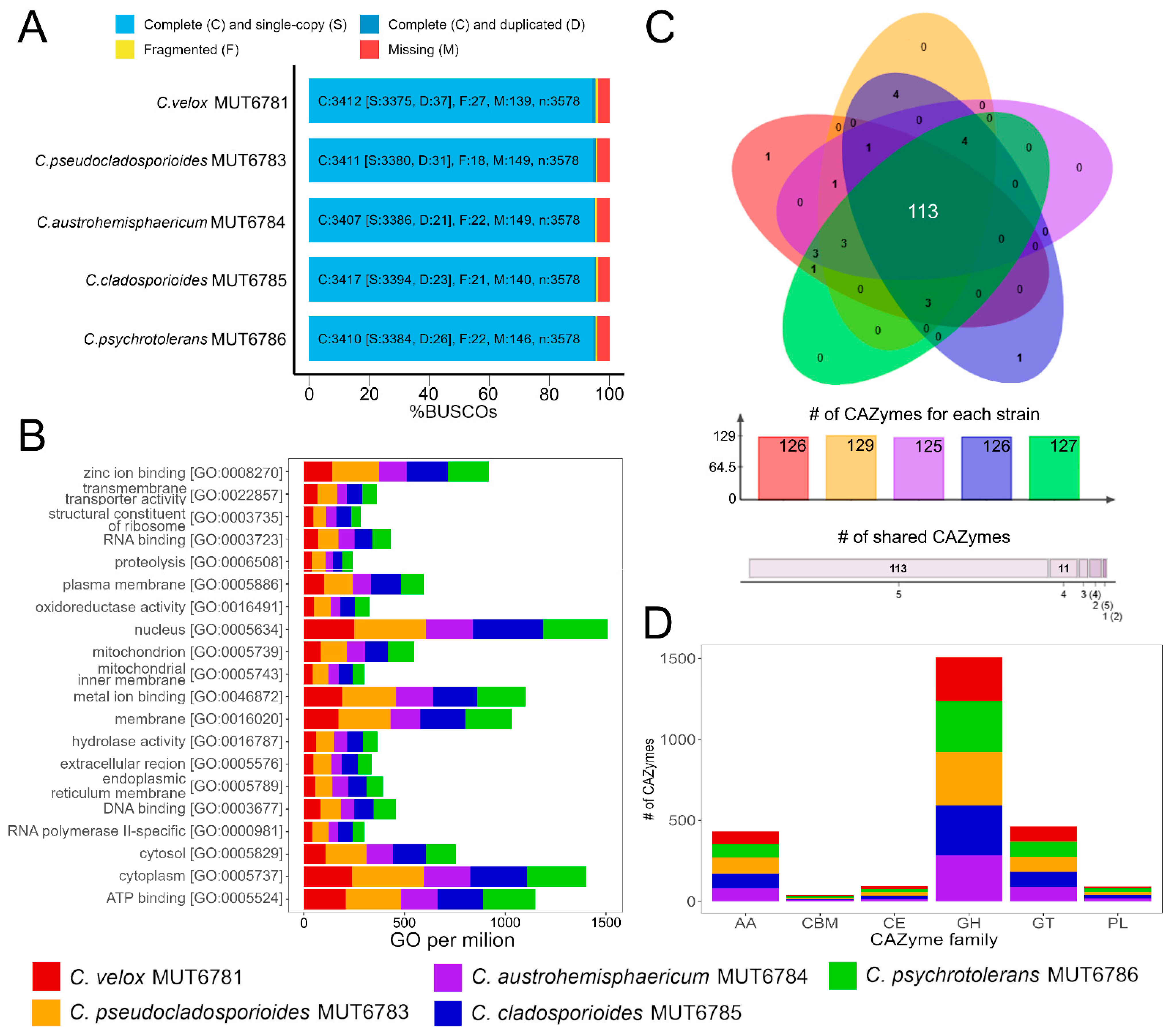
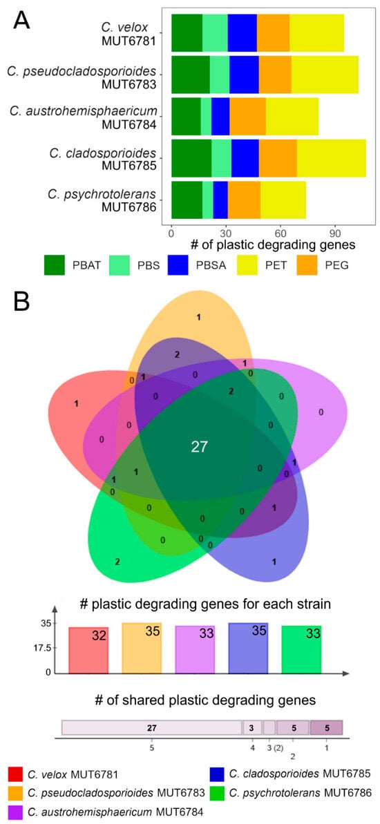
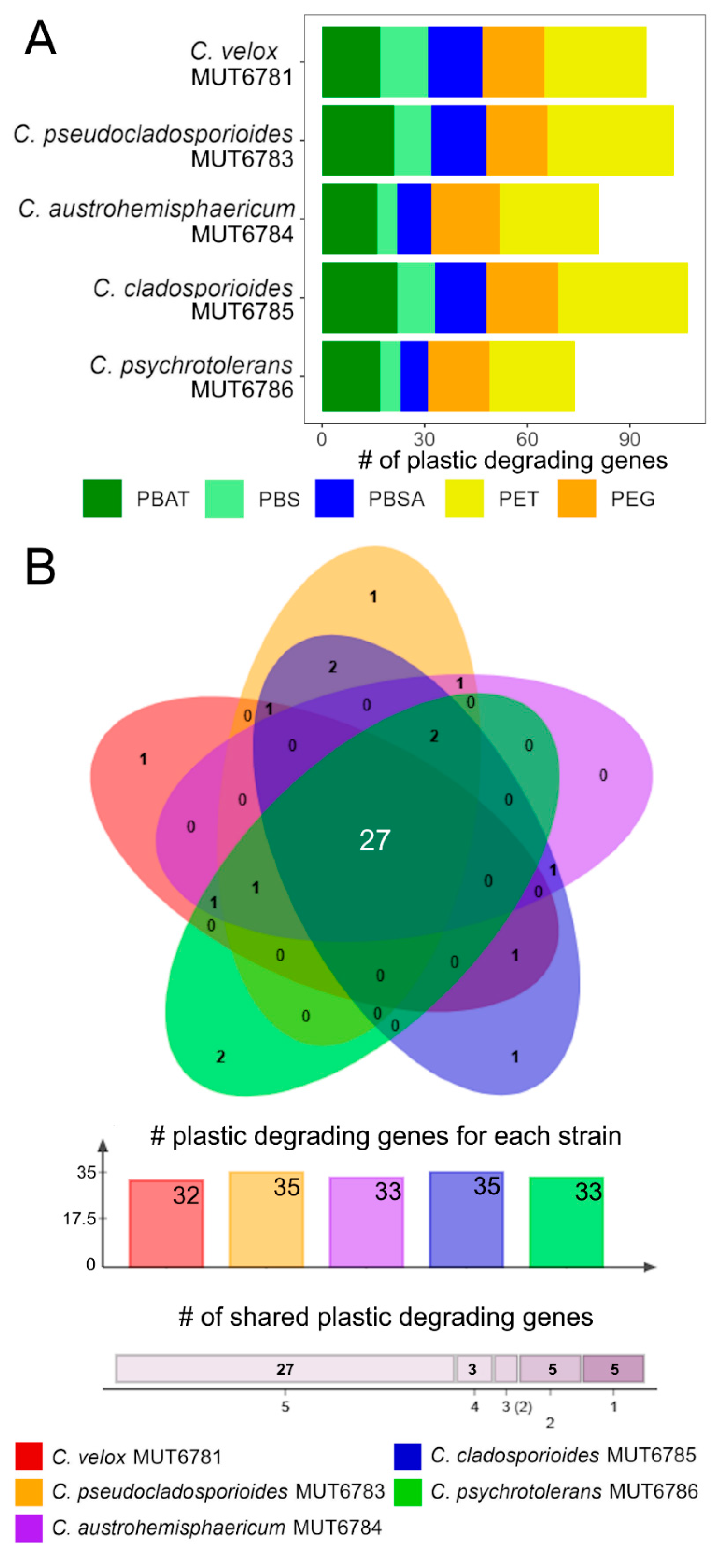
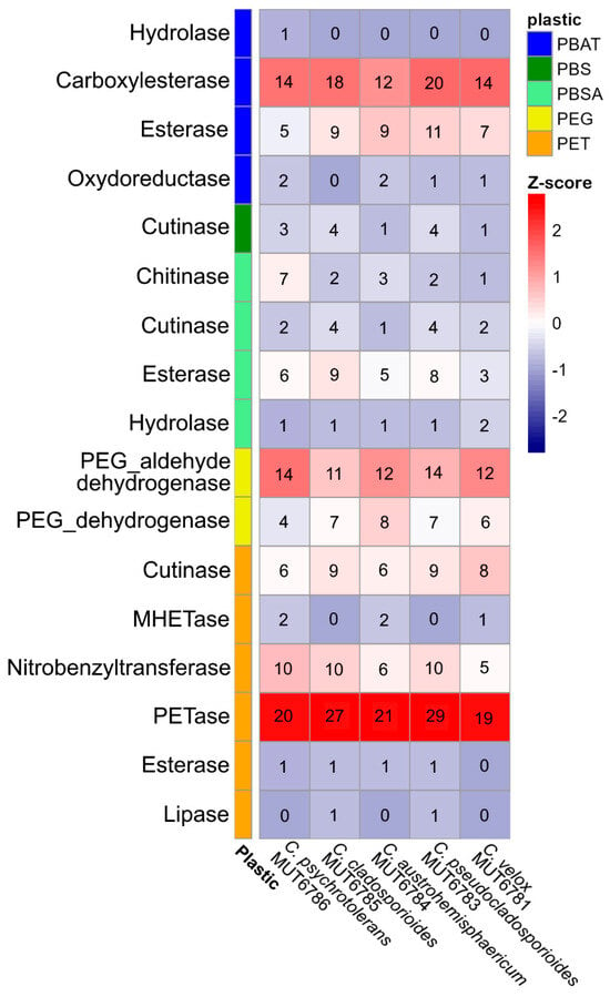
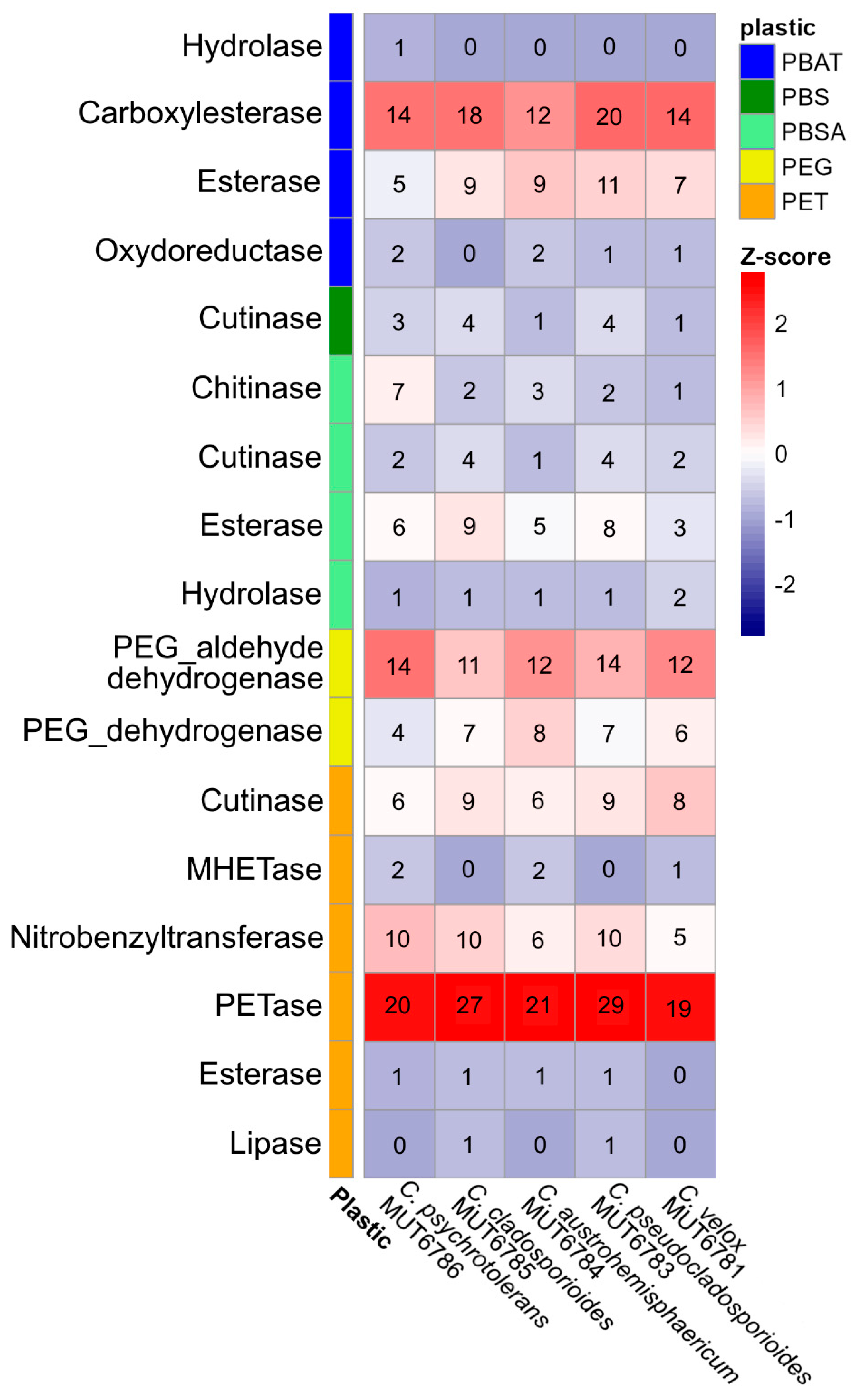
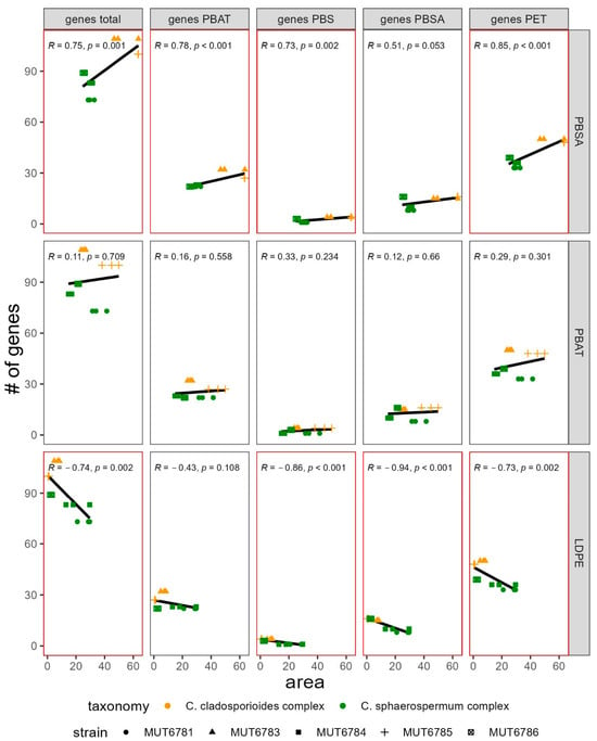
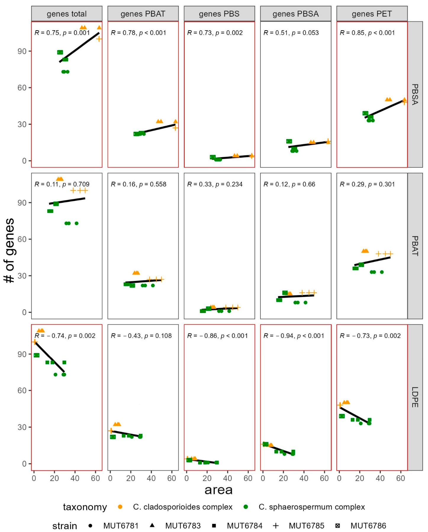

Disclaimer/Publisher’s Note: The statements, opinions and data contained in all publications are solely those of the individual author(s) and contributor(s) and not of MDPI and/or the editor(s). MDPI and/or the editor(s) disclaim responsibility for any injury to people or property resulting from any ideas, methods, instructions or products referred to in the content. |
© 2026 by the authors. Licensee MDPI, Basel, Switzerland. This article is an open access article distributed under the terms and conditions of the Creative Commons Attribution (CC BY) license.