

Med. Sci., Volume 6, Issue 4 (December 2018) – 38 articles
Cover Story (view full-size image):
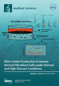
This study showed that human dermal fibroblasts produce significant levels of NO depending on stimulation and glucose conditions. The nitrite level in the media is not a reliable proxy for the actual level of NO produced by the cells. Using the CellNO trap, a novel measurement system developed in our lab, we are able to accurately measure NO produced by cells grown in culture without changing established culturing protocols. If we accurately measure the actual levels of NO produced by cells cultured under specific conditions, including the temporal NO release profile, we can understand and mimic the physiological NO release at different stages of wound healing and in different pathological states. This will facilitate a smooth transition of the wound from a chronic state into resolution, consequently shortening the time to complete the healing process of diabetic foot ulcers. View this paper.
- Issues are regarded as officially published after their release is announced to the table of contents alert mailing list.
- You may sign up for e-mail alerts to receive table of contents of newly released issues.
- PDF is the official format for papers published in both, html and pdf forms. To view the papers in pdf format, click on the "PDF Full-text" link, and use the free Adobe Reader to open them.
Previous Issue
Next Issue