


Abstract
Establishing a suitable propagation protocol is important for the conservation and potential utilization of Kaempferia chayanii (Zingiberaceae). However, no studies have reported on its micropropagation or phytochemical profiling. Explant initiation from rhizome buds was carried out on Murashige and Skoog (MS) medium (1962), supplemented with various exogenous plant growth regulators in both solid and liquid media. In addition, the phytochemical profile was assessed, with particular emphasis on HPLC analysis, to evaluate the antioxidant activity of different organs under varying growth conditions. In solid medium, the highest mean shoot proliferation (4.90 ± 0.38 shoots/explant) was obtained with 4 mg/L BA and 2 mg/L kinetin, representing 2.93 times the control value. In liquid medium, 2 mg/L BA combined with 1 mg/L TDZ produced the highest numerical mean shoot number (6.80 ± 0.47 shoots/explant), representing 1.89 times the corresponding control value. However, no significant differences were observed among the PGR-supplemented liquid-medium treatments. Principal component analysis (PCA) provided a clear overview of plant responses to plant growth regulators (PGRs). Wild leaves had the highest total phenolic content (TPC) (188.27 ± 3.49 mg GAE/g DW), rhizomes had the highest total flavonoid content (TFC) (19.99 ± 1.77 mg RE/g DW), while leaves from micropropagated plants (Ms1 medium treatment) showed high TPC (186.08 ± 5.94 mg GAE/g DW). Antioxidant activity revealed strong radical scavenging capacity, particularly in micropropagated plants, as determined by the 1,1-diphenyl-2-picrylhydrazyl (DPPH) assay and the ferric reducing antioxidant power (FRAP) assay. High-performance liquid chromatography (HPLC) analysis revealed distinct phenolic acid and flavonoid profiles among plant samples. This study represents the first report on the micropropagation and phytochemical profiling of K. chayanii, providing information for its sustainable propagation and potential utilization.
1. Introduction
Kaempferia is a large genus in the ginger family (Zingiberaceae) [1]. Several species are recognized in Thailand for their medicinal uses and as ingredients in local cuisine. For example, Kaempferia marginata Carey ex Roscoe and Kaempferia parviflora Wall. ex Baker are widely acknowledged for their valuable medicinal properties and pharmaceutical applications [2,3]. In addition, several species are valued for ornamental purposes, being noted for their attractive flowers, exotic foliage, and distinctive growth forms, such as Kaempferia elegans Wall. and Kaempferia roscoeana Wall. [2,4].
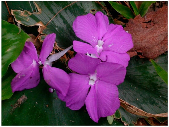
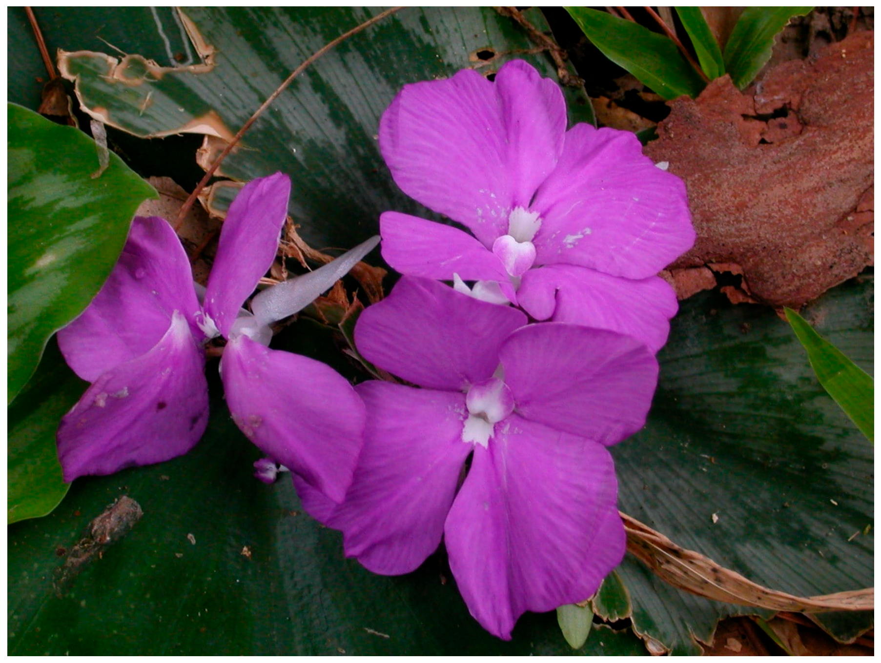
Kaempferia chayanii Koonterm (Figure 1) is a species native to Thailand and Laos and is currently known from limited localities in Tak and Sukhothai provinces of Thailand, as well as in Laos [5,6]. During fieldwork, the authors observed that this species was sold in local markets for ornamental purposes because of its attractive foliage and purple-pink flowers. Despite its ornamental potential, information regarding its cultivation and utilization remains limited. Although no formal conservation assessment or species-specific threat analysis has been reported for K. chayanii, further studies are needed to clarify its conservation status. In parallel, the development of efficient propagation methods may facilitate its cultivation and provide alternative planting materials, potentially reducing dependence on wild populations. More broadly, plant diversity is increasingly affected by habitat degradation, forest encroachment, and the collection of plants from natural habitats for commercial purposes [7,8]. These factors emphasize the importance of sustainable propagation strategies.
Figure 1.
Kaempferia chayanii Koonterm in its natural habitat. (Photo by Prof. Dr. Surapon Saensouk).
In this context, in vitro propagation techniques, particularly micropropagation and microrhizome induction, have been widely recognized as useful approaches for producing planting materials under controlled conditions [9,10]. Such approaches can support the sustainable utilization and ex situ conservation of valuable plant species by providing an alternative source of planting materials and reducing reliance on wild-collected populations. Although in vitro propagation protocols have been reported for several Kaempferia species [11,12,13,14,15,16,17,18,19], suitable plant growth regulator combinations for shoot proliferation in K. chayanii have not yet been established. Previous studies have also examined the phytochemical composition and antioxidant activity of other Kaempferia species [12,15,18,20]. However, comparable information on the phytochemical profiles of wild and in vitro-cultured K. chayanii remains unavailable.
Therefore, this study aimed to evaluate the effects of different plant growth regulator (PGR) combinations on shoot proliferation in K. chayanii using both solid and liquid Murashige and Skoog (MS) media. In addition, the phytochemical composition and antioxidant activities of wild plants and in vitro-cultured leaves were investigated to provide baseline information on the phytochemical properties and antioxidant potential of this species.
2. Materials and Methods
2.1. Initial Explants, Culture Medium, and Culture Conditions
Rhizome buds were used as initial explants for experiments. They were collected from Sukhothai Province, Thailand. The plant material was identified by Professor Dr. Surapon Saensouk at the Walai Rukhavej Botanical Research Institute (WRBRI), Mahasarakham University, Thailand. The specimens were deposited at the Vascular Plant Herbarium of Mahasarakham University (VMSU), Thailand. After decontamination with disinfectants (following the method of Saensouk et al., 2025 [18]), the explants were cultured on basal Murashige and Skoog (1962) (MS) medium [21] containing 2 mg/L 6-benzylaminopurine (BA; Sigma–Aldrich, Budapest, Hungary) and 0.1 mg/L 1-naphthaleneacetic acid (NAA; Sd Fine–Chem Limited, Mumbai, India) for 8 weeks. Culture media were prepared using MS stock solution supplemented with a solidifying agent (0.8% w/v agar) and sucrose (3% w/v). The pH was adjusted to approximately 5.8, and the media were sterilized by autoclaving for 15 minutes. The cultivation control room was maintained at 25 ± 2 °C with a 16-h photoperiod. Illumination was provided by fluorescent lamps (27 µmol m−2 s−1 photosynthetic photon flux density; Philips TLD 36W/54-765 Cool Daylight, Bangkok, Thailand).
2.2. Optimization of Shoot Proliferation
To optimize plantlet regeneration, new shoots of approximately 1.0 cm (stabilized on MS medium without plant growth regulators (PGRs) for 4 weeks) were induced by culturing on various combinations of PGRs at different concentrations. To establish a comprehensive formulation, auxins and cytokinins were applied either individually or in combination in both solid and liquid MS media. The treatments included BA, kinetin (Sigma–Aldrich, Buchs, Switzerland), thidiazuron (TDZ; Sigma–Aldrich, Munich, Germany), NAA, and indole-3-acetic acid (IAA; Sigma–Aldrich, Buchs, Switzerland). Details of these experiments are presented in Table 1 and Table 2.
Table 1.
Combination of PGRs in solid MS medium formulation to promote shoot proliferation.
Table 2.
Combination of PGRs in liquid MS medium formulation to promote shoot proliferation.
The containers used to support the medium and explants were culture jars (100 mL glass bottles) for solid medium experiments and culture flasks (250 mL Erlenmeyer flasks) for liquid medium experiments. In solid-medium experiments, one explant was cultured per culture jar, and ten biological replicates were used for each treatment. In liquid-medium experiments, three explants were cultured in each flask, with five flasks per treatment (15 explants in total). Liquid cultures were maintained on a rotary shaker at 120 rpm. After cultivation in the culture room, shoot and root production were observed and measured after 8 weeks of growth. For consistency with the solid-medium experiments, ten to fifteen explants (n = 10–15) were randomly selected from each treatment for statistical analysis.
Each experimental group included its own PGR-free MS medium control. Treatment responses were compared with the corresponding control within each group, as variation in shoot production was observed among the controls of different experimental series.
2.3. Plant Extraction for Bioactive Compound Evaluation
Whole wild plants were collected from Sukhothai Province, Thailand, in July 2020 during the flowering season. Fresh samples were cleaned with tap water to remove residues and subsequently rinsed with distilled water. To compare phytochemical contents among different plant parts, the plants were separated into leaves, pseudostems, rhizomes, and combined roots with storage roots. For in vitro-cultured plants, plantlets cultured on solid MS medium supplemented with 2 mg/L BA and 0.1 mg/L NAA (Ms1) for 8 weeks were compared with those grown on MS medium without PGRs (Ms0). For in vitro-cultured plants, only leaf segments were subjected to evaluation in this study. All samples were dried using the freeze-drying method for 48 h. The dried material was then ground into fine powder using a blender. Samples were stored at −20 °C until use.
Bioactive compounds were extracted using the shaking-maceration method, as described by Chumroenphat et al. (2019) [22] with some modification. In brief, dried powder samples were mixed with 80% methanol (2% w/v) and incubated at 37 °C with continuous shaking at 150 rpm for 15 h. After extraction, the filtrate was collected and stored in a refrigerator at 4 °C until use in experiments. The phytochemical and antioxidant analyses were performed in triplicate using the pooled samples.
2.4. Total Phenolic Contents (TPC) and Total Flavonoid Contents (TFC)
TPC in plant extract was determined using the Folin–Ciocalteu assay with modification, as described by Saensouk et al. [18]. The filtered extract was incubated with 10% (v/v) Folin–Ciocalteu solution (Sigma–Aldrich, Munich, Germany) at a 20:100 μL ratio in a 96-well plate for 5 min. Then, 10% (w/v) sodium carbonate (Na2CO3) was incubated for 2 h in a dark room, and data were recorded using a UV–Vis microplate reader (Varioskan™ LUX, Thermo Fisher Scientific Inc., Marsiling, Singapore) at 750 nm. The results are reported as mg gallic acid equivalent per gram of dry weight (mg GAE/g DW), with a calibration curve R2 of 0.9985.
For TFC analysis, an aluminum chloride colorimetric method was performed according to Saensouk et al. [18]. 125 μL of 1:4 ratio extract dilution (diluted with deionized water) was incubated with 10 µL of 5% (w/v) sodium nitrite (NaNO2) in well plate for 5 min. Subsequently, 15 µL of aluminum chloride hexahydrate (10% w/v, AlCl3·6H2O) was mixed and incubated for 6 min. To complete the reaction, 1 M sodium hydroxide (NaOH) and deionized water at 50 µL of each solution were added to the mixture. Absorbance of the reacted solution was measured at 510 nm with a microplate reader, and results are reported as mg rutin equivalent per gram of dry weight (mg RE/g DW), with a calibration curve R2 = 0.9980.
2.5. Assessment of Antioxidant Activity in Plant Extracts
2.5.1. DPPH Free Radical Inhibition Assay
The 1,1-diphenyl-2-picrylhydrazyl (DPPH, Alfa Aesar, Thermo Fisher Scientific, Waltham, MA, USA) solution was prepared to conduct the radical scavenging assay of plant extracts. This procedure followed Saensouk et al. [18]. Each plant extract (20 µL, 20 mg/mL) was pipetted into a well plate containing 180 µL of DPPH solution (0.15 mM, diluted in methanol). The mixture was incubated in the dark for 30 min, after which the absorbance was recorded at 517 nm with a microplate reader. The results are expressed as percentage inhibition of DPPH radicals, calculated according to the following equation 1:
represents the absorbance of the control (DPPH solution), whereas denotes the absorbance of the DPPH solution mixed with extract.
2.5.2. Ferric Reducing Antioxidant Power (FRAP) Assay
The Fe3+–TPTZ complex reagent was employed to evaluate the redox-linked colorimetric reaction, where Fe3+ ions are reduced to Fe2+ by antioxidants. This protocol was previously described by Saensouk et al. [18] and Kubola and Siriamornpun [23]. The FRAP reagent was prepared by mixing 300 mM acetate buffer (pH 3.6), 10 mM TPTZ in 40 mM HCl, and 20 mM FeCl3·6H2O (10:1:1, v/v/v), followed by incubation at 37 °C. Briefly, 180 µL of FRAP reagent was combined with 5 µL of plant extract (20 mg/mL). The mixture was allowed to stand for 1 min and subsequently incubated at 37 °C for 15 min. The absorbance was then recorded at 593 nm. Results are presented as mg ferrous sulfate (FeSO4) equivalent per gram of plant dry weight (mg FeSO4/g DW), with a calibration curve R2 = 0.9888.
2.6. HPLC Determination of Phenolic Acids and Flavonoid Compounds
2.6.1. Plant Extraction for HPLC Analysis
This protocol was adapted from Chumroenphat et al. [24]. Dried materials obtained from both wild and in vitro-cultured plants (Section 2.3) were subjected to extraction using the maceration method. Briefly, 0.20 g of dried powder sample was combined with 10 mL of methanolic hydrochloric acid solution (1% v/v) and incubated at 37 °C with continuous shaking at 150 rpm for 15 h. Before chemical determination, the resulting extract was passed through a 0.22 µm nylon membrane filter to remove particulates.
2.6.2. HPLC-Based Determination of Phenolic Acids and Flavonoids
The chromatography was performed on a Shimadzu LC–20A series system (Shimadzu Corp., Kyoto, Japan) fitted with a C18 column (4.6 mm × 250 mm, 5 µm; GL Sciences Inc., Tokyo, Japan). The mobile phase consisted of 0.1% (v/v) acetic acid (solvent A) and acetonitrile (solvent B) at a flow rate of 0.8 mL/min. The operating parameters followed the method reported by Kubola and Siriamornpun [23]. Compounds were tentatively identified by comparing their retention times (RTs) and UV spectra with those of reference standards. Quantification was performed using external calibration curves, with R2 values of approximately 0.999.
2.7. Experimental Design and Statistical Analysis
Micropropagation Experiments: Experiments in this section were performed under a completely randomized design (CRD). Each treatment was tested with ten to fifteen replicates, which were reported as mean value with standard error (mean ± SE). To compare the different groups of each culture medium, one-way analysis of variance (ANOVA) was conducted, and mean separations were determined using Duncan’s multiple range test (DMRT) as a post hoc test at a significance level of p < 0.05. Normality and homogeneity of variance assumptions (Shapiro–Wilk and Levene’s tests) were tested and confirmed before performing ANOVA.
Principal component analysis (PCA) was performed using treatment means from seven experimental series (S1–S5 and L1–L2), comprising 64 observations and four growth variables: shoot number, shoot length, root number, and root length. All variables were Z-score standardized prior to analysis. PCA was used to explore multivariate patterns across treatments, considering differences in experimental series and culture conditions, rather than to determine treatment superiority. The stability of PCA components was evaluated using leave-one-series-out sensitivity analysis, in which PCA was repeated after excluding each experimental series in turn. Variables were re-standardized for each PCA analysis, and the similarity between corresponding eigenvectors from the full and reduced datasets was assessed using cosine similarity after sign alignment [25]. Data analysis was carried out using SPSS software, version 29 (IBM Corp., Armonk, NY, USA), and OriginPro 2018 software (OriginLab Corp., Northampton, MA, USA.).
Phytochemical Profiling and Antioxidant Evaluation Experiments: Each experiment was analyzed with three technical replicates of pooled samples, and values were presented as mean ± standard error (SE), with mean comparisons determined by Duncan’s multiple range test (DMRT) at p < 0.05. Pearson correlation coefficient (r) analysis was performed using observations (n = 18), comprising six sample groups with three technical replicates (pooled samples) per group, to evaluate the correlation among TPC, TFC, DPPH, and FRAP. Statistical significance was assessed using two-tailed tests at p < 0.05. PCA biplots were used to explore patterns of variation in phytochemical content and antioxidant activity. Statistical analyses were performed using SPSS and OriginPro 2018.
3. Results
3.1. Effect of Exogenous PGRs on Shoot and Root Regeneration in Kaempferia chayanii
3.1.1. Shoot and Root Regeneration Under Different PGR Combinations in Solid Medium
Effects of cytokinin and auxin were studied to determine the optimum level for the induction of shoot and root regeneration in K. chayanii. The results of BA hormone (Experiment S1) on plant development are presented in Table 3 and Figure 2. No significant differences (p > 0.05) in shoot production were observed at BA concentrations ranging from 0.5 to 5 mg/L. However, BA at 3 mg/L (S1-B4 medium) produced higher plant regeneration than other concentrations, resulting in 3.60 ± 0.77 shoots/explant (Figure 2e) with an average shoot length of 2.85 ± 0.26 cm. Root production did not differ significantly from the control medium and remained low across treatments. Root regeneration ranged between 2.90 ± 0.60 and 4.30 ± 0.51 roots/explant, while root length varied from 1.40 ± 0.14 to 1.70 ± 0.11 cm.
Table 3.
Shoot and root production on solid MS medium containing different concentrations of BA in Kaempferia chayanii after 8 weeks of culture.
Figure 2.
Shoot and root development of Kaempferia chayanii after 8 weeks of culture on MS medium supplemented with different concentrations of BA. (a) MS medium without PGR; (b) 0.5 mg/L; (c) 1 mg/L; (d) 2 mg/L; (e) 3 mg/L; (f) 4 mg/L; (g) 5 mg/L. Scale bar = 2 cm.
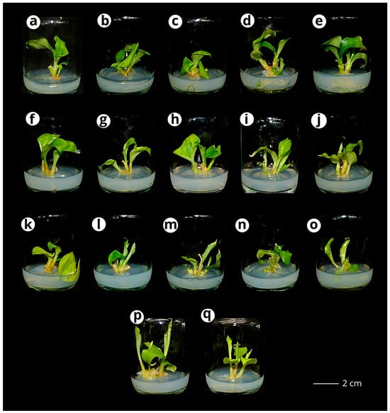
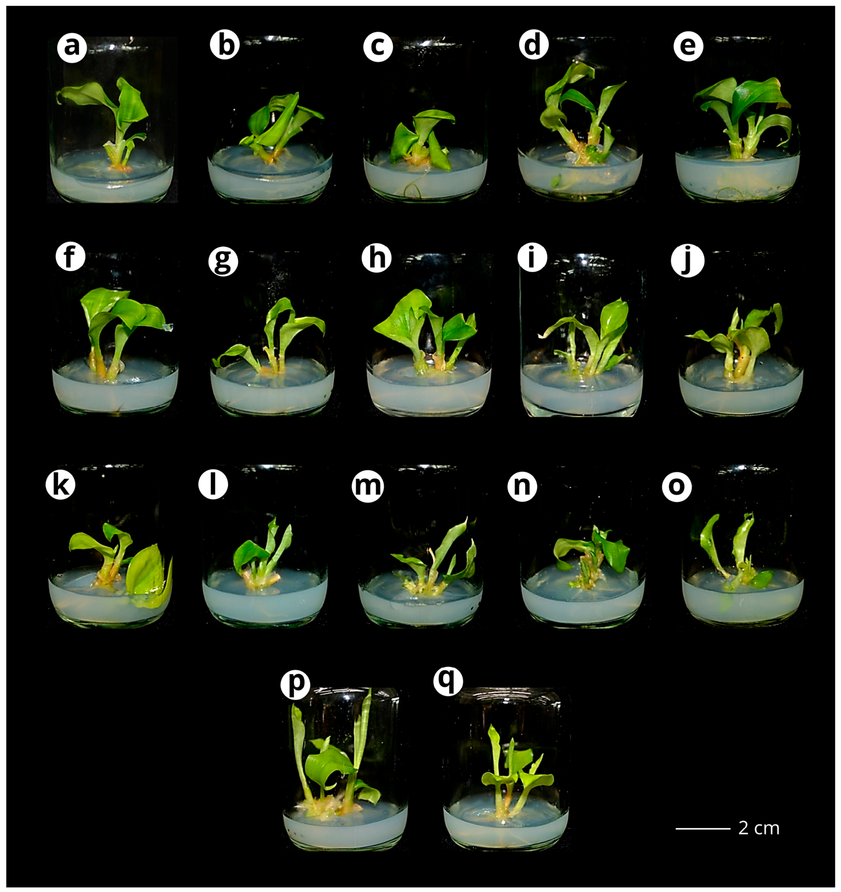
Based on these findings from BA alone, subsequent experiments were conducted to investigate the effects of combining BA with kinetin at different concentration levels. Results of the BA–kinetin combination (Experiment S2) are presented in Table 4 and Figure 3. This experiment showed that the S2-BK14 medium (4 mg/L BA + 2 mg/L kinetin) (Figure 3o) exhibited the highest shoot production, yielding 4.90 ± 0.38 shoots/explant with an average shoot length of 2.00 ± 0.27 cm. Root induction was maximized in the S2-BK5 medium (2 mg/L BA + 1 mg/L kinetin), producing 9.30 ± 1.21 roots/explant. The medium enriched with BA and kinetin showed a positive effect on shoot proliferation. However, increasing kinetin beyond 3 mg/L together with each concentration of BA tended to decrease shoot promotion. When compared with the BA-alone medium, this BA–kinetin medium combination enhanced root production (producing 4.22 ± 0.32 to 9.30 ± 1.21 roots/explant); however, root elongation remained short.
Table 4.
Shoot and root production on solid MS medium containing different concentrations of BA and kinetin in Kaempferia chayanii after 8 weeks of culture.
Figure 3.
Shoot proliferation of Kaempferia chayanii after 8 weeks of culture on MS medium supplemented with different concentrations of BA and kinetin (Kn). (a) MS medium without PGR; (b) 1BA + 1Kn; (c) 1BA + 2Kn; (d) 1BA + 3Kn; (e) 1BA + 4Kn; (f) 2BA + 1Kn; (g) 2BA + 2Kn; (h) 2BA + 3Kn; (i) 2BA + 4Kn; (j) 3BA + 1Kn; (k) 3BA + 2Kn; (l) 3BA + 3Kn; (m) 3BA + 4Kn; (n) 4BA + 1Kn; (o) 4BA + 2Kn; (p) 4BA + 3Kn; (q) 4BA + 4Kn mg/L. Scale bar = 2 cm.
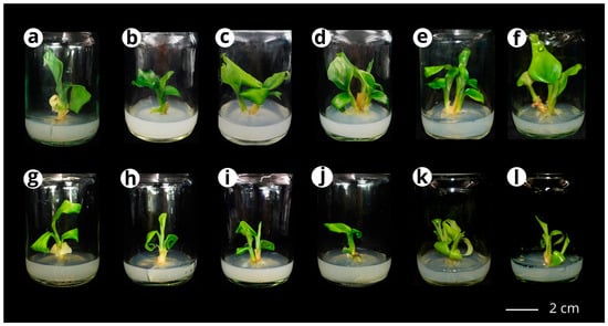
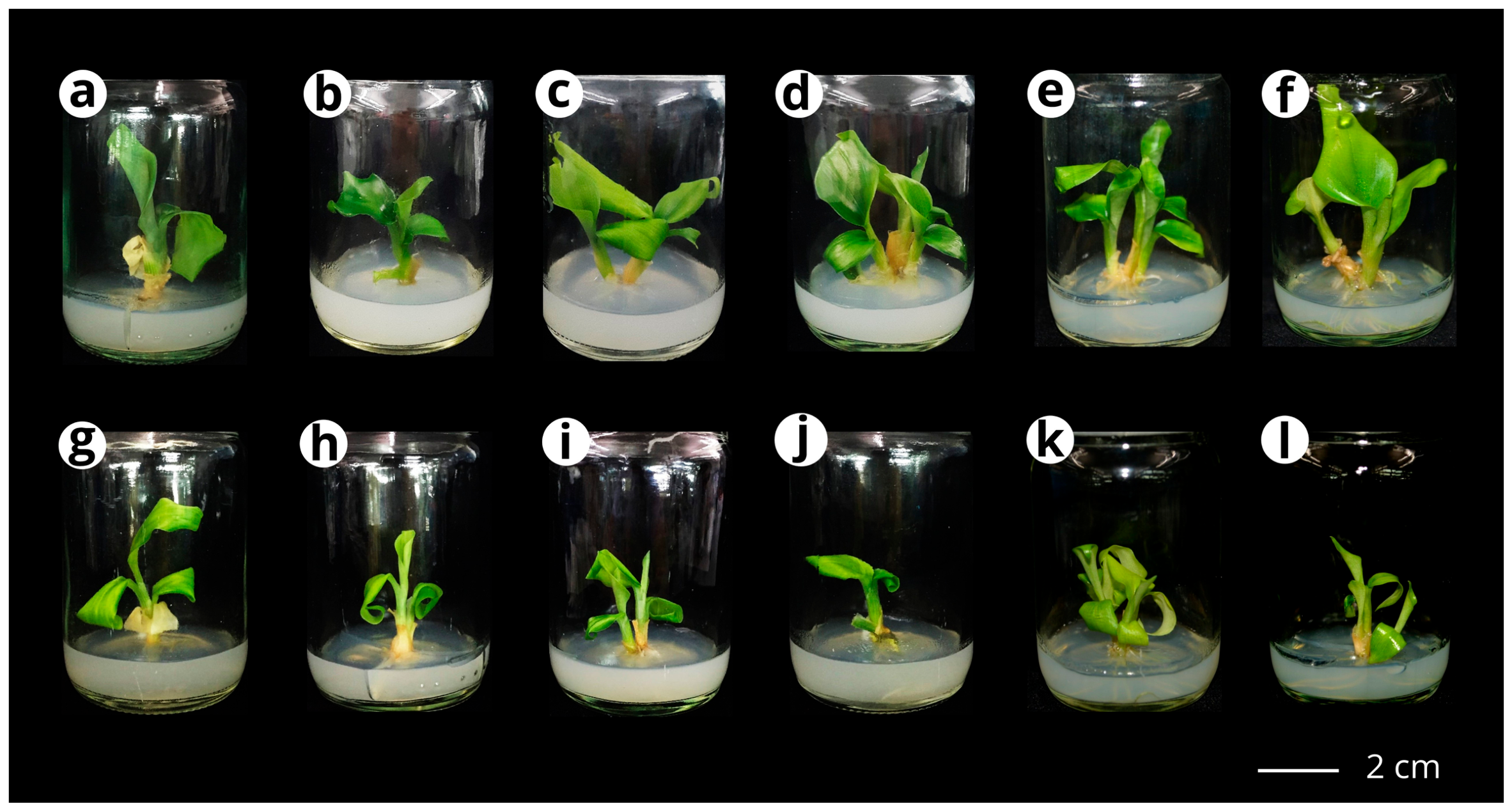
Because BA–kinetin treatments affected shoot and root responses differently, subsequent trials were conducted to assess whether the addition of auxin (NAA or IAA) could enhance regeneration efficiency. The results are presented in Table 5 and Figure 4. The medium supplemented with 5 mg/L BA and 0.5 mg/L NAA (S3-BN5 medium) promoted maximum shoot induction, yielding 2.82 ± 0.24 shoots per explant with a mean shoot length of 2.75 ± 0.21 cm (Table 5 and Figure 4f). However, varying concentrations of BA combined with NAA did not result in significant differences in shoot production. Notably, the S3-BN1 medium (0.5 mg/L BA + 0.5 mg/L NAA) exhibited optimal responses in both shoot and root production, including shoot length (4.18 ± 0.37 cm) and root number (12.90 ± 0.70 roots per explant). Root production in this combination resulted in short roots (1.02 ± 0.06 to 1.27 ± 0.19 cm). In addition, kinetin was also tested in combination with NAA. The combinations of 0.5–5 mg/L kinetin with 0.5 mg/L NAA did not result in significant differences in shoot induction and shoot length (p > 0.05). However, the medium supplemented with 4 mg/L kinetin and 0.5 mg/L NAA resulted in high root production, yielding 8.40 ± 0.73 roots per explant.
Table 5.
Shoot and root production on solid MS medium containing different concentrations of BA or kinetin in combination with NAA in Kaempferia chayanii after 8 weeks of culture.
Figure 4.
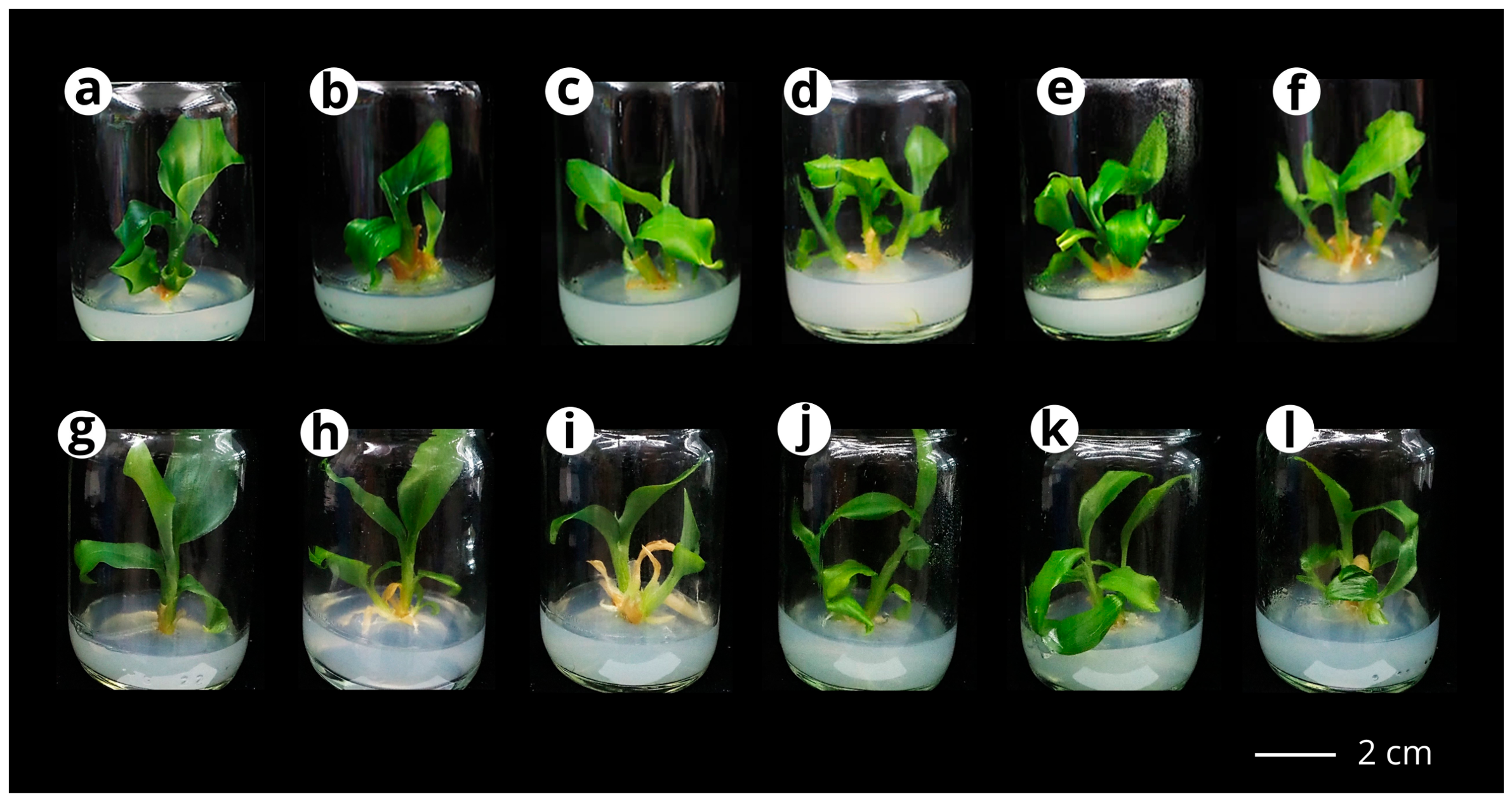
Shoot proliferation of Kaempferia chayanii after 8 weeks of culture on MS medium supplemented with different concentrations of BA or kinetin in combination with NAA. (a,g) MS medium without PGR; (b) 0.5BA + 0.5NAA; (c) 1BA + 0.5NAA; (d) 2BA + 0.5NAA; (e) 4BA + 0.5NAA; (f) 5BA + 0.5NAA; (h) 0.5Kn + 0.5NAA; (i) 1Kn + 0.5NAA; (j) 2Kn + 0.5NAA; (k) 4Kn + 0.5NAA; (l) 5Kn + 0.5NAA. Scale bar = 2 cm.
Based on the responses observed from previous combinations, subsequent experiments (S4) were conducted to evaluate the effects of BA or kinetin in combination with IAA on shoot and root regeneration. The response data are detailed in Table 6 and Figure 5. The S4-BA5 medium (5 mg/L BA + 0.5 mg/L IAA) gave the highest shoot promotion (4.50 ± 0.16 shoots/explant) with a shoot length of 3.33 ± 0.09 cm (Figure 5f). For root production, 3 mg/L BA mixed with 0.5 mg/L IAA resulted in the maximum value (9.40 ± 0.64 roots/explant) compared to all other media. However, root elongation showed no statistically significant variation (p > 0.05) among treatments. When compared with the kinetin combinations, the treatment with S4-KA2 medium (2 mg/L kinetin + 0.5 mg/L IAA) exhibited the maximum shoot induction (3.10 ± 0.23 shoots/explant). Remarkably, media containing kinetin at 1, 2, and 5 mg/L did not show significant differences in mean shoot production. Root production in media containing kinetin at concentrations ranging from 2 to 5 mg/L with IAA increased the number of roots, but the roots remained fewer and shorter compared with those produced in BA–IAA combinations.
Table 6.
Shoot and root production on solid MS medium containing different concentrations of BA or kinetin in combination with IAA in Kaempferia chayanii after 8 weeks of culture.
Figure 5.
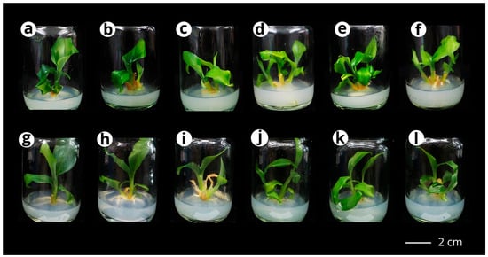
Shoot proliferation of Kaempferia chayanii after 8 weeks of culture on MS medium supplemented with different concentrations of BA or kinetin in combination with IAA. (a,g) MS medium without PGR; (b) 1BA + 0.5IAA; (c) 2BA + 0.5IAA; (d) 3BA + 0.5IAA; (e) 4BA + 0.5IAA; (f) 5BA + 0.5IAA; (h) 1Kn + 0.5IAA; (i) 2Kn + 0.5IAA; (j) 3Kn + 0.5IAA; (k) 4Kn + 0.5IAA; (l) 5Kn + 0.5IAA. Scale bar = 2 cm.
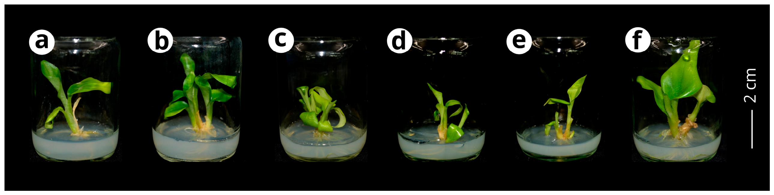
To observe the influence of NAA and IAA in combination with BA on plantlet responses, Experiment S5 was conducted (Table 7 and Figure 6). At BA concentrations of 1 and 3 mg/L combined with 0.5 mg/L NAA and IAA, maximum mean shoot production was observed (2.70 ± 0.30 and 2.70 ± 0.21 shoots/explant, respectively). Nonetheless, the number of shoots among the treatment groups (PGR-supplemented) did not differ significantly. Root multiplication among the treatment groups tended to decrease at higher BA levels (more than 2 mg/L), and the highest root production was observed in the S5-BA1 medium (1 mg/L BA + 0.5 mg/L NAA + 0.5 mg/L IAA). Notably, the root length in this experiment remained short (1.52 ± 0.19 cm to 2.13 ± 0.22 cm).
Table 7.
Shoot and root production on solid MS medium containing different concentrations of BA in combination with NAA and IAA in Kaempferia chayanii after 8 weeks of culture.
Figure 6.
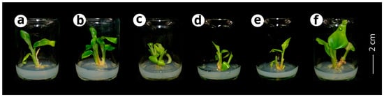
Shoot proliferation of Kaempferia chayanii after 8 weeks of culture on MS medium supplemented with different concentrations of BA in combination with NAA and IAA. (a) MS medium without PGR; (b) 1BA + 0.5NAA + 0.5IAA; (c) 2BA + 0.5NAA + 0.5IAA; (d) 3BA + 0.5NAA + 0.5IAA; (e) 4BA + 0.5NAA + 0.5IAA; (f) 5BA + 0.5NAA + 0.5IAA. Scale bar = 2 cm.
3.1.2. Shoot and Root Regeneration Under Different PGR Combinations in Liquid Medium
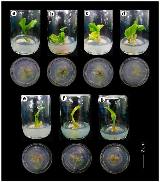
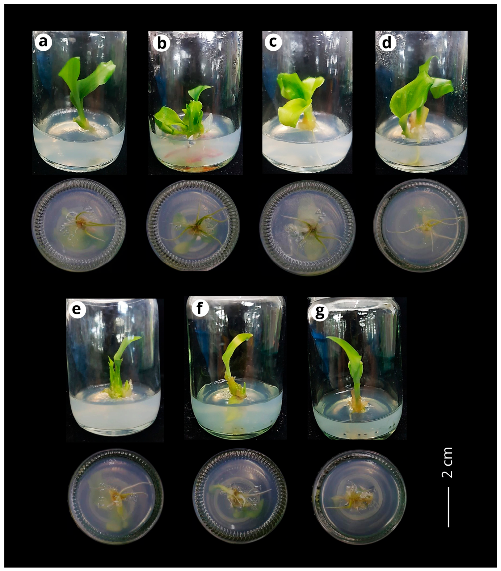
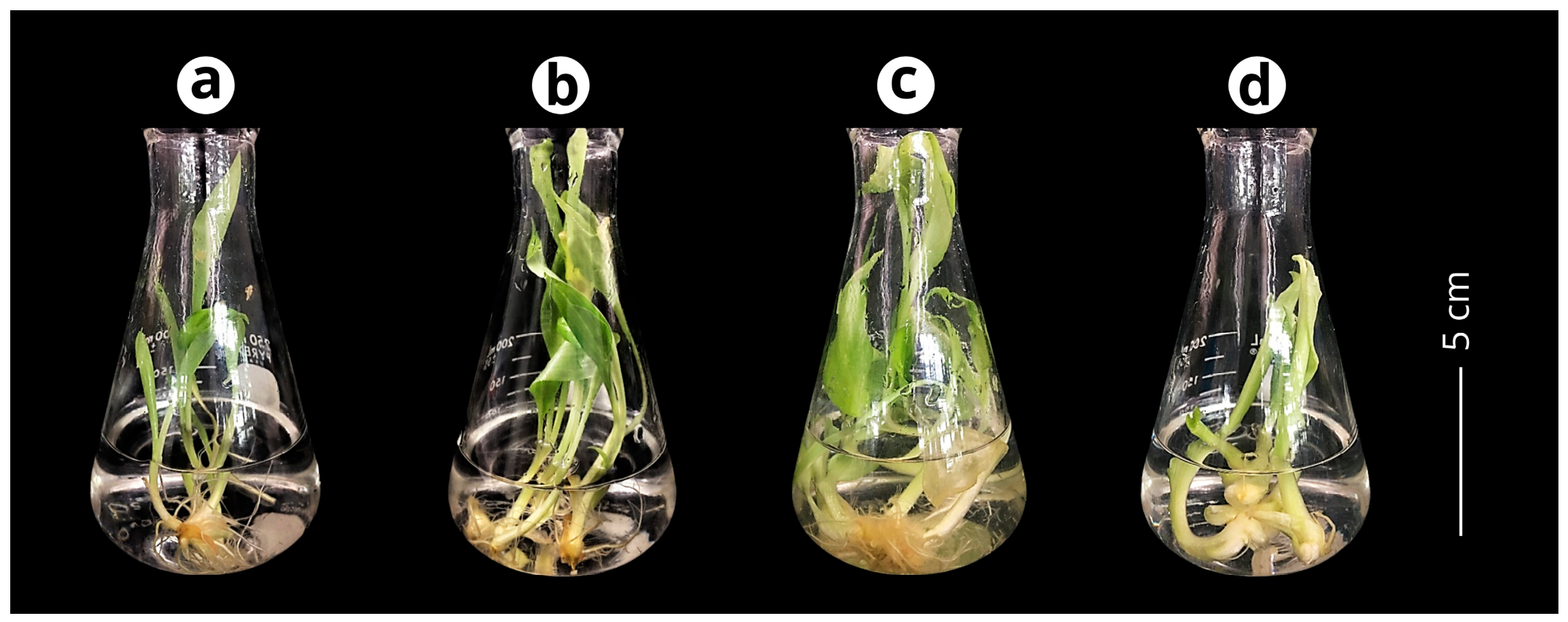
Previous studies have indicated that liquid medium can enhance shoot and root regeneration. To further evaluate this effect in K. chayanii, explants were cultured in liquid media supplemented with different cytokinins (BA, kinetin, or TDZ) in combination with 0.5 mg/L IAA. The results are presented in Table 8 and Figure 7. Among the L1 treatments, L1-C1 (1 mg/L BA + 0.5 mg/L IAA) showed the highest shoot induction (5.27 ± 0.45 shoots/explant) with shoot length (8.98 ± 0.41 cm) (Figure 7b). However, shoot and root length did not differ significantly among treatments (p > 0.05). Nevertheless, media supplemented with TDZ and IAA tended to yield shorter shoots and reduced root formation compared with the other treatments.
Table 8.
Shoot and root production on liquid MS medium containing different concentrations of BA, kinetin, and TDZ in combination with IAA in Kaempferia chayanii after 8 weeks of culture.
Figure 7.
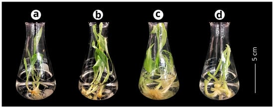
Shoot proliferation of Kaempferia chayanii after 8 weeks of culture on liquid MS medium supplemented with different concentrations of PGRs. (a) MS medium without PGR; (b) 1BA + 0.5IAA; (c) 1Kn + 0.5IAA; (d) 1TDZ + 0.5IAA. Scale bar = 5 cm.
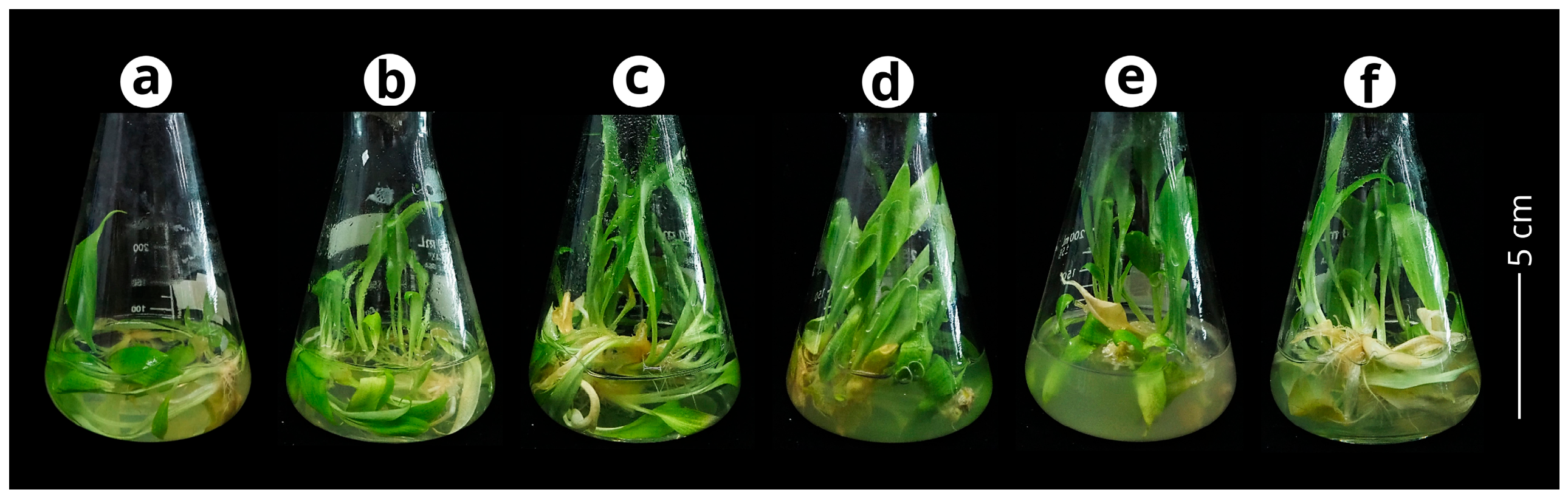
In the L2 experiment, the combination of BA (2 mg/L) and TDZ (1 mg/L) (L2-D3 medium) resulted in the highest numerical mean shoot production, with 6.80 ± 0.47 shoots/explant (Table 9 and Figure 8). The reported analysis indicated significant differences in shoot production among treatments, with PGR-supplemented treatments showing higher values than the control medium. However, no significant differences were observed among the PGR-supplemented treatments. Furthermore, shoot and root formation did not differ significantly in PGR-supplemented treatments. Compared with solid medium containing the same BA concentration (3 mg/L), liquid medium produced approximately twice as many shoots, reaching 6.70 ± 0.63 shoots/explant.
Table 9.
Shoot and root production on liquid MS medium containing different concentrations of BA and TDZ in combination with IAA in Kaempferia chayanii after 8 weeks of culture.
Figure 8.
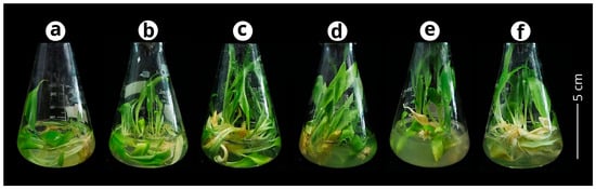
Shoot proliferation of Kaempferia chayanii after 8 weeks of culture on liquid MS medium supplemented with different concentrations of PGRs. (a) MS medium without PGR; (b) 1TDZ; (c) 1BA + 0.5IAA; (d) 2BA + 1TDZ; (e) 3BA; (f) 4BA. Scale bar = 5 cm.
3.1.3. PCA Biplot Analysis
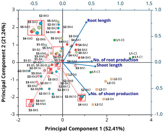
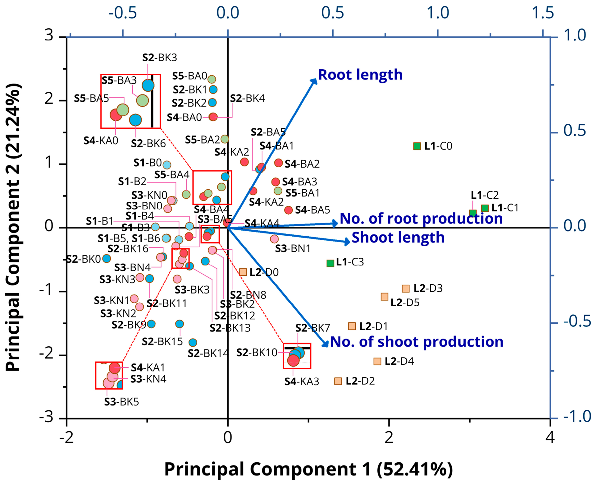
The first two principal components explained 73.65% of the total variance, with PC1 and PC2 accounting for 52.41% and 21.24%, respectively (Table 10 and Figure 9). PC1 was mainly associated with shoot length (eigenvector coefficient = 0.577) and root number (0.519), whereas PC2 was primarily associated with root length (0.779) and shoot number (−0.622).
Table 10.
Eigenvector coefficients of shoot and root traits on the first two principal components (PC1 and PC2).
Figure 9.
PCA biplot of culture medium treatments and plant growth responses. Vectors represent number of shoots, shoot length, number of roots, and root length. Circles denote solid media (S1–S5 series), while rectangles denote liquid media (L1 and L2 series). Percentages in parentheses indicate the variance explained by PC1 and PC2. Colors correspond to experimental groups, with identical colors representing the same group.
The PCA biplot revealed distribution patterns associated with experimental series and culture medium type (Figure 9). Most liquid-medium treatments, including the L1 (L1-C0 to L1-C3) and L2 (L2-D1 to L2-D5) series, were located on the positive side of PC1, whereas most solid-medium treatments were distributed on the negative side. Several L2 treatments (e.g., L2-D3, L2-D4, and L2-D5) were positioned in the lower-right quadrant, broadly aligned with the shoot-number vector.
Several BA-containing treatments (S4-BA1, S4-BA2, S4-BA3, and S4-BA5) were distributed in the upper-right region, generally in the direction of the root-length vector, whereas S5-BA1 was positioned on the positive side of PC1. In contrast, treatments such as S2-BK9, S2-BK11, S3-KN1, and S3-KN3 were located in the lower-left quadrant.
Leave-one-series-out sensitivity analysis indicated relatively stable PC1 directions (absolute cosine similarity = 0.986–1.000), whereas PC2 showed greater sensitivity (0.750–0.997), particularly following the exclusion of S2 (0.750) and L2 (0.826) (Tables S1 and S2).
3.2. Evaluation of TPC, TFC, and Antioxidant Activity
The total phenolic content (TPC) of polyphenol compounds was determined for screening bioactive sources using the Folin–Ciocalteu assay, and the results are presented in Table 11. In wild plants, TPC ranged from 65.70 to 188.27 mg GAE/g DW, with the highest value observed in leaves. Among in vitro-propagated plants, leaves cultured on PGR-supplemented MS medium (Ms1) exhibited significantly higher TPC (186.08 ± 5.94 mg GAE/g DW) than those cultured on PGR-free MS medium (Ms0) (153.58 ± 4.16 mg GAE/g DW). No significant difference was detected between wild leaves and Ms1 leaves. Furthermore, Ms0 leaves exhibited significantly higher TPC than wild pseudostems (65.70 ± 2.33 mg GAE/g DW) and rhizomes (109.42 ± 1.71 mg GAE/g DW) but did not differ significantly from combined roots and storage roots (157.03 ± 1.03 mg GAE/g DW).
Table 11.
Determination of total phenolic and flavonoid contents and antioxidant activity in Kaempferia chayanii extracts.
The aluminum chloride assay was used to measure the total flavonoid content of the extracts, facilitating the screening of flavones, flavanols, and other flavonoids within the polyphenol group. The results are presented in Table 11. In wild plants, TFC ranged from 4.39 to 19.99 mg RE/g DW, with the highest value observed in rhizomes. Among in vitro-propagated plants, leaves cultured on PGR-free MS medium (Ms0) exhibited significantly higher TFC (13.76 ± 1.10 mg RE/g DW) than those cultured on PGR-supplemented MS medium (Ms1) (7.18 ± 0.40 mg RE/g DW). However, the TFC of both in vitro-propagated samples was significantly lower than that of wild rhizomes.
The antioxidant activities of both wild and in vitro-cultured plants are presented in Table 11. In this study, DPPH and FRAP assays were conducted to evaluate the antioxidant properties of the extracts. When compared among samples, leaves derived from the Ms0 medium exhibited the highest values of DPPH inhibition and reducing power in the FRAP assay (30.99% and 19.07 ± 1.33 mg FeSO4/g DW, respectively), followed by leaves derived from the Ms1 medium (25.89% and 13.54 ± 0.59 mg FeSO4/g DW, respectively). In wild plants, the results showed that leaves (14.84%) and the combined roots and storage roots (13.05%) exhibited higher DPPH inhibition than rhizomes (7.82%) and pseudostems (5.92%). In contrast, FRAP values exhibited high activity in leaves (1.11 ± 0.03 mg FeSO4/g DW), with no significant differences observed among wild plant organs.
3.3. Correlation and Principal Component Analysis (PCA) of Phytochemical Compounds and Antioxidant Activities
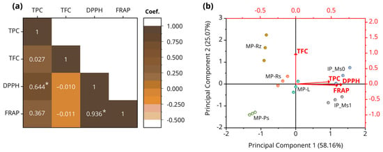
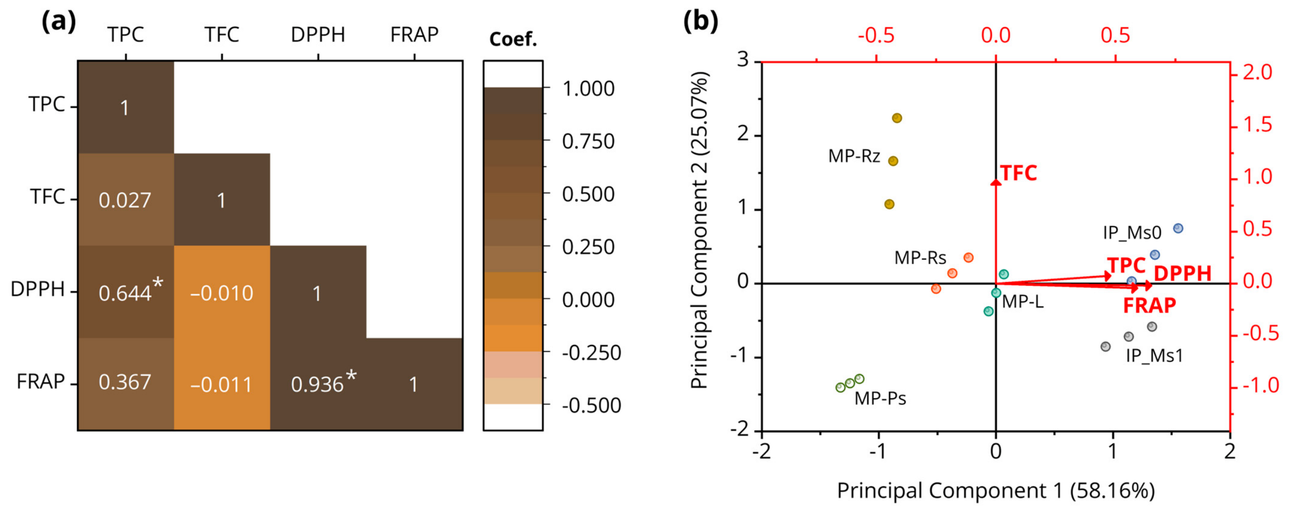
Pearson correlation analysis showed a significant positive correlation between TPC and DPPH activity (r = 0.644, p < 0.01) and a strong positive correlation between DPPH and FRAP activities (r = 0.936, p < 0.01) (Figure 10a). TPC also showed a moderate positive correlation with FRAP activity (r = 0.367), whereas TFC showed negligible correlations with DPPH (r = −0.010) and FRAP (r = −0.011).
Figure 10.
Pearson correlation and principal component analysis (PCA) of phytochemical contents and antioxidant activities in wild and in vitro-propagated Kaempferia chayanii. (a) Pearson correlation matrix of TPC, TFC, DPPH, and FRAP based on observations (n = 18). * indicates significance at p < 0.01 (two-tailed). (b) PCA biplot showing the distribution of six sample groups and associations among variables. PC1 and PC2 explained 58.16% and 25.07% of the total variance, respectively. Abbreviations: MP = wild plants; IP, in vitro-propagated plants; L, leaves; Ps, pseudostems; Rs, combined roots with storage roots; Rz, rhizomes; Ms0, PGR-free medium; Ms1, PGR-supplemented medium.
Principal component analysis (PCA) revealed variation among samples based on phytochemical contents and antioxidant activities. PC1 and PC2 explained 58.16% and 25.07% of the total variance, respectively, accounting for 83.23% of the overall variation. TPC, DPPH, and FRAP were positively associated with PC1, whereas TFC was more strongly associated with PC2. The PCA biplot illustrated differences in sample distribution (Figure 10b). MP-Rz was positioned on the positive side of PC2, whereas MP-Ps was located on the negative sides of both PC1 and PC2. MP-Rs and MP-L were positioned near the origin, while IP_Ms0 and IP_Ms1 were distributed mainly on the positive side of PC1.
3.4. Chromatographic Profiling of Bioactive Compounds in Kaempferia chayanii Using HPLC
Chromatographic profiles of phenolic acids in Kaempferia chayanii are summarized in Table 12. Wild plants showed organ-specific differences, and leaf samples were characterized by the presence of gallic acid (6.98 ± 0.34 μg/g DW), protocatechuic acid (0.63 ± 0.01 μg/g DW), p-hydroxybenzoic acid (3.35 ± 1.68 μg/g DW), vanillic acid (15.72 ± 0.48 μg/g DW), and sinapic acid (4.79 ± 0.40 μg/g DW). In pseudostem samples, phenolic acids were similarly detected, including gallic acid (5.66 ± 0.48 μg/g DW), protocatechuic acid (0.38 ± 0.02 μg/g DW), p-hydroxybenzoic acid (5.07 ± 0.54 μg/g DW), vanillic acid (2.40 ± 0.44 μg/g DW), caffeic acid (0.75 ± 0.32 μg/g DW), syringic acid (0.40 ± 0.02 μg/g DW), and sinapic acid (0.89 ± 0.38 μg/g DW). Rhizomes and combined roots with storage roots exhibited similar phenolic acid patterns; however, syringic acid (21.86 ± 0.30 μg/g DW), p-hydroxybenzoic acid (6.59 ± 0.03 μg/g DW), and vanillic acid (2.55 ± 0.01 μg/g DW) were most abundant in the combined roots with storage roots. In contrast, rhizome extracts contained sinapic acid (5.12 ± 0.10 μg/g DW), ferulic acid (3.65 ± 0.21 μg/g DW), and vanillic acid (2.93 ± 0.32 μg/g DW) as the most abundant compounds detected.
Table 12.
HPLC chromatographic analysis of phenolic acids in Kaempferia chayanii.
Leaf samples from in vitro-cultured plants (Ms0 and Ms1) showed identical phenolic acid profiles, comprising gallic acid, protocatechuic acid, p-coumaric acid, ferulic acid, and sinapic acid, though their levels differed. In the Ms0 sample, protocatechuic acid (15.19 ± 0.16 μg/g DW) showed the highest concentration among the analyzed phenolic acids, followed by ferulic acid (8.47 ± 0.73 μg/g DW). Conversely, in the Ms1 sample, gallic acid (37.44 ± 0.35 μg/g DW) was detected at the highest level within the tested compounds, followed by protocatechuic acid (30.11 ± 0.13 μg/g DW).
Among micropropagated plants, the total amount of phenolic acid demonstrated that leaves from the Ms1 treatment showed the highest level, yielding 74.64 ± 0.26 μg/g DW, followed by leaves from the Ms0 treatment exhibited 33.49 ± 0.39 μg/g DW. In wild plants, the highest value was observed in combined roots with storage roots (37.59 μg/g DW). When comparing wild plants and in vitro-cultured plants, significant differences in phenolic acid concentrations were observed across organs. Compounds such as vanillic acid, caffeic acid, sinapic acid, syringic acid, and p-coumaric acid were present at higher concentrations in wild plants compared with in vitro-cultured plants. In in vitro-cultured plants, ferulic acid, gallic acid, and protocatechuic acid exhibited significant differences relative to wild plant samples.
Flavonoid compounds have a high ability to scavenge free radicals, acting as antioxidant agents, and also exhibit anti-inflammatory, anticancer, antibacterial, and other biological activities [26]. In this study, rutin, myricetin, quercetin, and apigenin were analyzed in the samples (Table 13). Leaves from wild plants exhibited high concentrations of rutin and myricetin, with values of 132.18 ± 0.25 μg/g DW and 516.71 ± 2.55 μg/g DW, respectively. Similar to leaves, pseudostems from wild plants also exhibited rutin (23.63 ± 0.20 μg/g DW) and myricetin (23.73 ± 0.21 μg/g DW). Rhizome extracts contained rutin (5.15 ± 0.23 μg/g DW), myricetin (64.43 ± 0.08 μg/g DW), and apigenin (202.76 ± 6.67 μg/g DW), with apigenin noted as the highest level. In contrast, combined roots and storage roots detected quercetin (94.47 ± 1.30 μg/g DW) at the highest concentration, followed by myricetin (70.62 ± 0.52 μg/g DW) and rutin (5.86 ± 0.50 μg/g DW), respectively.
Table 13.
HPLC chromatographic analysis of flavonoid compounds in Kaempferia chayanii.
In vitro-cultured plants showed differences from wild plant extracts. Leaves from Ms0 revealed that apigenin (155.33 ± 2.22 μg/g DW) was the most abundant compound, followed by quercetin (123.41 ± 3.54 μg/g DW) and myricetin (118.20 ± 0.65 μg/g DW). In leaves from the Ms1 treatment, apigenin (299.11 ± 6.67 μg/g DW) and quercetin (127.17 ± 1.32 μg/g DW) were detected at the highest amounts, with myricetin (116.78 ± 6.88 μg/g DW) ranking next. Remarkably, rutin was not detected in leaves derived from micropropagation. The total detected flavonoid compounds indicated that leaves from wild plants (648.89 ± 1.40 μg/g DW) had the highest levels, followed by leaves from the Ms1 treatment (543.06 ± 4.96 μg/g DW). Significant differences in rutin and myricetin were observed, with the highest levels in wild plant leaves. Furthermore, myricetin and quercetin were detected in both treatments (leaves from Ms0 and Ms1) without significant differences, whereas apigenin in Ms1 exhibited approximately double the concentration compared to Ms0.
4. Discussion
4.1. Clonal Propagation of Kaempferia chayanii Under Different Combinations of Plant Growth Regulators and Medium Types
This study investigated the effects of various combinations of plant growth regulators (PGRs) on growth responses in K. chayanii. Our findings suggest that MS medium supplemented with appropriate PGR combinations effectively promoted shoot and root induction, highlighting the importance of optimizing the balance between growth regulators for successful micropropagation.
4.1.1. Micropropagation on Solid MS Medium
Under solid-medium conditions, BA alone promoted shoot induction, although root development remained poor. In contrast, combining BA with kinetin enhanced both shoot and root responses, with 4 mg/L BA supplemented with 2 mg/L kinetin achieving the highest shoot number, while 2 mg/L BA combined with 1 mg/L kinetin resulted in maximum root induction. Supplementation with auxins (NAA or IAA) further influenced regeneration efficiency, particularly at lower concentrations, although root elongation remained limited. These results indicate that cytokinin–auxin combination play a critical role in regulating morphogenesis in K. chayanii.
The enhanced shoot proliferation observed with BA–kinetin treatment in this study may reflect the combined effects of cytokinins, which are generally associated with promoting cell division and shoot initiation [27,28]. In the present study, solid MS medium supplemented with 4 mg/L BA and 2 mg/L kinetin produced 4.90 shoots/explant after 8 weeks. In comparison, K. marginata produced 4.90 shoots/explant on MS medium supplemented with 2 mg/L BA and 1 mg/L TDZ after 6 weeks [11], while K. parviflora produced 12.20 shoots/explant with 8 µM BA and 0.5 µM TDZ after 8 weeks [12]. Similarly, K. larsenii produced 13.10 shoots/explant on solid MS medium containing 2 mg/L BA, 3 mg/L TDZ, and 0.2 mg/L NAA after 8 weeks [18]. Although these studies reported different multiplication rates, direct comparisons should be made cautiously because of differences in explant materials and culture conditions.
Several species in this genus have been successfully propagated in solid medium supplemented with cytokinin–auxin combinations, including K. siamensis Sirirugsa [13], K. koratensis Picheans. [14], K. angustifolia Roscoe [15], K. rotunda L. [16], and K. grandifolia Saensouk & Jenjittikul [17]. On the other hand, cytokinin alone promoted the highest shoot number in K. spoliata Sirirugsa (synonym K. sisaketensis Picheans. & Koonterm) [29]. These differences suggest that PGR requirements for shoot proliferation may vary among Kaempferia species, depending on explant characteristics and culture conditions. Previous studies have shown that exogenous PGRs can influence endogenous hormone balance in other plant species [30], although this mechanism was not examined in K. chayanii. Therefore, optimization of PGR combinations for individual Kaempferia species is important for improving in vitro shoot proliferation and may support future conservation efforts.
4.1.2. Micropropagation in Liquid MS Medium
Liquid medium enhances nutrient uptake and increases the contact surface between explants and the culture medium. In the present study, shoot production in K. chayanii was observed at higher levels in PGR-free liquid medium (3.20–3.60 shoots/explant) compared with PGR-free solid medium (1.10–1.90 shoots/explant). Consistent with previous reports in K. koratensis [14] and K. fallax [19], our findings suggest that liquid culture may provide favorable conditions for shoot regeneration in Kaempferia. Furthermore, BA or kinetin-containing media supplemented with IAA generally promoted better shoot regeneration than TDZ-containing treatments, indicating that cytokinin–auxin combination may play an important role in regulating morphogenesis in this species. Taken together, these findings suggest that liquid MS medium containing 1 to 3 mg/L BA, combined with low concentrations of a co-cytokinin or auxin, may represent a suitable option for shoot multiplication in K. chayanii.
However, the BA–TDZ combination that produced the highest mean shoot number did not differ significantly from other PGR-supplemented treatments. Although shoot elongation and root development were evaluated in this study, shoot quality and plantlet survival after acclimatization were not assessed. Therefore, shoot production alone is insufficient to determine the most suitable propagation treatment. Furthermore, because the formulations and other experimental factors varied (e.g., explant density and vessel configuration), the results should be interpreted with caution. This variability is recognized as a limitation of the present study. Clarification of the relative influence of medium type may be achieved in future investigations through standardized factorial designs with appropriate controls.
4.1.3. Principal Component Analysis of Growth Responses
Principal component analysis (PCA) provided an exploratory overview of plant growth responses under different PGR combinations and culture media. PC1 was mainly associated with shoot length and root number, whereas PC2 reflected contrasting associations with root length and shoot number. The distribution of treatments in the PCA biplot suggested distinct patterns associated with medium type and experimental series. However, these patterns should be interpreted cautiously because the pooled dataset included observations from different experimental series. Furthermore, leave-one-series-out sensitivity analysis indicated relatively stable PC1 directions, whereas PC2 showed greater sensitivity to the exclusion of certain experimental series, suggesting that the PCA structure was partly influenced by dataset composition. Similar applications of PCA to explore relationships between culture conditions and growth parameters have been reported in Rhus coriaria L. [31]. Therefore, PCA should be considered a complementary exploratory approach for understanding multivariate growth responses rather than independent evidence of treatment effectiveness. The identification of suitable culture conditions should primarily rely on experimental observations and appropriate statistical comparisons. These observations highlight the value of multivariate approaches and suggest their potential utility in refining micropropagation protocols for K. chayanii and related taxa.
These findings suggest that the growth responses of K. chayanii varied among the tested PGR combinations and culture conditions. The shoot proliferation observed in liquid medium suggests its potential for propagating this species. Future research should focus on evaluating other types of exogenous PGRs, different medium types (such as semi-solid medium), Temporary Immersion Bioreactor (TIB) system, variations in MS salt strength, and advanced culture systems to support large-scale propagation and potential commercial utilization of this plant. In addition, ex vitro acclimatization should be conducted as a subsequent step to ensure the survival and successful adaptation of clonally propagated plantlets, followed by an assessment of genetic fidelity in comparison with wild plants.
4.2. Phytochemical Composition and Their Antioxidant Activity of Kaempferia chayanii
The bioactive constituents of this plant were first explored to assess antioxidant potential and possible applications in pharmaceutical and related areas. To determine which parts of this species accumulate phytochemicals, wild plants were specifically examined in each organ, including leaves, pseudostems, rhizomes, and combined roots with storage roots. Furthermore, the phytochemical profiles of leaves from tissue-cultured plants were also compared with those of wild plants to examine differences between the two sample sources.
4.2.1. Total Phenolic and Flavonoid Evaluation
Our study showed that the phytochemical content, including total phenolics (TPC) and total flavonoids (TFC), was highly accumulated in K. chayanii. In wild plants, TPC concentrations ranged from 65.70 to 188.27 mg GAE/g DW, while TFC concentrations ranged from 4.39 to 19.99 mg RE/g DW, respectively. Previous studies have reported phytochemical profiling within this genus and suggested that the content of phytochemical compounds could be utilized for bioactive purposes. Nonthalee et al. [20] documented that K. grandifolia presented high TPC and TFC values in 95% ethanolic extracts obtained by Soxhlet extraction, with TPC concentrations ranging from 164.38 to 170.24 mg GAE/g DW and TFC concentrations ranging from 22.91 to 70.24 mg RE/g DW. Furthermore, the 95% ethanolic extract of K. siamensis exhibited TPC concentrations ranging from 183.00 to 233.51 mg GAE/g DW and TFC concentrations ranging from 41.28 to 137.35 mg RE/g DW. In addition, K. parviflora (black ginger) extracted using the Pressurized Liquid Extraction (PLE) method exhibited TPC ranging from 72.22 to 211.13 mg GAE/g DW, and TFC ranging from 11.29 to 155.03 mg quercetin equivalent (QE)/g DW [32]. K. larsenii also displayed high TPC (201.88–716.03 mg GAE/g DW) and TFC (7.01–47.89 mg QE/g DW) when extracted with 80% methanol [18]. Thus, the genus Kaempferia represents an important source of phenolic and flavonoid compounds with high potential for diverse biological applications, particularly as antioxidants and bioactive products. Crucially, this study highlights the significant phytochemical potential of K. chayanii.
Different plant organs exhibited varying levels of phenolic accumulation, with wild leaves showing the highest TPC values among the organs examined. The higher TPC values in leaves may be partly associated with their direct exposure to environmental stressors. Consequently, plants often accumulate phenolic compounds in leaf tissues to protect against harmful factors such as UV radiation, temperature fluctuations, oxidative agents, and pathogen attack [33]. Similar findings have been reported in other Kaempferia species, including K. parviflora [32], K. larsenii [18], K. grandifolia [20], K. siamensis [20], K. galanga L. [34], and K. pulchra Ridl. [35]. However, some studies have reported higher accumulation of phenolic and flavonoid compounds in the rhizomes of ginger species. Such variation may be associated with organ-specific physiological functions and environmental conditions. For instance, methanolic extracts of K. angustifolia showed higher TPC values in rhizomes than in other plant parts [15]. In the present study, total flavonoid content (TFC) was numerically higher in rhizomes and combined roots with storage roots than in aerial organs. This pattern may be associated with the storage and protective functions of underground organs, which play important roles in nutrient accumulation and responses to environmental stress.
Importantly, our study showed that leaves from in vitro-cultured plants exhibited TPC values similar to those of wild leaves and higher than those observed in certain organs of wild plants. These findings suggest that substantial phenolic accumulation can occur under in vitro conditions, although differences in developmental stage and growing conditions should be considered when comparing wild and in vitro-cultured plants. Leaves, as major sites of biosynthesis, play an important role in producing phenolic compounds, which may partly explain the relatively high TPC values observed in in vitro-cultured plants. In particular, samples from the Ms1 medium treatment exhibited higher TPC values than those from the Ms0 medium treatment. This finding suggests that exogenous plant growth regulators (PGRs) in the Ms1 medium may influence phenolic accumulation, potentially through elicitor-like effects [36]. Similar effects of medium composition and elicitor treatments on phenolic and flavonoid accumulation have been reported in Kaempferia [18], Curcuma [37], and Zingiber [38,39,40] species. However, the Folin–Ciocalteu and aluminum chloride assays are affected by nonphenolic reducing compounds and flavonoid structure, respectively; thus, TPC and TFC represent assay-dependent estimates rather than absolute concentrations. Similarly, DPPH and FRAP assays reflect in vitro antioxidant activity rather than physiological or therapeutic effects. Further biological studies are needed to assess potential health-related applications.
4.2.2. Antioxidant Activity and Their Relationship with Phytochemical Contents
The antioxidant activity of K. chayanii in 80% methanolic extracts from wild plants at a concentration of 20 mg/mL was moderate. In contrast, extracts from in vitro-cultured plants exhibited higher antioxidant activity under the conditions examined. These findings may reflect differences in phytochemical composition, particularly phenolic and flavonoid compounds, which are known to contribute to antioxidant activity. Our study revealed a significant positive correlation between TPC and DPPH activity (r = 0.644), whereas TFC showed negligible correlations with DPPH and FRAP activities. Saensouk et al. [18] reported that 80% methanolic extracts of wild K. larsenii (100 mg DW/mL) exhibited moderate to high antioxidant activity, with 45.43–59.04% radical scavenging in the DPPH assay. Similarly, crude extracts of wild K. grandifolia at 1.0 mg/mL exhibited 24.77–29.98% DPPH inhibition [20]. Our observations suggest a potential contribution of phenolic compounds to radical scavenging activity, consistent with previous reports in medicinal and aromatic plants [35,41,42]. The positive association between TPC and DPPH activity suggests that phenolic compounds may contribute to antioxidant capacity, as samples with higher TPC tended to exhibit greater radical scavenging activity. In contrast, no significant correlation was observed between TFC and DPPH radical inhibition. Nevertheless, differences in antioxidant activity between wild and in vitro-cultured plants should be interpreted cautiously due to potential differences in developmental stages and growing conditions. Furthermore, PCA revealed variation in phytochemical contents and antioxidant activities among samples. PC1 was primarily associated with TPC, DPPH, and FRAP, whereas PC2 was more closely associated with TFC. These patterns suggest differences in phytochemical and antioxidant profiles among plant organs and sample sources. However, these associations should be interpreted as exploratory patterns rather than evidence of direct relationships between phytochemical composition and antioxidant activity. Given the limited number of biological replicates, these findings should be considered preliminary and require further confirmation using additional independent samples.
4.2.3. HPLC Profiles of Phenolic Acids and Flavonoids
HPLC analysis of K. chayanii extracts revealed several phenolic acids and flavonoids in both wild and in vitro-cultured plants. These findings provide information on the phytochemical composition of the species. In wild plants, gallic acid, vanillic acid, sinapic acid, rutin, and myricetin were detected in the aerial parts. The underground organs also contained several compounds, including vanillic acid, syringic acid, ferulic acid, sinapic acid, quercetin, and apigenin. In contrast, leaves from in vitro-cultured plants contained gallic acid, protocatechuic acid, ferulic acid, and sinapic acid, together with flavonoids such as apigenin, quercetin, and myricetin. These compounds have also been reported in various medicinal and edible plants [43,44], although their contributions to antioxidant activity in the present study remain unclear.
The higher concentrations of gallic acid, protocatechuic acid, and apigenin in leaves derived from Ms1 compared with Ms0 suggest that exogenous PGRs may influence phenolic accumulation, possibly through changes in secondary metabolism. In our study, the underlying mechanisms were not directly investigated. Previous studies in other medicinal plants have reported that PGR treatments can affect phenylpropanoid and flavonoid biosynthesis, leading to changes in bioactive compound accumulation [45,46,47]. However, the mechanisms underlying these changes remain unclear and require further investigation.
These HPLC findings should also be considered alongside the TPC results. Although in vitro-cultured leaves showed high TPC values, the amounts of phenolic acids quantified by HPLC were relatively low. This difference may reflect the distinct analytical targets of these methods, differences in extraction procedures, and the presence of unmeasured compounds. Therefore, the observed antioxidant activity cannot be directly attributed to individual compounds without further evidence.
The observed differences in phytochemical composition and antioxidant activity between wild and in vitro-cultured plants should also be interpreted with caution. Differences in plant maturity, physiological age, and growth conditions may have influenced these results, making it difficult to determine the effects of in vitro culture alone. Future studies should compare plants at similar growth stages under controlled conditions. In addition, quantitative HPLC analysis was limited by incomplete method validation. Further validation and optimization are needed to improve the reliability of compound quantification for future applications.
5. Conclusions
This study successfully propagated Kaempferia chayanii using in vitro culture techniques. Our findings suggest that plantlet explants cultured on solid MS medium supplemented with BA and kinetin exhibited enhanced shoot proliferation compared with BA alone. The combination of auxins and cytokinins also influenced shoot and root development, particularly root formation. Moreover, liquid MS medium supported shoot proliferation, with the BA–TDZ combination producing the highest mean shoot number, although this response did not differ significantly from other PGR-supplemented treatments. The phenolic and flavonoid compositions observed in wild and in vitro-cultured plants, together with their antioxidant activities, provide baseline information on the phytochemical properties of K. chayanii. Thus, this study may support future research on the sustainable utilization of this species and its potential applications in bioproducts and herbal medicines. The developed propagation protocol may also support future ex situ conservation efforts, although further evaluation of acclimatization, ex vitro survival, genetic fidelity, microrhizome induction, and plantlet establishment is needed to determine its practical value and potential to reduce collection pressure on wild populations.
Supplementary Materials
The following supporting information can be downloaded at: https://www.mdpi.com/article/10.3390/life16111799/s1, Table S1: Sensitivity of principal component 1 (PC1) directions and individual eigenvector coefficients under leave-one-series-out analysis.; Table S2: Sensitivity of principal component 2 (PC2) directions and individual eigenvector coefficients under leave-one-series-out analysis.
Author Contributions
Conceptualization, P.S. (Piyaporn Saensouk) and S.S. (Surapon Saensouk); methodology, P.S. (Piyaporn Saensouk), S.S. (Surapon Saensouk), R.T., S.N. and S.S. (Sittisak Saliwan); software, P.S. (Phiphat Sonthongphithak); validation, P.S. (Piyaporn Saensouk), S.S. (Surapon Saensouk), P.S. (Phiphat Sonthongphithak), T.C.; formal analysis, R.T., S.N., and S.S. (Sittisak Saliwan); investigation, R.T., S.N., and S.S. (Sittisak Saliwan); resources, S.S. (Surapon Saensouk), P.S. (Piyaporn Saensouk) and T.C.; data curation, P.S. (Piyaporn Saensouk), S.S. (Surapon Saensouk), P.S. (Phiphat Sonthongphithak), S.N. and T.C.; writing—original draft preparation, P.S. (Piyaporn Saensouk) and P.S. (Phiphat Sonthongphithak); writing—review and editing, P.S. (Piyaporn Saensouk), S.S. (Surapon Saensouk), P.S. (Phiphat Sonthongphithak), R.T., S.N., S.S. (Sittisak Saliwan) and T.C.; visualization, P.S. (Piyaporn Saensouk), S.S. (Surapon Saensouk), P.S. (Phiphat Sonthongphithak), R.T., S.N., and S.S. (Sittisak Saliwan); supervision, P.S. (Piyaporn Saensouk) and S.S. (Surapon Saensouk); project administration, P.S. (Piyaporn Saensouk) and S.S. (Surapon Saensouk); funding acquisition, P.S. (Piyaporn Saensouk) and S.S. (Surapon Saensouk). All authors have read and agreed to the published version of the manuscript.
Funding
This research project was financially supported by Mahasarakham University.
Data Availability Statement
All data supporting the findings of this study are contained within the article. Additional datasets are available from the corresponding author upon reasonable request.
Acknowledgments
The authors would like to thank the Department of Biology, Faculty of Science, and the Laboratory Equipment Center at Mahasarakham University, Maha Sarakham, Thailand, for providing the necessary facilities for this research. We also extend our sincere gratitude to Witsuta Koetchaiyaphum for her valuable assistance with the plant tissue culture experiments.
Conflicts of Interest
The authors declare no conflicts of interest.
References
- POWO. Plants of the World Online. Facilitated by the Royal Botanic Gardens, Kew. 2026. Available online: https://powo.science.kew.org/ (accessed on 1 August 2026).
- Boonma, T.; Saensouk, S.; Saensouk, P. Biogeography, conservation status, and traditional uses of Zingiberaceae in Saraburi Province, Thailand, with Kaempferia chaveerachiae sp. nov. Horticulturae 2024, 10, 934. [Google Scholar] [CrossRef] [Scilit]
- Jitpromma, T.; Saensouk, S.; Saensouk, P.; Boonma, T. Diversity, traditional uses, economic values, and conservation status of Zingiberaceae in Kalasin Province, Northeastern Thailand. Horticulturae 2025, 11, 247. [Google Scholar] [CrossRef] [Scilit]
- Picheansoonthon, C.; Koonterm, S. Note on the genus Kaempferia L. (Zingiberaceae) in Thailand. J. Thai Trad. Alt. Med. 2008, 6, 73–83. [Google Scholar]
- Koonterm, S. Kaempferia chayanii (Zingiberaceae), a new species from Southern Laos. Folia Malays. 2008, 9, 17–22. [Google Scholar]
- Newman, M.F.; Barfod, A.S.; Esser, H.J.; Simpson, D.; Parnell, J.A.N. Flora of Thailand; The Forest Herbarium, Department of National Parks, Wildlife and Plant Conservation: Bangkok, Thailand, 2023; Volume 16, Part 2. [Google Scholar]
- Junsongduang, A.; Saensouk, S.; Balslev, H. Amnat Charoen healers in Thailand and their medicinal plants. Plants 2025, 14, 602. [Google Scholar] [CrossRef] [Scilit] [PubMed]
- Struebig, M.J.; Lee, J.S.H.; Deere, N.J.; Gevaña, D.T.; Ingram, D.J.; Lwin, N.; Nguyen, T.; Santika, T.; Seaman, D.J.I.; Supriatna, J.; et al. Drivers and solutions to Southeast Asia’s biodiversity crisis. Nat. Rev. Biodivers. 2025, 1, 497–514. [Google Scholar] [CrossRef] [Scilit]
- Hasnain, A.; Naqvi, S.A.H.; Ayesha, S.I.; Khalid, F.; Ellahi, M.; Iqbal, S.; Hassan, M.Z.; Abbas, A.; Adamski, R.; Markowska, D.; et al. Plants in vitro propagation with its applications in food, pharmaceuticals and cosmetic industries; current scenario and future approaches. Front. Plant Sci. 2022, 13, 1009395. [Google Scholar] [CrossRef] [Scilit] [PubMed]
- Khezri, M.; Asghari-Zakaria, R.; Zare, N. Plant cell and tissue culture: Propagation, improvement, and conservation of medicinal plants. In Biosynthesis of Natural Products in Plants; Kumar, N., Ed.; Springer: Singapore, 2024; pp. 1–20. [Google Scholar] [CrossRef] [Scilit] [PubMed]
- Saensouk, P.; Muangsan, N.; Saensouk, S.; Sirinajun, P. In vitro propagation of Kaempferia marginata Carey ex Roscoe, a native plant species to Thailand. J. Anim. Plant Sci. 2016, 26, 1405–1410. [Google Scholar]
- Park, H.Y.; Kim, K.S.; Ak, G.; Zengin, G.; Cziáky, Z.; Jekő, J.; Adaikalam, K.; Song, K.; Kim, D.H.; Sivanesan, I. Establishment of a rapid micropropagation system for Kaempferia parviflora Wall. ex Baker: Phytochemical analysis of leaf extracts and evaluation of biological activities. Plants 2021, 10, 698. [Google Scholar] [CrossRef] [Scilit] [PubMed]
- Nonthalee, S.; Saensouk, S.; Maneechai, S.; Saensouk, P. In vitro propagation, microrhizome induction, and evaluation of genetic variation by RAPD markers of Kaempferia siamensis Sirirugsa. Propag. Ornam. Plants 2022, 22, 11–12. [Google Scholar]
- Saliwan, S.; Saensouk, S.; Saensouk, P. In vitro propagation of Kaempferia koratensis Picheans., An endemic plant of Thailand. Koch Cha Sarn J. Sci. 2022, 44, 1–13. [Google Scholar]
- Saensouk, S.; Yaowachai, W.; Chumroenphat, T.; Nonthalee, S.; Saensouk, P. In vitro regeneration, transplantation and phytochemical profiles of Kaempferia angustifolia Roscoe. Not. Bot. Horti. Agrobo. 2023, 51, 13190. [Google Scholar] [CrossRef] [Scilit]
- Sahoo, S.; Lenka, J.; Kar, B.; Nayak, S. Clonal fidelity and phytochemical analysis of in vitro propagated Kaempferia rotunda Linn.—An endangered medicinal plant. Vitr. Cell. Dev. Biol.-Plant 2023, 59, 329–339. [Google Scholar] [CrossRef] [Scilit]
- Nonthalee, S.; Saensouk, S.; Maneechai, S.; Souladeth, P.; Saensouk, P. A tissue culture technique for rapid clonal propagation, microrhizome induction, and RAPD analysis of Kaempferia grandifolia Saensouk & Jenjittikul—A rare plant species in Thailand. Horticulturae 2025, 11, 6. [Google Scholar] [CrossRef] [Scilit]
- Saensouk, S.; Sonthongphithak, P.; Chumroenphat, T.; Muangsan, N.; Souladeth, P.; Saensouk, P. In vitro multiplication, antioxidant activity, and phytochemical profiling of wild and in vitro-cultured plants of Kaempferia larsenii Sirirugsa—A rare plant species in Thailand. Horticulturae 2025, 11, 281. [Google Scholar] [CrossRef] [Scilit]
- Saensouk, S.; Sonthongphithak, P.; Pudpong, J.; Muangsan, N.; Saensouk, P. Micropropagation of Kaempferia fallax Gagnep.—A rare species from Thailand. Trop. J. Nat. Prod. Res. 2026, 10, 7531–7539. [Google Scholar] [CrossRef] [Scilit]
- Nonthalee, S.; Maneechai, S.; Saensouk, S.; Saensouk, P. Comparative phytochemical profiling (GC–MS and HPLC) and evaluation of antioxidant activities of wild, in vitro cultured and greenhouse plants of Kaempferia grandifolia Saensouk and Jenjitt. and Kaempferia siamensis Sirirugsa; Rare plant species in Thailand. Pharmacogn. Mag. 2023, 19, 156–167. [Google Scholar] [CrossRef] [Scilit]
- Murashige, T.; Skoog, F. A revised medium for rapid growth and bioassay tobacco tissue culture. Physiol. Plant. 1962, 15, 473–497. [Google Scholar] [CrossRef] [Scilit]
- Chumroenphat, T.; Sombonwatthanakul, I.; Saensouk, S.; Siriamornpun, S. The diversity of biologically active compounds in the rhizomes of recently discovered Zingiberaceae plants native to Northeastern Thailand. Pharmacogn. J. 2019, 11, 1014–1022. [Google Scholar] [CrossRef] [Scilit]
- Kubola, J.; Siriamornpun, S. Phytochemicals and antioxidant activity of different fruit fractions (peel, pulp, aril and seed) of Thai gac (Momordica cochinchinensis Spreng). Food Chem. 2011, 127, 1138–1145. [Google Scholar] [CrossRef] [Scilit] [PubMed]
- Chumroenphat, T.; Somboonwatthanakul, I.; Saensouk, S.; Siriamornpun, S. Changes in curcuminoids and chemical components of turmeric (Curcuma longa L.) under freeze–drying and low–temperature drying methods. Food Chem. 2021, 339, 128121. [Google Scholar] [CrossRef] [Scilit] [PubMed]
- Krzanowski, W.J. Between-groups comparison of principal components. J. Am. Stat. Assoc. 1979, 74, 703–707. [Google Scholar] [CrossRef]
- Zahra, M.; Abrahamse, H.; George, B.P. Flavonoids: Antioxidant powerhouses and their role in nanomedicine. Antioxidants 2024, 13, 922. [Google Scholar] [CrossRef] [Scilit] [PubMed]
- Shirin, F.; Rana, P.K. In vitro plantlet regeneration from nodal explants of field-grown culms in Bambusa glaucescens Willd. Plant Biotechol. Rep. 2007, 1, 141–147. [Google Scholar] [CrossRef] [Scilit]
- Ashraf, M.F.; Aziz, M.A.; Kemat, N.; Ismail, I. Effect of cytokinin types, concentrations and their interactions on in vitro shoot regeneration of Chlorophytum borivilianum Sant. & Fernandez. Electron. J. Biotechnol. 2014, 17, 275–279. [Google Scholar] [CrossRef] [Scilit]
- Thungjan, R.; Saensouk, S.; Saensouk, P. Effect of different concentration of plant growth regulators on in vitro culture of Kaempferia sisaketensis Picheans. & Koonterm. Burapha Sci. J. 2024, 29, 74–98. [Google Scholar]
- Ai, Y.; Chen, Y.; Zhu, S.; Jiang, L.; Chen, J.; Li, C.; Li, P.; Zeng, W.; Kuang, D.; Liu, Q.; et al. The impacts of plant growth regulators on the rapid propagation of Gardenia jasminoides Ellis. in tissue culture. Forests 2024, 15, 446. [Google Scholar] [CrossRef] [Scilit]
- Palaz, E.B.; Adali, S.; Yilmaz, A. Pioneering in vitro micropropagation of sumac (Rhus coriaria L.) using promising SPM medium in bioreactor system. Vitr. Cell. Dev. Biol.-Plant 2025, 61, 402–412. [Google Scholar] [CrossRef] [Scilit]
- Doan, M.D.; Phan, T.K.P.; Phan, T.Q.; Nguyen, T.T.; Nguyen, M.T.; Ngo, T.N.Y.; To, N.B.; Nguyen, Q.V.; Nguyen, A.D.; Nguyen, V.B. First comprehensive analysis of different parts used of Kaempferia parviflora from Vietnam’s Central Highlands: Green extraction, phytochemistry, and diverse medical potential. Chem. Biodivers. 2026, 23, e02713. [Google Scholar] [CrossRef] [Scilit] [PubMed]
- Anjali; Kumar, S.; Korra, T.; Thakur, R.; Arutselvan, R.; Kashyap, A.S.; Nehela, Y.; Chaplygin, V.; Minkina, T.; Keswani, C. Role of plant secondary metabolites in defence and transcriptional regulation in response to biotic stress. Plant Stress 2023, 8, 100154. [Google Scholar] [CrossRef] [Scilit]
- Khuntia, S.; Sahoo, D.; Kar, B.; Sahoo, S. Metabolic profiling of Kaempferia galanga leaf and rhizome extract using GC-MS. J. Appl. Adv. Res. 2022, 7, 35–41. [Google Scholar] [CrossRef] [Scilit]
- Solanki, H.K.; Dabhade, K.A.; Bupesh, G.; Arumugam, V.A.; Visvanathan, P.; Bhaskar, M.; Saravanan, K.M. Phytoconstituent screening and in vitro hypoglycemic and antioxidant properties of terpenoid fraction of Kaempferia pulchra extracts in Indian traditional medicine. J. Herbmed Pharmacol. 2023, 12, 194–201. [Google Scholar] [CrossRef] [Scilit]
- Ibrahim, M. Role of endogenous and exogenous hormones in bioactive compounds production in medicinal plants via in vitro culture technique. In Plant Hormones—Recent Advances, New Perspectives and Applications; Christophe, H., Ed.; IntechOpen: London, UK, 2022; pp. 131–144. [Google Scholar] [CrossRef] [Scilit]
- Sangsuwan, P.; Hanmeng, O.; Wisoram, W. Using elicitors to induce curcuminoid biosynthesis in Curcuma longa via in vitro micropropagation. Malays. Appl. Biol. 2026, 55, 86–91. [Google Scholar] [CrossRef] [Scilit]
- Widyastuti, D.A.; Santosa, D.; Nuringtyas, T.R.; Rohman, A. Callus induction of red ginger (Zingiber officinale var. rubrum) with 2,4-D and kinetin combination to enhance total phenolic and flavonoid content. IOP Conf. Ser. Earth Environ. Sci. 2024, 1364, 012051. [Google Scholar] [CrossRef] [Scilit]
- Alwakil, N.H.; Mohamad Annuar, M.S.; Jalil, M. Synergistic effects of plant growth regulators and elicitors on α-humulene and zerumbone production in Zingiber zerumbet Smith adventitious root cultures. Molecules 2022, 27, 4744. [Google Scholar] [CrossRef] [Scilit] [PubMed]
- Macalalad, E.A.; Robidillo, C.J.T.; Marfori, E.C. Influence of different cytokinins on the growth, [6]-gingerol production and antioxidant activity of in vitro multiple shoot culture of ginger (Zingiber officinale Roscoe). Res. J. Med. Plants 2016, 10, 194–200. [Google Scholar] [CrossRef] [Scilit]
- Chavan, J.J.; Gaikwad, N.B.; Kshirsagar, P.R.; Dixit, G.B. Total phenolics, flavonoids and antioxidant properties of three Ceropegia species from Western Ghats of India. S. Afr. J. Bot. 2013, 88, 273–277. [Google Scholar] [CrossRef] [Scilit]
- Khairullah, A.R.; Solikhah, T.I.; Ansori, A.N.M.; Hanisia, R.H.; Puspitarani, G.A.; Fadholly, A.; Ramandinianto, S.C. Medicinal importance of Kaempferia galanga L. (Zingiberaceae): A comprehensive review. J. Herbmed Pharmacol. 2021, 10, 281–288. [Google Scholar] [CrossRef] [Scilit]
- Al-Khayri, J.M.; Sahana, G.R.; Nagella, P.; Joseph, B.V.; Alessa, F.M.; Al-Mssallem, M.Q. Flavonoids as potential anti-inflammatory molecules: A review. Molecules 2022, 27, 2901. [Google Scholar] [CrossRef] [Scilit] [PubMed]
- Zhang, Y.; Cai, P.; Cheng, G.; Zhang, Y. A Brief review of phenolic compounds identified from plants: Their extraction, analysis, and biological activity. Nat. Prod. Commun. 2022, 17, 1934578X211069721. [Google Scholar] [CrossRef] [Scilit]
- Subramanian, M.; Gantait, S.; Jaafar, J.N.; Ismail, M.F.; Sinniah, U.R. Micropropagation of white turmeric (Curcuma zedoaria (Christm.) Roscoe) and establishment of adventitious root culture for the production of phytochemicals. Ind. Crops Prod. 2025, 223, 120101. [Google Scholar] [CrossRef] [Scilit]
- Verma, V.; Priti; Thakur, D.; Bhargava, B. Dual role of meta-topolin in mediating in vitro propagation and phytochemical elicitation in Zephyranthes grandiflora. J. Plant Growth Regul. 2025, 44, 4628–4644. [Google Scholar] [CrossRef] [Scilit]
- Hassan, K.M.; Mohamed, N.N.I.; Emam, T.M.; Abdelkader, M.; Abdelhamid, A.N. Exogenous benzyladenine reinforces the antioxidant activity, phytochemical content, and macronutrients of Tagetes erecta plants. Sci. Rep. 2026, 16, 8616. [Google Scholar] [CrossRef] [Scilit] [PubMed]

Disclaimer/Publisher’s Note: The statements, opinions and data contained in all publications are solely those of the individual author(s) and contributor(s) and not of MDPI and/or the editor(s). MDPI and/or the editor(s) disclaim responsibility for any injury to people or property resulting from any ideas, methods, instructions or products referred to in the content. |
© 2026 by the authors. Licensee MDPI, Basel, Switzerland. This article is an open access article distributed under the terms and conditions of the Creative Commons Attribution (CC BY) license.