Next Issue
Volume 12, May-2
Previous Issue
Volume 12, April-2


Cells, Volume 12, Issue 9 (May-1 2023) – 133 articles

Cover Story (view full-size image): 
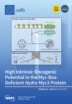
The characterization of a novel paralogous myc gene in Hydra, termed myc3, with conserved biochemical and oncogenic functions reveals a high complexity of the Myc network in this cnidarian. Based on its specific expression pattern, myc3 appears to function in nerve and gland cell differentiation, where its protein product could act as a dominant negative competitor of the stem-cell-specific Myc2 protein to enforce the committed state. In contrast to its highly divergent N‑terminus, the Myc3 C-terminus displays the best conservation grade compared to vertebrate Myc proteins, which may explain its strong intrinsic oncogenicity based on defined structural features. A comparison of different Myc isoforms in terms of structure and function can help to identify potentially druggable surfaces on Myc, which is an oncogenic driver in many human tumors. View this paper
  • Issues are regarded as officially published after their release is announced to the table of contents alert mailing list.
  • You may sign up for e-mail alerts to receive table of contents of newly released issues.
  • PDF is the official format for papers published in both, html and pdf forms. To view the papers in pdf format, click on the "PDF Full-text" link, and use the free Adobe Reader to open them.
Order results
Result details
Section
Select all
Export citation of selected articles as:
23 pages, 1393 KiB  
Review
The Role of Polo-Like Kinase 1 in Regulating the Forkhead Box Family Transcription Factors
by Xavier T. R. Moore, Lilia Gheghiani and Zheng Fu
Cells 2023, 12(9), 1344; https://doi.org/10.3390/cells12091344 - 08 May 2023
Cited by 2 | Viewed by 2385
Abstract
Polo-like kinase 1 (PLK1) is a serine/threonine kinase with more than 600 phosphorylation substrates through which it regulates many biological processes, including mitosis, apoptosis, metabolism, RNA processing, vesicle transport, and G2 DNA-damage checkpoint recovery, among others. Among the many PLK1 targets are [...] Read more.
Polo-like kinase 1 (PLK1) is a serine/threonine kinase with more than 600 phosphorylation substrates through which it regulates many biological processes, including mitosis, apoptosis, metabolism, RNA processing, vesicle transport, and G2 DNA-damage checkpoint recovery, among others. Among the many PLK1 targets are members of the FOX family of transcription factors (FOX TFs), including FOXM1, FOXO1, FOXO3, and FOXK1. FOXM1 and FOXK1 have critical oncogenic roles in cancer through their antagonism of apoptotic signals and their promotion of cell proliferation, metastasis, angiogenesis, and therapeutic resistance. In contrast, FOXO1 and FOXO3 have been identified to have broad functions in maintaining cellular homeostasis. In this review, we discuss PLK1-mediated regulation of FOX TFs, highlighting the effects of PLK1 on the activity and stability of these proteins. In addition, we review the prognostic and clinical significance of these proteins in human cancers and, more importantly, the different approaches that have been used to disrupt PLK1 and FOX TF-mediated signaling networks. Furthermore, we discuss the therapeutic potential of targeting PLK1-regulated FOX TFs in human cancers. Full article
(This article belongs to the Section Cell Proliferation and Division)
Show Figures

Figure 1

15 pages, 2984 KiB  
Article
Multiple and Consecutive Genome Editing Using i-GONAD and Breeding Enrichment Facilitates the Production of Genetically Modified Mice
by Carolina R. Melo-Silva, Cory J. Knudson, Lingjuan Tang, Samita Kafle, Lauren E. Springer, Jihae Choi, Christopher M. Snyder, Yajing Wang, Sangwon V. Kim and Luis J. Sigal
Cells 2023, 12(9), 1343; https://doi.org/10.3390/cells12091343 - 08 May 2023
Cited by 2 | Viewed by 1589
Abstract
Genetically modified (GM) mice are essential tools in biomedical research. Traditional methods for generating GM mice are expensive and require specialized personnel and equipment. The use of clustered regularly interspaced short palindromic repeats (CRISPR) coupled with improved-Genome editing via Oviductal Nucleic Acids Delivery [...] Read more.
Genetically modified (GM) mice are essential tools in biomedical research. Traditional methods for generating GM mice are expensive and require specialized personnel and equipment. The use of clustered regularly interspaced short palindromic repeats (CRISPR) coupled with improved-Genome editing via Oviductal Nucleic Acids Delivery (i-GONAD) has highly increased the feasibility of producing GM mice in research laboratories. However, genetic modification in inbred mouse strains of interest such as C57BL/6 (B6) is still challenging because of their low fertility and embryo fragility. We have successfully generated multiple novel GM mouse strains in the B6 background while attempting to optimize i-GONAD. We found that i-GONAD reduced the litter size in superovulated pregnant females but did not impact pregnancy rates. Natural mating or low-hormone dose did not increase the low fertility rate observed in superovulated B6 females. However, diet enrichment had a positive effect on pregnancy success. We also optimized breeding conditions to increase the survival of small litters by co-housing i-GONAD-treated pregnant B6 females with synchronized pregnant FVB/NJ companion mothers. Thus, GM mice generation was increased by an enriched diet and shared pup rearing with highly fertile females such as FVB/NJ. In the present study, we generated 16 GM mice using a CRISPR/Cas system to target individual and multiple loci simultaneously or consecutively. We also compared homology-directed repair efficiency using different methods for LoxP insertion for conditional knockout mouse production. We found that a two-step serial LoxP insertion, in which each LoxP sequence was inserted individually in different i-GONAD procedures, was a low-risk high-efficiency method for generating floxed mice. Full article
Show Figures

Graphical abstract

13 pages, 839 KiB  
Review
Retinal Cell Damage in Diabetic Retinopathy
by Jing Zhou and Bo Chen
Cells 2023, 12(9), 1342; https://doi.org/10.3390/cells12091342 - 08 May 2023
Cited by 13 | Viewed by 3146
Abstract
Diabetic retinopathy (DR), the most common microvascular complication that occurs in diabetes mellitus (DM), is the leading cause of vision loss in working-age adults. The prevalence of diabetic retinopathy is approximately 30% of the diabetic population and untreated DR can eventually cause blindness. [...] Read more.
Diabetic retinopathy (DR), the most common microvascular complication that occurs in diabetes mellitus (DM), is the leading cause of vision loss in working-age adults. The prevalence of diabetic retinopathy is approximately 30% of the diabetic population and untreated DR can eventually cause blindness. For decades, diabetic retinopathy was considered a microvascular complication and clinically staged by its vascular manifestations. In recent years, emerging evidence has shown that diabetic retinopathy causes early neuronal dysfunction and neurodegeneration that may precede vascular pathology and affect retinal neurons as well as glial cells. This knowledge leads to new therapeutic strategies aiming to prevent dysfunction of retinal neurons at the early stage of DR. Early detection and timely treatment to protect retinal neurons are critical to preventing visual loss in DR. This review provides an overview of DR and the structural and functional changes associated with DR, and discusses neuronal degeneration during diabetic retinopathy, the mechanisms underlying retinal neurodegeneration and microvascular complications, and perspectives on current and future clinic therapies. Full article
(This article belongs to the Special Issue Retinal Cell Biology in Health and Disease)
Show Figures

Figure 1

24 pages, 2569 KiB  
Review
Oxidative Stress and MicroRNAs in Endothelial Cells under Metabolic Disorders
by Morgan Minjares, Wendy Wu and Jie-Mei Wang
Cells 2023, 12(9), 1341; https://doi.org/10.3390/cells12091341 - 08 May 2023
Cited by 4 | Viewed by 2787
Abstract
Reactive oxygen species (ROS) are radical oxygen intermediates that serve as important second messengers in signal transduction. However, when the accumulation of these molecules exceeds the buffering capacity of antioxidant enzymes, oxidative stress and endothelial cell (EC) dysfunction occur. EC dysfunction shifts the [...] Read more.
Reactive oxygen species (ROS) are radical oxygen intermediates that serve as important second messengers in signal transduction. However, when the accumulation of these molecules exceeds the buffering capacity of antioxidant enzymes, oxidative stress and endothelial cell (EC) dysfunction occur. EC dysfunction shifts the vascular system into a pro-coagulative, proinflammatory state, thereby increasing the risk of developing cardiovascular (CV) diseases and metabolic disorders. Studies have turned to the investigation of microRNA treatment for CV risk factors, as these post-transcription regulators are known to co-regulate ROS. In this review, we will discuss ROS pathways and generation, normal endothelial cell physiology and ROS-induced dysfunction, and the current knowledge of common metabolic disorders and their connection to oxidative stress. Therapeutic strategies based on microRNAs in response to oxidative stress and microRNA’s regulatory roles in controlling ROS will also be explored. It is important to gain an in-depth comprehension of the mechanisms generating ROS and how manipulating these enzymatic byproducts can protect endothelial cell function from oxidative stress and prevent the development of vascular disorders. Full article
(This article belongs to the Special Issue The Role of Oxidative Stress in Cardiovascular Diseases)
Show Figures

Figure 1

12 pages, 2213 KiB  
Article
T Regulatory Cell-Associated Tolerance Induction by High-Dose Immunoglobulins in an HLA-Transgenic Mouse Model of Pemphigus
by Christoph Hudemann, Jochen Hoffmann, Enno Schmidt, Michael Hertl and Rüdiger Eming
Cells 2023, 12(9), 1340; https://doi.org/10.3390/cells12091340 - 08 May 2023
Viewed by 1760
Abstract
Pemphigus vulgaris (PV) is a potentially lethal autoimmune bullous skin disorder caused by IgG autoantibodies against desmoglein 3 (Dsg3) and Dsg1. During the last three decades, high-dose intravenous immunoglobulins (IVIgs) have been applied as an effective and relatively safe treatment regime in severe, [...] Read more.
Pemphigus vulgaris (PV) is a potentially lethal autoimmune bullous skin disorder caused by IgG autoantibodies against desmoglein 3 (Dsg3) and Dsg1. During the last three decades, high-dose intravenous immunoglobulins (IVIgs) have been applied as an effective and relatively safe treatment regime in severe, therapy-refractory PV. This prompted us to study T- and B- cell polarization by IVIg in a human-Dsg3-dependent mouse model for PV. Using humanized mice transgenic for HLA-DRB1*04:02, which is a highly prevalent haplotype in PV, we employed IVIg in two different experimental approaches: in prevention and quasi-therapeutic settings. Our data show that intraperitoneally applied IVIg was systemically distributed for up to 42 days or longer. IVIg-treated Dsg3-immunized mice exhibited, in contrast to Dsg3-immunized mice without IVIg, significantly less Dsg3-specific IgG, and showed induction of T regulatory cells in lymphatic tissue. Ex vivo splenocyte analysis upon Dsg3-specific stimulation revealed an initial, temporarily reduced antigen-induced cell proliferation, as well as IFN-γ secretion that became less apparent over the course of time. Marginal-zone B cells were initially reduced in the preventive approach but re-expanded over time. In contrast, in the quasi-therapeutic approach, a robust down-regulation in both spleen and lymph nodes was observed. We found a significant down-regulation of the immature transitional 1 (T1) B cells in IVIg-treated mice in the quasi-therapeutic approach, while T2 and T3, representing a healthy stage of B-cell development, appeared to be up-regulated by IVIg. In summary, in two experimental settings employing an active PV mouse model, we demonstrate distinct alterations of T- and B-cell populations upon IVIg treatment, compatible with a tolerance-associated polarization in lymphatic tissue. Our data suggest that the clinical efficacy of IVIg is at least modulated by distinct alterations of T- and B-cell populations compatible with a tolerance-associated polarization in lymphatic tissue. Full article
(This article belongs to the Special Issue Autoimmune Diseases: Molecular Mechanisms and Therapies)
Show Figures

Graphical abstract

16 pages, 1468 KiB  
Review
Identification of Exo-miRNAs: A Summary of the Efforts in Translational Studies Involving Triple-Negative Breast Cancer
by Jarline Encarnación-Medina, Lenin Godoy, Jaime Matta and Carmen Ortiz-Sánchez
Cells 2023, 12(9), 1339; https://doi.org/10.3390/cells12091339 - 07 May 2023
Cited by 2 | Viewed by 1775
Abstract
Triple-negative breast cancer (TNBC) accounts for about 10–15% of all breast cancers (BC) in the US and its diagnosis is associated with poor survival outcomes. A better understanding of the disease etiology is crucial to identify target treatment options to improve patient outcomes. [...] Read more.
Triple-negative breast cancer (TNBC) accounts for about 10–15% of all breast cancers (BC) in the US and its diagnosis is associated with poor survival outcomes. A better understanding of the disease etiology is crucial to identify target treatment options to improve patient outcomes. The role of exo-miRNAs in TNBC has been studied for more than two decades. Although some studies have identified exo-miR candidates in TNBC using clinical samples, consensus regarding exo-miR candidates has not been achieved. The purpose of this review is to gather information regarding exo-miR candidates reported in TNBC translational studies along with the techniques used to isolate and validate the potential targets. The techniques suggested in this review are based on the use of commercially available materials for research and clinical laboratories. We expect that the information included in this review can add additional value to the recent efforts in the development of a liquid biopsy to identify TNBC cases and further improve their survival outcomes. Full article
Show Figures

Figure 1

17 pages, 3008 KiB  
Article
The E1a Adenoviral Gene Upregulates the Yamanaka Factors to Induce Partial Cellular Reprogramming
by Gracia Mendoza, Rebeca González-Pastor, Juan Miguel Sánchez, Altamira Arce-Cerezo, Miguel Quintanilla, Gema Moreno-Bueno, Anna Pujol, Carolina Belmar-López, Alba de Martino, Efrén Riu, Tristan A. Rodriguez and Pilar Martin-Duque
Cells 2023, 12(9), 1338; https://doi.org/10.3390/cells12091338 - 07 May 2023
Viewed by 2198
Abstract
The induction of pluripotency by enforced expression of different sets of genes in somatic cells has been achieved with reprogramming technologies first described by Yamanaka’s group. Methodologies for generating induced pluripotent stem cells are as varied as the combinations of genes used. It [...] Read more.
The induction of pluripotency by enforced expression of different sets of genes in somatic cells has been achieved with reprogramming technologies first described by Yamanaka’s group. Methodologies for generating induced pluripotent stem cells are as varied as the combinations of genes used. It has previously been reported that the adenoviral E1a gene can induce the expression of two of the Yamanaka factors (c-Myc and Oct-4) and epigenetic changes. Here, we demonstrate that the E1a-12S over-expression is sufficient to induce pluripotent-like characteristics closely to epiblast stem cells in mouse embryonic fibroblasts through the activation of the pluripotency gene regulatory network. These findings provide not only empirical evidence that the expression of one single factor is sufficient for partial reprogramming but also a potential mechanistic explanation for how viral infection could lead to neoplasia if they are surrounded by the appropriate environment or the right medium, as happens with the tumorogenic niche. Full article
Show Figures

Figure 1

18 pages, 3264 KiB  
Article
Functional Characterisation of the Circular RNA, circHTT(2-6), in Huntington’s Disease
by Laura Gantley, Brett W. Stringer, Vanessa M. Conn, Youichirou Ootsuka, Duncan Holds, Mark Slee, Kamelya Aliakbari, Kirsty Kirk, Rebecca J. Ormsby, Stuart T. Webb, Adrienne Hanson, He Lin, Luke A. Selth and Simon J. Conn
Cells 2023, 12(9), 1337; https://doi.org/10.3390/cells12091337 - 07 May 2023
Cited by 1 | Viewed by 2142
Abstract
Trinucleotide repeat disorders comprise ~20 severe, inherited, human neuromuscular and neurodegenerative disorders, which result from an abnormal expansion of repetitive sequences in the DNA. The most common of these, Huntington’s disease (HD), results from expansion of the CAG repeat region in exon 1 [...] Read more.
Trinucleotide repeat disorders comprise ~20 severe, inherited, human neuromuscular and neurodegenerative disorders, which result from an abnormal expansion of repetitive sequences in the DNA. The most common of these, Huntington’s disease (HD), results from expansion of the CAG repeat region in exon 1 of the HTT gene via an unknown mechanism. Since non-coding RNAs have been implicated in the initiation and progression of many diseases, herein we focused on a circular RNA (circRNA) molecule arising from non-canonical splicing (backsplicing) of HTT pre-mRNA. The most abundant circRNA from HTT, circHTT(2-6), was found to be more highly expressed in the frontal cortex of HD patients, compared with healthy controls, and positively correlated with CAG repeat tract length. Furthermore, the mouse orthologue (mmu_circHTT(2-6)) was found to be enriched within the brain and specifically the striatum, a region enriched for medium spiny neurons that are preferentially lost in HD. Transgenic overexpression of circHTT(2-6) in two human cell lines—SH-SY5Y and HEK293—reduced cell proliferation and nuclear size without affecting cell cycle progression or cellular size, or altering the CAG repeat region length within HTT. CircHTT(2-6) overexpression did not alter total HTT protein levels, but reduced its nuclear localisation. As these phenotypic and genotypic changes resemble those observed in HD patients, our results suggest that circHTT(2-6) may play a functional role in the pathophysiology of this disease. Full article
(This article belongs to the Special Issue Non-coding RNAs and Neurological Diseases 2022)
Show Figures

Graphical abstract

24 pages, 4416 KiB  
Article
The Pesticide Chlordecone Promotes Parkinsonism-like Neurodegeneration with Tau Lesions in Midbrain Cultures and C. elegans Worms
by Valeria Parrales-Macias, Patrick P. Michel, Aurore Tourville, Rita Raisman-Vozari, Stéphane Haïk, Stéphane Hunot, Nicolas Bizat and Annie Lannuzel
Cells 2023, 12(9), 1336; https://doi.org/10.3390/cells12091336 - 07 May 2023
Cited by 1 | Viewed by 2504
Abstract
Chlordecone (CLD) is an organochlorine pesticide (OCP) that is currently banned but still contaminates ecosystems in the French Caribbean. Because OCPs are known to increase the risk of Parkinson’s disease (PD), we tested whether chronic low-level intoxication with CLD could reproduce certain key [...] Read more.
Chlordecone (CLD) is an organochlorine pesticide (OCP) that is currently banned but still contaminates ecosystems in the French Caribbean. Because OCPs are known to increase the risk of Parkinson’s disease (PD), we tested whether chronic low-level intoxication with CLD could reproduce certain key characteristics of Parkinsonism-like neurodegeneration. For that, we used culture systems of mouse midbrain dopamine (DA) neurons and glial cells, together with the nematode C. elegans as an in vivo model organism. We established that CLD kills cultured DA neurons in a concentration- and time-dependent manner while exerting no direct proinflammatory effects on glial cells. DA cell loss was not impacted by the degree of maturation of the culture. The use of fluorogenic probes revealed that CLD neurotoxicity was the consequence of oxidative stress-mediated insults and mitochondrial disturbances. In C. elegans worms, CLD exposure caused a progressive loss of DA neurons associated with locomotor deficits secondary to alterations in food perception. L-DOPA, a molecule used for PD treatment, corrected these deficits. Cholinergic and serotoninergic neuronal cells were also affected by CLD in C. elegans, although to a lesser extent than DA neurons. Noticeably, CLD also promoted the phosphorylation of the aggregation-prone protein tau (but not of α-synuclein) both in midbrain cell cultures and in a transgenic C. elegans strain expressing a human form of tau in neurons. In summary, our data suggest that CLD is more likely to promote atypical forms of Parkinsonism characterized by tau pathology than classical synucleinopathy-associated PD. Full article
(This article belongs to the Collection Advances in Neurodegenerative Disease)
Show Figures

Graphical abstract

39 pages, 19619 KiB  
Article
Centrosome Formation in the Bovine Early Embryo
by Rustem Uzbekov, Galina N. Singina, Ekaterina N. Shedova, Charles Banliat, Tomer Avidor-Reiss and Svetlana Uzbekova
Cells 2023, 12(9), 1335; https://doi.org/10.3390/cells12091335 - 07 May 2023
Cited by 8 | Viewed by 1973
Abstract
Centrosome formation during early development in mice and rats occurs due to the appearance of centrioles de novo. In contrast, in humans and other non-rodent mammals, centrioles are thought to be derived from spermatozoa. Ultrastructural study of zygotes and early embryos of cattle [...] Read more.
Centrosome formation during early development in mice and rats occurs due to the appearance of centrioles de novo. In contrast, in humans and other non-rodent mammals, centrioles are thought to be derived from spermatozoa. Ultrastructural study of zygotes and early embryos of cattle at full series of ultrathin sections show that the proximal centriole of the spermatozoon disappears by the end of the first cleavage division. Centrioles appear in two to four cell embryos in fertilized oocytes and in parthenogenetic embryos. Centriole formation includes the appearance of atypical centrioles with randomly arranged triplets and centrioles with microtubule triplets of various lengths. After the third cleavage, four centriolar cylinders appear for the first time in the blastomeres while each embryo still has two atypical centrioles. Our results showed that the mechanisms of centriole formation in different groups of mammals are universal, differing only in the stage of development in which they occur. Full article
(This article belongs to the Special Issue The Regulation of the Cell Cycle II)
Show Figures

Figure 1

4 pages, 209 KiB  
Editorial
Role of Autophagy in Ehrlichia-Induced Liver Injury
by Aditya Kumar Sharma and Nahed Ismail
Cells 2023, 12(9), 1334; https://doi.org/10.3390/cells12091334 - 07 May 2023
Cited by 1 | Viewed by 1104
Abstract
Autophagy is a cellular process that involves the cell breakdown and recycling of cellular components, such as old, damaged, or abnormal proteins, for important cellular functions including development, immune function, stress, and starvation [...] Full article
(This article belongs to the Special Issue Autophagy in the Liver)
12 pages, 1466 KiB  
Article
Ratio of Urinary Proteins to Albumin Excretion Shifts Substantially during Progression of the Podocytopathy Alport Syndrome, and Spot Urine Is a Reliable Method to Detect These Pathologic Changes
by Jan Boeckhaus, Lea Mohr, Hassan Dihazi, Burkhard Tönshoff, Lutz T. Weber, Lars Pape, Kay Latta, Henry Fehrenbach, Baerbel Lange-Sperandio, Matthias Kettwig, Hagen Staude, Sabine König, Ulrike John-Kroegel, Jutta Gellermann, Bernd Hoppe, Matthias Galiano, Dieter Haffner, Heidrun Rhode and Oliver Gross
Cells 2023, 12(9), 1333; https://doi.org/10.3390/cells12091333 - 07 May 2023
Cited by 1 | Viewed by 1574
Abstract
The urinary albumin- and protein-to-creatinine ratios (UACR and UPCR, respectively) are key endpoints in most clinical trials assessing risk of progression of chronic kidney disease (CKD). For the first time, the current study compares the UACR versus the UPCR head-to-head at early stages [...] Read more.
The urinary albumin- and protein-to-creatinine ratios (UACR and UPCR, respectively) are key endpoints in most clinical trials assessing risk of progression of chronic kidney disease (CKD). For the first time, the current study compares the UACR versus the UPCR head-to-head at early stages of CKD, taking use of the hereditary podocytopathy Alport syndrome (AS) as a model disease for any CKD. Urine samples originated from the prospective randomized, controlled EARLY PRO-TECT Alport trial (NCT01485978). Urine samples from 47 children with confirmed diagnoses of AS at very early stages of CKD were divided according to the current stage of AS: stage 0 (UACR < 30 mg/g), stage 1 (30–300 mg/g) or stage 2 (>300 mg/g). The range of estimated glomerular filtration rate was 75–187.6 mL/min. The mean age was 10.4 ± 4.5 years. In children at stage 0, proteinuria in spot urine, confirmed in 24 h urine, was almost ten times higher than albuminuria (106.4 ± 42.2 vs. 12.5 ± 9.7; p < 0.05); it was “only” about three times higher in stage 1 (328.5 ± 210.1 vs. 132.3 ± 80.5; p < 0.05) and almost equal in stage 2 (1481.9 ± 983.4 vs. 1109.7 ± 873.6; p = 0.36). In 17 children, UACRs and UPCRs were measured simultaneously in 24 h urine and spot urine in the same study visit. Interestingly, the UACR (and UPCR) in 24 h urine vs. in spot urine varied by less than 10% (266.8 ± 426.4 vs. 291.2 ± 530.2). In conclusion, our study provides the first evidence that in patients with normal glomerular filtration rate (GFR) and low amounts of albuminuria, especially in children with podocytopathies such as AS, measuring the UACR and UPCR in spot urine is a reliable and convenient alternative to 24 h urine collection. Our study advocates both the UACR and the UPCR as relevant diagnostic biomarkers in future clinical trials in children with glomerular diseases because the UPCR seems to be a very significant parameter at very early stages of podocytopathies. The German Federal Ministry of Education and Research funded this trial (01KG1104). Full article
(This article belongs to the Special Issue New Aspects for Understanding Podocytopathies II)
Show Figures

Figure 1

22 pages, 4014 KiB  
Article
Novel Therapeutic Target Critical for SARS-CoV-2 Infectivity and Induction of the Cytokine Release Syndrome
by William W. Harless, Beth Lewis, Bessi Qorri, Samar Abdulkhalek and Myron R. Szewczuk
Cells 2023, 12(9), 1332; https://doi.org/10.3390/cells12091332 - 07 May 2023
Cited by 1 | Viewed by 1739
Abstract
We discovered a novel therapeutic target critical for SARS-CoV-2, cellular infectivity and the induction of the cytokine release syndrome. Here, we show that the mammalian enzyme neuraminidase-1 (Neu-1) is part of a highly conserved signaling platform that regulates the dimerization and activation of [...] Read more.
We discovered a novel therapeutic target critical for SARS-CoV-2, cellular infectivity and the induction of the cytokine release syndrome. Here, we show that the mammalian enzyme neuraminidase-1 (Neu-1) is part of a highly conserved signaling platform that regulates the dimerization and activation of the ACE2 receptors and the Toll-like receptors (TLRs) implicated in the cytokine release syndrome (CRS). Activated Neu-1 cleaves glycosylated residues that provide a steric hindrance to both ACE2 and TLR dimerization, a process critical to both viral attachment to the receptor and entry into the cell and TLR activation. Blocking Neu-1 inhibited ACE2 receptor dimerization and internalization, TLR dimerization and activation, and the expression of several key inflammatory molecules implicated in the CRS and death from ARDS. Treatments that target Neu-1 are predicted to be highly effective against infection with SARS-CoV-2, given the central role played by this enzyme in viral cellular entry and the induction of the CRS. Full article
(This article belongs to the Special Issue Angiotensin-Converting Enzyme 2 in Health and Diseases)
Show Figures

Figure 1

26 pages, 6301 KiB  
Article
“Combo” Multi-Target Pharmacological Therapy and New Formulations to Reduce Inflammation and Improve Endogenous Remyelination in Traumatic Spinal Cord Injury
by Marzia Moretti, Riccardo Caraffi, Luca Lorenzini, Ilaria Ottonelli, Michele Sannia, Giuseppe Alastra, Vito Antonio Baldassarro, Alessandro Giuliani, Jason Thomas Duskey, Maura Cescatti, Barbara Ruozi, Luigi Aloe, Maria Angela Vandelli, Luciana Giardino, Giovanni Tosi and Laura Calzà
Cells 2023, 12(9), 1331; https://doi.org/10.3390/cells12091331 - 06 May 2023
Cited by 2 | Viewed by 2288
Abstract
Spinal cord injury (SCI) is characterized by a cascade of events that lead to sensory and motor disabilities. To date, this condition is irreversible, and no cure exists. To improve myelin repair and limit secondary degeneration, we developed a multitherapy based on nanomedicines [...] Read more.
Spinal cord injury (SCI) is characterized by a cascade of events that lead to sensory and motor disabilities. To date, this condition is irreversible, and no cure exists. To improve myelin repair and limit secondary degeneration, we developed a multitherapy based on nanomedicines (NMeds) loaded with the promyelinating agent triiodothyronine (T3), used in combination with systemic ibuprofen and mouse nerve growth factor (mNGF). Poly-L-lactic-co-glycolic acid (PLGA) NMeds were optimized and loaded with T3 to promote sustained release. In vitro experiments confirmed the efficacy of T3-NMeds to differentiate oligodendrocyte precursor cells. In vivo rat experiments were performed in contusion SCI to explore the NMed biodistribution and efficacy of combo drugs at short- and long-term post-lesion. A strong anti-inflammatory effect was observed in the short term with a reduction of type M1 microglia and glutamate levels, but with a subsequent increase of TREM2. In the long term, an improvement of myelination in NG2-IR, an increase in MBP content, and a reduction of the demyelination area were observed. These data demonstrated that NMeds can successfully be used to obtain more controlled local drug delivery and that this multiple treatment could be effective in improving the outcome of SCIs. Full article
(This article belongs to the Collection Cell Biology of Spinal Cord Injury and Repair)
Show Figures

Figure 1

22 pages, 2625 KiB  
Article
Randomized Clinical Trial of Antioxidant Therapy Patients with Septic Shock and Organ Dysfunction in the ICU: SOFA Score Reduction by Improvement of the Enzymatic and Non-Enzymatic Antioxidant System
by Alfredo Aisa-Álvarez, Israel Pérez-Torres, Verónica Guarner-Lans, Linaloe Manzano-Pech, Randall Cruz-Soto, Ricardo Márquez-Velasco, Sergio Casarez-Alvarado, Juvenal Franco-Granillo, Marcela Elizabeth Núñez-Martínez and María Elena Soto
Cells 2023, 12(9), 1330; https://doi.org/10.3390/cells12091330 - 06 May 2023
Cited by 3 | Viewed by 2228
Abstract
Background and aim: Here, we assess the effect of adjuvant antioxidant therapies in septic shock patients with organ dysfunction and their effect on the enzymatic and non-enzymatic antioxidant systems. Methods: Randomized clinical trial run between 2018 and 2022. One hundred and thirty-one patients [...] Read more.
Background and aim: Here, we assess the effect of adjuvant antioxidant therapies in septic shock patients with organ dysfunction and their effect on the enzymatic and non-enzymatic antioxidant systems. Methods: Randomized clinical trial run between 2018 and 2022. One hundred and thirty-one patients with septic shock were included in five groups with 25, 27, 24, 26 and 29 patients each. Group 1 received vitamin C (Vit C), Group 2 vitamin E (Vit E), Group 3 n-acetylcysteine (NAC), Group 4 melatonin (MT) and group 5 no treatment. All antioxidants were administered orally or through a nasogastric tube for 5 days as an adjuvant to standard therapy. Results: All patients had multiple organ failure (MOF) and low Vit C levels. Vit C therapy decreased CRP, PCT and NO3/NO2 but increased Vit C levels. The SOFA score decreased with MT in 75%, Vit C 63% and NAC 50% vs. controls 33% (p = 0.0001, p = 0.03 and p = 0.001 respectively). MT diminished lipid peroxidation (LPO) (p = 0.01) and improved total antioxidant capacity (TAC) (p = 0.04). Vit E increased thiol levels (p = 0.02) and tended to decrease LPO (p = 0.06). Selenium levels were decreased in the control group (p = 0.04). Conclusions: Antioxidants used as an adjuvant therapy in the standard treatment of septic shock decrease MOF and oxidative stress markers. They increase the TAC and thiols, and maintain selenium levels. Full article
(This article belongs to the Special Issue Immunopathogenesis of Bacterial Infection)
Show Figures

Figure 1

13 pages, 996 KiB  
Opinion
Cellular and Mitochondrial NAD Homeostasis in Health and Disease
by Jaylyn Waddell, Rehana Khatoon and Tibor Kristian
Cells 2023, 12(9), 1329; https://doi.org/10.3390/cells12091329 - 06 May 2023
Cited by 3 | Viewed by 2760
Abstract
The mitochondrion has a unique position among other cellular organelles due to its dynamic properties and symbiotic nature, which is reflected in an active exchange of metabolites and cofactors between the rest of the intracellular compartments. The mitochondrial energy metabolism is greatly dependent [...] Read more.
The mitochondrion has a unique position among other cellular organelles due to its dynamic properties and symbiotic nature, which is reflected in an active exchange of metabolites and cofactors between the rest of the intracellular compartments. The mitochondrial energy metabolism is greatly dependent on nicotinamide adenine dinucleotide (NAD) as a cofactor that is essential for both the activity of respiratory and TCA cycle enzymes. The NAD level is determined by the rate of NAD synthesis, the activity of NAD-consuming enzymes, and the exchange rate between the individual subcellular compartments. In this review, we discuss the NAD synthesis pathways, the NAD degradation enzymes, and NAD subcellular localization, as well as NAD transport mechanisms with a focus on mitochondria. Finally, the effect of the pathologic depletion of mitochondrial NAD pools on mitochondrial proteins’ post-translational modifications and its role in neurodegeneration will be reviewed. Understanding the physiological constraints and mechanisms of NAD maintenance and the exchange between subcellular compartments is critical given NAD’s broad effects and roles in health and disease. Full article
(This article belongs to the Special Issue Mitochondria at the Crossroad of Health and Disease)
Show Figures

Figure 1

16 pages, 5067 KiB  
Article
Validation and Functional Analysis of Reference and Tissue-Specific Genes in Adipose Tissue of Freshwater Drum, Aplodinotus grunniens, under Starvation and Hypothermia Stress
by Miaomiao Xue, Haibo Wen, Pao Xu, Jianxiang Chen, Qingyong Wang, Yongkai Tang, Xueyan Ma, Guohua Lv, Hongxia Li and Changyou Song
Cells 2023, 12(9), 1328; https://doi.org/10.3390/cells12091328 - 06 May 2023
Cited by 1 | Viewed by 1914
Abstract
Adipose tissue is critical to the growth, development, and physiological health of animals. Reference genes play an essential role in normalizing the expression of mRNAs. Tissue-specific genes are preferred for their function and expression in specific tissues or cell types. Identification of these [...] Read more.
Adipose tissue is critical to the growth, development, and physiological health of animals. Reference genes play an essential role in normalizing the expression of mRNAs. Tissue-specific genes are preferred for their function and expression in specific tissues or cell types. Identification of these genes contributes to understanding the tissue–gene relationship and the etiology and discovery of new tissue-specific targets. Therefore, reference genes and tissue-specific genes in the adipose tissue of Aplodinotus grunniens were identified to explore their function under exogenous starvation (1 d, 2 w, 6 w) and hypothermic stress (18 °C and 10 °C for 2 d and 8 d) in this study. Results suggest that 60SRP was the most stable reference gene in adipose tissue. Meanwhile, eight genes were validated as tissue-specific candidates from the high-throughput sequencing database, while seven of them (ADM2, β2GP1, CAMK1G, CIDE3, FAM213A, HSL, KRT222, and NCEH1) were confirmed in adipose tissue. Additionally, these seven tissue-specific genes were active in response to starvation and hypothermic stress in a time- or temperature-dependent manner. These results demonstrate that adipose-specific genes can be identified using stable internal reference genes, thereby identifying specific important functions under starvation and hypothermic stress, which provides tissue-specific targets for adipose regulation in A. grunniens. Full article
(This article belongs to the Special Issue The Molecular and Cellular Basis for Fish Health)
Show Figures

Figure 1

14 pages, 4694 KiB  
Article
Embryonic Ethanol but Not Cannabinoid Exposure Affects Zebrafish Cardiac Development via Agrin and Sonic Hedgehog Interaction
by Chengjin Zhang, Natalie Ezem, Shanta Mackinnon and Gregory J. Cole
Cells 2023, 12(9), 1327; https://doi.org/10.3390/cells12091327 - 06 May 2023
Viewed by 2037
Abstract
Recent studies demonstrate the adverse effects of cannabinoids on development, including via pathways shared with ethanol exposure. Our laboratory has shown that both the nervous system and cardiac development are dependent on agrin modulation of sonic hedgehog (shh) and fibroblast growth factor (Fgf) [...] Read more.
Recent studies demonstrate the adverse effects of cannabinoids on development, including via pathways shared with ethanol exposure. Our laboratory has shown that both the nervous system and cardiac development are dependent on agrin modulation of sonic hedgehog (shh) and fibroblast growth factor (Fgf) signaling pathways. As both ethanol and cannabinoids impact these signaling molecules, we examined their role on zebrafish heart development. Zebrafish embryos were exposed to a range of ethanol and/or cannabinoid receptor 1 and 2 agonist concentrations in the absence or presence of morpholino oligonucleotides that disrupt agrin or shh expression. In situ hybridization was employed to analyze cardiac marker gene expression. Exposure to cannabinoid receptor agonists disrupted midbrain–hindbrain boundary development, but had no effect on heart development, as assessed by the presence of cardiac edema or the altered expression of cardiac marker genes. In contrast, exposure to 1.5% ethanol induced cardiac edema and the altered expression of cardiac marker genes. Combined exposure to agrin or shh morpholino and 0.5% ethanol disrupted the cmlc2 gene expression pattern, with the restoration of the normal expression following shh mRNA overexpression. These studies provide evidence that signaling pathways critical to heart development are sensitive to ethanol exposure, but not cannabinoids, during early zebrafish embryogenesis. Full article
(This article belongs to the Special Issue Modeling Developmental Processes and Disorders in Zebrafish)
Show Figures

Figure 1

26 pages, 1414 KiB  
Review
The Role of Eosinophil-Derived Neurotoxin and Vascular Endothelial Growth Factor in the Pathogenesis of Eosinophilic Asthma
by Maciej Tota, Julia Łacwik, Julia Laska, Łukasz Sędek and Krzysztof Gomułka
Cells 2023, 12(9), 1326; https://doi.org/10.3390/cells12091326 - 06 May 2023
Cited by 2 | Viewed by 3226
Abstract
Asthma is a chronic complex pulmonary disease characterized by airway inflammation, remodeling, and hyperresponsiveness. Vascular endothelial growth factor (VEGF) and eosinophil-derived neurotoxin (EDN) are two significant mediators involved in the pathophysiology of asthma. In asthma, VEGF and EDN levels are elevated and correlate [...] Read more.
Asthma is a chronic complex pulmonary disease characterized by airway inflammation, remodeling, and hyperresponsiveness. Vascular endothelial growth factor (VEGF) and eosinophil-derived neurotoxin (EDN) are two significant mediators involved in the pathophysiology of asthma. In asthma, VEGF and EDN levels are elevated and correlate with disease severity and airway hyperresponsiveness. Diversity in VEGF polymorphisms results in the variability of responses to glucocorticosteroids and leukotriene antagonist treatment. Targeting VEGF and eosinophils is a promising therapeutic approach for asthma. We identified lichochalcone A, bevacizumab, azithromycin (AZT), vitamin D, diosmetin, epigallocatechin gallate, IGFBP-3, Neovastat (AE-941), endostatin, PEDF, and melatonin as putative add-on drugs in asthma with anti-VEGF properties. Further studies and clinical trials are needed to evaluate the efficacy of those drugs. AZT reduces the exacerbation rate and may be considered in adults with persistent symptomatic asthma. However, the long-term effects of AZT on community microbial resistance require further investigation. Vitamin D supplementation may enhance corticosteroid responsiveness. Herein, anti-eosinophil drugs are reviewed. Among them are, e.g., anti-IL-5 (mepolizumab, reslizumab, and benralizumab), anti-IL-13 (lebrikizumab and tralokinumab), anti-IL-4 and anti-IL-13 (dupilumab), and anti-IgE (omalizumab) drugs. EDN over peripheral blood eosinophil count is recommended to monitor the asthma control status and to assess the efficacy of anti-IL-5 therapy in asthma. Full article
(This article belongs to the Special Issue Asthma: Pathogenesis, Diagnosis, and Treatment)
Show Figures

Figure 1

19 pages, 2894 KiB  
Article
Protein Networks Associated with Native Metabotropic Glutamate 1 Receptors (mGlu1) in the Mouse Cerebellum
by Mahnaz Mansouri, Leopold Kremser, Thanh-Phuong Nguyen, Yu Kasugai, Laura Caberlotto, Martin Gassmann, Bettina Sarg, Herbert Lindner, Bernhard Bettler, Lucia Carboni and Francesco Ferraguti
Cells 2023, 12(9), 1325; https://doi.org/10.3390/cells12091325 - 05 May 2023
Viewed by 1842
Abstract
The metabotropic glutamate receptor 1 (mGlu1) plays a pivotal role in synaptic transmission and neuronal plasticity. Despite the fact that several interacting proteins involved in the mGlu1 subcellular trafficking and intracellular transduction mechanisms have been identified, the protein network associated [...] Read more.
The metabotropic glutamate receptor 1 (mGlu1) plays a pivotal role in synaptic transmission and neuronal plasticity. Despite the fact that several interacting proteins involved in the mGlu1 subcellular trafficking and intracellular transduction mechanisms have been identified, the protein network associated with this receptor in specific brain areas remains largely unknown. To identify novel mGlu1-associated protein complexes in the mouse cerebellum, we used an unbiased tissue-specific proteomic approach, namely co-immunoprecipitation followed by liquid chromatography/tandem mass spectrometry analysis. Many well-known protein complexes as well as novel interactors were identified, including G-proteins, Homer, δ2 glutamate receptor, 14-3-3 proteins, and Na/K-ATPases. A novel putative interactor, KCTD12, was further investigated. Reverse co-immunoprecipitation with anti-KCTD12 antibodies revealed mGlu1 in wild-type but not in KCTD12-knock-out homogenates. Freeze-fracture replica immunogold labeling co-localization experiments showed that KCTD12 and mGlu1 are present in the same nanodomain in Purkinje cell spines, although at a distance that suggests that this interaction is mediated through interposed proteins. Consistently, mGlu1 could not be co-immunoprecipitated with KCTD12 from a recombinant mammalian cell line co-expressing the two proteins. The possibility that this interaction was mediated via GABAB receptors was excluded by showing that mGlu1 and KCTD12 still co-immunoprecipitated from GABAB receptor knock-out tissue. In conclusion, this study identifies tissue-specific mGlu1-associated protein clusters including KCTD12 at Purkinje cell synapses. Full article
(This article belongs to the Special Issue The Role of Metabotropic Glutamate Receptors in Health and Disease)
Show Figures

Figure 1

17 pages, 1038 KiB  
Review
Pericyte–Glioblastoma Cell Interaction: A Key Target to Prevent Glioblastoma Progression
by Ana Pombero, Raquel Garcia-Lopez and Salvador Martínez
Cells 2023, 12(9), 1324; https://doi.org/10.3390/cells12091324 - 05 May 2023
Cited by 3 | Viewed by 2075
Abstract
Multiple biological processes rely on direct intercellular interactions to regulate cell proliferation and migration in embryonic development and cancer processes. Tumor development and growth depends on close interactions between cancer cells and cells in the tumor microenvironment. During embryonic development, morphogenetic signals and [...] Read more.
Multiple biological processes rely on direct intercellular interactions to regulate cell proliferation and migration in embryonic development and cancer processes. Tumor development and growth depends on close interactions between cancer cells and cells in the tumor microenvironment. During embryonic development, morphogenetic signals and direct cell contacts control cell proliferation, polarity, and morphogenesis. Cancer cells communicate with cells in the tumor niche through molecular signals and intercellular contacts, thereby modifying the vascular architecture and antitumor surveillance processes and consequently enabling tumor growth and survival. While looking for cell-to-cell signaling mechanisms that are common to both brain development and cancer progression, we have studied the infiltration process in glioblastoma multiforme (GBM), which is the most malignant primary brain tumor and with the worst prognosis. Cell-to-cell contacts, by means of filopodia-like structures, between GBM cells and brain pericytes (PCs) are necessary for adequate cell signaling during cancer infiltration; similarly, contacts between embryonic regions, via cytonemes, are required for embryo regionalization and development. This GBM–PC interaction provokes two important changes in the physiological function of these perivascular cells, namely, (i) vascular co-option with changes in cell contractility and vascular malformation, and (ii) changes in the PC transcriptome, modifying the microvesicles and protein secretome, which leads to the development of an immunosuppressive phenotype that promotes tumor immune tolerance. Moreover, the GTPase Cdc42 regulates cell polarity across organisms, from yeast to humans, playing a central role in GBM cell–PC interaction and maintaining vascular co-option. As such, a review of the molecular and cellular mechanisms underlying the development and maintenance of the physical interactions between cancer cells and PCs is of particular interest. Full article
Show Figures

Figure 1

20 pages, 7226 KiB  
Article
New Insights into the Impact of Human Papillomavirus on Oral Cancer in Young Patients: Proteomic Approach Reveals a Novel Role for S100A8
by Marisol Miranda-Galvis, Carolina Carneiro Soares, Carolina Moretto Carnielli, Jaqueline Ramalho Buttura, Raisa Sales de Sá, Estela Kaminagakura, Fabio Albuquerque Marchi, Adriana Franco Paes Leme, Clóvis A. Lópes Pinto, Alan Roger Santos-Silva, Rogerio Moraes Castilho, Luiz Paulo Kowalski and Cristiane Helena Squarize
Cells 2023, 12(9), 1323; https://doi.org/10.3390/cells12091323 - 05 May 2023
Cited by 1 | Viewed by 1890
Abstract
Human papillomavirus (HPV) infection has recently been linked to a subset of cancers affecting the oral cavity. However, the molecular mechanisms underlying HPV-driven oral squamous cell carcinoma (OSCC) onset and progression are poorly understood. Methods: We performed MS-based proteomics profiling based on HPV [...] Read more.
Human papillomavirus (HPV) infection has recently been linked to a subset of cancers affecting the oral cavity. However, the molecular mechanisms underlying HPV-driven oral squamous cell carcinoma (OSCC) onset and progression are poorly understood. Methods: We performed MS-based proteomics profiling based on HPV status in OSCC in young patients, following biological characterization and cell assays to explore the proteome functional landscape. Results: Thirty-nine proteins are differentially abundant between HPV (+) and HPV (−) OSCC. Among them, COPS3, DYHC1, and S100A8 are unfavorable for tumor recurrence and survival, in contrast to A2M and Serpine1, low levels of which show an association with better DFS. Remarkably, S100A8 is considered an independent prognostic factor for lower survival rates, and at high levels, it alters tumor-associated immune profiling, showing a lower proportion of M1 macrophages and dendritic cells. HPV (+) OSCC also displayed the pathogen-associated patterns receptor that, when activated, triggered the S100A8 and NFκB inflammatory responses. Conclusion: HPV (+) OSCC has a peculiar microenvironment pattern distinctive from HPV (−), involving the expression of pathogen-associated pattern receptors, S100A8 overexpression, and NFκB activation and responses, which has important consequences in prognosis and may guide therapeutic decisions. Full article
(This article belongs to the Collection Tumor Metabolism and Therapy)
Show Figures

Figure 1

34 pages, 6609 KiB  
Review
Role of Macroautophagy in Mammalian Male Reproductive Physiology
by Doaa Kirat, Ahmed Mohamed Alahwany, Ahmed Hamed Arisha, Adel Abdelkhalek and Taku Miyasho
Cells 2023, 12(9), 1322; https://doi.org/10.3390/cells12091322 - 05 May 2023
Cited by 1 | Viewed by 3196
Abstract
Physiologically, autophagy is an evolutionarily conserved and self-degradative process in cells. Autophagy carries out normal physiological roles throughout mammalian life. Accumulating evidence shows autophagy as a mechanism for cellular growth, development, differentiation, survival, and homeostasis. In male reproductive systems, normal spermatogenesis and steroidogenesis [...] Read more.
Physiologically, autophagy is an evolutionarily conserved and self-degradative process in cells. Autophagy carries out normal physiological roles throughout mammalian life. Accumulating evidence shows autophagy as a mechanism for cellular growth, development, differentiation, survival, and homeostasis. In male reproductive systems, normal spermatogenesis and steroidogenesis need a balance between degradation and energy supply to preserve cellular metabolic homeostasis. The main process of autophagy includes the formation and maturation of the phagophore, autophagosome, and autolysosome. Autophagy is controlled by a group of autophagy-related genes that form the core machinery of autophagy. Three types of autophagy mechanisms have been discovered in mammalian cells: macroautophagy, microautophagy, and chaperone-mediated autophagy. Autophagy is classified as non-selective or selective. Non-selective macroautophagy randomly engulfs the cytoplasmic components in autophagosomes that are degraded by lysosomal enzymes. While selective macroautophagy precisely identifies and degrades a specific element, current findings have shown the novel functional roles of autophagy in male reproduction. It has been recognized that dysfunction in the autophagy process can be associated with male infertility. Overall, this review provides an overview of the cellular and molecular basics of autophagy and summarizes the latest findings on the key role of autophagy in mammalian male reproductive physiology. Full article
Show Figures

Figure 1

20 pages, 8180 KiB  
Article
Novel Filamin C Myofibrillar Myopathy Variants Cause Different Pathomechanisms and Alterations in Protein Quality Systems
by Dominik Sellung, Lorena Heil, Nassam Daya, Frank Jacobsen, Janine Mertens-Rill, Heidi Zhuge, Kristina Döring, Misagh Piran, Hendrik Milting, Andreas Unger, Wolfgang A. Linke, Rudi Kley, Corinna Preusse, Andreas Roos, Dieter O. Fürst, Peter F. M. van der Ven and Matthias Vorgerd
Cells 2023, 12(9), 1321; https://doi.org/10.3390/cells12091321 - 05 May 2023
Cited by 2 | Viewed by 2159
Abstract
Myofibrillar myopathies (MFM) are a group of chronic muscle diseases pathophysiologically characterized by accumulation of protein aggregates and structural failure of muscle fibers. A subtype of MFM is caused by heterozygous mutations in the filamin C (FLNC) gene, exhibiting progressive muscle [...] Read more.
Myofibrillar myopathies (MFM) are a group of chronic muscle diseases pathophysiologically characterized by accumulation of protein aggregates and structural failure of muscle fibers. A subtype of MFM is caused by heterozygous mutations in the filamin C (FLNC) gene, exhibiting progressive muscle weakness, muscle structural alterations and intracellular protein accumulations. Here, we characterize in depth the pathogenicity of two novel truncating FLNc variants (p.Q1662X and p.Y2704X) and assess their distinct effect on FLNc stability and distribution as well as their impact on protein quality system (PQS) pathways. Both variants cause a slowly progressive myopathy with disease onset in adulthood, chronic myopathic alterations in muscle biopsy including the presence of intracellular protein aggregates. Our analyses revealed that p.Q1662X results in FLNc haploinsufficiency and p.Y2704X in a dominant-negative FLNc accumulation. Moreover, both protein-truncating variants cause different PQS alterations: p.Q1662X leads to an increase in expression of several genes involved in the ubiquitin-proteasome system (UPS) and the chaperone-assisted selective autophagy (CASA) system, whereas p.Y2704X results in increased abundance of proteins involved in UPS activation and autophagic buildup. We conclude that truncating FLNC variants might have different pathogenetic consequences and impair PQS function by diverse mechanisms and to varying extents. Further studies on a larger number of patients are necessary to confirm our observations. Full article
(This article belongs to the Special Issue Autophagy, Mitophagy and Disease)
Show Figures

Figure 1

21 pages, 4369 KiB  
Article
Poly-L-Lactic Acid Fillers Improved Dermal Collagen Synthesis by Modulating M2 Macrophage Polarization in Aged Animal Skin
by Seyeon Oh, Je Hyuk Lee, Hyoung Moon Kim, Sosorburam Batsukh, Mi Jeong Sung, Tae Hwan Lim, Myoung Hoon Lee, Kuk Hui Son and Kyunghee Byun
Cells 2023, 12(9), 1320; https://doi.org/10.3390/cells12091320 - 05 May 2023
Cited by 6 | Viewed by 4207
Abstract
Poly-L-lactic acid (PLLA) fillers correct cutaneous volume loss by stimulating fibroblasts to synthesize collagen and by augmenting the volume. PLLA triggers the macrophage-induced activation of fibroblasts that secrete transforming growth factor-β (TGF-β). However, whether M2 macrophage polarization is involved in PLLA-induced collagen synthesis [...] Read more.
Poly-L-lactic acid (PLLA) fillers correct cutaneous volume loss by stimulating fibroblasts to synthesize collagen and by augmenting the volume. PLLA triggers the macrophage-induced activation of fibroblasts that secrete transforming growth factor-β (TGF-β). However, whether M2 macrophage polarization is involved in PLLA-induced collagen synthesis via fibroblast activation in aged skin is not known. Therefore, we evaluated the effect of PLLA on dermal collagen synthesis via M2 polarization in an H2O2-induced cellular senescence model and aged animal skin. H2O2-treated macrophages had increased expression levels of the M1 marker CD80 and decreased expression levels of the M2 marker CD163, which were reversed by PLLA. The expression levels of interleukin (IL)-4 and IL-13, which mediate M2 polarization, were decreased in H2O2-treated macrophages and increased upon the PLLA treatment. CD163, IL-4, and IL-13 expression levels were decreased in aged skin, but increased after the PLLA treatment. The expression levels of TGF-β, pSMAD2/SMAD2, connective tissue growth factor (CTGF), alpha-smooth muscle actin (α-SMA), collagen type 1A1 (COL1A1), and COL3A1 were also decreased in aged skin, but increased after the PLLA treatment. Moreover, PLLA upregulated phosphatidylinositol 3-kinase p85α (PI3-kinase p85α)/protein kinase B (AKT) signaling, leading to fibroblast proliferation. PLLA decreased the expression of matrix metalloproteinase (MMP) 2 and MMP3, which destroy collagen and elastin fibers in aged skin. The amount of collagen and elastin fibers in aged skin increased following the PLLA treatment. In conclusion, PLLA causes M2 polarization by increasing IL-4 and IL-13 levels and upregulating TGF-β expression and collagen synthesis in aged skin. Full article
Show Figures

Figure 1

22 pages, 4508 KiB  
Article
NET Proteome in Established Type 1 Diabetes Is Enriched in Metabolic Proteins
by Samal Bissenova, Darcy Ellis, Aïsha Callebaut, Guy Eelen, Rita Derua, Mijke Buitinga, Chantal Mathieu, Conny Gysemans and Lut Overbergh
Cells 2023, 12(9), 1319; https://doi.org/10.3390/cells12091319 - 05 May 2023
Cited by 2 | Viewed by 1874
Abstract
Background and aims: Type 1 diabetes (T1D) is a chronic autoimmune disease characterized by a T-cell-mediated destruction of the pancreatic insulin-producing beta cells. A growing body of evidence suggests that abnormalities in neutrophils and neutrophil extracellular trap (NET) formation (NETosis) are associated with [...] Read more.
Background and aims: Type 1 diabetes (T1D) is a chronic autoimmune disease characterized by a T-cell-mediated destruction of the pancreatic insulin-producing beta cells. A growing body of evidence suggests that abnormalities in neutrophils and neutrophil extracellular trap (NET) formation (NETosis) are associated with T1D pathophysiology. However, little information is available on whether these changes are primary neutrophil defects or related to the environmental signals encountered during active disease. Methods: In the present work, the NET proteome (NETome) of phorbol 12-myristate 13-acetate (PMA)- and ionomycin-stimulated neutrophils from people with established T1D compared to healthy controls (HC) was studied by proteomic analysis. Results: Levels of NETosis, in addition to plasma levels of pro-inflammatory cytokines and NET markers, were comparable between T1D and HC subjects. However, the T1D NETome was distinct from that of HC in response to both stimuli. Quantitative analysis revealed that the T1D NETome was enriched in proteins belonging to metabolic pathways (i.e., phosphoglycerate kinase, glyceraldehyde-3-phosphate dehydrogenase, and UTP-glucose-1-phosphate uridylyltransferase). Complementary metabolic profiling revealed that the rate of extracellular acidification, an approximate measure for glycolysis, and mitochondrial respiration were similar between T1D and HC neutrophils in response to both stimuli. Conclusion: The NETome of people with established T1D was enriched in metabolic proteins without an apparent alteration in the bio-energetic profile or dysregulated NETosis. This may reflect an adaptation mechanism employed by activated T1D neutrophils to avoid impaired glycolysis and consequently excessive or suboptimal NETosis, pivotal in innate immune defence and the resolution of inflammation. Full article
(This article belongs to the Special Issue New Insights into Neutrophil Biology)
Show Figures

Figure 1

20 pages, 1088 KiB  
Systematic Review
Vascularisation in Deep Endometriosis: A Systematic Review with Narrative Outcomes
by Simon G. Powell, Priyanka Sharma, Samuel Masterson, James Wyatt, Ilyas Arshad, Shakil Ahmed, Gendie Lash, Michael Cross and Dharani K. Hapangama
Cells 2023, 12(9), 1318; https://doi.org/10.3390/cells12091318 - 05 May 2023
Cited by 2 | Viewed by 2140
Abstract
Deep endometriosis (DE) is the most severe subtype of endometriosis, with the hallmark of lesions infiltrating adjacent tissue. Abnormal vascularisation has been implicated in contributing to endometriosis lesion development in general, and how vascularisation influences the pathogenesis of DE, in particular, is of [...] Read more.
Deep endometriosis (DE) is the most severe subtype of endometriosis, with the hallmark of lesions infiltrating adjacent tissue. Abnormal vascularisation has been implicated in contributing to endometriosis lesion development in general, and how vascularisation influences the pathogenesis of DE, in particular, is of interest. This systematic review followed the PRISMA guidelines to elucidate and examine the evidence for DE-specific vascularisation. A literature search was performed using MEDLINE, Embase, PubMed, Scopus, Cochrane CENTRAL Library and Europe PubMed Central databases. The databases were searched from inception to the 13 March 2023. A total of 15 studies with 1125 patients were included in the review. The DE lesions were highly vascularised, with a higher microvessel density (MVD) than other types of endometriotic lesions, eutopic endometrium from women with endometriosis and control tissue. Vascular endothelial growth factor, its major subtype (VEGF-A) and associated receptor (VEGFR-2) were significantly increased in the DE lesions compared to superficial endometriosis, eutopic endometrium and control tissue. Progestin therapy was associated with a significant decrease in the MVD of the DE lesions, explaining their therapeutic effect. This review comprehensively summarises the available literature, reporting abnormal vascularisation to be intimately related to the pathogenesis of DE and presents potentially preferential therapeutic targets for the medical management of DE. Full article
(This article belongs to the Special Issue Molecular Advances and New Therapeutic Approaches in Endometriosis)
Show Figures

Graphical abstract

17 pages, 4006 KiB  
Article
Extracellular Vesicle-Packaged miR-195-5p Sensitizes Melanoma to Targeted Therapy with Kinase Inhibitors
by Nathalia L. Santos, Silvina O. Bustos, Patricia P. Reis, Roger Chammas and Luciana N. S. Andrade
Cells 2023, 12(9), 1317; https://doi.org/10.3390/cells12091317 - 05 May 2023
Cited by 4 | Viewed by 1531
Abstract
Management of advanced melanoma remains challenging, with most BRAF (B-Raf proto-oncogene, serine/threonine kinase)-mutated metastatic patients relapsing within a few months upon MAPK inhibitors treatment. Modulation of tumor-derived extracellular vesicle (EVs) cargo with enrichment of antitumoral molecules is a promising strategy to impair tumor [...] Read more.
Management of advanced melanoma remains challenging, with most BRAF (B-Raf proto-oncogene, serine/threonine kinase)-mutated metastatic patients relapsing within a few months upon MAPK inhibitors treatment. Modulation of tumor-derived extracellular vesicle (EVs) cargo with enrichment of antitumoral molecules is a promising strategy to impair tumor progression and increase treatment response. Herein, we report that restored expression of miR-195-5p, down-regulated in melanoma favoring drug resistance, increases the release of EVs enriched in the tumor suppressor miRNAs, miR-195-5p, miR-152-3p, and miR-202-3p. Incorporating these EVs by bystander tumor cells resulted in decreased proliferation and viability, accompanied by a reduction in CCND1 and YAP1 mRNA levels. Upon treatment with MAPK inhibitors, miR-195 EVs significantly decreased BCL2-L1 protein levels and increased cell death ratio and treatment efficacy. Additionally, EVs exogenously loaded with miR-195-5p by electroporation reduced tumor volume in vivo and impaired engraftment and growth of xenografts implanted with melanoma cells exposed to MAPK inhibitors. Our study shows that miR-195-5p antitumoral activity can be spread to bystander cells through EVs, improving melanoma response to targeted therapy and revealing a promising EV-based strategy to increase clinical response in patients harboring BRAF mutations. Full article
Show Figures

Graphical abstract

9 pages, 854 KiB  
Review
Rethinking Oncologic Treatment Strategies with Interleukin-2
by Brian Ko, Naoko Takebe, Omozusi Andrews, Monish Ram Makena and Alice P. Chen
Cells 2023, 12(9), 1316; https://doi.org/10.3390/cells12091316 - 05 May 2023
Cited by 4 | Viewed by 2676
Abstract
High-dose recombinant human IL-2 (rhIL-2, aldesleukin) emerged as an important treatment option for selected patients with metastatic melanoma and metastatic renal cell carcinoma, producing durable and long-lasting antitumor responses in a small fraction of patients and heralding the potential of cancer immunotherapy. However, [...] Read more.
High-dose recombinant human IL-2 (rhIL-2, aldesleukin) emerged as an important treatment option for selected patients with metastatic melanoma and metastatic renal cell carcinoma, producing durable and long-lasting antitumor responses in a small fraction of patients and heralding the potential of cancer immunotherapy. However, the adoption of high-dose rhIL-2 has been restricted by its severe treatment-related adverse event (TRAE) profile, which necessitates highly experienced clinical providers familiar with rhIL-2 administration and readily accessible critical care medicine support. Given the comparatively wide-ranging successes of immune checkpoint inhibitors and chimeric antigen receptor T cell therapies, there have been concerted efforts to significantly improve the efficacy and toxicities of IL-2-based immunotherapeutic approaches. In this review, we highlight novel drug development strategies, including biochemical modifications and engineered IL-2 variants, to expand the narrow therapeutic window of IL-2 by leveraging downstream activation of the IL-2 receptor to selectively expand anti-tumor CD8-positive T cells and natural killer cells. These modified IL-2 cytokines improve single-agent activity in solid tumor malignancies beyond the established United States Food and Drug Administration (FDA) indications of metastatic melanoma and renal cell carcinoma, and may also be safer in rational combinations with established treatment modalities, including anti-PD-(L)1 and anti-CTLA-4 immunotherapy, chemotherapies, and targeted therapy approaches. Full article
Show Figures

Figure 1

18 pages, 1726 KiB  
Review
The miR-183 Cluster: Biogenesis, Functions, and Cell Communication via Exosomes in Cancer
by Shuhui Li, Wei Meng, Ziyi Guo, Min Liu, Yanyun He, Yanli Li and Zhongliang Ma
Cells 2023, 12(9), 1315; https://doi.org/10.3390/cells12091315 - 05 May 2023
Cited by 2 | Viewed by 1995
Abstract
Cancer is one of the leading causes of human death. MicroRNAs have been found to be closely associated with cancer. The miR-183 cluster, comprising miR-183, miR-96, and miR-182, is transcribed as a polycistronic miRNA cluster. Importantly, in most cases, these clusters promote cancer [...] Read more.
Cancer is one of the leading causes of human death. MicroRNAs have been found to be closely associated with cancer. The miR-183 cluster, comprising miR-183, miR-96, and miR-182, is transcribed as a polycistronic miRNA cluster. Importantly, in most cases, these clusters promote cancer development through different pathways. Exosomes, as extracellular vesicles, play an important role in cellular communication and the regulation of the tissue microenvironment. Interestingly, the miR-183 cluster can be detected in exosomes and plays a functional regulatory role in tumor development. Here, the biogenesis and functions of the miR-183 cluster in highly prevalent cancers and their relationship with other non-coding RNAs are summarized. In addition, the miR-183 cluster in exosomes has also been discussed. Finally, we discuss the miR-183 cluster as a promising target for cancer therapy. This review is expected to provide a new direction for cancer treatment. Full article
Show Figures

Figure 1

Previous Issue
Next Issue
Back to TopTop