


Abstract
The cotton bollworm, Helicoverpa armigera, is a major polyphagous agricultural pest that is notoriously difficult to control due to its broad host range, scattered oviposition, larval boring behavior, and strong resistance to both chemical insecticides and Bt proteins. Melatonin, an endogenous hormone ubiquitously present in living organisms, plays critical regulatory roles in various physiological processes, including metabolism, immunity, stress responses, and reproduction. Moreover, numerous studies have demonstrated that exogenous melatonin plays an important role in helping plants defend against diverse environmental stresses. In the present study, an artificial diet supplementation method was employed to investigate the effects of different concentrations of exogenous melatonin on the growth, development, and reproduction of H. armigera, with the aim of evaluating its potential application in integrated pest management (IPM). Larvae were treated with melatonin at concentrations of 0, 0.02, 0.2, 2, and 20 μg/g. The results showed that with increasing melatonin concentration, larval developmental duration was significantly prolonged, whereas pupal duration was significantly shortened (p < 0.05). At 20 μg/g, the longest larval duration (16.90 d) was recorded, representing an extension of 2.69 d compared with the control, while pupal duration (8.04 d) was reduced by 2.16 d relative to the control. Furthermore, higher melatonin concentrations significantly decreased larval head capsule width, larval body and pupal weight, and also significantly shortened adult longevity and oviposition period, while markedly reducing both total fecundity and daily fecundity per female. At 20 μg/g, the mean total fecundity per female reached its minimum (224.13 eggs), representing a reduction of 62.81% compared with the control. Collectively, these findings demonstrate that exogenous melatonin can significantly inhibit the growth, development, and reproduction of H. armigera, highlighting its potential application as a biorational tool in crop protection and integrated pest management.
1. Introduction
As the world’s predominant staple crop, Zea mays is indispensable for maintaining both food and feed security [1]. The Hexi Corridor of Gansu Province, a key irrigated agricultural zone in China, serves as a major production base for hybrid corn seeds, annually contributing 60% of the nation’s high-quality seeds. However, the expansion of maize cultivation in this region has been accompanied by the emergence of H. armigera (Lepidoptera: Noctuidae) as a major limiting factor threatening the sustainable development of the corn industry. H. armigera is a cosmopolitan, polyphagous pest that infests over 200 host plants, including corn, cotton, soybean and various vegetables [2]. Its larvae feed on both vegetative and reproductive structures of host plants, causing substantial economic losses [3]. In particular, larval feeding causes mechanical wounds to corn ears and kernels, providing direct infection courts for ear rot pathogens such as Fusarium verticillioides and Fusarium proliferatum, thereby significantly increasing the risk of kernel contamination with hazardous mycotoxins, including fumonisins [4]. The difficulty in managing H. armigera stems primarily from its polyphagy, oviposition dispersal, larval boring behavior, and pronounced resistance to both chemical insecticides and Bt proteins [5,6,7]. Therefore, there is an urgent need to explore scientifically sound and effective control strategies for this pest.
Melatonin (N-acetyl-5-methoxytryptamine) is an important indoleamine hormone that exerts pleiotropic physiological functions in plants, animals, and fungi [8,9,10]. Melatonin was first isolated from the bovine pineal gland in 1958 and named for its ability to blanch tadpole skin [11]. In organisms, melatonin synthesis universally proceeds from tryptophan through hydroxylation, decarboxylation, N-acetylation, and methylation [12,13,14]. Melatonin is now widely recognized as a key physiological signaling molecule in higher vertebrates, where it perceives and transmits external photoperiodic changes and plays a central role in coordinating circadian rhythms with environmental cues [15]. In addition, it exerts significant regulatory functions in various animal physiological processes, including metabolism, immunity, and reproduction [16,17,18]. In plants, melatonin acts as both an antioxidant and a growth regulator, and has been extensively applied to enhance crop tolerance to abiotic stresses—such as salt stress [19,20], chilling [21], heat [22], and drought [23,24,25]—as well as biotic stress, including fungal infection [26]. Given the broad stress-alleviating properties of melatonin, whether exogenous melatonin can enhance crop resistance to insect pests has become a pertinent question. In recent years, the physiological roles of melatonin in insects have received increasing attention. Accumulating evidence indicates that melatonin regulates diverse biological processes in insects, particularly development and reproduction. For instance, Niva and Takeda reported that melatonin influences nymphal development, polyphenism, and reproduction in Halyomorpha halys [27]. Similarly, Zhang et al. demonstrated that exogenous melatonin significantly alters the growth, development, and reproduction of Aphis craccivora [28]. Furthermore, melatonin can also affect insect performance by modifying insect behavior and inducing host plant resistance, as observed in Aphis gossypii [9]. These findings collectively suggest that exogenous melatonin has great potential as a strategy for pest management.
Although melatonin is known to regulate diverse biological processes in insects, its direct effects on the biology of H. armigera, a major lepidopteran agricultural pest, remain poorly understood. Most previous studies have focused on plant-mediated effects or on other insect species, and the extent to which dietary melatonin influences the growth, development, and reproduction of H. armigera under controlled conditions has not been systematically evaluated. Therefore, this study aimed to assess the direct effects of exogenous melatonin in an artificial diet on the biological characteristics of H. armigera, thereby providing baseline information for future research on its potential role in integrated pest management.
2. Materials and Methods
2.1. Insect Rearing and Diet Preparation
H. armigera larvae used in this study were originally collected from a cornfield in Tiaoshan Town, Jingtai County, Gansu Province, China (104°03′ E, 37°13′ N). The colony was reared on an artificial diet for multiple generations in a controlled-environment growth chamber (SPX-250-GB, Shanghai Yuejin Medical Instruments Co., Ltd., Shanghai, China) prior to use. The artificial diet was prepared following the method of Zhang et al. [29]. Briefly, 800 mL of distilled water was heated to boiling, and 12.5 g of agar was added as a gelling agent with continuous stirring. After the agar had completely dissolved, the following ingredients were sequentially added: 7.5 g of ascorbic acid as an additional vitamin source, 0.75 g of sorbic acid and 2.5 mL of propionic acid as antimicrobial agents, 1.25 g of citric acid as a pH adjuster, half a tablet of multivitamin complex, and 0.12 g of streptomycin as an antibiotic. The mixture was thoroughly homogenized, followed by the addition of 50 g of soybean powder and 150 g of cornmeal as protein and carbohydrate sources, and 50 g of dry yeast as a vitamin supplement. The mixture was stirred until it reached a semi-solid consistency, poured into molds, allowed to cool, and then stored at 4 °C until use. All rearing procedures were conducted under controlled conditions at 27 ± 1 °C, 70 ± 10% relative humidity, and a photoperiod of 16L:8D.
2.2. Preparation of Artificial Diets Supplemented with Exogenous Melatonin
To quantitatively assess the effects of exogenous melatonin at varying concentrations on the growth, development, and reproductive performance of H. armigera, a series of preliminary experiments were conducted. These pre-tests revealed that during the active feeding period (late-instar larvae), the maximum daily diet consumption per larva was less than 1 g. Furthermore, it was determined that applying 100 μL of solution onto a 1-g diet cube allowed for complete absorption without causing deformation of the diet matrix or posing a risk of drowning for early-instar larvae, thereby establishing the methodological basis for subsequent assays.
Melatonin (purity > 98.0%; Merck Chemical Technology (Shanghai) Co., Ltd., Shanghai, China) was dissolved in deionized water to prepare stock solutions at concentrations of 200, 20, 2, and 0.2 mg/L, which were stored at 4 °C until use. Aliquots of precisely weighed artificial diet (1.0 g each) were placed into individual wells of sterile 12-well plates. An aliquot of 100 μL from each melatonin solution was then uniformly pipetted onto the surface of each diet cube, yielding final melatonin concentrations of 0.02, 0.2, 2, and 20 μg per gram of diet. These supplemented diets were subsequently used for larval feeding bioassays.
2.3. Measurement of Biological Parameters
Newly hatched H. armigera larvae (<2 h old) were individually reared in 12-well plates containing the melatonin-supplemented diets prepared as described in Section 2.2, with one larva per well. Each treatment comprised 30 larvae and was replicated three times. Larval development was monitored daily, and the diet was replaced as necessary. From day 3 post-hatching onward, 10 larvae were randomly sampled from each treatment and weighed once a day using an analytical balance (0.0001 g precision) until the prepupal stage. Survival rate, molting events, and the developmental duration of each instar were recorded throughout the larval period. Following pupation, pupal duration was also documented. To further elucidate the effects of exogenous melatonin treatment on pupal weight in both sexes, pupae were sexed 4 days after pupation based on the location of the genital pores on the ventral surface of the 8th or 9th abdominal segment. Subsequently, 10 female and 10 male pupae were randomly selected from each treatment for pupal weight measurement.
Upon adult emergence, moths were supplied with 10% honey water as a supplementary nutrient source. To assess ovarian development, unmated 2-day-old females from each treatment were dissected, and their ovarian morphology was photographed under a stereomicroscope. Ovarian developmental stages were determined based on a combination of criteria, including ovarian size and shape, the developmental progression of oocytes within ovarioles, the extent of yolk protein deposition, the number of mature eggs at the base of ovarioles, and the morphological changes in surrounding fat body cells, following the classification system of Zhang et al. [30]. For oviposition assays, ten pairs of newly emerged male and female adults from each treatment were placed into rearing cages (15 cm × 11 cm × 9 cm), the openings of which were covered with 200-mesh nylon gauze to facilitate ventilation and prevent escape. Daily observations were conducted to record egg deposition and adult survival until the death of all individuals. Reproductive parameters, including adult longevity, oviposition period, total fecundity, and mean daily fecundity per female, were subsequently calculated for each treatment.
2.4. Measurement of Head Capsule Width
Head capsules shed by larvae at each instar from each treatment during the rearing period described in Section 2.3 were collected. The head capsules were mounted onto glass slides using ultra-thin, low-viscosity double-sided adhesive tape. Photographs were taken and head capsule widths were measured under a stereomicroscope (OLYMPUS SZX16; Olympus Corporation, Tokyo, Japan) using EPview 5.0 software, following the methodology described by Chakravarty et al. [31]. For each melatonin concentration treatment, 20 head capsules per instar were measured.
2.5. Data Analysis
All experimental data were statistically analyzed using Microsoft Excel 2019 and SPSS 27.0. Prior to parametric analyses, the assumptions of normality and homogeneity of variances were assessed using the Shapiro–Wilk test and Levene’s test, respectively. As all dependent variables met both assumptions (p > 0.05), no data transformations were applied. One-way ANOVA was employed to determine the significance of differences among the melatonin treatment groups and the control group with respect to larval developmental duration, pupal developmental duration, larval head capsule width, larval body weight, adult longevity, oviposition duration, and fecundity. When ANOVA indicated significant overall effects, Duncan’s multiple range test was used for post hoc multiple comparisons at a significance level of p < 0.05. Within each treatment, differences in pupal weight between female and male pupae were assessed using independent-samples t-tests. All data are presented as mean ± standard error (SE).
3. Results
3.1. Influence of Exogenous Melatonin on the Developmental Duration
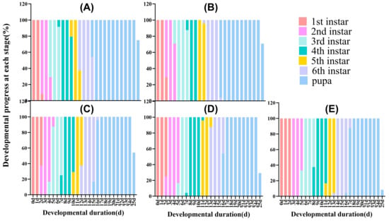
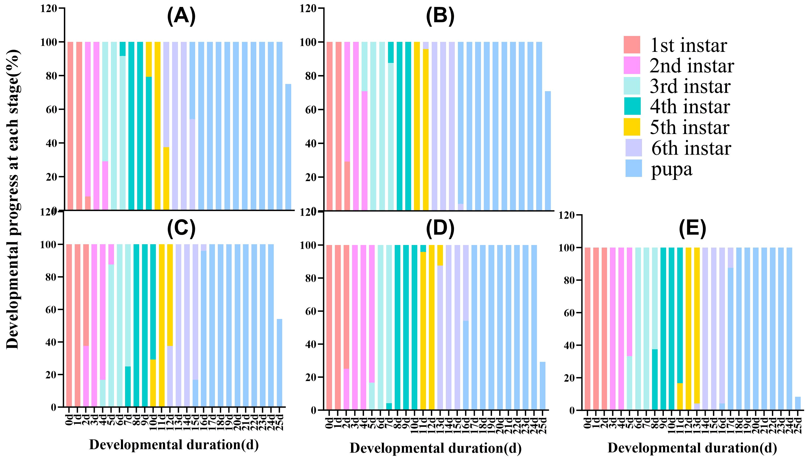
Observational data revealed that larvae exposed to all tested concentrations of exogenous melatonin successfully completed development to adulthood, with a 100% survival rate recorded across all treatment groups. Despite this uniform survival, exogenous melatonin application exerted a significant modulatory effect on both larval and pupal developmental durations of H. armigera (Table A1, Figure 1). With increasing melatonin concentration, the larval stage was significantly prolonged, whereas the pupal stage was markedly shortened (larval duration: F4,115 = 235.122, p < 0.05; pupal duration: F4,115 = 166.243, p < 0.05). Specifically, in the control group, 45.83% of the sixth instar larvae had developed to the pupal stage by day 14, whereas under the 20 µg/g melatonin treatment, only 4.17% of the sixth instar larvae reached the pupal stage by day 16. The mean larval and pupal developmental durations in the control group were 14.20 d and 10.2 d, respectively, whereas those in the 20 µg/g treatment group were 16.90 d and 8.04 d, representing an extension of 2.69 d in larval duration and a reduction of 2.16 d in pupal duration relative to the control. Collectively, these findings indicate that exogenous melatonin is associated with retarded larval development and accelerated pupal metamorphosis, with a more pronounced shortening effect on pupal duration.
Figure 1.
Effects of different concentrations of exogenous melatonin on the development of H. armigera. (A): 0(CK) μg/g; (B): 0.02 μg/g; (C): 0.2 μg/g; (D): 2 μg/g; (E): 20 μg/g.
3.2. Influence of Exogenous Melatonin on the Head Capsule Width of Larvae
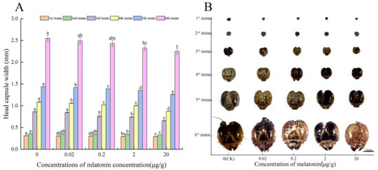
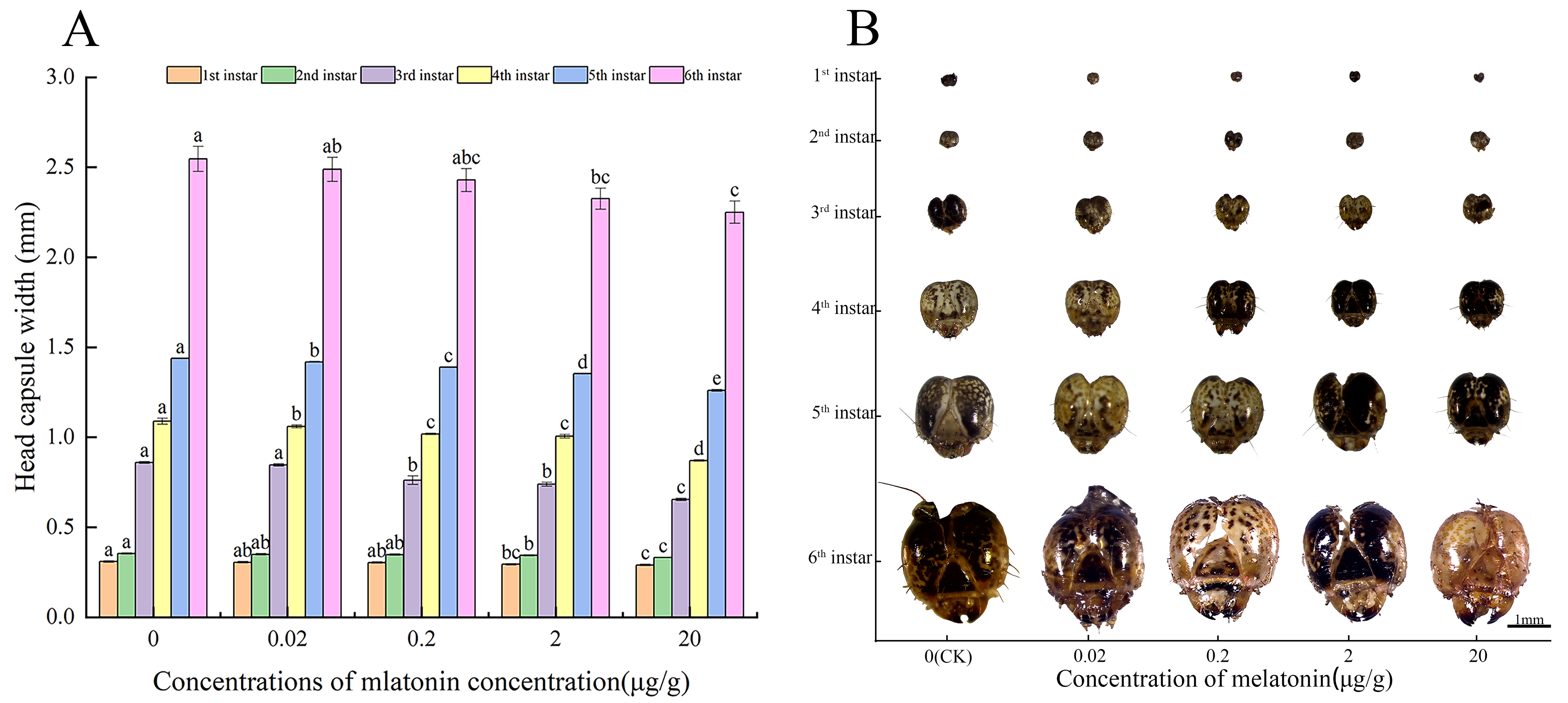
Larval head capsule width increased with instar progression across all exogenous melatonin treatment groups (Figure 2A). However, at the same instar, head capsule width decreased significantly with increasing melatonin concentration in the diet compared with the control (Figure 2B). At the first instar, larvae fed with 2 µg/g and 20 µg/g melatonin exhibited significantly reduced head capsule widths relative to the control (p < 0.05), whereas no significant differences were observed among the other treatment groups (p > 0.05). At the second instar, head capsule width in the 2 µg/g and 20 µg/g groups was significantly lower than that in the other three groups (first instar: F4,95 = 3.354, p > 0.05; second instar: F4,95 = 6.284, p < 0.05; third instar: F4,95 = 44.959, p < 0.05; fourth instar: F4.95 = 66.807, p < 0.05; fifth instar: F4,95 = 701.230, p < 0.05; sixth instar: F4,95 = 3.471, p > 0.05). At the sixth (final) instar, the mean head capsule width of control larvae reached 2.547 mm, whereas those treated with 2 µg/g and 20 µg/g melatonin measured only 2.325 mm and 2.250 mm, corresponding to reductions of 8.71% and 11.67%, respectively, relative to the control. Collectively, these results demonstrate that exogenous melatonin significantly reduces larval head capsule width in a concentration-dependent manner across all instars, indicating a marked effect on the body size of H. armigera larvae.
Figure 2.
Effects of different concentrations of exogenous melatonin on the larval head capsule width of H. armigera. Different lowercase letters denote significant differences (Duncan’s multiple range test, p < 0.05) in head capsule width among concentrations within the same instar. (A) Head capsule width (mm) of 1st to 6th instar larvae under different melatonin concentrations; (B) representative photographs of head capsules from 1st to 6th instar larvae treated with different melatonin concentrations.
3.3. Influence of Exogenous Melatonin on the Body Weight of Larvae, Pupal Weight of Females and Males
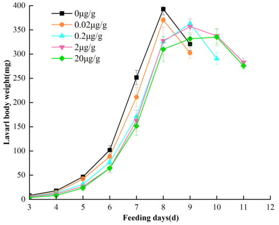
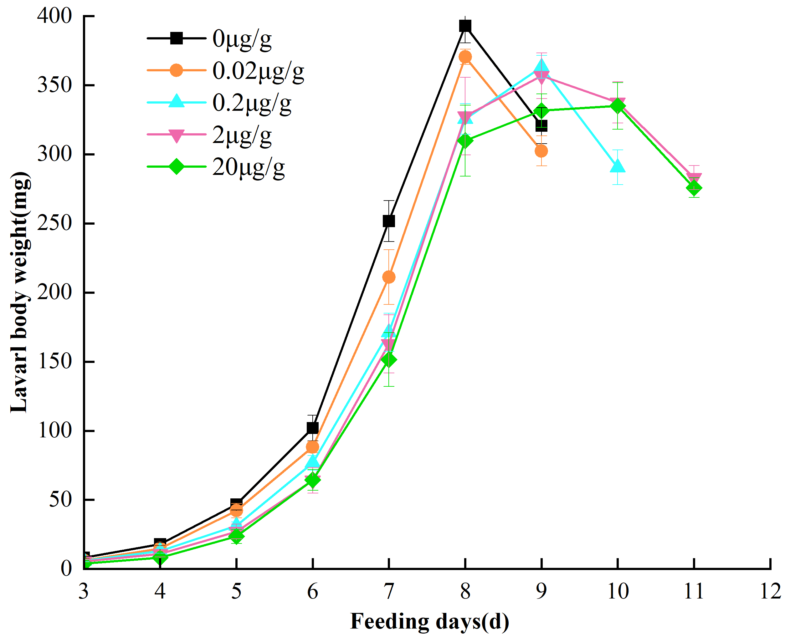
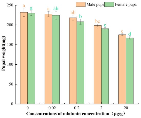
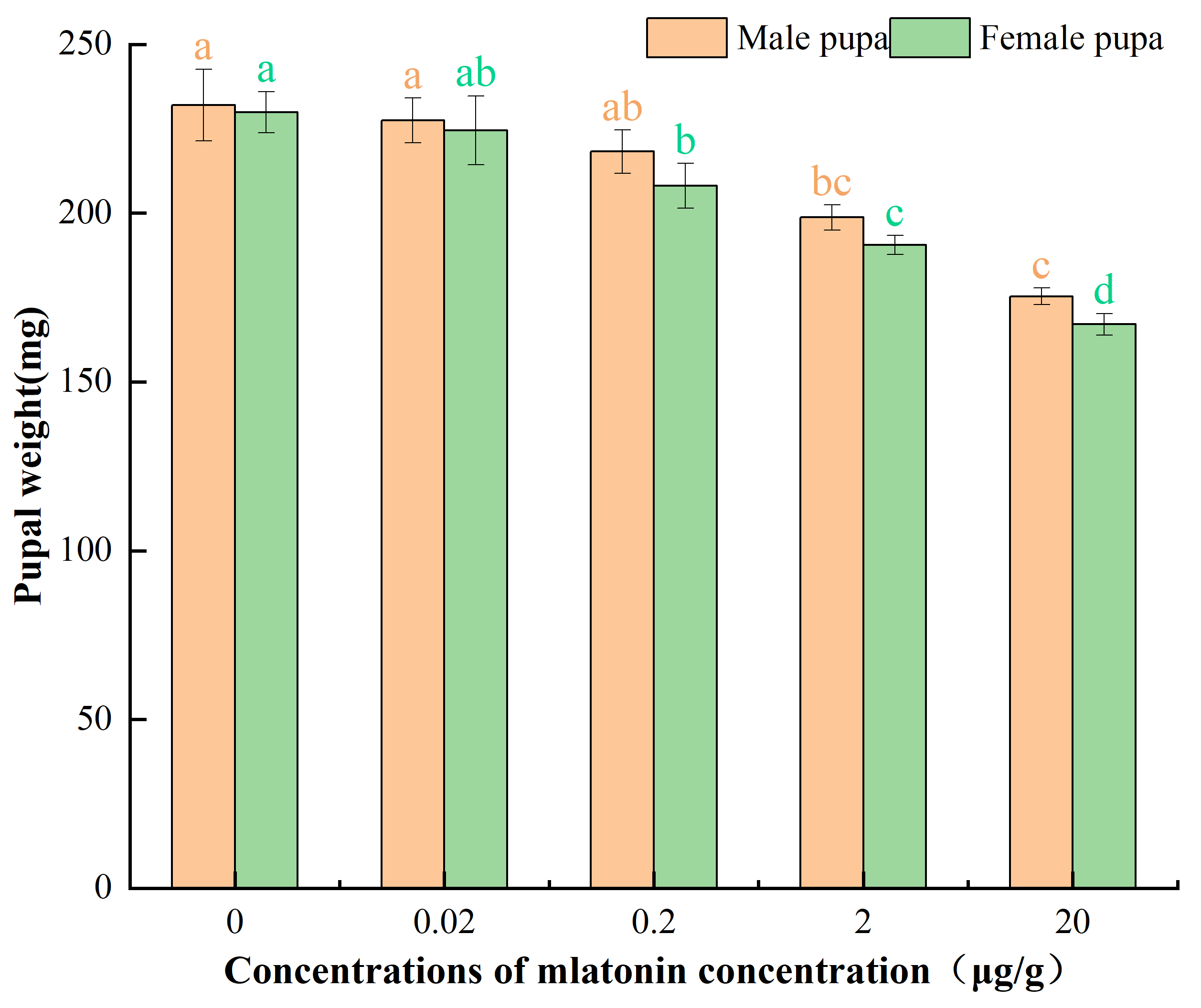
Compared with the control group, larval body weight in all melatonin-treated groups decreased significantly on the same feeding days, and the magnitude of reduction increased with rising melatonin concentration (Table A2, Figure 3). Across all treatment groups, larval body weight reached its peak 1–2 days prior to pupation; however, the timing of peak weight was progressively delayed with increasing melatonin concentration. In the control and 0.02 μg/g groups, maximum larval weights (392.99 mg and 370.49 mg, respectively) were recorded on day 8 of feeding. In the 0.2 μg/g and 2 μg/g groups, peak weights (363.21 mg and 356.84 mg, respectively) occurred on day 9, corresponding to reductions of 7.57% and 9.11% relative to the control. The 20 μg/g group attained its maximum weight (335.01 mg) on day 10, representing a 14.75% decrease compared with the control. Exogenous melatonin treatment also had a significant effect on the pupal weight of both female and male H. armigera, with pupal weights gradually decreasing as exogenous melatonin concentration increased (female pupal weight: F4,43 = 13.414, p < 0.05; male pupal weight: F4,38 = 7.529, p < 0.05; Figure 4). The female and male pupal weights in the control group were 229.88 mg and 231.99 mg, respectively, whereas both female (167.05 mg) and male (175.33 mg) pupal weights in the 20 μg/g treatment group were the smallest, representing reductions of 27.33% and 24.42%, respectively, compared with the control. Under the same treatment concentration, although male pupal weight was consistently higher than female pupal weight, the difference between female and male pupal weights was not significant (p > 0.05). These results indicate that exogenous melatonin not only prolonged larval developmental duration and delayed pupation but also significantly reduced larval body weight and pupal weight of both female and male moths.
Figure 3.
Changes in average daily body weight of the larvae of H. armigera under different concentrations of exogenous melatonin treatment.
Figure 4.
Effects of different concentrations of exogenous melatonin on the male and female pupal weight of H. armigera. Different lowercase letters denote significant differences (Duncan’s multiple range test, p < 0.05) in pupal weight among concentrations within the same sex. Letters in green denote comparisons among females, and letters in orange denote comparisons among males.
3.4. Influence of Exogenous Melatonin on the Adult Longivity and Fecundity
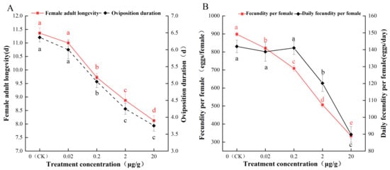
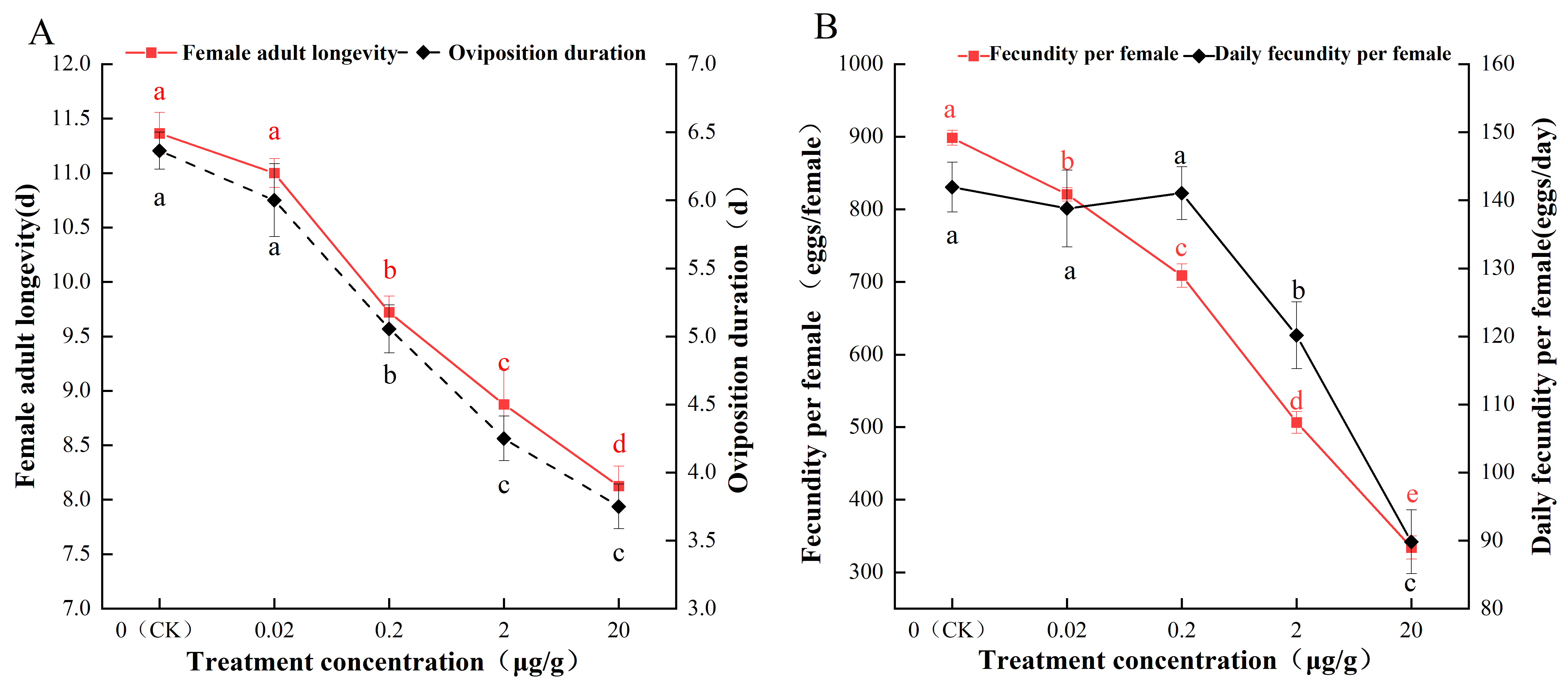
Exogenous melatonin at different concentrations significantly affected female adult longevity, oviposition duration, total fecundity per female, and average daily egg production per female in H. armigera (female adult longevity: F4,39 = 45.601, p < 0.05; oviposition duration: F4,39 = 38.250, p < 0.05; total fecundity per female: F4,39 = 326.712, p < 0.05; average daily egg production per female: F4,39 = 21.530, p < 0.05). As shown in Figure 5A, both adult longevity and oviposition duration decreased significantly with increasing melatonin concentration, with the shortest values recorded at 20 µg/g (8.13 days and 3.75 days, respectively), representing reductions of 3.23 days and 2.61 days compared with the control (11.36 days and 6.36 days, respectively). Furthermore, total fecundity per female and average daily egg production per female also declined significantly with increasing melatonin concentration (Figure 5B), reaching their lowest values at 20 µg/g (224.13 eggs/female and 89.81 eggs/day, respectively), corresponding to reductions of 75.05% and 36.72% relative to the control (898.45 eggs/female and 141.96 eggs/day, respectively). Collectively, these results demonstrate that increasing concentrations of exogenous melatonin lead to a concentration-dependent decrease in adult longevity, oviposition duration, total fecundity per female, and average daily egg production per female in H. armigera.
Figure 5.
Effects of different concentrations of exogenous melatonin on the adult longevity, oviposition duration (A) and total and daily fecundity (B) of H. armigera. Different lowercase letters above the data points indicate significant differences among different treatment concentrations (p < 0.05).
3.5. Influence of Exogenous Melatonin at Different Concentrations on Ovarian Development of H. armigera
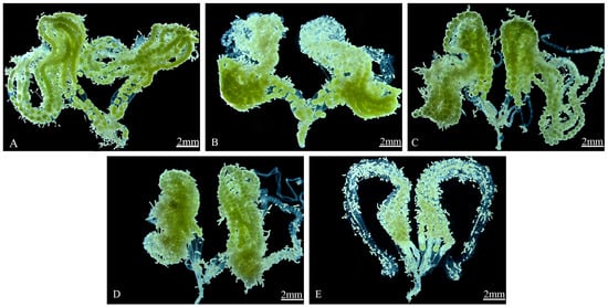
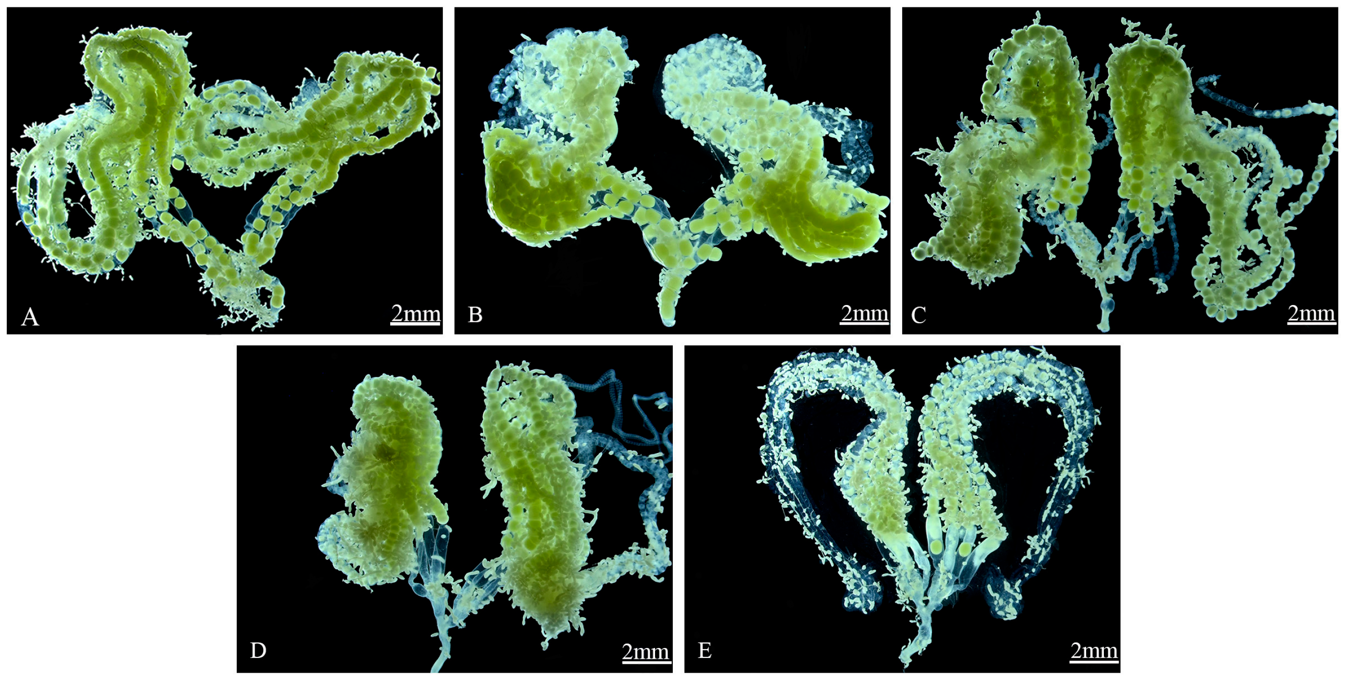
Ovarian maturation stages of H. armigera females under different exogenous melatonin concentrations were assessed based on established ovarian development criteria, as shown in Figure 6. In the control group, two-day-old female moths exhibited ovaries at the late vitellogenesis stage, transitioning toward the mature egg-laying phase. The ovarian tubules appeared yellowish-white, with oocytes clearly discernible and regularly arranged; oocytes aggregated at the basal region, and mature oocytes were present near the main oviduct (Figure 6A). At 0.02 µg/g melatonin, the ovaries remained at the late vitellogenesis stage; vitellogenesis had initiated in the ovarian tubules, and immature eggs were observed at the distal end of the lateral oviduct (Figure 6B). In the 0.2 µg/g treatment, ovaries were at the middle vitellogenesis stage; the ovarian tubules had not yet expanded to form distinct egg chambers, and immature eggs were located at the oviduct terminus (Figure 6C). At 2 µg/g, ovarian development progressed to the early-to-middle vitellogenesis stage; immature eggs were arranged in a linear series within the oviduct, with no mature eggs detected in the main oviduct, which remained relatively slender and contained numerous fat globules (Figure 6D). In the 20 µg/g group, the ovarian tubules were thin, and both the tubules and the oviduct exhibited a milky-white, translucent appearance; oocytes werescarce, with only a limited number of visible eggs, while the fat body was abundant and milky in color, indicating that the ovaries were at the early vitellogenesis stage (Figure 6E).
Figure 6.
Effects of different concentrations of exogenous melatonin on ovaries of H. armigera. (A): 0 (CK) μg/g; (B): 0.02 μg/g; (C): 0.2 μg/g; (D): 2 μg/g; (E): 20 μg/g.
4. Discussion
Melatonin is a naturally occurring compound ubiquitously distributed across living organisms, where it plays conserved and vital regulatory roles in metabolism, immunity, stress responses, and reproduction [14,32]. Owing to its versatile biological activities and application potential, coupled with the rapid development of synthetic biology, microbial fermentation has become a green and efficient route for melatonin production, enabling the extensive utilization of exogenous melatonin in both production and practice [12]. In the medical field, exogenous melatonin is employed to boost immunity and retard aging in humans and animals [33,34]. In higher animal production, it is used to regulate nutrient partitioning, growth and development, and seasonal reproductive physiology [17,35]. In plant production, it is primarily utilized to enhance crop tolerance to both biotic and abiotic environmental stresses [7,36]. These multifaceted functions confer upon melatonin considerable potential for agricultural applications in plants and control of herbivorous insects [37,38]. In this study, our results demonstrated that increasing melatonin concentrations significantly prolonged larval developmental duration (Figure 1), reduced head capsule width (Figure 2A,B), decreased larval body weight and pupal weight (Figure 3 and Figure 4), collectively demonstrating a dose-dependent inhibitory effect on larval growth of H. armigera. These results are consistent with prior studies on Acyrthosiphon pisum [39], Halyomorpha halys [27], and Aphis craccivora [28]. A likely mechanism is that exogenous melatonin disrupts nutrient accumulation and energy metabolism, thereby impairing normal development. Aceituno-Medina et al. reported that in Anastrepha ludens, the protein:carbohydrate:lipid ratio in the larval diet differentially affects the survival and fecundity of male and female adults [40]. Bhavanam and Trewick reported that in Ephestia kuehniella, larval nutrient limitation affects male phenotype, reproductive investment, and adult longevity [41]. Thus, the prolonged larval duration of H. armigera demonstrated observed under high melatonin concentrations may stem from altered nutrient deposition. Concurrently, increasing melatonin concentrations significantly shortened pupal developmental duration, suggesting accelerated pupal development and earlier adult eclosion. This finding aligns with Wang et al. [42], who demonstrated that melatonin injection terminated diapause in Antheraea pernyi pupae by mimicking photoperiodic signals, thereby hastening adult emergence under short-day conditions. Notably, under high melatonin treatment, adult female longevity and oviposition duration were each reduced by approximately 3 days, and total fecundity per female dropped by over 60% compared to the control (p < 0.05; Figure 5). Ovarian dissection confirmed that high melatonin concentrations markedly inhibited ovarian maturation (Figure 6). These reproductive impairments are highly consistent with observations in H. halys [27], A. craccivora [28], further supporting the view that exogenous melatonin exerts conserved adverse effects on herbivorous insects, with particularly pronounced negative impacts on their reproductive performance. However, Bonilla et al. found that after feeding newly eclosed male adults of Drosophila melanogaster with a melatonin-containing diet, exogenous melatonin significantly extended their lifespan [43]. The extended longevity of male D. melanogaster adults may be associated with the antioxidant properties of melatonin, as numerous studies have shown that melatonin can act as an antioxidant to delay senescence [16,33]. Therefore, discrepancies in the findings reported by different researchers might reflect involvement of species- and stage-specific physiological mechanisms underlying melatonin influence on lifespan.
It is widely recognized that nutrient reserves acquired during the larval stage profoundly influence developmental rate, adult longevity, and reproductive output, with fecundity serving as a key determinant of future population dynamics [44,45]. The accumulation of nutrients during this stage provides the essential material foundation for reproduction; greater nutrient reserves are generally associated with enhanced reproductive potential and accelerated population growth. In this study, honey was provided as a nutritional supplement for adults of H. armigera during the adult stage. Nevertheless, differences in adult longevity and reproductive output persisted between the control and exogenous melatonin treatments, indicating that exogenous melatonin affects nutrient accumulation during the larval stage (as reflected by reduced body weight), which in turn influences adult longevity and reproduction. Richter et al. [46] reported that melatonin functions as a neurohormonal releasing factor in Periplaneta americana, where it binds to specific receptors in the brain and stimulates the release of prothoracicotropic hormone (PTTH), thereby indirectly modulating ecdysteroid biosynthesis. This observation suggests that melatonin may participate in the regulation of insect growth and development, potentially affecting nutrient accumulation. Similarly, Girish et al. [47] demonstrated that melatonin induces ecdysteroidogenesis, methyl farnesoate synthesis, and the expression of ecdysteroid receptor (EcR) and retinoid X receptor (RXR) in the hepatopancreas and ovary of the Scylla serrata. Furthermore, Díaz López et al. [48] reported that melatonin exerts significant effects on the neuroendocrine reproductive axis by binding to specific receptors within the hypothalamic–pituitary–gonadal (HPG) axis and modulating the secretion of gonadotrophin-releasing hormone (GnRH), which in turn regulates the synthesis and release of luteinizing hormone (LH) and follicle-stimulating hormone (FSH). These findings indicate that melatonin may be involved in the regulation of gonadal development and consequently affect reproductive function, particularly fecundity. Taken together, the shortened adult longevity and reduced fecundity observed in H. armigera in the present study may be partially attributed to impaired nutrient accumulation during the larval stage. In addition, melatonin-induced disruption of the endocrine system—especially the modulation of juvenile hormone and ecdysteroid titers—may further interfere with metamorphic and reproductive processes [49,50]. Nevertheless, the specific effects of exogenous melatonin treatment on the titers of juvenile hormone, ecdysteroid, and follicle-stimulating hormone, as well as the underlying regulatory mechanisms in H. armigera, remain to be elucidated and warrant further investigation.
In summary, this study investigated the effects of exogenous melatonin on the biological characteristics of H. armigera by rearing larvae on an artificial diet supplemented with exogenous melatonin under laboratory conditions. The results demonstrated that exogenous melatonin exerted significant inhibitory effects on the growth, development, and reproduction of H. armigera, indicating an adverse impact on population development and highlighting its potential application value in integrated pest management. Given that the present findings were obtained under laboratory conditions using an artificial diet, field experiments are warranted to evaluate both the control efficacy of exogenous melatonin against target pests and its nontarget effects, thereby determining its practical application value. Nevertheless, these findings offer new perspectives and a foundation for future research on the application of melatonin in crop protection and integrated pest management.
Author Contributions
J.H.: Writing—original draft, Methodology, Investigation, Data curation, Formal analysis. X.S.: Methodology, Data curation. C.G.: Methodology, Investigation. X.Q.: Conceptualization, Methodology, Writing—review and editing. T.Z.: Conceptualization, Methodology, Formal analysis, Writing—review and editing, Funding acquisition, Supervision. All authors have read and agreed to the published version of the manuscript.
Funding
This research was funded by National Science Foundation of China (No. 32560261, 31960227) and Special Project for the Construction of Maize Industry Technology System for Modern Characteristic Cold and Drought-Resistant Agriculture in Gansu Province (GSARS08).
Data Availability Statement
The raw data supporting the conclusions of this article will be made available by the authors on request. Further inquiries can be directed to the corresponding author.
Conflicts of Interest
The authors declare no conflicts of interest.
Appendix A
Table A1.
Effects of different concentrations of exogenous melatonin on the development of H. armigera.
Table A2.
Effects of different concentrations of exogenous melatonin on the larval weight of H. armigera.
References
- Fathipour, Y.; Babaei, M.; Bagheri, A.; Talebi, A.A.; Yazdanpanah, S. Demographic parameters of Helicoverpa armigera on ten corn hybrids-mediated artificial diets reveals striking differences. J. Crop Prot. 2021, 10, 363–374. [Google Scholar]
- Yadav, S.P.S.; Lahutiya, V.; Paudel, P. A review on the biology, ecology, and management tactics of Helicoverpa armigera (Lepidoptera: Noctuidae). Turk. J. Agric.-Food Sci. Technol. 2022, 10, 2467–2476. [Google Scholar] [CrossRef] [Scilit]
- Scholz, B.C.G.; Monsour, C.J.; Zalucki, M.P. An evaluation of selective Helicoverpa armigera control options in sweet corn. Aust. J. Exp. Agric. 1998, 38, 601–607. [Google Scholar] [CrossRef] [Scilit]
- Negruț, G.N.; Cotuna, O.; Sǎrǎțeanu, V.; Durău, C.C. Research regarding the relationship among the pests Ostrinia nubilalis, Helicoverpa armigera and the fungi Fusarium verticillioides, Aspergillus flavus in corn in the climatic conditions from Lovrin (Timiș County). Res. J. Agric. Sci. 2019, 51, 282–291. [Google Scholar]
- Folcher, L.; Weissenberger, A.; Delos, M. Quantitative relationships between Ostrinia nubilalis activity and deoxynivalenol contamination in French maize. Int. J. Pest Manag. 2012, 58, 302–309. [Google Scholar] [CrossRef] [Scilit]
- Yang, Y.; Li, Y.; Wu, Y. Current status of insecticide resistance in Helicoverpa armigera after 15 years of Bt cotton planting in China. J. Econ. Entomol. 2013, 106, 375–381. [Google Scholar] [CrossRef] [Scilit] [PubMed]
- Muthusamy, R.; Ramkumar, G.; Kumarasamy, S.; Kumar, T.C.; Albeshr, M.F.; Alrefaei, A.F.; Nhung, T.C.; B, B.; Karuppusamy, I. Effect of melatonin and luzindole antagonist on fipronil toxicity, detoxification and antioxidant enzyme system in different tissues of Helicoverpa armigera (Lepidoptera: Noctuidae). Environ. Res. 2023, 231, 116130. [Google Scholar] [CrossRef] [Scilit] [PubMed]
- Moustafa-Farag, M.; Almoneafy, A.; Mahmoud, A.; Elkelish, A.; Arnao, M.B.; Li, L.; Ai, S. Melatonin and its protective role against biotic stress impacts on plants. Biomolecules 2019, 10, 54. [Google Scholar] [CrossRef] [Scilit] [PubMed]
- Liu, X.; Du, C.; Yue, C.; Tan, Y.; Fan, H. Exogenously applied melatonin alleviates the damage in cucumber plants caused by Aphis goosypii through altering the insect behavior and inducing host plant resistance. Pest Manag. Sci. 2022, 79, 140–151. [Google Scholar] [CrossRef] [Scilit] [PubMed]
- Yin, L.; Wang, P.; Li, M.; Ke, X.; Li, C.; Liang, D.; Wu, S.; Ma, X.; Li, C.; Zou, Y.; et al. Exogenous melatonin improves Malus resistance to Marssonina apple blotch. J. Pineal Res. 2013, 54, 426–434. [Google Scholar] [CrossRef] [Scilit] [PubMed]
- Lerner, A.B.; Case, J.D.; Takahashi, Y.; Lee, T.H.; Mori, W. Isolation of melatonin, the pineal gland factor that lightens melanocyteS1. J. Am. Chem. Soc. 1958, 80, 2587. [Google Scholar] [CrossRef] [Scilit]
- Zhao, D.; Yu, Y.; Shen, Y.; Liu, Q.; Zhao, Z.; Sharma, R.; Reiter, R.J. Melatonin synthesis and function: Evolutionary history in animals and plants. Front. Endocrinol. 2019, 10, 441357. [Google Scholar] [CrossRef] [Scilit] [PubMed]
- Andronachi, V.C.; Simeanu, C.; Matei, M.; Radu-Rusu, R.M.; Simeanu, D. Melatonin: An overview on the synthesis processes and on its multiple bioactive roles played in animals and humans. Agriculture 2025, 15, 273. [Google Scholar] [CrossRef] [Scilit]
- Mannino, G.; Pernici, C.; Serio, G.; Gentile, C.; Bertea, C.M. Melatonin and phytomelatonin: Chemistry, biosynthesis, metabolism, distribution and bioactivity in plants and animals—An overview. Int. J. Mol. Sci. 2021, 22, 9996. [Google Scholar] [CrossRef] [Scilit] [PubMed]
- Arendt, J.; Skene, D.J. Melatonin as a chronobiotic. Sleep Med. Rev. 2005, 9, 25–39. [Google Scholar] [CrossRef] [Scilit] [PubMed]
- Anisimov, V.N. Effects of exogenous melatonin—A review. Toxicol. Pathol. 2003, 31, 589–603. [Google Scholar] [CrossRef] [Scilit] [PubMed]
- Reiter, R.J.; Tan, D.X.; Manchester, L.C.; Paredes, S.D.; Mayo, J.C.; Sainz, R.M. Melatonin and reproduction revisited. Biol. Reprod. 2009, 81, 445–456. [Google Scholar] [CrossRef] [Scilit] [PubMed]
- Wang, L.Y.; Liu, J.L.; Wang, W.X.; Sun, Y. Exogenous melatonin improves growth and photosynthetic capacity of cucumber under salinity-induced stress. Photosynthetica 2016, 54, 19–27. [Google Scholar] [CrossRef] [Scilit]
- Zhang, T.; Shi, Z.; Zhang, X.; Zheng, S.; Wang, J.; Mo, J. Alleviating effects of exogenous melatonin on salt stress in cucumber. Sci. Hortic. 2020, 262, 109070. [Google Scholar] [CrossRef] [Scilit]
- Jiang, D.; Lu, B.; Liu, L.; Duan, W.; Meng, Y.; Li, J.; Zhang, K.; Sun, H.; Zhang, Y.; Dong, H.; et al. Exogenous melatonin improves the salt tolerance of cotton by removing active oxygen and protecting photosynthetic organs. BMC Plant Biol. 2021, 21, 331. [Google Scholar] [CrossRef] [Scilit] [PubMed]
- Ding, F.; Liu, B.; Zhang, S. Exogenous melatonin ameliorates cold-induced damage in tomato plants. Sci. Hortic. 2017, 219, 264–271. [Google Scholar] [CrossRef] [Scilit]
- Fan, X.; Zhao, J.; Sun, X.; Zhu, Y.; Li, Q.; Zhang, L.; Zhao, D.; Huang, L.; Zhang, C.; Liu, Q. Exogenous melatonin improves the quality performance of rice under high temperature during grain filling. Agronomy 2022, 12, 949. [Google Scholar] [CrossRef] [Scilit]
- Li, J.; Zeng, L.; Cheng, Y.; Lu, G.; Fu, G.; Ma, H.; Liu, Q.; Zhang, X.; Zou, X.; Li, C. Exogenous melatonin alleviates damage from drought stress in Brassica napus L. (rapeseed) seedlings. Acta Physiol. Plant. 2018, 40, 43. [Google Scholar] [CrossRef] [Scilit]
- Huang, B.; Chen, Y.E.; Zhao, Y.Q.; Ding, C.B.; Liao, J.Q.; Hu, C.; Yuan, S.; Yuan, M. Exogenous melatonin alleviates oxidative damages and protects photosystem II in maize seedlings under drought stress. Front. Plant Sci. 2019, 10, 677. [Google Scholar] [CrossRef] [Scilit] [PubMed]
- Imran, M.; Latif Khan, A.; Shahzad, R.; Aaqil Khan, M.; Bilal, S.; Khan, A.; Kang, S.; Lee, I.J. Exogenous melatonin induces drought stress tolerance by promoting plant growth and antioxidant defence system of soybean plants. AoB Plants 2021, 13, plab026. [Google Scholar] [CrossRef] [Scilit] [PubMed]
- Liu, C.; Chen, L.; Zhao, R.; Li, R.; Zhang, S.; Yu, W.; Sheng, J.; Shen, L. Melatonin induces disease resistance to Botrytis cinerea in tomato fruit by activating jasmonic acid signaling pathway. J. Agric. Food Chem. 2019, 67, 6116–6124. [Google Scholar] [CrossRef] [Scilit] [PubMed]
- Niva, C.C.; Takeda, M. Effects of photoperiod, temperature and melatonin on nymphal development, polyphenism and reproduction in Halyomorpha halys (Heteroptera: Pentatomidae). Zool. Sci. 2003, 20, 963–970. [Google Scholar] [CrossRef] [Scilit] [PubMed]
- Zhang, Y.X.; Zhang, T.W.; Shi, L.; Yuan, Y.; Liu, C.Z. Effects of exogenous melatonin on the growth, development, and reproduction of Aphis craccivora. Chin. J. Ecol. 2023, 42, 1150–1154. [Google Scholar]
- Zhang, Y.L.; Zhang, K.X.; Ma, Y.; Zhang, Q.Y.; Liu, W.H.; Jiang, H.X.; Yang, L.K.; Liu, C.Z. Feeding selectivity of Spodoptera frugiperda on different maize cultivars and its relationship with chemical substances in maize leaves. China Plant Prot. 2021, 41, 10–16. [Google Scholar]
- Zhang, W.N.; Xiao, H.J.; Liang, G.M.; Guo, Y.Y. Observation on ovarian morphology and oogenesis in the cotton bollworm, Helicoverpa armigera (Lepidoptera: Noctuidae). Acta Entomol. Sin. 2013, 56, 358–364. [Google Scholar]
- Chakravarty, S.; Ganguly, S.; Padwal, K.G.; Keval, R.; Srivastava, C.P. Morphology of immature stages and adults of Helicoverpa armigera. Indian J. Entomol. 2022, 84, 588–593. [Google Scholar]
- Hussain, B.; Haouala, F.; Fariduddin, Q. From night hormone to green signal: The journey of melatonin. Protoplasma 2026, 263, 1117–1140. [Google Scholar] [CrossRef] [Scilit] [PubMed]
- Bocheva, G.; Bakalov, D.; Iliev, P.; Tafradjiiska-Hadjiolova, R. The vital role of melatonin and its metabolites in the neuroprotection and retardation of brain aging. Int. J. Mol. Sci. 2024, 25, 5122. [Google Scholar] [CrossRef] [Scilit] [PubMed]
- Das, J.; Ghosh, S.; Ghosh, B.; Sarkar, S.; Sarkar, S. Exploring the role of melatonin in disease prevention and anti-aging. In Biotech Miracles: Harnessing the Power of Microbes; Integrated Publications: New Delhi, India, 2024; Volume 14, p. 888292. [Google Scholar]
- Barrett, P.; Bolborea, M. Molecular pathways involved in seasonal body weight and reproductive responses governed by melatonin. J. Pineal Res. 2012, 52, 376–388. [Google Scholar] [CrossRef] [Scilit] [PubMed]
- Cai, H.; Li, J.; Li, J.; Teng, H. Melatonin—Angel of plant growth regulation and protection. Adv. Agrochem. 2025, 4, 114–122. [Google Scholar] [CrossRef] [Scilit]
- Janas, K.M.; Posmyk, M.M. Melatonin, an underestimated natural substance with great potential for agricultural application. Acta Physiol. Plant. 2013, 35, 3285–3292. [Google Scholar] [CrossRef] [Scilit]
- Khan, M.; Hussain, A.; Yun, B.W.; Mun, B.G. Melatonin: The multifaceted molecule in plant growth and defense. Int. J. Mol. Sci. 2024, 25, 6799. [Google Scholar] [CrossRef] [Scilit] [PubMed]
- Gao, N.; Hardie, J. Melatonin and the pea aphid, Acyrthosiphon pisum. J. Insect Physiol. 1997, 43, 615–620. [Google Scholar] [CrossRef] [Scilit] [PubMed]
- Aceituno-Medina, M.; Hernández, E.; Rincón-Betancurt, O.; García-Fajardo, L.V.; Diego-García, E. Larval nutritional effects on male and female survival and fecundity in Anastrepha ludens. Entomol. Exp. Appl. 2024, 172, 2–14. [Google Scholar] [CrossRef] [Scilit]
- Bhavanam, S.; Trewick, S. Effects of larval crowding and nutrient limitation on male phenotype, reproductive investment and strategy in Ephestia kuehniella Zeller (Insecta: Lepidoptera). J. Stored Prod. Res. 2017, 71, 64–71. [Google Scholar] [CrossRef] [Scilit]
- Wang, Q.; Egi, Y.; Takeda, M.; Oishi, K.; Sakamoto, K. Melatonin pathway transmits information to terminate pupal diapause in the Chinese oak silkmoth Antheraea pernyi and through reciprocated inhibition of dopamine pathway functions as a photoperiodic counter. Entomol. Sci. 2015, 18, 74–84. [Google Scholar] [CrossRef] [Scilit]
- Bonilla, E.; Medina-Leendertz, S.; Díaz, S. Extension of life span and stress resistance of Drosophila melanogaster by long-term supplementation with melatonin. Exp. Gerontol. 2002, 37, 629–638. [Google Scholar] [CrossRef] [Scilit] [PubMed]
- Yan, J.; Kibech, R.; Stone, C.M. Differential effects of larval and adult nutrition on female survival, fecundity, and size of the yellow fever mosquito, Aedes aegypti. Front. Zool. 2021, 18, 10. [Google Scholar] [CrossRef] [Scilit] [PubMed]
- Tigreros, N.; Sass, E.M.; Lewis, S.M. Sex-specific response to nutrient limitation and its effects on female mating success in a gift-giving butterfly. Evol. Ecol. 2013, 27, 1145–1158. [Google Scholar] [CrossRef] [Scilit]
- Richter, K.; Pesche, E.; Pesche, D. Effect of melatonin on the release of prothoracicotropic hormone from the brain of Periplaneta americana (Blattodea: Blattidae). EJE 2013, 96, 341–345. [Google Scholar]
- Girish, B.P.; Swetha, C.H.; Reddy, P.S. Induction of ecdysteroidogenesis, methyl farnesoate synthesis and expression of ecdysteroid receptor and retinoid X receptor in the hepatopancreas and ovary of the giant mud crab, Scylla serrata by melatonin. Gen. Comp. Endocrinol. 2015, 217, 37–42. [Google Scholar] [CrossRef] [Scilit] [PubMed]
- Díaz López, B.; Rodríguez, E.D.; Urquijo, C.; Alvarez, C.F. Melatonin influences on the neuroendocrine-reproductive axis. Ann. N. Y. Acad. Sci. 2005, 1057, 337–364. [Google Scholar] [CrossRef] [Scilit] [PubMed]
- Czesak, M.E.; Fox, C.W. Evolutionary ecology of egg size and number in a seed beetle: Genetic trade-off differs between environments. Evolution 2003, 57, 1121–1132. [Google Scholar] [CrossRef] [Scilit] [PubMed]
- Parthasarathy, R.; Palli, S.R. Molecular analysis of nutritional and hormonal regulation of female reproduction in the red flour beetle, Tribolium castaneum. Insect Biochem. Mol. Biol. 2011, 41, 294–305. [Google Scholar] [CrossRef] [Scilit] [PubMed]
Disclaimer/Publisher’s Note: The statements, opinions and data contained in all publications are solely those of the individual author(s) and contributor(s) and not of MDPI and/or the editor(s). MDPI and/or the editor(s) disclaim responsibility for any injury to people or property resulting from any ideas, methods, instructions or products referred to in the content. |
© 2026 by the authors. Licensee MDPI, Basel, Switzerland. This article is an open access article distributed under the terms and conditions of the Creative Commons Attribution (CC BY) license.