


Abstract
Eight H-bonded salts of arsenic acid and nitrogen bases (2,4,6-trimethylpyridine, pyridine-2,6-diamine, pyridin-4-ol, 4-methoxypyridine, 4-methoxyaniline, 1,3,5-triazine-2,4,6-triamine, diethylamine and N1,N1,N2,N2-tetraethylethane-1,2-diamine) were studied in the solid state by single crystal X-ray diffraction technique and DFT calculations. In all cases quite short (≤2.65 Å) OHO bonds were found in the self-assembled supramolecular ribbons or 2D networks of dihydrogen arsenates, constituting a repertoire of five different H-bonding patterns (motifs). The electron localization function maps revealed the spots of the nucleophilic sites on oxygen atoms that determine the preferable directions for H-bonding of H2AsO4− anions observed in the crystal packing. Analysis of the electrostatic potential maps for isolated species has demonstrated that upon H-bonding between H2AsO4− anions and protonated nitrogen bases, NH+⋯−OAsO(OH)2, the redistribution of electron density within the anion provides otherwise virtually non-existent electrophilic sites on hydrogen atoms, which balances the Coulomb repulsion and allows for the anion⋯anion pairing within the crystal. The topological analysis of the calculated crystalline electron density after relaxation of the hydrogen atoms’ positions was used to classify the OHO bonds as moderately strong ones (with an interaction energy up to 65 kJ/mol) and revealed a high degree of ionicity of molecular moieties within ion pairs (with an absolute charge up to 0.87 e). For the strongest OHO and NHO bonds, the noticeable covalent character was shown by using the crystal orbital Hamiltonian population analysis.
1. Introduction
In recent years, inorganic acidic anions and organic–inorganic hybrids have attracted increasing attention for their role in functional materials, particularly for applications related to proton conductivity and to nonlinear optical properties (in the presence of non-centrosymmetric crystal structures) [1]. In this context, it is now well recognized that efficient proton conduction requires well-organized H-bond networks capable of facilitating proton transfer via thermal hopping and phonon-assisted tunneling mechanisms along supramolecular chains [2,3,4,5,6,7]. A particularly interesting aspect of these systems is the H-bond-mediated anion⋯anion self-assembly, an apparently counterintuitive phenomenon as it involves units with the same net charge. While these anion⋯anion interactions have been called ‘anti-electrostatic’ H-bonds (AEHB) [8] in relation to the same negative charges of the interacting units, the Coulomb’s law remains valid as the interpenetration of van der Waals radii of involved atoms is enabled by the fact that, in the crystal packing, anions polarization changes the electrostatic potential at H atoms from negative to positive and by the overcompensation of the electrostatic repulsion between the net charges by other attractive forces [9]. The H-bond-mediated anion⋯anion interactions enable the formation of dimers, infinite chains, or other extended networks. Depending on the dimensionality of these supramolecular architectures, H-bond networks can be classified as 0D (discrete adducts), 1D, or 2D, according to the extent of the anionic assembly [8,10]. H-bond-driven anion⋯anion self-assembly is well documented for systems containing anions such as HSO4−, H2PO4−, and HCO3− [8,9,10,11,12]. Structural data reported in the Cambridge Structural Database indicate that such phenomena are not rare and lead to infinite or discrete supramolecular adducts stabilized by H-bond networks [13,14,15]. More recent studies have also shown that alternative interactions, such as σ-hole interactions, can contribute to the formation of anion⋯anion supramolecular adducts [16,17,18,19], occasionally giving rise to hybrid motifs in which multiple types of different interactions cooperate in stabilizing the supramolecular anion adducts [20].
In this context, dihydrogen arsenate anions (H2AsO4−) represent a system of great interest but remain relatively less explored compared to their sulphate and selenate analogs. Several literature examples show that H2AsO4− anions can self-assemble into extended supramolecular structures through AsO–H⋯OAs H-bonds in the presence of nitrogen-containing organic cations, both aromatic and aliphatic. These architectures, often featuring non-centrosymmetric infinite chains, appear particularly promising as candidates for high proton-conductivity materials [21,22,23,24,25,26]. Moreover, extended H-bond networks based on arsenate anion⋯anion assembly have also been linked to proton delocalization phenomena relevant for photonic applications [27,28]. Proton conductivity in these materials is strongly influenced by several factors, including H-bond strength, cooperative coupling between adjacent bonds, and the degree of proton delocalization within O–H⋯O bonds [29]. A thorough understanding of these aspects is essential for the rational design and functional optimization of materials based on H2AsO4−⋯H2AsO4− supramolecular synthons.
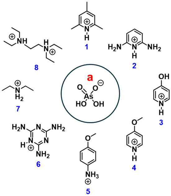
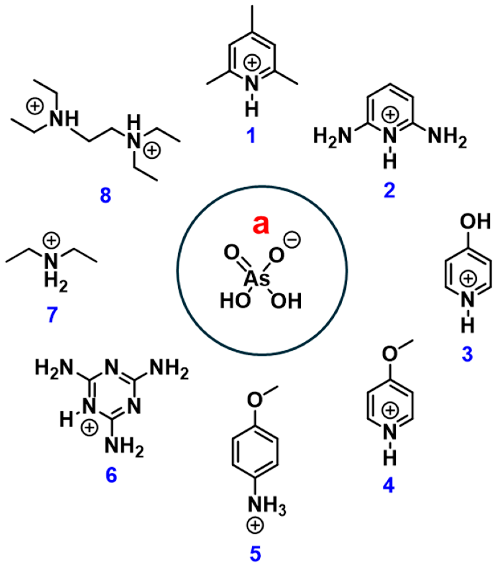
In this work, we report a systematic investigation of H-bond-mediated anion⋯anion motifs in the supramolecular assemblies of dihydrogen arsenates 1·a–8·a (Scheme 1). Eight salts obtained from arsenic acid with various nitrogen bases were investigated in the solid state using single-crystal X-ray diffraction and density functional theory (DFT) calculations. Structural analysis revealed the presence of particularly short O–H⋯O bonds, organized into ribbons or 2D networks defining distinct H-bond motifs. The integration of crystallographic data with theoretical analyses of electron density, electrostatic potential, and bonding interactions allows elucidation of the structural and electronic factors underlying anion⋯anion self-assembly in dihydrogen arsenates, as well as the key role of proton delocalization in stabilizing these architectures.
Scheme 1.
Structural formulas of the components in the crystal structures described in this work.
2. Materials and Methods
2.1. Materials
All compounds were purchased from commercial suppliers (TCI America, Tokyo (Japan) and Merck, Darmstadt (Germany)) and used without further purification. The arsenic acid (H3AsO4) solution was prepared by dissolving 1.150 g of arsenic pentoxide (As2O5) in 100 mL of distilled water, followed by reflux at 100 °C for 5 weeks [30]. The concentration of the resulting acid solution was determined by titration with NaOH using a pH meter and found to be 0.1 M, consistent with the quantitative hydrolysis of As2O5 to H3AsO4.
Crystal sample preparation. All salts were prepared according to the following procedure. Separate aqueous solutions (0.1 mmol) containing equimolar amounts of arsenic acid and the base were mixed under stirring. The solutions were then left to slowly evaporate at room temperature in clear 4 mL borosilicate vials. Single crystals suitable for X-ray diffraction were obtained in nearly quantitative yields (<95%) within two weeks.
2.2. Methods
X-ray diffraction analysis. Data collections were performed at 300 K or 100 K using an XtaLAB Synergy diffractometer (Rigaku, Tokyo, Japan) equipped with a HyPix detector (Rigaku, Tokyo, Japan). Unit cell refinement and data reduction were performed using the CrysAlisPro (version 1.171.41.98a) software [31]. Structures were solved by direct methods using the SHELXT [32] program and refined by full-matrix least-squares on F2 with anisotropic displacement parameters for the non-hydrogen atoms using the SHELXL [33] program incorporated in the Olex2 (version 1.5) software [34]. Hydrogen atoms were placed in idealized positions and first refined using a riding model; subsequently, they were additionally refined as described in the next subsection. Absorption correction was performed based on the multiscan procedure. X-ray structures were depicted using capped sticks representation (Mercury, version 2022.3.0) software [35]. CCDC deposition numbers: 2521265, 2521276, 2521277, 2521278, 2521279, 2521280, 2521281, 2521284 contain the validated [36] supplementary crystallographic data for this paper (see the electronic Supporting Information File, ESI). These data are provided free of charge by the joint Cambridge Crystallographic Data Centre and Fachinformationszentrum Karlsruhe Access Structures service.
Solid-state calculations. Electronic calculations of 1·a–8·a were performed using the CRYSTAL17 (version 1.0.2) [37] software. Constrained geometry optimizations were done at the B3LYP-D*/POB-TZVP [38,39,40,41,42,43] level of theory. The chosen combination of the DFT functional and the atomic basis set has been shown to reproduce the H-bond geometric features of the supramolecular chains of hydrogen selenates (HSeO4−) with sufficient accuracy, see our recent work [29]. Accuracy of calculation of the Coulomb and Hartree–Fock exchange series was controlled by a set of overlap tolerances, which were taken to be (10−9, 10−9, 10−9, 10−9, 10−18) [44]. The maximum order of multipole expansion [45,46] in the long-range zone for the electron-electron Coulomb interaction was set to 6. Calculation of the DFT exchange-correlation contribution over the unit cell volume was done with the XLGRID DFT grid specification [44,47]; tolerances for the DFT density and the DFT grid weight were taken to be 10−10 and 10−18, respectively. Integration in the Brillouin zone was performed using the Monkhorst–Pack scheme [48] for an isotropic 6 × 6 × 6 k-point grid for 1·a, and for an anisotropic 8 × 6 × 6 (for 2·a and 8·a), 4 × 8 × 2 (for 3·a), 6 × 8 × 3 (for 4·a), 4 × 6 × 6 (for 5·a) 8 × 4 × 4 (for 6·a) and 8 × 5 × 8 (for 7·a) k-point grids. Tolerance on energy controlling the SCF convergence (using the DIIS accelerator method [49]) for geometry optimizations and single-point calculations was set to 10−8 and 10−9 Hartree, respectively.
Single-crystal structure parameters of the above crystals from the X-ray diffraction data at 300 and 100 K were used as the starting points for the DFT calculations. While keeping fixed the unit cell parameters and heavy atoms’ positions, all hydrogen atoms’ positions were optimized with tight convergence criteria (maximum/RMS forces and displacements smaller than 0.000075/0.000050 a.u. and 0.000225/0.000150 a.u., in the order) and symmetry constraint imposed by the corresponding space group: triclinic P-1 for 2·a, 6·a, and 8·a; monoclinic P21/n for 4·a and 7·a, P21/m for 1·a, and P21/c for 3·a and 5·a. For 1·a featuring dynamically disordered hydrogen atoms of methyl groups of the protonated 2,4,6-trimethylpyridine molecules, the structures of eight different ordered isomorphs were subjected to the above constrained geometry optimization; afterwards, only one of the optimized structures (see Figures S29 and S30) was selected for further calculations, which are described onwards. Analytical energy gradients [50] within a quasi-Newton approach combined with the BFGS [51] algorithm for Hessian updating were employed. Consistency between the optimized geometries and the aforementioned overlap tolerances for integrals’ evaluation was ensured by the FINALRUN option with the value of 4 [44].
Topological analysis [52] of the periodic electron density (ED) was performed using the quantum theory of atoms in molecules (QTAIM) by means of the TOPOND14 module [53,54,55] implemented in the CRYSTAL17 code [56]. AIM atomic charges were evaluated by integration of the three-dimensional density distribution over the atomic basins, whose enclosing zero-flux surfaces were determined manually to the nearest preliminary retrieved bond critical point for a given nucleus using an eigenvector following approach with the tolerance value of 0.002 a.u. [57,58,59].
Crystal orbital Hamiltonian population projected onto local atomic orbitals pertaining to two selected groups of H and A (A = O or N) atoms, involved in the H⋯A hydrogen-bonding, was calculated over the energy range spanned from the first valence band, occupied by the non-core electrons, up to the highest occupied valence band, i.e., up to the Fermi energy level. The Hamiltonian eigenvectors were preliminary recalculated using a fine isotropic 12 × 12 × 12 k-point grid.
Molecular calculations. Electronic calculations of the molecular adducts analyzed in this paper were performed using the Gaussian16 (version A.01) software [60]. Single-point calculations with standard tolerance on energy controlling the SCF convergence were performed at the PW6B95-D3(BJ)/def2-TZVPD level of theory [61,62,63,64]. The basis set superposition error for the calculation of complexation (or interaction) energies was corrected using the counterpoise method [65]. The ED, molecular ESP [66], and electron localization function (ELF [67]) surfaces were calculated using the MultiWFN (version 3.8) software [68,69]. The ELF isosurfaces were mapped at 0.87 iso value.
Manipulation and visualization of the crystal and molecular structures were done using BIOVIA’s Materials Studio (version 17.1.0.48) [70] and GaussView (version 6.0.16) software [71], respectively. Graphs were plotted using the Origin (version b9.5.1.195) software [72].
3. Results and Discussion
In this work, eight crystal structures are reported, all containing a single unique ion pair consisting of the H2AsO4− anion H-bonded to the corresponding protonated nitrogen base within the unit cell. The crystals belong to triclinic (2·a, 6·a, 8·a) or monoclinic (1·a, 3·a, 4·a, 5·a, 7·a) lattices. The presence of only one independent ion pair in the unit cell accounts for the observation of only two—or, in the case of 1·a, only one—distinct O–H⋯O hydrogen bonds between the anions, depending on the relative positions of the cations.
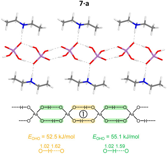
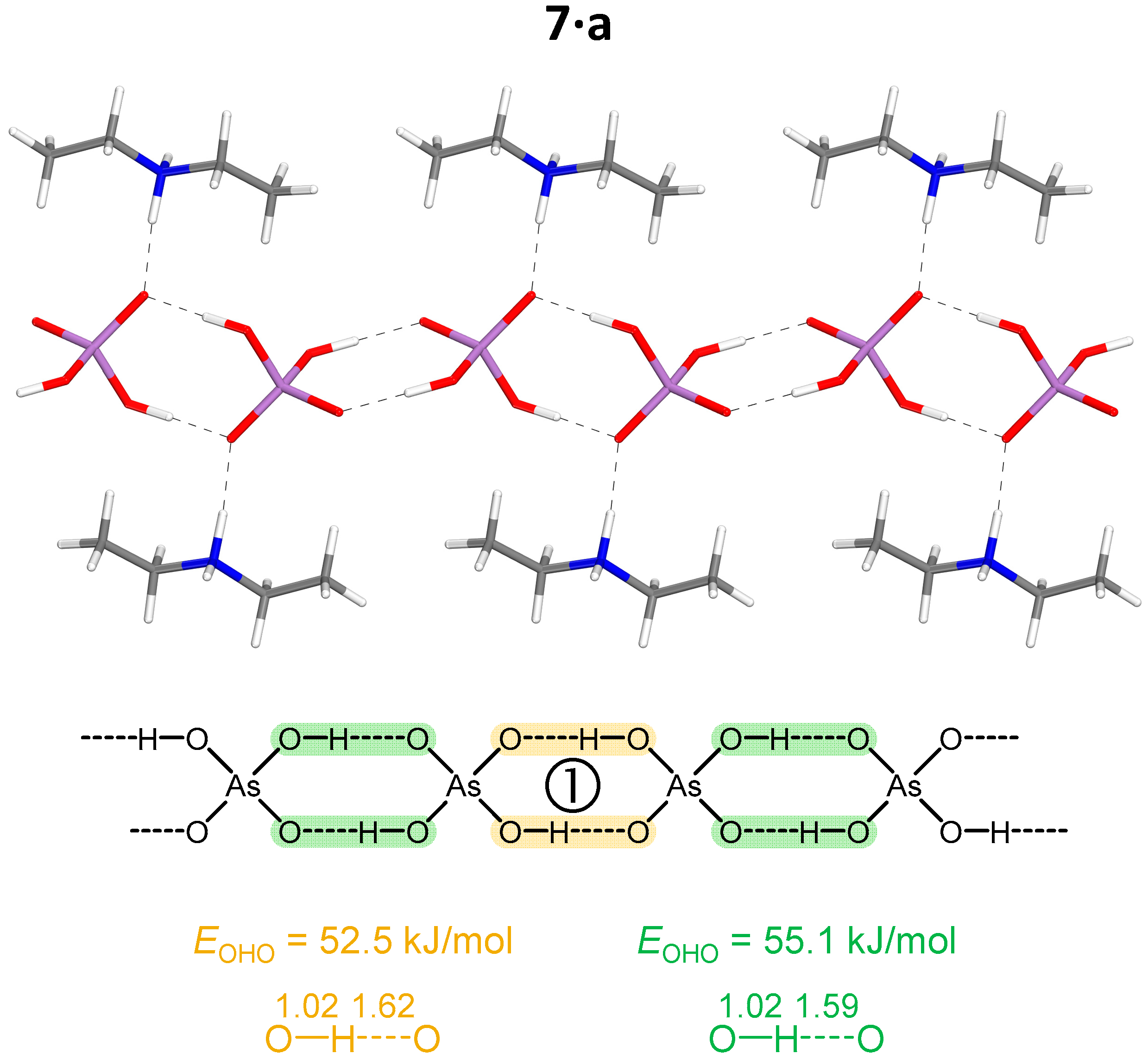
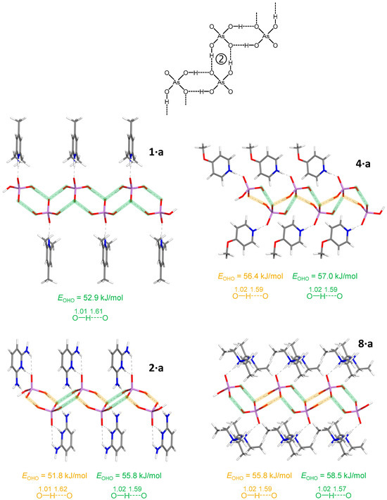
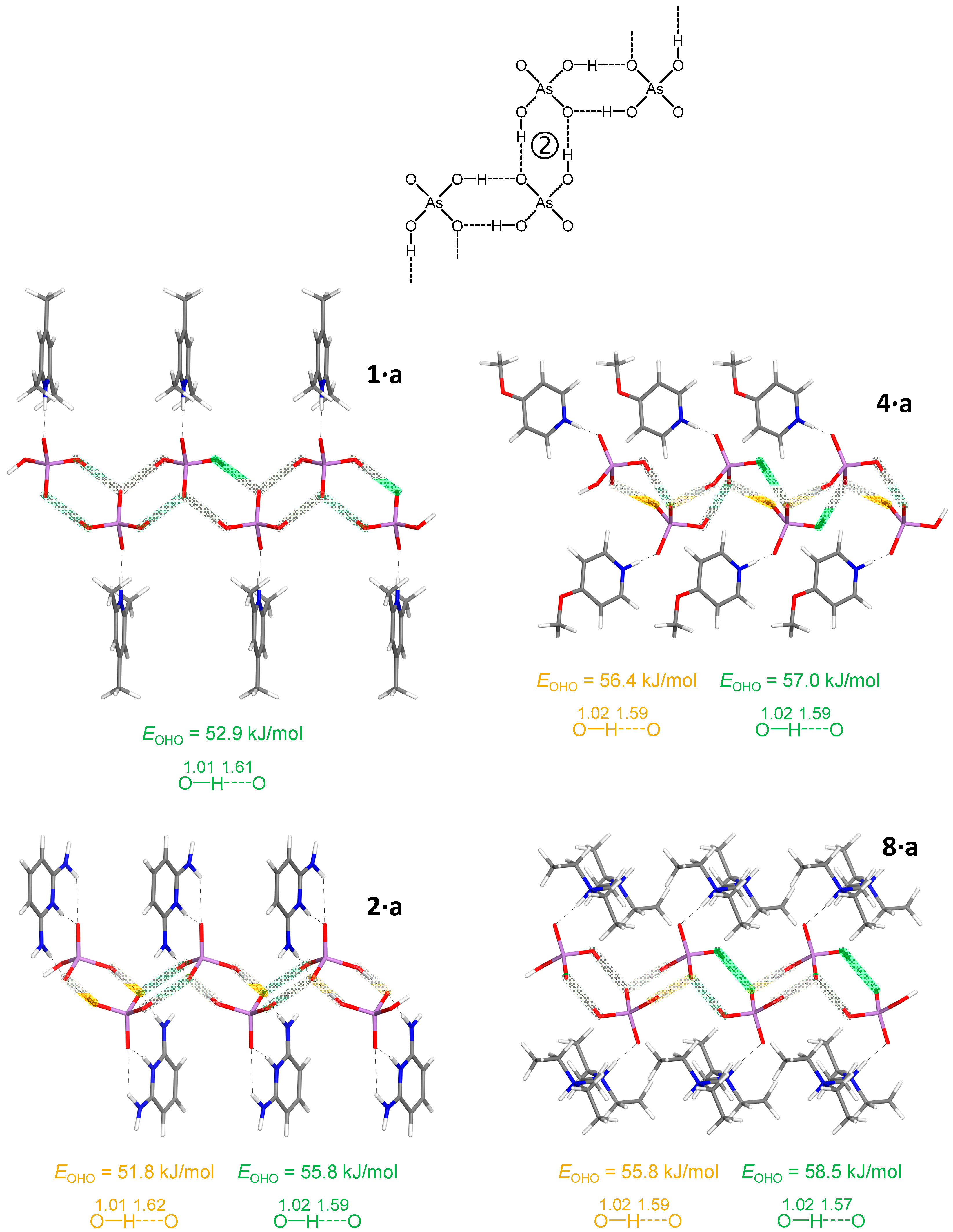
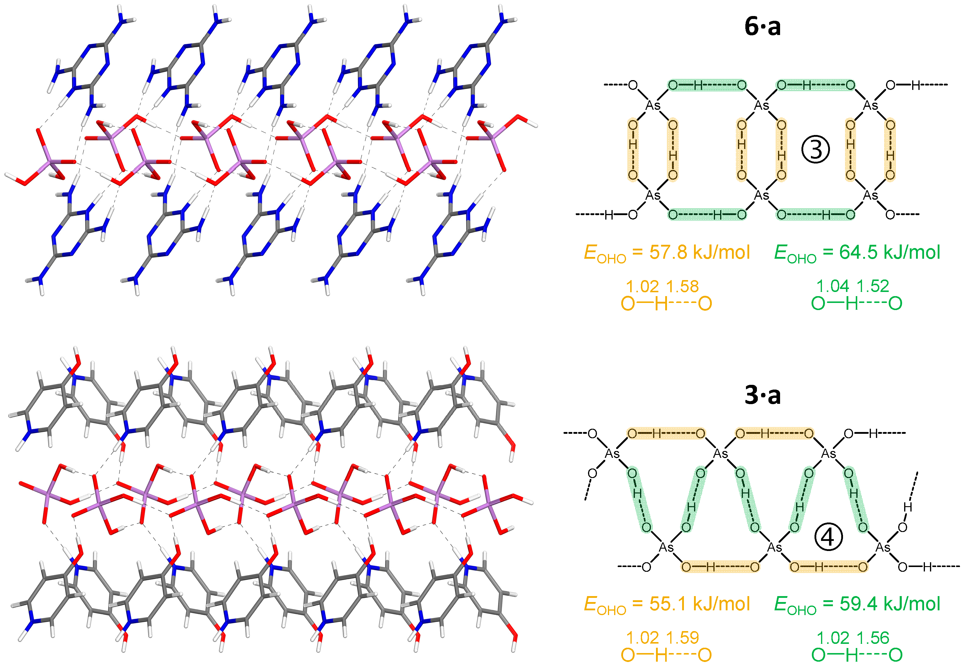
From a self-assembly viewpoint, compounds 1·a, 2·a, 4·a, 7·a, and 8·a show the presence of anion⋯anion ribbons wherein any H2AsO4− unit is H-bonded to two adjacent H2AsO4− units. These 1D networks are assembled via different supramolecular synthons: In 7·a, the ribbon is formed via the involvement of all the four O−/OH residues of H2AsO4− units (Figure 1), while in 1·a, 2·a, 4·a, and 8·a the ribbon is formed via the involvement of only three O−/OH residues (Figure 2). Anion⋯anion ribbons are also present in the crystals of also 3·a and 6·a, but different supramolecular synthons are adopted, and along these ribbons any anion is H-bonded to four adjacent anions in 3·a and to three adjacent anions in 6·a (Figure 3).
Figure 1.
Partial representation of the H-bonded ribbon in 7·a with optimized positions of the hydrogen atoms (motif ① of H2AsO4− anions’ assembling). Color coding (top representation): whitish, hydrogen; grey, carbon; blue, nitrogen; red, oxygen; violet, arsenic. Interatomic distances are given in Å. Bond energies, EOHO, are estimated from the electronic kinetic energy density values at BCP, Gbcp.
Figure 2.
Partial representation of the H-bonded chains in 1·a, 2·a, 4·a and 8·a with optimized positions of the hydrogen atoms (motif ② of H2AsO4− anions’ assembling). Color coding (top representation): whitish, hydrogen; grey, carbon; blue, nitrogen; red, oxygen; violet, arsenic. Interatomic distances are given in Å. EOHO values are estimated from the Gbcp values.
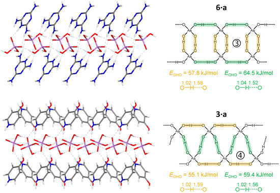
Figure 3.
Partial view of the H-bonded ribbons in 6·a and 3·a with optimized positions of the hydrogen atoms (motifs ③ and ④ of H2AsO4− anions’ assembling, respectively). Color coding (top representation): whitish, hydrogen; grey, carbon; blue, nitrogen; red, oxygen; violet, arsenic. Interatomic distances are given in Å. EOHO values are estimated from the Gbcp values.
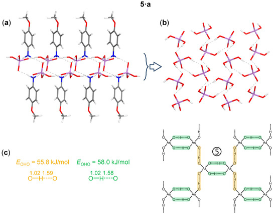
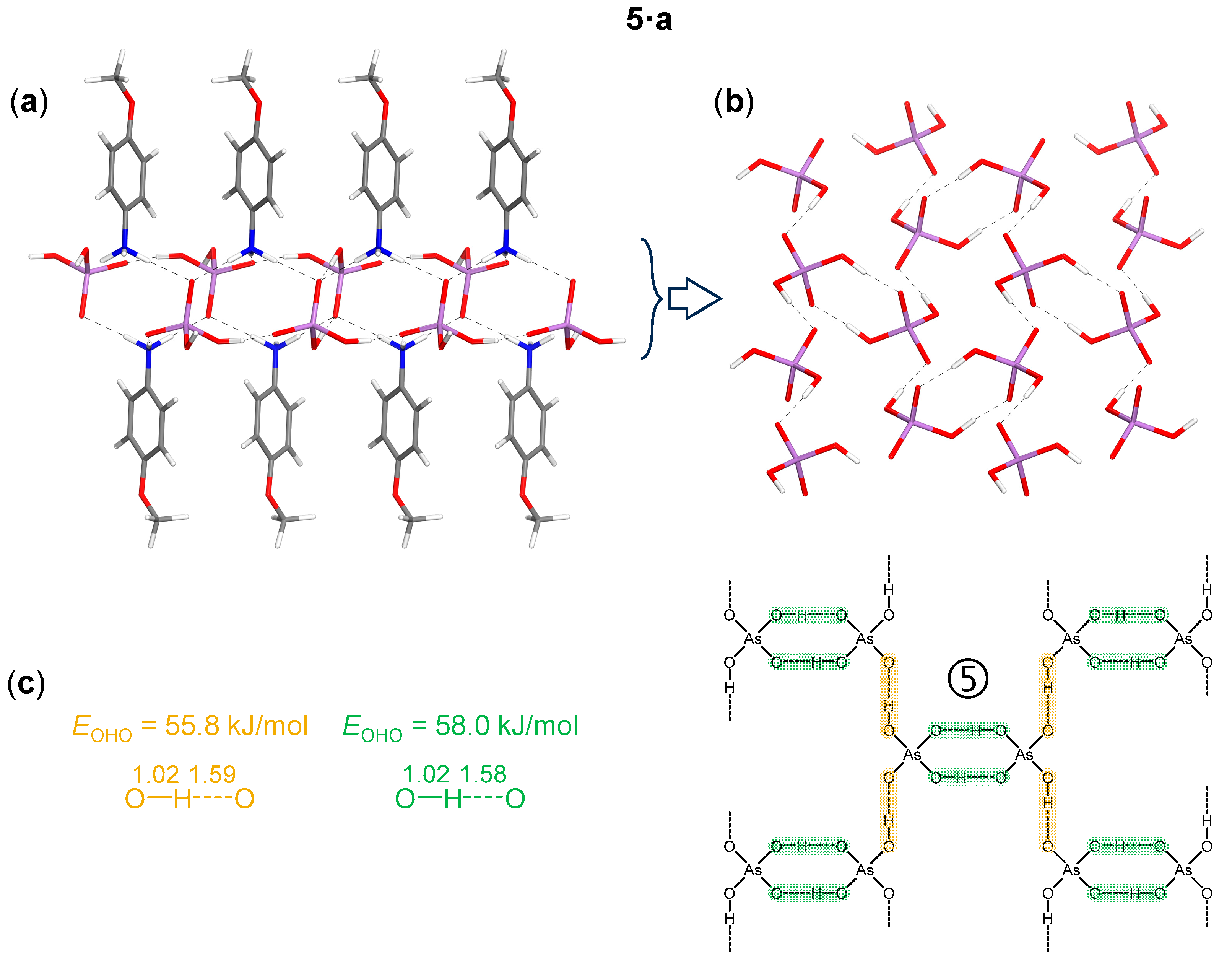
Compound 5·a represents a unique case, displaying an anion⋯anion self-assembly organized into a two-dimensional (2D) network (Figure 4).
Figure 4.
Partial view of the chainmail-like 2D H-bond network in 5·a with optimized positions of the hydrogen atoms (motif ⑤ of H2AsO4− anions’ assembling). (a): side view, (b): top view. Color coding (top representation): whitish, hydrogen; grey, carbon; blue, nitrogen; red, oxygen; violet, arsenic. Interatomic distances are given in Å. EOHO values (c) are estimated from the Gbcp values.
2,4,6-Trimethylpyridinium dihydrogen arsenate (1·a) crystallizes in the P21/m space group. In this compound, the average O⋯O distance within the AsO–H⋯OAs H-bond is 2.622 Å, a remarkably short value indicative of a strong H-bond, which overcomes the electrostatic anion–anion repulsion and enables the H2AsO4− anions to self-assemble. As summarized in Table 1, even shorter O⋯O distances are observed in compounds 2·a (already known in another polymorphic form corresponding to the monoclinic space group [73] and nevertheless exhibiting the same motif of H-bonds as described onwards), 3·a–8·a. This consistent trend toward short O⋯O separations suggests the presence of a robust network of H2AsO4− anions stabilized by H-bonding.
Table 1.
Space groups and O⋯O distances in the obtained hybrid organic–inorganic salts 1·a–8·a.
The overall crystal structures are stabilized by electrostatic attraction between oppositely charged cations and anions, as well as by a variety of non-covalent interactions spanning a wide range of lengths and strengths. These include: (i) major O–H⋯O and N–H⋯O hydrogen bonds, (ii) π-stacking interactions between N-heterocycles, observed in compounds 1·a–6·a, and (iii) numerous additional minor C–H⋯O contacts. In all eight salts, close contacts between anions and cations, characterized by N⋯O distances of 2.604–2.855 Å, contribute to the redistribution of negative charge within the anion, thereby enabling the formation of short inter-anionic OHO hydrogen bonds (see Table 1). This arrangement facilitates the anion⋯anion pairing through H-bonding observed in their crystal structures. Among the studied crystals, 1·a, 2·a, and 4·a show that aromatic cations engage in π–π stacking interactions, leading to the formation of spatially segregated cationic and anionic regions.
All major H-bonds involving H2AsO4− anions are fairly linear (except for the weakest NHO bonds), with angles in the ranges 169° < ∠(OHO) < 179° and 131° < ∠(NHO) < 179° in the computationally optimized structures. The crystal packings of the studied compounds are shown in Figures S1–S8. The association motifs of the amphoteric dihydrogen arsenate anions, which can simultaneously act as H-bond donors and acceptors, are more readily discussed in conjunction with the results of DFT calculations performed on the same crystals. Acknowledging the intrinsic difficulty of X-ray diffraction techniques in accurately locating hydrogen atoms, the hydrogen positions were selectively relaxed while keeping the remaining unit cell parameters fixed (see Table S1 and the resulting structures in Figure 1, Figure 2, Figure 3 and Figure 4). Analysis of these optimized structures allows five distinct motifs of H-bonding (supramolecular synthons) to be identified. These motifs can be described as: ribbons of cyclic dimers double-linked to each other (motifs ①, ②, and ③ in Figure 1, Figure 2 and Figure 3), ribbons of cyclic trimers fused to each other (motif ④ in Figure 3), and 2-D networks of cyclic dimers double-linked to each other (motif ⑤ in Figure 4).
The spatial arrangement of the H-bond networks in the studied crystals is constrained by the anisotropy of the outer electronic shell of the proton-accepting oxygen atoms, which dictates the preferred directions for the formation of O–H⋯O and N–H⋯O bonds involving the H2AsO4− anions. This anisotropy originates from the lone-pair domains, which give rise to toroidal and bean-shaped isosurfaces of the electron localization function (ELF) in the As–Oδ− and As–O–H groups, respectively (see Figures S9–S16). The ELF isosurfaces are very similar for the isolated anion and for the corresponding ion pair considered in the X-ray-derived geometry.
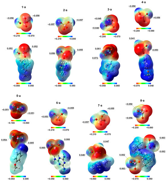
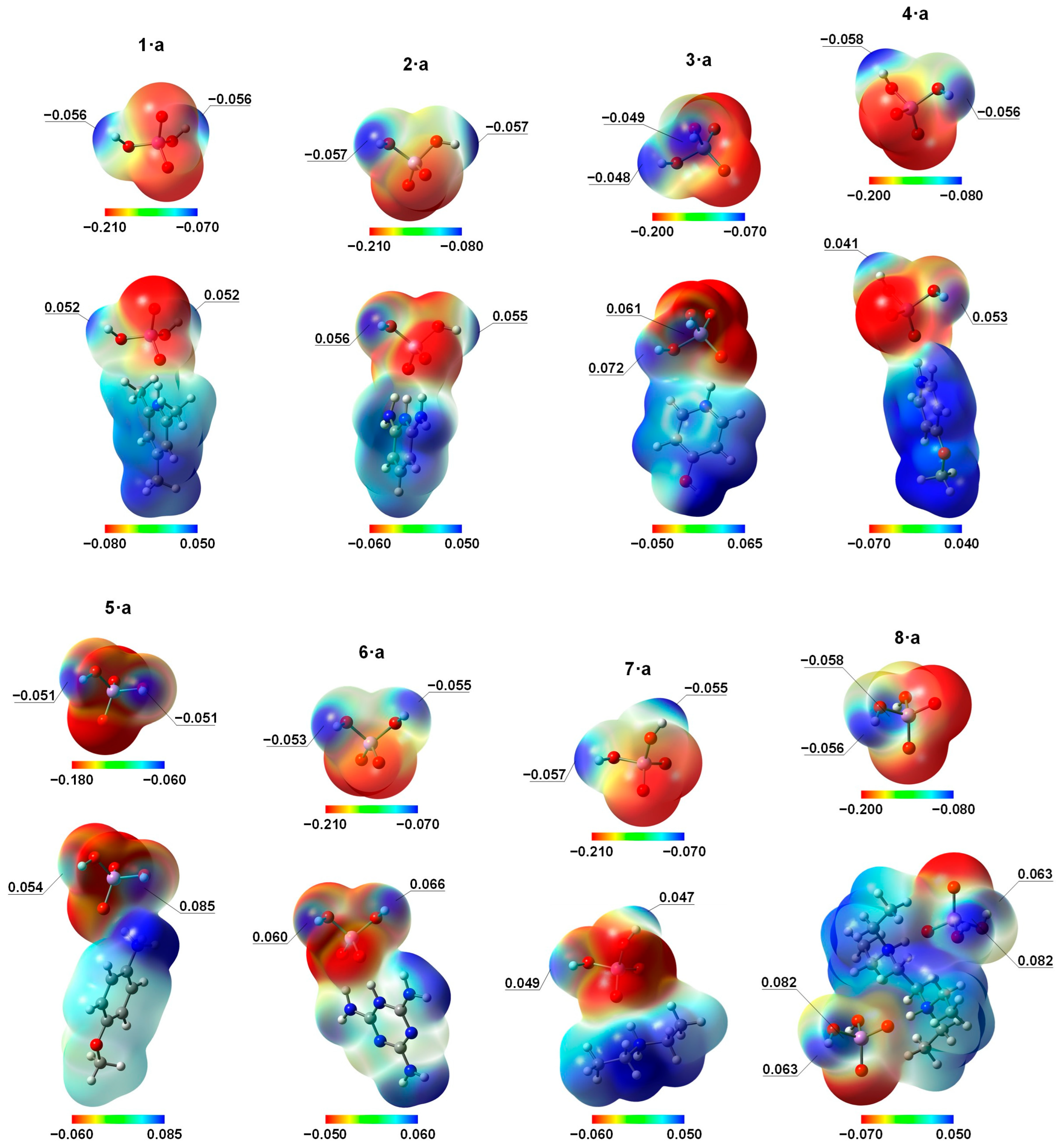
The 3D ESP maps represented via ED isosurfaces, color-coded with the ESP values, are shown for isolated H2AsO4− anions (Figure 5, upper stripe) and their complexes with counter cations (Figure 5, lower stripe) in crystalline geometry. Note that the color-coded ranges of ESP values are not symmetric and different in each case, being selected for maximum contrast between various regions. The key difference between isolated anions and ion pairs is in the electrophilicity of the H2AsO4− unit. The entire surface is negative for the isolated anions, but polarization effects in the ion pair create electrophilic sites on the hydrogen atoms of the As–O–H groups: the negative extremum of ESP around H atoms changes to the positive one (see the values given in Figure 5). Thus, the ESP maps demonstrate that the redistribution of the negative charge of H2AsO4− anion by adjacent cation is crucial to drive the self-assembly of the anions in all studied crystals. Indeed, the complexation between isolated H2AsO4− anions in crystalline geometry is energetically unfavorable: significant electrostatic repulsion hampers self-association, as evidenced by the results of additional calculations shown in Figure S17.
Figure 5.
ED 0.001 a.u. isosurfaces mapped by the molecular electrostatic potential (MESP) for H2AsO4− anion and the corresponding zwitterion in the X-ray geometries of the studied crystals (calculated at PW6B95-D3(BJ)/def2-TZVPD level of theory) but with preoptimized positions of the hydrogen atoms (at B3LYP-D*/POB-TZVP level of theory). Different color scales are used for the MESP (in a.u.).
On the example of 1·a, we further checked whether an additional stabilization of the anion⋯anion pairing takes place, emanating from the non-additive polarization effects upon sequential formation of the supramolecular chain of ion pairs, which might make the oxygen acceptor more negatively charged and the proton itself more positively charged in the H2AsO4− anion [74,75]. As can be gleaned from Figures S18 and S19, the ESP values on oxygen and hydrogen atoms along the H-bond direction change non-monotonically with an increase in the number of ion pair units in the chain, but reach a plateau—negative for the oxygen atoms and positive for the hydrogen atoms—at six bound units of 1·a.
Within the QTAIM methodology, the nature of an H-bond can be described by the topological properties of the ED, ρ, and various functions derived from it at the bond critical point (BCP) between the bridging proton and the proton-accepting atom [62,63,64,65,66,67,68,69,70,71,72,73,74,75,76,77,78,79,80,81,82,83,84]. For example, the signs of ▽2ρbcp (i.e., the Laplacian of the ED) and Hbcp = Gbcp + Vbcp (i.e., the total electronic energy density as a sum of the kinetic and potential energy densities Gbcp and Vbcp at the BCP) are used to differentiate between three types of interactions: non-covalent (region I, ▽2ρbcp > 0, Hbcp > 0), covalent (region II, ▽2ρbcp < 0, Hbcp < 0), and partially covalent (region III, ▽2ρbcp > 0, Hbcp < 0). Since V < 0 and G > 0, the sign of Hbcp reveals whether Vbcp or Gbcp dominates in the bonding region: a dominance of Vbcp (Hbcp < 0) indicates that accumulation of electronic charge in the internuclear region is stabilizing; conversely, if Gbcp > |Vbcp|, (Hbcp > 0), the internuclear charge accumulation is destabilizing, which is typical for a non-covalent interaction. The boundaries between regions I, II, and III are defined by Hbcp = 0 (i.e., |Vbcp|/Gbcp = 1) and by ▽2ρbcp = 0 (i.e., |Vbcp|/Gbcp = 2 from the virial equation). Within region I, the higher the bond degree index (BDI) Hbcp/ρbcp, the weaker the interaction. Similarly, in regions II and III, the higher the BDI, the higher the covalent character of the interaction.
For all studied structures, the calculated values of the above outlined parameters at the intermolecular BCPs of the OHO bonds between H2AsO4− anions are listed in Table 2. These bonds, being similar to each other, exhibit quite large ρbcp values of approximately 0.06 a.u. but positive ▽2ρbcp values indicating a locally charge-depleted domain, which places these bonds in region III at the border with region I, characterizing them as non-covalent interactions with a small degree of covalency [85]. This finding is in agreement with the relative values of other H-bond descriptors, meeting the inequalities 1 < |Vbcp|/Gbcp < 2, Gbcp/ρbcp < 1 (the kinetic energy per electron, in a.u.), and Hbcp/ρbcp < 0. The H-bond energies, EOHO, estimated using the equation EOHO = 0.429 Gbcp [86,87], fall in the range 51 < EOHO < 65 kJ/mol with an average value of 56 kJ/mol. Noteworthy, the full set of QTAIM parameters, the estimated H-bond energies, and the O⋯O interatomic distances for the interacting H2AsO4− anions are similar to those for the recently studied interacting HSeO4− anions [29], for which we have predicted the thermally accessible vibrational motion of the bridging protons from one heavy (oxygen) atom to another in the double-well potentials. For completeness, in ESI we provide QTAIM parameters at the intermolecular BCPs of the NHO bonds between cations and anions (as well as analogous OHO bonds in the case of 3·a), and also NHN bonds between cations in 6·a (see Figures S20–S27 and Table S2). In a nutshell, the ρ, ▽2ρ, V, G, |V|/G, H and H/ρ values indicate that the appertaining H-bonds in studied crystals can be classified as follows: (i) OHO, moderately strong bond (72 kJ/mol); (ii) NHO, from weak to moderately strong bonds (9 < ENHO < 66 kJ/mol); (iii) NHN, weak bonds (21 < ENHN < 24 kJ/mol). Despite the short N⋯O distances between cations and anions, the strong proton displacements towards nitrogen atoms (almost complete proton transfers) lead to a large degree of charge separation: the absolute values of AIM charges of the interacting molecular moieties are 0.81–0.87 e (see Table S3). Furthermore, an AIM charge of the bridging hydrogen atom might be considered as an additional descriptor of the H-bond strength that performs well over a sufficiently wide sampling, which is shown for NHO bonds in Figure S28a.
Table 2.
QTAIM parameters (in a.u.) at the intermolecular BCPs of the OHO bonds between H2AsO4− anions (electron density ρbcp and its Laplacian ▽2ρbcp; kinetic, potential, and total electronic energy densities Gbcp, Vbcp, and Hbcp, respectively; derived parameters Gbcp/ρbcp, |Vbcp|/Gbcp, and Hbcp/ρbcp), and estimated corresponding bond energies (in kJ/mol), EOHO = 0.429 Gbcp [86,87] for 1·a–8·a.
It is well-known that the electronic density of states (DOS) analysis provides valuable insight into the contribution of particular local atomic orbitals (AOs) to the overall electronic structure of solids over a specified energy range [88,89,90,91,92]. Projected crystal orbital Hamiltonian population (COHP) analysis is an extension of the DOS analysis aimed to characterize pairwise interactions between two atoms (two discrete groups of AOs) in solids [92,93,94,95,96]. By weighing DOS by the Hamiltonian matrix, COHP partitions the band energies into bonding (stabilizing) and antibonding (destabilizing) regions, thus providing information regarding the partial covalent character of hydrogen⋯acceptor interactions in molecular solids [97,98]. In this context, COHP analysis has proven itself to be complementary to the QTAIM analysis. Both approaches, being fundamentally different, share the same concept of gauging the nature and strength of H-bonds as a tangible quantity in a solid directly from its underlying electronic structure. An estimate of the covalent contribution to H-bonding interaction can be made by integrating stabilizing (negative) and destabilizing (positive) regions of the projected COHP values up to the highest occupied band (i.e., the Fermi energy level, εF), giving so-called IpCOHP values.
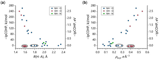
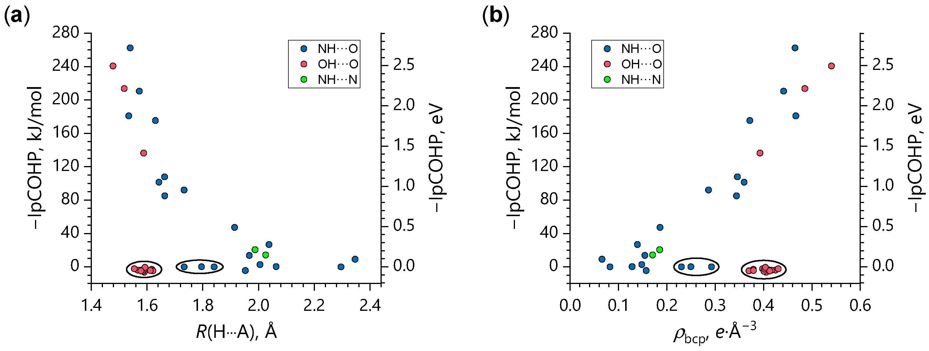
In Figure 6, –IpCOHP values are plotted against the corresponding H-bond lengths R(AH⋯O) and ρbcp densities for pairs of atoms involved in the AH⋯O interactions (A = O or N) for all studied structures. One obtains a variety of stabilizing interaction energies in the range 0–260 kJ/mol. On the one hand, the larger values in this set correlate reasonably well with the R(AH⋯O) and ρbcp values, similarly to what was previously observed for short OHO and OHN bonds (see Figures 2 and 3 in Ref. [97]). It is also appreciable that weak NHN bonds in 6·a, although there are only two unique ones of them, follow the same trend (green symbols in Figure 6). On the other hand, there is a group of −IpCOHP values that tightly cluster around zero (circled data points in Figure 6) regardless of the OH⋯O (NH⋯O) interatomic distance or the electronic charge concentration at the BCP. For OHO bonds (red symbols in Figure 6), this means that stabilizing and destabilizing pCOHP regions effectively cancel each other over the entire valence region, which indicates that the H-bonding anion–anion interactions are essentially closed-shell electrostatic in nature. This is in concordance with the positive ▽2ρbcp values (see Table 2), implying that the electronic charge is concentrated within the H and O atomic basins, rather than being shared between them. An exception is the following three OHO bonds with significant covalent contribution: the strongest among motifs 1–5 OHO bonds linking H2AsO4− dimers in 6·a (highlighted in green in Figure 3), the strongest and unique among studied structures OHO bonds between protonated pyridinium-4-ol cations and H2AsO4− anions (see Figure S22) in 3·a, and moderately strong OHO bonds in the tightly linked ribbons of H2AsO4− cyclic trimers in the same structure (highlighted in pale orange in Figure 3). In other words, it seems that the covalent character of an anion⋯anion interaction becomes detectable by the –IpCOHP values if either the bond is very strong (short) or the negative charge of the anion is strongly shifted towards the tightly bound cation. In the case of 3·a, such charge redistribution is facilitated by the fact that both OHO bonds in question (anion⋯anion and anion⋯cation ones) share the same acceptor atom. Previously, it was shown that not only the electrostatic interactions, but also orbital interactions (i.e., inter-fragment charge transfer and electron exchange) contribute significantly to the stabilizing energy of anion⋯anion H-bonds [99,100,101]. For NHO bonds (blue symbols in Figure 6), the −IpCOHP values are close to zero for three data points (circled in Figure 6), which correspond to the H-bonds that involve one H atom of the NH2+ group in 7·a and two H atoms of the NH3+ group in 5·a (see Figures S26 and S24, respectively). In other words, among the anti-cooperatively coupled H-bonds formed by NH2+/NH3+ groups, the shortest bond has a significant covalent character, while the other one(s) do not, though they remain relatively short. On the one hand, within the O1⋯H1–N–H2⋯O2 chain, the charge transfer from O1 to H1 makes H2 more negative, thus reducing its acidity and hence its tendency to bond with O2. On the other hand, the short H2⋯O2 distance could be due to the crystal packing effects. Beyond these remarks, it is not clear which feature of these interactions leads to such small −IpCOHP values.
Figure 6.
Dependencies of the negative integrated to the εF level projected COHP values (−IpCOHP) for the OH⋯O, NH⋯O, and NH⋯N interactions in all studied crystals on the corresponding interatomic R(H⋯A) distances (A = O, N) (a) and ρbcp densities (b).
Overall, comparison of the OH⋯O, NH⋯O, and NH⋯N interaction energies obtained by QTAIM and COHP analyses reveals that in crystals held by multiple non-covalent interactions, a gradual increase in the covalent contribution to the interaction energy upon the bond shortening is not always fulfilled, in contrast to what is often observed for systems with a single dominant H-bonding interaction [102].
4. Conclusions
Eight crystalline salts of arsenic acid with nitrogen-containing bases were investigated to analyze H-bond-mediated anion⋯anion self-assembly in dihydrogen arsenates. Single-crystal X-ray diffraction shows that, in all structures, H2AsO4− anions are involved in short and nearly linear O–H⋯O bonds (O⋯O ≤ 2.65 Å), leading to the formation of extended supramolecular architectures. Depending on the crystal packing, these interactions generate ribbon-like assemblies or a two-dimensional anionic network. On this basis, five recurrent H-bonding motifs were identified. DFT calculations were used to rationalize the experimental observations. ELF analysis indicates that the anisotropic distribution of the lone-pair electron density on oxygen atoms governs the preferred directions of H-bonding. Electrostatic potential maps show that interactions with protonated nitrogen bases induce a redistribution of electron density within the H2AsO4− anion, creating electrophilic regions on the As–O–H hydrogen atoms that enable anion⋯anion association despite Coulombic repulsion. Topological analysis of the crystalline electron density classifies the inter-anionic O–H⋯O bonds as moderately strong interactions, with estimated energies in the range of 50–65 kJ/mol and a limited covalent contribution. The ion pair systems display a high degree of ionicity, with AIM charges approaching ±0.87 e. Complementary COHP analysis indicates that most anion⋯anion H-bonds are predominantly electrostatic, while a considerable covalent component is observed only for the shortest O–H⋯O interactions or in cases of pronounced cation-induced charge redistribution. Overall, this study provides a systematic structural and electronic description of an H-bond-driven anion⋯anion self-assembly of dihydrogen arsenates.
Supplementary Materials
The following supporting information can be downloaded at: https://www.mdpi.com/article/10.3390/cryst16030162/s1, References [86,87] are cited in the supplementary materials. Figure S1: Crystal packing of 1·a; Figure S2: Crystal packing of 2·a; Figure S3: Crystal packing of 3·a; Figure S4: Crystal packing of 4·a; Figure S5: Crystal packing of 5·a; Figure S6: Crystal packing of 6·a; Figure S7: Crystal packing of 7·a; Figure S8: Crystal packing of 8·a; Figure S9: Isosurfaces of the ELF for oxygen atoms in H2AsO4− anion and zwitterion in 1·a; Figure S10: Isosurfaces of the ELF for oxygen atoms in H2AsO4− anion and zwitterion in 2·a; Figure S11: Isosurfaces of the ELF for oxygen atoms in H2AsO4− anion and zwitterion in 3·a; Figure S12: Isosurfaces of the ELF for oxygen atoms in H2AsO4− anion and zwitterion in 4·a; Figure S13: Isosurfaces of the ELF for oxygen atoms in H2AsO4− anion and zwitterion in 5·a; Figure S14: Isosurfaces of the ELF for oxygen atoms in H2AsO4− anion and zwitterion in 6·a; Figure S15: Isosurfaces of the ELF for oxygen atoms in H2AsO4− anion and zwitterion in 7·a; Figure S16: Isosurfaces of the ELF for oxygen atoms in H2AsO4− anion and zwitterion in 8·a; Figure S17: Complexation energies of the cyclic dimers/trimers of H2AsO4− anions in 1·a–8·a; Figure S18: MESP maps for an isolated H2AsO4− anion and clusters of zwitterions of 1·a; Figure S19: Dependencies of the ESP values on O and H atoms on the number of bound zwitterions of 1·a; Figure S20: Optimized interatomic distances of the NHO bonds and their energies in 1·a; Figure S21: Optimized interatomic distances of the NHO bonds and their energies in 2·a; Figure S22: Optimized interatomic distances of the NHO and OHO bonds and their energies in 3·a; Figure S23: Optimized interatomic distances of the NHO bonds and their energies in 4·a; Figure S24: Optimized interatomic distances of the NHO bonds and their energies in 5·a; Figure S25: Optimized interatomic distances of the NHO and NHN bonds and their energies in 6·a; Figure S26: Optimized interatomic distances of the NHO bonds and their energies in 7·a; Figure S27: Optimized interatomic distances of the NHO bonds and their energies in 8·a; Figure S28: Dependencies of the NHO, OHO, and NHN bonds’ energies on the AIM charges of the bridging protons and the H-bond lengths in the studied crystals; Figure S29: Unit cells of the disordered X-ray structure and the DFT-D* calculated ordered structures (isomorphs) of 1·a; Figure S30: Relative total energies of the isomorphs of 1·a; Table S1: Lengths of the XH (X = C, N, O) covalent bonds in 1·a–8·a according to the X-ray diffraction data and DFT-D* calculations; Table S2: QTAIM parameters at the intermolecular bond critical points of the NHO, OHO, and NHN bonds in 1·a–8·a; Table S3: AIM charges of the protonated N-bases and H2AsO4− anions in 1·a–8·a; Table S4: Crystal data and structure refinement for 1·a at 300 K; Table S5: Crystal data and structure refinement for 2·a at 300 K; Table S6: Crystal data and structure refinement for 3·a at 300 K; Table S7: Crystal data and structure refinement for 4·a at 300 K; Table S8: Crystal data and structure refinement for 5·a at 300 K; Table S9: Crystal data and structure refinement for 6·a at 100 K; Table S10: Crystal data and structure refinement for 7·a at 300 K; Table S11: Crystal data and structure refinement for 8·a at 300 K.
Author Contributions
Conceptualization, A.P., P.M.T., and G.R.; Methodology, P.M.T.; Formal analysis, E.R.C. and A.P.; Investigation, C.L.I., E.R.C., R.B. and A.B.; Data curation, A.P.; Writing—original draft, E.R.C.; Writing—review & editing, C.L.I., A.B., A.P. and P.M.T.; Supervision, A.P., P.M.T. and G.R. All authors have read and agreed to the published version of the manuscript.
Funding
This work received financial support from the Russian Science Foundation (RSF) under Grant No. 23-13-00095. A.P. and G.R. thank PRIN-2022, project GEMSTONE, no. 2022SK7JPA and PRIN-2022, project FLIPPER, no. 202224KAX8.
Data Availability Statement
The data supporting this article have been included as part of the Supplementary Information. Crystallographic data of compounds described in this paper have been deposited at the CCDC under Deposition Numbers 2521278 (for 1·a), 2521277 (for 2·a), 2521281 (for 3·a), 2521276 (for 4·a), 2521284 (for 5·a), 2521280 (for 6·a), 2521265 (for 7·a) and 2521279 (for 8·a) can be obtained free of charge from https://www.ccdc.cam.ac.uk/ (accessed on 10 February 2026).
Acknowledgments
The authors gratefully acknowledge the Computing Center of St. Petersburg State University Research Park (https://researchpark.spbu.ru/) for providing computational facilities.
Conflicts of Interest
There are no conflicts of interest.
References
- Stejskal, J.; Havlíček, D.; Císařová, I.; Matulková, I. Vibrational Spectroscopic and X-Ray Single Crystal Diffraction Investigation of Tetra-n-Alkylammonium Hydrogen Selenates. J. Chem. Crystallogr. 2017, 47, 59–68. [Google Scholar] [CrossRef] [Scilit]
- Belushkin, A.V.; Carlile, C.J.; Shuvalov, L.A. A Quasielastic Neutron Scattering Study of Protonic Transport in Hydrogen-Bonded Alkali Metal Hydrogen Sulphates and Selenates. Ferroelectrics 1995, 167, 21–31. [Google Scholar] [CrossRef] [Scilit]
- Ikeda, S.; Yamada, Y. Phase Transition in Hydrogen-Bonded Ferroelectric Compounds—Quantum Fluctuations versus Thermal Fluctuations. Phys. B Condens. Matter 1995, 213–214, 652–657. [Google Scholar] [CrossRef] [Scilit]
- Pavlenko, N.I. Protonic Conductivity at the Superionic Phase Transitions in the M3H(XO4)2 Crystal Group. J. Phys. Condens. Matter 1999, 11, 5099–5110. [Google Scholar] [CrossRef] [Scilit]
- Pavlenko, N.; Pietraszko, A.; Pawlowski, A.; Polomska, M.; Stasyuk, I.V.; Hilczer, B. Hydrogen Transport in Superionic System Rb3H(SeO4)2: A Revised Cooperative Migration Mechanism. Phys. Rev. B Condens. Matter 2011, 84, 064303. [Google Scholar] [CrossRef] [Scilit]
- Matsui, H.; Shimatani, K.; Ikemoto, Y.; Sasaki, T.; Matsuo, Y. Phonon-Assisted Proton Tunneling in the Hydrogen-Bonded Dimeric Selenates of Cs3H(SeO4)2. J. Chem. Phys. 2020, 152, 154502. [Google Scholar] [CrossRef] [Scilit]
- Matsui, H.; Fukuda, K.; Takano, S.; Ikemoto, Y.; Sasaki, T.; Matsuo, Y. Mechanisms of the Antiferro-Electric Ordering in Superprotonic Conductors Cs3H(SeO4)2 and Cs3D(SeO4)2. J. Chem. Phys. 2022, 156, 204504. [Google Scholar] [CrossRef] [Scilit] [PubMed]
- Zhao, W.; Flood, A.H.; White, N.G. Recognition and Applications of Anion-Anion Dimers Based on Anti-Electrostatic Hydrogen Bonds (AEHBs). Chem. Soc. Rev. 2020, 49, 7893–7906. [Google Scholar] [CrossRef] [Scilit] [PubMed]
- Martín-Fernández, C.; Montero-Campillo, M.M.; Alkorta, I. Hydrogen Bonds Are Never of an “Anti-Electrostatic” Nature: A Brief Tour of a Misleading Nomenclature. J. Phys. Chem. Lett. 2024, 15, 4105–4110. [Google Scholar] [CrossRef] [Scilit]
- Rajbanshi, A.; Wan, S.; Custelcean, R. Dihydrogen Phosphate Clusters: Trapping H2PO4− Tetramers and Hexamers in Urea-Functionalized Molecular Crystals. Cryst. Growth Des. 2013, 13, 2233–2237. [Google Scholar] [CrossRef] [Scilit]
- Drozd, M.; Baran, J. Polarized IR-Microscope Spectra of Guanidinium Hydrogenselenate Single Crystal. Spectrochim. Acta A Mol. Biomol. Spectrosc. 2005, 61, 2953–2965. [Google Scholar] [CrossRef] [Scilit]
- Akriche, S.; Rzaigui, M. 2-Amino-3-Nitro-Pyridinium Hydrogen Selenate. Acta Cryst. E 2009, 65, o1648. [Google Scholar] [CrossRef] [Scilit] [PubMed]
- White, N.G. Antielectrostatically hydrogen bonded anion dimers: Counter-intuitive, common and consistent. CrystEngComm 2019, 21, 4855–4858. [Google Scholar] [CrossRef] [Scilit]
- Lorenc, J.; Bryndal, I.; Marchewka, M.; Kucharska, E.; Lis, T.; Hanuza, J. Crystal and Molecular Structure of 2-Amino-5-Chloropyridinium Hydrogen Selenate—Its IR and Raman Spectra, DFT Calculations and Physicochemical Properties. J. Raman Spectrosc. 2008, 39, 863–872. [Google Scholar] [CrossRef] [Scilit]
- Maalej, W.; Ben Rached, A.; Mhiri, T.; Daoud, A.; Zouari, N.; Elaoud, Z. Vibrational Study, Phase Transitions and Electrical Properties of 4-Benzylpyridinium Monohydrogenselenate. J. Phys. Chem. Solids 2016, 96–97, 92–99. [Google Scholar] [CrossRef] [Scilit]
- Pizzi, A.; Dhaka, A.; Beccaria, R.; Resnati, G. Anion⋯anion Self-Assembly under the Control of σ- and π-Hole Bonds. Chem. Soc. Rev. 2024, 53, 6654. [Google Scholar] [CrossRef] [Scilit]
- Lo Iacono, C.; Pizzi, A.; Mahmudov, K.T.; Gomila, R.M.; Frontera, V.; Resnati, G. When CuCl42− and CuBr42− Form Anion⋯Anion Networks Assembled via Cu⋯Cl/Br Regium Bonds. Cryst. Growth Des. 2025, 25, 4338−4347. [Google Scholar] [CrossRef] [Scilit]
- Semyonov, O.; Antonkin, N.S.; Radzhabov, A.D.; Pizzi, A.; Lo Iacono, C.; Guselnikova, O.; Burguera, S.; Frontera, A.; Resnati, G.; Postnikov, P.S. Matere bond identification in non-crystalline materials by using X-ray photoelectron spectroscopy. Chem. Sci. 2025, 16, 18928–18935. [Google Scholar] [CrossRef] [Scilit]
- Gomila, R.M.; Frontera, A. Noble gas bonds facilitate anion⋯anion supramolecular assemblies: Insights from CSD and DFT analysis. CrystEngComm 2025, 27, 989–996. [Google Scholar] [CrossRef] [Scilit]
- Beccaria, R.; Dhaka, A.; Calabrese, M.; Pizzi, A.; Frontera, A.; Resnati, G. Chalcogen and Hydrogen Bond Team up in Driving Anion⋯Anion Self-Assembly. Chem. Eur. J. 2024, 30, e202303641. [Google Scholar] [CrossRef] [Scilit] [PubMed]
- Wilkinson, H.S.; Harrison, W.T.A. Guanidinium Dihydrogenarsenate. Acta Crystallogr. Sect. E Struct. Rep. Online 2005, 61, 2023–2025. [Google Scholar] [CrossRef] [Scilit]
- Wilkinson, H.S.; Harrison, W.T.A. Creatininium Dihydrogenarsenate. Acta Crystallogr. Sect. E Struct. Rep. Online 2006, 61, 1228–1230. [Google Scholar] [CrossRef] [Scilit]
- Chtioui, A.; Benhamada, L.; Jouini, A. Crystal Structure, Thermal Analysis and IR Spectroscopic Investigation of (C6H9N2)H2XO4 (X = As, P). Mater. Res. Bull. 2005, 40, 2243–2255. [Google Scholar] [CrossRef] [Scilit]
- Oueslati, A.; Rayes, A.; Ben Nasr, C.; Lefebvre, F. Synthesis and Characterization of 2-Amino-3-Methylpyridinium Dihydrogenomonoarsenate. Mater. Res. Bull. 2005, 40, 1680–1689. [Google Scholar] [CrossRef] [Scilit]
- Janczak, J.; Perpétuo, G.J. Hydrogen-Bonded Networks in Crystals of 1-(Diamino-Methyl-Ene)-Thio-Uron-1- Ium Perchlorate, Hydrogen Sulfate, Dihydrogen Phosphate and Dihydrogen Arsenate. Acta Crystallogr. Sect. C Cryst. Struct. Commun. 2008, 64, 330–334. [Google Scholar] [CrossRef] [Scilit]
- Kanagathara, N.; MaryAnjalin, F.; Ragavendran, V.; Dhanasekaran, D.; Usha, R.; Rao, R.G.S.; Marchewka, M.K. Experimental and Theoretical (DFT) Investigation of Crystallographic, Spectroscopic and Hirshfeld Surface Analysis of Anilinium Arsenate. J. Mol. Struct. 2021, 1223, 128965. [Google Scholar] [CrossRef] [Scilit]
- Anbalagan, G.; Marchewka, M.K.; Pawlus, K.; Kanagathara, N. Crystal Structure and Vibrational Spectra of Melaminium Arsenate. J. Mol. Struct. 2015, 1079, 407–413. [Google Scholar] [CrossRef] [Scilit]
- Roshini, S.R.A.; Kanagathara, N.; Marchewka, M.K.; Janczak, J.; Jayalakshmi, D. Growth, Structural, Optical, Morphological, Thermal, Laser Damage Threshold and Hardness Properties of Organic-Inorganic Crystal: L-Argininium (Bis)Dihydrogenarsenate. J. Mol. Struct. 2026, 1352, 144499. [Google Scholar] [CrossRef] [Scilit]
- Beccaria, R.; Pizzi, A.; Chakalov, E.; Resnati, G.; Tolstoy, P. Proton Delocalization in Short Hydrogen Bonds Assembling HSeO4− Anions into Supramolecular Adducts. Phys. Chem. Chem. Phys. 2025, 27, 13601–13617. [Google Scholar] [CrossRef] [Scilit] [PubMed]
- Shikina, N.D.; Zotov, A.B.; Tagirov, B.R. Influence of Pressure in the 0.1–100 MPa Interval on the First Dissociation Constant of Arsenous Acid in Water Solutions at 298.15 K. Russ. J. Phys. Chem. A 2010, 84, 1076–1078. [Google Scholar] [CrossRef] [Scilit]
- Rigaku. CrysAlis PRO; Rigaku Oxford Diffraction; Rigaku Corporation: Tokyo, Japan, 2021. [Google Scholar]
- Sheldrick, G.M. SHELXT—Integrated Space-Group and Crystal-Structure Determination. Acta Crystallogr. A Found. Adv. 2015, 71, 3–8. [Google Scholar] [CrossRef] [Scilit]
- Sheldrick, G.M. Crystal Structure Refinement with SHELXL. Acta Crystallogr. C Struct. Chem. 2015, 71, 3–8. [Google Scholar] [CrossRef] [Scilit]
- Dolomanov, O.V.; Bourhis, L.J.; Gildea, R.J.; Howard, J.A.K.; Puschmann, H. OLEX2: A Complete Structure Solution, Refinement and Analysis Program. J. Appl. Crystallogr. 2009, 42, 339–341. [Google Scholar] [CrossRef] [Scilit]
- MacRae, C.F.; Sovago, I.; Cottrell, S.J.; Galek, P.T.A.; McCabe, P.; Pidcock, E.; Platings, M.; Shields, G.P.; Stevens, J.S.; Towler, M.; et al. Mercury 4.0: From Visualization to Analysis, Design and Prediction. J. Appl. Crystallogr. 2020, 53, 226–235. [Google Scholar] [CrossRef] [Scilit]
- Spek, A.L. Structure Validation in Chemical Crystallography. Acta Crystallogr. D Biol. Crystallogr. 2009, 65, 148–155. [Google Scholar] [CrossRef] [Scilit] [PubMed]
- Dovesi, R.; Erba, A.; Orlando, R.; Zicovich-Wilson, C.M.; Civalleri, B.; Maschio, L.; Rérat, M.; Casassa, S.; Baima, J.; Salustro, S.; et al. Quantum-Mechanical Condensed Matter Simulations with CRYSTAL. Wiley Interdiscip. Rev. Comput. Mol. Sci. 2018, 8, e1360. [Google Scholar] [CrossRef] [Scilit]
- Becke, A.D. A New Mixing of Hartree-Fock and Local Density-Functional Theories. J. Chem. Phys. 1993, 98, 1372–1377. [Google Scholar] [CrossRef] [Scilit]
- Becke, A.D. Density-Functional Thermochemistry. III. The Role of Exact Exchange. J. Chem. Phys. 1993, 98, 5648–5652. [Google Scholar] [CrossRef] [Scilit]
- Vilela Oliveira, D.; Laun, J.; Peintinger, M.F.; Bredow, T. BSSE-Correction Scheme for Consistent Gaussian Basis Sets of Double- and Triple-Zeta Valence with Polarization Quality for Solid-State Calculations. J. Comput. Chem. 2019, 40, 2364–2376. [Google Scholar] [CrossRef] [Scilit]
- CRYSTAL—Basis Sets Library. Available online: https://www.crystal.unito.it/basis_sets.html (accessed on 10 February 2026).
- Grimme, S. Semiempirical GGA-Type Density Functional Constructed with a Long-Range Dispersion Correction. J. Comput. Chem. 2006, 27, 1787–1799. [Google Scholar] [CrossRef] [Scilit]
- Civalleri, B.; Zicovich-Wilson, C.M.; Valenzano, L.; Ugliengo, P. B3LYP Augmented with an Empirical Dispersion Term (B3LYP-D*) as Applied to Molecular Crystals. CrystEngComm 2008, 10, 405–410. [Google Scholar] [CrossRef] [Scilit]
- Dovesi, R.; Saunders, V.R.; Roetti, C.; Orlando, R.; Zicovich-Wilson, C.M.; Pascale, F.; Civalleri, B.; Doll, K.; Harrison, N.M.; Bush, I.J.; et al. CRYSTAL17 User’s Manual 2017; University of Torino: Torino, Italy, 2018. [Google Scholar]
- Ewald, P.P. Die Berechnung Optischer Und Elektrostatischer Gitterpotentiale. Ann. Phys. 1921, 369, 253–287. [Google Scholar] [CrossRef] [Scilit]
- Dovesi, R.; Pisani, C.; Roetti, C. Treatment of Coulomb Interactions in Hartree-Fock Calculations of Periodic Systems. Phys. Rev. B Condens. Matter Mater. Phys. 1983, 28, 5781–5792. [Google Scholar] [CrossRef] [Scilit]
- Towler, M.D.; Zupan, A.; Causà, M. Density Functional Theory in Periodic Systems Using Local Gaussian Basis Sets. Comput. Phys. Commun. 1996, 98, 181–205. [Google Scholar] [CrossRef] [Scilit]
- Monkhorst, H.J.; Pack, J.D. Special Points for Brillouin-Zone Integrations*. Phys. Rev. B 1976, 13, 5188–5192. [Google Scholar] [CrossRef] [Scilit]
- Maschio, L. Direct Inversion of the Iterative Subspace (DIIS) Convergence Accelerator for Crystalline Solids Employing Gaussian Basis Sets. Theor. Chem. Acc. 2018, 137, 60. [Google Scholar] [CrossRef] [Scilit]
- Doll, K.; Saunders, V.R.; Harrison, N.M. Analytical Hartree–Fock Gradients for Periodic Systems. Int. J. Quantum Chem. 2001, 82, 1–13. [Google Scholar] [CrossRef] [Scilit]
- Schlegel, H.B. Geometry Optimization. Wiley Interdiscip. Rev. Comput. Mol. Sci. 2011, 1, 790–809. [Google Scholar] [CrossRef] [Scilit]
- Bader, R.F.W. Atoms in Molecules—A Quantum Theory, Vol. 22 of International Series of Monographs in Chemistry; Oxford University Press: Oxford, UK, 1990. [Google Scholar]
- Bertini, L.; Cargnoni, F.; Gatti, C. Chemical Insight into Electron Density and Wave Functions: Software Developments and Applications to Crystals, Molecular Complexes and Materials Science. Theor. Chem. Acc. 2007, 117, 847–884. [Google Scholar] [CrossRef] [Scilit]
- Gatti, C.; Saunders, V.R.; Roetti, C. Crystal Field Effects on the Topological Properties of the Electron Density in Molecular Crystals: The Case of Urea. J. Chem. Phys. 1994, 101, 10686–10696. [Google Scholar] [CrossRef] [Scilit]
- Gatti, C.; Casassa, S. TOPOND14 User’s Manual (CNR-ISTM Milano, Milano, 2014); University of Milano: Milano, Italy, 2017. [Google Scholar]
- Casassa, S.; Erba, A.; Baima, J.; Orlando, R. Electron Density Analysis of Large (Molecular and Periodic) Systems: A Parallel Implementation. J. Comput. Chem. 2015, 36, 1940–1946. [Google Scholar] [CrossRef] [Scilit]
- Banerjee, A.; Adams, N.; Simons, J. Search for Stationary Points on Surfaces. J. Phys. Chem. 1985, 89, 52–57. [Google Scholar] [CrossRef] [Scilit]
- Keith, T.A. Molecules in Magnetic Fields. Ph.D. Thesis, McMaster University, Hamilton, ON, Canada, 1993. [Google Scholar]
- Popelier, P.L.A. A Robust Algorithm to Locate Automatically All Types of Critical Points in the Charge Density and Its Laplacian. Chem. Phys. Lett. 1994, 228, 160–164. [Google Scholar] [CrossRef] [Scilit]
- Frisch, M.J.; Trucks, G.W.; Schlegel, H.B.; Scuseria, G.E.; Robb, M.A.; Cheeseman, J.R.; Scalmani, G.; Barone, V.; Petersson, G.A.; Nakatsuji, H.; et al. Gaussian 16, Revision A.03; Gaussian, Inc.: Wallingford, CT, USA, 2016.
- Zhao, Y.; Truhlar, D.G. Design of Density Functionals That Are Broadly Accurate for Thermochemistry, Thermochemical Kinetics, and Nonbonded Interactions. J. Phys. Chem. A 2005, 109, 5656–5667. [Google Scholar] [CrossRef] [Scilit] [PubMed]
- Rappoport, D.; Furche, F. Property-Optimized Gaussian Basis Sets for Molecular Response Calculations. J. Chem. Phys. 2010, 133, 134105. [Google Scholar] [CrossRef] [Scilit]
- Pritchard, B.P.; Altarawy, D.; Didier, B.; Gibson, T.D.; Windus, T.L. New Basis Set Exchange: An Open, Up-to-Date Resource for the Molecular Sciences Community. J. Chem. Inf. Model. 2019, 59, 4814–4820. [Google Scholar] [CrossRef] [Scilit]
- Grimme, S.; Ehrlich, S.; Goerigk, L. Effect of the Damping Function in Dispersion Corrected Density Functional Theory. J. Comput. Chem. 2011, 32, 1456–1465. [Google Scholar] [CrossRef] [Scilit]
- Boys, S.F.; Bernardi, F. The Calculation of Small Molecular Interactions by the Differences of Separate Total Energies. Some Procedures with Reduced Errors. Mol. Phys. Int. J. Interface Between Chem. Phys. 1970, 19, 553–566. [Google Scholar] [CrossRef] [Scilit]
- Murray, J.S.; Politzer, P. The Electrostatic Potential: An Overview. Wiley Interdiscip. Rev. Comput. Mol. Sci. 2011, 1, 153–163. [Google Scholar] [CrossRef] [Scilit]
- Becke, A.D.; Edgecombe, K.E. A Simple Measure of Electron Localization in Atomic and Molecular Systems. J. Chem. Phys. 1990, 92, 5397–5403. [Google Scholar] [CrossRef] [Scilit]
- Lu, T.; Chen, F. Multiwfn: A Multifunctional Wavefunction Analyzer. J. Comput. Chem. 2012, 33, 580–592. [Google Scholar] [CrossRef] [Scilit]
- Zhang, J.; Lu, T. Efficient Evaluation of Electrostatic Potential with Computerized Optimized Code. Phys. Chem. Chem. Phys. 2021, 23, 20323–20328. [Google Scholar] [CrossRef] [Scilit]
- BIOVIA. Materials Studio 2017, version 17.1.0.48; Dassault Systèmes: San Diego, CA, USA, 2017. [Google Scholar]
- Dennington, R.D., II; Keith, T.A.; Millam, J.M. GaussView, version 6.0.16; Semichem Inc.: Shawnee, KS, USA, 2016. [Google Scholar]
- Origin. Origin 2018; OriginLab: Northampton, MA, USA, 2018. [Google Scholar]
- Bouaziz, E.; Ben Hassen, C.; Chniba-Boudjada, N.; Daoud, A.; Mhiri, T.; Boujelbene, M. Crystal Structure, Hirshfeld Surface Analysis, Vibrational, Thermal Behavior and UV Spectroscopy of (2,6-Diaminopyridinium) Dihydrogen Arsenate. J. Mol. Struct. 2017, 1145, 121–131. [Google Scholar] [CrossRef] [Scilit]
- Filot, I.A.W.; Palmans, A.R.A.; Hilbers, P.A.J.; van Santen, R.A.; Pidko, E.A.; de Greef, T.F. Understanding Cooperativity in Hydrogen-Bond-Induced Supramolecular Polymerization: A Density Functional Theory Study. J. Phys. Chem. B 2010, 114, 13667–13674. [Google Scholar] [CrossRef] [Scilit] [PubMed]
- Hoepfner, V.; Deringer, V.L.; Dronskowski, R. Hydrogen-Bonding Networks from First-Principles: Exploring the Guanidine Crystal. J. Phys. Chem. A 2012, 116, 4551–4559. [Google Scholar] [CrossRef] [Scilit] [PubMed]
- Bader, R.F.W.; Essén, H. The Characterization of Atomic Interactions. J. Chem. Phys. 1983, 80, 1943–1960. [Google Scholar] [CrossRef] [Scilit]
- Cremer, D.; Kraka, E. A Description of the Chemical Bond in Terms of Local Properties of Electron Density and Energy. Croat. Chem. Acta 1984, 57, 1259–1281. [Google Scholar]
- Koch, U.; Popelier, P.L.A. Characterization of C-H-O Hydrogen Bonds on the Basis of the Charge Density. J. Phys. Chem. 1995, 99, 9747–9754. [Google Scholar] [CrossRef] [Scilit]
- Espinosa, E.; Molins, E.; Lecomte, C. Hydrogen Bond Strengths Revealed by Topological Analyses of Experimentally Observed Electron Densities. Chem. Phys. Lett. 1998, 285, 170–173. [Google Scholar] [CrossRef] [Scilit]
- Jenkins, S.; Morrison, I. The Chemical Character of the Intermolecular Bonds of Seven Phases of Ice as Revealed by Ab Initio Calculation of Electron Densities. Chem. Phys. Lett. 2000, 317, 97–102. [Google Scholar] [CrossRef] [Scilit]
- Gatti, C.; May, E.; Destro, R.; Cargnoni, F. Fundamental Properties and Nature of CH··O Interactions in Crystals on the Basis of Experimental and Theoretical Charge Densities. The Case of 3,4-Bis(Dimethylamino)-3-Cyclobutene-1,2-Dione (DMACB) Crystal. J. Phys. Chem. A 2002, 106, 2707–2720. [Google Scholar] [CrossRef] [Scilit]
- Gatti, C. Chemical Bonding in Crystals: New Directions. Z. Krist. 2005, 220, 399–457. [Google Scholar] [CrossRef] [Scilit]
- Grabowski, S.J. What Is the Covalency of Hydrogen Bonding? Chem. Rev. 2011, 111, 2597–2625. [Google Scholar] [CrossRef] [Scilit]
- Espinosa, E.; Alkorta, I.; Elguero, J.; Molins, E. From Weak to Strong Interactions: A Comprehensive Analysis of the Topological and Energetic Properties of the Electron Density Distribution Involving X-H⋯F-Y Systems. J. Chem. Phys. 2002, 117, 5529–5542. [Google Scholar] [CrossRef] [Scilit]
- Vener, M.V.; Manaev, A.V.; Egorova, A.N.; Tsirelson, V.G. QTAIM Study of Strong H-Bonds with the O−H⋯A Fragment (A = O, N) in Three-Dimensional Periodical Crystals. J. Phys. Chem. A 2007, 111, 1155–1162. [Google Scholar] [CrossRef] [Scilit]
- Mata, I.; Alkorta, I.; Espinosa, E.; Molins, E. Relationships between Interaction Energy, Intermolecular Distance and Electron Density Properties in Hydrogen Bonded Complexes under External Electric Fields. Chem. Phys. Lett. 2011, 507, 185–189. [Google Scholar] [CrossRef] [Scilit]
- Vener, M.V.; Egorova, A.N.; Churakov, A.V.; Tsirelson, V.G. Intermolecular Hydrogen Bond Energies in Crystals Evaluated Using Electron Density Properties: DFT Computations with Periodic Boundary Conditions. J. Comput. Chem. 2012, 33, 2303–2309. [Google Scholar] [CrossRef] [Scilit]
- Bandura, A.V.; Kubicki, J.D.; Sofo, J.O. Comparisons of Multilayer H2O Adsorption onto the (110) Surfaces of α-TiO2 and SnO2 as Calculated with Density Functional Theory. J. Phys. Chem. B 2008, 112, 11616–11624. [Google Scholar] [CrossRef] [Scilit]
- Evarestov, R.A.; Bandura, A.V.; Losev, M.V.; Piskunov, S.; Zhukovskii, Y.F. Titania Nanotubes Modeled from 3- and 6-Layered (1 0 1) Anatase Sheets: Line Group Symmetry and Comparative Ab Initio LCAO Calculations. Phys. E Low Dimens. Syst. Nanostruct. 2010, 43, 266–278. [Google Scholar] [CrossRef] [Scilit]
- Laskowski, R.; Blaha, P. Understanding of 33S NMR Shielding in Inorganic Sulfides and Sulfates. J. Phys. Chem. C 2015, 119, 731–740. [Google Scholar] [CrossRef] [Scilit]
- Treviño, P.; Garcia-Castro, A.C.; López-Moreno, S.; Bautista-Hernández, A.; Bobocioiu, E.; Reynard, B.; Caracas, R.; Romero, A.H. Anharmonic Contribution to the Stabilization of Mg(OH)2 from First Principles. Phys. Chem. Chem. Phys. 2018, 20, 17799–17808. [Google Scholar] [CrossRef] [Scilit]
- Evarestov, R.A.; Kuzmin, A. Topological Analysis of Chemical Bonding in the Layered FePSe3 upon Pressure-Induced Phase Transitions. J. Comput. Chem. 2020, 41, 2610–2623. [Google Scholar] [CrossRef] [Scilit] [PubMed]
- Dronskowski, R.; Peter, E.B. Crystal Orbital Hamilton Populations (COHP). Energy-Resolved Visualization of Chemical Bonding in Solids Based on Density-Functional Calculations. J. Phys. Chem. 1993, 97, 8617–8624. [Google Scholar] [CrossRef] [Scilit]
- Deringer, V.L.; Tchougréeff, A.L.; Dronskowski, R. Crystal Orbital Hamilton Population (COHP) Analysis as Projected from Plane-Wave Basis Sets. J. Phys. Chem. A 2011, 115, 5461–5466. [Google Scholar] [CrossRef] [Scilit]
- Maintz, S.; Deringer, V.L.; Tchougréeff, A.L.; Dronskowski, R. Analytic Projection from Plane-Wave and PAW Wavefunctions and Application to Chemical-Bonding Analysis in Solids. J. Comput. Chem. 2013, 34, 2557–2567. [Google Scholar] [CrossRef] [Scilit]
- Ruggiero, M.T.; Erba, A.; Korter, T.M. Origins of Contrasting Copper Coordination Geometries in Crystalline Copper Sulfate Pentahydrate. Phys. Chem. Chem. Phys. 2015, 17, 31023–31029. [Google Scholar] [CrossRef] [Scilit]
- Deringer, V.L.; Englert, U.; Dronskowski, R. Covalency of Hydrogen Bonds in Solids Revisited. Chem. Commun. 2014, 50, 11547–11549. [Google Scholar] [CrossRef] [Scilit]
- Deringer, V.L.; George, J.; Dronskowski, R.; Englert, U. Plane-Wave Density Functional Theory Meets Molecular Crystals: Thermal Ellipsoids and Intermolecular Interactions. Acc. Chem. Res. 2017, 50, 1231–1239. [Google Scholar] [CrossRef] [Scilit]
- Frenking, G.; Caramori, G.F. No Need for a Re-Examination of the Electrostatic Notation of the Hydrogen Bonding: A Comment. Angew. Chemie Int. Ed. 2015, 54, 2596–2599. [Google Scholar] [CrossRef] [Scilit] [PubMed]
- Horn, P.R.; Mao, Y.; Head-Gordon, M. Probing Non-Covalent Interactions with a Second Generation Energy Decomposition Analysis Using Absolutely Localized Molecular Orbitals. Phys. Chem. Chem. Phys. 2016, 18, 23067–23079. [Google Scholar] [CrossRef] [Scilit] [PubMed]
- Wang, C.; Fu, Y.; Zhang, L.; Danovich, D.; Shaik, S.; Mo, Y. Hydrogen- and Halogen-Bonds between Ions of like Charges: Are They Anti-Electrostatic in Nature? J. Comput. Chem. 2018, 39, 481–487. [Google Scholar] [CrossRef] [Scilit] [PubMed]
- Gilli, G.; Gilli, P. Towards an Unified Hydrogen-Bond Theory. J. Mol. Struct. 2000, 552, 1–15. [Google Scholar] [CrossRef] [Scilit]

Disclaimer/Publisher’s Note: The statements, opinions and data contained in all publications are solely those of the individual author(s) and contributor(s) and not of MDPI and/or the editor(s). MDPI and/or the editor(s) disclaim responsibility for any injury to people or property resulting from any ideas, methods, instructions or products referred to in the content. |
© 2026 by the authors. Licensee MDPI, Basel, Switzerland. This article is an open access article distributed under the terms and conditions of the Creative Commons Attribution (CC BY) license.