Next Issue
Volume 17, January-1
Previous Issue
Volume 16, December-1


Cancers, Volume 16, Issue 24 (December-2 2024) – 164 articles

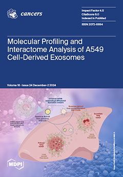
Cover Story (view full-size image): Exosomes, small extracellular vesicles, play a crucial role in intercellular communication. Cancer cell-derived exosomes exhibit tumorigenic properties, modulating the tumor microenvironment and promoting metastatic signaling in distant cells. This study aims to decipher the molecular profile and interactome of lung adenocarcinoma A549 cell-derived exosomes using multi-omics and bioinformatics approaches. We isolated and characterized exosomes from A549 cells, and through high-throughput proteomic and miRNA profiling, identified key molecules and their roles in recipient cell physiology. Comparative miRNA profiling with normal lung fibroblast (MRC-5) exosomes revealed tumor-associated miRNAs with potential further exploration, advancing our understanding of exosomal molecular components. View this paper
  • Issues are regarded as officially published after their release is announced to the table of contents alert mailing list.
  • You may sign up for e-mail alerts to receive table of contents of newly released issues.
  • PDF is the official format for papers published in both, html and pdf forms. To view the papers in pdf format, click on the "PDF Full-text" link, and use the free Adobe Reader to open them.
Order results
Result details
Section
Select all
Export citation of selected articles as:
26 pages, 1853 KiB  
Systematic Review
The Effect of Resistance and/or Aerobic Training on Quality of Life, Fitness, and Body Composition in Prostate Cancer Patients—A Systematic Review and Meta-Analysis
by Shimon Kempin, Alexander Buchner, Sarah Frederike Brose, Nina Schmidt-Hegemann, Matthias May, Ingmar Wolff, Anton Kravchuk, Christian Stief, Sabine D. Brookman-May and Benazir Enzinger
Cancers 2024, 16(24), 4286; https://doi.org/10.3390/cancers16244286 - 23 Dec 2024
Viewed by 743
Abstract
Background: Prostate cancer (PC) and its treatment are often associated with side effects such as fatigue, muscle loss, and diminished quality of life (QoL). Physical exercise, particularly resistance training (RT) and aerobic training (AT), has been suggested as a strategy to mitigate [...] Read more.
Background: Prostate cancer (PC) and its treatment are often associated with side effects such as fatigue, muscle loss, and diminished quality of life (QoL). Physical exercise, particularly resistance training (RT) and aerobic training (AT), has been suggested as a strategy to mitigate these effects. However, the comparative efficacy of RT, AT, and combined RT/AT on QoL, body composition, physical fitness, and laboratory markers in PC patients is still insufficiently understood. Methods: Randomized controlled trials (RCTs) investigating structured RT, AT, or combined RT/AT programs in PC patients undergoing various treatments were included. The primary outcome was QoL, assessed using EORTC QLQ-C30 and EORTC QLQ-PR25 questionnaires. Secondary outcomes included body composition, fitness, and laboratory parameters. The studies were sourced from PubMed, Embase, and CENTRAL through May 2024. The effect sizes were pooled using random-effects models, and the risk of bias was systematically assessed following the GRADE approach. Results: A total of 30 RCTs, encompassing 2216 PC patients, were analyzed. Combined RT/AT significantly improved QoL subdomains, including global health, and cognitive and sexual function, while reducing fatigue and urinary symptoms. RT alone improved body composition by increasing lean body mass and reducing body fat percentage. Both RT and combined RT/AT enhanced strength (chest and leg press) and VO2peak. No significant changes were observed in laboratory markers, such as PSA or lipid levels. The effects of isolated AT remain unclear due to limited data. Conclusions: RT and combined RT/AT significantly improve QoL, fitness, and body composition in PC patients, with no detectable effect on PSA or lipid levels. Further research is needed to elucidate the specific effects of AT and to investigate long-term outcomes. Full article
(This article belongs to the Special Issue Prostate Cancer Therapy: Supporting Strategies and Management Options)
Show Figures

Figure 1

15 pages, 4754 KiB  
Article
Discoidin Domain Receptor 2 Contributes to Breast Cancer Progression and Chemoresistance by Interacting with Collagen Type I
by Ai Sato, Kiyoshi Takagi, Momoka Yoshida, Mio Yamaguchi-Tanaka, Mikoto Sagehashi, Yasuhiro Miki, Minoru Miyashita and Takashi Suzuki
Cancers 2024, 16(24), 4285; https://doi.org/10.3390/cancers16244285 - 23 Dec 2024
Viewed by 591
Abstract
Background: Chemoresistance is an important issue to be solved in breast cancer. It is well known that the content and morphology of collagens in tumor tissues are drastically altered following chemotherapy, and discoidin domain receptor 2 (DDR2) is a unique type of [...] Read more.
Background: Chemoresistance is an important issue to be solved in breast cancer. It is well known that the content and morphology of collagens in tumor tissues are drastically altered following chemotherapy, and discoidin domain receptor 2 (DDR2) is a unique type of receptor tyrosine kinase (RTK). This RTK is activated by collagens, playing important roles in human malignancies. However, the contribution to the chemoresistance of DDR2 in terms of the association with collagens remains largely unclear in breast cancer. Methods: We immunolocalized DDR2 and collagen type I in 224 breast cancer tissues and subsequently conducted in vitro studies to confirm the role of DDR2 in breast cancer chemoresistance using chemosensitive and chemoresistant cell lines. Results: DDR2 immunoreactivity was positively correlated with aggressive behaviors of breast cancer and was significantly associated with an increased risk of recurrence, especially in those who received chemotherapy. Moreover, in vitro experiments demonstrated that DDR2 promoted the proliferative activity of breast cancer cells, and cell viability after epirubicin treatment was significantly maintained by DDR2 in a collagen I-dependent manner. Conclusions: These data suggested that DDR2 could be a poor prognostic factor associated with cell proliferation and chemotherapy resistance in human breast cancer. Full article
(This article belongs to the Special Issue Hormones and Tumors)
Show Figures

Figure 1

16 pages, 1342 KiB  
Article
Diffusion-Weighted MRI and Human Papillomavirus (HPV) Status in Oropharyngeal Cancer
by Heleen Bollen, Rüveyda Dok, Frederik De Keyzer, Sarah Deschuymer, Annouschka Laenen, Johannes Devos, Vincent Vandecaveye and Sandra Nuyts
Cancers 2024, 16(24), 4284; https://doi.org/10.3390/cancers16244284 - 23 Dec 2024
Viewed by 595
Abstract
Background: This study aimed to explore the differences in quantitative diffusion-weighted (DW) MRI parameters in oropharyngeal squamous cell carcinoma (OPC) based on Human Papillomavirus (HPV) status before and during radiotherapy (RT). Methods: Echo planar DW sequences acquired before and during (chemo)radiotherapy (CRT) of [...] Read more.
Background: This study aimed to explore the differences in quantitative diffusion-weighted (DW) MRI parameters in oropharyngeal squamous cell carcinoma (OPC) based on Human Papillomavirus (HPV) status before and during radiotherapy (RT). Methods: Echo planar DW sequences acquired before and during (chemo)radiotherapy (CRT) of 178 patients with histologically proven OPC were prospectively analyzed. The volumetric region of interest (ROI) was manually drawn on the apparent diffusion coefficient (ADC) map, and 105 DW-MRI radiomic parameters were extracted. Change in ADC values (Δ ADC) was calculated as the difference between baseline and during RT at week 4, normalized by the baseline values. Results: Pre-treatment first-order 10th percentile ADC and Gray Level co-occurrence matrix (GLCM)-correlation were significantly lower in HPV-positive compared with HPV-negative tumors (82.4 × 10⁻⁵ mm2/s vs. 90.3 × 10⁻⁵ mm2/s, p = 0.03 and 0.18 vs. 0.30, p < 0.01). In the fourth week of RT, all first-order ADC values were significantly higher in HPV-positive tumors (p < 0.01). Δ ADC mean was significantly higher for the HPV-positive compared with the HPV-negative OPC group (95% vs. 55%, p < 0.01). A predictive model for HPV status based on smoking status, alcohol consumption, GLCM correlation, and mean ADC and 10th percentile ADC values yielded an area under the curve of 0.77 (95% CI 0.70–0.84). Conclusions: Our results highlight the potential of DW-MR imaging as a non-invasive biomarker for the prediction of HPV status, although its current role remains supplementary to pathological confirmation. Full article
(This article belongs to the Special Issue Advances in Radiotherapy for Head and Neck Cancer)
Show Figures

Figure 1

14 pages, 3967 KiB  
Article
Clinical Introduction of Stem Cell Sparing Radiotherapy to Reduce the Risk of Xerostomia in Patients with Head and Neck Cancer
by Maria I. van Rijn-Dekker, Arjen van der Schaaf, Sanne W. Nienhuis, Antoinette S. Arents-Huls, Rachel B. Ger, Olga Hamming-Vrieze, Frank J. P. Hoebers, Mischa de Ridder, Sabrina Vigorito, Ellen M. Zwijnenburg, Johannes A. Langendijk, Peter van Luijk and Roel J. H. M. Steenbakkers
Cancers 2024, 16(24), 4283; https://doi.org/10.3390/cancers16244283 - 23 Dec 2024
Viewed by 546
Abstract
Background/Objectives: Studies have shown that dose to the parotid gland stem cell rich (SCR) regions should be reduced to lower the risk of xerostomia after radiotherapy (RT). This study aimed to assess whether stem cell sparing (SCS)-RT can be adopted in routine clinical [...] Read more.
Background/Objectives: Studies have shown that dose to the parotid gland stem cell rich (SCR) regions should be reduced to lower the risk of xerostomia after radiotherapy (RT). This study aimed to assess whether stem cell sparing (SCS)-RT can be adopted in routine clinical practice. Methods: Multiple planning studies were performed to compare SCS-RT with standard (ST)-RT using 30 head and neck cancer patients. Shifts in mean dose to the SCR regions (Dmean,SCR) and other organs at risk and their estimated impact on normal tissue complication probability (NTCP) for side-effects were compared using Wilcoxon signed-rank test. A multicenter study was performed (eight institutions, three patients) to test the generalizability of SCS-RT using the Friedman test. Results: Using photons, Dmean,SCR was reduced with median 4.1/3.5 Gy for ipsilateral/contralateral (p < 0.001). The largest reductions were when the SCR regions overlapped less with target volumes. Subsequently, NTCPs for xerostomia decreased (p < 0.001). Using protons, Dmean,SCR was also reduced (2.2/1.9 Gy for ipsilateral/contralateral, p < 0.002). Nevertheless, SCS-RT did not further decrease NTCPs for xerostomia (p > 0.17). Target coverage and prevention of other side-effects were not compromised. However, increased mean oral cavity dose was observed in some patients. Lastly, in the multicenter study Dmean,SCR could be reduced by slightly adjusting the standard optimization. Contralateral Dmean,SCR reductions differed between centers (p = 0.01), which was attributed to differences in ST-RT plans. Conclusions: Stem cell sparing radiotherapy can be clinically introduced by making small adjustments to the optimization strategy and can reduce the risk of xerostomia. Full article
(This article belongs to the Special Issue Genetic Analysis and Clinical Therapy in Head and Neck Cancers)
Show Figures

Figure 1

12 pages, 795 KiB  
Article
Association Between Reconstruction Technique and Clinical Outcomes in Advanced Gastric Cancer Patients Undergoing Proximal Gastrectomy
by Katarzyna Sędłak, Karol Rawicz-Pruszyński, Zuzanna Pelc, Radosław Mlak, Katarzyna Gęca, Magdalena Skórzewska, Krzysztof Zinkiewicz, Katarzyna Chawrylak and Wojciech P. Polkowski
Cancers 2024, 16(24), 4282; https://doi.org/10.3390/cancers16244282 - 23 Dec 2024
Viewed by 541
Abstract
Background: There is an upward shift in the incidence and localization of gastric cancer (GC). Proximal gastrectomy (PG) has been advocated as an alternative operation for upper-third GC. An uneventful postoperative course is currently measured using a well-defined textbook outcome (TO), which represents [...] Read more.
Background: There is an upward shift in the incidence and localization of gastric cancer (GC). Proximal gastrectomy (PG) has been advocated as an alternative operation for upper-third GC. An uneventful postoperative course is currently measured using a well-defined textbook outcome (TO), which represents a composite of surgical quality metrics. The aim of this study was to compare TO after two reconstruction methods following PG: double-tract reconstruction (DTR) and posterior esophagogastrostomy with partial neo-fundoplication (EGF). Materials and Methods: Primary proximal gastric adenocarcinoma patients who had undergone PG with DTR or EGF were included in this study. In a prospectively collected database, DTR and EGF were identified in 30 and 30 patients, respectively. Results: Patients with DTR had a 5.5-fold higher chance of achieving TO compared to those with EGF (OR = 5.67; p = 0.0266). No statistically significant differences in overall survival were noted when both reconstruction methods were compared. Conclusion: In patients with proximal GC undergoing PG, TO is more likely to be achieved using DTR compared to EGF, with similar overall survival. Randomized controlled trials are warranted to indicate the preferred reconstruction technique after PG. Full article
(This article belongs to the Section Clinical Research of Cancer)
Show Figures

Figure 1

18 pages, 2705 KiB  
Article
Cardiotoxicity in Breast Cancer: Impact of Clinical Classifications and Treatment on Heart Health
by Sergiu Ioan Murg, Loredana Matiș, Andrada Florina Moldovan, Andrada Florina Schwarz-Madar, Daniela Florina Trifan, Timea Claudia Ghitea and Mircea Ioachim Popescu
Cancers 2024, 16(24), 4281; https://doi.org/10.3390/cancers16244281 - 23 Dec 2024
Viewed by 603
Abstract
Background/Objectives: Cardio-oncology has become essential in addressing cardiovascular complications from cancer therapies. While advancements in treatments have improved survival rates, they also increase cardiovascular risks. This study evaluates the cardiotoxic effects of cytostatic treatments, examining the relationship between tumor characteristics, such as histopathology [...] Read more.
Background/Objectives: Cardio-oncology has become essential in addressing cardiovascular complications from cancer therapies. While advancements in treatments have improved survival rates, they also increase cardiovascular risks. This study evaluates the cardiotoxic effects of cytostatic treatments, examining the relationship between tumor characteristics, such as histopathology and TNM classification, and cardiovascular complications, aiming to improve cardiotoxicity prevention and management in oncology patients. Methods: We conducted a retrospective analysis of cancer patients treated with anthracyclines, HER2-targeted therapies, and radiotherapy. Cardiac function was monitored via echocardiography, focusing on global longitudinal strain and left ventricular ejection fraction (LVEF). Cardiac troponins and natriuretic peptides were measured to detect subclinical cardiotoxicity, with patients stratified by TNM cancer stage and histopathology. Results: Our analysis identified a significant association between certain cytostatic treatments, such as anthracyclines and HER2-targeted therapies, and a reduction in LVEF, particularly in patients with advanced-stage cancer. Biomarker assessments indicated early signs of cardiotoxicity in patients before clinical symptoms emerged. The findings also demonstrated a higher prevalence of cardiovascular complications in patients with pre-existing risk factors. Conclusions: This study highlights the importance of personalized treatment protocols in minimizing cardiotoxicity and improving the quality of life for oncology patients. Regular cardiac monitoring, combined with the use of biomarkers, can help identify high-risk patients early, allowing for timely interventions. Future research should focus on optimizing cardioprotective strategies to mitigate the cardiovascular risks associated with modern cancer therapies. Clinical Trial Registration: N/A (retrospective study). Full article
(This article belongs to the Section Clinical Research of Cancer)
Show Figures

Figure 1

13 pages, 2019 KiB  
Article
Efficacy of Neoadjuvant Hypofractionated Chemoradiotherapy in Elderly Patients with Locally Advanced Rectal Cancer: A Single-Center Retrospective Analysis
by Jae Seung Kim, Jaram Lee, Hyeung-min Park, Soo Young Lee, Chang Hyun Kim and Hyeong Rok Kim
Cancers 2024, 16(24), 4280; https://doi.org/10.3390/cancers16244280 - 23 Dec 2024
Viewed by 668
Abstract
Background/Objectives: The application of long-course chemoradiotherapy (LCRT) in elderly patients with locally advanced rectal cancer (LARC) can be challenging due to increased risks of complications associated with comorbidities and reduced functional status. This study aimed to assess the efficacy of neoadjuvant hypofractionated [...] Read more.
Background/Objectives: The application of long-course chemoradiotherapy (LCRT) in elderly patients with locally advanced rectal cancer (LARC) can be challenging due to increased risks of complications associated with comorbidities and reduced functional status. This study aimed to assess the efficacy of neoadjuvant hypofractionated chemoradiotherapy (HCRT) in elderly patients with mid-to-low LARC. Methods: We performed a retrospective review of patients diagnosed with LARC from January 2013 to December 2020 and included those aged 70 years or older. Patients were categorized into three groups based on their treatment strategies: neoadjuvant HCRT (33 or 35 Gy in 10 fractions), neoadjuvant LCRT, and upfront surgery. Comparative analyses were performed on clinicopathological characteristics, short-term outcomes, and long-term survival outcomes among these groups. Results: Among the 296 patients included, 30 (10.1%) received HCRT, 195 (65.9%) underwent standard LCRT, and 71 (24.0%) underwent upfront surgery. The baseline characteristics showed that the HCRT group had a higher American Society of Anesthesiologists (ASA) score (ASA score 3 or 4, HCRT 43.3% vs. LCRT 16.9% vs. upfront surgery 15.5%, p = 0.002). The HCRT group showed a significantly lower incidence of radiotherapy-related complications than the LCRT group (16.7% vs. 48.7%, p = 0.001). However, the rate of pathological complete response was significantly lower in the HCRT group (10.0% vs. 15.4%, p = 0.002). The 3-year relapse-free survival (83.0% vs. 77.2% vs. 83.2%; p = 0.411), 3-year local recurrence-free survival (93.1% vs. 93.2% vs. 93.5%; p = 0.464), and 5-year overall survival (65.1% vs. 67.0% vs. 67.7%; p = 0.682) were not significantly different between the three groups. Multivariate analysis also showed that the treatment strategy was not associated with survival outcomes. Conclusions: Neoadjuvant HCRT demonstrated reduced radiotherapy-related complications and acceptable long-term oncologic outcomes. Therefore, neoadjuvant HCRT may be considered as a viable alternative for elderly patients with LARC. Full article
(This article belongs to the Special Issue The Surgical Management of Colorectal Cancer)
Show Figures

Figure 1

21 pages, 501 KiB  
Systematic Review
Sentinel Lymph Node Biopsy: Is There a Role in Non-Melanoma Skin Cancer? A Systematic Review
by Lorenzo Borgognoni, Pietro Susini, Gianni Gerlini, Paola Brandani, Vanni Giannotti and Serena Sestini
Cancers 2024, 16(24), 4279; https://doi.org/10.3390/cancers16244279 - 23 Dec 2024
Viewed by 483
Abstract
Background/Objectives: Sentinel Lymph Node Biopsy (SLNB) aims at identifying clinically occult nodal metastases. It is the standard staging procedure for patients with T1b to T4 primary cutaneous melanoma. Moreover, it is recommended whenever the risk of a positive SLNB is >5%, according to [...] Read more.
Background/Objectives: Sentinel Lymph Node Biopsy (SLNB) aims at identifying clinically occult nodal metastases. It is the standard staging procedure for patients with T1b to T4 primary cutaneous melanoma. Moreover, it is recommended whenever the risk of a positive SLNB is >5%, according to the National Comprehensive Cancer Network Melanoma guidelines. When considering Non-Melanoma Skin Cancer (NMSC), the SLNB could play a role in tumors that mainly spreads via lymphatics, but strong evidence is missing. In this paper, the hot topics and controversies are reviewed; Methods: A PRISMA systematic review was carried out on the PubMed (MEDLINE) library from 2004–2024, searching for studies on SLNB in NMSC; Results: Seventy articles and 6379 patients undergoing SLNB for Squamous Cell Carcinoma (SCC), Merkel Cell Carcinoma (MCC), and Porocarcinoma were included. Overall, the SLNB positivity rate in these NMSCs was 24.4%, with an SNLB detection rate of 97.6%. Specifically, the SLNB positivity rate was 12.3% for high-risk cutaneous SCC, 24.4% for anogenital SCC, 29.3% for MCC, and 30.6% for Porocarcinoma. Most papers concluded that SLNB is safe, feasible, and significant in these malignancies; Conclusions: SLNB should be discussed and offered to every patient with MCC, and it should be discussed and considered in “high risk” SCC and Porocarcinoma for staging and prognostic purposes, aiming to identify a subgroup of patients who may benefit the most from early treatments. Full article
(This article belongs to the Special Issue Advances in Skin Cancer: Diagnosis, Treatment and Prognosis)
Show Figures

Figure 1

9 pages, 785 KiB  
Article
Effectiveness of the Korean National Cancer Screening Program in Reducing Colorectal Cancer Mortality
by Hyeon Ji Lee, Kyeongmin Lee, Byung Chang Kim, Jae Kwan Jun, Kui Son Choi and Mina Suh
Cancers 2024, 16(24), 4278; https://doi.org/10.3390/cancers16244278 - 23 Dec 2024
Viewed by 547
Abstract
Background: Whether colorectal cancer (CRC) screening with a fecal immunochemical test (FIT) reduces mortality remains unclear. In South Korea, CRC screening with a FIT for individuals aged ≥ 50 years has been part of the Korean National Cancer Screening Program (KNCSP) since 2004. [...] Read more.
Background: Whether colorectal cancer (CRC) screening with a fecal immunochemical test (FIT) reduces mortality remains unclear. In South Korea, CRC screening with a FIT for individuals aged ≥ 50 years has been part of the Korean National Cancer Screening Program (KNCSP) since 2004. The aim of this study was to evaluate the effectiveness of the KNCSP in reducing CRC-specific mortality. Methods: We conducted a nested case-control study using cohort-based data derived from the KNCSP database. The cohort included 5,944,540 colorectal cancer-free individuals aged ≥ 50 years as of 2004. Individuals who died after CRC diagnosis were defined as cases (n = 29,992) and their sociodemographic characteristics were matched to those of the selected controls. The effects of screening exposure, frequency, and time interval on CRC-specific mortality were analyzed according to age group. Conditional logistic regression analysis was performed. Results: Compared with individuals who had never been screened, those who had ever been screened showed an OR of 0.74 (95% CI, 0.71–0.76) for CRC-specific mortality. CRC-specific mortality decreased as the number of screenings increased. Similar results were observed for those aged 50–79 years; however, the results for those aged 75–79 years were not statistically significant. Moreover, those aged ≥ 80 years had the opposite results. Conclusions: CRC mass screening using FIT is effective for individuals aged 50–74 years; therefore, this study suggests that countries considering introducing national CRC screening implement FIT for those within this age range. Full article
(This article belongs to the Section Cancer Causes, Screening and Diagnosis)
Show Figures

Figure 1

1 pages, 144 KiB  
Correction
Correction: Tsuneki et al. Deep Learning-Based Screening of Urothelial Carcinoma in Whole Slide Images of Liquid-Based Cytology Urine Specimens. Cancers 2023, 15, 226
by Masayuki Tsuneki, Makoto Abe and Fahdi Kanavati
Cancers 2024, 16(24), 4277; https://doi.org/10.3390/cancers16244277 - 23 Dec 2024
Viewed by 323
Abstract
There were errors in the original publication [...] Full article
(This article belongs to the Special Issue Artificial Intelligence in Cancer Screening)
15 pages, 1486 KiB  
Review
Saliva in Balancing Oral and Systemic Health, Oral Cancer, and Beyond: A Narrative Review
by Kohei Okuyama and Souichi Yanamoto
Cancers 2024, 16(24), 4276; https://doi.org/10.3390/cancers16244276 - 23 Dec 2024
Viewed by 652
Abstract
Saliva plays a multifaceted role in oral health and systemic well-being. It supports digestion, protects oral tissues, maintains a healthy oral microbiome, and facilitates wound healing. Additionally, saliva serves as a diagnostic tool that reflects systemic health and disease/therapeutic states. Furthermore, although saliva [...] Read more.
Saliva plays a multifaceted role in oral health and systemic well-being. It supports digestion, protects oral tissues, maintains a healthy oral microbiome, and facilitates wound healing. Additionally, saliva serves as a diagnostic tool that reflects systemic health and disease/therapeutic states. Furthermore, although saliva shows a protective effect against oral cancer development, once tumor formation occurs, it may be involved in tumor progression and metastasis via exosomes and microRNAs. This review discusses the essential role of saliva; its relationship with the development, progression, and metastasis of head and neck squamous cell carcinoma (HNSCC); liquid biopsy tools for early diagnosis and monitoring of HNSCC; and the potential of exosomes as therapeutic agents. Full article
(This article belongs to the Special Issue Oral Potentially Malignant Disorders and Oral Cavity Cancer)
Show Figures

Figure 1

17 pages, 4889 KiB  
Article
Crosstalk Between Omental Adipose-Derived Stem Cells and Gastric Cancer Cells Regulates Cancer Stemness and Chemotherapy Resistance
by Jun Kinoshita, Kenta Doden, Yusuke Sakimura, Saki Hayashi, Hiroto Saito, Toshikatsu Tsuji, Daisuke Yamamoto, Hideki Moriyama, Toshinari Minamoto and Noriyuki Inaki
Cancers 2024, 16(24), 4275; https://doi.org/10.3390/cancers16244275 - 23 Dec 2024
Viewed by 581
Abstract
Background: Peritoneal metastasis (PM) remains a major challenge in patients with gastric cancer (GC) and occurs preferentially in adipose-rich organs, such as the omentum. Adipose-derived stem cells (ASCs) may influence cancer behavior. This study aimed to investigate whether ASCs isolated from the omentum [...] Read more.
Background: Peritoneal metastasis (PM) remains a major challenge in patients with gastric cancer (GC) and occurs preferentially in adipose-rich organs, such as the omentum. Adipose-derived stem cells (ASCs) may influence cancer behavior. This study aimed to investigate whether ASCs isolated from the omentum can act as progenitors of cancer-associated fibroblasts (CAFs) and analyze their effects on the cancer stem cell (CSC) niche and the treatment resistance of GC cells. Methods: ASCs were isolated from the human omentum and their cellular characteristics were analyzed during co-culturing with GC cells. Results: ASCs express CAF markers and promote desmoplasia in cancer stroma in a mouse xenograft model. When co-cultured with GC cells, ASCs enhanced the sphere-forming efficiency of MKN45 and MKN74 cells. ASCs increased IL-6 secretion and enhanced the expression of Nanog and CD44v6 in GC cells; however, these changes were suppressed by the inhibition of IL-6. Xenograft mouse models co-inoculated with MKN45 cells and ASCs showed enhanced CD44v6 and Nanog expression and markedly reduced apoptosis induced by 5-FU treatment. Conclusion: This study improves our understanding of ASCs’ role in PM treatment resistance and has demonstrated the potential for new treatment strategies targeting ASCs. Full article
(This article belongs to the Special Issue Insights into Cancer Stem Cells)
Show Figures

Figure 1

12 pages, 722 KiB  
Article
At-Home Care Program for Acute Myeloid Leukemia Induction Phase in Patients Treated with Venetoclax-Based Low-Intensity Regimens
by Alexandra Martínez-Roca, Carlos Jiménez-Vicente, Beatriz Merchán, Sandra Castaño-Diez, Inés Zugasti, Helena Brillembourg, Álex Bataller, Francesca Guijarro, Albert Cortés-Bullich, Ana Trigueros, Amanda Isabel Pérez-Valencia, Cristina Gallego, Nuria Ballestar, Luis Gerardo Rodríguez-Lobato, Esther Carcelero, Marina Díaz-Beyá, Jordi Esteve and Francesc Fernández-Avilés
Cancers 2024, 16(24), 4274; https://doi.org/10.3390/cancers16244274 - 23 Dec 2024
Viewed by 744
Abstract
Background: Even though venetoclax in combination with azacitidine (VenAza) is considered a low-intensity regimen, its patients present a high incidence of cytopenia and infections during the first courses, making the initial management a challenging phase. Methods: This difficulty in our center led to [...] Read more.
Background: Even though venetoclax in combination with azacitidine (VenAza) is considered a low-intensity regimen, its patients present a high incidence of cytopenia and infections during the first courses, making the initial management a challenging phase. Methods: This difficulty in our center led to the establishment of an At-Home (AH) program for ramp-up and follow-up patients during the VenAza combination induction phase focused on therapy administration, patient and caregiver education, and management of adverse events (AEs). A total of 70 patients with newly diagnosed acute myeloid leukemia (ND-AML) or relapsed/refractory AML (R/R AML) were treated with VenAza from March 2019 to May 2022. We compared outcomes between patients managed with a hospital-based (inpatient) approach and those managed through the AH program. Results: Despite most patients experiencing grade 3–4 cytopenias (96.9%), the incidence of serious infections and other AEs was comparable between both groups, with no significant difference in febrile neutropenia (42.3% vs. 27.8%, p = 0.38). Overall, the AH cohort demonstrated a significantly lower hospital readmission rate after ramp-up (29.5% vs. 84.6%, p = 0.001). Moreover, the inpatient cohort’s admission days were longer than in the AH cohort (13 vs. 8, p = 0.28). Conclusions: AH management was feasible and safe, leading to better resource use, enhanced patient comfort, and improved treatment compliance. The potential of AH programs for managing low-intensity chemotherapy regimens can reduce hospital admissions and subsequently improve patient and caregiver well-being. Full article
(This article belongs to the Collection Acute Myeloid Leukemia (AML))
Show Figures

Figure 1

16 pages, 8703 KiB  
Article
Disrupted Lipid Metabolism, Cytokine Signaling, and Dormancy: Hallmarks of Doxorubicin-Resistant Triple-Negative Breast Cancer Models
by Radhakrishnan Vishnubalaji and Nehad M. Alajez
Cancers 2024, 16(24), 4273; https://doi.org/10.3390/cancers16244273 - 23 Dec 2024
Viewed by 682
Abstract
Background: Chemoresistance in triple-negative breast cancer (TNBC) presents a significant clinical hurdle, limiting the efficacy of treatments like doxorubicin. This study aimed to explore the molecular changes associated with doxorubicin resistance and identify potential therapeutic targets to overcome this resistance, thereby improving treatment [...] Read more.
Background: Chemoresistance in triple-negative breast cancer (TNBC) presents a significant clinical hurdle, limiting the efficacy of treatments like doxorubicin. This study aimed to explore the molecular changes associated with doxorubicin resistance and identify potential therapeutic targets to overcome this resistance, thereby improving treatment outcomes for TNBC patients. Methods: Doxorubicin-resistant (DoxR) TNBC models (MDA-MB-231 and BT-549) were generated by exposing cells to increasing concentrations of doxorubicin. RNA sequencing (RNA-Seq) was performed using the Illumina platform, followed by bioinformatics analysis with CLC Genomics Workbench and iDEP. Functional assays assessed proliferation, sphere formation, migration, and cell cycle changes. Protein expression and phosphorylation were confirmed via Western blotting. Pathway and network analyses were conducted using Ingenuity Pathway Analysis (IPA) and STRING, while survival analysis was performed using Kaplan–Meier Plotter database. Results: DoxR cells exhibited reduced proliferation, sphere formation, and migration, but showed enhanced tolerance to doxorubicin. Increased CHK2 and p53 phosphorylation indicated cellular dormancy as a resistance mechanism. RNA-Seq analysis revealed upregulation of cytokine signaling and stress-response pathways, while cholesterol and lipid biosynthesis were suppressed. Activation of the IL1β cytokine network was prominent in DoxR cells, and CRISPR-Cas9 screens data identified dependencies on genes involved in rRNA biogenesis and metabolism. A 27-gene signature associated with doxorubicin resistance was linked to worse clinical outcomes in a large breast cancer cohort (HR = 1.76, FDR p < 2.0 × 10⁻¹³). Conclusions: This study uncovers potential therapeutic strategies for overcoming TNBC resistance, including dormancy reversal and targeting onco-ribosomal pathways and cytokine signaling networks, to improve the efficacy of doxorubicin-based treatments. Full article
(This article belongs to the Special Issue Molecular Insights into Drug Resistance in Cancer)
Show Figures

Graphical abstract

10 pages, 241 KiB  
Article
Safety and Efficacy of Initiating Parenteral Nutrition at Home, Home Start PN, in Advanced Peritoneal Metastasis
by Chunmeng Zhang, Ujwal Yanala, Mounika Addula, Sherry Adams, Louise Ocken, Patricia Skiendziel, Tia Bodkins and Jason M. Foster
Cancers 2024, 16(24), 4272; https://doi.org/10.3390/cancers16244272 - 23 Dec 2024
Viewed by 633
Abstract
Background: Patients with peritoneal carcinomatosis often experience intestinal failure throughout the course of their disease, and total parenteral nutrition (TPN) can be used as a temporary solution or as a bridge to definitive cytoreductive surgery. Guidelines for TPN are well established for inpatients [...] Read more.
Background: Patients with peritoneal carcinomatosis often experience intestinal failure throughout the course of their disease, and total parenteral nutrition (TPN) can be used as a temporary solution or as a bridge to definitive cytoreductive surgery. Guidelines for TPN are well established for inpatients and in 2014, guidelines were established for the initiation of TPN for outpatients in a home setting. However, the safety and efficacy of home start TPN in advanced oncology patients remain unknown. This study aims to explore the safety and efficacy of starting TPN in the home setting for patients with peritoneal carcinomatosis. Method: Health records of advanced cancer patients receiving TPN during 2009–2020 were retrospectively reviewed. Data pertaining to diagnosis, demographics, nutritional parameters, and outcomes including hospital readmission rates were collected. Safety was measured based on catheter-related complications and hospital admissions related to electrolyte or fluid imbalance due to TPN. Efficacy was determined by weight gain/stability and pre-albumin and albumin levels. The Fisher’s exact and Kruskal–Wallis tests were used to analyze the data. Results: Seventy TPN patients were identified, of which forty-two were home start (HS) and twenty-eight were in hospital (HP). The two groups were not significantly different in age, (HS: mean = 58.3 ± 13.9; HP: mean = 58.0 ± 13; p = 0.95), baseline body weight (p = 0.13), baseline albumin (p = 0.26) or pre-albumin (p = 0.48). At the end of treatment, the HS and HP groups had similar percentages of patients experiencing weight gain/stability (75% vs. 47%, p = 0.1), stable/increased pre-albumin (68% vs. 65%, p = 1), and stable/increased albumin levels (48% vs. 59%, p = 0.58). There was no difference in observed readmission between the groups (p = 0.79). At the end of treatment, 48% of the HS group and 36% of the HP group resumed an oral diet. Conclusions: This is the first study to present a comparison between home and hospital start TPN in advanced cancer patients, demonstrating that the initiation of outpatient TPN in the home setting is as safe and efficacious as TPN initiated in the hospital. Full article
(This article belongs to the Special Issue Advances in the Management of Peritoneal Surface Malignancies)
21 pages, 911 KiB  
Review
Insights into the Relationship Between the Gut Microbiome and Immune Checkpoint Inhibitors in Solid Tumors
by Sona Ciernikova, Aneta Sevcikova, Maria Novisedlakova and Michal Mego
Cancers 2024, 16(24), 4271; https://doi.org/10.3390/cancers16244271 - 23 Dec 2024
Viewed by 882
Abstract
Immunotherapy with immune checkpoint inhibitors represents a revolutionary approach to the treatment of solid tumors, including malignant melanoma, lung cancer, and gastrointestinal malignancies. Anti-CTLA-4 and anti-PD-1/PDL-1 therapies provide prolonged survival for cancer patients, but their efficacy and safety are highly variable. This review [...] Read more.
Immunotherapy with immune checkpoint inhibitors represents a revolutionary approach to the treatment of solid tumors, including malignant melanoma, lung cancer, and gastrointestinal malignancies. Anti-CTLA-4 and anti-PD-1/PDL-1 therapies provide prolonged survival for cancer patients, but their efficacy and safety are highly variable. This review focuses on the crucial role of the gut microbiome in modulating the efficacy and toxicity of immune checkpoint blockade. Studies suggest that the composition of the gut microbiome may influence the response to immunotherapy, with specific bacterial strains able to promote an anti-tumor immune response. On the other hand, dysbiosis may increase the risk of adverse effects, such as immune-mediated colitis. Interventions aimed at modulating the microbiome, including the use of probiotics, prebiotics, fecal microbial transplantation, or dietary modifications, represent promising strategies to increase treatment efficacy and reduce toxicity. The combination of immunotherapy with the microbiome-based strategy opens up new possibilities for personalized treatment. In addition, factors such as physical activity and nutritional supplementation may indirectly influence the gut ecosystem and consequently improve treatment outcomes in refractory patients, leading to enhanced patient responses and prolonged survival. Full article
(This article belongs to the Special Issue Immunotherapy of Solid Tumors and New Ideas of Anti-tumor Metastasis)
Show Figures

Graphical abstract

11 pages, 1167 KiB  
Article
Evaluation of Ex Vivo Shear Wave Elastography of Axillary Sentinel Lymph Nodes in Patients with Early Breast Cancer
by Riku Togawa, Helena Dahm, Manuel Feisst, Peter Sinn, André Hennigs, Juliane Nees, André Pfob, Benedikt Schäfgen, Anne Stieber, Oliver Zivanovic, Jörg Heil, Michael Golatta and Fabian Riedel
Cancers 2024, 16(24), 4270; https://doi.org/10.3390/cancers16244270 - 22 Dec 2024
Viewed by 670
Abstract
Background: The pretherapeutic assessment of axillary lymph node status is crucial in staging early breast cancer patients, significantly influencing their further treatment and prognosis. According to current guidelines, patients with clinically unsuspicious axillary status regularly undergo a biopsy of sentinel lymph nodes [...] Read more.
Background: The pretherapeutic assessment of axillary lymph node status is crucial in staging early breast cancer patients, significantly influencing their further treatment and prognosis. According to current guidelines, patients with clinically unsuspicious axillary status regularly undergo a biopsy of sentinel lymph nodes (SLNs), whereby metastasis is detected in up to 20% of cases. In recent years, the use of shear wave elastography (SWE) has been studied as an additional ultrasound tool for the non-invasive assessment of tumors in the breast parenchyma and axillary lymph nodes. Previous studies (examining the axilla in patients) have shown that metastases have significantly higher SWE values than benign nodes. Methods: This study aims to evaluate whether SWE can differentiate between tumor-free and metastatic-affected SLN ex vivo, i.e., by examining the pathological specimen. SWE was performed ex vivo on SLN specimens and compared with final histopathological results. Results: A total of 168 SLNs from 105 patients were measured using ex vivo SWE and subjected to standard histopathological processing. In this group, 17 metastases in 17 patients (16.19%) were detected. Tumor-free SLNs had a mean velocity of 1.33 ± 0.23 m/s, while metastatic nodes showed a mean velocity of 1.35 ± 0.29 m/s (p = 0.724). There was no significant difference in ex vivo SWE between benign and malignant SLNs in this population. Conclusions: Contrary to previous studies, this study did not find SWE effective in differentiating lymph node metastases. Further research is needed to clarify SWE’s potential role in axillary staging. Full article
(This article belongs to the Section Cancer Causes, Screening and Diagnosis)
Show Figures

Figure 1

25 pages, 3265 KiB  
Review
Anesthetic Approaches and Their Impact on Cancer Recurrence and Metastasis: A Comprehensive Review
by Hoon Choi and Wonjung Hwang
Cancers 2024, 16(24), 4269; https://doi.org/10.3390/cancers16244269 - 22 Dec 2024
Viewed by 909
Abstract
Cancer recurrence and metastasis remain critical challenges following surgical resection, influenced by complex perioperative mechanisms. This review explores how surgical stress triggers systemic changes, such as neuroendocrine responses, immune suppression, and inflammation, which promote the dissemination of residual cancer cells and circulating tumor [...] Read more.
Cancer recurrence and metastasis remain critical challenges following surgical resection, influenced by complex perioperative mechanisms. This review explores how surgical stress triggers systemic changes, such as neuroendocrine responses, immune suppression, and inflammation, which promote the dissemination of residual cancer cells and circulating tumor cells. Key mechanisms, such as epithelial–mesenchymal transition and angiogenesis, further enhance metastasis, while hypoxia-inducible factors and inflammatory responses create a microenvironment conducive to tumor progression. Anesthetic agents and techniques modulate these mechanisms in distinct ways. Inhaled anesthetics, such as sevoflurane, may suppress immune function by increasing catecholamines and cytokines, thereby promoting cancer progression. In contrast, propofol-based total intravenous anesthesia mitigates stress responses and preserves natural killer cell activity, supporting immune function. Opioids suppress immune surveillance and promote angiogenesis through the activation of the mu-opioid receptor. Opioid-sparing strategies using NSAIDs show potential in preserving immune function and reducing recurrence risk. Regional anesthesia offers benefits by reducing systemic stress and immune suppression, though the clinical outcomes remain inconsistent. Additionally, dexmedetomidine and ketamine exhibit dual effects, both enhancing and inhibiting tumor progression depending on the dosage and context. This review emphasizes the importance of individualized anesthetic strategies to optimize long-term cancer outcomes. While retrospective studies suggest potential benefits of propofol-based total intravenous anesthesia and regional anesthesia, further large-scale trials are essential to establish the definitive role of anesthetic management in cancer recurrence and survival. Full article
(This article belongs to the Special Issue Perioperative Management and Cancer Outcome)
Show Figures

Figure 1

36 pages, 4187 KiB  
Review
Advances for Managing Pancreatic Cystic Lesions: Integrating Imaging and AI Innovations
by Deniz Seyithanoglu, Gorkem Durak, Elif Keles, Alpay Medetalibeyoglu, Ziliang Hong, Zheyuan Zhang, Yavuz B. Taktak, Timurhan Cebeci, Pallavi Tiwari, Yuri S. Velichko, Cemal Yazici, Temel Tirkes, Frank H. Miller, Rajesh N. Keswani, Concetto Spampinato, Michael B. Wallace and Ulas Bagci
Cancers 2024, 16(24), 4268; https://doi.org/10.3390/cancers16244268 - 22 Dec 2024
Viewed by 834
Abstract
Pancreatic cystic lesions (PCLs) represent a spectrum of non-neoplasms and neoplasms with varying malignant potential, posing significant challenges in diagnosis and management. While some PCLs are precursors to pancreatic cancer, others remain benign, necessitating accurate differentiation for optimal patient care. Conventional approaches to [...] Read more.
Pancreatic cystic lesions (PCLs) represent a spectrum of non-neoplasms and neoplasms with varying malignant potential, posing significant challenges in diagnosis and management. While some PCLs are precursors to pancreatic cancer, others remain benign, necessitating accurate differentiation for optimal patient care. Conventional approaches to PCL management rely heavily on radiographic imaging, and endoscopic ultrasound (EUS) guided fine-needle aspiration (FNA), coupled with clinical and biochemical data. However, the observer-dependent nature of image interpretation and the complex morphology of PCLs can lead to diagnostic uncertainty and variability in patient management strategies. This review critically evaluates current PCL diagnosis and surveillance practices, showing features of the different lesions and highlighting the potential limitations of conventional methods. We then explore the potential of artificial intelligence (AI) to transform PCL management. AI-driven strategies, including deep learning algorithms for automated pancreas and lesion segmentation, and radiomics for analyzing heterogeneity, can improve diagnostic accuracy and risk stratification. These advanced techniques can provide more objective and reproducible assessments, aiding clinicians in decision-making regarding follow-up intervals and surgical interventions. Early results suggest that AI-driven methods can significantly improve patient outcomes by enabling earlier detection of high-risk lesions and reducing unnecessary procedures for benign cysts. Finally, this review emphasizes that AI-driven approaches could potentially reshape the landscape of PCL management, ultimately leading to improved pancreatic cancer prevention. Full article
(This article belongs to the Special Issue Medical Imaging and Artificial Intelligence in Cancer)
Show Figures

Figure 1

14 pages, 2289 KiB  
Article
Per-Irradiation Monitoring by kV-2D Acquisitions in Stereotactic Treatment of Spinal and Non-Spinal Bony Metastases Using an On-Board Imager of a Linear Accelerator
by Ahmed Hadj Henni, Geoffrey Martinage, Lucie Lebret and Ilias Arhoun
Cancers 2024, 16(24), 4267; https://doi.org/10.3390/cancers16244267 - 22 Dec 2024
Viewed by 508
Abstract
Background/Objectives: An on-board imager on a linear accelerator allows the acquisition of kV-2D images during irradiation. Overlaying specific structures on these images enables the visual verification of movement at regular frequencies. Our aim was to validate this tracking method for the stereotactic treatment [...] Read more.
Background/Objectives: An on-board imager on a linear accelerator allows the acquisition of kV-2D images during irradiation. Overlaying specific structures on these images enables the visual verification of movement at regular frequencies. Our aim was to validate this tracking method for the stereotactic treatment of bone metastases. Methods: Shifts in three translational directions were simulated using an anthropomorphic phantom. For these simulated shifts, planar images were acquired at different angles of incidence, with overlaid volumes of interest. A blinded test was then administered to the 18 participants to evaluate their decisions regarding whether to stop treatment. The results considered the experience of the operators. Quantitative analyses were performed on the intra-fractional images of 29 patients. Results: Participants analyzed each image with an average (standard deviation) decision time of 3.0 s (2.3). For offsets of 0.0, 1.0, 1.5, and 2.0 mm, the results were 78%, 93%, 90%, and 100% for the expert group and 78%, 70%, 79%, and 88% for the less-experienced group. Clinical feedback confirmed this guidance technique and extended it to non-spinal bony metastases. Sudden movements exceeding the 2.0 mm threshold occurred in 3.3% of the analyzed fractions, with a detection rate of 97.8% for vertebral locations. For non-vertebral bone locations, movements exceeding a threshold of 3.0 mm occurred in 3.5% of cases and were detected in 96.5%. Conclusions: The clinical use of planar OBI and superimposed structures for visual-image guidance in bone stereotactic treatment was validated using an anthropomorphic phantom and clinical feedback. Full article
Show Figures

Figure 1

24 pages, 4058 KiB  
Review
Targeting of Non-Classical Human Leukocyte Antigens as Novel Therapeutic Strategies in Cancer
by Javier David Benitez Fuentes, Jorge Bartolome Arcilla, Kauzar Mohamed Mohamed, Alfonso Lopez de Sa, Alicia de Luna Aguilar, Kissy Guevara-Hoyer, Pablo Ballestin Martinez, Antonio David Lazaro Sanchez, Edgardo D. Carosella, Alberto Ocaña and Silvia Sánchez-Ramon
Cancers 2024, 16(24), 4266; https://doi.org/10.3390/cancers16244266 - 22 Dec 2024
Viewed by 977
Abstract
Human leukocyte antigens (HLAs) are essential regulators of immune responses against cancer, with classical HLAs well-documented for their role in tumor recognition and immune surveillance. In recent years, non-classical HLAs—including HLA-E, HLA-F, HLA-G, and HLA-H—have emerged as critical players in the immune landscape [...] Read more.
Human leukocyte antigens (HLAs) are essential regulators of immune responses against cancer, with classical HLAs well-documented for their role in tumor recognition and immune surveillance. In recent years, non-classical HLAs—including HLA-E, HLA-F, HLA-G, and HLA-H—have emerged as critical players in the immune landscape of cancer due to their diverse and less conventional functions in immune modulation. These molecules exhibit unique mechanisms that enable tumors to escape immune detection, promote tumor progression, and contribute to therapeutic resistance. This review provides a comprehensive examination of the current understanding of non-classical HLAs in solid cancers, focusing on their specific roles in shaping the tumor microenvironment and influencing immune responses. By analyzing how HLA-E, HLA-F, HLA-G, and HLA-H modulate interactions with immune cells, such as T cells, natural killer cells, and antigen-presenting cells, we highlight key pathways through which these molecules contribute to immune evasion and metastasis. Additionally, we review promising therapeutic strategies aimed at targeting non-classical HLAs, including emerging immunotherapies that could potentially enhance cancer treatment outcomes by reversing immune suppression within tumors. Understanding the influence of these non-classical HLAs in solid cancers may offer new insights into cancer immunology and may lead to the development of innovative and more effective immunotherapeutic approaches. This review underscores the importance of non-classical HLAs as potential therapeutic targets, providing a necessary foundation for future studies in the evolving field of cancer immunotherapy. Full article
Show Figures

Figure 1

11 pages, 1790 KiB  
Article
Minimizing Long-Term Toxicities for Patients with Primary Mediastinal B-Cell Lymphoma Undergoing Modern Radiotherapy: Results from a Monocentric Biophysical Risk Evaluation
by Andrea Baehr, Sebastian Schäfer, Maria Jäckel, Saskia Alexandra Becker, Susanne Ghandili, Maximilian Grohmann, Hans Theodor Eich and Michael Oertel
Cancers 2024, 16(24), 4265; https://doi.org/10.3390/cancers16244265 - 22 Dec 2024
Viewed by 618
Abstract
Introduction: Primary mediastinal B-cell lymphoma (PMBCL) is a rare form of aggressive B-cell lymphoma with a predominant onset in young patients. The minimization of potential (late) side effects is of cardinal interest for these patients. An anticipation of the individual risk profile is [...] Read more.
Introduction: Primary mediastinal B-cell lymphoma (PMBCL) is a rare form of aggressive B-cell lymphoma with a predominant onset in young patients. The minimization of potential (late) side effects is of cardinal interest for these patients. An anticipation of the individual risk profile is desirable to counsel the patient on the putative impact of radiotherapy (RT). Methods: RT plans for a cohort of 25 patients with PMBCL were prospectively designed. One plan with two parallel- opposing fields (APPA) and another with volume-modulated arc therapy (VMAT) technique with 40 Gy in 2 Gy fractions each. Normal The normal tissue complication probability (NTCP) was calculated using the Lyman-–Kutcher-–Burman model for heart, lung and oesophageal toxicity. Results: APPA planning resulted in lower median doses (Dmedian) for the heart and lungs, whereas all other dose metrics for heart, lungs and esophagus were lower in VMAT planning. A significant difference in the mean NTCPs when comparing the APPA to VMAT plans was seen for increased cardiac mortality, pneumonitis and esophagitis. PTV size correlated with increased cardiac mortality and esophagitis in both plan variations and with pneumonitis for VMAT plans. Dmean, Dmedian, and V20Gy correlated with the risk for pneumonitis, and Dmean, Dmedian, and V1% with the risk for esophagitis in both variants. Conclusions: We showed decreased risk of different NTCPs for VMAT and APPA planning for thoracic toxicities. The use of an IMRT technique like VMAT showed advantages for several DVH metrics in organs at risk and should therefore be recommended for radiation treatment of PMBCL. Full article
(This article belongs to the Section Cancer Therapy)
Show Figures

Figure 1

12 pages, 440 KiB  
Article
The Importance of Early Detection and Prevention of Atypical Skin Lesions and Other Melanoma Risk Factors in a Younger Population
by Paulina Karp, Katarzyna Karp, Marcelina Kądziela, Radosław Zajdel and Agnieszka Żebrowska
Cancers 2024, 16(24), 4264; https://doi.org/10.3390/cancers16244264 - 22 Dec 2024
Viewed by 533
Abstract
Background/Objectives: Skin cancer is becoming increasingly common due to increasing risk factors such as excessive ultraviolet (UV) radiation, genetic predisposition, fair skin, and a history of sunburn. Melanoma accounts for only 1% of cases but causes most skin cancer deaths. Dysplastic nevi (DN) [...] Read more.
Background/Objectives: Skin cancer is becoming increasingly common due to increasing risk factors such as excessive ultraviolet (UV) radiation, genetic predisposition, fair skin, and a history of sunburn. Melanoma accounts for only 1% of cases but causes most skin cancer deaths. Dysplastic nevi (DN) are important precursors of melanoma. The aim of this study was to investigate the influence of these risk factors on the incidence and stage of skin cancer. Methods: The study included 591 patients aged 18 to 64 who visited the Department of Dermatology and Venereology in 2022–2023 for skin examinations. Each patient completed a questionnaire regarding the risk factors for melanoma and atypical melanocytic nevi and then underwent a dermatoscopic examination of the whole body using a digital videodermatoscope. Results: Dermatoscopic examination revealed a lesion suggestive of melanoma in 1.69% of the patients. Risk factors for developing melanoma included male gender, family history of melanoma, number of skin moles, sunburn in childhood, sun-dependent hobby, using a tanning bed, using low sun protection factor (SPF) cream, not avoiding sun exposure, and co-occurrence of actinic keratosis. Conclusions: Risk factors for melanoma and dysplastic nevi are still common among patients, but the situation has been improving over the years. Early intervention and education on sun safety can play pivotal roles in reducing the incidence of atypical moles and potentially preventing malignant transformations. Full article
(This article belongs to the Special Issue Dermoscopy in Skin Cancer)
Show Figures

Figure 1

24 pages, 1178 KiB  
Review
Current Clinical Applications of PSMA-PET for Prostate Cancer Diagnosis, Staging, and Treatment
by Franz von Stauffenberg, Cédric Poyet, Stephan Beintner-Skawran, Alexander Maurer and Florian A. Schmid
Cancers 2024, 16(24), 4263; https://doi.org/10.3390/cancers16244263 - 21 Dec 2024
Viewed by 1003
Abstract
Over the past decade, prostate-specific membrane antigen positron emission tomography (PSMA-PET) has revolutionized prostate cancer (PCa) imaging, offering greater sensitivity and specificity compared to conventional imaging modalities such as CT, MRI, and bone scintigraphy. PSMA-PET is particularly valuable in staging newly diagnosed patients [...] Read more.
Over the past decade, prostate-specific membrane antigen positron emission tomography (PSMA-PET) has revolutionized prostate cancer (PCa) imaging, offering greater sensitivity and specificity compared to conventional imaging modalities such as CT, MRI, and bone scintigraphy. PSMA-PET is particularly valuable in staging newly diagnosed patients with intermediate- and high-risk disease, detecting biochemical recurrence, and evaluating metastatic cases. By utilizing radiotracers that accumulate specifically in PSMA-expressing cells, even small metastases can be detected, offering a detailed assessment of cancer extent and enabling more targeted diagnostic evaluations. Among the most utilized radiotracers, [68Ga]- and [18F]-labeled PSMA tracers enable precise imaging even with low disease burden. This diagnostic precision also supports advanced therapeutic approaches, including metastasis-directed therapy for oligometastatic cases and systemic treatment options, such as radioligand therapy, which presents new treatment perspectives for metastatic, castration-resistant PCa. This review examines the evolution of PSMA-PET in the diagnostics and therapy of PCa while comparing the current recommendations from leading clinical guidelines. The integration of PSMA-PET into clinical practice has redefined the management of PCa, improving diagnostic accuracy and enabling personalized treatment strategies, while lacking prospective long-term outcome data. As PSMA-PET continues to expand in clinical application, this review highlights its significant advancements while critically addressing limitations to ensure balanced and evidence-based implementation in prostate cancer care. Full article
(This article belongs to the Special Issue PSMA PET/CT in Prostate Cancer)
Show Figures

Figure 1

34 pages, 2622 KiB  
Article
Relative Survival, Conditional Survival, and Causes of Death in Patients with Early Gastric Cancer, with a Focus on Differences Between Cardia and Non-Cardia Cancer
by Anas Elgenidy, Omar Alomari, Mohamed Marey Hesn, Anas Khaled, Sarah A. Nada, Mostafa Elsayed, Ali Mahmoud, Mohammed Al-mahdi Al-kurdi, Ahmed M. Afifi and George Cholankeril
Cancers 2024, 16(24), 4262; https://doi.org/10.3390/cancers16244262 - 21 Dec 2024
Viewed by 957
Abstract
Background: Many researchers believe that cardia (CGC) and non-cardia (NCGC) are two different types of tumors, having different features like incidence rate, risk factors, geographical location, and socioeconomic status. This study aims to investigate the causes of death (COD) survival rates among [...] Read more.
Background: Many researchers believe that cardia (CGC) and non-cardia (NCGC) are two different types of tumors, having different features like incidence rate, risk factors, geographical location, and socioeconomic status. This study aims to investigate the causes of death (COD) survival rates among early gastric cancer patients with a focus on differences between CGC and NCGC. Methods: This retrospective study employed SEER*stat software (version 8.3.92) to analyze the SEER 17 plus dataset (2000–2019). Standardized mortality ratios (SMR) were computed. Relative survival and conditional survival post-diagnosis were calculated using R software (version 4.1.0) among the different subgroups. Results: Within the follow-up period, 55.4% (5381) died, predominantly within the initial year post-diagnosis. Esophageal cancer was the leading non-gastric cancer cause in CGC, while miscellaneous tumors dominated in NCGC. The 1-year and 5-year relative survival for CGC patients were 76.4% and 48.9% respectively, while for NCGC were 80.4% and 63.9%. The 3-year conditional survival after 1 year and 5e years of survival for CGC were 68.7% and 88.8%, respectively, while for NCGC were 82.2% and 93.5%, respectively. This means that the longer a person has survived after diagnosis with cancer, the greater the likelihood that person will survive for another 3 years. Conclusions: This study sheds light on the substantial impact of non-cancer COD in GC patients, underscoring the necessity of considering comorbidities in their comprehensive management and follow-up. Impact: This study contributes valuable insights for clinical decision-making and informs future research directions regarding CGC and NCGC. Full article
(This article belongs to the Special Issue Gastric Cancer Surgery: Gastrectomy, Risk, and Related Prognosis)
Show Figures

Figure 1

17 pages, 2734 KiB  
Article
Isolation of Plasma Extracellular Vesicles for High-Depth Analysis of Proteomic Biomarkers in Metastatic Castration-Resistant Prostate Cancer Patients
by Ali T. Arafa, Megan Ludwig, Onur Tuncer, Lily Kollitz, Ava Gustafson, Ella Boytim, Christine Luo, Barbara Sabal, Daniel Steinberger, Yingchun Zhao, Scott M. Dehm, Zuzan Cayci, Justin Hwang, Peter W. Villalta, Emmanuel S. Antonarakis and Justin M. Drake
Cancers 2024, 16(24), 4261; https://doi.org/10.3390/cancers16244261 - 21 Dec 2024
Viewed by 808
Abstract
Introduction: Prostate cancer treatment has been revolutionized by targeted therapies, including PARP inhibitors, checkpoint immunotherapies, and PSMA-targeted radiotherapies. Despite such advancements, accurate patient stratification remains a challenge, with current methods relying on genomic markers, tissue staining, and imaging. Extracellular vesicle (EV)-derived proteins [...] Read more.
Introduction: Prostate cancer treatment has been revolutionized by targeted therapies, including PARP inhibitors, checkpoint immunotherapies, and PSMA-targeted radiotherapies. Despite such advancements, accurate patient stratification remains a challenge, with current methods relying on genomic markers, tissue staining, and imaging. Extracellular vesicle (EV)-derived proteins offer a novel non-invasive alternative for biomarker discovery, holding promise for improving treatment precision. However, the characterization of plasma-derived EVs in prostate cancer patients remains largely unexplored. Methods: We conducted proteomic analyses on EVs isolated from plasma in 27 metastatic castration-resistant prostate cancer (mCRPC) patients. EVs were purified using ultracentrifugation and analyzed via mass spectrometry. Proteomic data were correlated with clinical markers such as serum prostate-specific antigen (PSA) and bone lesion counts. Statistical significance was assessed using Mann–Whitney t-tests and Spearman correlation. Results: The median age of patients was 74 (range: 44–94) years. At the time of blood collection, the median PSA level was 70 (range: 0.5–1000) ng/mL. All patients had bone metastasis. A total of 5213 proteins were detected, including EV-related proteins (CD9, CD81, CD63, FLOT1, TSG101) and cancer-related proteins (PSMA, B7-H3, PD-L1). Proteomic profiling of plasma EVs revealed a significant correlation between specific EV-derived proteins and clinical prognostic markers. B7-H3, LAT1, and SLC29A1 showed a strong association with serum PSA levels and number of bone lesions, indicating potential for these proteins to serve as biomarkers of disease burden and therapy response. Conclusions: Our findings demonstrate the potential of EV-based proteomics for identifying biomarkers in mCRPC patients. Proteins such as B7-H3 and LAT1 could guide precision oncology approaches, improving patient stratification. Future research incorporating outcomes data and EV subpopulation analysis is needed to establish clinical relevance. Full article
(This article belongs to the Special Issue New Insights into Urologic Oncology)
Show Figures

Figure 1

20 pages, 5318 KiB  
Review
Targeting Perineural Invasion in Pancreatic Cancer
by Ingrid Garajová and Elisa Giovannetti
Cancers 2024, 16(24), 4260; https://doi.org/10.3390/cancers16244260 - 21 Dec 2024
Viewed by 760
Abstract
Pancreatic cancer is an aggressive tumor with dismal prognosis. Neural invasion is one of the pathological hallmarks of pancreatic cancer. Peripheral nerves can modulate the phenotype and behavior of the malignant cells, as well as of different components of the tumor microenvironment, and [...] Read more.
Pancreatic cancer is an aggressive tumor with dismal prognosis. Neural invasion is one of the pathological hallmarks of pancreatic cancer. Peripheral nerves can modulate the phenotype and behavior of the malignant cells, as well as of different components of the tumor microenvironment, and thus affect tumor growth and metastasis. From a clinical point of view, neural invasion is translated into intractable pain and represents a predictor of tumor recurrence and poor prognosis. Several molecules are implicated in neural invasion and pain onset in PDAC, including neutrophins (e.g., NGF), chemokines, adhesion factors, axon-guidance molecules, different proteins, and neurotransmitters. In this review, we discuss the role of nerves within the pancreatic cancer microenvironment, highlighting how infiltrating nerve fibers promote tumor progression and metastasis, while tumor cells, in turn, drive nerve outgrowth in a reciprocal interaction that fuels tumor advancement. We outline key molecules involved in neural invasion in pancreatic cancer and, finally, explore potential therapeutic strategies to target neural invasion, aiming to both inhibit cancer progression and alleviate cancer-associated pain. Full article
(This article belongs to the Special Issue Clinical Applications of Molecular Subtyping of Pancreatic Cancer)
Show Figures

Figure 1

22 pages, 2813 KiB  
Article
A Proteomic Examination of Plasma Extracellular Vesicles Across Colorectal Cancer Stages Uncovers Biological Insights That Potentially Improve Prognosis
by Abidali Mohamedali, Benjamin Heng, Ardeshir Amirkhani, Shivani Krishnamurthy, David Cantor, Peter Jun Myung Lee, Joo-Shik Shin, Michael Solomon, Gilles J. Guillemin, Mark S. Baker and Seong Beom Ahn
Cancers 2024, 16(24), 4259; https://doi.org/10.3390/cancers16244259 - 21 Dec 2024
Viewed by 751
Abstract
Background: Recent advancements in understanding plasma extracellular vesicles (EVs) and their role in disease biology have provided additional unique insights into the study of Colorectal Cancer (CRC). Methods: This study aimed to gain biological insights into disease progression from plasma-derived extracellular vesicle proteomic [...] Read more.
Background: Recent advancements in understanding plasma extracellular vesicles (EVs) and their role in disease biology have provided additional unique insights into the study of Colorectal Cancer (CRC). Methods: This study aimed to gain biological insights into disease progression from plasma-derived extracellular vesicle proteomic profiles of 80 patients (20 from each CRC stage I–IV) against 20 healthy age- and sex-matched controls using a high-resolution SWATH-MS proteomics with a reproducible centrifugation method to isolate plasma EVs. Results: We applied the High-Stringency Human Proteome Project (HPP) guidelines for SWATH-MS analysis, which refined our initial EV protein identification from 1362 proteins (10,993 peptides) to a more reliable and confident subset of 853 proteins (6231 peptides). In early-stage CRC, we identified 11 plasma EV proteins with differential expression between patients and healthy controls (three up-regulated and eight down-regulated), many of which are involved in key cancer hallmarks. Additionally, within the same cohort, we analysed EV proteins associated with tumour recurrence to identify potential prognostic indicators for CRC. A subset of up-regulated proteins associated with extracellular vesicle formation (GDI1, NSF, and TMED9) and the down-regulation of TSG101 suggest that micro-metastasis may have occurred earlier than previously anticipated. Discussion: By employing stringent proteomic analysis and a robust SWATH-MS approach, we identified dysregulated EV proteins that potentially indicate early-stage CRC and predict recurrence risk, including proteins involved in metabolism, cytoskeletal remodelling, and immune response. While our findings underline discrepancies with other studies due to differing isolation and stringency parameters, they provide valuable insights into the complexity of the EV proteome, emphasising the need for standardised protocols and larger, well-controlled studies to validate potential biomarkers. Full article
(This article belongs to the Special Issue Plasma Proteomics Analysis Predicts Cancer Biomarkers)
Show Figures

Figure 1

14 pages, 3863 KiB  
Article
Quantitative Structural Analysis of Hyperchromatic Crowded Cell Groups in Cervical Cytology: Overcoming Diagnostic Pitfalls
by Shinichi Tanaka, Tamami Yamamoto and Norihiro Teramoto
Cancers 2024, 16(24), 4258; https://doi.org/10.3390/cancers16244258 - 21 Dec 2024
Viewed by 441
Abstract
Background: The diagnostic challenges presented by hyperchromatic crowded cell groups (HCGs) in cervical cytology often result in either overdiagnosis or underdiagnosis due to their densely packed, three-dimensional structures. The objective of this study is to characterize the structural differences among HSIL-HCGs, AGC-HCGs, and [...] Read more.
Background: The diagnostic challenges presented by hyperchromatic crowded cell groups (HCGs) in cervical cytology often result in either overdiagnosis or underdiagnosis due to their densely packed, three-dimensional structures. The objective of this study is to characterize the structural differences among HSIL-HCGs, AGC-HCGs, and NILM-HCGs using quantitative texture analysis metrics, with the aim of facilitating the differentiation of benign from malignant cases. Methods: A total of 585 HCGs images were analyzed, with assessments conducted on 8-bit gray-scale value, thickness, skewness, and kurtosis across various groups. Results: HSIL-HCGs are distinctly classified based on 8-bit gray-scale value. Significant statistical differences were observed in all groups, with HSIL-HCGs exhibiting higher cellular density and cluster thickness compared to NILM and AGC groups. In the AGC group, HCGs shows statistically significant differences in 8-bit gray-scale value compared to NILM-HCGs, but the classification performance by 8-bit gray-scale value is not high because the cell density and thickness are almost similar. These variations reflect the characteristic cellular structures unique to each group and substantiate the potential of 8-bit gray-scale value as an objective diagnostic indicator, especially for HSIL-HCGs. Conclusion: Our findings indicate that the integration of gray-scale-based texture analysis has the potential to improve diagnostic accuracy in cervical cytology and break through current diagnostic limitations in the identification of high-risk lesions. Full article
(This article belongs to the Special Issue Advances in Molecular Oncology and Therapeutics)
Show Figures

Figure 1

11 pages, 2211 KiB  
Article
A Novel Triplet of Alisertib Plus Ibrutinib Plus Rituximab Is Active in Mantle Cell Lymphoma
by Baskaran Subramani, Patrick J. Conway, Aisha Al-Khinji, Kun Zhang, Ritu Pandey and Daruka Mahadevan
Cancers 2024, 16(24), 4257; https://doi.org/10.3390/cancers16244257 - 21 Dec 2024
Viewed by 692
Abstract
Background/Objectives: Aurora (AK) A/B are oncogenic mitotic kinases that when over-expressed are poor prognostic markers in mantle cell lymphoma (MCL). Methods and Results: Alisertib, an AK-A inhibitor, has anti-tumor activity in relapsed/refractory (r/r) MCL patients. We evaluated alisertib plus ibrutinib in [...] Read more.
Background/Objectives: Aurora (AK) A/B are oncogenic mitotic kinases that when over-expressed are poor prognostic markers in mantle cell lymphoma (MCL). Methods and Results: Alisertib, an AK-A inhibitor, has anti-tumor activity in relapsed/refractory (r/r) MCL patients. We evaluated alisertib plus ibrutinib in MCL to abrogate ibrutinib resistance. Alisertib plus ibrutinib was therapeutically synergistic on both Granta-519 insensitive to ibrutinib and JeKo-1 cells sensitive to ibrutinib. Alisertib decreased PI-3K, BTK, p38, HCK, and RSK kinases, indicative of its multipotent effect on cellular proliferation and growth. A mouse xenograft model of Granta-519 demonstrated that alisertib plus ibrutinib had a comparable anti-tumor response to ibrutinib plus rituximab. However, alisertib plus ibrutinib plus rituximab demonstrated significantly stronger tumor growth inhibition than the doublets. Conclusions: Both double and triple combinations showed enhanced survival versus ibrutinib alone. Ibrutinib insensitivity can be disrupted by alisertib plus ibrutinib in MCL. Full article
Show Figures

Figure 1

Previous Issue
Back to TopTop