


Simple Summary
Lung and colon cancer are among the leading causes of cancer-related deaths globally, emphasizing the critical need for early and precise detection to improve treatment and patient outcomes. This research introduces a novel deep learning model for efficient lung and colon cancer detection, aiming to address the limitations of existing computationally intensive models. This study proposes a lightweight, parameter-efficient model suitable for mobile devices, utilizing a 1D convolutional neural network enhanced with Squeeze-and-Excitation layers. By achieving 100% accuracy on a large dataset of histopathological images, this research provides a significant advancement in cancer detection technology. The findings hold the potential to revolutionize medical diagnostics by enabling faster, more accessible, and reliable cancer screening, potentially leading to earlier interventions and improved survival rates.
Abstract
Lung and colon cancers are among the leading causes of cancer-related mortality worldwide. Early and accurate detection of these cancers is crucial for effective treatment and improved patient outcomes. False or incorrect detection is harmful. Accurately detecting cancer in a patient’s tissue is crucial to their effective treatment. While analyzing tissue samples is complicated and time-consuming, deep learning techniques have made it possible to complete this process more efficiently and accurately. As a result, researchers can study more patients in a shorter amount of time and at a lower cost. Much research has been conducted to investigate deep learning models that require great computational ability and resources. However, none of these have had a accurate detection rate for these life-threatening malignancies. Misclassified or falsely detecting cancer can have very harmful consequences. This research proposes a new lightweight, parameter-efficient, and mobile-embedded deep learning model based on a 1D convolutional neural network with squeeze-and-excitation layers for efficient lung and colon cancer detection. This proposed model diagnoses and classifies lung squamous cell carcinomas and adenocarcinoma of the lung and colon from digital pathology images. Extensive experiment demonstrates that our proposed model achieves accuracy for detecting lung, colon, and lung and colon cancers from the histopathological (LC25000) lung and colon datasets, which is considered the best accuracy for around million trainable parameters and around million flops. Compared with the existing results, our proposed architecture shows state-of-the-art performance in lung, colon, and lung and colon cancer detection.
1. Introduction
Cancer is a disease in which cells grow uncontrollably and spread throughout the body [1]. Trillions of cells are living in a healthy body. Normal cells unceasingly reproduce only if needed and instructed by other cells, ensuring fixed sizes of each tissue. On the other hand, cancer cells, with their ability to migrate and invade nearby tissues, increase the masses of tissue [2]. These cells can develop tumors that can be malignant or benign. Cancerous tumors, which are also known as malignant tumors, invade neighboring tissues and travel throughout the body to generate new tumors (a process known as metastasis). Many malignancies produce solid tumors, while blood cancers, such as leukemia, do not. Benign tumors do not spread to or infect surrounding tissues. They barely reproduce after removal, although malignant tumors do. However, benign tumors can grow quite large. Some, like benign brain tumors, can cause severe symptoms or even death [3].
According to the World Health Organization (WHO), there were estimated to be about 20 million new cancer diagnoses and million fatalities in 2022 [4]. After a cancer diagnosis, an estimated million people are expected to survive for five years. About one in every five individuals will experience cancer in their lifetime, and the disease is fatal for one in every nine men and one in every twelve women. According to an estimation from the IARC’s Global Cancer Observatory, the three most common cancer types worldwide in 2022 were lung, breast, and colorectal cancers. The data covered 185 nations and 36 different types of cancer, and showed that ten specific cancers account for around two-thirds of all new cases and fatalities worldwide. Lung cancer led the list, accounting for million new cases, or of all new cancer cases. Breast cancer was second with 2.3 million cases (), followed by colorectal cancer with million (). Other primary cancers were prostate cancer (1.5 million cases) and stomach cancer (970,000 instances). In terms of mortality, lung cancer was the leading cause, accounting for million fatalities (% of total cancer deaths), followed by colorectal cancer, liver cancer, breast cancer, and stomach cancer. The high incidence of lung cancer, particularly in Asia, is associated with continued tobacco use. There are significant disparities in cancer incidence and death between sexes. Breast cancer was the most often diagnosed cancer and the leading cause of cancer mortality in women, whereas lung cancer held both distinctions in males. Prostate and colorectal cancers are the most common diagnoses in men after lung cancer, and liver and colorectal cancers are the second and third leading causes of death, respectively. In women, lung and colorectal cancers are the second and third most common causes of new cases and fatalities, respectively. In 2024, it is expected that there will be 2,001,140 new cancer cases in the United States, with 611,720 deaths from the disease. For men, prostate, lung, and colorectal cancers are predicted to account for of all cancer cases. Similarly, breast, lung, and colorectal cancers are expected to account for of all diagnoses in women, indicating their widespread influence on the population [3].
Cancer cells differ significantly from normal cells. They can develop without external growth signals, whereas normal cells require such signals to divide. They also disregard signals that generally stop cell division, trigger apoptosis, or programmed cell death. Furthermore, cancer cells invade neighboring tissues and can spread to other areas of the body, but normal cells stick to their specific territory and rarely move. They can stimulate the growth of blood vessels, leading to tumors and providing a continuous supply of nutrients and oxygen while assisting in waste removal. These cells can also evade the immune system, which typically destroys aberrant cells, and influence immunological responses to promote their survival and growth. Furthermore, cancer cells often have significant chromosomal changes, including duplication and deletion, and may have double the number of chromosomes as normal cells. They also absorb and utilize nutrients differently, enabling faster growth and multiplication compared to other cells [3].
Carcinomas, the most prevalent type of cancer, are caused by epithelial cells that cover both the internal and external surfaces of the body. Under a microscope, these cells generally appear to be column-shaped. Various carcinomas are called after the kind of epithelial cell involved. Adenocarcinoma develops from epithelial cells that produce fluids or mucus and is common in breast, colon, and prostate cancers. Basal cell carcinoma begins in the basal layer of the epidermis, the skin’s outermost layer. Meanwhile, squamous cell carcinoma develops from squamous cells, which are flat and scale-like and found just beneath the skin’s surface and lining various organs such as the stomach, intestines, lungs, bladder, and kidneys. This form is also known as epidermoid carcinoma. Adenocarcinoma, squamous cell carcinoma, and large cell carcinoma are identified as non-small cell lung cancer (NSCLC) due to their similarities in treatment and prognoses. They accounted for of lung cancer types. On the other hand, small cell lung cancer (SLCL) accounted for up to . SCLC grows and spreads much faster than NSCLC, and when patients are diagnosed, the cancer will have already spread beyond the lungs [5].
Colorectal cancer occurs when cells in the colon or rectum grow out of control, which is also known as “colon cancer”. Abnormal growths, known as polyps, can arise in the colon or rectum. Over time, certain polyps may develop into cancer. Cancer cells spread from the innermost layer of the colon and rectum’s wall to the outer layers. Even though colorectal cancer can be completely treated if detected early, it can still spread to other organs, especially the lungs, which is known as lung metastasis. The American College of Surgeons found that of colon cancer patients, in of them cancer had spread to the lungs. That is, a patient with colon cancer might have a high chance of having lung cancer synchronously [6].
Symptoms can manifest in the very early stages of cancer. However, they are often not significantly noticeable as these symptoms are commonly mistaken for a common cold or flu, displaying signs such as loss of appetite and coughing. This underscores the importance of regular screening tests to detect and remove abnormalities such as polyps before they develop into cancer. Imaging tests or histopathology images, such as X-rays, ultrasound, MRI, and CT scans, create detailed internal body images. These tests serve multiple purposes: identifying potential cancer locations, measuring cancer spread, assessing ongoing treatment effectiveness, and monitoring for cancer reappearance post-treatment. A computed tomography (CT or CAT) scan, which uses X-rays to produce accurate cross-sectional pictures, is particularly useful in diagnosing whether colon cancer has spread to lymph nodes or essential organs such as the liver, lungs, or others. In the past, doctors had to go through a lengthy and laborious procedure to review histological pictures and identify cancer cases; however, with the continuous development of technology, this process may now be completed much faster with the vital assistance of artificial intelligence (AI) [7].
AI has shown exceptional abilities in medical diagnosis, analyzing various tests such as CT scans, MRI scans, X-rays, blood tests, and biopsies using AI techniques. However, this paper also analyzes test images using our proposed architecture. The diagnostic process involves collecting samples and integrating and interpreting information to provide a diagnosis, which forms the basis for implementing the appropriate treatment plan. Given that people are prone to errors, it is not surprising that overdiagnosis is more common among patients, leading to unnecessary treatment and impacting health and the economy [8]. AI can significantly aid the healthcare system in timely and accurately identifying and diagnosing diseases. A branch of AI, machine learning (ML), focuses on using data as input resources [9] and performs tasks without explicit programming. Healthcare experts implement the most recent machine learning in triage to highlight abnormal cells and prioritize life-threatening patients [10]. Applying specified mathematical functions produces a result (classification or regression) often impossible for people to achieve [8]. The evolution of deep learning (DL) algorithms has enabled machines to evaluate complicated, high-dimensional data, such as images, multidimensional anatomy scans, and videos. DL, a subset of machine learning (ML), is a collection of algorithms meant to replicate the structure and function of the human brain. This improves their capacity to comprehend and learn from massive quantities of data [11]. DL algorithms can identify patterns and abnormalities that may not be visible to the human eye. In recent decades, DL has optimized using artificial neural networks (ANNs), support vector machines (SVMs), etc., to improve its pattern identification abilities.This paper introduces a novel mobile-embedded deep learning architecture with a 1D convolutional neural network (CNN) and squeeze-and-excitation layers to detect lung and colon cancer from the histopathological images dataset (LC25000). Our proposed model achieved state-of-the-art accuracy in detecting cancerous cells and promises to bring this advancement to global healthcare for better medical diagnostics.
The rest of the paper is organized in the following order: Section 2 provides an insight into previous works that contribute to our achievement in this paper. Section 3 briefly overviews the techniques to construct our proposed model. Section 4 elaborates our CNN model architecture. Section 5 details the methods used to evaluate the results. Section 6 outlines the dataset and methodologies and reports the potential outcome of our research. Finally, Section 7 discusses the work that has been undertaken in this article as well as promising future work.
2. Related Work
2.1. Colon Cancer
Sena et al. [12] took a ’direct’ method, labeling raw photos rather than segmenting them in 2019. A total accuracy of was reached, with most mislabeling related to a nearby category. Tests on an external dataset with a different resolution produced more than accuracies. This study proved that a properly trained neural network may give fast, accurate, and reproducible labeling for colon cancer images, thereby improving the quality and timeliness of medical diagnostics. In 2019, Yoon et al. developed some improved systems based on the Visual Geometry Group (VGG), which won the classification task in the 2014 ImageNet Large Scale Visual Recognition Competition (ILSVRC), and performed two tests [13]. Firstly, they found the optimal modified VGG configuration for their incomplete dataset, yielding , , , , and accuracies. And the second experiment used the best adjusted VGG configuration to assess the performance of the CNN model. Their proposed modified VGG-E configuration demonstrated the highest performance in terms of accuracy, loss, sensitivity, and specificity, achieving accuracy, a loss of , sensitivity, and specificity across the entire dataset. In a study in 2019, Kather et al. looked into whether deep convolutional neural networks (CNNs) might derive prognosticators directly from these widely available photos [14]. They manually identified single-tissue regions in 86 CRC tissue slides from 25 CRC patients, giving over HE image patches, and utilized these to train a CNN using transfer learning, achieving an accuracy of more than .
Wei et al. proposed a paper in 2020 where the prognostic analysis used histopathologic slides gathered from the Dartmouth-Hitchcock Medical Center in Lebanon, New Hampshire [15]. This dataset consisted of 326 slides for training, 157 for internal evaluation, and 25 for validation. The deep neural network had a mean accuracy of ( CI, –) in the internal evaluation of 157 slides compared to local pathologists’ accuracy of ( CI, –). For the external data collection, 238 slides for 179 different patients were received from 24 institutions in 13 states. The deep neural network attained an accuracy of ( CI, –), comparable to the accuracy of local pathologists of 86.6% (95% CI, 82.3–90.9%) on the external dataset. In 2020, Iizuka et al. trained convolutional neural networks (CNNs) and recurrent neural networks (RNNs) on biopsy histopathology whole-slide images (WSIs) from the stomach and colon [16]. The models were taught to categorize WSI as adenocarcinoma, adenoma, or non-neoplastic. They examined their models on three separate test sets, reaching AUCs of 0.96 and 0.99 for colonic cancer and adenoma, respectively. The results show that their models are generalizable and have considerable potential for use in a practical histopathological diagnostic workflow system. In the same year, Xu et al. introduced a deep learning-based technique for colorectal cancer identification and segmentation using digitized H&E-stained histology slides [17]. This study showed that the neural network approach achieved a median accuracy of for normal slides and for cancer slides when compared to pathologist-based diagnosis using H&E-stained slides digitized from clinical samples.
In 2021, Hamida et al. published research where they proposed two DL models using CNN-based histopathological image classification to diagnose colon cancer [18]. They achieved impressive patch-level classification results, with ResNet reaching a accuracy rate. Their ResNet model was evaluated on , , and merged datasets and showed effectiveness with accuracy rates of , , and , respectively. They evaluated these datasets with SegNet and achieved accuracy rates of , , and , respectively. Researchers, including Babu and Tina, worked on automatically extracting high-level characteristics from colon biopsy images for automated patient diagnosis and prognosis using transfer learning architectures for colon cancer detection this year [19]. This study utilized a pre-trained CNN to extract visual features, which were then used to train a Bayesian optimal support vector machine classifier. Furthermore, this optimal network for colon cancer detection was examined using pre-trained neural networks, such as Inception-V3, VGG-16, and Alexnet. Additionally, four datasets were tested to assess the proposed framework: two were from Indian hospitals and were categorized as different magnifications (4×, 10×, 20×, and 40×), while the other two were public datasets of colon images. Based on public datasets analysis using the above-mentioned models, the Inception-V3 network achieved an accuracy range of to and outperformed the other tested frameworks. Tasnim et al. used CNN with pooling layers and MobileNetV2 models for colon cell image categorization [20]. The models were trained and tested at different epochs to determine the learning rate. The max pooling and average pooling layers were found to be and accurate, respectively. MobileNetV2 surpassed the other two models, with the highest accuracy of and a data loss rate of .
Sakr et al. proposed a lightweight deep learning method in 2022, utilizing CNNs to efficiently detect colon cancer histopathological images and normalizing input before training [21]. The system achieved an accuracy of , which was considered remarkable after comparative analysis with existing methods, highlighting its potential for improving colon cancer detection. Hasan et al. also used CNNs to analyze digital images of colon tissue to accurately classify adenocarcinomas in 2022 [22]. Automated AI diagnosis could accelerate assessments and reduce associated costs, leveraging modern DL and digital image processing techniques. The results showed accuracy rates of up to , indicating that implementation of this approach could lead to automated systems for detecting various forms of colon cancer. This year, Talukder et al. introduced a hybrid ensemble feature extraction model aimed to efficiently detect colon cancer using machine learning and deep learning techniques [23]. Integrating deep feature extraction and ensemble learning with high-performance filtering for cancer image datasets, the computer-based model achieved impressive accuracy rates of for colon cancer detection on the histopathological LC25000 dataset.
A study carried out by Bostanci’s research team in 2023 analyzed RNA-seq data from the extracellular vesicles of healthy individuals and colon cancer patients to develop predictive models for cancer presence and stage classification [24]. The study achieved high accuracy rates by utilizing both canonical machine learning and deep learning classifiers, including KNN, LMT, RT, RC, RF, 1-D CNN, LSTM, and BiLSTM. Canonical ML algorithms reached up to accuracy for cancer prediction and for cancer stage classification, while DL models achieved and accuracies, respectively. The results indicate that both ML and DL models can effectively predict and classify colon cancer stages, varying their performance depending on the number of features.
2.2. Lung Cancer
In 2019, Zhang et al. introduced a three-dimensional CNN that detects and classifies lung nodules as malignant or benign based on histological and laboratory results [25]. The well-trained model has a sensitivity of ( CI, –) and specificity of ( CI, –). Smaller nodules (<10 mm) have high sensitivity and specificity compared to bigger nodules (10–30 mm). Manual assessments from various doctor grades were compared to three-dimensional CNN results to validate the model. The results suggest that the CNN model outperformed the manual assessment. Pham et al. created a revolutionary two-step deep learning system to address the problem of false-positive prediction while retaining accurate cancer diagnosis [26]. Three hundred and forty-nine whole-slide lung cancer lymph node pictures were gathered, including 233 slides for training, 10 for validation, and 106 for testing. The first step was using a deep learning algorithm to exclude often misclassified noncancerous areas (lymphoid follicles). The second phase involved developing a deep-learning classifier to detect cancer cells. These two-step strategies decreased errors by on average and up to on slides containing reactive lymphoid follicles. Furthermore, sensitivity was achieved in macro-metastases, micro-metastases, and isolated tumor cells.
Gertych et al. developed a pipeline that used a CNN and soft-voting as the decision function to identify solid, micro-papillary, acinar, and cribriform growth patterns, as well as non-tumor areas [27]. Slides from the main LAC were received from the Cedars-Sinai Medical Center (CSMC), the Military Institute of Medicine in Warsaw, and the TCGA portal. Several CNN models trained with image tiles taken from 78 slides (MIMW and CSMC) were tested on 128 test slides from the three locations based on the F1-score and pathologist-manual tumor annotations. The best CNN produced F1-scores of (solid), (micropapillary), (acinar), (cribriform), and (non-tumor), respectively. The overall accuracy in recognizing the five tissue classifications was percent. Slide-based accuracy in the CSMC set () was considerably higher (p < 2.3 ) than in the MIMW () and TCGA (), indicating superior slide quality. Hatuwal and Thapa proposed a CNN to categorize an image as benign, adenocarcinoma, or squamous cell carcinoma in 2020 [28]. The model achieved and accuracies during training and validation, respectively. The model’s performance was evaluated using precision, F1-score, recall, and a confusion matrix.
Saif et al. sought to use and modify the current pre-trained CNN-based model to detect lung and colon cancer using histopathology pictures and improve augmentation strategies [29]. Eight distinct pre-trained CNN models were trained on the LC25000 dataset: VGG16, NASNetMobile, InceptionV3, InceptionResNetV2, ResNet50, Xception, MobileNet, and DenseNet169. The model’s performance was evaluated using precision, recall, F1-score, and accuracy. GradCAM and SmoothGrad were used to represent the pre-trained CNN models’ attention images that identify malignant and benign images. After training and testing on 1500 photos, the suggested model achieved an overall accuracy of , whereas the VGG16 model achieved . The proposed model had a sensitivity of for adenocarcinoma, for benign, and for squamous cells. Abbas et al. used several off-the-shelf pre-trained (on ImageNet dataset) CNNs to classify the histopathological slides into three classes: lung benign tissue, squamous cell carcinoma, and adenocarcinoma [30]. The F1-scores of AlexNet, VGG-19, ResNet-18, ResNet-34, ResNet-50, and ResNet-101 on the test dataset showed results of , , , , , and , respectively. Srinidhi et al. created the first deep learning-based classifier to classify lung adenocarcinoma, lung squamous cell carcinoma, small cell lung carcinoma, pulmonary tuberculosis, organizing pneumonia, and normal lung in 2021 [31]. The EfficientNet-B5 model outperformed ResNet-50 and was chosen as the classifier’s backbone. Four medical centers tested 1067 slides with a classifier showing consistently high AUCs of , , , and . The intraclass correlation coefficients were greater than . In the same year, Han et al. used 50 top-ranked feature subset selection techniques for categorization [32]. The LDA (AUROC: ; accuracy: ) and SVM (AUROC: ; accuracy: ) classifiers, along with the NR feature selection approach, performed optimally. Our investigation found that the random forest (RF) classifier (AUROC: ; accuracy: ) and the NR feature selection approach (AUROC: ; accuracy: ) performed well on average. Furthermore, the VGG16 DL algorithm (AUROC: ; accuracy: ) beat all other machine learning methods when combined with radiomics.
In a work in 2021, P Marentakis et al. wanted to look at the potential of NSCLC histological classification into AC and SCC using various feature extraction and classification approaches on pre-treatment CT scans [33]. The picture dataset used (102 patients) was obtained from the publicly available cancer imaging archive collection (TCIA). They looked at four different technique families: (a) radiomics with two classifiers (kNN and SVM), (b) four cutting-edge CNNs with transfer learning and fine tuning (Alexnet, ResNet101, Inceptionv3, and InceptionResnetv2), (c) a CNN combined with a long short-term memory (LSTM) network to fuse information about the spatial coherency of tumor CT slices, and (d) combinatorial models (LSTM + CNN + radiomics). Additionally, two qualified radiologists independently assessed the CT pictures. Our findings indicated that Inception was the best CNN (accuracy = , auc = ). LSTM + Inception outperformed all other algorithms (accuracy = , auc = ). Additionally, LSTM + Inception beat experts by 7–25% (p < 0.05).
Abdul Rahaman Wahab Sait developed a deep-learning model for lung cancer detection using PET/CT images comprising 31,562 annotated images in 2022 [34]. He addressed challenges like computational complexity by employing techniques such as preprocessing, augmentation, and model optimization. CNN-based DenseNet-121 and MobileNetV3 models were constructed to extract features and identify the types of lung cancer. His model achieved a high accuracy of and a Cohen’s Kappa value of 95.8 with fewer parameters and can potentially aid in early-stage lung cancer detection. In 2022, Shandilya and Nayak formulated a computer-aided diagnostic (CAD) approach for classifying histopathological images of lung tissues [35]. Utilizing a publicly available dataset of samples of histopathological photographs, they extracted image features and assessed seven pre-trained convolutional neural network models, including MobileNEt, VGG-19, ResNet-101, DenseNet-121, DenseNet-169, InceptionV3, Inception ResNet-V2, and MobileNetV2 for histopathological images classification of 15,000 samples. Among them, ResNet-101 attained the highest accuracy of . In the same year, Ameer et al. developed a deep learning model for automated lung cancer cell detection in histopathological tissue images [36]. They used several models encompassing InceptionV3, Random Forest, and CNNs. These models were trained meticulously to extract important features from the images, thereby improving the efficiency and accuracy of lung cancer cell detection. The proposed model achieved remarkable accuracy of , precision of , recall of , F1-score of , and specificity measures of .
In 2023, Priyadarsini et al. proposed a framework designed to detect and categorize lung cancer using deep learning models trained on X-ray and CT scan images [37]. Three deep learning models—sequential, functional, and transfer models—were implemented and trained on open-source datasets to improve patient treatment. Emphasizing deep learning methods, particularly CNNs, they extracted specific features from image datasets. The functional model stood out with accuracy and specificity for lung cancer detection while requiring fewer parameters and computational resources than existing models. Siddiqui et al. introduced a pioneering method for lung CT image classification, focusing on enhancing efficiency and accuracy in 2023 [38]. The method employed an enhanced Gabor filter for preprocessing, reducing parameters using the Gauss–Kuzmin distribution to maintain detail while minimizing computational load. Feature selection was conducted via an enhanced deep belief network (E-DBN) with two cascaded restricted Boltzmann machines (RBMs), followed by evaluation with five classifiers, leading to the selection of a support vector machine (SVM) for optimal performance. The experimental results demonstrated superior accuracy and sensitivity compared to existing methods, with the proposed approach achieving an F1-score of and accuracy of . These findings suggest promising advancements in lung cancer diagnosis through advanced image processing techniques. Wahid et al. proposed a CAD in 2023 utilizing CNNs to detect lung cancer within the LC25000 dataset, encompassing 25,000 histopathological color image samples [39]. Four CNN models, including ShuffleNet-V2, GoogLeNet, ResNet-18, and a customized CNN model, were used. Among them, ShuffleNet-V2 achieved the highest accuracy of and exhibited the shortest training time of s.
2.3. Lung and Colon Cancer
A study by Masud et al. sought to offer a computer-aided diagnosis system for diagnosing squamous cell carcinomas, lung adenocarcinomas, and colon adenocarcinomas using convolutional neural networks and digital pathology pictures in 2020 [40]. A shallow neural network design was employed to identify the histological slides as squamous cell carcinomas, adenocarcinomas, or benign lung. A similar methodology was used to classify adenocarcinomas and benign colon tumors. The diagnosis accuracy for the lung and colon was around and , respectively. Garg et al. also published a work in 2020 that sought to use and modify the current pre-trained CNN-based model to detect lung and colon cancer using histopathology pictures and improved augmentation strategies [41]. This article trained eight distinct pre-trained CNN models on the LC25000 dataset: VGG16, NASNetMobile, InceptionV3, InceptionResNetV2, ResNet50, Xception, MobileNet, and DenseNet169. The model’s performance was evaluated using precision, recall, F1-score, accuracy, and AUROC scores. The results show that all eight models achieved significant outcomes, ranging from to accuracy. GradCAM and SmoothGrad were then utilized to represent the attention images of pre-trained CNN models that identify malignant and benign images.
Ali et al. presented a novel multi-input dual-stream capsule network in 2021 that used the powerful feature learning capabilities of conventional and separable convolutional layers to classify histopathological images of lung and colon cancer into five categories (three malignant and two benign) [42]. They preprocessed the dataset using a novel color balancing technique that attempts to adjust three color channels before gamma correction and sharpening the most noticeable features. The suggested model was given two inputs simultaneously (one with original photos and the other with preprocessed images), allowing it to learn features more effectively. The provided findings reveal that the model had an overall accuracy of and an F1-score of .
In research published in 2021, Mehedi et al. described a unique DL-based supervised learning approach that uses pathological image analysis to identify five distinct tissue types (two noncancerous, three cancerous) present in lung and colon tumors [40]. The LC25000 dataset was utilized for both training and validation techniques. Two different kinds of domain transformations were used to obtain four sets of features. The resulting features were concatenated to create a combined collection of features with both kinds of information. The results confirm that the model is accurate and reliable ( F1-score) for identifying lung and colon cancer, with a peak classification accuracy of .
In 2022, Hage et al. developed CADs using artificial intelligence to accurately classify different types of colon and lung tissues based on histopathological images [43]. The researchers utilized machine learning models, including XGBoost, SVM, RF, LDA, MLP, and LightGBM, to classify histopathological images that they obtained from the LC25000 dataset. The results showed that the models achieved satisfactory accuracy and precision in identifying lung and colon cancer subtypes, among which the XGBoost model performed the best, with an accuracy of and an F1-score of . Talukder et al. developed a hybrid ensemble model for the efficient detection of lung and colon cancer, which combined deep feature extraction and ensemble learning techniques to analyze histopathological image datasets using a set of metrics (LC25000) [23]. The model was evaluated using high-performance filtering and achieved high accuracy rates for detecting lung and colon cancer of . Mehmood et al. also developed a highly accurate and computationally efficient model for the rapid and precise diagnosis of lung and colon cancer in 2022 [44]. They utilized a dataset consisting of images divided into five classes. To train the model, they modified four layers of the pre-trained neural network, AlexNet, and achieved an overall accuracy of . They further enhanced the image quality through contrast enhancement techniques, resulting in an improved accuracy of .
In 2023, Singh et al. presented an ensemble classifier that combined random forest, support vector machine (SVM), and logistic regression [45]. The deep features from lung and colon cancer images, obtained from the LC25000 dataset, were extracted using VGG16 and binary pattern methods. These methods yielded the initial relevant features for the ensemble classifier. The proposed methodology achieved an average accuracy of , precision of , and recall of . Bhattacharya et al. proposed a framework that combined deep learning and meta-heuristic approaches for the accurate prediction of lung and colon cancer from histopathological images in which they trained deep learning models, ResNet-18 and EfficientNet-b4-wide, on the LC25000 dataset and extracted deep features [46]. They developed the AdBet-WOA hybrid meta-heuristic optimization algorithm to remove redundancy in the feature vector. They used the SVM classifier to distinguish lung and colon cancer, achieving an impressive accuracy of . Al-Jabbar et al. developed three strategies, each with two systems, to analyze the dataset in 2023 [47]. The GoogLeNet and VGG-19 models were used to enhance the images and increase the contrast of affected areas, followed by dimensionality reduction using the PCA method to retain essential features. They used ANN with fusion features of CNN models and handcrafted models and reached a high sensitivity of , precision of , accuracy of , specificity of , and AUC value of , indicating the effectiveness of the proposed approach for the early diagnosis of lung and colon cancer.
3. Background Work
3.1. Residual 1D Convolution Networks
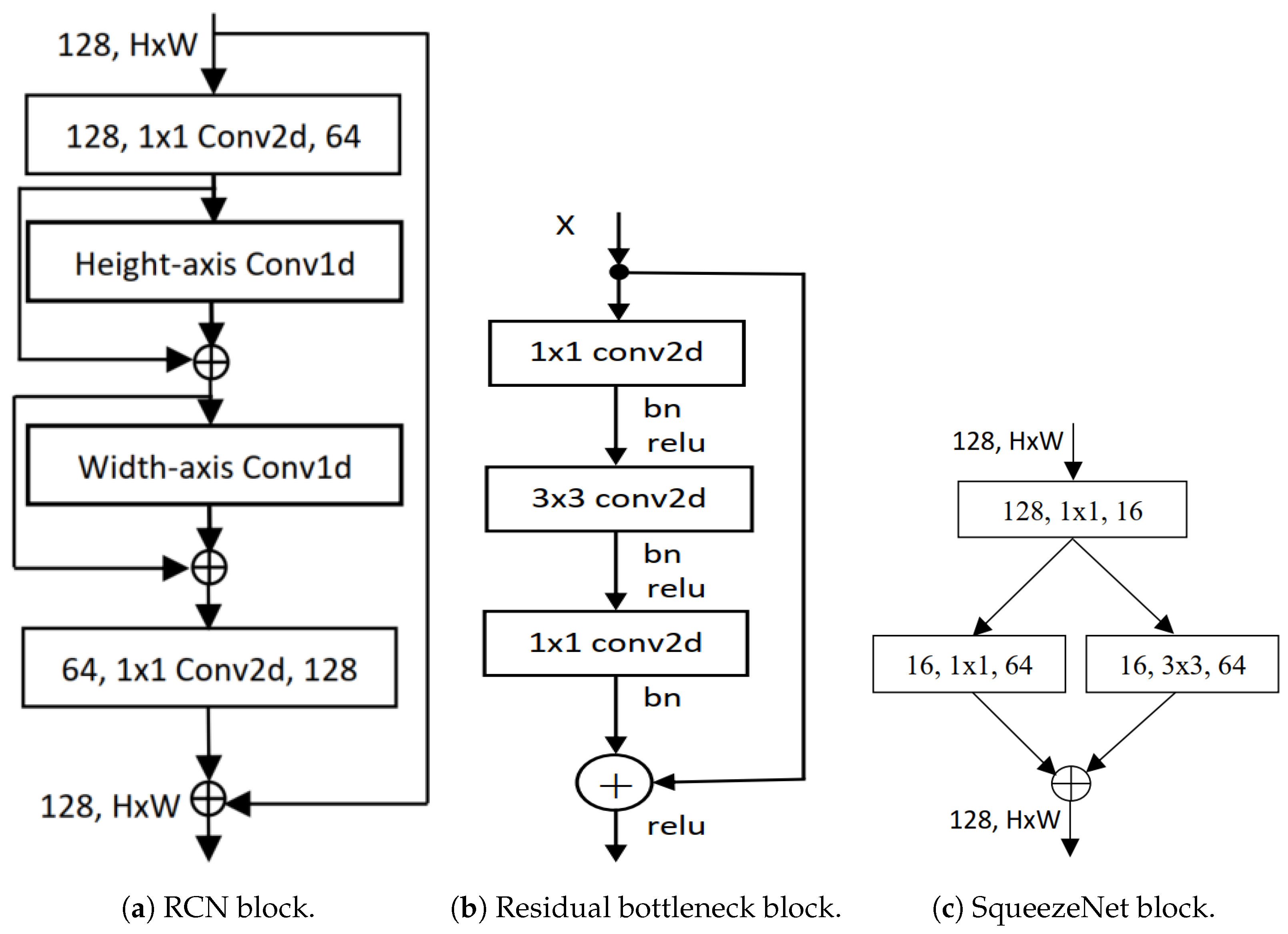
Residual 1D convolution networks (RCNs), as shown in Figure 1a, are a technique introduced by Shahadat and Maida where 2D convolution operation is replaced by 1D CNN layers with residual connections [48]. Residual bottleneck blocks in this particular network type focus on learning residual functions rather than full transformations, as shown in Figure 1b. They operate over one-dimensional data, such as a time-series signal, where the convolutional filter moves along the time axis. Its architecture comprises an input layer for 1D sequential data, convolutional layers with activation functions, residual connections for skip connections, a pooling layer for downsampling the sequence, and fully connected layers for final classification. The 1D CNN layer processes 1D input at a time whereas the 2D CNN layer takes . This way, the input cost becomes 2H instead of , and the operation is explained by the equation given as,
where the neighborhood of pixel i with a spatial extent of k is , and the trainable weight is the shared weight to compute the output for all pixel positions i. Additionally, the n-th channel of the trainable weight W is applied to the n-th channel of the input X in order to generate the n-th channel of the output feature map , where the computational cost is calculated as,
The total cost is multiplied by 2 for the two 1D CNN layers. This type of network is used to mitigate the vanishing gradient problem, making it easier to train deep networks and helping in learning identity mappings.
3.2. Squeeze-And-Excitation Networks
The squeeze-and-excitation network (SENet) is designed to improve CNNs by capturing channel interdependencies with minimal computational overhead [49]. SENet, depicted in Figure 1c, introduces parameters to each channel within a convolutional block, enabling the network to adaptively adjust the weighting of each feature map. The network gives equal weight to each channel when generating the output feature maps. It consists of two main operations named as “squeeze” and “excitation”. During the squeeze operation, the spatial dimensions of the input feature maps are reduced while retaining the channel-wise information. This process involves generating a channel descriptor and usually includes global pooling operations. On the other hand, the excitation operation utilizes the channel descriptor to calculate channel-wise scaling factors that determine how much emphasis should be placed on each channel.
Figure 1.
Illustration of block diagrams found in (a) Residual network [50], (b) Residual 1D convolutional network [48], and (c) SqueezeNet [49].
The factors are computed using a small neural network, such as ReLU or sigmoid. It can be constructed for any transformation as,
where is a convolutional operator. If we suppose that is a set of learned filter channels, then we can write the outputs of as , where
In the equation above, represents convolution. is a 2D spatial kernel. It acts as a single channel of , which operates on the corresponding channel of X. The output is the result of a summation across all channels, encompassing channel dependencies within . However, these dependencies are intertwined with the spatial correlation captured by the filters. The expanded diagram of SENet is shown in Figure 2.
Figure 2.
SqueezeNet block [49].
3.3. SqueezeNext Architecture
The SqueezeNext architecture is a compact network designed to be trained with few model parameters from the beginning, rather than relying on compression methods to reduce the parameter count. One of the methods used by the SqueezeNext architecture to implement this efficiently is utilizing low-rank filters. Assuming that the input to the i-th layer of the network with convolution filters is , the output activation of is produced. This layer transformation consumes cost and the filters consist of tensors of size . The goal is to reduce the parameters, W, using a low-rank basis, , in post-training compression. However, upon examining the trained network weights, it becomes evident that they usually do not exhibit a low-rank structure. Therefore, many networks necessitate some form of retraining. Instead of doing that, redesigning the network using the low-rank decomposition from the outset encourages the network to learn a low-rank structure from the beginning. This is the strategy adopted by the SqueezeNext architecture. Firstly, the K-convolutions are decomposed into two separable convolutions of size and , which reduces the number of parameters from to and also increases the network’s depth. Both of these convolutions have a ReLU activation and a batch normalization layer [51]. The block diagram is depicted in Figure 3a. This SqueezeNext block is stacked together to construct the SqueezeNext network architecture; a 23-layer network architecture is depicted in Figure 4.
Figure 3.
Illustration of block diagrams found in (a) SqueezeNext [51], and (b) SEC [52].
Figure 4.
SqueezeNext network architecture (23 layers) [51].
3.4. Residual 1D Block with SE Layer
Introduced by Shahadat, “Squeeze-and-Excitation-based 1D Convolutional Networks” (SECs) represent a parameter-efficient, mobile-embedded deep learning architecture. The SEC architecture, shown in Figure 3b, replaces the 1D CNN layer with a squeeze-and-excitation (SE) block to enhance cost efficiency and reduce computational complexity [52]. As the SEC replaces an RCN layer in RCNs, the cost reduction is directly analyzed between these two layers.
The cost comparison between the computational costs of the residual 1D CNN and the SE block is expressed as:
which equals , where is the ratio comparing the computational costs between the original residual 1D convolutional layer and the SE block. Also, k is the kernel, , and denote the number of input and output channels.
3.5. Reduced CNN Layer Network
A reduced CNN layer network functions similarly to traditional CNNs, achieving comparable performance while minimizing computational cost and model size [53]. The bottleneck, SENets, and channel squeezing are network architectures that utilize reduced CNN layers. The fundamental architecture of ResNet includes the basic residual block, which consists of two convolutional layers and a residual connection. A bottleneck residual block also incorporates a convolution, depicted in Figure 1b. The computational cost of the basic block is twice the cost of the spatial CNN layer, which is . Another essential component of the ResNet architecture is the bottleneck layer, which includes two pointwise convolution layers: a first, known as the ConvDown layer, and a final, known as the ConvUp layer. The first layer reduces , passed through the spatial convolution layer, whereas the final layer is responsible for increasing of the spatial CNN layer. In the reduced CNN block, this ConvUp layer in SEC [52] is replaced by the channel concatenation layer shown in Figure 5a.
Figure 5.
Illustration of block diagrams found in (a) Reduced CNN layers Network [48,53,54], (b) our proposed network architectures.
4. Proposed Architecture
The proposed parameter-efficient architecture is a novel, lightweight, mobile-embedded network designed for the accurate detection of lung and colon cancer subtypes using histopathological images. The primary objective of this architecture is to reduce computational costs while maintaining high accuracy, making it suitable for deployment on mobile devices. We utilize a combination of residual 1D convolutional neural networks (Conv1D) [48] and squeeze-and-excitation (SE) [49] blocks as their fundamental building blocks and construct our proposed block architecture, depicted in Figure 5b. Unlike traditional 2D CNNs, which are computationally expensive, the proposed architecture employs 1D CNNs along the width axis, called residual 1D CNN (RCN) [48]. SE blocks are integrated into the architecture to implement a channel-wise attention mechanism. This mechanism allows the network to selectively emphasize important channels and suppress less relevant ones, improving feature representation and overall efficiency.
Regarding computing, our suggested architecture is more economical than the lightweight SqueezeNext block [51]. We only used one instead of two pointwise CNN layers in the SqueezeNext block. Savings of at least costs are beneficial. We use the RCN and SE layers to replace the two separable CNN layers ( and ). These changes’ cost comparisons are described as follows:
where the number of input channels, height, width, and output height are represented by the variables , h, w, and , the kernel size is k. Equation (6) shows that our proposed block is times more cost-effective than the separable CNN layers in the SqueezeNext block.
Our modifications are not limited to these. To decrease the complexity of the network, we also replace the ConvUp layer (the CNN layer is used to increase the number of output channels) using channel concatenation. The absence of the channel-based weight layer from our channel concatenation results in decreased performance attributed to the pointwise CNN layer. We employ the SE layer, which helps to improve performance by utilizing a channel-wise attention method, to get around this restriction. In addition, this SE layer is less expensive than the CNN layer, which is described as,
The above equation describes the computational cost reductions by our proposed block compared to the SqueezeNext block with a factor of . So, our proposed architecture is more parameter-efficient and cost-effective than the well-known compact SqueezeNext block.
We utilize precisely two SE layers to boost performance. The SE layers take output feature maps from the RCN layer as input and produce better output feature maps using channel-wise feature recalibration. These channel-wise feature recalibrations improve the model’s performance, reduce overfitting, and focus on important channels. We stack this proposed block in the SqueezeNext network architecture to construct the proposed network.
5. Performance Evaluation
Evaluating performance is crucial for assessing how accurately a model predicts outcomes. It confirms that the model fits the training data well and is also effective for new and unseen data. Common evaluation metrics include accuracy, precision, recall (sensitivity), and the F1-score.
5.1. Accuracy
Accuracy is a crucial performance evaluation metric used in machine learning and statistics for classifying problems. It measures the correctness of the trained parameters or cases and assesses the proportion of correct observations among the total observations. The accuracy is calculated as,
where TP is true positives, which is the number of correctly predicted positive instances by the model. For example, if a person has the disease, the test is positive; TN stands for true negatives, which represents the number of negative instances correctly predicted by the model. This means that if the person does not have the disease, the test results are negative; FP stands for false positives, which is the number of negative instances incorrectly predicted as positive by the given model. This means that the test can show a positive result even if the person is not diseased. Lastly, FN stands for false negatives, representing the number of positive instances that the model incorrectly predicts as negative. This means that even if a person is diseased, the test results are negative. When the numbers of TP and TN are high compared to the total predictions, the accuracy is high. On the other hand, if the numbers of TP and TN are low compared to the total predictions, the accuracy is low. The total prediction is the sum of all the predictions: TP + TN + FP + FN. Therefore, we can conclude that higher accuracy is needed to enhance the overall reliability of the model’s predictions. By improving accuracy, we can decrease the occurrences of FP and FN, leading to more reliable and effective decision-making.
5.2. Precision
Another way to measure the performance of machine learning models is using the precision, which is calculated as the ratio of true positives to the sum of true positives and false positives. The equation to calculate the precision is defined as,
In the equation, a high precision value indicates that when the model predicts a positive outcome, it is usually correct. This suggests that the model has a low number of false positives, making its positive predictions reliable. Conversely, a low precision value indicates that the positive predictions made by the model are incorrect. Therefore, we can conclude that low precision can have a negative impact.
5.3. Recall
Recall, also known as sensitivity, is another important metric used to assess the performance of machine learning models. It quantifies the model’s capability to accurately predict all the positive instances in a dataset. It is calculated as the ratio of true positives to the sum of true positives and false negatives. The formula to calculate recall is as follows,
Based on the above equation, we understand that a high recall signifies that the model predicts most actual positives, while a low recall score indicates a high number of false negatives, leading to the model failing to predict actual positives. A low recall score could result in significant issues, particularly when conducting disease screenings, and can lead to severe repercussions.
5.4. F1-Score
In the statistical analysis of binary classification, the F1-score is used to measure predictive performance. It is calculated as the ratio of two times precision and recall (both determined using the previously mentioned equations) to the sum of precision and recall. The F1-score can also be defined as the harmonic mean of precision and recall. Its values range from 0 to 1, where 0 indicates the lowest performance and 1 indicates the highest performance.
Based on the equation above, a high F1-score indicates that the model identifies a greater proportion of positive instances while minimizing false positives. Conversely, a low F1-score suggests that the model struggles to accurately predict positive instances.
6. Experimental Results
6.1. Dataset Description
This research uses the lung and colon cancer histopathological images LC25000 dataset [55]. This dataset comprises two main categories of cancer cells: colon adenocarcinoma and benign colon tissue, and lung adenocarcinoma, lung squamous cell carcinoma, and benign lung tissue. The sample images of these lung and colon cancer categories are depicted in Figure 6.
Figure 6.
Randomly selected lung and colon cancer histopathological images from the LC25000 dataset [55].
They contain 25,000 color cancer cell images from five classes of lung and colon cancer’s benign and malignant tissue images. Initially, 1250 photos were taken from cancer tissues on pathology glass slides at the James A. Haley Veterans’ Hospital in Tampa, Florida, with 250 images for each category [40]. The original LC25000 dataset includes 750 lung tissue samples, comprising 250 adenocarcinoma, 250 squamous cell carcinomas, and 250 benign tissue samples. The dataset includes 500 colon tissue samples, with 250 adenocarcinoma and 250 benign tissue samples. These photos were then augmented with techniques like rotation and flipping, resulting in a collection of 5000 images per class and totaling 25000 images for lung and colon cancers.
The photos were originally 1024 × 768 pixels but were cropped to 768 × 768 pixels before augmentation to retain a uniform square shape. All photos are HIPAA-compliant, vetted, and freely available to AI researchers, making them an invaluable resource for creating more effective diagnostic tools. In our experimental analysis, we resized all the images to 256 × 256 pixels and randomly cropped them to 224 × 224 pixels. After resizing, we normalized the images using their mean and standard deviation. Moreover, we split the main dataset (LC25000) into two parts: for training and for testing samples. The image distribution of the lung and colon dataset is explained in Table 1.
Table 1.
Distributions of lung and colon cancer LC25000 histopathological images dataset.
6.2. Methodology
Similar hyperparameters were used to the original SqueezeNet architectures [51]. We then examined 23-layer architectures with the block multipliers “[6, 6, 8, 1]”. Likewise, we constructed 44-layer architectures with the block multipliers “[12, 12, 16, 2]”. All the proposed architectures were trained using various batch sizes, including 8, 16, 32, 64, and 128.
The LC25000 dataset contains 25,000 images resized and cropped to 224 × 224 pixels. Mean/std normalization was applied to preprocess our image data. All models were trained using the stochastic gradient descent (SGD) optimizer. We applied warmed-up linear learning for the first ten epochs, followed by cosine learning scheduling from epochs 11 to 120.
6.3. Results Analysis
This section evaluates the outcomes of using our proposed model. It is important to note that the accuracy does not significantly improve or decline as the batch sizes increase. Additionally, our model showed excellent performance for a smaller number of epochs. Furthermore, our model showed exceptional accuracy in colon cancer detection and achieved nearly perfect results in all tests. It takes fewer epochs to show state-of-the-art performance than the lung cancer detection using our model. The different batch sizes effectively train our models, demonstrating that our model can process varying amounts of input data without sacrificing effectiveness. Finally, Table 2 and Table 3 present an overview of our results, indicating that our model achieved the best performance for all datasets with a fixed batch size of 64.
Table 2.
The performance on the LC25000 dataset to detect lung and colon cancer using our proposed networks.
Table 3.
The overall performance on the LC25000 dataset using our proposed networks.
6.4. Results Comparisons
Table 4, Table 5, Table 6 and Table 7 compare our proposed method and several previous well-known studies and relevant network architectures. Table 4 compares several models and demonstrates the direct effectiveness of our modified architecture. It is crucial to note that these studies are based on using different datasets and imaging with different numbers of epochs, and batch sizes, which makes direct comparison a bit more difficult. However, our proposed method achieves perfect scores in all evaluation metrics, i.e., accuracy rate, precision, sensitivity, and F1-score, showcasing its versatility in detecting lung and colon cancer. This comparison also offers a contextual understanding of our proposed model in relation to other methodologies.
Table 4.
The performance on the LC25000 dataset to detect lung and colon cancer using some relevant networks and our proposed network [56].
Table 5.
Comparison of our proposed method results with other methods on the colon cancer detection dataset. “HI” and “CRAG” stand for histopathological images and colorectal adenocarcinoma gland, respectively.
Table 6.
Comparison of our proposed result with other methods on the lung cancer detection dataset. “CAD”, “ML”, “GO”, and “LR” stand for clustering KNN-classifier, machine learning, genetic optimization, and logistic regression, respectively.
Table 7.
Comparison of our proposed result with other methods on lung and colon cancer detection dataset. Here, “IQ-OTHNCCD” is a lung cancer dataset.
6.4.1. Lung Cancer
Here, we discuss the comparison of lung cancer detection between various published works and our proposed approach. Our mobile-supported and parameter-efficient method, a 1D convolutional neural network with an SE layer, outperforms all of the other methods as seen in Table 6. Our method utilized CT scan imaging of the publicly available LC25000 dataset and achieved perfect metrics, i.e., accuracy rate, precision, recall, and an F1-score of . Remarkably, despite using a different dataset from LIDC-IDRI and a different network known as AlexNet and GoogLeNet, the method proposed by Vinod Kumar and Brijesh Bakariya [121] also achieved the same precision score as ours using computer-based models. Nonetheless, our achievements in terms of accuracy exceed all of the previous work, as detailed in Table 6.
6.4.2. Colon Cancer
This section portrays the comparative efficacy of various methodologies in detecting colon cancer, as enumerated in Table 5. Utilizing CT scan imaging from the LC25000 dataset, our 1D convolutional network with SE layers achieves a accuracy rate, precision, recall, and F1-score in the detection of colon cancer. Our proposed method surpasses nearly all studies referenced in Table 5, demonstrating the model’s capability to identify all instances of colon cancer presence or absence across both trained and new datasets. Additionally, our model maintains consistent performance across 30 epochs and 64 batch sizes, with million parameters. It is worth noting that other computer-based models, such as the CNN model described by Dabass et al. (2022) [87], also achieved accuracy scores using a computer-based deep learning model.
6.4.3. Lung and Colon Cancer
We compared lung and colon cancer detection among various published methods as presented in Table 7. The proposed method, 1D CNN with squeeze-and-excitation (SE) layers, used CT scans imaging from the publicly available LC25000 dataset as the input. The 1D CNN with SE layers is trained on this data. The model identifies features related to the presence of lung and colon cancer. Our proposed method achieved a remarkable accuracy rate, precision, recall, and F1-score in lung and colon cancer detection. This high accuracy indicates that the proposed model consistently correctly identifies all the instances of colon and lung cancer present or absent in the dataset. It consistently performs well over 50 epochs with a batch size of 64, using million parameters. In machine learning, “epochs” refers to the number of times the entire dataset is passed through the model during training. On the other hand, “batch size” refers to the number of samples processed before the model’s internal parameters are updated. This means the model can be trained on various datasets, not just a specific set. Additionally, it outperforms almost all the other methods mentioned in Table 7. Additionally, the LightGBM computer-based model [136] proposed by Indu Chhillar and Ajmer Singh in 2024 also achieved the same accuracy as our model using CT scan imaging from the LC25000 dataset [136].
6.5. Final Remarks
Our model outperformed existing models significantly, which emphasizes the efficiency of our approach. Surprisingly, our model accomplished accuracy in detecting colon cancer across all batch sizes. It also achieved accuracy in detecting lung cancer. It achieved perfect scores in detecting lung and colon cancer types with a batch size of 64, while maintaining consistently high results in other areas. While existing models have attained comparable performance, none have consistently reached the best accuracy across three cancer types as our model has. Despite implementing the same SqueezeNet network as the studies by Mohamed et al. (2023) [91] and Suominen et al. (2024) [134], our models achieved higher accuracy rates. Moreover, this suggests that combining SE layers and 1D convolutional networks can effectively enhance feature extraction capabilities and achieve state-of-the-art performance in medical image analysis where timely and accurate results are crucial. Our model achieved accuracy for lung cancer, colon cancer, and both lung and colon cancers. We analyzed the overfitting of our model by examining the training and validation loss, as shown in Figure 7a–c. This analysis was performed for each epoch across all categories of lung and colon cancer. Our findings indicate that the model does not overfit on the datasets for lung cancer, colon cancer, and the combined lung and colon cancer images.
Figure 7.
Training vs. validation loss diagrams to analyze overfitting in our proposed model across different numbers of epochs.
To create a successful machine learning (ML) model, focusing on the network’s ability to generalize and its reliability is essential. A large and diverse dataset is needed for effective training, so we used data augmentation to artificially expand the dataset by making small changes to the original data. This expansion allowed our model to recognize and generalize from a wider range of patterns and abnormal details in histopathological images. It significantly contributed to the model’s performance across different batches and resulted in the highest accuracy. Additionally, it helped us overcome overfitting (as shown in Figure 7a–c), which is crucial for learning detailed features of cancerous diseases without being misled by identical patterns.
7. Conclusions and Future Work
According to the World Health Organization (WHO), lung and colon cancer were the leading causes of death in 2020 [137]. Early diagnosis is crucial to overcome this issue. A study, proposed by a CNN network, constructed with 1D convolutional networks and SE layers to detect lung and colon cancer features from the large LC25000 dataset. Historically, diagnosis was a complex and lengthy process. The aim was to propose an approach that is not only efficient but also computationally economical by minimizing parameters; in this case, only M parameters were used. Overall, we achieved a accuracy, precision, recall, and F1-score across various batch sizes and epochs, indicating significant progress and the reliability of our model.
However, the limitation of this work is that the model has only been applied to the classification of lung and colon cancers, even though it shows potential for detecting them. Further analysis of the results indicates that there is still room for improvement in detecting other types of cancer in order to achieve optimal performance. Our comparisons show that our method outperforms almost all previous studies. Implementing this mobile-supported identification method in healthcare will assist pathologists in diagnosing lung and colon cancer more easily and reliably. In the future, we plan to apply our diagnostic method on other medical disease detection datasets to expand the scope of our study and improve the accuracy rate. This will enable us to extend our contributions to the detection of other types of cancer.
Author Contributions
Conceptualization, N.S.; methodology, N.S.; software, N.S. and R.L.; validation, N.S. and R.L.; formal analysis, N.S., R.L. and A.N.; investigation, N.S., R.L. and A.N.; resources, N.S., R.L. and A.N.; writing—original draft preparation, N.S., R.L. and A.N.; writing—review and editing, N.S. and R.L.; visualization, N.S. and R.L.; supervision, N.S.; project administration, N.S.; funding acquisition, N.S. and R.L. All authors have read and agreed to the published version of the manuscript.
Funding
This research was funded by the TruScholar Summer Undergraduate Research Program.
Institutional Review Board Statement
Not applicable.
Informed Consent Statement
Patient consent was waived, as this was a retrospective analysis without any patient intervention. Patients were completely anonymized throughout the entire process.
Data Availability Statement
Data is publicly available [55] and the details are attached in the manuscript.
Conflicts of Interest
The authors declare no conflicts of interest.
References
- National Cancer Institute. Cancer Statistics. 2024. Available online: https://www.cancer.gov/about-cancer/understanding/statistics (accessed on 10 June 2024).
- Weinberg, R.A. How cancer arises. Sci. Am. 1996, 275, 62–70. [Google Scholar] [CrossRef] [Scilit] [PubMed]
- National Cancer Institute. What is Cancer? 2024. Available online: https://www.cancer.gov/about-cancer/understanding/what-is-cancer (accessed on 10 June 2024).
- WHO. Global Cancer Burden Growing Amidst Mounting Need for Services, 2024. Available online: https://www.who.int/news-room/fact-sheets/detail/cancer (accessed on 10 June 2024).
- American Cancer Society. What is Lung Cancer? 2024. Available online: https://www.cancer.org/cancer/types/lung-cancer/about/what-is.html (accessed on 10 June 2024).
- American Cancer Society. What is Colon Cancer? 2024. Available online: https://www.cancer.org/cancer/types/colon-rectal-cancer/about/what-is-colorectal-cancer.html (accessed on 10 June 2024).
- Al-Antari, M.A. AI Algorithms in Medical Diagnostics. 2023. Available online: https://www.ncbi.nlm.nih.gov/pmc/articles/PMC9955430/#:~:text=AI%20algorithms%20can%20analyze%20medical,diseases%20more%20accurately%20and%20quickly (accessed on 10 June 2024).
- National Center for Biotechnology Information. Research Article Title Placeholder 2024. Available online: https://www.ncbi.nlm.nih.gov/pmc/articles/PMC8950225/ (accessed on 10 June 2024).
- Stafford, I.S.; Kellermann, M.; Mossotto, E.; Beattie, R.M.; MacArthur, B.D.; Ennis, S. A systematic review of the applications of artificial intelligence and machine learning in autoimmune diseases. NPJ Digit. Med. 2020, 3, 30. [Google Scholar] [CrossRef] [Scilit] [PubMed]
- Technology, K. Machine Learning and Medical Diagnosis: An Introduction to How AI Improves Disease Detection. 2024. Available online: https://kili-technology.com/data-labeling/machine-learning/machine-learning-and-medical-diagnosis-an-introduction-to-how-ai-improves-disease-detection (accessed on 10 June 2024).
- Schmidhuber, J. Deep learning in neural networks: An overview. Neural Netw. 2015, 61, 85–117. [Google Scholar] [CrossRef] [Scilit] [PubMed]
- Sena, P.; Fioresi, R.; Faglioni, F.; Losi, L.; Faglioni, G.; Roncucci, L. Deep learning techniques for detecting preneoplastic and neoplastic lesions in human colorectal histological images. Oncol. Lett. 2019, 18, 6101–6107. [Google Scholar] [CrossRef] [Scilit]
- Yoon, H.; Lee, J.; Oh, J.E.; Kim, H.R.; Lee, S.; Chang, H.J.; Sohn, D.K. Tumor identification in colorectal histology images using a convolutional neural network. J. Digit. Imaging 2019, 32, 131–140. [Google Scholar] [CrossRef] [Scilit]
- Kather, J.N.; Krisam, J.; Charoentong, P.; Luedde, T.; Herpel, E.; Weis, C.A.; Gaiser, T.; Marx, A.; Valous, N.A.; Ferber, D.; et al. Predicting survival from colorectal cancer histology slides using deep learning: A retrospective multicenter study. PloS Med. 2019, 16, e1002730. [Google Scholar] [CrossRef] [Scilit]
- Wei, J.W.; Suriawinata, A.A.; Vaickus, L.J.; Ren, B.; Liu, X.; Lisovsky, M.; Tomita, N.; Abdollahi, B.; Kim, A.S.; Snover, D.C.; et al. Evaluation of a deep neural network for automated classification of colorectal polyps on histopathologic slides. JAMA Netw. Open 2020, 3, e203398. [Google Scholar] [CrossRef] [Scilit]
- Iizuka, O.; Kanavati, F.; Kato, K.; Rambeau, M.; Arihiro, K.; Tsuneki, M. Deep learning models for histopathological classification of gastric and colonic epithelial tumors. Sci. Rep. 2020, 10, 1504. [Google Scholar] [CrossRef] [Scilit] [PubMed]
- Xu, L.; Walker, B.; Liang, P.I.; Tong, Y.; Xu, C.; Su, Y.C.; Karsan, A. Colorectal cancer detection based on deep learning. J. Pathol. Inform. 2020, 11, 28. [Google Scholar] [CrossRef] [Scilit]
- Hamida, A.B.; Devanne, M.; Weber, J.; Truntzer, C.; Derangère, V.; Ghiringhelli, F.; Forestier, G.; Wemmert, C. Deep learning for colon cancer histopathological images analysis. Comput. Biol. Med. 2021, 136, 104730. [Google Scholar] [CrossRef] [Scilit]
- Babu, T.; Singh, T.; Gupta, D.; Hameed, S. Colon cancer prediction on histological images using deep learning features and Bayesian optimized SVM. J. Intell. Fuzzy Syst. 2021, 41, 5275–5286. [Google Scholar] [CrossRef] [Scilit]
- Tasnim, Z.; Chakraborty, S.; Shamrat, F.J.M.; Chowdhury, A.N.; Nuha, H.A.; Karim, A.; Zahir, S.B.; Billah, M.M. Deep learning predictive model for colon cancer patient using CNN-based classification. Int. J. Adv. Comput. Sci. Appl. 2021, 12, 687–696. [Google Scholar] [CrossRef] [Scilit]
- Sakr, A.S.; Soliman, N.F.; Al-Gaashani, M.S.; Pławiak, P.; Ateya, A.A.; Hammad, M. An efficient deep learning approach for colon cancer detection. Appl. Sci. 2022, 12, 8450. [Google Scholar] [CrossRef] [Scilit]
- Hasan, M.I.; Ali, M.S.; Rahman, M.H.; Islam, M.K.; Islam, M.K. Automated detection and characterization of colon cancer with deep convolutional neural networks. J. Healthc. Eng. 2022, 2022. [Google Scholar] [CrossRef] [Scilit]
- Talukder, M.A.; Islam, M.M.; Uddin, M.A.; Akhter, A.; Hasan, K.F.; Moni, M.A. Machine learning-based lung and colon cancer detection using deep feature extraction and ensemble learning. Expert Syst. Appl. 2022, 205, 117695. [Google Scholar] [CrossRef] [Scilit]
- Bostanci, E.; Kocak, E.; Unal, M.; Guzel, M.S.; Acici, K.; Asuroglu, T. Machine learning analysis of RNA-seq data for diagnostic and prognostic prediction of colon cancer. Sensors 2023, 23, 3080. [Google Scholar] [CrossRef] [Scilit]
- Zhang, C.; Sun, X.; Dang, K.; Li, K.; Guo, X.w.; Chang, J.; Yu, Z.q.; Huang, F.y.; Wu, Y.s.; Liang, Z.; et al. Toward an expert level of lung cancer detection and classification using a deep convolutional neural network. Oncologist 2019, 24, 1159–1165. [Google Scholar] [CrossRef] [Scilit]
- Pham, H.H.N.; Futakuchi, M.; Bychkov, A.; Furukawa, T.; Kuroda, K.; Fukuoka, J. Detection of lung cancer lymph node metastases from whole-slide histopathologic images using a two-step deep learning approach. Am. J. Pathol. 2019, 189, 2428–2439. [Google Scholar] [CrossRef] [Scilit]
- Gertych, A.; Swiderska-Chadaj, Z.; Ma, Z.; Ing, N.; Markiewicz, T.; Cierniak, S.; Salemi, H.; Guzman, S.; Walts, A.E.; Knudsen, B.S. Convolutional neural networks can accurately distinguish four histologic growth patterns of lung adenocarcinoma in digital slides. Sci. Rep. 2019, 9, 1483. [Google Scholar] [CrossRef] [Scilit]
- Hatuwal, B.K.; Thapa, H.C. Lung cancer detection using convolutional neural network on histopathological images. Int. J. Comput. Trends Technol. 2020, 68, 21–24. [Google Scholar] [CrossRef] [Scilit]
- Saif, A.; Qasim, Y.R.H.; Al-Sameai, H.A.M.; Ali, O.A.F.; Hassan, A.A.M. Multi paths technique on convolutional neural network for lung cancer detection based on histopathological images. Int. J. Adv. Netw. Appl. 2020, 12, 4549–4554. [Google Scholar] [CrossRef] [Scilit]
- Abbas, M.A.; Bukhari, S.U.K.; Syed, A.; Shah, S.S.H. The Histopathological Diagnosis of Adenocarcinoma & Squamous Cells Carcinoma of Lungs by Artificial intelligence: A comparative study of convolutional neural networks. MedRxiv 2020, 2020-05. [Google Scholar]
- Srinidhi, C.L.; Ciga, O.; Martel, A.L. Deep neural network models for computational histopathology: A survey. Med. Image Anal. 2021, 67, 101813. [Google Scholar] [CrossRef] [Scilit] [PubMed]
- Han, Y.; Ma, Y.; Wu, Z.; Zhang, F.; Zheng, D.; Liu, X.; Tao, L.; Liang, Z.; Yang, Z.; Li, X.; et al. Histologic subtype classification of non-small cell lung cancer using PET/CT images. Eur. J. Nucl. Med. Mol. Imaging 2021, 48, 350–360. [Google Scholar] [CrossRef] [Scilit] [PubMed]
- Marentakis, P.; Karaiskos, P.; Kouloulias, V.; Kelekis, N.; Argentos, S.; Oikonomopoulos, N.; Loukas, C. Lung cancer histology classification from CT images based on radiomics and deep learning models. Med. Biol. Eng. Comput. 2021, 59, 215–226. [Google Scholar] [CrossRef] [Scilit] [PubMed]
- Wahab Sait, A.R. Lung Cancer Detection Model Using Deep Learning Technique. Appl. Sci. 2023, 13, 12510. [Google Scholar] [CrossRef] [Scilit]
- Shandilya, S.; Nayak, S.R. Analysis of lung cancer by using deep neural network. In Proceedings of the 2nd Innovation in Electrical Power Engineering, Communication, and Computing Technology (IEPCCT 2021), Online, 25–28 May 2021; pp. 427–436. [Google Scholar]
- Abd Al-Ameer, A.A.; Hussien, G.A.; Al Ameri, H.A. Lung cancer detection using image processing and deep learning. Indones. J. Electr. Eng. Comput. Sci. 2022, 28, 987–993. [Google Scholar] [CrossRef] [Scilit]
- Jasmine Pemeena Priyadarsini, M.; Kotecha, K.; Rajini, G.; Hariharan, K.; Utkarsh Raj, K.; Bhargav Ram, K.; Indragandhi, V.; Subramaniyaswamy, V.; Pandya, S. Lung Diseases Detection Using Various Deep Learning Algorithms. J. Healthc. Eng. 2023, 2023, 3563696. [Google Scholar] [CrossRef] [Scilit]
- Siddiqui, E.A.; Chaurasia, V.; Shandilya, M. Detection and classification of lung cancer computed tomography images using a novel improved deep belief network with Gabor filters. Chemom. Intell. Lab. Syst. 2023, 235, 104763. [Google Scholar] [CrossRef] [Scilit]
- Wahid, R.R.; Nisa, C.; Amaliyah, R.P.; Puspaningrum, E.Y. Lung and colon cancer detection with convolutional neural networks on histopathological images. In Proceedings of the 7th International Conference on Science and Technology, Yogyakarta, Indonesia, 7–8 September 2021; Volume 2654. [Google Scholar]
- Masud, M.; Sikder, N.; Nahid, A.A.; Bairagi, A.K.; AlZain, M.A. A machine learning approach to diagnosing lung and colon cancer using a deep learning-based classification framework. Sensors 2021, 21, 748. [Google Scholar] [CrossRef] [Scilit]
- Garg, S.; Garg, S. Prediction of lung and colon cancer through analysis of histopathological images by utilizing Pre-trained CNN models with visualization of class activation and saliency maps. In Proceedings of the 2020 3rd Artificial Intelligence and Cloud Computing Conference, Kyoto, Japan, 18–20 December 2020; pp. 38–45. [Google Scholar]
- Ali, M.; Ali, R. Multi-input dual-stream capsule network for improved lung and colon cancer classification. Diagnostics 2021, 11, 1485. [Google Scholar] [CrossRef] [Scilit] [PubMed]
- Hage Chehade, A.; Abdallah, N.; Marion, J.M.; Oueidat, M.; Chauvet, P. Lung and colon cancer classification using medical imaging: A feature engineering approach. Phys. Eng. Sci. Med. 2022, 45, 729–746. [Google Scholar] [CrossRef] [Scilit]
- Mehmood, S.; Ghazal, T.M.; Khan, M.A.; Zubair, M.; Naseem, M.T.; Faiz, T.; Ahmad, M. Malignancy detection in lung and colon histopathology images using transfer learning with class selective image processing. IEEE Access 2022, 10, 25657–25668. [Google Scholar] [CrossRef] [Scilit]
- Singh, O.; Singh, K.K. An approach to classify lung and colon cancer of histopathology images using deep feature extraction and an ensemble method. Int. J. Inf. Technol. 2023, 15, 4149–4160. [Google Scholar] [CrossRef] [Scilit]
- Bhattacharya, A.; Saha, B.; Chattopadhyay, S.; Sarkar, R. Deep feature selection using adaptive β-Hill Climbing aided whale optimization algorithm for lung and colon cancer detection. Biomed. Signal Process. Control. 2023, 83, 104692. [Google Scholar] [CrossRef] [Scilit]
- Al-Jabbar, M.; Alshahrani, M.; Senan, E.M.; Ahmed, I.A. Histopathological Analysis for Detecting Lung and Colon Cancer Malignancies Using Hybrid Systems with Fused Features. Bioengineering 2023, 10, 383. [Google Scholar] [CrossRef] [Scilit] [PubMed]
- Shahadat, N.; Maida, A.S. Deep Residual Axial Networks. arXiv 2023, arXiv:2301.04631. [Google Scholar]
- Hu, J.; Shen, L.; Sun, G. Squeeze-and-excitation networks. In Proceedings of the IEEE Conference on Computer Vision and Pattern Recognition, Salt Lake City, UT, USA, 18–23 June 2018; pp. 7132–7141. [Google Scholar]
- He, K.; Zhang, X.; Ren, S.; Sun, J. Deep residual learning for image recognition. In Proceedings of the IEEE conference on computer vision and pattern recognition, Las Vegas, NV, USA, 27–30 June 2016; pp. 770–778. [Google Scholar]
- Gholami, A.; Kwon, K.; Wu, B.; Tai, Z.; Yue, X.; Jin, P.; Zhao, S.; Keutzer, K. Squeezenext: Hardware-aware neural network design. In Proceedings of the IEEE Conference on Computer Vision and Pattern Recognition Workshops, Salt Lake City, UT, USA, 18–23 June 2018; pp. 1638–1647. [Google Scholar]
- Shahadat, N. Lung Image Analysis using Squeeze and Excitation Based Convolutional Networks. In Proceedings of the 2023 26th International Conference on Computer and Information Technology (ICCIT), Cox’s Bazar, Bangladesh, 13–15 December 2023. [Google Scholar] [CrossRef] [Scilit]
- Shahadat, N. Convolutional Layer Reduction from Deep Convolutional Networks. In Proceedings of the 2023 26th International Conference on Computer and Information Technology (ICCIT), Cox’s Bazar, Bangladesh, 13–15 December 2023. [Google Scholar]
- Shahadat, N. Mobile-Based Deep Convolutional Networks for Malaria Parasites Detection from Blood Cell Images. In Proceedings of the 2023 26th International Conference on Computer and Information Technology (ICCIT), Cox’s Bazar, Bangladesh, 13–15 December 2023. [Google Scholar] [CrossRef] [Scilit]
- Borkowski, A.A.; Bui, M.M.; Thomas, L.B.; Wilson, C.P.; DeLand, L.A.; Mastorides, S.M. LC25000 Lung and colon histopathological image dataset. arXiv 2021, arXiv:1912.12142. [Google Scholar]
- Shahadat, N. Lung and Colon Cancer Histopathological Image Classification Using 1D Convolutional Channel-based Attention Networks. In Proceedings of the International FLAIRS Conference, Sandestin Beach, FL, USA, 19–21 May 2024; Volume 37. [Google Scholar]
- Hilado, S.D.F.; Lim, L.A.G.; Naguib, R.N.; Dadios, E.P.; Avila, J.M.C. Implementation of wavelets and artificial neural networks in colonic histopathological classification. J. Adv. Comput. Intell. Intell. Inform. 2014, 18, 792–797. [Google Scholar] [CrossRef] [Scilit]
- Rathore, S.; Hussain, M.; Iftikhar, M.A.; Jalil, A. Ensemble classification of colon biopsy images based on information rich hybrid features. Comput. Biol. Med. 2014, 47, 76–92. [Google Scholar] [CrossRef] [Scilit]
- Xu, Y.; Mo, T.; Feng, Q.; Zhong, P.; Lai, M.; Eric, I.; Chang, C. Deep learning of feature representation with multiple instance learning for medical image analysis. In Proceedings of the 2014 IEEE International Conference on Acoustics, Speech and Signal Processing (ICASSP), Florence, Italy, 4–9 May 2014; pp. 1626–1630. [Google Scholar]
- Gheonea, D.I.; Streba, C.T.; Vere, C.C.; Șerbănescu, M.; Pirici, D.; Comănescu, M.; Streba, L.A.M.; Ciurea, M.E.; Mogoantă, S.; Rogoveanu, I.; et al. Diagnosis system for hepatocellular carcinoma based on fractal dimension of morphometric elements integrated in an artificial neural network. BioMed Res. Int. 2014, 2014, 239706. [Google Scholar] [CrossRef] [Scilit] [PubMed]
- Akbar, B.; Gopi, V.P.; Babu, V.S. Colon cancer detection based on structural and statistical pattern recognition. In Proceedings of the 2015 2nd International Conference on Electronics and Communication Systems (ICECS), Coimbatore, India, 26–27 February 2015; pp. 1735–1739. [Google Scholar]
- Rathore, S.; Hussain, M.; Khan, A. Automated colon cancer detection using hybrid of novel geometric features and some traditional features. Comput. Biol. Med. 2015, 65, 279–296. [Google Scholar] [CrossRef] [Scilit] [PubMed]
- Rathore, S.; Hussain, M.; Iftikhar, M.A.; Jalil, A. Novel structural descriptors for automated colon cancer detection and grading. Comput. Methods Programs Biomed. 2015, 121, 92–108. [Google Scholar] [CrossRef] [Scilit]
- Kourou, K.; Exarchos, T.P.; Exarchos, K.P.; Karamouzis, M.V.; Fotiadis, D.I. Machine learning applications in cancer prognosis and prediction. Comput. Struct. Biotechnol. J. 2015, 13, 8–17. [Google Scholar] [CrossRef] [Scilit]
- Xu, J.; Luo, X.; Wang, G.; Gilmore, H.; Madabhushi, A. A deep convolutional neural network for segmenting and classifying epithelial and stromal regions in histopathological images. Neurocomputing 2016, 191, 214–223. [Google Scholar] [CrossRef] [Scilit]
- Liu, J.; Wang, D.; Wei, Z.; Lu, L.; Kim, L.; Turkbey, E.; Summers, R.M. Colitis detection on computed tomography using regional convolutional neural networks. In Proceedings of the 2016 IEEE 13th International Symposium on Biomedical Imaging (ISBI), Prague, Czech Republic, 3–16 April 2016; pp. 863–866. [Google Scholar]
- Ştefănescu, D.; Streba, C.; Cârţână, E.T.; Săftoiu, A.; Gruionu, G.; Gruionu, L.G. Computer aided diagnosis for confocal laser endomicroscopy in advanced colorectal adenocarcinoma. PLoS ONE 2016, 11, e0154863. [Google Scholar] [CrossRef] [Scilit] [PubMed]
- Godkhindi, A.M.; Gowda, R.M. Automated detection of polyps in CT colonography images using deep learning algorithms in colon cancer diagnosis. In Proceedings of the 2017 International Conference on Energy, Communication, Data Analytics and Soft Computing (ICECDS), Chennai, India, 1–2 August 2017; pp. 1722–1728. [Google Scholar]
- Bardhi, O.; Sierra-Sosa, D.; Garcia-Zapirain, B.; Elmaghraby, A. Automatic colon polyp detection using Convolutional encoder-decoder model. In Proceedings of the 2017 IEEE International Symposium on Signal Processing and Information Technology (ISSPIT), Bilbao, Spain, 18–20 December 2017; pp. 445–448. [Google Scholar]
- Trebeschi, S.; van Griethuysen, J.J.; Lambregts, D.M.; Lahaye, M.J.; Parmar, C.; Bakers, F.C.; Peters, N.H.; Beets-Tan, R.G.; Aerts, H.J. Deep learning for fully-automated localization and segmentation of rectal cancer on multiparametric MR. Sci. Rep. 2017, 7, 5301. [Google Scholar] [CrossRef] [Scilit]
- Haj-Hassan, H.; Chaddad, A.; Harkouss, Y.; Desrosiers, C.; Toews, M.; Tanougast, C. Classifications of Multispectral Colorectal Cancer Tissues Using Convolution Neural Network. J. Pathol. Inform. 2017, 8, 1. [Google Scholar] [CrossRef] [Scilit]
- Basha, S.S.; Ghosh, S.; Babu, K.K.; Dubey, S.R.; Pulabaigari, V.; Mukherjee, S. Rccnet: An efficient convolutional neural network for histological routine colon cancer nuclei classification. In Proceedings of the 2018 15th International Conference on Control, Automation, Robotics, and Vision (ICARCV), Singapore, 18–21 November 2018; pp. 1222–1227. [Google Scholar]
- Ponzio, F.; Macii, E.; Ficarra, E.; Di Cataldo, S. Colorectal cancer classification using deep convolutional networks. In Proceedings of the 11th International Joint Conference on Biomedical Engineering Systems and Technologies, Funchal, Madeira, Portugal, 19–21 January 2018; Volume 2, pp. 58–66. [Google Scholar]
- Tang, J.; Li, J.; Xu, X. Segnet-based gland segmentation from colon cancer histology images. In Proceedings of the 2018 33rd Youth Academic Annual Conference of Chinese Association of Automation (YAC), Nanjing, China, 18–20 May 2018; pp. 1078–1082. [Google Scholar]
- Dos Santos, L.F.S.; Neves, L.A.; Rozendo, G.B.; Ribeiro, M.G.; do Nascimento, M.Z.; Tosta, T.A.A. Multidimensional and fuzzy sample entropy (SampEnMF) for quantifying H&E histological images of colorectal cancer. Comput. Biol. Med. 2018, 103, 148–160. [Google Scholar]
- Gupta, P.; Chiang, S.F.; Sahoo, P.K.; Mohapatra, S.K.; You, J.F.; Onthoni, D.D.; Hung, H.Y.; Chiang, J.M.; Huang, Y.; Tsai, W.S. Prediction of colon cancer stages and survival period with machine learning approach. Cancers 2019, 11, 2007. [Google Scholar] [CrossRef] [Scilit]
- Wang, Y.H.; Nguyen, P.A.; Islam, M.M.; Li, Y.C.; Yang, H.C. Development of Deep Learning Algorithm for Detection of Colorectal Cancer in EHR Data. MedInfo 2019, 264, 438–441. [Google Scholar]
- Blanes-Vidal, V.; Baatrup, G.; Nadimi, E.S. Addressing priority challenges in the detection and assessment of colorectal polyps from capsule endoscopy and colonoscopy in colorectal cancer screening using machine learning. Acta Oncol. 2019, 58, S29–S36. [Google Scholar] [CrossRef] [Scilit]
- Qasim, Y.; Al-Sameai, H.; Ali, O.; Hassan, A. Convolutional neural networks for automatic detection of colon adenocarcinoma based on histopathological images. In Proceedings of the International Conference of Reliable Information and Communication Technology, Langkawi, Malaysia, 21–22 December 2020; pp. 19–28. [Google Scholar]
- Bukhari, S.U.K.; Syed, A.; Bokhari, S.K.A.; Hussain, S.S.; Armaghan, S.U.; Shah, S.S.H. The histological diagnosis of colonic adenocarcinoma by applying partial self supervised learning. MedRxiv 2020, 2020-08. [Google Scholar]
- Liang, M.; Ren, Z.; Yang, J.; Feng, W.; Li, B. Identification of colon cancer using multi-scale feature fusion convolutional neural network based on shearlet transform. IEEE Access 2020, 8, 208969–208977. [Google Scholar] [CrossRef] [Scilit]
- Gupta, P.; Huang, Y.; Sahoo, P.K.; You, J.F.; Chiang, S.F.; Onthoni, D.D.; Chern, Y.J.; Chao, K.Y.; Chiang, J.M.; Yeh, C.Y.; et al. Colon tissues classification and localization in whole slide images using deep learning. Diagnostics 2021, 11, 1398. [Google Scholar] [CrossRef] [Scilit] [PubMed]
- Tamang, L.D.; Kim, B.W. Deep learning approaches to colorectal cancer diagnosis: A review. Appl. Sci. 2021, 11, 10982. [Google Scholar] [CrossRef] [Scilit]
- Albashish, D. Ensemble of adapted convolutional neural networks (CNN) methods for classifying colon histopathological images. PeerJ Comput. Sci. 2022, 8, e1031. [Google Scholar] [CrossRef] [Scilit]
- Ho, C.; Zhao, Z.; Chen, X.F.; Sauer, J.; Saraf, S.A.; Jialdasani, R.; Taghipour, K.; Sathe, A.; Khor, L.Y.; Lim, K.H.; et al. A promising deep learning-assistive algorithm for histopathological screening of colorectal cancer. Sci. Rep. 2022, 12, 2222. [Google Scholar] [CrossRef] [Scilit] [PubMed]
- Akilandeswari, A.; Sungeetha, D.; Joseph, C.; Thaiyalnayaki, K.; Baskaran, K.; Jothi Ramalingam, R.; Al-Lohedan, H.; Al-Dhayan, D.M.; Karnan, M.; Meansbo Hadish, K.; et al. Automatic detection and segmentation of colorectal cancer with deep residual convolutional neural network. Evid.-Based Complement. Altern. Med. 2022, 2022, 3415603. [Google Scholar] [CrossRef] [Scilit]
- Dabass, M.; Vashisth, S.; Vig, R. A convolution neural network with multi-level convolutional and attention learning for classification of cancer grades and tissue structures in colon histopathological images. Comput. Biol. Med. 2022, 147, 105680. [Google Scholar] [CrossRef] [Scilit]
- Kumar, V.R.P.; Arulselvi, M.; Sastry, K. Comparative assessment of colon cancer classification using diverse deep learning approaches. J. Data Sci. Intell. Syst. 2023, 1, 128–135. [Google Scholar] [CrossRef] [Scilit]
- Rajput, A.; Subasi, A. Automated detection of colon cancer using deep learning. In Applications of Artificial Intelligence in Medical Imaging; Elsevier: Amsterdam, The Netherlands, 2023; pp. 265–281. [Google Scholar]
- Azar, A.T.; Tounsi, M.; Fati, S.M.; Javed, Y.; Amin, S.U.; Khan, Z.I.; Alsenan, S.; Ganesan, J. Automated system for colon cancer detection and segmentation based on deep learning techniques. Int. J. Sociotechnology Knowl. Dev. (IJSKD) 2023, 15, 1–28. [Google Scholar] [CrossRef] [Scilit]
- Mohamed, A.A.A.; Hançerlioğullari, A.; Rahebi, J.; Ray, M.K.; Roy, S. Colon disease diagnosis with convolutional neural network and grasshopper optimization algorithm. Diagnostics 2023, 13, 1728. [Google Scholar] [CrossRef] [Scilit] [PubMed]
- Keshani, M.; Azimifar, Z.; Tajeripour, F.; Boostani, R. Lung nodule segmentation and recognition using SVM classifier and active contour modeling: A complete intelligent system. Comput. Biol. Med. 2013, 43, 287–300. [Google Scholar] [CrossRef] [Scilit]
- Naresh, P.; Shettar, D.R. Early detection of lung cancer using neural network techniques. Int. J. Eng. 2014, 4, 78–83. [Google Scholar]
- Al-Absi, H.R.; Belhaouari, S.B.; Sulaiman, S. A Computer Aided Diagnosis System for Lung Cancer based on Statistical and Machine Learning Techniques. J. Comput. 2014, 9, 425–431. [Google Scholar] [CrossRef] [Scilit]
- Wang, X.p.; Zhang, W.; Cui, Y. Tumor segmentation in lung CT images based on support vector machine and improved level set. Optoelectron. Lett. 2015, 11, 395–400. [Google Scholar] [CrossRef] [Scilit]
- Kumar, D.; Wong, A.; Clausi, D.A. Lung nodule classification using deep features in CT images. In Proceedings of the 2015 12th Conference on Computer and Robot Vision, Halifax, NS, Canada, 3–5 June 2015; pp. 133–138. [Google Scholar]
- Cai, Z.; Xu, D.; Zhang, Q.; Zhang, J.; Ngai, S.M.; Shao, J. Classification of lung cancer using ensemble-based feature selection and machine learning methods. Mol. Biosyst. 2015, 11, 791–800. [Google Scholar] [CrossRef] [Scilit]
- Bar, Y.; Diamant, I.; Wolf, L.; Lieberman, S.; Konen, E.; Greenspan, H. Chest pathology detection using deep learning with non-medical training. In Proceedings of the 2015 IEEE 12th International Symposium on Biomedical Imaging ISBI, Brooklyn, NY, USA, 16–19 April 2015; pp. 294–297. [Google Scholar]
- Sun, W.; Zheng, B.; Qian, W. Computer-aided lung cancer diagnosis with deep learning algorithms. In Proceedings of the Medical Imaging 2016: Computer-Aided Diagnosis, Orlando, FL, USA, 13–16 February 2017; pp. 241–248. [Google Scholar]
- Golan, R.; Jacob, C.; Denzinger, J. Lung nodule detection in CT images using deep convolutional neural networks. In Proceedings of the 2016 International Joint Conference on Neural Networks (IJCNN), Vancouver, BC, Canada, 24–29 July 2016; pp. 243–250. [Google Scholar]
- Asuntha, A.; Brindha, A.; Indirani, S.; Srinivasan, A. Lung cancer detection using SVM algorithm and optimization techniques. J. Chem. Pharm. Sci 2016, 9, 3198–3203. [Google Scholar]
- Yang, H.; Yu, H.; Wang, G. Deep learning for the classification of lung nodules. arXiv 2016, arXiv:1611.06651. [Google Scholar]
- Paul, R.; Hawkins, S.H.; Hall, L.O.; Goldgof, D.B.; Gillies, R.J. Combining deep neural network and traditional image features to improve survival prediction accuracy for lung cancer patients from diagnostic CT. In Proceedings of the 2016 IEEE International Conference on Systems, Man, and Cybernetics (SMC), Budapest, Hungary, 9–12 October 2016; pp. 2570–2575. [Google Scholar]
- Ciompi, F.; Chung, K.; Van Riel, S.J.; Setio, A.A.A.; Gerke, P.K.; Jacobs, C.; Scholten, E.T.; Schaefer-Prokop, C.; Wille, M.M.; Marchiano, A.; et al. Towards automatic pulmonary nodule management in lung cancer screening with deep learning. Sci. Rep. 2017, 7, 46479. [Google Scholar] [CrossRef] [Scilit] [PubMed]
- Song, Q.; Zhao, L.; Luo, X.; Dou, X. Using deep learning for classification of lung nodules on computed tomography images. J. Healthc. Eng. 2017, 2017. [Google Scholar] [CrossRef] [Scilit] [PubMed]
- Alakwaa, W.; Nassef, M.; Badr, A. Lung cancer detection and classification with 3D convolutional neural network (3D-CNN). Int. J. Adv. Comput. Sci. Appl. 2017, 8, 8. [Google Scholar] [CrossRef] [Scilit]
- Sun, W.; Zheng, B.; Qian, W. Automatic feature learning using multichannel ROI based on deep structured algorithms for computerized lung cancer diagnosis. Comput. Biol. Med. 2017, 89, 530–539. [Google Scholar] [CrossRef] [Scilit]
- Wu, Q.; Zhao, W. Small-cell lung cancer detection using a supervised machine learning algorithm. In Proceedings of the 2017 International Symposium on Computer Science and Intelligent Controls (ISCSIC), Budapest, Hungary, 20–22 October 2017; pp. 88–91. [Google Scholar]
- Tekade, R.; Rajeswari, K. Lung cancer detection and classification using deep learning. In Proceedings of the 2018 Fourth International Conference on Computing Communication Control and Automation (ICCUBEA), Pune, India, 16–18 August 2018; pp. 1–5. [Google Scholar]
- Ausawalaithong, W.; Thirach, A.; Marukatat, S.; Wilaiprasitporn, T. Automatic lung cancer prediction from chest X-ray images using the deep learning approach. In Proceedings of the 2018 11th Biomedical Engineering International Conference (BMEiCON), Chiang Mai, Thailand, 21–24 November 2018; pp. 1–5. [Google Scholar]
- Coudray, N.; Ocampo, P.S.; Sakellaropoulos, T.; Narula, N.; Snuderl, M.; Fenyö, D.; Moreira, A.L.; Razavian, N.; Tsirigos, A. Classification and mutation prediction from non–small cell lung cancer histopathology images using deep learning. Nat. Med. 2018, 24, 1559–1567. [Google Scholar] [CrossRef] [Scilit]
- Sharma, M.; Bhatt, J.S.; Joshi, M.V. Early detection of lung cancer from CT images: Nodule segmentation and classification using deep learning. In Proceedings of the Tenth International Conference on Machine Vision, Wien, Austria, 13–15 November 2017; Volume 10696, pp. 226–233. [Google Scholar]
- Shakeel, P.M.; Burhanuddin, M.A.; Desa, M.I. Lung cancer detection from CT image using improved profuse clustering and deep learning instantaneously trained neural networks. Measurement 2019, 145, 702–712. [Google Scholar] [CrossRef] [Scilit]
- Sang, J.; Alam, M.S.; Xiang, H. Automated detection and classification for early stage lung cancer on CT images using deep learning. In Proceedings of the Pattern Recognition and Tracking XXX, Baltimor, MD, USA, 15–16 April 2019; Volume 10995, pp. 200–207. [Google Scholar]
- Lakshmanaprabu, S.; Mohanty, S.N.; Shankar, K.; Arunkumar, N.; Ramirez, G. Optimal deep learning model for classification of lung cancer on CT images. Future Gener. Comput. Syst. 2019, 92, 374–382. [Google Scholar]
- Nasser, I.M.; Abu-Naser, S.S. Lung cancer detection using artificial neural network. Int. J. Eng. Inf. Syst. (IJEAIS) 2019, 3, 17–23. [Google Scholar]
- Elnakib, A.; Amer, H.M.; Abou-Chadi, F.E. Early lung cancer detection using deep learning optimization. Int. Assoc. Online Eng. 2020, 82–94. [Google Scholar] [CrossRef] [Scilit]
- Ahmed, T.; Parvin, M.S.; Haque, M.R.; Uddin, M.S. Lung cancer detection using CT image based on 3D convolutional neural network. J. Comput. Commun. 2020, 8, 35. [Google Scholar] [CrossRef]
- Subramanian, R.R.; Mourya, R.N.; Reddy, V.P.T.; Reddy, B.N.; Amara, S. Lung cancer prediction using deep learning framework. Int. J. Control. Autom. 2020, 13, 154–160. [Google Scholar]
- Heuvelmans, M.A.; van Ooijen, P.M.; Ather, S.; Silva, C.F.; Han, D.; Heussel, C.P.; Hickes, W.; Kauczor, H.U.; Novotny, P.; Peschl, H.; et al. Lung cancer prediction by Deep Learning to identify benign lung nodules. Lung Cancer 2021, 154, 1–4. [Google Scholar] [CrossRef] [Scilit]
- Kumar, V.; Bakariya, B. Classification of malignant lung cancer using deep learning. J. Med. Eng. Technol. 2021, 45, 85–93. [Google Scholar] [CrossRef] [Scilit] [PubMed]
- Chaunzwa, T.L.; Hosny, A.; Xu, Y.; Shafer, A.; Diao, N.; Lanuti, M.; Christiani, D.C.; Mak, R.H.; Aerts, H.J. Deep learning classification of lung cancer histology using CT images. Sci. Rep. 2021, 11, 1–12. [Google Scholar] [CrossRef] [Scilit]
- Tripathi, S.; Shetty, S.; Jain, S.; Sharma, V. Lung disease detection using deep learning. Int. J. Innov. Technol. Explor. Eng 2021, 10, 1–10. [Google Scholar]
- Shafi, I.; Din, S.; Khan, A.; Díez, I.D.L.T.; Casanova, R.d.J.P.; Pifarre, K.T.; Ashraf, I. An effective method for lung cancer diagnosis from ct scan using deep learning-based support vector network. Cancers 2022, 14, 5457. [Google Scholar] [CrossRef] [Scilit] [PubMed]
- Manickavasagam, R.; Selvan, S.; Selvan, M. CAD system for lung nodule detection using deep learning with CNN. Med. Biol. Eng. Comput. 2022, 60, 221–228. [Google Scholar] [CrossRef] [Scilit]
- Shah, A.A.; Malik, H.A.M.; Muhammad, A.; Alourani, A.; Butt, Z.A. Deep learning ensemble 2D CNN approach towards the detection of lung cancer. Sci. Rep. 2023, 13, 2987. [Google Scholar] [CrossRef] [Scilit]
- Mangal, S.; Chaurasia, A.; Khajanchi, A. Convolution neural networks for diagnosing colon and lung cancer histopathological images. arXiv 2020, arXiv:2009.03878. [Google Scholar]
- Adu, K.; Yu, Y.; Cai, J.; Owusu-Agyemang, K.; Twumasi, B.A.; Wang, X. DHS-CapsNet: Dual horizontal squash capsule networks for lung and colon cancer classification from whole slide histopathological images. Int. J. Imaging Syst. Technol. 2021, 31, 2075–2092. [Google Scholar] [CrossRef] [Scilit]
- Toğaçar, M. Disease type detection in lung and colon cancer images using the complement approach of inefficient sets. Comput. Biol. Med. 2021, 137, 104827. [Google Scholar] [CrossRef] [Scilit]
- Kumar, N.; Sharma, M.; Singh, V.P.; Madan, C.; Mehandia, S. An empirical study of handcrafted and dense feature extraction techniques for lung and colon cancer classification from histopathological images. Biomed. Signal Process. Control. 2022, 75, 103596. [Google Scholar] [CrossRef] [Scilit]
- Attallah, O.; Aslan, M.F.; Sabanci, K. A framework for lung and colon cancer diagnosis via lightweight deep learning models and transformation methods. Diagnostics 2022, 12, 2926. [Google Scholar] [CrossRef] [Scilit] [PubMed]
- Provath, M.A.M.; Deb, K.; Dhar, P.K.; Shimamura, T. Classification of Lung and Colon Cancer Histopathological Images Using Global Context Attention Based Convolutional Neural Network. IEEE Access 2023, 11, 110164–110183. [Google Scholar] [CrossRef] [Scilit]
- Hadiyoso, S.; Aulia, S.; Irawati, I.D. Diagnosis of lung and colon cancer based on clinical pathology images using convolutional neural network and CLAHE framework. Int. J. Appl. Sci. Eng. 2023, 20, 1–7. [Google Scholar] [CrossRef] [Scilit]
- Suominen, M.; Subasi, M.E.; Subasi, A. Automated detection of colon cancer from histopathological images using deep neural networks. In Applications of Artificial Intelligence Healthcare and Biomedicine; Elsevier: Amsterdam, The Netherlands, 2024; pp. 243–287. [Google Scholar]
- Singh, O.; Kashyap, K.L.; Singh, K.K. Lung and Colon Cancer Classification of Histopathology Images Using Convolutional Neural Network. Comput. Sci. 2024, 5, 223. [Google Scholar] [CrossRef] [Scilit]
- Chhillar, I.; Singh, A. A feature engineering-based machine learning technique to detect and classify lung and colon cancer from histopathological images. Med. Biol. Eng. Comput. 2024, 62, 913–924. [Google Scholar] [CrossRef] [Scilit]
- World Health Organization. Cancer. 2023. Available online: https://www.who.int/news-room/fact-sheets/detail/cancer (accessed on 26 June 2024).
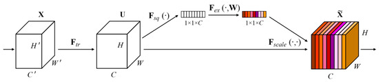
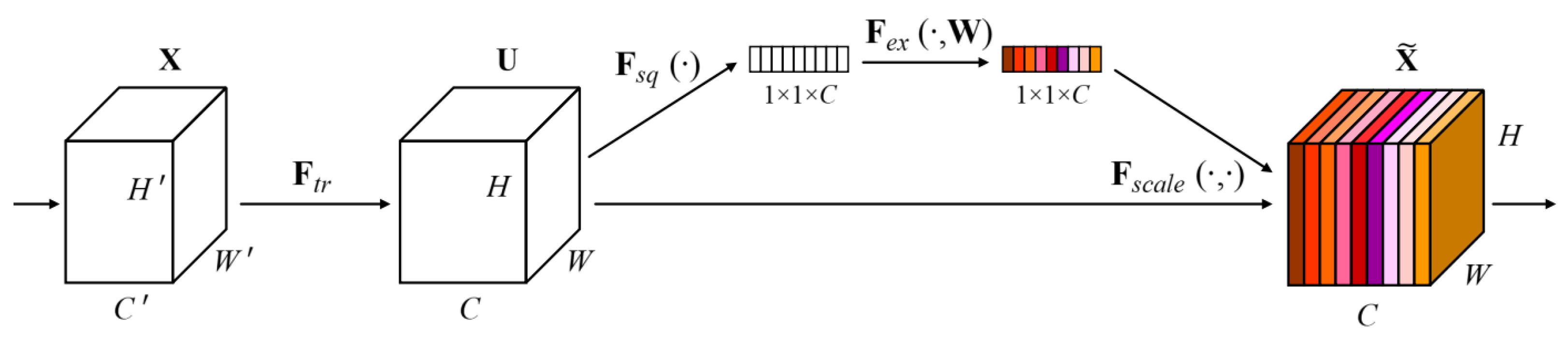
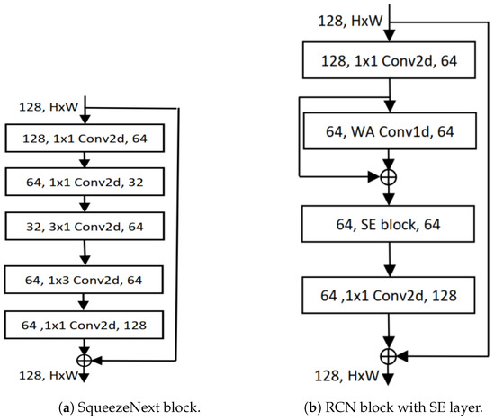
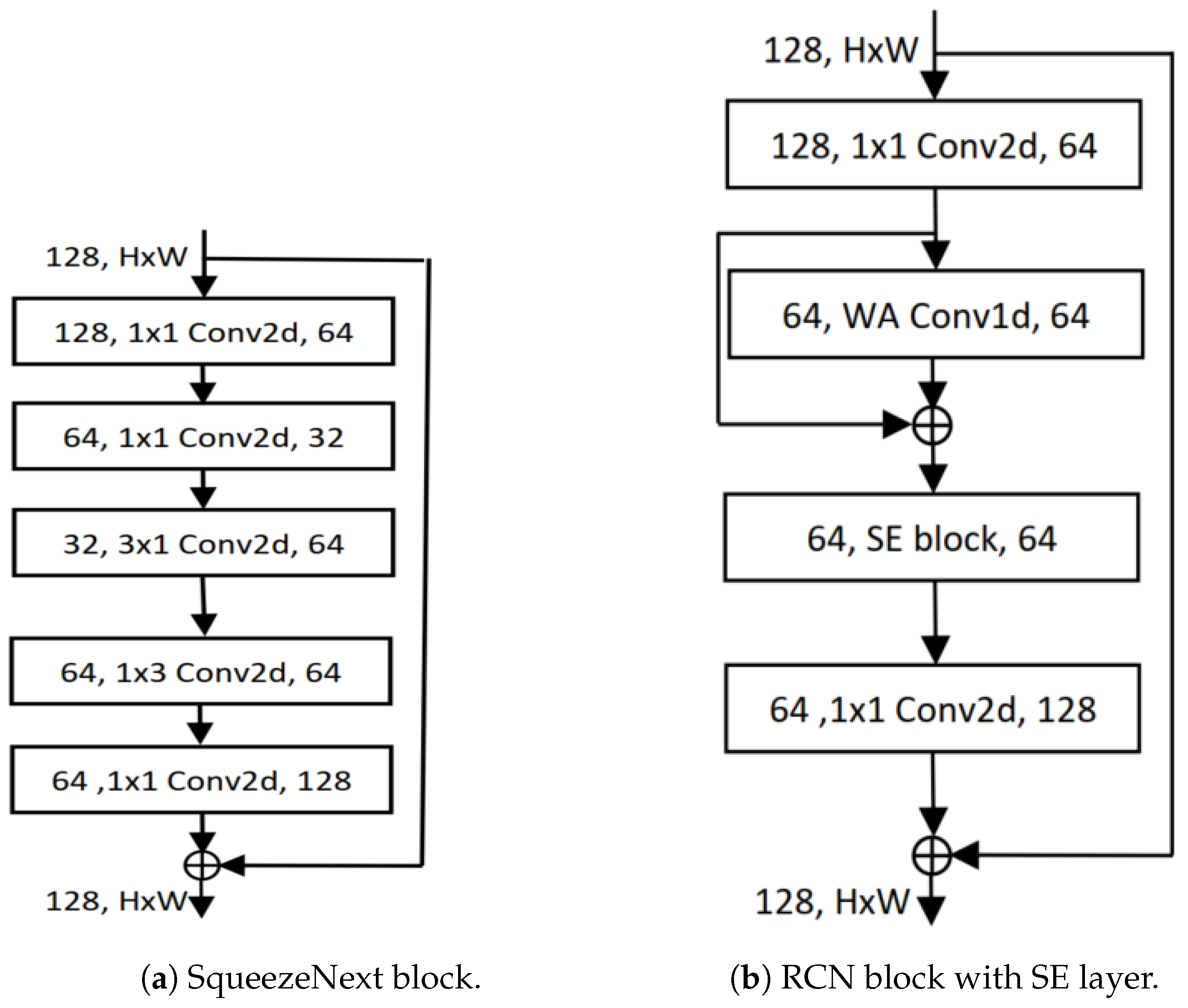
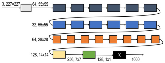
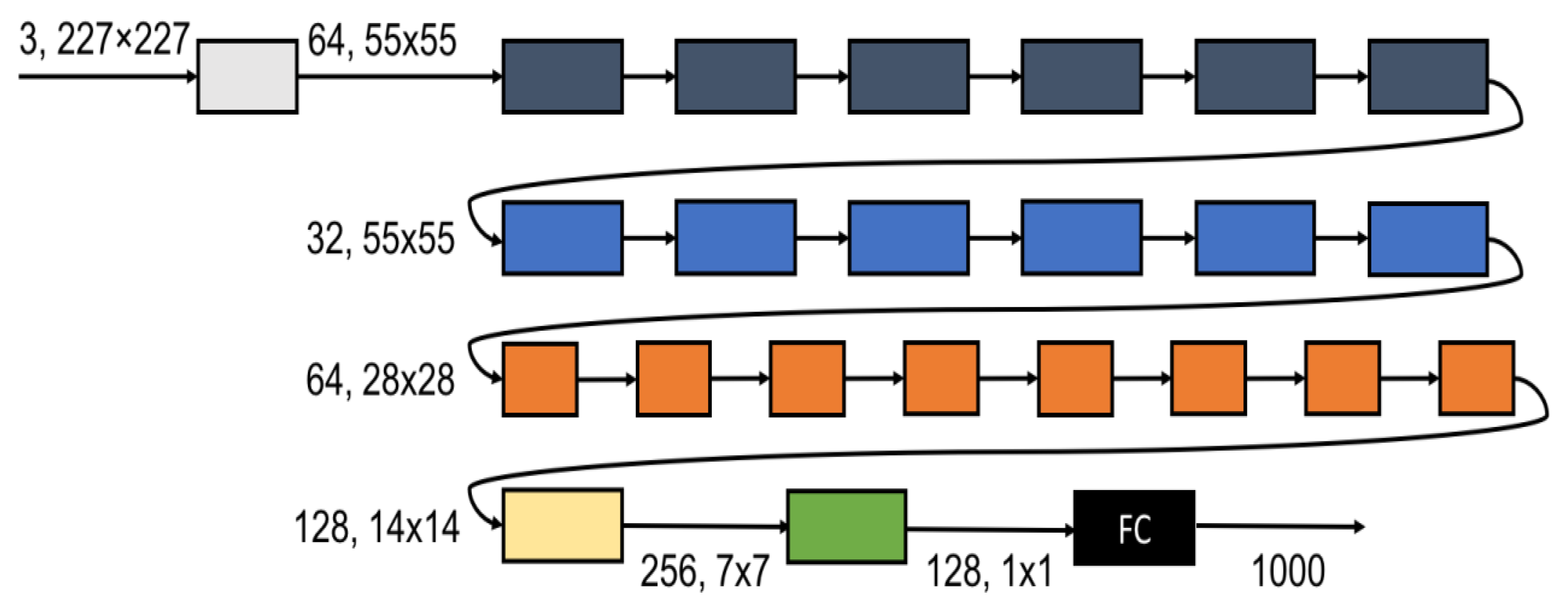
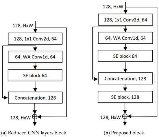
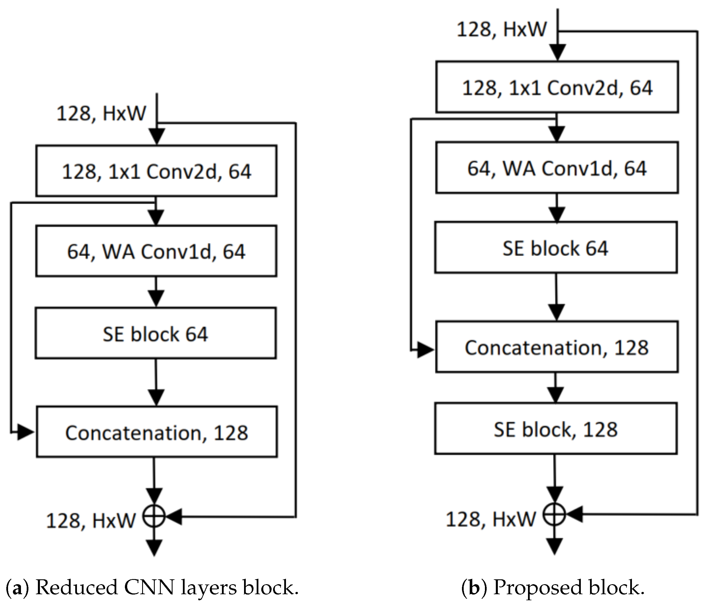
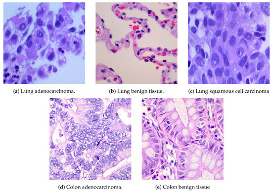
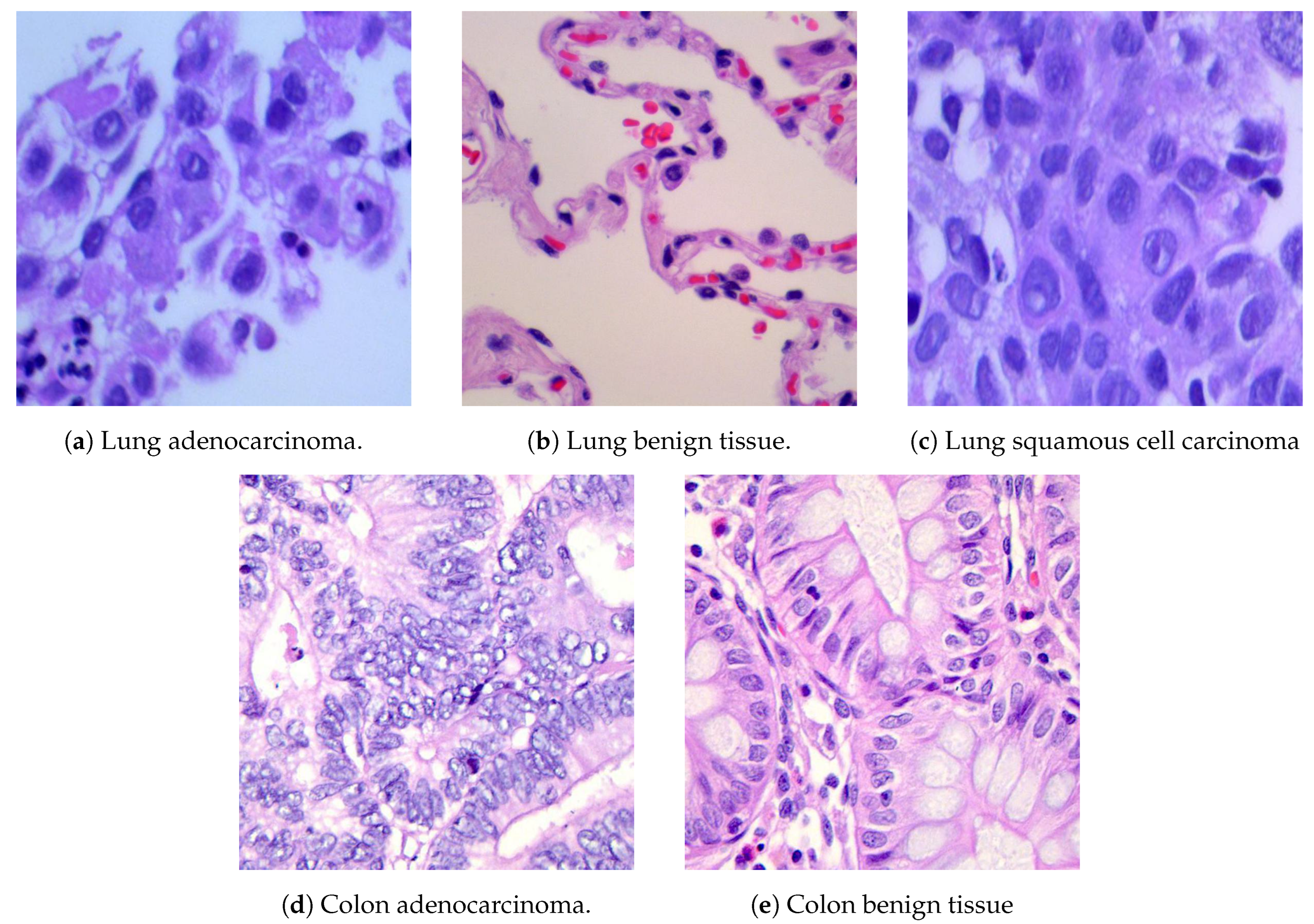
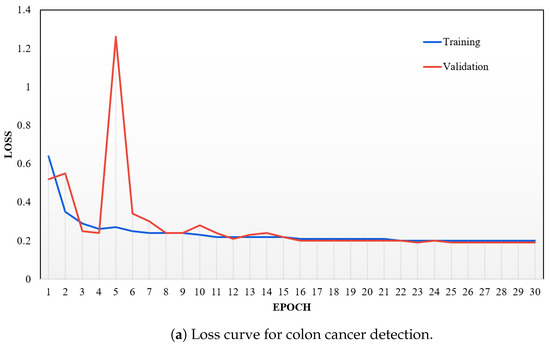
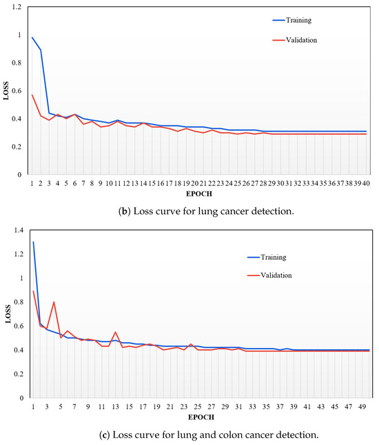
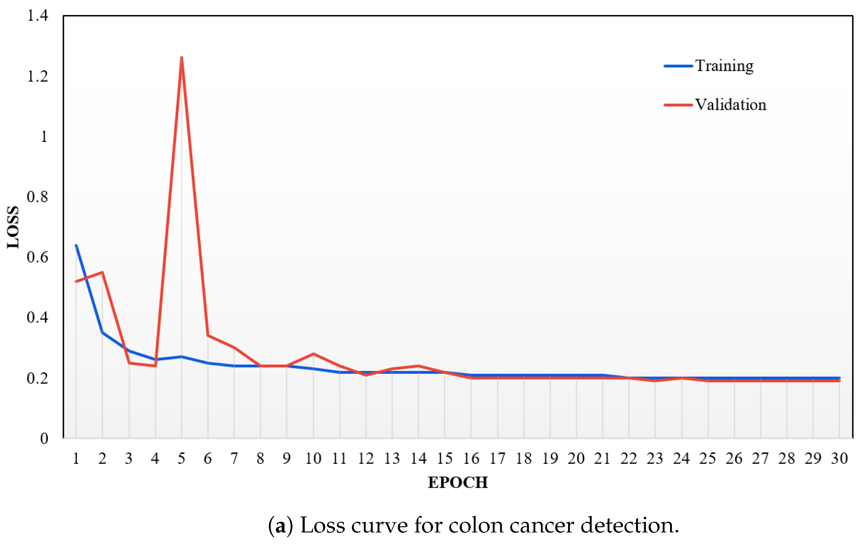
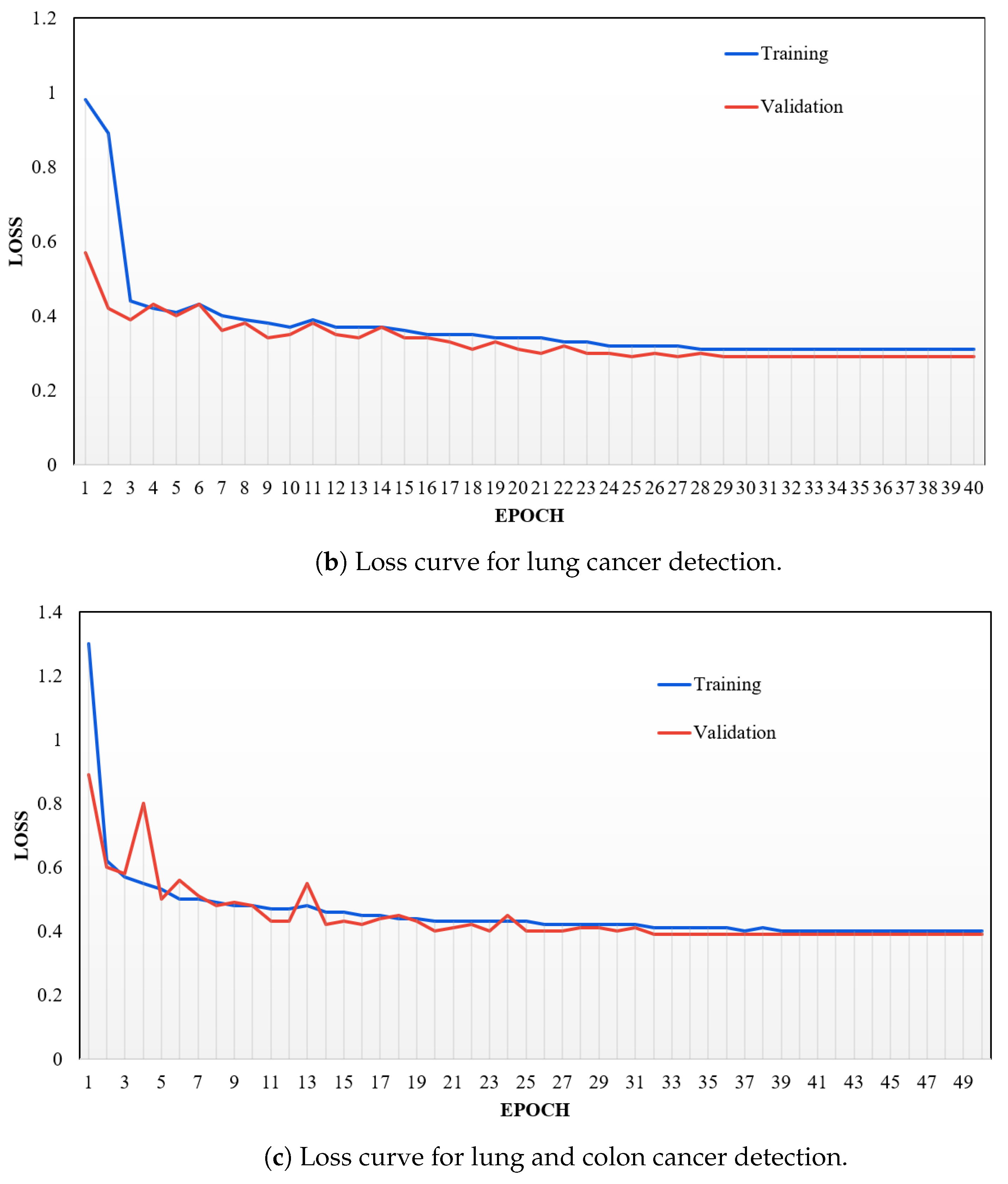

Disclaimer/Publisher’s Note: The statements, opinions and data contained in all publications are solely those of the individual author(s) and contributor(s) and not of MDPI and/or the editor(s). MDPI and/or the editor(s) disclaim responsibility for any injury to people or property resulting from any ideas, methods, instructions or products referred to in the content. |
© 2024 by the authors. Licensee MDPI, Basel, Switzerland. This article is an open access article distributed under the terms and conditions of the Creative Commons Attribution (CC BY) license (https://creativecommons.org/licenses/by/4.0/).