Next Issue
Volume 13, April-2
Previous Issue
Volume 13, March-2


Cancers, Volume 13, Issue 7 (April-1 2021) – 262 articles

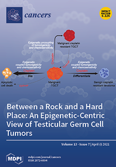
Cover Story (view full-size image): Testicular germ cell tumors are the most common malignancy of young men, and the incidence is rising worldwide. These tumors are curable with conventional chemotherapy, although a significant number of patients are resistant and die of progressive disease. Epigenetics appears to play an especially important role in the pathogenesis and therapeutic responses of testicular germ cell tumors. Singh et al. review the current evidence that supports a central role for epigenetics in the etiology, progression, and treatment of this common form of testicular cancer. A working model is developed that postulates that epigenetic features that drive testicular cancer malignancy also enable these tumors to be cured at a high rate with chemotherapy. This model provides new insights related to testicular cancer chemoresistance and potential vulnerabilities to epigenetic-based therapies. View this paper
  • Issues are regarded as officially published after their release is announced to the table of contents alert mailing list.
  • You may sign up for e-mail alerts to receive table of contents of newly released issues.
  • PDF is the official format for papers published in both, html and pdf forms. To view the papers in pdf format, click on the "PDF Full-text" link, and use the free Adobe Reader to open them.
Order results
Result details
Section
Select all
Export citation of selected articles as:
20 pages, 6074 KiB  
Article
SIPA1 Is a Modulator of HGF/MET Induced Tumour Metastasis via the Regulation of Tight Junction-Based Cell to Cell Barrier Function
by Chang Liu, Wenguo Jiang, Lijian Zhang, Rachel Hargest and Tracey A. Martin
Cancers 2021, 13(7), 1747; https://doi.org/10.3390/cancers13071747 - 6 Apr 2021
Cited by 5 | Viewed by 3127
Abstract
Background: Lung cancer is the leading cause of cancer death. SIPA1 is a mitogen induced GTPase activating protein (GAP) and may hamper cell cycle progression. SIPA1 has been shown to be involved in MET signaling and may contribute to tight junction (TJ) function [...] Read more.
Background: Lung cancer is the leading cause of cancer death. SIPA1 is a mitogen induced GTPase activating protein (GAP) and may hamper cell cycle progression. SIPA1 has been shown to be involved in MET signaling and may contribute to tight junction (TJ) function and cancer metastasis. Methods: Human lung tumour cohorts were analyzed. In vitro cell function assays were performed after knock down of SIPA1 in lung cancer cells with/without treatment. Quantitative polymerase chain reaction (qPCR) and western blotting were performed to analyze expression of HGF (hepatocyte growth factor), MET, and their downstream markers. Immunohistochemistry (IHC) and immunofluorescence (IFC) staining were performed. Results: Higher expression of SIPA1 in lung tumours was associated with a poorer prognosis. Knockdown of SIPA1 decreased invasiveness and proliferation of in vitro cell lines, and the SIPA1 knockdown cells demonstrated leaky barriers. Knockdown of SIPA1 decreased tight junction-based barrier function by downregulating MET at the protein but not the transcript level, through silencing of Grb2, SOCS, and PKCμ (Protein kinase Cµ), reducing the internalization and recycling of MET. Elevated levels of SIPA1 protein are correlated with receptor tyrosine kinases (RTKs), especially HGF/MET and TJs. The regulation of HGF on barrier function and invasion required the presence of SIPA1. Conclusions: SIPA1 plays an essential role in lung tumourigenesis and metastasis. SIPA1 may be a diagnostic and prognostic predictive biomarker. SIPA1 may also be a potential therapeutic target for non-small cell lung cancer (NSCLC) patients with aberrant MET expression and drug resistance. Full article
(This article belongs to the Special Issue Cell Adhesion Molecules in Metastasis)
Show Figures

Figure 1

27 pages, 1396 KiB  
Review
Epigenetics in a Spectrum of Myeloid Diseases and Its Exploitation for Therapy
by Michael Maher, Jeannine Diesch, Marguerite-Marie Le Pannérer and Marcus Buschbeck
Cancers 2021, 13(7), 1746; https://doi.org/10.3390/cancers13071746 - 6 Apr 2021
Cited by 7 | Viewed by 4237
Abstract
Mutations in genes encoding chromatin regulators are early events contributing to developing asymptomatic clonal hematopoiesis of indeterminate potential and its frequent progression to myeloid diseases with increasing severity. We focus on the subset of myeloid diseases encompassing myelodysplastic syndromes and their transformation to [...] Read more.
Mutations in genes encoding chromatin regulators are early events contributing to developing asymptomatic clonal hematopoiesis of indeterminate potential and its frequent progression to myeloid diseases with increasing severity. We focus on the subset of myeloid diseases encompassing myelodysplastic syndromes and their transformation to secondary acute myeloid leukemia. We introduce the major concepts of chromatin regulation that provide the basis of epigenetic regulation. In greater detail, we discuss those chromatin regulators that are frequently mutated in myelodysplastic syndromes. We discuss their role in the epigenetic regulation of normal hematopoiesis and the consequence of their mutation. Finally, we provide an update on the drugs interfering with chromatin regulation approved or in development for myelodysplastic syndromes and acute myeloid leukemia. Full article
Show Figures

Figure 1

10 pages, 398 KiB  
Article
Clinical Validation of the Comprehensive Complication Index in Colon Cancer Surgery
by Nicolò Tamini, Davide Bernasconi, Lorenzo Ripamonti, Giulia Lo Bianco, Marco Braga and Luca Nespoli
Cancers 2021, 13(7), 1745; https://doi.org/10.3390/cancers13071745 - 6 Apr 2021
Cited by 9 | Viewed by 2490
Abstract
(1) Introduction: To date, the sensitivity of the Comprehensive Complication Index (CCI) in a homogeneous cohort of colonic resections for oncologic purposes has not been reported. The present study aims to compare the CCI with the conventional Clavien–Dindo classification (CDC) in colon cancer [...] Read more.
(1) Introduction: To date, the sensitivity of the Comprehensive Complication Index (CCI) in a homogeneous cohort of colonic resections for oncologic purposes has not been reported. The present study aims to compare the CCI with the conventional Clavien–Dindo classification (CDC) in colon cancer patients. (2) Methods: The clinical data of patients submitted to an elective colectomy for adenocarcinoma were retrieved from a prospectively maintained database. Postoperative complications and length of stay were reviewed, and CDC and CCI scores were calculated for each patient. The association of the CCI and the CDC with the length of stay, prolongation of stay and readmission rate were assessed and compared. (3) Results: The overall postoperative morbidity was 26.9%. In particular, 157 (20.4%) patients had more than one complication. A strong correlation between the two scoring systems was observed (r = 99.4%; 95%CI: 99.3–99.5%). In multivariate analysis, CCI had a higher predictive ability for all endpoints. Regarding subgroup analysis, the difference between the CCI and CDC was progressively increased when evaluating outcome measures in complicated and multi-complicated patients. (4) Conclusion: Both scoring systems adequately report the overall burden of postoperative complications. The CCI showed a greater ability than the CDC to predict hospital stay, particularly in patients with multiple postoperative complications. Full article
(This article belongs to the Section Cancer Therapy)
Show Figures

Figure 1

24 pages, 3749 KiB  
Article
Modulated Electro-Hyperthermia Induces a Prominent Local Stress Response and Growth Inhibition in Mouse Breast Cancer Isografts
by Csaba András Schvarcz, Lea Danics, Tibor Krenács, Pedro Viana, Rita Béres, Tamás Vancsik, Ákos Nagy, Attila Gyenesei, József Kun, Marko Fonović, Robert Vidmar, Zoltán Benyó, Tamás Kaucsár and Péter Hamar
Cancers 2021, 13(7), 1744; https://doi.org/10.3390/cancers13071744 - 6 Apr 2021
Cited by 15 | Viewed by 3557
Abstract
Modulated electro-hyperthermia (mEHT) is a selective cancer treatment used in human oncology complementing other therapies. During mEHT, a focused electromagnetic field (EMF) is generated within the tumor inducing cell death by thermal and nonthermal effects. Here we investigated molecular changes elicited by mEHT [...] Read more.
Modulated electro-hyperthermia (mEHT) is a selective cancer treatment used in human oncology complementing other therapies. During mEHT, a focused electromagnetic field (EMF) is generated within the tumor inducing cell death by thermal and nonthermal effects. Here we investigated molecular changes elicited by mEHT using multiplex methods in an aggressive, therapy-resistant triple negative breast cancer (TNBC) model. 4T1/4T07 isografts inoculated orthotopically into female BALB/c mice were treated with mEHT three to five times. mEHT induced the upregulation of the stress-related Hsp70 and cleaved caspase-3 proteins, resulting in effective inhibition of tumor growth and proliferation. Several acute stress response proteins, including protease inhibitors, coagulation and heat shock factors, and complement family members, were among the most upregulated treatment-related genes/proteins as revealed by next-generation sequencing (NGS), Nanostring and mass spectrometry (MS). pathway analysis demonstrated that several of these proteins belong to the response to stimulus pathway. Cell culture treatments confirmed that the source of these proteins was the tumor cells. The heat-shock factor inhibitor KRIBB11 reduced mEHT-induced complement factor 4 (C4) mRNA increase. In conclusion, mEHT monotherapy induced tumor growth inhibition and a complex stress response. Inhibition of this stress response is likely to enhance the effectiveness of mEHT and other cancer treatments. Full article
(This article belongs to the Special Issue Regional Hyperthermia as Treatment of Solid Tumors)
Show Figures

Graphical abstract

25 pages, 1548 KiB  
Review
Combined Effects of Myeloid Cells in the Neuroblastoma Tumor Microenvironment
by Jennifer Frosch, Ilia Leontari and John Anderson
Cancers 2021, 13(7), 1743; https://doi.org/10.3390/cancers13071743 - 6 Apr 2021
Cited by 12 | Viewed by 3786
Abstract
Despite multimodal treatment, survival chances for high-risk neuroblastoma patients remain poor. Immunotherapeutic approaches focusing on the activation and/or modification of host immunity for eliminating tumor cells, such as chimeric antigen receptor (CAR) T cells, are currently in development, however clinical trials have failed [...] Read more.
Despite multimodal treatment, survival chances for high-risk neuroblastoma patients remain poor. Immunotherapeutic approaches focusing on the activation and/or modification of host immunity for eliminating tumor cells, such as chimeric antigen receptor (CAR) T cells, are currently in development, however clinical trials have failed to reproduce the preclinical results. The tumor microenvironment is emerging as a major contributor to immune suppression and tumor evasion in solid cancers and thus has to be overcome for therapies relying on a functional immune response. Among the cellular components of the neuroblastoma tumor microenvironment, suppressive myeloid cells have been described as key players in inhibition of antitumor immune responses and have been shown to positively correlate with more aggressive disease, resistance to treatments, and overall poor prognosis. This review article summarizes how neuroblastoma-driven inflammation induces suppressive myeloid cells in the tumor microenvironment and how they in turn sustain the tumor niche through suppressor functions, such as nutrient depletion and generation of oxidative stress. Numerous preclinical studies have suggested a range of drug and cellular therapy approaches to overcome myeloid-derived suppression in neuroblastoma that warrant evaluation in future clinical studies. Full article
(This article belongs to the Special Issue The Tumor Microenvironment of Neuroblastoma)
Show Figures

Figure 1

23 pages, 729 KiB  
Review
Roles of microRNAs in Regulating Cancer Stemness in Head and Neck Cancers
by Melysa Fitriana, Wei-Lun Hwang, Pak-Yue Chan, Tai-Yuan Hsueh and Tsai-Tsen Liao
Cancers 2021, 13(7), 1742; https://doi.org/10.3390/cancers13071742 - 6 Apr 2021
Cited by 9 | Viewed by 4224
Abstract
Head and neck squamous cell carcinomas (HNSCCs) are epithelial malignancies with 5-year overall survival rates of approximately 40–50%. Emerging evidence indicates that a small population of cells in HNSCC patients, named cancer stem cells (CSCs), play vital roles in the processes of tumor [...] Read more.
Head and neck squamous cell carcinomas (HNSCCs) are epithelial malignancies with 5-year overall survival rates of approximately 40–50%. Emerging evidence indicates that a small population of cells in HNSCC patients, named cancer stem cells (CSCs), play vital roles in the processes of tumor initiation, progression, metastasis, immune evasion, chemo-/radioresistance, and recurrence. The acquisition of stem-like properties of cancer cells further provides cellular plasticity for stress adaptation and contributes to therapeutic resistance, resulting in a worse clinical outcome. Thus, targeting cancer stemness is fundamental for cancer treatment. MicroRNAs (miRNAs) are known to regulate stem cell features in the development and tissue regeneration through a miRNA–target interactive network. In HNSCCs, miRNAs act as tumor suppressors and/or oncogenes to modulate cancer stemness and therapeutic efficacy by regulating the CSC-specific tumor microenvironment (TME) and signaling pathways, such as epithelial-to-mesenchymal transition (EMT), Wnt/β-catenin signaling, and epidermal growth factor receptor (EGFR) or insulin-like growth factor 1 receptor (IGF1R) signaling pathways. Owing to a deeper understanding of disease-relevant miRNAs and advances in in vivo delivery systems, the administration of miRNA-based therapeutics is feasible and safe in humans, with encouraging efficacy results in early-phase clinical trials. In this review, we summarize the present findings to better understand the mechanical actions of miRNAs in maintaining CSCs and acquiring the stem-like features of cancer cells during HNSCC pathogenesis. Full article
(This article belongs to the Special Issue MicroRNA and Cancer)
Show Figures

Figure 1

20 pages, 7583 KiB  
Article
Effects of Epstein-Barr Virus Infection on the Risk and Prognosis of Primary Laryngeal Squamous Cell Carcinoma: A Hospital-Based Case-Control Study in Taiwan
by Li-Ang Lee, Tuan-Jen Fang, Hsueh-Yu Li, Hai-Hua Chuang, Chung-Jan Kang, Kai-Ping Chang, Chun-Ta Liao, Tse-Ching Chen, Chung-Guei Huang and Tzu-Chen Yen
Cancers 2021, 13(7), 1741; https://doi.org/10.3390/cancers13071741 - 6 Apr 2021
Cited by 8 | Viewed by 3287
Abstract
Mounting molecular evidence supports Epstein–Barr virus (EBV) involvement in the pathogenesis of laryngeal squamous cell carcinoma (LSCC); however, the epidemiological data are inconsistent. In this retrospective case-control study, we aimed to determine whether EBV infection underlies the risk and prognosis of LSCC. The [...] Read more.
Mounting molecular evidence supports Epstein–Barr virus (EBV) involvement in the pathogenesis of laryngeal squamous cell carcinoma (LSCC); however, the epidemiological data are inconsistent. In this retrospective case-control study, we aimed to determine whether EBV infection underlies the risk and prognosis of LSCC. The prevalence of EBV infection, as analyzed using an EBV DNA polymerase chain reaction assay, was significantly higher in 42 Taiwanese patients with newly diagnosed primary LSCC, compared to 39 age- and sex-matched control patients without cancer (48% vs. 19%). Furthermore, most of the EBER signals detected using in situ hybridization were localized to the nuclei of tumor-infiltrating lymphocytes. In multivariate analysis, EBV DNA positivity, age ≥ 55 years, cigarette smoking, and high BCL-2, B2M, and CD161 expression (assessed using immunohistochemistry) were identified as independent risk factors for LSCC. Furthermore, five-year local recurrence and disease-free survival rates were 34% and 58%, respectively, with a high EBER signal and low CD3 expression independently predicting five-year local recurrence and disease-free survival. Our comprehensive profiling data accurately identified patients at risk for LSCC development, local recurrence, or disease-free survival. The information obtained in this study improves our understanding of EBV infection in LSCC, and may guide precision medicine for patients with LSCC. Full article
(This article belongs to the Special Issue Epstein-Barr Virus Infection in Cancer)
Show Figures

Figure 1

14 pages, 5166 KiB  
Article
Circulating Tumor DNA Reflects Uveal Melanoma Responses to Protein Kinase C Inhibition
by John J. Park, Russell J. Diefenbach, Natalie Byrne, Georgina V. Long, Richard A. Scolyer, Elin S. Gray, Matteo S. Carlino and Helen Rizos
Cancers 2021, 13(7), 1740; https://doi.org/10.3390/cancers13071740 - 6 Apr 2021
Cited by 19 | Viewed by 3678
Abstract
The prognosis for patients with UM is poor, and recent clinical trials have failed to prolong overall survival (OS) of these patients. Over 95% of UM harbor activating driver mutations, and this allows for the investigation of ctDNA. In this study, we investigated [...] Read more.
The prognosis for patients with UM is poor, and recent clinical trials have failed to prolong overall survival (OS) of these patients. Over 95% of UM harbor activating driver mutations, and this allows for the investigation of ctDNA. In this study, we investigated the value of ctDNA for adaptive clinical trial design in metastatic UM. Longitudinal plasma samples were analyzed for ctDNA in 17 metastatic UM patients treated with PKCi-based therapy in a phase 1 clinical trial setting. Plasma ctDNA was assessed using digital droplet PCR (ddPCR) and a custom melanoma gene panel for targeted next generation sequencing (NGS). Baseline ctDNA strongly correlated with baseline lactate dehydrogenase (LDH) (p < 0.001) and baseline disease burden (p = 0.002). Early during treatment (EDT) ctDNA accurately predicted patients with clinical benefit to PKCi using receiver operator characteristic (ROC) curves (AUC 0.84, [95% confidence interval 0.65–1.0, p = 0.026]). Longitudinal ctDNA assessment was informative for establishing clinical benefit and detecting disease progression with 7/8 (88%) of patients showing a rise in ctDNA and targeted NGS of ctDNA revealed putative resistance mechanisms prior to radiological progression. The inclusion of longitudinal ctDNA monitoring in metastatic UM can advance adaptive clinical trial design. Full article
(This article belongs to the Special Issue Cancer Biomarkers in Body Fluids)
Show Figures

Figure 1

26 pages, 3118 KiB  
Article
Understanding the Molecular Mechanism of miR-877-3p Could Provide Potential Biomarkers and Therapeutic Targets in Squamous Cell Carcinoma of the Cervix
by Saioa Mendaza, Joaquín Fernández-Irigoyen, Enrique Santamaría, Imanol Arozarena, David Guerrero-Setas, Tamara Zudaire, Rosa Guarch, August Vidal, José-Santos Salas, Xavier Matias-Guiu, Karina Ausín, Carmen Gil, Rubén Hernández-Alcoceba and Esperanza Martín-Sánchez
Cancers 2021, 13(7), 1739; https://doi.org/10.3390/cancers13071739 - 6 Apr 2021
Cited by 4 | Viewed by 3103
Abstract
No therapeutic targets and molecular biomarkers are available in cervical cancer (CC) management. In other cancer types, micro-RNA-877-3p (miR-877-3p) has been associated with events relevant for CC development. Thus, we aimed to determine miR-877-3p role in CC. miR-877-3p levels were examined by quantitative-PCR [...] Read more.
No therapeutic targets and molecular biomarkers are available in cervical cancer (CC) management. In other cancer types, micro-RNA-877-3p (miR-877-3p) has been associated with events relevant for CC development. Thus, we aimed to determine miR-877-3p role in CC. miR-877-3p levels were examined by quantitative-PCR in 117 cervical lesions and tumors. Effects on CC cell proliferation, migration, and invasion were evaluated upon anti-miR-877-3p transfection. miR-877-3p dependent molecular mechanism was comprehensively explored by proteomics, dual-luciferase reporter assay, western blot, and immunohistochemistry. Cervical tumors expressed higher miR-877-3p levels than benign lesions. miR-877-3p promoted CC cell migration and invasion, at least partly by modulating cytoskeletal protein folding through the chaperonin-containing T-complex protein 1 complex. Notably, miR-877-3p silencing synergized with paclitaxel. Interestingly, miR-877-3p downregulated the levels of an in silico-predicted target, ZNF177, whose expression and subcellular location significantly distinguished high-grade squamous intraepithelial lesions (HSILs) and squamous cell carcinomas of the cervix (SCCCs). Cytoplasmic ZNF177 was significantly associated with worse progression-free survival in SCCC. Our results suggest that: (i) miR-877-3p is a potential therapeutic target whose inhibition improves paclitaxel effects; (ii) the expression and location of its target ZNF177 could be diagnostic biomarkers between HSIL and SCCC; and (iii) cytoplasmic ZNF177 is a poor-prognosis biomarker in SCCC. Full article
Show Figures

Graphical abstract

20 pages, 5091 KiB  
Article
Blockade of AMPK-Mediated cAMP–PKA–CREB/ATF1 Signaling Synergizes with Aspirin to Inhibit Hepatocellular Carcinoma
by Hongying Zhang, Songpeng Yang, Jiao Wang and Yangfu Jiang
Cancers 2021, 13(7), 1738; https://doi.org/10.3390/cancers13071738 - 6 Apr 2021
Cited by 22 | Viewed by 5147
Abstract
Aspirin can prevent or inhibit inflammation-related cancers, such as colorectal cancer and hepatocellular carcinoma (HCC). However, the effectiveness of chemotherapy may be compromised by activating oncogenic pathways in cancer cells. Elucidation of such chemoresistance mechanisms is crucial to developing novel strategies to maximize [...] Read more.
Aspirin can prevent or inhibit inflammation-related cancers, such as colorectal cancer and hepatocellular carcinoma (HCC). However, the effectiveness of chemotherapy may be compromised by activating oncogenic pathways in cancer cells. Elucidation of such chemoresistance mechanisms is crucial to developing novel strategies to maximize the anti-cancer effects of aspirin. Here, we report that aspirin markedly induces CREB/ATF1 phosphorylation in HCC cells, which compromises aspirin’s anti-HCC effect. Inhibition of AMP-activated protein kinase (AMPK) abrogates the induction of CREB/ATF1 phosphorylation by aspirin. Mechanistically, activation of AMPK by aspirin results in decreased expression of the urea cycle enzyme carbamoyl-phosphate synthase 1 (CPS1) in HCC cells and xenografts. Treatment with aspirin or CPS1 knockdown stimulates soluble adenylyl cyclase expression, thereby increasing cyclic AMP (cAMP) synthesis and stimulating PKA–CREB/ATF1 signaling. Importantly, abrogation of aspirin-induced CREB/ATF1 phosphorylation could sensitize HCC to aspirin. The bis-benzylisoquinoline alkaloid berbamine suppresses the expression of cancerous inhibitor of protein phosphatase 2A (CIP2A), leading to protein phosphatase 2A-mediated downregulation of CREB/ATF1 phosphorylation. The combination of berbamine and aspirin significantly inhibits HCC in vitro and in vivo. These data demonstrate that the regulation of cAMP-PKA-CREB/ATF1 signaling represents a noncanonical function of CPS1. Targeting the PKA–CREB/ATF1 axis may be a strategy to improve the therapeutic effects of aspirin on HCC. Full article
(This article belongs to the Special Issue Targeting Signal Transduction Pathways in Cancer)
Show Figures

Figure 1

19 pages, 7501 KiB  
Review
Plasma-Treated Solutions (PTS) in Cancer Therapy
by Hiromasa Tanaka, Sander Bekeschus, Dayun Yan, Masaru Hori, Michael Keidar and Mounir Laroussi
Cancers 2021, 13(7), 1737; https://doi.org/10.3390/cancers13071737 - 6 Apr 2021
Cited by 75 | Viewed by 5614
Abstract
Cold physical plasma is a partially ionized gas generating various reactive oxygen and nitrogen species (ROS/RNS) simultaneously. ROS/RNS have therapeutic effects when applied to cells and tissues either directly from the plasma or via exposure to solutions that have been treated beforehand using [...] Read more.
Cold physical plasma is a partially ionized gas generating various reactive oxygen and nitrogen species (ROS/RNS) simultaneously. ROS/RNS have therapeutic effects when applied to cells and tissues either directly from the plasma or via exposure to solutions that have been treated beforehand using plasma processes. This review addresses the challenges and opportunities of plasma-treated solutions (PTSs) for cancer treatment. These PTSs include plasma-treated cell culture media in experimental research as well as clinically approved solutions such as saline and Ringer’s lactate, which, in principle, already qualify for testing in therapeutic settings. Several types of cancers were found to succumb to the toxic action of PTSs, suggesting a broad mechanism of action based on the tumor-toxic activity of ROS/RNS stored in these solutions. Moreover, it is indicated that the PTS has immuno-stimulatory properties. Two different routes of application are currently envisaged in the clinical setting. One is direct injection into the bulk tumor, and the other is lavage in patients suffering from peritoneal carcinomatosis adjuvant to standard chemotherapy. While many promising results have been achieved so far, several obstacles, such as the standardized generation of large volumes of sterile PTS, remain to be addressed. Full article
(This article belongs to the Special Issue Advances in Plasma Oncology toward Clinical Translation)
Show Figures

Figure 1

14 pages, 3238 KiB  
Article
Epithelial and Mesenchymal Markers in Adrenocortical Tissues: How Mesenchymal Are Adrenocortical Tissues?
by Iuliu Sbiera, Stefan Kircher, Barbara Altieri, Martin Fassnacht, Matthias Kroiss and Silviu Sbiera
Cancers 2021, 13(7), 1736; https://doi.org/10.3390/cancers13071736 - 6 Apr 2021
Cited by 7 | Viewed by 2473
Abstract
A clinically relevant proportion of adrenocortical carcinoma (ACC) cases shows a tendency to metastatic spread. The objective was to determine whether the epithelial to mesenchymal transition (EMT), a mechanism associated with metastasizing in several epithelial cancers, might play a crucial role in ACC. [...] Read more.
A clinically relevant proportion of adrenocortical carcinoma (ACC) cases shows a tendency to metastatic spread. The objective was to determine whether the epithelial to mesenchymal transition (EMT), a mechanism associated with metastasizing in several epithelial cancers, might play a crucial role in ACC. 138 ACC, 29 adrenocortical adenomas (ACA), three normal adrenal glands (NAG), and control tissue samples were assessed for the expression of epithelial (E-cadherin and EpCAM) and mesenchymal (N-cadherin, SLUG and SNAIL) markers by immunohistochemistry. Using real-time RT-PCR we quantified the alternative isoform splicing of FGFR 2 and 3, another known indicator of EMT. We also assessed the impact of these markers on clinical outcome. Results show that both normal and neoplastic adrenocortical tissues lacked expression of epithelial markers but strongly expressed mesenchymal markers N-cadherin and SLUG. FGFR isoform splicing confirmed higher similarity of adrenocortical tissues to mesenchymal compared to epithelial tissues. In ACC, higher SLUG expression was associated with clinical markers indicating aggressiveness, while N-cadherin expression inversely associated with these markers. In conclusion, we could not find any indication of EMT as all adrenocortical tissues lacked expression of epithelial markers and exhibited closer similarity to mesenchymal tissues. However, while N-cadherin might play a positive role in tissue structure upkeep, SLUG seems to be associated with a more aggressive phenotype. Full article
(This article belongs to the Special Issue Endocrine and Neuroendocrine Cancers)
Show Figures

Figure 1

16 pages, 2020 KiB  
Article
Matched-Pair Analysis of Survival in the Patients with Advanced Laryngeal and Hypopharyngeal Squamous Cell Carcinoma Treated with Induction Chemotherapy Plus Chemo-Radiation or Total Laryngectomy
by Patricia García-Cabo, Fernando López, Mario Sánchez-Canteli, Laura Fernández-Vañes, César Álvarez-Marcos, José Luis Llorente, Maria Ángeles de la Rúa, Pilar Blay and Juan P. Rodrigo
Cancers 2021, 13(7), 1735; https://doi.org/10.3390/cancers13071735 - 6 Apr 2021
Cited by 11 | Viewed by 2216
Abstract
Background: We performed a comparative analysis between an organ-preservation protocol and surgery followed by radiotherapy in patients with locally advanced squamous cell carcinoma of the larynx and hypopharynx; Methods: 60 previously untreated patients who were treated with induction chemotherapy followed by chemoradiotherapy in [...] Read more.
Background: We performed a comparative analysis between an organ-preservation protocol and surgery followed by radiotherapy in patients with locally advanced squamous cell carcinoma of the larynx and hypopharynx; Methods: 60 previously untreated patients who were treated with induction chemotherapy followed by chemoradiotherapy in responders were compared with a control group of 60 patients treated with up-front surgery. Both groups were statistically comparable, according to the subsite, TNM (tumor-node-metastasis) stage, age, and sex; Results: Mean age was 58 years and 92% were male. No significant statistical difference was observed for overall survival (OS) (HR 0.75; 95% CI 0.48–1.18; P = 0.22) and disease-specific survival (DSS) (HR 0.98; 95% CI 0.52–1.83, P = 0.96). Also, there was no significant difference for recurrence-free survival (HR 0.931; 95% CI 0.57–1.71; P = 0.81), metastases-free survival (HR 2.23; 95% CI 0.67–7.41; P = 0.19), and the appearance of second primary tumors (HR 1.22; 95% CI 0.51–2.88; P = 0.64); Conclusions: The results of the organ-preservation approach did not appear inferior to those of surgery plus (chemo)radiotherapy for patients with T3/T4a larynx and T2–T4a hypopharynx cancer with respect to OS and DSS, locoregional control and metastases-free survival. Full article
(This article belongs to the Special Issue Surgical Treatment of Head and Neck Squamous Cell Carcinomas (HNSCC))
Show Figures

Figure 1

15 pages, 15305 KiB  
Article
Role for the Histone Demethylase KDM4B in Rhabdomyosarcoma via CDK6 and CCNA2: Compensation by KDM4A and Apoptotic Response of Targeting Both KDM4B and KDM4A
by Zoë S. Walters, Ewa Aladowicz, Barbara Villarejo-Balcells, Gary Nugent, Joanna L. Selfe, Paul Eve, Julian Blagg, Olivia Rossanese and Janet Shipley
Cancers 2021, 13(7), 1734; https://doi.org/10.3390/cancers13071734 - 6 Apr 2021
Cited by 4 | Viewed by 2968
Abstract
Histone demethylases are epigenetic modulators that play key roles in regulating gene expression related to many critical cellular functions and are emerging as promising therapeutic targets in a number of tumor types. We previously identified histone demethylase family members as overexpressed in the [...] Read more.
Histone demethylases are epigenetic modulators that play key roles in regulating gene expression related to many critical cellular functions and are emerging as promising therapeutic targets in a number of tumor types. We previously identified histone demethylase family members as overexpressed in the pediatric sarcoma, rhabdomyosarcoma. Here we show high sensitivity of rhabdomyosarcoma cells to a pan-histone demethylase inhibitor, JIB-04 and identify a key role for the histone demethylase KDM4B in rhabdomyosarcoma cell growth through an RNAi-screening approach. Decreasing KDM4B levels affected cell cycle progression and transcription of G1/S and G2/M checkpoint genes including CDK6 and CCNA2, which are bound by KDM4B in their promoter regions. However, after sustained knockdown of KDM4B, rhabdomyosarcoma cell growth recovered. We show that this can be attributed to acquired molecular compensation via recruitment of KDM4A to the promoter regions of CDK6 and CCNA2 that are otherwise bound by KDM4B. Furthermore, upfront silencing of both KDM4B and KDM4A led to RMS cell apoptosis, not seen by reducing either alone. To circumvent compensation and elicit stronger therapeutic responses, our study supports targeting histone demethylase sub-family proteins through selective poly-pharmacology as a therapeutic approach. Full article
Show Figures

Figure 1

14 pages, 1738 KiB  
Article
Cancer Cells’ Metabolism Dynamics in Renal Cell Carcinoma Patients’ Outcome: Influence of GLUT-1-Related hsa-miR-144 and hsa-miR-186
by Mariana Morais, Francisca Dias, Inês Nogueira, Anabela Leão, Nuno Gonçalves, Luís Araújo, Sara Granja, Fátima Baltazar, Ana L Teixeira and Rui Medeiros
Cancers 2021, 13(7), 1733; https://doi.org/10.3390/cancers13071733 - 6 Apr 2021
Cited by 14 | Viewed by 3076
Abstract
The cancer cells’ metabolism is altered due to deregulation of key proteins, including glucose transporter 1 (GLUT-1), whose mRNA levels are influenced by microRNAs (miRNAs). Renal cell carcinoma (RCC) is the most common and lethal neoplasia in the adult kidney, mostly due to [...] Read more.
The cancer cells’ metabolism is altered due to deregulation of key proteins, including glucose transporter 1 (GLUT-1), whose mRNA levels are influenced by microRNAs (miRNAs). Renal cell carcinoma (RCC) is the most common and lethal neoplasia in the adult kidney, mostly due to the lack of accurate diagnosis and follow-up biomarkers. Being a metabolic associated cancer, this study aimed to understand the hsa-miR-144-5p and hsa-miR-186-3p’s potential as biomarkers of clear cell RCC (ccRCC), establishing their role in its glycolysis status. Using three ccRCC lines, the intra- and extracellular levels of both miRNAs, GLUT-1’s mRNA expression and protein levels were assessed. Glucose consumption and lactate production were evaluated as glycolysis markers. A decrease of intracellular levels of these miRNAs and increase of their excretion was observed, associated with an increase of GLUT-1’s levels and glycolysis’ markers. Through a liquid biopsy approach, we found that RCC patients present higher plasmatic levels of hsa-miR-186-3p than healthy individuals. The Hsa-miR144-5p’s higher levels were associated with early clinical stages. When patients were stratified according to miRNAs plasmatic levels, low plasmatic levels of hsa-miR-144-5p and high plasmatic levels of hsa-miR-186-3p (high-risk group) showed the worst overall survival. Thus, circulating levels of these miRNAs may be potential biomarkers of ccRCC prognosis. Full article
(This article belongs to the Special Issue The Roles of microRNAs in Cancer Aggressiveness and Drug Resistance)
Show Figures

Figure 1

12 pages, 1929 KiB  
Article
Substance P Antagonism Prevents Chemotherapy-Induced Cardiotoxicity
by Ashiq Legi, Emma Rodriguez, Thomas K. Eckols, Cyrus Mistry and Prema Robinson
Cancers 2021, 13(7), 1732; https://doi.org/10.3390/cancers13071732 - 6 Apr 2021
Cited by 13 | Viewed by 2787
Abstract
Background: Doxorubicin (DOX), used in chemotherapeutic regimens in many cancers, has been known to induce, cardiotoxicity and life-threatening heart failure or acute coronary syndromes in some patients. We determined the role of Substance P (SP), a neuropeptide and its high affinity receptor, [...] Read more.
Background: Doxorubicin (DOX), used in chemotherapeutic regimens in many cancers, has been known to induce, cardiotoxicity and life-threatening heart failure or acute coronary syndromes in some patients. We determined the role of Substance P (SP), a neuropeptide and its high affinity receptor, NK-1R in chemotherapy associated cardiotoxicity in mice. We determined if NK-1R antagonism will prevent DOX-induced cardiotoxicity in vivo. Methods: C57BL/6 mice (6- week old male) were injected intraperitoneally with DOX (5 mg per kilogram of body weight once a week for 5 weeks) with or without treatment with aprepitant (a NK-1R antagonist, Emend, Merck & Co., Kenilworth, NJ, USA). Five different dosages of aprepitant were administered in the drinking water five days before the first injection of DOX and then continued until the end of the experiment. Each of these 5 doses are as follows; Dose 1 = 0.9 µg/mL, Dose 2 = 1.8 µg/mL, Dose 3 = 3.6 µg/mL, Dose 4 = 7.2 µg/mL, Dose 5 = 14.4 µg/mL. Controls consisted of mice injected with PBS (instead of DOX) with or without aprepitant treatment. The experiment was terminated 5 weeks post-DOX administration and various cardiac functional parameters were determined. Following euthanization, we measured heart weight to body weight ratios and the following in the hearts, of mice treated with and without DOX and aprepitant; (a) levels of SP and NK1R, (b) cardiomyocyte diameter (to determine evidence of cardiomyocyte hypertrophy), (c) Annexin V levels (to determine evidence of cardiac apoptosis), and (d) ratios of reduced glutathione (GSH) to oxidized glutathione (GSSG) (to determine evidence of oxidative stress). Results: We demonstrated that the levels of SP and NK1R were significantly increased respectively by 2.07 fold and 1.86 fold in the hearts of mice treated with versus without DOX. We determined that DOX-induced cardiac dysfunction was significantly attenuated by treatment with aprepitant. Cardiac functional parameters such as fractional shortening (FS), ejection fraction (EF) and stroke volume (SV) were respectively decreased by 27.6%, 21.02% and 21.20% compared to the vehicle treated group (All, p < 0.05, ANOVA). Importantly, compared to treatment with DOX alone, treatment with lower doses of aprepitant in DOX treated mice significantly reduced the effects of DOX on FS, EF and SV to values not significantly different from sham (vehicle treated) mice (All, p < 0.05, ANOVA). The levels of, apoptosis marker (Annexin V), oxidative stress (ratio of GSH with GSSG) and cardiomyocyte hypertrophy were respectively increased by 47.61%, 91.43% and 47.54% in the hearts of mice treated with versus without DOX. Compared to the DOX alone group, treatment with DOX and Dose 1, 2 and 3 of aprepitant significantly decreased the levels of each of these parameters (All p < 0.05, ANOVA). Conclusions: Our studies indicate that the SP/NK1-R system is a key mediator that induces, DOX-induced, cardiac dysfunction, cardiac apoptosis, cardiac oxidative stress and cardiomyocyte hypertrophy. These studies implicate that NK-1R antagonists may serve as a novel therapeutic tool for prevention of chemotherapy induced cardiotoxicity in cancer. Full article
(This article belongs to the Special Issue Involvement of the Substance P/Neurokinin-1 Receptor System in Cancer)
Show Figures

Figure 1

17 pages, 2131 KiB  
Article
Inhibition of Liver Metastasis in Colorectal Cancer by Targeting IL-13/IL13Rα2 Binding Site with Specific Monoclonal Antibodies
by Marta Jaén, Rubén A. Bartolomé, Carmen Aizpurua, Ángela Martin-Regalado, J. Ignacio Imbaud and J. Ignacio Casal
Cancers 2021, 13(7), 1731; https://doi.org/10.3390/cancers13071731 - 6 Apr 2021
Cited by 8 | Viewed by 2932
Abstract
Background: IL13Rα2 is reportedly a promising therapeutic target in different cancers. Still, no specific antagonists have reached the clinics yet. We investigated the use of a IL-13/IL13Rα2 binding motif, called D1, as a new target for the development of therapeutic monoclonal antibodies (mAbs) [...] Read more.
Background: IL13Rα2 is reportedly a promising therapeutic target in different cancers. Still, no specific antagonists have reached the clinics yet. We investigated the use of a IL-13/IL13Rα2 binding motif, called D1, as a new target for the development of therapeutic monoclonal antibodies (mAbs) for colorectal cancer (CRC) metastasis. Methods: IL13Rα2 D1 peptides were prepared and used for immunization and antibody development. Antibodies were tested for inhibition of cellular invasion through Matrigel using CRC cell lines. Effects of the mAbs on cell signaling, receptor internalization and degradation were determined by western blot and flow cytometry. Swiss nude mice were used for survival analysis after treatment with IL13Rα2-specific mAbs and metastasis development. Results: IL13Rα2 D1 peptides were used to generate highly selective mAbs that blocked IL13/IL13Rα2-mediated SRC activation and cell invasion in colorectal cancer cells. Antibodies also provoked a significant reduction in cell adhesion and proliferation of metastatic cancer cells. Treatment with mAbs impaired the FAK, SRC and PI3K/AKT pathway activation. Blocking effectivity was shown to correlate with the cellular IL13Rα2 expression level. Despite mAb 5.5.4 partially blocked IL-13 mediated receptor internalization from the cancer cell surface it still promotes receptor degradation. Compared with other IL13Rα2-specific antibodies, 5.5.4 exhibited a superior efficacy to inhibit metastatic growth in vivo, providing a complete mouse survival in different conditions, including established metastasis. Conclusions: Monoclonal antibody 5.5.4 showed a highly selective blocking capacity for the interaction between IL-13 and IL13Rα2 and caused a complete inhibition of liver metastasis in IL13Rα2-positive colorectal cancer cells. This capacity might be potentially applicable to other IL13Rα2-expressing tumors. Full article
(This article belongs to the Special Issue Targeting Cancer Metastasis)
Show Figures

Graphical abstract

21 pages, 5068 KiB  
Article
Identification and Validation of an Immune-Associated RNA-Binding Proteins Signature to Predict Clinical Outcomes and Therapeutic Responses in Glioma Patients
by Ruotong Tian, Yimin Li, Qian Liu and Minfeng Shu
Cancers 2021, 13(7), 1730; https://doi.org/10.3390/cancers13071730 - 6 Apr 2021
Cited by 12 | Viewed by 2991
Abstract
The prognosis of patients with glioma is largely related to both the tumor-infiltrating immune cells and the expression of RNA-binding proteins (RBPs) that are able to regulate various pro-inflammatory and oncogenic mediators. However, immune-associated RBPs in glioma remain unexplored. In this study, we [...] Read more.
The prognosis of patients with glioma is largely related to both the tumor-infiltrating immune cells and the expression of RNA-binding proteins (RBPs) that are able to regulate various pro-inflammatory and oncogenic mediators. However, immune-associated RBPs in glioma remain unexplored. In this study, we captured patient data from The Cancer Genome Atlas (TCGA) and divided them into two immune subtype groups according to the difference in infiltration of immune cells. After differential expression and co-expression analysis, we identified 216 RBPs defined as immune-associated RBPs. After narrowing down processes, eight RBPs were selected out to construct a risk signature that proven to be a novel and independent prognostic factor. The patients were divided into high- and low-risk groups on the basis of risk score. Higher risk scores meant worse overall survival and higher expression of human leukocyte antigen and immune checkpoints such as PD1 and CTLA4. In addition, analyses of pathway enrichment, somatic mutation, copy number variations and immuno-/chemotherapeutic response prediction were performed in high- and low-risk groups and compared with each other. For the first time, we demonstrated a novel signature composed of eight immune-associated RBPs that was valuable in predicting the survival of glioma patients and directing immunotherapy and chemotherapy. Full article
Show Figures

Graphical abstract

20 pages, 1086 KiB  
Review
Tertiary Prevention of HCC in Chronic Hepatitis B or C Infected Patients
by Wei Teng, Yen-Chun Liu, Wen-Juei Jeng and Chien-Wei Su
Cancers 2021, 13(7), 1729; https://doi.org/10.3390/cancers13071729 - 6 Apr 2021
Cited by 15 | Viewed by 6237
Abstract
Hepatocellular carcinoma (HCC) ranks as a leading cause of common cancer and cancer-related death. The major etiology of HCC is due to chronic hepatitis virus including HBV and HCV infections. Scheduled HCC surveillance in high risk populations improves the early detection rate and [...] Read more.
Hepatocellular carcinoma (HCC) ranks as a leading cause of common cancer and cancer-related death. The major etiology of HCC is due to chronic hepatitis virus including HBV and HCV infections. Scheduled HCC surveillance in high risk populations improves the early detection rate and the feasibility of curative treatment. However, high HCC recurrence rate still accounts for the poor prognosis of HCC patients. In this article, we critically review the pathogenesis of viral hepatitis-related hepatocellular carcinoma and the evidence of tertiary prevention efficacy by current available antiviral treatment, and discuss the knowledge gap in viral hepatitis-related HCC tertiary prevention. Full article
(This article belongs to the Special Issue Hepatobiliary Cancers)
Show Figures

Figure 1

13 pages, 1544 KiB  
Article
Predictive Factors for RAI-Refractory Disease and Short Overall Survival in PDTC
by David Kersting, Robert Seifert, Lukas Kessler, Ken Herrmann, Sarah Theurer, Tim Brandenburg, Henning Dralle, Frank Weber, Lale Umutlu, Dagmar Führer-Sakel, Rainer Görges, Christoph Rischpler and Manuel Weber
Cancers 2021, 13(7), 1728; https://doi.org/10.3390/cancers13071728 - 6 Apr 2021
Cited by 11 | Viewed by 2928
Abstract
Background: The clinical phenotype of poorly differentiated thyroid cancer (PDTC) can vary substantially. We aim to evaluate risk factors for radioiodine refractory (RAI-R) disease and reduced overall survival (OS). Methods: We retrospectively screened our institutional database for PDTC patients. For the assessment of [...] Read more.
Background: The clinical phenotype of poorly differentiated thyroid cancer (PDTC) can vary substantially. We aim to evaluate risk factors for radioiodine refractory (RAI-R) disease and reduced overall survival (OS). Methods: We retrospectively screened our institutional database for PDTC patients. For the assessment of RAI-R disease, we included patients who underwent dual imaging with 18F-FDG-PET and 124I-PET/131I scintigraphy that met the internal standard of care. We tested primary size, extrathyroidal extension (ETE), and age >55 years as risk factors for RAI-R disease at initial diagnosis and during the disease course using uni- and multivariate analyses. We tested metabolic tumor volume (MTV), total lesion glycolysis (TLG) on 18F-FDG-PET, and the progression of stimulated thyroglobulin within 4–6 months of initial radioiodine therapy as prognostic markers for OS. Results: Size of primary >40 mm and ETE were significant predictors of RAI-R disease in the course of disease in univariate (81% vs. 27%, p = 0.001; 89% vs. 33%, p < 0.001) and multivariate analyses. Primary tumor size was an excellent predictor of RAI-R disease (AUC = 0.90). TLG/MTV > upper quartile and early thyroglobulin progression were significantly associated with shorter median OS (29.0 months vs. 56.9 months, p < 0.05; 57.8 months vs. not reached p < 0.005, respectively). Discussion: PDTC patients, especially those with additional risk factors, should be assessed for RAI-R disease at initial diagnosis and in the course of disease, allowing for early implementation of multimodal treatment. Primary tumor size >40 mm, ETE, and age >55 are significant risk factors for RAI-R disease. High MTV/TLG is a significant risk factor for premature death and can help identify patients requiring intervention. Full article
(This article belongs to the Section Cancer Biomarkers)
Show Figures

Figure 1

23 pages, 3811 KiB  
Article
Succinate Anaplerosis Has an Onco-Driving Potential in Prostate Cancer Cells
by Ana Carolina B. Sant’Anna-Silva, Juan A. Perez-Valencia, Marco Sciacovelli, Claude Lalou, Saharnaz Sarlak, Laura Tronci, Efterpi Nikitopoulou, Andras T. Meszaros, Christian Frezza, Rodrigue Rossignol, Erich Gnaiger and Helmut Klocker
Cancers 2021, 13(7), 1727; https://doi.org/10.3390/cancers13071727 - 6 Apr 2021
Cited by 16 | Viewed by 4190
Abstract
Tumor cells display metabolic alterations when compared to non-transformed cells. These characteristics are crucial for tumor development, maintenance and survival providing energy supplies and molecular precursors. Anaplerosis is the property of replenishing the TCA cycle, the hub of carbon metabolism, participating in the [...] Read more.
Tumor cells display metabolic alterations when compared to non-transformed cells. These characteristics are crucial for tumor development, maintenance and survival providing energy supplies and molecular precursors. Anaplerosis is the property of replenishing the TCA cycle, the hub of carbon metabolism, participating in the biosynthesis of precursors for building blocks or signaling molecules. In advanced prostate cancer, an upshift of succinate-driven oxidative phosphorylation via mitochondrial Complex II was reported. Here, using untargeted metabolomics, we found succinate accumulation mainly in malignant cells and an anaplerotic effect contributing to biosynthesis, amino acid, and carbon metabolism. Succinate also stimulated oxygen consumption. Malignant prostate cells displayed higher mitochondrial affinity for succinate when compared to non-malignant prostate cells and the succinate-driven accumulation of metabolites induced expression of mitochondrial complex subunits and their activities. Moreover, extracellular succinate stimulated migration, invasion, and colony formation. Several enzymes linked to accumulated metabolites in the malignant cells were found upregulated in tumor tissue datasets, particularly NME1 and SHMT2 mRNA expression. High expression of the two genes was associated with shorter disease-free survival in prostate cancer cohorts. Moreover, in-vitro expression of both genes was enhanced in prostate cancer cells upon succinate stimulation. In conclusion, the data indicate that uptake of succinate from the tumor environment has an anaplerotic effect that enhances the malignant potential of prostate cancer cells. Full article
(This article belongs to the Special Issue Mitochondria Metabolism and Cancer Therapy)
Show Figures

Graphical abstract

19 pages, 2892 KiB  
Article
Is Carboxypeptidase B1 a Prognostic Marker for Ductal Carcinoma In Situ?
by Charu Kothari, Alisson Clemenceau, Geneviève Ouellette, Kaoutar Ennour-Idrissi, Annick Michaud, Caroline Diorio and Francine Durocher
Cancers 2021, 13(7), 1726; https://doi.org/10.3390/cancers13071726 - 6 Apr 2021
Cited by 4 | Viewed by 3341
Abstract
Ductal carcinoma in situ (DCIS) is considered a non-obligatory precursor for invasive ductal carcinoma (IDC). Around 70% of women with atypical ductal hyperplasia (ADH) undergo unnecessary surgery due to the difficulty in differentiating ADH from low-grade DCIS. If untreated, 14–60% of DCIS progress [...] Read more.
Ductal carcinoma in situ (DCIS) is considered a non-obligatory precursor for invasive ductal carcinoma (IDC). Around 70% of women with atypical ductal hyperplasia (ADH) undergo unnecessary surgery due to the difficulty in differentiating ADH from low-grade DCIS. If untreated, 14–60% of DCIS progress to IDC, highlighting the importance of identifying a DCIS gene signature. Human transcriptome data of breast tissue samples representing each step of BC progression were analyzed and high expression of carboxypeptidase B1 (CPB1) expression strongly correlated with DCIS. This was confirmed by quantitative PCR in breast tissue samples and cell lines model. High CPB1 expression correlated with better survival outcome, and mRNA level was highest in DCIS than DCIS adjacent to IDC and IDC. Moreover, loss of CPB1 in a DCIS cell line led to invasive properties associated with activation of HIF1α, FN1, STAT3 and SPP1 and downregulation of SFRP1 and OS9. The expression of CPB1 could predict 90.1% of DCIS in a cohort consisting of DCIS and IDC. We identified CPB1, a biomarker that helps differentiate DCIS from ADH or IDC and in predicting if a DCIS is likely to progress to IDC, thereby helping clinicians in their decisions. Full article
(This article belongs to the Special Issue The Biomarkers for the Diagnosis and Prognosis in Cancer)
Show Figures

Figure 1

27 pages, 4766 KiB  
Review
The Yin and Yang of Discoidin Domain Receptors (DDRs): Implications in Tumor Growth and Metastasis Development
by Sandra Majo and Patrick Auguste
Cancers 2021, 13(7), 1725; https://doi.org/10.3390/cancers13071725 - 6 Apr 2021
Cited by 22 | Viewed by 4736
Abstract
The tumor microenvironment is a complex structure composed of the extracellular matrix (ECM) and nontumoral cells (notably cancer-associated fibroblasts (CAFs) and immune cells). Collagens are the main components of the ECM and they are extensively remodeled during tumor progression. Some collagens are ligands [...] Read more.
The tumor microenvironment is a complex structure composed of the extracellular matrix (ECM) and nontumoral cells (notably cancer-associated fibroblasts (CAFs) and immune cells). Collagens are the main components of the ECM and they are extensively remodeled during tumor progression. Some collagens are ligands for the discoidin domain receptor tyrosine kinases, DDR1 and DDR2. DDRs are involved in different stages of tumor development and metastasis formation. In this review, we present the different roles of DDRs in these processes and discuss controversial findings. We conclude by describing emerging DDR inhibitory strategies, which could be used as new alternatives for the treatment of patients. Full article
(This article belongs to the Special Issue Matrix Effectors and Cancer)
Show Figures

Figure 1

26 pages, 1581 KiB  
Review
The Tumour Vasculature as a Target to Modulate Leucocyte Trafficking
by Yang Zhao, Ka Ka Ting, Paul Coleman, Yanfei Qi, Jinbiao Chen, Mathew Vadas and Jennifer Gamble
Cancers 2021, 13(7), 1724; https://doi.org/10.3390/cancers13071724 - 6 Apr 2021
Cited by 11 | Viewed by 3370
Abstract
The effectiveness of immunotherapy against solid tumours is dependent on the appropriate leucocyte subsets trafficking and accumulating in the tumour microenvironment (TME) with recruitment occurring at the endothelium. Such recruitment involves interactions between the leucocytes and the endothelial cells (ECs) of the vessel [...] Read more.
The effectiveness of immunotherapy against solid tumours is dependent on the appropriate leucocyte subsets trafficking and accumulating in the tumour microenvironment (TME) with recruitment occurring at the endothelium. Such recruitment involves interactions between the leucocytes and the endothelial cells (ECs) of the vessel and occurs through a series of steps including leucocyte capture, their rolling, adhesion, and intraluminal crawling, and finally leucocyte transendothelial migration across the endothelium. The tumour vasculature can curb the trafficking of leucocytes through influencing each step of the leucocyte recruitment process, ultimately producing an immunoresistant microenvironment. Modulation of the tumour vasculature by strategies such as vascular normalisation have proven to be efficient in facilitating leucocyte trafficking into tumours and enhancing immunotherapy. In this review, we discuss the underlying mechanisms of abnormal tumour vasculature and its impact on leucocyte trafficking, and potential strategies for overcoming the tumour vascular abnormalities to boost immunotherapy via increasing leucocyte recruitment. Full article
(This article belongs to the Special Issue The Role of the Vasculature in Immunotherapy)
Show Figures

Graphical abstract

12 pages, 1003 KiB  
Review
Small Cell Lung Cancer: State of the Art of the Molecular and Genetic Landscape and Novel Perspective
by Valeria Denninghoff, Alessandro Russo, Diego de Miguel-Pérez, Umberto Malapelle, Amin Benyounes, Allison Gittens, Andres Felipe Cardona and Christian Rolfo
Cancers 2021, 13(7), 1723; https://doi.org/10.3390/cancers13071723 - 6 Apr 2021
Cited by 18 | Viewed by 8298
Abstract
Small cell lung cancer (SCLC) is a highly proliferative lung cancer that is not amenable to surgery in most cases due to the high metastatic potential. Precision medicine has not yet improved patients’ survival due to the lack of actionable mutations. Intra- and [...] Read more.
Small cell lung cancer (SCLC) is a highly proliferative lung cancer that is not amenable to surgery in most cases due to the high metastatic potential. Precision medicine has not yet improved patients’ survival due to the lack of actionable mutations. Intra- and intertumoral heterogeneity allow the neoplasms to adapt to various microenvironments and treatments. Further studying this heterogeneous cancer might yield the discovery of actionable mutations. First-line SCLC treatment has added immunotherapy to its armamentarium. There has been renewed interest in SCLC, and numerous clinical trials are underway with novel therapeutic approaches. Understanding the molecular and genetic landscape of this heterogeneous and lethal disease will pave the way for novel drug development. Full article
(This article belongs to the Special Issue Small Cell Lung Cancer: A New Era Is Beginning?)
Show Figures

Figure 1

4 pages, 201 KiB  
Editorial
Treatment Strategies for Metastatic Soft Tissue Sarcomas
by Lisette M. Wiltink, Rick L. M. Haas, Hans Gelderblom and Michiel A. J. van de Sande
Cancers 2021, 13(7), 1722; https://doi.org/10.3390/cancers13071722 - 6 Apr 2021
Cited by 7 | Viewed by 2131
Abstract
Soft tissue sarcomas (STS) are a diverse group of rare tumors of mesenchymal origin with different clinical, histologic and molecular characteristics [...] Full article
(This article belongs to the Special Issue Metastatic Soft-Tissue Sarcomas)
23 pages, 7762 KiB  
Article
The Consumption of Cholesterol-Enriched Diets Conditions the Development of a Subtype of HCC with High Aggressiveness and Poor Prognosis
by Arturo Simoni-Nieves, Soraya Salas-Silva, Lisette Chávez-Rodríguez, Alejandro Escobedo-Calvario, Matthis Desoteux, Leticia Bucio, Verónica Souza, Roxana U. Miranda-Labra, Linda E. Muñoz-Espinosa, Cédric Coulouarn, María Concepción Gutiérrez-Ruiz, Jens U. Marquardt and Luis E. Gomez-Quiroz
Cancers 2021, 13(7), 1721; https://doi.org/10.3390/cancers13071721 - 6 Apr 2021
Cited by 15 | Viewed by 4806
Abstract
Non-alcoholic fatty liver disease (NAFLD) and progression to non-alcoholic steatohepatitis (NASH) result as a consequence of diverse conditions, mainly unbalanced diets. Particularly, high-fat and cholesterol content, as well as carbohydrates, such as those commonly ingested in Western countries, frequently drive adverse metabolic alterations [...] Read more.
Non-alcoholic fatty liver disease (NAFLD) and progression to non-alcoholic steatohepatitis (NASH) result as a consequence of diverse conditions, mainly unbalanced diets. Particularly, high-fat and cholesterol content, as well as carbohydrates, such as those commonly ingested in Western countries, frequently drive adverse metabolic alterations in the liver and promote NAFLD development. Lipid liver overload is also one of the main risk factors for initiation and progression of hepatocellular carcinoma (HCC), but detailed knowledge on the relevance of high nutritional cholesterol remains elusive. We were aimed to characterize HCC development in mice fed with a Western diet (high in lipids and cholesterol) and to identify molecular alterations that define a subtype of liver cancer induced by lipid overload. Mice under western or high cholesterol diets more frequently developed tumors with a more aggressive phenotype than animals fed with a chow diet. Associated changes involved macrophage infiltration, angiogenesis, and stemness features. RNA-seq revealed a specific gene expression signature (Slc41a; Fabp5; Igdcc4 and Mthfd1l) resembling the adverse phenotypic features and poor clinical outcomes seen in patients with HCC. In conclusion; consumption of lipid enriched diets; particularly cholesterol; could accelerate HCC development with an aggressive phenotype and poor prognosis Full article
Show Figures

Graphical abstract

22 pages, 27529 KiB  
Article
Specific Compositions of Cannabis sativa Compounds Have Cytotoxic Activity and Inhibit Motility and Colony Formation of Human Glioblastoma Cells In Vitro
by Hadar Peeri, Nurit Shalev, Ajjampura C. Vinayaka, Rephael Nizar, Gila Kazimirsky, Dvora Namdar, Seegehalli M. Anil, Eduard Belausov, Chaya Brodie and Hinanit Koltai
Cancers 2021, 13(7), 1720; https://doi.org/10.3390/cancers13071720 - 5 Apr 2021
Cited by 15 | Viewed by 6884
Abstract
Glioblastoma multiforme (GBM) is the most lethal subtype of glioma. Cannabis sativa is used for the treatment of various medical conditions. Around 150 phytocannabinoids have been identified in C. sativa, among them Δ-9-tetrahydrocannabinol (THC) and cannabidiol (CBD) that trigger GBM cell death. [...] Read more.
Glioblastoma multiforme (GBM) is the most lethal subtype of glioma. Cannabis sativa is used for the treatment of various medical conditions. Around 150 phytocannabinoids have been identified in C. sativa, among them Δ-9-tetrahydrocannabinol (THC) and cannabidiol (CBD) that trigger GBM cell death. However, the optimal combinations of cannabis molecules for anti-GBM activity are unknown. Chemical composition was determined using high-performance liquid chromatography (HPLC) and gas chromatography mass spectrometry (GC/MS). Cytotoxic activity was determined by XTT and lactate dehydrogenase (LDH) assays and apoptosis and cell cycle by fluorescence-activated cell sorting (FACS). F-actin structures were observed by confocal microscopy, gene expression by quantitative PCR, and cell migration and invasion by scratch and transwell assays, respectively. Fractions of a high-THC cannabis strain extract had significant cytotoxic activity against GBM cell lines and glioma stem cells derived from tumor specimens. A standard mix (SM) of the active fractions F4 and F5 induced apoptosis and expression of endoplasmic reticulum (ER)-stress associated-genes. F4 and F5 inhibited cell migration and invasion, altered cell cytoskeletons, and inhibited colony formation in 2 and 3-dimensional models. Combinations of cannabis compounds exert cytotoxic, anti-proliferative, and anti-migratory effects and should be examined for efficacy on GBM in pre-clinical studies and clinical trials. Full article
Show Figures

Graphical abstract

20 pages, 4429 KiB  
Article
Integrative RNA-Seq and H3 Trimethylation ChIP-Seq Analysis of Human Lung Cancer Cells Isolated by Laser-Microdissection
by Quang Ong, Shingo Sakashita, Emi Hanawa, Naomi Kaneko, Masayuki Noguchi and Masafumi Muratani
Cancers 2021, 13(7), 1719; https://doi.org/10.3390/cancers13071719 - 5 Apr 2021
Cited by 1 | Viewed by 3538
Abstract
Our previous integrative study in gastric cancer discovered cryptic promoter activation events that drive the expression of important developmental genes. However, it was unclear if such cancer-associated epigenetic changes occurred in cancer cells or other cell types in bulk tissue samples. An integrative [...] Read more.
Our previous integrative study in gastric cancer discovered cryptic promoter activation events that drive the expression of important developmental genes. However, it was unclear if such cancer-associated epigenetic changes occurred in cancer cells or other cell types in bulk tissue samples. An integrative analysis consisting of RNA-Seq and H3K4me3 ChIP-Seq was used. This workflow was applied to a set of matched normal lung tissues and non-small cell lung cancer (NSCLC) tissues, for which the stroma and tumor cell parts could be isolated by laser-microdissection microscopy (LMD). RNA-Seq analysis showed subtype-specific differential expressed genes and enriched pathways in NSCLC. ChIP-Seq analysis results suggested that the proximal altered H3K4me3 regions were located at differentially expressed genes involved in cancer-related pathways, while altered distal H3K4me3 regions were annotated with enhancer activity of cancer regulatory genes. Interestingly, integration with ENCODE data revealed that proximal tumor-gained promoters were associated with EZH2 and SUZ12 occupancies, which are the core components of polycomb repressive complex 2 (PRC2). This study used LMD on clinical samples for an integrative analysis to overcome the tissue heterogeneity problem in cancer research. The results also contribute to the overall understanding of genetic and epigenetic dysregulation of lung malignancy. Full article
Show Figures

Figure 1

24 pages, 3450 KiB  
Review
Unveiling Cancer Metabolism through Spontaneous and Coherent Raman Spectroscopy and Stable Isotope Probing
by Jiabao Xu, Tong Yu, Christos E. Zois, Ji-Xin Cheng, Yuguo Tang, Adrian L. Harris and Wei E. Huang
Cancers 2021, 13(7), 1718; https://doi.org/10.3390/cancers13071718 - 5 Apr 2021
Cited by 37 | Viewed by 8014
Abstract
Metabolic reprogramming is a common hallmark in cancer. The high complexity and heterogeneity in cancer render it challenging for scientists to study cancer metabolism. Despite the recent advances in single-cell metabolomics based on mass spectrometry, the analysis of metabolites is still a destructive [...] Read more.
Metabolic reprogramming is a common hallmark in cancer. The high complexity and heterogeneity in cancer render it challenging for scientists to study cancer metabolism. Despite the recent advances in single-cell metabolomics based on mass spectrometry, the analysis of metabolites is still a destructive process, thus limiting in vivo investigations. Being label-free and nonperturbative, Raman spectroscopy offers intrinsic information for elucidating active biochemical processes at subcellular level. This review summarizes recent applications of Raman-based techniques, including spontaneous Raman spectroscopy and imaging, coherent Raman imaging, and Raman-stable isotope probing, in contribution to the molecular understanding of the complex biological processes in the disease. In addition, this review discusses possible future directions of Raman-based technologies in cancer research. Full article
(This article belongs to the Special Issue Novel Perspectives on Hypoxia in Cancer)
Show Figures

Figure 1

Previous Issue
Next Issue
Back to TopTop