


Abstract
Background/Objectives: An increasing number of studies have reported liver involvement in both children and adults with celiac disease (CD). This often manifests as isolated hypertransaminasemia or hepatic steatosis (HS). The aim of this study was to define the prevalence of hypertransaminasemia and HS in a pediatric population with CD before starting a gluten-free diet (GFD) and to analyze how the introduction of a GFD could modify this condition. We also conducted a state-of-the-art literature review of the association between hypertransaminasemia, metabolic dysfunction-associated steatotic liver disease (MASLD) and CD. Methods: We retrospectively reviewed the clinical charts of pediatric CD patients diagnosed in three different pediatric units of Sicily, analyzing clinical, laboratory, ultrasound, and histology data before and 12 months after the introduction of a GFD. Results: A total of 160 patients (65.0% females, median age 6.4 (0.8–13.2) years) were included; hypertransaminasemia and HS prevalences at diagnosis were 8.1% and 6.1%, respectively. Subjects with hypertransaminasemia were younger (p = 0.01) than those without and had higher frequencies of HS (p = 0.034) and anti-tissue transglutaminase (tTg) immunoglobulin (Ig)G positivity (p = 0.046). Subjects with HS were younger (p = 0.0001) and had a higher frequency of hypertransaminasemia (p = 0.029) compared to non-steatotic ones. After 12 months of a GFD, hypertransaminasemia and HS persisted in 53.8% and 50.0% of patients, respectively. Conclusions: The prevalences of hypertransaminasemia and HS in Sicilian pediatric CD patients seem to be lower than those reported in other geographical areas. A GFD can reverse the trend of liver involvement, although periods of longer than 12 months may be necessary. However, a GFD has been associated with an increased prevalence of HS, and so regular follow-up involving a nutritionist should be recommended to guide physicians in patient management.
1. Introduction
The definition of non-alcoholic fatty liver disease (NAFLD) presents a number of limitations. In fact, it has been proven that in both children and adults, NAFLD is strictly associated with a metabolic syndrome (MetS), and so the term metabolic-associated fatty liver disease (MAFLD) was first proposed instead [1,2] to define a condition of hepatic steatosis (HS) in addition to the presence of overweight or obesity, diabetes mellitus, or metabolic dysfunction [3,4]. More recently, it was renamed metabolic dysfunction-associated steatotic liver disease (MASLD) [5], a steatotic liver disease (SLD) in the presence of one or more cardiometabolic risk factor(s) and the absence of harmful alcohol intake [6].
The current clinical practice guidelines (CPGs) for the diagnosis, treatment, and follow-up of individuals with MASLD have been developed as a joint effort by the European Association for the Study of the Liver (EASL), European Association for the Study of Diabetes (EASD), and European Association for the Study of Obesity (EASO) [5], updating the multi-society NAFLD CPGs released in 2016 [7]. MASLD has become the most common chronic liver disease, and its prevalence will likely continue to rise. The estimated global prevalence of MASLD in the general population has risen from 25% in 2016 [8] to currently more than 30%, and its incidence is continually increasing [8,9,10]. It has been estimated that in approximately 10–30% of subjects with isolated steatosis progress to steatohepatitis and advanced liver disease, but this risk is much higher in the presence of type 2 diabetes (T2D) (42–65% have steatosis). The MASLD spectrum comprises various conditions, including isolated liver steatosis (metabolic dysfunction-associated steatotic liver, MASL), metabolic dysfunction-associated steatohepatitis (MASH, previously non-alcoholic steatohepatitis, NASH), as well as fibrosis, cirrhosis, and MASH-related hepatocellular carcinoma (HCC).
Celiac disease (CD), an autoimmune enteropathy triggered by the intake of gluten, is a widespread pathology, with a worldwide estimated prevalence of approximately 1%, and it is characterized by both gastrointestinal and systemic symptoms, both in adults and children, which are usually resolved by eliminating gluten from the diet (gluten-free diet, GFD) [11]. Although it is often associated with malabsorption symptoms (steatorrhea, weight loss, nutritional deficiencies, etc.), a growing number of sufferers are overweight or frankly obese [12]. One of the conditions that is most frequently detected in paucisymptomatic/asymptomatic subjects is an increase in transaminase levels, which often regresses completely after a GFD is started [13].
A greater risk of NAFLD has emerged in recent years (even though today we should more properly talk about MASLD) in both children and adults with CD adhering to a gluten-free diet (GFD) compared to the general population [14,15] so that the European Society for the Study of CD (ESsCD) has indicated NAFLD as a possible comorbidity of CD [12]. Several pathogenetic mechanisms have been suggested to explain LS development in CD subjects: intestinal mucosa inflammation, increased intestinal permeability (IP), changes in the gut microbiota, and exocrine pancreatic insufficiency [16,17,18,19,20].
Despite the growing amount of evidence, to date, few studies have focused on this condition in the pediatric age group, some reporting that the prevalence of CD among children with NAFLD could be comparable to the prevalence of CD in the general pediatric population [21]. Nevertheless, a study conducted on 11,488 children (0–19 years) affected by CD compared with 57,029 healthy children (of the same age and sex) supports the hypothesis of a close association between CD and NAFLD in childhood [14]. Even less evidence about possible pathogenic mechanisms and/or pre or post-GFD variables associated with MASLD development in CD children has been reported.
In this scenario, we wanted to analyze how this specific condition is characterized, both before and after GFD, in a CD pediatric population attending the main third-level centers for the diagnosis and treatment of CD in western Sicily (Italy). The primary aim of this study was to retrospectively define the prevalence of hypertransaminasemia (and potentially associated HS) in a pediatric population with CD before starting a GFD and to analyze how the introduction of the GFD could modify this condition. The secondary aim was to define the main demographic, clinical, laboratory, and histological features of pediatric CD subjects that correlate with hypertransaminasemia. Finally, in light of these retrospective data, we also conducted a state-of-the-art literature review of the associations between hypertransaminasemia, MASLD, and CD in both children and adults to increase physicians’ awareness and allow an early diagnosis and prompt treatment.
2. Materials and Methods
2.1. Population and Study Design
This was a retrospective and multicenter study performed on pediatric patients suffering from CD who were consecutively diagnosed according to the European Society for Paediatric Gastroenterology Hepatology and Nutrition (ESPGHAN) criteria [11] between 01/2018 and 10/2023 in three different pediatric departments of Palermo, Italy: Pediatric Gastroenterology Unit, “Di Cristina” Hospital; Department of Pediatrics, University Hospital of Palermo; and Pediatrics Unit, “V. Cervello” Hospital, Ospedali Riuniti “Villa Sofia-Cervello”.
The clinical records of the patients were retrospectively reviewed, and the data obtained were entered into a computerized database.
2.1.1. Population and Study Design
The clinical charts of children aged 1–14 years who were consecutively diagnosed with CD according the ESPGHAN criteria [11] in the three study centers were screened to be included in the study according to the criteria below.
2.1.2. Inclusion Criteria
- 1.
- Age > 0.5 and <15 years;
- 2.
- Complete CD serological panel (anti-gliadin antibody (AGA) immunoglobulin (Ig) A and G, tissue transglutaminase (tTg) IgA and IgG, and anti-endomysial antibodies (EMA));
- 3.
- Liver transaminase assay performed at diagnosis.
2.1.3. Exclusion Criteria
- 1.
- Self-exclusion of gluten from the diet before CD diagnosis;
- 2.
- Type I and type II refractory CD diagnosis;
- 3.
- Diagnosis of chronic inflammatory bowel disease or other organic pathologies affecting the digestive system (e.g., wheat allergy, microscopic colitis, diverticulitis, segmental colitis associated with diverticulosis, etc.), neurological diseases, major psychiatric disorders, infectious diseases, immunological deficiencies, and impairments limiting physical activity;
- 4.
- Incomplete clinical records, lacking the data considered for the present study;
- 5.
- Lack of clinical follow-up for at least 12 months after diagnosis with >2 outpatient visits during the follow-up period;
- 6.
- Alcohol intake (no threshold) by the child and/or by the mother during pregnancy or breastfeeding;
- 7.
- Chronic hepatotropic virus infections (hepatitis B virus (HBV) and hepatitis C virus (HCV));
- 8.
- Autoimmune liver diseases;
- 9.
- Congenital metabolic liver diseases (e.g., alpha-1 antitrypsin deficiency, hemochromatosis, Wilson’s disease, porphyria, other storage diseases, etc.);
- 10.
- Chronic long-term treatment with drugs associated with hepatic damage and liver enzyme modification (e.g., paracetamol, antibiotics, antiepileptics, etc.) [22,23];
- 11.
- Chronic long-term treatments with drugs associated with both macrovesicular (glucocorticoids, estrogens, tamoxifen, amiodarone, methotrexate, and 5-fluorouracil) and microvesicular (glucocorticoids, valproic acid, tetracycline, and zidovudine) steatosis [24].
In accordance with the ESPGHAN CPGs, the CD diagnosis was performed with the ‘no biopsy approach’ [11]. However, in a subgroup of patients, esophago-gastro-duodenoscopy (EDS) with a biopsy was performed, whenever required by good clinical practice, and the histological findings were categorized according to the Marsh–Oberhuber classification [25,26].
2.2. Outcomes
2.2.1. Primary Outcome
To establish the prevalence of hypertransaminasemia not related to other diseases in the CD children, aspartate aminotransferase (AST) and alanine aminotransferase (ALT) data were collected for all the subjects. Data were analyzed both before the start of the GFD and after 1 year of adherence to analyze how the introduction of the GFD could modify this condition. Hypertransaminasemia, differentiated by the age and sex of the children, was defined according to the international validated threshold values [27,28,29].
Accurate anamnesis and specific tests (e.g., study of autoimmune disease, search for genetic hypertransaminasemia causes, etc.) were performed to exclude all possible causes of hypertransaminasemia not related to CD, according to the multi-society CPGs [30].
All the subjects with hypertransaminasemia underwent a liver US examination, both before and after 1 year of the GFD to assess the possible association with HS, according to the methods proposed by Shannon et al. [31] and recently endorsed by multi-society CPGs for pediatric steatotic liver disease [30].
A liver US examination at baseline (i.e., before starting the GFD) was also performed in subjects with atypical CD during the diagnostic workup to exclude possible alternative conditions that might have been responsible for the symptoms reported by the children. The liver US was repeated after 1 year of the GFD in all the patients who had HS at baseline.
2.2.2. Secondary Outcome
To evaluate any conditions that could be associated with hypertransaminasemia, the following baseline (i.e., before the CD diagnosis, on a gluten-containing diet) demographic, clinical, laboratory, and histological (only when EDS was performed, see above) features were analyzed:
- Sex, age at diagnosis, and ethnicity;
- Body mass index (BMI) divided into subclasses according to pediatric standards [32];
- Familial history of CD;
- CD clinical presentation, including type of CD (typical, atypical, or silent), type of stool disorder (diarrhea, constipation, or mixed bowel movements), abdominal pain, weight loss, growth retardation, evidence of coexisting autoimmune disorders, extraintestinal symptoms, hypertension, and anemia (defined according to age and sex) [33];
- Complete blood count;
- Iron metabolism parameters (serum total iron, ferritin, and transferrin levels);
- Liver function and cholestasis indexes (serum albumin, total and direct bilirubin, alkaline phosphatase (ALP), and gamma glutamyl transpeptidase (GGT) levels, and the international normalized ratio (INR));
- Non-invasive hepatic fibrosis predictive scores (AST-to-platelet ratio index (APRI) [34] and fibrosis-4 (FIB-4) index [35]);
- Fasting blood glucose level;
- Lipid metabolism indexes (total cholesterol, high-density lipoprotein (HDL), low-density lipoprotein (LDL), and triglyceride levels);
- Thyroid function indexes (thyroid-stimulating hormone (TSH), triiodothyronine (fT3), and thyroxine (fT4) levels);
- CD autoantibodies (AGA IgA and IgG, tTg IgA and IgG, EMA);
- HLA status;
- Marsh histology score (only in patients who underwent EDS during the diagnostic work-up).
- In addition, the following clinical and laboratory parameters were collected and analyzed after 12 months of the GFD to establish a putative correlation with the disappearance or persistence of hypertransaminasemia:
- CD symptom modification (asymptomatic at diagnosis, disappearance, reduction, unchanged, or increase; a 0–10 point visual analog scale (VAS) was used due to the differences in symptoms reported at baseline, which included both intestinal and extraintestinal symptoms, and to the lack of a specific validated score for CD symptoms in a pediatric setting; an increase/reduction of ≥2 points was considered a significant modification);
- GFD adherence score (the 0–4-level Biagi/Pavia score was used) [36];
- CD autoantibodies (AGA IgA and IgG, tTg IgA and IgG, EMA).
2.3. Statistical Analysis
The data are expressed as the means ± standard deviations (SDs) when the distribution was Gaussian, and the Student’s t test was used to evaluate differences between groups. Otherwise, the data are expressed as medians and interquartile ranges (IQR) and were analyzed with the Mann–Whitney U tests. Normality was assessed with the Shapiro–Wilk test.
The paired tests were performed with Student’ T test for paired data when the distribution was Gaussian, otherwise with the Wilcoxon test.
The χ2 test and Fisher’s exact test were used to compare frequency values in the various population groups, and the McNemar test was used to compare paired proportions.
IBM SPSS Statistics, version 27.0 software (IBM SPSS Inc., Chicago, IL, USA), and MedCalc, version 22.0 (MedCalc Software, Acacialaan, Ostend, Belgium) were used for the statistical analyses.
Values of p < 0.05 were statistically significant. All subjects agreed to participate in the study and informed consent forms were signed by the children’s parents or legal guardians. The protocol was approved by the Ethics Committee 2 of Palermo (report n. 806/11_27_2024) and the study was registered on the ClinicalTrials.gov website (protocol n. NCT06206616).
2.4. Review of the Literature
For this narrative review, the research group evaluated associations between CD and liver damage, ranging from isolated hypertransaminasemia to liver cirrhosis. The main literature sources were PubMed/MEDLINE and Scopus, and the literature search was conducted in November 2024 by combining free text words and medical subject headings (MeSH). Each keyword was then combined using the Boolean operators “AND” and “OR”.
3. Results
The clinical records of 317 children diagnosed with CD between 01/2018 and 10/2023 were reviewed. After the application of the inclusion/exclusion criteria, 160 patients were found eligible and then recruited (Figure 1).
Figure 1.
Flow chart of the study. CD: Celiac disease.
3.1. Study Population
Most of the children were female (65.0%) and had a median age of 6.4 (0.8–13.2) years. The majority of them presented with the typical manifestations of CD (50.6%), although only a minority (18.1%) reported diarrhea, but extraintestinal manifestations (25.0%) and anemia (22.5%) were also recorded (see Supplementary Table S1). In our study population considered as a whole, no particular abnormalities were found in the main blood chemistry parameters (see Supplementary Table S2), with the exception of the obvious positivity for CD antibody markers, as well as the HLA DQ2/DQ8 configuration. Only 26 subjects were required to undergo EDS plus biopsy to confirm the diagnosis (or rule out the coexistence of other conditions), thus the ‘no biopsy approach’ was applied in 83.8% of the children enrolled (see Supplementary Table S3).
3.2. Hypertransaminasemia and HS Before the Start of the GFD
The prevalences of hypertransaminasemia and HS at diagnosis (i.e., before starting the GFD) were, respectively, 8.1% (n: 13) and 6.1% (n: 6, considering only the 99 patients who underwent a US examination before diagnosis).
An analysis of the baseline features of the patients with hypertransaminasemia compared to all the other CD children showed a higher prevalence of patients with a lower age (p = 0.01), the presence of HS (p = 0.034), and anti-tTg IgG positivity (p = 0.046).
Similarly, in the subjects with HS at the US examination, a higher prevalence was found for those with a lower age (p = 0.0001), as well as higher frequency of hypertransaminasemia (p = 0.029).
No patients had either an APRI or FIB-4 indicative of a potential evolution towards liver fibrosis development.
Table 1.
Baseline (T0) demographic and clinical features of the CD population according to hypertransaminasemia or US liver steatosis evidence.
Table 2.
Baseline (T0) laboratory, US, and histological features of the CD population according to hypertransaminasemia or US liver steatosis evidence.
Table 3.
Baseline (T0) CD immunological and histological features according to hypertransaminasemia or US liver steatosis evidence.
3.3. A GFD and Its Effects on Hypertransaminasemia and HS
The data from all the enrolled patients were reassessed after 12 months of the GFD. Overall, dietary adherence was high, with over 90% of subjects showing strict adherence (Biagi score of 3–4), which corresponded to a reduction in symptoms (completely disappearing in 53.1% of cases and reduced in 24.4%), and a significant reduction in CD immunological biomarkers (see Supplementary Tables S4 and S5).
The dietary intervention in the hypertransaminasemia group led to a reduction (or even the complete disappearance of symptoms) in 84.6% of patients, with a perfect correspondence between adherence to the diet and symptom improvement. Similarly, both transaminase levels and CD immunological biomarkers were significantly reduced after 12 months of the GFD, although hypertransaminasemia persisted in 53.8% (n.: 7) of patients (see Table 4 and Table 5). No de novo increases in liver transaminase levels were reported.
Table 4.
Comparison of clinical, laboratory, and US features at baseline (T0) and follow-up (T12) in CD patients with hypertransaminasemia.
Table 5.
Comparison of US and laboratory features at baseline (T0) and follow-up (T12) in CD patients with hypertransaminasemia.
A comparison of the main clinical, US, and laboratory features, both at baseline (T0) and after 12 months of the GFD (T12), of patients with hypertransaminasemia persistence to those of non-persistent hypertransaminasemia patients showed no main differences, apart from a greater reduction in liver transaminase levels in the non-persistent hypertransaminasemia group (see Table 6).
Table 6.
Comparison of clinical, laboratory, and US features at baseline (T0) and follow-up (T12) in CD patients with persistent vs. non-persistent hypertransaminasemia.
Finally, it should be noted that in half of the patients, all belonging to the subgroup without hypertransaminasemia at baseline, there was a complete resolution of HS after 12 months of the GFD, whereas in all three patients with hypertransaminasemia and HS at diagnosis, the presence of HS was confirmed after 12 months of the GFD. However, the small number of patients did not permit reliable correlation analyses.
4. Discussion
CD is a chronic autoimmune disorder triggered by the ingestion of the gliadin fraction of gluten, which induces intestinal permeability (IP) alterations in genetically predisposed individuals. It is a very widespread disease, with a prevalence of around 0.5–2% in the general population [37,38,39]. Despite the increase in documented incidence in the last few decades, CD is still underdiagnosed today, with a prevalence of around 0.37% in Italy and 0.35% in Sicily [40].
The pathogenesis of CD is not entirely understood, but it is thought to be due to a combination of genetic, environmental, and immunological factors. The most important genetic susceptibility factors are HLA-DQ2 and HLA-DQ8, whereas gluten is the primary trigger for the development of an immune response in the gut epithelium [41]. The intestinal enzyme tTg2 modifies gluten peptides, which bind to HLA-DQ2 or HLA-DQ8 on the surface of antigen-presenting cells. These trigger a T-cell response, with the release of proinflammatory cytokines, leading to mucosal inflammation and damage to the epithelium. These can induce a B-cell response, leading to the production of anti-tTG antibodies as well. Peptides are capable of directly activating epithelial cells to produce cytokines, such as interleukin (IL)-15. This enhances the cytolytic activity of intraepithelial lymphocytes and ultimately disrupts the lining and increases IP [42].
CD is characterized by gastrointestinal symptoms (diarrhea, abdominal pain, bloating, etc.) and/or systemic symptoms (asthenia, headache, skin manifestations, anemia, hypertransaminasemia, etc.) both in adults and in children [43,44,45,46,47,48,49]; it is nowadays considered a systemic disorder, and is associated with other systemic diseases, such as type 1 diabetes [50], autoimmune thyroiditis, Sjögren’s syndrome, psoriasis, microscopic colitis, and dermatitis herpetiformis [51,52]. The pathogenesis behind the extraintestinal manifestations of CD is still not entirely understood, but it is thought to be due to autoantibodies that target and bind Tg2 in intestinal and extraintestinal tissues. Overexpression of Tg2 in the liver, causing IgA antibody deposition, could potentially explain the liver damage in CD patients [53].
4.1. Liver Damage in Celiac Disease: From Isolated Hypertransaminasemia to Liver Cirrhosis
CD has been found in up to 9% of patients with elevated liver enzyme levels in the absence of other causes [54,55,56]. Therefore, screening for CD should be part of the workup of patients with an otherwise unexplained increase in liver enzymes levels if other, more common, causes of liver disease have been ruled out [56,57]. If the tests for CD are positive, then a small-bowel biopsy should be performed, in keeping with the guidelines [25,58,59,60].
Conversely, isolated hypertransaminasemia has been observed in 15–55% of asymptomatic or paucisymptomatic CD patients [55,61,62], and in most of them, liver enzymes normalized on a GFD [13]. Bonamico et al. reported increased liver enzyme levels in 60% of CD children [63]; later, many studies detected hypertransaminasemia in 9.2–47.7% of CD pediatric patients [64,65,66,67,68,69]. Only in one study enrolling 149 CD children [70] was hypertransaminasemia lower (6.7%) and it was correlated with lower folic acid levels compared with patients with normal liver enzymes. In a recent Russian study [71], increased liver enzyme levels were observed in 55.9% of CD patients and were correlated with elevated anti-endomysial antibody (EMA) levels. In a recent systematic review and meta-analysis [72] including 42 studies, hypertransaminasemia was identified in 21.42% of CD patients (95% CI: 17.02–26.59, I2 = 94%) overall, with a similar prevalence in adults (21.20%) and children (21.51%); 86.4% of them showed a response to the GFD.
“Celiac hepatitis”, the term specifically used to identify liver injury in CD patients that resolves after a GFD [38], is characterized by Kupffer cell hyperplasia on liver histology, with a preserved architecture and a mild mononuclear infiltrate of the portal and lobular tracts [53,73,74]. It ranges from mild to severe hepatitis; intraepithelial lymphocytes can also be seen in the interlobular bile ducts, as well as in the small bowel [62].
The mechanisms underlying the development of abnormal liver enzyme levels in CD patients are still speculative. A predisposition to autoimmunity and the systemic effects of abnormal IP are thought to play pathogenic roles [38].
Gliadin induces an increase in IP and MyD88- dependent zonulin release by binding to the chemokine receptor CXCR3 [75,76,77]; zonulin reversibly regulates IP by modulating intercellular tight junctions. Liver damage might be a consequence of the increased IP, which allows toxins, cytokines, and antigens to reach the liver via the portal circulation and cause liver injury through the release of pro-inflammatory mediators. Toll-like receptors expressed in liver cells (such as Kupffer cells, endothelial cells, dendritic cells, hepatic stellate cells and hepatocytes) can recognize lipopolysaccharides (from Gram-negative bacteria) and mount an immune response [78]. Abnormal IP has been shown in patients with CD and their relatives [79]. In the study by Novacek et al. [62], IP was studied in CD patients before the start of a GFD using the lactulose–mannitol ratio (LaMa) test; interestingly, it was greater in patients with elevated serum liver enzyme levels than in those with normal ones (p = 0.0001) and it correlated with AST levels. Similar findings were reported in the study by Cooper B.T. [80], where the LaMa test was significantly higher in CD patients compared to nonceliac participants. Moreover, there was a positive correlation with an improvement in the severity of histology findings after a GFD; patients with a lower LaMa excretion ratio after treatment had mostly grade I histological gradings and higher ratios of the villus height to total mucosal thickness, suggesting an association between histological changes and IP.
Another theory hypothesizes that chronic intestinal mucosal inflammation may be the primary trigger; a similar effect has been proposed in patients with other forms of gastrointestinal inflammation, such as ulcerative colitis [81].
It has been reported that after 12 months on a GFD, the abnormal liver enzyme levels completely normalized in 95–96% of CD patients [55,62]. As there is usually another explanation for the hypertransaminasemia in patients whose liver enzyme levels do not improve on a GFD, other causes must be investigated [82], including viral, autoimmune, and metabolic liver diseases. The study of Ludvigsson J.F. et al. [83] suggested that individuals with CD are at increased risk of liver disease, such as acute and chronic hepatitis, primary sclerosing cholangitis (PSC), fatty liver, liver failure, liver cirrhosis or liver fibrosis and primary biliary cirrhosis (PBC); moreover, prior liver disease was associated with a statistically significant 4- to 6-fold increased risk of later CD. Mounajjed et al. [74] studied thirty patients with CD who had undergone liver biopsy and found nine patients with autoimmune hepatitis (AIH), three with PBC and seven with PSC. CD patients with hypertransaminasemia are usually asymptomatic and may not have any CD manifestations or symptoms [38,84].
The association between CD and PBC seems reasonably well established. Logan et al. [85], Ginn et al. [86], and Neuberger et al. [87] reported cases of patients with concomitant CD and PBC where a GFD alone did not normalize aminotransferase levels. In a well-designed epidemiological study from the UK [88], the prevalence of PBC in 143 CD patients was 3% and the prevalence of CD in 67 patients with PBC was 6%. Similar findings were reported in the study by Dickey et al. [89], where both CD and PBC were present in 7% of 57 patients with PBC, whereas EMAs were found in 11% of patients. Almost all patients had no gastrointestinal symptoms. The prevalence of PBC was reported to be 2- to 3-fold higher in the CD patients, while the CD prevalence in the PBC patients ranged from 3 to 7% [90].
The association between PSC and CD was first noted in 1988 by Hay et al. [91]. More recently, another two studies examined this relationship; in the first [92], CD was found in about 3% of patients with PSC, in the second [93], sixty-one patients with PSC were screened for CD and only one of these patients was positive for EMA.
To evaluate the frequency of CD in patients with autoimmune hepatitis (AIH), Volta et al. [94] tested sera from one hundred eighty-one AIH patients for CD autoantibodies, detecting EMA IgA in eight patients (4%). This prevalence is at least eight times greater than the incidence of CD in the general population. Duodenal biopsy was performed in five of these eight subjects, and CD features were present in all of them. Additionally, Di Biase et al. studied [95] seven children with both known CD on a GFD and AIH with mild fibrosis and necrosis at liver biopsy, who had been treated with steroids and azathioprine for 5 years; aminotransferase levels normalized in all of them at 5 years and there was a significant improvement in liver histology. The prevalence of CD in AIH patients was higher compared to that in the general population and was thought to be around 4–6.4% [78].
Other forms of autoimmune liver disease may also be associated with CD; one case report suggested an association between CD and autoimmune cholangitis [96].
In the 1980s, at least three case reports described various forms of fatty liver in patients with CD [97,98,99]. In all cases, patients had severe HS with hepatomegaly and marked elevations in liver enzyme levels; there was clear evidence of malabsorption secondary to advanced CD in most cases. In all cases, a clinical and biochemical improvement was noted shortly after the start of a GFD, and regression of the fatty infiltration was seen in patients who agreed to a repeat liver biopsy. More convincingly, another two studies showed an association between CD and HS; in a study by Bardella et al. [100] three out of thirteen patients with CD (23%) with elevated liver enzyme levels had steatosis on an ultrasound examination, and in a study by Jacobsen et al. [61], seven of twenty-five CD patients (28%) with abnormal liver enzyme levels who underwent a biopsy had more than 25% steatosis.
Furthermore, Wakim-Fleming et al. [101] tested for CD in 204 patients with biopsy-proven cirrhosis. Five patients were found to be positive for CD on a duodenal biopsy. These patients had cirrhosis secondary to NASH, cryptogenic liver disease, PSC, AIH, and alcoholic liver disease. Four of these patients started a GFD and followed it for 2 years. The biochemical/serological abnormalities and small bowel histology normalized following treatment. Model for end-stage liver disease (MELD) scores improved in three patients.
Moreover, CD has been described in patients with end-stage liver disease (ESLD) [102]. In a study from Finland, four patients with severe liver failure awaiting liver transplantation were discovered to have CD. All patients showed a marked clinical improvement in their liver disease following the introduction of a GFD. The investigators then screened one hundred eighty-five patients from their liver transplant population and found that eight patients (4.3%) had CD. As in the other reports, most of these patients did not have significant gastrointestinal symptoms suggestive of CD. Thus, patients with ESLD of known or unknown causes should probably be screened for CD and, if found to be positive, undergo a confirmatory small-bowel biopsy and start a GFD.
In conclusion, in CD patients, if there is evidence for another cause of liver disease, then the treatment of this specific cause should be commenced (if available) and the GFD should be continued to control CD. However, what effect GFD adherence has on the progression of the underlying liver disease is uncertain [57].
4.2. Celiac Disease and MASLD: A Link Between Intestinal Damage and Systemic Manifestations
Historically, liver steatosis has been reported to be a possible manifestation of CD in patients still on a gluten-containing diet [98]; the association between CD and fatty infiltration of the liver is notoriously secondary to rapid weight loss or malabsorption in patients with typical CD, characterized by malabsorption symptoms (steatorrhea, weight loss, nutritional deficiencies, etc.) [37,45,103].
In recent years, the clinical presentation of CD has changed and the typical symptoms, such as diarrhea, nutritional deficits and weight loss, have become less frequent, especially in adult patients [103,104], who are more frequently overweight or obese than underweight at diagnosis [12,105]. In a retrospective study [106], 54.8% of treatment-naive CD patients had a body mass index (BMI) within normal limits, although 8.1% of the remainder were either overweight or obese. Several other studies have made similar observations, and up to 44% of patients have been found to be overweight or obese at the CD diagnosis [107,108,109,110].
Moreover, a close correlation between CD and NAFLD has been observed, so that it is now included among the possible CD comorbidities [12]. This evidence should be considered in the light of data reporting NAFLD as a risk factor for liver cirrhosis and primary liver cancer development [7]. Alarmingly, recent evidence has suggested that NAFLD also increases long-term mortality for cancer, liver disease, and CVD (cardiovascular disease) in children and young adults [111].
Since the revision of the nomenclature from NAFLD to MASLD is a very recent development [5], most of the studies in this review used the first definition (NAFLD); for methodological rigor we will therefore continue to refer to NAFLD, although it would be more correct to adopt the MASLD definition. In addition, several re-examinations of existing cohort studies indicate that NAFLD-related findings can be fully extrapolated to individuals with MASLD. As an example, analyses of data of a large tertiary care NAFLD cohort in the National Health Nutrition Examination Survey (NHANES III) database found a nearly complete overlap between NAFLD and MASLD populations, with only 5.3% of individuals with NAFLD not fulfilling the MASLD criteria; the concordance between NAFLD and MAFLD was excellent, with a 0.968 Cohen’s kappa coefficient (95% CI 0.962–0.973) [112].
The prevalence of CD in NAFLD patients has been reported as 2.2–7.9% [56,113,114] and among these patients, the BMI was often in the normal range [114]. In adults, a CD prevalence of around 2–14% has been observed in NAFLD patients. Only a few studies have analyzed this correlation in a pediatric population, reporting the same prevalence of CD in children with NAFLD and in the general pediatric population [21].
Conversely, little is known about the risk of NAFLD in those already diagnosed with CD. Many studies suggest that CD have a three-fold higher risk of developing NAFLD [14,15,56,98,113,114].
Metabolic risk factors and IP abnormalities are potential triggers for NAFLD development in CD patients. Based on the current evidence, alteration of the gut–liver axis is the main hypothesis supporting an association between CD and NAFLD [115,116,117,118]; in particular, altered IP and small intestinal bacterial overgrowth (SIBO) are common findings [115] in CD patients [76] and have also been described in NAFLD patients [116,117], suggesting a possible pathogenic link [118,119].
Miele et al. [116] demonstrated on duodenal biopsy specimens that NAFLD patients had a lower intensity of duodenal ZO-1 staining, which suggests less intact tight junctions, possibly inducing increased IP, bacterial translocation, and hepatic fat accumulation. The gut–liver axis via the portal system has also been proposed as a potential route for inflammatory cytokines, which may trigger the onset of NASH (now renamed MASH, metabolic dysfunction-associated steatohepatitis). In CD patients, the IP alteration induced by gluten is associated with an inflammatory response [120] and a growing translocation of bacteria via the portal circulation from the intestinal lumen to the liver, where they trigger inflammatory processes through NF-κB, activated by the Toll-like receptor 4 (TLR4)–lipopolysaccharide (LPS)–LPS binding protein (LPB) complex, that produces proinflammatory cytokines (tumor necrosis factor (TNF)-α, interleukin (IL)-1β, IL-6, IL-12, and IL-18) [121,122]. Additionally, SIBO is common in CD patients [115] and it can be associated with persistent symptoms in patients adhering to a GFD; intestinal microbiota alterations may modify nutrient absorption, increase mucosal damage [123], and contribute to NAFLD pathogenesis [117]. Levels of endotoxin (i.e., LPS), derived from intestinal Gram-negative microbiota, are elevated in the sera of adults [124] and children [125] with NAFLD, suggesting increased IP in these individuals as well. SIBO is more common among patients with NAFLD than healthy individuals [116] and it is associated with higher TNF-α levels, independent of increases in gut permeability markers [117]. Whether a compromised intestinal barrier is a cause or effect of NAFLD requires additional study, though the prevailing theory is that bacteria-derived endotoxins and related cytokines may serve as a “second hit” in a subgroup of patients with HS, leading to NASH development [126]. This hypothesis is further supported by Kamal et al. [127], whose patients with concomitant NAFLD and CD showed a suboptimal and slower histological intestinal improvement after commencing a GFD compared to patients with CD alone.
In the Italian study by Tortora et al. [119], in treatment-naive CD patients, the prevalence of metabolic syndrome (MetS) was 2%, while in a recent Indian study [128], it was relatively high (11.4%); the difference could be related to the definition of MetS (Adult Treatment Panel III criteria used by Tortora vs. the consensus definition for Asian Indians used in the latter study). Furthermore, both studies demonstrated a high prevalence of fatty liver at the CD diagnosis (18% with HS on ultrasonography in the first study and 14.3% observed on FibroScan® in the second study). In the Indian study, 13% of patients had a BMI in the overweight/obese categories at the CD diagnosis.
In a more recent Italian study [129], MetS was reported in 3.24% of the patients at the CD diagnosis, and HS was reported in 1.7%.
In a retrospective study of 221 newly diagnosed CD patients [130], 65 (29.4%) presented NAFLD at the CD diagnosis, while 32 (14.5%) met the criteria for MAFLD (κ = 0.57). There were no significant differences between NAFLD and MAFLD, except for the higher rate of insulin resistance (IR) of the MAFLD patients (75% vs. 33.8%, p < 0.001). At 2 years of follow-up, 46.6% of patients developed NAFLD, while 32.6% had MAFLD (κ = 0.71). MAFLD subjects had higher transaminase levels (p = 0.03), LDL cholesterol levels (p = 0.04), BMI, waist circumference, and IR than NAFLD patients. MAFLD patients showed higher non-invasive liver fibrosis scores than NAFLD subjects (AST-to-platelet ratio index (APRI) = 1.43 ± 0.56 vs. 0.91 ± 0.62, p < 0.001; NAFLD fibrosis score (NFS) = −1.72 ± 1.31 vs. −2.18 ± 1.41, p = 0.03; and Fibrosis (FIB)-4 = 1.27 ± 0.77 vs. 1.04 ± 0.74, p = 0.04).
In a recent systematic review and meta-analysis [131], the pooled prevalences of fatty liver and MetS in treatment-naive patients with CD were 18.2% (95% CI 8.3–30.8%, n = 1237) and 4.3% (95% CI 2.4–6.7, n = 1239), respectively.
Studies dealing with CD and MASLD in treatment-naive CD patients are summarized in Table 7.
Table 7.
Studies analyzing correlations between CD, metabolic syndrome, and MAFLD in treatment-naive (i.e., gluten-containing diet) CD subjects.
4.3. MASLD in CD Patients on a GFD: When Treatment Becomes the Origin of New Problems
CD requires a lifelong GFD [103]. Overall, this diet is considered safe, and it can reduce the long-term risks of serious neoplastic and non-neoplastic complications [137], but it must not be forgotten that it is linked to increased lipid [138,139,140,141,142] and carbohydrate [138,143,144,145] intake, and so many adults and children with CD become overweight after commencing a GFD [109,132,133,146]. Recent evidence has also shown that a GFD, being high in fats and simple sugars, can worsen glucose tolerance and induce MetS and fatty liver (MASLD) in a proportion of CD patients [119,129], potentially increasing their CV (cardiovascular) risk [135].
In a case–control study [138], a total of 98 CD subjects (aged 10–23 years) were matched by age, sex, and BMI with 98 nonceliac participants. A nonconsecutive 3-day food record was completed to assess energy, nutrient, and food intake; compared with the control group, the CD patients reported a significantly higher consumption of added sugar (p < 0.001) and total fat (p < 0.017).
Several studies of adults and children with CD indicate that obesity/overweight at diagnosis is not unusual and that there is a trend toward the development of overweight/obesity in subjects who comply with a GFD [12,110,134,147], increasing the risks of developing MASLD. According to the World Health Organization (WHO), BMI cut-offs of 25–29.9 kg/m2 and >30 kg/m2 define overweight and obesity in non-Asians [148]. The presence, duration, and severity of obesity are associated with an increased risk of disease progression in patients with MASLD [148]. An American study [146] confirmed that BMI increases in CD patients after they start a GFD, especially in the GFD-adherent group. The visceral fat distribution, i.e., abdominal obesity, is a major risk factor for cardiometabolic disorders. Waist circumference is a crude index of abdominal obesity and visceral fat accumulation; the current cut-offs of >94 cm in men and >80 cm in women for Caucasians (and adjusted for other ethnicities) are associated with an increased cardiometabolic risk [149] and, consequently, an increased risk of MASLD development.
More recently, two systematic reviews including only studies of adults on GFD on the correlation between overweight/obesity and CD were published. In the first, Nikniaz et al. [150] performed a meta-analysis, reporting a statistically significant increase in BMI during the follow-up period (standard mean difference = 0.26; 95%CI, 0.17–0.35; p < 0.001), although the mean BMI remained in the normal-weight category. In the second, Potter et al. [151] focused mainly on the effect of a GFD on CV risk factors and reported an increase in BMI, though patients remained within the normal-weight category.
For the first time in the scientific literature, a recent systematic review with a meta-analysis [136] compared adult and pediatric populations with CD on a GFD, showing the prevalence of normal and underweight BMI to be significantly higher in children and the prevalence of an overweight/obese BMI to be significantly higher in adults; moreover, after commencing a GFD, an increase in the BMI category was significantly more frequent in adults, while the opposite was observed in children. Overall, only 9% of CD patients moved up from an underweight/normal BMI category to an overweight/obese BMI category, whereas 20% changed in the opposite direction, moving from the overweight/obese BMI category to the underweight/normal BMI category. The change of category was significantly higher in the pediatric population than in the adult population. This result agrees with the observation that children are more prone to be symptomatic at disease presentation and therefore are more frequently affected by malabsorption, whereas adults more frequently have an atypical or oligosymptomatic presentation [44]. Moreover, this finding is in line with the WHO’s global assessment of overweight and obesity, which shows an increased prevalence of obesity with increasing age [152]).
Evidence of a higher risk of NAFLD in both children and adults with CD adhering to a GFD compared to the general population emerged in the last decade. In a study based on the Swedish Patient Registry, Reilly et al. [14] compared 11,488 CD children (out of 26,816 CD patients) on a GFD and 57,029 healthy children (out of 130,051 healthy individuals) of the same age and sex, observing in the former an increased risk of NAFLD (HR 4.6; confidence interval 95% = 2.3–9.1; p = 0.059). The excess risk value was highest in the first year after the CD diagnosis (HR 13.3, 95% CI = 3.5-50.3, p < 0.001), it decreased after 5 years (HR 4.2, 95% CI 2.5–7.2, p < 0.001), remaining superimposable after 10–15 years (HR 4.6, 95% CI 2.0–10.6, p < 0.001), and persisted through 15 years after the diagnosis (HR 2.5, 95% CI = 1.0–5.9, p = 0.043). Later on, in a case–control study [15] with a prospective enrolment of CD adult outpatients on a GFD and healthy controls, more than one-third of the CD patients had concurrent NAFLD, which accounted for a three-fold higher risk compared to the general population (in a binary logistic regression model, the adjusted odds ratio was 2.90, 95% CI: 1.64–5.15, p < 0.001); the raw prevalence of NAFLD was 34.7% and 21.8% in the CD and control groups, respectively (p = 0.006). Additionally, the relative risk of NAFLD was notably higher in the non-overweight CD patients than in the lean controls, with a difference in the multivariate analysis that accounted for a six-fold increase in risk (adjusted odds ratio = 5.71, 95% CI: 2.3–14.19, p < 0.001); CD patients developed NAFLD with fewer metabolic risk factors compared to controls. On the contrary, there were no differences in NAFLD prevalence between the two populations when overweight and obese patients were considered, suggesting that traditional metabolic risk factors may mask the effects of the GFD on these patients.
The association between CD and MASLD, especially in patients on a GFD, has still not been explored in much detail. Children [135] and adults [119,128,153,154] with CD on a GFD may have an increased cardiovascular disease (CVD) risk, which overlaps with those for MASLD.
In a multicenter study enrolling 114 CD children on a GFD for at least one year, Norsa et al. [135] identified three or more concomitant CVD risk factors (BMI, waist circumference, LDL cholesterol levels, triglyceride levels, blood pressure, and insulin resistance) in 14% of patients; the most common were high fasting triglyceride levels (34.8%), elevated blood pressure (29.4%), and high concentrations of calculated LDL cholesterol (24.1%); four children (3.5%) had insulin resistance. The prevalence of borderline LDL cholesterol levels (24%) was higher than at the diagnosis (10%) (p = 0.090). Moreover, trends toward increases in overweight (from 8.8% to 11.5%) and obesity (from 5.3% to 8.8%) were also seen in patients on the GFD. These findings suggest that screening for CVD risk factors in CD children both at diagnosis and during follow-up is important, as is the need for dietary counseling targeting CVD prevention.
In the study by Emilsson L. et al. [154], CD patients were at higher risks of CVD (hazard ratio, 1.10; 95% confidence interval [CI], 1.03–1.28) and stroke (odds ratio, 1.11; 95% CI, 1.02–1.20).
In an observational prospective study including 98 consecutive adult CD patients (>18 years) diagnosed at the Gastrointestinal Unit of the University of Naples, Tortora et al. [119] observed that CD patients had an increased prevalence of MetS 1 year after starting a GFD (from 2% at diagnosis to 29.5% after 1 year of the GFD) and of HS on US (from 18% at diagnosis to 28.5% after 1 year of the GFD); moreover, 2/98 patients (2%) fulfilled the diagnostic criteria for MetS at diagnosis [155] compared to 29/98 patients (29.5%) after 12 months of the GFD (p < 0.01; OR: 20). With regard to the MetS sub-categories, the authors reported the following data 1 year after patients started the GFD compared to the baseline, respectively: in 72 vs. 48 patients the waist circumference cut-off was exceeded (p < 0.01; OR: 2.8); 18 vs. 4 patients had high blood pressure (p < 0.01; OR: 5.2); 25 vs. 7 patients exceeded the glycemic threshold (p = 0.01; OR: 4.4); 34 vs. 32 patients had reduced levels of HDL cholesterol (p = 0.7); and 16 vs. 7 patients had high levels of triglycerides (p = 0.05). In this study, many CD patients had a high or normal BMI at diagnosis (mean value 22.9 ± 4) but this value increased after the start of the GFD 24.1 ± 4; p = 0.01). Moreover, of the whole cohort of 98 patients, 18 (18%) showed HS on US at the CD diagnosis, rising to 28 after 1 year of the GFD (18% vs. 28.5%; p = 0.1). HS was present in 19 of the 29 patients with MetS and in 9 of the 69 patients without MetS (65% vs. 13%; p < 0.01; OR: 19), suggesting that patients with MetS had a higher risk of excessive fat accumulation in the liver than patients without.
This finding confirms the experience of other authors who demonstrated that HS is also strongly associated with MetS [156,157,158] in CD patients.
In an Italian study [129], adult CD patients showed an increased risk of developing both MetS and HS after following a GFD. MetS was reported in 3.24% of cases at the CD diagnosis and in 14.6% after a GFD (p < 0.0001). HS was reported in 1.7% at diagnosis and in 11.1% after starting a GFD (p < 0.0001). Regarding the metabolic sub-categories, the prevalences of increased waist circumference, hypertension, reduced HDL cholesterol levels, hyperglycemia, hypercholesterolemia, and a BMI > 25 were significantly higher after starting a GFD compared to the baseline values at the CD diagnosis (p = 0.0001 for each parameter).
In a recent Indian study [128], the prevalences of MetS and fatty liver were assessed in two cohorts of CD patients. After 1 year of a GFD, MetS prevalence increased from 11.4% at diagnosis to 18.2% (p = 0.219) and the prevalence of fatty liver rose from 14.3% at diagnosis to 29.5% (p = 0.002). The prevalence of HS in the CD patients who had been following a GFD for more than 1 year was 23%. Moreover, the proportion of patients with BMI in the overweight/obese categories increased to approximately 25% after 1 year of a GFD. Interestingly, the increase in BMI was mainly in body fat rather than in muscle mass, as suggested by the increases in body fat percentage and BMI. Among the individual parameters of the MetS after 1 year of a GFD: the number of patients with hyperglycemia increased from five (11.9%) to thirteen (30.9%) (p = 0.039) and the number with hypertriglyceridemia increased from four (9.1%) to seven (15.9%) (p = 0.375); one patient developed hypertension. A slight decrease in the proportion of patients with low HDL levels was reported (before the GFD 74.4% vs. after 63.6%; p = 0.392), mainly in male patients; there was also a significant rise in body weight (before 49.0 ± 13.7 kg vs. after 53.2 ± 13.3 kg; p < 0.001), thus resulting in a significant increase in BMI (p = 0.003). The percentage of overweight and obese patients increased from 13.6% at baseline to 25% after 1 year of a GFD. There was also an increase in the number of patients with an elevated waist circumference (before 22.7% vs. after 31.8%; p = 0.125), with increases in both the mean waist circumference (before 76.9 ± 10.9 cm vs. after 80.1 ± 12.5 cm; p = 0.003) and the mean hip circumference (before 84.6 ± 7.9 cm vs. 88.0 ± 8.7 cm after; p < 0.001). The number of patients with fatty liver increased from six (14.3%) at baseline to thirteen (29.5%) (p = 0.002), with a significant increase in mean CAP values from 213.5 ± 46.9 dB to 230.8 ± 50.1 dB (p = 0.001), while no significant differences in liver stiffness were observed (p = 0.098). Furthermore, there was an increase in the proportion of patients with a more severe degree of fatty liver, with eight patients (18.2%) having grade III fatty liver after 1 year of a GFD compared to one (2.4%) at baseline.
In a recent meta-analysis of 11 cross-sectional and longitudinal studies (analyzing data from 2578 subjects) [131], the pooled prevalence of fatty liver and MetS in CD patients on a GFD for varying durations (from 6 months to 36 years) was 28.2% (95% CI 20.7–36.4%, n = 1368) and 21.3% (95% CI 11.7–32.9%, n = 2193), respectively, compared to 18.2% (95% CI 8.3–30.8%) and 4.3% (95% CI 2.4–6.7) before the start of the GFD.
Studies dealing with CD and MASLD in patients on a GDF are summarized in Table 8.
Table 8.
Studies analyzing correlations between CD, metabolic syndrome, and MAFLD in subjects with CD after starting a GFD.
Despite the emerging evidence that a sizeable proportion of CD patients on a GFD gain weight and eventually develop MetS, the pathophysiological mechanisms of this condition are not clear. Persistent gut–liver axis alterations and an unfavorable composition of the GFD have been proposed as potential factors [119]. To support the first hypothesis, a phenomenon of hyperabsorption has been described after the restoration of intestinal mucosal function with the GFD in CD patients [159]; a differential secretion pattern of brain–gut axis hormones has also been described in patients with both untreated and treated CD, which may underlie the weight gain and development of MetS [159,160]. For the second, the nutritional composition of packaged gluten-free foods (PGFF) has been proposed as a possible cause of overweight/obesity [161]. Several studies have confirmed that a long-term GFD may not be nutritionally balanced [14,161]; a GFD is high in fat and sugar and low in fiber (pasta and cereal-based food, sweet snacks, cookies, cakes, and bread and bakery products) and induces a higher intake of simple sugars with a higher glycemic index and higher protein and saturated fat contents, and the intake of complex carbohydrates [162,163,164]. Many gluten-free foods are characterized by a glycemic index that is thought to be higher than that of equivalent gluten-containing foods [165,166], although this report has been refuted by some authors [167]; the higher glycemic index of gluten-free products could be partially explained by the impairment caused by gluten to the amylase-mediated hydrolysis of starch granules in the lumen of the small intestine (reduction of ‘accessibility’), thus decreasing starch adsorption. Furthermore, the high quantity of saturated fat used in preparing processed food for a GFD to make it palatable also adds to the higher calorie intake. Finally, the unpalatability of some gluten-free foods may induce a preference towards hyperproteic and hyperlipidemic foods [162,168], which, in turn, may lead to an increased energy intake and subsequent weight gain.
Moreover, the association between overweight/obesity and insulin resistance in CD patients on a GFD has been explained by two immunological/inflammatory hypotheses. First, tumor necrosis factor alpha (TNFα) expression is increased in obese humans and is thought to participate in insulin resistance mechanisms by inhibiting tyrosine kinase activity at the insulin receptor; therefore, the increased BMI during a GFD might explain the higher prevalence of insulin resistance in CD patients at follow-up [169,170,171]. According to the second hypothesis, which is supported by the study of Luciani et al. [172], tTG drives inflammation in CD patients via the downregulation of peroxisome proliferator-activated receptor gamma (PPARG). As PPARG upregulation is involved in type 2 diabetes susceptibility [173], it is possible that by reducing inflammation, the GFD might also influence this pathway. However, this theory needs to be corroborated by further research.
Figure 2 summarizes the possible mechanisms that might be involved in MASLD development in CD patients both before and after the introduction of the GFD.
Figure 2.
Possible pathogenic links between celiac disease and MASLD development both before and after the GFD introduction. AI: autoimmune; Ab: antibodies; APC: antigen-presenting cell; CD: celiac disease; CXCR: C-X-C motif chemokine receptor; FA: fatty acids; HDL: high-density lipoproteins; IEL: intraepithelial lymphocytes; IFN: interferon; Ig: immunoglobulin; IL: interleukin; IP: intestinal permeability; LPS: lipopolysaccharide; MASLD: metabolic dysfunction-associated steatotic liver disease; NF-kB: nuclear factor kappa-light-chain-enhancer of activated B cells; TNF: tumor necrosis factor; TG: triglyceride; TLR: Toll-like receptor; tTg: tissue transglutaminase.
4.4. Another Small Piece Toward Completing the Puzzle: Hypertransaminasemia and HS in a Cohort of Sicilian Children with Celiac Disease
In light of the data reported in the international literature, but above all of the gaps in our understanding that are still present today, particularly in the pediatric population, we decided to investigate the prevalence of hypertransaminasemia and HS in children presenting to the main referral centers for CD in western Sicily.
In the 160 children with CD enrolled, prevalence values of 8.1% and 6.1% were observed for hypertransaminasemia and HS, respectively.
As regards hypertransaminasemia, the data for our population differed significantly from many of the data reported in the literature, which attest prevalence rates between 9.2% and 47.7% in various geographical areas (from the USA to Eastern Europe and the Middle East) [64,65,66,67,68,69], but which, overall, seem to indicate that approximately 21% of children with CD have hypertransaminasemia [72]. However, not all the international data differ from what we have highlighted. In a Polish study, which was also conducted retrospectively and on a population similar to ours in terms of numbers, sex, and average age, the prevalence of increased transaminase levels was 6.7% and evidence of HS was 8.1% [70]. In that study, the authors identified a positive correlation between IgA anti-TG2 concentrations and serum ALT activity, whereas our data proved just a small but significantly higher frequency of anti-tTg IgG positivity in subjects with hypertransaminasemia. Similarly to other studies, which, however, had a higher prevalence of hypertransaminasemia, in our pediatric population, children with increased levels of liver function-related enzymes presented at an earlier age [64,66]. Of note is the close correlation between high transaminase values and US evidence of HS (23.1% in patients with hypertransaminasemia vs. 3.5% in patients without hypertransaminasemia), as well as the absence of any association with the classic risk factors of MetS (e.g., obesity, hypertriglyceridemia, etc.). This fully reflects what has already been reported in many other studies on children with celiac disease [64,65,66,72], supporting the hypothesis that in pediatric patients, metabolic factors play a minor/irrelevant part in liver disorders associated with CD (unlike in adults, where their role is more significant), leaving the main role to the immunological factors linked to the increase in IP and the translocation of enteric products with consequent immunogenic activity [76,115,116,117,118,119].
As regards US evidence of HS, although the small number of our patients is a non-negligible factor and that the US method is not the gold standard for the study of this condition, particularly in children, our study is one of the very few [64,68,70] to report the prevalence of this condition in children with celiac disease before starting a GFD, setting it between 5% and 8%. This result differs significantly from those found in adults, where it is estimated that approximately 18% of celiac patients already have HS at diagnosis [131]. Similarly to what has already been reported for hypertransaminasemia, there were correlations between HS and the lower age group and the presence of hypertransaminasemia, while no specific associations were found with the clinical, biochemical, or immunological features that characterize MetS, further strengthening the hypothesis of the low relevance of metabolic factors as a cause of hepatic involvement in children with celiac disease.
A 12-month GFD intake resulted in mainly clinical benefits in patients with hypertransaminasemia, with a reduction or disappearance of symptoms in over 80%, but transaminase levels normalized in less than 50%, and steatosis did not disappear in any of the patients with both hypertransaminasemia and HS (vs. a complete regression in patients in whom HS was not associated with hypertransaminasemia). The persistence of hypertransaminasemia in our group of patients seems to be in contrast with other studies [65,66,67,68], such as, for example, the study by Farre et al. [64], in which transaminase levels normalized in 100% of pediatric patients with CD following a GFD, either before or at the time of negativization of the CD immunological markers. In our analysis, no significant differences were identified either at baseline or after 12 months of a GFD in patients with or without normalized transaminase levels. However, it should be emphasized that our follow-up stopped at 12 months of a GFD and that, therefore, longer periods of the diet could have led to a complete regression of the liver involvement.
Finally, although the value of the predictive scores for the risk of developing liver fibrosis (i.e., APRI and FIB-4) has been recognized by the main scientific hepatology societies as being severely limited in the pediatric population, we believe it is relevant to mention that none of the subjects, whether with or without hypertransaminasemia or with or without HS, presented high scores. This finding, which potentially minimizes the risk of evolution toward more severe forms of liver involvement in children with CD, must be confirmed with scores specific for the pediatric age group and using elastography methods; neither of these were included in our study due to its retrospective nature.
This study suffers from some limitations that need to be mentioned. The retrospective design of the study made it impossible to obtain multiple data that could have clarified the clinical and etiopathogenetic aspects of the conditions analyzed. In particular, the absence of specific biomarkers to evaluate intestinal bacterial growth, IP, tissue damage, and immunological activation did not allow us to draw conclusions on the pathophysiological mechanisms involved, but only to hypothesize possible explanations consistent with reports already existing in the literature. Similarly, and again due to the retrospective nature of the study, the most appropriate methods for analyzing both HS and hypothetical fibrotic damage to the liver were not used. A further limitation is related to the small number of patients, who were enrolled at third-level centers, which could have created an implicit selection bias by including in the analysis patients with more complex clinical and immunological features, and, therefore, the results may not be extendable to the entire population of children with celiac disease. Finally, the limited follow-up period considered in this study does not allow us to exclude the possibility that longer periods of a GFD may improve patient outcomes or that new elements, not considered in our analysis, may emerge.
5. Conclusions
To date, a GFD is the only available treatment for CD; even if it is considered a quite safe therapeutic option, it is associated with increases in overweight/obesity, MetS, and HS (MASLD). Most CD patients start a GFD at a young age, and so a long-term GFD can lead to a perpetual and progressive worsening of metabolic disorders, predisposing CD patients to a higher CVD risk.
In the last few decades, many studies have suggested high risks of overweight/obesity and NAFLD in CD patients, which increase after a GFD is started. A few emerging studies have also suggested an increased risk of MASLD in CD patients on a GFD; therefore, the nutritional and metabolic features of these patients should be assessed at regular intervals.
However, the liver involvement in CD patients is not solely secondary to GFD intake, but already exists before the CD diagnosis, with prevalence rates differing in children and adults. Only a few conflicting works have analyzed hypertransaminasemia and HS in pediatric CD patients. Our study found prevalences of 8.1% and 6.1% for hypertransaminasemia and HS, respectively, which were below the values reported in the literature, although these seem to confirm some of the clinical features (a greater association with a lower age at diagnosis already reported by other authors). Of note, the absence of the typical parameters of MetS in these patients seems to exclude the metabolic origin of these disorders in a pre-GFD state, while giving prominence to the immunological one. A GFD can reverse the trend of liver involvement, although periods longer than 12 months may be necessary.
Consequently, appropriate preventive strategies should be initiated, including early screening and counselling to promote physical activities and a well-balanced GFD. CD patients should be advised to avoid adding excess fats and sugars to homemade GFD products and avoid buying commercially available products high in fats and sugars. All medical units diagnosing CD should contemplate including in their staff a nutritionist with experience in this particular clinical field. Dietary advice provided using a patient-tailored approach should assist CD/MASLD/HS patients in achieving an appropriate nutritional intake whilst reducing the risk of long-term liver-related events. It is also important for health authorities to legislate on and monitor the calories and fat contained in gluten-free products.
Supplementary Materials
The following supporting information can be downloaded at: https://www.mdpi.com/article/10.3390/nu17010085/s1, Table S1: Baseline (T0) demographic and clinical features of CD population; Table S2: Baseline (T0) laboratory, US and histological features of the CD population; Table S3: baseline (T0) CD immunological and histological features; Table S4: Clinical, laboratory and US features at follow-up (i.e. after 12 months of GFD) of CD population; Table S5: Comparison of CD immunological features at baseline (i.e. before start of GFD) and at follow-up (i.e. after 12 months of GFD).
Author Contributions
Conceptualization: A.S., F.L., and A.C.; methodology, A.S., F.L., P.M., and A.C.; software, M.S.; validation, S.A., F.C., M.L.L., and M.C. (Michele Citrano); formal analysis, M.S.; investigation, M.P., A.G., L.B., G.C., M.C. (Marta Chiavetta), F.F., A.N., A.T., L.V., A.A., M.L.L., M.C. (Michele Citrano), and D.D.L.; writing—original draft preparation, A.S., and F.L.; writing—review and editing, A.S., F.L., M.P., S.A., F.C., P.M., and A.C.; supervision, A.C.; funding acquisition, A.C. All authors have read and agreed to the published version of the manuscript.
Funding
This research was funded by the University of Palermo “Incentivi attività di ricerca interdisciplinare Misura_B” D.R.4186/2023 (prot. 90342-06/15/2023) assigned in 2023 to Prof. Antonio Carroccio with a project entitled ‘NAFLD in Pediatric CD: a pilot to establish the potential pathophysiological basis of an under-attended condition’ (CdA resolution 07/07–09/14/2023 prot. 135120/2023). The study sponsor had no role in the study design; in the collection, analysis and interpretation of the data; in writing the manuscript; or in the decision to submit the paper for publication.
Institutional Review Board Statement
The study was approved by the Ethics Committee 2 of Palermo (report n. 806/11_27_2024, 13 November 2024) and registered on the ClinicalTrials.gov website (protocol n. NCT06206616).
Informed Consent Statement
Informed consent was obtained from all subjects involved in the study.
Data Availability Statement
The data presented in this study are available upon request from the corresponding author. The data are not publicly available due to privacy restrictions.
Acknowledgments
We wish to thank native English speaker Carole Greenall for revising the text.
Conflicts of Interest
The authors declare no conflicts of interest.
References
- Eslam, M.; Newsome, P.N.; Sarin, S.K.; Anstee, Q.M.; Targher, G.; Romero-Gomez, M.; Zelber-Sagi, S.; Wai-Sun Wong, V.; Dufour, J.-F.; Schattenberg, J.M.; et al. A New Definition for Metabolic Dysfunction-Associated Fatty Liver Disease: An International Expert Consensus Statement. J. Hepatol. 2020, 73, 202–209. [Google Scholar] [CrossRef] [Scilit] [PubMed]
- Shiha, G.; Alswat, K.; Al Khatry, M.; Sharara, A.I.; Örmeci, N.; Waked, I.; Benazzouz, M.; Al-Ali, F.; Hamed, A.E.; Hamoudi, W.; et al. Nomenclature and Definition of Metabolic-Associated Fatty Liver Disease: A Consensus from the Middle East and North Africa. Lancet Gastroenterol. Hepatol. 2021, 6, 57–64. [Google Scholar] [CrossRef] [Scilit] [PubMed]
- Kaya, E.; Yilmaz, Y. Metabolic-Associated Fatty Liver Disease (MAFLD): A Multi-Systemic Disease Beyond the Liver. J. Clin. Transl. Hepatol. 2022, 10, 329–338. [Google Scholar] [CrossRef] [Scilit]
- Pipitone, R.M.; Ciccioli, C.; Infantino, G.; La Mantia, C.; Parisi, S.; Tulone, A.; Pennisi, G.; Grimaudo, S.; Petta, S. MAFLD: A Multisystem Disease. Ther. Adv. Endocrinol. Metab. 2023, 14, 20420188221145549. [Google Scholar] [CrossRef] [Scilit]
- Tacke, F.; Horn, P.; Wai-Sun Wong, V.; Ratziu, V.; Bugianesi, E.; Francque, S.; Zelber-Sagi, S.; Valenti, L.; Roden, M.; Schick, F.; et al. EASL–EASD–EASO Clinical Practice Guidelines on the Management of Metabolic Dysfunction-Associated Steatotic Liver Disease (MASLD). J. Hepatol. 2024, 81, 492–542. [Google Scholar] [CrossRef] [Scilit]
- Rinella, M.E.; Lazarus, J.V.; Ratziu, V.; Francque, S.M.; Sanyal, A.J.; Kanwal, F.; Romero, D.; Abdelmalek, M.F.; Anstee, Q.M.; Arab, J.P.; et al. A Multisociety Delphi Consensus Statement on New Fatty Liver Disease Nomenclature. Hepatology 2023, 78, 1966–1986. [Google Scholar] [CrossRef] [Scilit]
- European Association for the Study of the Liver (EASL); European Association for the Study of Diabetes (EASD); European Association for the Study of Obesity (EASO). EASL–EASD–EASO Clinical Practice Guidelines for the Management of Non-Alcoholic Fatty Liver Disease. J. Hepatol. 2016, 64, 1388–1402. [Google Scholar] [CrossRef] [Scilit] [PubMed]
- Younossi, Z.M.; Golabi, P.; Paik, J.M.; Henry, A.; Van Dongen, C.; Henry, L. The Global Epidemiology of Nonalcoholic Fatty Liver Disease (NAFLD) and Nonalcoholic Steatohepatitis (NASH): A Systematic Review. Hepatology 2023, 77, 1335–1347. [Google Scholar] [CrossRef] [Scilit] [PubMed]
- Quek, J.; Chan, K.E.; Wong, Z.Y.; Tan, C.; Tan, B.; Lim, W.H.; Tan, D.J.H.; Tang, A.S.P.; Tay, P.; Xiao, J.; et al. Global Prevalence of Non-Alcoholic Fatty Liver Disease and Non-Alcoholic Steatohepatitis in the Overweight and Obese Population: A Systematic Review and Meta-Analysis. Lancet Gastroenterol. Hepatol. 2023, 8, 20–30. [Google Scholar] [CrossRef] [Scilit] [PubMed]
- Le, M.H.; Le, D.M.; Baez, T.C.; Wu, Y.; Ito, T.; Lee, E.Y.; Lee, K.; Stave, C.D.; Henry, L.; Barnett, S.D.; et al. Global Incidence of Non-Alcoholic Fatty Liver Disease: A Systematic Review and Meta-Analysis of 63 Studies and 1,201,807 Persons. J. Hepatol. 2023, 79, 287–295. [Google Scholar] [CrossRef] [Scilit]
- Husby, S.; Koletzko, S.; Korponay-Szabó, I.; Kurppa, K.; Mearin, M.L.; Ribes-Koninckx, C.; Shamir, R.; Troncone, R.; Auricchio, R.; Castillejo, G.; et al. European Society Paediatric Gastroenterology, Hepatology and Nutrition Guidelines for Diagnosing Coeliac Disease 2020. J. Pediatr. Gastroenterol. Nutr. 2020, 70, 141–156. [Google Scholar] [CrossRef] [Scilit] [PubMed]
- Al-Toma, A.; Volta, U.; Auricchio, R.; Castillejo, G.; Sanders, D.S.; Cellier, C.; Mulder, C.J.; Lundin, K.E.A. European Society for the Study of Coeliac Disease (ESsCD) Guideline for Coeliac Disease and Other Gluten-related Disorders. United Eur. Gastroenterol. J. 2019, 7, 583–613. [Google Scholar] [CrossRef] [Scilit]
- Yoosuf, S.; Singh, P.; Khaitan, A.; Strand, T.A.; Ahuja, V.; Makharia, G.K. Prevalence of Celiac Disease in Patients with Liver Diseases: A Systematic Review and Meta-Analyses. Am. J. Gastroenterol. 2023, 118, 820–832. [Google Scholar] [CrossRef] [Scilit]
- Reilly, N.R.; Lebwohl, B.; Hultcrantz, R.; Green, P.H.R.; Ludvigsson, J.F. Increased Risk of Non-Alcoholic Fatty Liver Disease after Diagnosis of Celiac Disease. J. Hepatol. 2015, 62, 1405–1411. [Google Scholar] [CrossRef] [Scilit]
- Tovoli, F.; Negrini, G.; Farì, R.; Guidetti, E.; Faggiano, C.; Napoli, L.; Bolondi, L.; Granito, A. Increased Risk of Nonalcoholic Fatty Liver Disease in Patients with Coeliac Disease on a Gluten-free Diet: Beyond Traditional Metabolic Factors. Aliment. Pharmacol. Ther. 2018, 48, 538–546. [Google Scholar] [CrossRef] [Scilit] [PubMed]
- Farnetti, S.; Zocco, M.A.; Garcovich, M.; Gasbarrini, A.; Capristo, E. Functional and Metabolic Disorders in Celiac Disease: New Implications for Nutritional Treatment. J. Med. Food 2014, 17, 1159–1164. [Google Scholar] [CrossRef] [Scilit]
- Cardoso-Silva, D.; Delbue, D.; Itzlinger, A.; Moerkens, R.; Withoff, S.; Branchi, F.; Schumann, M. Intestinal Barrier Function in Gluten-Related Disorders. Nutrients 2019, 11, 2325. [Google Scholar] [CrossRef] [Scilit] [PubMed]
- Marasco, G.; Di Biase, A.R.; Schiumerini, R.; Eusebi, L.H.; Iughetti, L.; Ravaioli, F.; Scaioli, E.; Colecchia, A.; Festi, D. Gut Microbiota and Celiac Disease. Dig. Dis. Sci. 2016, 61, 1461–1472. [Google Scholar] [CrossRef] [Scilit]
- Hoffmanová, I.; Sánchez, D.; Tučková, L.; Tlaskalová-Hogenová, H. Celiac Disease and Liver Disorders: From Putative Pathogenesis to Clinical Implications. Nutrients 2018, 10, 892. [Google Scholar] [CrossRef] [Scilit] [PubMed]
- Valvano, M.; Longo, S.; Stefanelli, G.; Frieri, G.; Viscido, A.; Latella, G. Celiac Disease, Gluten-Free Diet, and Metabolic and Liver Disorders. Nutrients 2020, 12, 940. [Google Scholar] [CrossRef] [Scilit]
- Yodoshi, T.; Orkin, S.; Arce-Clachar, A.C.; Bramlage, K.; Xanthakos, S.A.; Valentino, P.L.; Mouzaki, M. Alternative Etiologies of Liver Disease in Children with Suspected NAFLD. Pediatrics 2021, 147, e2020009829. [Google Scholar] [CrossRef] [Scilit] [PubMed]
- Monge-Urrea, F.; Montijo-Barrios, E. Drug-Induced Liver Injury in Pediatrics. J. Pediatr. Gastroenterol. Nutr. 2022, 75, 391–395. [Google Scholar] [CrossRef] [Scilit] [PubMed]
- Liu, Y.; Li, H.; Huang, L.; Wan, C.; Wang, H.; Jiao, X.; Zeng, L.; Jia, Z.; Cheng, G.; Zhang, L.; et al. Liver Injury in Children: Signal Analysis of Suspected Drugs Based on the Food and Drug Administration Adverse Event Reporting System. BMC Pediatr. 2023, 23, 492. [Google Scholar] [CrossRef] [Scilit] [PubMed]
- Kolaric, T.O.; Nincevic, V.; Kuna, L.; Duspara, K.; Bojanic, K.; Vukadin, S.; Raguz-Lucic, N.; Wu, G.Y.; Smolic, M. Drug-Induced Fatty Liver Disease: Pathogenesis and Treatment. J. Clin. Transl. Hepatol. 2021, 9, 731. [Google Scholar] [CrossRef] [Scilit]
- Marsh, M.N. Gluten, Major Histocompatibility Complex, and the Small Intestine. A Molecular and Immunobiologic Approach to the Spectrum of Gluten Sensitivity (’celiac Sprue’). Gastroenterology 1992, 102, 330–354. [Google Scholar] [CrossRef] [Scilit]
- Oberhuber, G.; Granditsch, G.; Vogelsang, H. The Histopathology of Coeliac Disease. Eur. J. Gastroenterol. Hepatol. 1999, 11, 1185. [Google Scholar] [CrossRef] [Scilit]
- Vajro, P.; Lenta, S.; Socha, P.; Dhawan, A.; McKiernan, P.; Baumann, U.; Durmaz, O.; Lacaille, F.; McLin, V.; Nobili, V. Diagnosis of Nonalcoholic Fatty Liver Disease in Children and Adolescents. J. Pediatr. Gastroenterol. Nutr. 2012, 54, 700–713. [Google Scholar] [CrossRef] [Scilit] [PubMed]
- England, K.; Thorne, C.; Pembrey, L.; Tovo, P.; Newell, M. Age- and Sex-related Reference Ranges of Alanine Aminotransferase Levels in Children: European Paediatric HCV Network. J. Pediatr. Gastroenterol. Nutr. 2009, 49, 71–77. [Google Scholar] [CrossRef] [Scilit]
- Vos, M.B.; Abrams, S.H.; Barlow, S.E.; Caprio, S.; Daniels, S.R.; Kohli, R.; Mouzaki, M.; Sathya, P.; Schwimmer, J.B.; Sundaram, S.S.; et al. NASPGHAN Clinical Practice Guideline for the Diagnosis and Treatment of Nonalcoholic Fatty Liver Disease in Children. J. Pediatr. Gastroenterol. Nutr. 2017, 64, 319–334. [Google Scholar] [CrossRef] [Scilit]
- European Society for Pediatric Gastroenterology, Hepatology and Nutrition (ESPGHAN); European Association for the Study of the Liver (EASL); North American Society for Pediatric Gastroenterology, Hepatology, and Nutrition (NASPGHAN); Latin-American Society for Pediatric Gastroenterology, Hepatology, and Nutrition (LASPGHAN); Asian Pan-Pacific Society for Pediatric Gastroenterology, Hepatology and Nutrition (APPSPGHAN); Pan Arab Society for Pediatric Gastroenterology and Nutrition (PASPGHAN); Commonwealth Association of Paediatric Gastroenterology & Nutrition (CAPGAN); Federation of International Societies of Pediatric Hepatology, Gastroenterology and Nutrition (FISPGHAN). Paediatric Steatotic Liver Disease Has Unique Characteristics: A Multisociety Statement Endorsing the New Nomenclature. J. Pediatr. Gastroenterol. Nutr. 2024, 78, 1190–1196. [Google Scholar] [CrossRef] [Scilit]
- Shannon, A.; Alkhouri, N.; Carter-Kent, C.; Monti, L.; Devito, R.; Lopez, R.; Feldstein, A.E.; Nobili, V. Ultrasonographic Quantitative Estimation of Hepatic Steatosis in Children with NAFLD. J. Pediatr. Gastroenterol. Nutr. 2011, 53, 190–195. [Google Scholar] [CrossRef] [Scilit]
- Prentice, A.M. Body Mass Index Standards for Children. Are Useful for Clinicians but Not yet for Epidemiologists. BMJ 1998, 317, 1401–1402. [Google Scholar] [CrossRef] [Scilit]
- Braat, S.; Fielding, K.L.; Han, J.; Jackson, V.E.; Zaloumis, S.; Xu, J.X.H.; Moir-Meyer, G.; Blaauwendraad, S.M.; Jaddoe, V.W.; Gaillard, R.; et al. Haemoglobin Thresholds to Define Anaemia from Age 6 Months to 65 Years: Estimates from International Data Sources. Lancet Haematol. 2024, 11, e253–e264. [Google Scholar] [CrossRef] [Scilit] [PubMed]
- Wai, C.-T.; Greenson, J.K.; Fontana, R.J.; Kalbfleisch, J.D.; Marrero, J.A.; Conjeevaram, H.S.; Lok, A.S.-F. A Simple Noninvasive Index Can Predict Both Significant Fibrosis and Cirrhosis in Patients with Chronic Hepatitis C. Hepatology 2003, 38, 518–526. [Google Scholar] [CrossRef] [Scilit]
- Sterling, R.K.; Lissen, E.; Clumeck, N.; Sola, R.; Correa, M.C.; Montaner, J.; Sulkowski, M.S.; Torriani, F.J.; Dieterich, D.T.; Thomas, D.L.; et al. Development of a Simple Noninvasive Index to Predict Significant Fibrosis in Patients with HIV/HCV Coinfection. Hepatology 2006, 43, 1317–1325. [Google Scholar] [CrossRef] [Scilit]
- Biagi, F.; Bianchi, P.I.; Marchese, A.; Trotta, L.; Vattiato, C.; Balduzzi, D.; Brusco, G.; Andrealli, A.; Cisarò, F.; Astegiano, M.; et al. A Score That Verifies Adherence to a Gluten-Free Diet: A Cross-Sectional, Multicentre Validation in Real Clinical Life. Br. J. Nutr. 2012, 108, 1884–1888. [Google Scholar] [CrossRef] [Scilit] [PubMed]
- Evans, K.E.; Sanders, D.S. Celiac Disease. Gastroenterol. Clin. N. Am. 2012, 41, 639–650. [Google Scholar] [CrossRef] [Scilit]
- Rubio-Tapia, A.; Murray, J.A. Liver Involvement in Celiac Disease. Minerva Med. 2008, 99, 595–604. [Google Scholar] [PubMed]
- Gatti, S.; Rubio-Tapia, A.; Makharia, G.; Catassi, C. Patient and Community Health Global Burden in a World with More Celiac Disease. Gastroenterology 2024, 167, 23–33. [Google Scholar] [CrossRef] [Scilit]
- Mansueto, P.; Spagnuolo, G.; Calderone, S.; D’Agate, C.C.; Cosenza, S.; Leonardi, G.; Camilleri, S.; Pistone, M.; Seminara, G.; Alaimo, C.; et al. Improving the Diagnostic Approach to Celiac Disease: Experience from a Regional Network. Dig. Liver Dis. 2022, 54, 771–775. [Google Scholar] [CrossRef] [Scilit]
- Kupfer, S.S.; Jabri, B. Pathophysiology of Celiac Disease. Gastrointest Endosc Clin. N. Am. 2012, 22, 639–660. [Google Scholar] [CrossRef] [Scilit] [PubMed]
- Cukrowska, B.; Sowińska, A.; Bierła, J.B.; Czarnowska, E.; Rybak, A.; Grzybowska-Chlebowczyk, U. Intestinal Epithelium, Intraepithelial Lymphocytes and the Gut Microbiota—Key Players in the Pathogenesis of Celiac Disease. World J. Gastroenterol. 2017, 23, 7505–7518. [Google Scholar] [CrossRef] [Scilit]
- Carroccio, A.; Iannitto, E.; Cavataio, F.; Montalto, G.; Tumminello, M.; Campagna, P.; Lipari, M.G.; Notarbartolo, A.; Iacono, G. Sideropenic Anemia and Celiac Disease: One Study, Two Points of View. Dig. Dis. Sci. 1998, 43, 673–678. [Google Scholar] [CrossRef] [Scilit] [PubMed]
- Vivas, S.; Ruiz de Morales, J.M.; Fernandez, M.; Hernando, M.; Herrero, B.; Casqueiro, J.; Gutierrez, S. Age-Related Clinical, Serological, and Histopathological Features of Celiac Disease. Am. J. Gastroenterol. 2008, 103, 2360–2365. [Google Scholar] [CrossRef] [Scilit]
- Iwańczak, B.; Matusiewicz, K.; Iwańczak, F. Clinical Picture of Classical, Atypical and Silent Celiac Disease in Children and Adolescents. Adv. Clin. Exp. Med. 2013, 22, 667–673. [Google Scholar] [PubMed]
- Ehsani-Ardakani, M.J.; Rostami Nejad, M.; Villanacci, V.; Volta, U.; Manenti, S.; Caio, G.; Giovenali, P.; Becheanu, G.; Diculescu, M.; Pellegrino, S.; et al. Gastrointestinal and Non-Gastrointestinal Presentation in Patients with Celiac Disease. Arch. Iran Med. 2013, 16, 78–82. [Google Scholar]
- Anania, C.; De Luca, E.; De Castro, G.; Chiesa, C.; Pacifico, L. Liver Involvement in Pediatric Celiac Disease. World J. Gastroenterol. 2015, 21, 5813–5822. [Google Scholar] [CrossRef] [Scilit]
- Seidita, A.; Mansueto, P.; Compagnoni, S.; Castellucci, D.; Soresi, M.; Chiarello, G.; Cavallo, G.; De Carlo, G.; Nigro, A.; Chiavetta, M.; et al. Anemia in Celiac Disease: Prevalence, Associated Clinical and Laboratory Features, and Persistence after Gluten-Free Diet. J. Pers. Med. 2022, 12, 1582. [Google Scholar] [CrossRef] [Scilit] [PubMed]
- Pastore, L.; Carroccio, A.; Compilato, D.; Panzarella, V.; Serpico, R.; Lo Muzio, L. Oral Manifestations of Celiac Disease. J. Clin. Gastroenterol. 2008, 42, 224–232. [Google Scholar] [CrossRef] [Scilit]
- Lorini, R.; Scaramuzza, A.; Vitali, L.; d’Annunzio, G.; Antonietta Avanzini, M.; De Giacomo, C.; Severi, F. Clinical Aspects of Coeliac Disease in Children with Insulin-Dependent Diabetes Mellitus. J. Pediatr. Endocrinol. Metab. 1996, 9, 101–112. [Google Scholar] [CrossRef] [Scilit] [PubMed]
- Lauret, E.; Rodrigo, L. Celiac Disease and Autoimmune-Associated Conditions. BioMed. Res. Int. 2013, 2013, 1–17. [Google Scholar] [CrossRef] [Scilit] [PubMed]
- Elli, L.; Villalta, D.; Roncoroni, L.; Barisani, D.; Ferrero, S.; Pellegrini, N.; Bardella, M.T.; Valiante, F.; Tomba, C.; Carroccio, A.; et al. Nomenclature and Diagnosis of Gluten-Related Disorders: A Position Statement by the Italian Association of Hospital Gastroenterologists and Endoscopists (AIGO). Dig. Liver Dis. 2017, 49, 138–146. [Google Scholar] [CrossRef] [Scilit]
- Marciano, F.; Savoia, M.; Vajro, P. Celiac Disease-Related Hepatic Injury: Insights into Associated Conditions and Underlying Pathomechanisms. Dig. Liver Dis. 2016, 48, 112–119. [Google Scholar] [CrossRef] [Scilit]
- Volta, U.; De Franceschi, L.; Lari, F.; Molinaro, N.; Zoli, M.; Bianchi, F.B. Coeliac Disease Hidden by Cryptogenic Hypertransaminasaemia. Lancet 1998, 352, 26–29. [Google Scholar] [CrossRef] [Scilit]
- Bardella, M.T.; Fraquelli, M.; Quatrini, M.; Molteni, N.; Bianchi, P.; Conte, D. Prevalence of Hypertransaminasemia in Adult Celiac Patients and Effect of Gluten-Free Diet. Hepatology 1995, 22, 833–836. [Google Scholar]
- Iacono, O.L.; Petta, S.; Venezia, G.; Di Marco, V.; Tarantino, G.; Barbaria, F.; Mineo, C.; De Lisi, S.; Almasio, P.L.; Craxi, A. Anti-Tissue Transglutaminase Antibodies in Patients with Abnormal Liver Tests: Is It Always Coeliac Disease? Am. J. Gastroenterol. 2005, 100, 2472–2477. [Google Scholar] [CrossRef] [Scilit]
- Abdo, A.; Meddings, J.; Swain, M. Liver Abnormalities in Celiac Disease. Clin. Gastroenterol. Hepatol. 2004, 2, 107–112. [Google Scholar] [CrossRef] [Scilit]
- Walker-Smith, J.A. Management of Infantile Gastroenteritis. Arch. Dis. Child. 1990, 65, 917–918. [Google Scholar] [CrossRef] [Scilit] [PubMed]
- Di Sabatino, A.; Corazza, G.R. Coeliac Disease. Lancet 2009, 373, 1480–1493. [Google Scholar] [CrossRef] [Scilit]
- Zingone, F.; Maimaris, S.; Auricchio, R.; Caio, G.P.I.; Carroccio, A.; Elli, L.; Galliani, E.; Montagnani, M.; Valiante, F.; Biagi, F. Guidelines of the Italian Societies of Gastroenterology on the Diagnosis and Management of Coeliac Disease and Dermatitis Herpetiformis. Dig. Liver Dis. 2022, 54, 1304–1319. [Google Scholar] [CrossRef] [Scilit] [PubMed]
- Jacobsen, M.B.; Fausa, O.; Elgjo, K.; Schrumpf, E. Hepatic Lesions in Adult Coeliac Disease. Scand. J. Gastroenterol. 1990, 25, 656–662. [Google Scholar] [CrossRef] [Scilit]
- Novacek, G.; Miehsler, W.; Wrba, F.; Ferenci, P.; Penner, E.; Vogelsang, H. Prevalence and Clinical Importance of Hypertransaminasaemia in Coeliac Disease. Eur. J. Gastroenterol. Hepatol. 1999, 11, 283–288. [Google Scholar] [CrossRef] [Scilit] [PubMed]
- Bonamico, M.; Pitzalis, G.; Culasso, F.; Vania, A.; Monti, S.; Benedetti, C.; Mariani, P.; Signoretti, A. Hepatic Damage in Celiac Disease in Children. Minerva Pediatr. 1986, 38, 959–962. [Google Scholar] [PubMed]
- Farre, C.; Esteve, M.; Curcoy, A.; Cabré, E.; Arranz, E.; Amat, L.L.; Garcia-Tornel, S. Hypertransaminasemia in Pediatric Celiac Disease Patients and Its Prevalence as a Diagnostic Clue. Am. J. Gastroenterol. 2002, 97, 3176–3181. [Google Scholar] [CrossRef] [PubMed]
- Alavi Moghaddam, M.; Rostami Nejad, M.; Shalmani, H.M.; Rostami, K.; Nazemalhosseini Mojarad, E.; Aldulaimi, D.; Zali, M.R. The Effects of Gluten-Free Diet on Hypertransaminasemia in Patients with Celiac Disease. Int. J. Prev. Med. 2013, 4, 700–704. [Google Scholar]
- Lee, G.J.; Boyle, B.; Ediger, T.; Hill, I. Hypertransaminasemia in Newly Diagnosed Pediatric Patients with Celiac Disease. J. Pediatr. Gastroenterol. Nutr. 2016, 63, 340–343. [Google Scholar] [CrossRef] [Scilit]
- Bukowski, J.S.; Mazan, A.; Mitrowski, M.; Gawrońska, A.; Banasiuk, M.; Banaszkiewicz, A. Assessment of Hypertransaminasaemia in Children with Newly Diagnosed Coeliac Disease. Pediatr. Pol. 2018, 93, 139–143. [Google Scholar] [CrossRef] [Scilit]
- Altay, D.; Doğan, Y. Liver Involvement in Children during the Diagnosis of Celiac Disease: A Single-Center Experience from Turkey. Middle East J. Dig. Dis. 2022, 14, 200–206. [Google Scholar] [CrossRef] [Scilit]
- Regev, A.; Ben-Tov, A.; Yerushalmy-Feler, A.; Weintraub, Y.; Moran-Lev, H.; Cohen, S.; Amir, A.Z. Elevated Liver Enzymes of Newly Diagnosed Pediatric Celiac Patients—A Prospective-Observational Study. Eur. J. Pediatr. 2022, 181, 753–762. [Google Scholar] [CrossRef] [Scilit]
- Kwiatek-Średzińska, K.A.; Kondej-Muszyńska, K.; Uścinowicz, M.; Werpachowska, I.; Sobaniec-Łotowska, M.; Lebensztejn, D. Liver Pathology in Children with Newly Diagnosed Celiac Disease. Clin. Exp. Hepatol. 2019, 5, 129–132. [Google Scholar] [CrossRef] [Scilit]
- Cherkasova, E.A.; Klimov, L.Y.; Kuryaninova, V.A.; Yagupova, A.V.; Ivenskaya, T.A.; Gliva, A.V. Liver Damage in Children and Adolescents with Newly Diagnosed Celiac Disease: Clinical and Anamnestic, Serological and Morphological Patterns. Ter. Arkh. 2023, 95, 158–163. [Google Scholar] [CrossRef] [Scilit] [PubMed]
- Jena, A.; Kumar-M, P.; Kumar, A.; Birda, C.L.; Choudhury, A.; Kumar, N.; Ramai, D.; Facciorusso, A.; Samanta, J. Liver Abnormalities in Celiac Disease and Response to Gluten Free Diet: A Systematic Review and Meta-analysis. J. Gastroenterol. Hepatol. 2023, 38, 11–22. [Google Scholar] [CrossRef] [Scilit] [PubMed]
- Zali, M.R.; Rostami Nejad, M.; Rostami, K.; Alavian, S.M. Liver Complications in Celiac Disease. Hepat. Mon. 2011, 11, 333–341. [Google Scholar]
- Mounajjed, T.; Oxentenko, A.; Shmidt, E.; Smyrk, T. The Liver in Celiac Disease: Clinical Manifestations, Histologic Features, and Response to Gluten-Free Diet in 30 Patients. Am. J. Clin. Pathol. 2011, 136, 128–137. [Google Scholar] [CrossRef] [Scilit]
- Davison, S. Coeliac Disease and Liver Dysfunction. Arch. Dis. Child. 2002, 87, 293–296. [Google Scholar] [CrossRef] [Scilit]
- Drago, S.; El Asmar, R.; Di Pierro, M.; Grazia Clemente, M.; Sapone, A.T.A.; Thakar, M.; Iacono, G.; Carroccio, A.; D’Agate, C.; Not, T.; et al. Gliadin, Zonulin and Gut Permeability: Effects on Celiac and Non-Celiac Intestinal Mucosa and Intestinal Cell Lines. Scand. J. Gastroenterol. 2006, 41, 408–419. [Google Scholar] [CrossRef] [Scilit] [PubMed]
- Lammers, K.M.; Lu, R.; Brownley, J.; Lu, B.; Gerard, C.; Thomas, K.; Rallabhandi, P.; Shea-Donohue, T.; Tamiz, A.; Alkan, S.; et al. Gliadin Induces an Increase in Intestinal Permeability and Zonulin Release by Binding to the Chemokine Receptor CXCR3. Gastroenterology 2008, 135, 194–204.e3. [Google Scholar] [CrossRef] [Scilit] [PubMed]
- Iqbal, U.; Chaudhary, A.; Karim, M.A.; Siddiqui, M.A.; Anwar, H.; Merrell, N. Association of Autoimmune Hepatitis and Celiac Disease: Role of Gluten-Free Diet in Reversing Liver Dysfunction. J. Investig. Med. High Impact Case Rep. 2017, 5, 2324709617705679. [Google Scholar] [CrossRef] [Scilit]
- van Elburg, R.M.; Uil, J.J.; Mulder, C.J.; Heymans, H.S. Intestinal Permeability in Patients with Coeliac Disease and Relatives of Patients with Coeliac Disease. Gut 1993, 34, 354–357. [Google Scholar] [CrossRef] [Scilit] [PubMed]
- Cooper, B.T. Small Intestinal Permeability as an Indicator of Jejunal Mucosal Recovery in Patients with Celiac Sprue on a Gluten-Free Diet. J. Clin. Gastroenterol. 1985, 7, 232–236. [Google Scholar] [CrossRef] [Scilit]
- Broome, U.; Glaumann, H.; Hellers, G.; Nilsson, B.; Sorstad, J.; Hultcrantz, R. Liver Disease in Ulcerative Colitis: An Epidemiological and Follow up Study in the County of Stockholm. Gut 1994, 35, 84–89. [Google Scholar] [CrossRef] [Scilit] [PubMed]
- Villavicencio Kim, J.; Wu, G.Y. Celiac Disease and Elevated Liver Enzymes: A Review. J. Clin. Transl. Hepatol. 2020, 9, 116. [Google Scholar] [CrossRef] [Scilit]
- Ludvigsson, J.F.; Elfström, P.; BroomÉ, U.; Ekbom, A.; Montgomery, S.M. Celiac Disease and Risk of Liver Disease: A General Population-Based Study. Clin. Gastroenterol. Hepatol. 2007, 5, 63–69.e1. [Google Scholar] [CrossRef] [Scilit]
- Vajro, P.; Fontanella, A.; Mayer, M.; De Vincenzo, A.; Terracciano, L.M.; D’Armiento, M.; Vecchione, R. Elevated Serum Aminotransferase Activity as an Early Manifestation of Gluten-Sensitive Enteropathy. J. Pediatr. 1993, 122, 416–419. [Google Scholar] [CrossRef] [Scilit]
- Logan, R.F.A.; Ferguson, A.; Finlayson, N.D.C.; Weir, D.G. Primary biliary cirrhosis and cœliac disease. Lancet 1978, 311, 230–233. [Google Scholar] [CrossRef] [Scilit] [PubMed]
- Ginn, P.; Workman, R.D. Primary Biliary Cirrhosis and Adult Celiac Disease. West J. Med. 1992, 156, 547–549. [Google Scholar] [PubMed]
- NEUBERGER, J. PBC and the Gut: The Villi Atrophy, the Plot Thickens. Gut 1999, 44, 594–595. [Google Scholar] [CrossRef] [Scilit]
- Kingham, J.G.C.; Parker, D.R. The Association between Primary Biliary Cirrhosis and Coeliac Disease: A Study of Relative Prevalences. Gut 1998, 42, 120–122. [Google Scholar] [CrossRef] [Scilit] [PubMed]
- Dickey, W.; McMillan, S.A.; Callender, M.E. High Prevalence of Celiac Sprue among Patients with Primary Biliary Cirrhosis. J. Clin. Gastroenterol. 1997, 25, 328–329. [Google Scholar] [CrossRef] [Scilit]
- Vajro, P.; Paolella, G.; Maggiore, G.; Giordano, G. Pediatric Celiac Disease, Cryptogenic Hypertransaminasemia, and Autoimmune Hepatitis. J. Pediatr. Gastroenterol. Nutr. 2013, 56, 663–670. [Google Scholar] [CrossRef] [Scilit] [PubMed]
- Hay, J.E. Primary Sclerosing Cholangitis and Celiac Disease. Ann. Intern. Med. 1988, 109, 713. [Google Scholar] [CrossRef] [Scilit]
- Schrumpf, E.; Abdelnoor, M.; Fausa, O.; Elgjo, K.; Jenssen, E.; Kolmannskog, F. Risk Factors in Primary Sclerosing Cholangitis. J. Hepatol. 1994, 21, 1061–1066. [Google Scholar] [CrossRef] [Scilit]
- Volta, U.; Rodrigo, L.; Granito, A.; Petrolini, N.; Muratori, P.; Muratori, L.; Linares, A.; Veronesi, L.; Fuentes, D.; Zauli, D.; et al. Celiac Disease in Autoimmune Cholestatic Liver Disorders. Am. J. Gastroenterol. 2002, 97, 2609–2613. [Google Scholar] [CrossRef]
- Volta, U.; De Franceschi, L.; Molinaro, N.; Cassani, F.; Muratori, L.; Lenzi, M.; Bianchi, F.B.; Czaja, A.J. Frequency and Significance of Anti-Gliadin and Anti-Endomysial Antibodies in Autoimmune Hepatitis. Dig. Dis. Sci. 1998, 43, 2190–2195. [Google Scholar] [CrossRef] [Scilit]
- Di Biase, A.R.; Colecchia, A.; Scaioli, E.; Berri, R.; Viola, L.; Vestito, A.; Balli, F.; Festi, D. Autoimmune Liver Diseases in a Paediatric Population with Coeliac Disease—A 10-year Single-centre Experience. Aliment. Pharmacol. Ther. 2010, 31, 253–260. [Google Scholar] [CrossRef] [Scilit]
- Gogos, C.A.; Nikolopoulou, V.; Zolota, V.; Siampi, V.; Vagenakis, A. Autoimmune Cholangitis in a Patient with Celiac Disease: A Case Report and Review of the Literature. J. Hepatol. 1999, 30, 321–324. [Google Scholar] [CrossRef] [Scilit]
- Capron, J.P.; Sevenet, F.; Quénum, C.; Doutrellot, C.; Capron-Chivrac, D.; Delamarre, J. Massive Hepatic Steatosis Disclosing Adult Celiac Disease. Study of a Case and Review of the Literature. Gastroenterol. Clin. Biol 1983, 7, 256–260. [Google Scholar]
- Naschitz, J.E.; Yeshurun, D.; Zuckerman, E.; Arad, E.; Boss, J.H. Massive Hepatic Steatosis Complicating Adult Celiac Disease: Report of a Case and Review of the Literature. Am. J. Gastroenterol. 1987, 82, 1186–1189. [Google Scholar]
- Sood, A.; Midha, V.; Sood, N. Nonalcoholic Steatohepatitis, Obesity and Celiac Disease. Indian J. Gastroenterol. 2003, 22, 156. [Google Scholar] [PubMed]
- Bardella, M.T.; Vecchi, M.; Conte, D.; Del Ninno, E.; Fraquelli, M.; Pacchetti, S.; Minola, E.; Landoni, M.; Cesana, B.M.; De Franchis, R. Chronic Unexplained Hypertransaminasemia May Be Caused by Occult Celiac Disease. Hepatology 1999, 29, 654–657. [Google Scholar] [CrossRef] [Scilit]
- Wakim-Fleming, J.; Pagadala, M.R.; McCullough, A.J.; Lopez, R.; Bennett, A.E.; Barnes, D.S.; Carey, W.D. Prevalence of Celiac Disease in Cirrhosis and Outcome of Cirrhosis on a Gluten Free Diet: A Prospective Study. J. Hepatol. 2014, 61, 558–563. [Google Scholar] [CrossRef] [Scilit]
- Kaukinen, K.; Halme, L.; Collin, P.; Färkkilä, M.; Mäki, M.; Vehmanen, P.; Partanen, J.; Höckerstedt, K. Celiac Disease in Patients with Severe Liver Disease: Gluten-Free Diet May Reverse Hepatic Failure. Gastroenterology 2002, 122, 881–888. [Google Scholar] [CrossRef] [Scilit]
- Ludvigsson, J.F.; Leffler, D.A.; Bai, J.C.; Biagi, F.; Fasano, A.; Green, P.H.R.; Hadjivassiliou, M.; Kaukinen, K.; Kelly, C.P.; Leonard, J.N.; et al. The Oslo Definitions for Coeliac Disease and Related Terms. Gut 2013, 62, 43–52. [Google Scholar] [CrossRef] [Scilit]
- Dickey, W.; McMillan, S.A. Increasing Numbers at a Specialist Coeliac Clinic: Contribution of Serological Testing in Primary Care. Dig. Liver Dis. 2005, 37, 928–933. [Google Scholar] [CrossRef] [Scilit]
- Dickey, W.; Bodkin, S. Prospective Study of Body Mass Index in Patients with Coeliac Disease. BMJ 1998, 317, 1290. [Google Scholar] [CrossRef] [Scilit]
- Singh, I.; Agnihotri, A.; Sharma, A.; Verma, A.K.; Das, P.; Thakur, B.; Sreenivas, V.; Gupta, S.D.; Ahuja, V.; Makharia, G.K. Patients with Celiac Disease May Have Normal Weight or May Even Be Overweight. Indian J. Gastroenterol. 2016, 35, 20–24. [Google Scholar] [CrossRef] [Scilit] [PubMed]
- Tucker, E.; Rostami, K.; Prabhakaran, S.; Al Dulaimi, D. Patients with Coeliac Disease Are Increasingly Overweight or Obese on Presentation. J. Gastrointestin Liver Dis. 2012, 21, 11–15. [Google Scholar]
- Stein, A.C.; Liao, C.; Paski, S.; Polonsky, T.; Semrad, C.E.; Kupfer, S.S. Obesity and Cardiovascular Risk in Adults with Celiac Disease. J. Clin. Gastroenterol. 2016, 50, 545–550. [Google Scholar] [CrossRef] [Scilit]
- Reilly, N.R.; Aguilar, K.; Hassid, B.G.; Cheng, J.; DeFelice, A.R.; Kazlow, P.; Bhagat, G.; Green, P.H. Celiac Disease in Normal-weight and Overweight Children. J. Pediatr. Gastroenterol. Nutr. 2011, 53, 528–531. [Google Scholar] [CrossRef] [Scilit] [PubMed]
- Dickey, W.; Kearney, N. Overweight in Celiac Disease: Prevalence, Clinical Characteristics, and Effect of a Gluten-Free Diet. Am. J. Gastroenterol. 2006, 101, 2356–2359. [Google Scholar] [CrossRef] [Scilit]
- Simon, T.G.; Roelstraete, B.; Hartjes, K.; Shah, U.; Khalili, H.; Arnell, H.; Ludvigsson, J.F. Non-Alcoholic Fatty Liver Disease in Children and Young Adults Is Associated with Increased Long-Term Mortality. J. Hepatol. 2021, 75, 1034–1041. [Google Scholar] [CrossRef] [Scilit] [PubMed]
- Younossi, Z.M.; Paik, J.M.; Stepanova, M.; Ong, J.; Alqahtani, S.; Henry, L. Clinical Profiles and Mortality Rates Are Similar for Metabolic Dysfunction-Associated Steatotic Liver Disease and Non-Alcoholic Fatty Liver Disease. J. Hepatol. 2024, 80, 694–701. [Google Scholar] [CrossRef] [Scilit]
- Bardella, M.T.; Valenti, L.; Pagliari, C.; Peracchi, M.; Farè, M.; Fracanzani, A.L.; Fargion, S. Searching for Coeliac Disease in Patients with Non-Alcoholic Fatty Liver Disease. Dig. Liver Dis. 2004, 36, 333–336. [Google Scholar] [CrossRef] [Scilit] [PubMed]
- Rahimi, A.; Daryani, N.E.; Ghofrani, H.; Taher, M.; Pashaei, M.R.; Abdollahzade, S.; Kalani, M.; Ajdarkosh, H. The Prevalence of Celiac Disease among Patients with Non-Alcoholic Fatty Liver Disease in Iran. Turk. J. Gastroenterol. 2011, 22, 300–304. [Google Scholar] [CrossRef] [Scilit]
- Rubio-Tapia, A.; Barton, S.H.; Rosenblatt, J.E.; Murray, J.A. Prevalence of Small Intestine Bacterial Overgrowth Diagnosed by Quantitative Culture of Intestinal Aspirate in Celiac Disease. J. Clin. Gastroenterol. 2009, 43, 157–161. [Google Scholar] [CrossRef] [Scilit] [PubMed]
- Miele, L.; Valenza, V.; La Torre, G.; Montalto, M.; Cammarota, G.; Ricci, R.; Mascianà, R.; Forgione, A.; Gabrieli, M.L.; Perotti, G.; et al. Increased Intestinal Permeability and Tight Junction Alterations in Nonalcoholic Fatty Liver Disease. Hepatology 2009, 49, 1877–1887. [Google Scholar] [CrossRef] [Scilit]
- Wigg, A.J. The Role of Small Intestinal Bacterial Overgrowth, Intestinal Permeability, Endotoxaemia, and Tumour Necrosis Factor Alpha in the Pathogenesis of Non-Alcoholic Steatohepatitis. Gut 2001, 48, 206–211. [Google Scholar] [CrossRef] [Scilit] [PubMed]
- Abenavoli, L.; Luigiano, C.; Larussa, T.; Milic, N.; De Lorenzo, A.; Stelitano, L.; Morace, C.; Consolo, P.; Miraglia, S.; Fagoonee, S.; et al. Liver Steatosis in Celiac Disease: The Open Door. Minerva Gastroenterol. Dietol. 2013, 59, 89–95. [Google Scholar]
- Tortora, R.; Capone, P.; De Stefano, G.; Imperatore, N.; Gerbino, N.; Donetto, S.; Monaco, V.; Caporaso, N.; Rispo, A. Metabolic Syndrome in Patients with Coeliac Disease on a Gluten-free Diet. Aliment. Pharmacol. Ther. 2015, 41, 352–359. [Google Scholar] [CrossRef] [Scilit]
- Heyman, M.; Abed, J.; Lebreton, C.; Cerf-Bensussan, N. Intestinal Permeability in Coeliac Disease: Insight into Mechanisms and Relevance to Pathogenesis. Gut 2012, 61, 1355–1364. [Google Scholar] [CrossRef] [Scilit]
- Spadoni, I.; Zagato, E.; Bertocchi, A.; Paolinelli, R.; Hot, E.; Di Sabatino, A.; Caprioli, F.; Bottiglieri, L.; Oldani, A.; Viale, G.; et al. A Gut-Vascular Barrier Controls the Systemic Dissemination of Bacteria. Science 2015, 350, 830–834. [Google Scholar] [CrossRef] [Scilit]
- Miura, K.; Ishioka, M.; Iijima, K. The Roles of the Gut Microbiota and Toll-like Receptors in Obesity and Nonalcoholic Fatty Liver Disease. J. Obes. Metab. Syndr. 2017, 26, 86–96. [Google Scholar] [CrossRef] [Scilit] [PubMed]
- Woodhouse, C.A.; Patel, V.C.; Singanayagam, A.; Shawcross, D.L. Review Article: The Gut Microbiome as a Therapeutic Target in the Pathogenesis and Treatment of Chronic Liver Disease. Aliment. Pharmacol. Ther. 2018, 47, 192–202. [Google Scholar] [CrossRef] [Scilit] [PubMed]
- Volynets, V.; Küper, M.A.; Strahl, S.; Maier, I.B.; Spruss, A.; Wagnerberger, S.; Königsrainer, A.; Bischoff, S.C.; Bergheim, I. Nutrition, Intestinal Permeability, and Blood Ethanol Levels Are Altered in Patients with Nonalcoholic Fatty Liver Disease (NAFLD). Dig. Dis. Sci. 2012, 57, 1932–1941. [Google Scholar] [CrossRef] [Scilit]
- Alisi, A.; Manco, M.; Devito, R.; Piemonte, F.; Nobili, V. Endotoxin and Plasminogen Activator Inhibitor-1 Serum Levels Associated with Nonalcoholic Steatohepatitis in Children. J. Pediatr. Gastroenterol. Nutr. 2010, 50, 645–649. [Google Scholar] [CrossRef] [Scilit]
- Day, C.P.; James, O.F.W. Steatohepatitis: A Tale of Two “Hits”? Gastroenterology 1998, 114, 842–845. [Google Scholar] [CrossRef] [Scilit] [PubMed]
- Kamal, S.; Aldossari, K.K.; Ghoraba, D.; Abdelhakam, S.M.; Kamal, A.H.; Bedewi, M.; Nabegh, L.; Bahnasy, K.; Hafez, T. Clinicopathological and Immunological Characteristics and Outcome of Concomitant Coeliac Disease and Non-Alcoholic Fatty Liver Disease in Adults: A Large Prospective Longitudinal Study. BMJ Open Gastroenterol. 2018, 5, e000150. [Google Scholar] [CrossRef] [Scilit] [PubMed]
- Agarwal, A.; Singh, A.; Mehtab, W.; Gupta, V.; Chauhan, A.; Rajput, M.S.; Singh, N.; Ahuja, V.; Makharia, G.K. Patients with Celiac Disease Are at High Risk of Developing Metabolic Syndrome and Fatty Liver. Intest. Res. 2021, 19, 106–114. [Google Scholar] [CrossRef] [Scilit] [PubMed]
- Ciccone, A.; Gabrieli, D.; Cardinale, R.; Di Ruscio, M.; Vernia, F.; Stefanelli, G.; Necozione, S.; Melideo, D.; Viscido, A.; Frieri, G.; et al. Metabolic Alterations in Celiac Disease Occurring after Following a Gluten-Free Diet. Digestion 2019, 100, 262–268. [Google Scholar] [CrossRef] [Scilit]
- Rispo, A.; Imperatore, N.; Guarino, M.; Tortora, R.; Alisi, A.; Cossiga, V.; Testa, A.; Ricciolino, S.; Fiorentino, A.; Morisco, F. Metabolic-associated Fatty Liver Disease (MAFLD) in Coeliac Disease. Liver Int. 2021, 41, 788–798. [Google Scholar] [CrossRef] [Scilit] [PubMed]
- Aggarwal, N.; Agarwal, A.; Alarouri, H.; Dwarakanathan, V.; Dang, S.; Ahuja, V.; Makharia, G.K. Patients with Celiac Disease Have High Prevalence of Fatty Liver and Metabolic Syndrome. Dig. Dis. Sci. 2024, 69, 3029–3042. [Google Scholar] [CrossRef] [Scilit] [PubMed]
- Valletta, E.; Fornaro, M.; Cipolli, M.; Conte, S.; Bissolo, F.; Danchielli, C. Celiac Disease and Obesity: Need for Nutritional Follow-up after Diagnosis. Eur. J. Clin. Nutr. 2010, 64, 1371–1372. [Google Scholar] [CrossRef] [Scilit] [PubMed]
- Cheng, J.; Brar, P.S.; Lee, A.R.; Green, P.H.R. Body Mass Index in Celiac Disease. J. Clin. Gastroenterol. 2010, 44, 267–271. [Google Scholar] [CrossRef] [Scilit]
- Ukkola, A.; Mäki, M.; Kurppa, K.; Collin, P.; Huhtala, H.; Kekkonen, L.; Kaukinen, K. Changes in Body Mass Index on a Gluten-Free Diet in Coeliac Disease: A Nationwide Study. Eur. J. Intern. Med. 2012, 23, 384–388. [Google Scholar] [CrossRef] [Scilit] [PubMed]
- Norsa, L. Cardiovascular Disease Risk Factor Profiles in Children with Celiac Disease on Gluten-Free Diets. World J. Gastroenterol. 2013, 19, 5658. [Google Scholar] [CrossRef] [Scilit] [PubMed]
- Barone, M.; Iannone, A.; Cristofori, F.; Dargenio, V.N.; Indrio, F.; Verduci, E.; Di Leo, A.; Francavilla, R. Risk of Obesity during a Gluten-Free Diet in Pediatric and Adult Patients with Celiac Disease: A Systematic Review with Meta-Analysis. Nutr. Rev. 2023, 81, 252–266. [Google Scholar] [CrossRef] [Scilit] [PubMed]
- Haines, M.L.; Anderson, R.P.; Gibson, P.R. Systematic Review: The Evidence Base for Long-term Management of Coeliac Disease. Aliment. Pharmacol. Ther. 2008, 28, 1042–1066. [Google Scholar] [CrossRef] [Scilit]
- Babio, N.; Alcázar, M.; Castillejo, G.; Recasens, M.; Martínez-Cerezo, F.; Gutiérrez-Pensado, V.; Masip, G.; Vaqué, C.; Vila-Martí, A.; Torres-Moreno, M.; et al. Patients with Celiac Disease Reported Higher Consumption of Added Sugar and Total Fat Than Healthy Individuals. J. Pediatr. Gastroenterol. Nutr. 2017, 64, 63–69. [Google Scholar] [CrossRef] [Scilit] [PubMed]
- Bardella, M.T.; Fredella, C.; Prampolini, L.; Molteni, N.; Giunta, A.M.; Bianchi, P.A. Body Composition and Dietary Intakes in Adult Celiac Disease Patients Consuming a Strict Gluten-Free Diet. Am. J. Clin. Nutr. 2000, 72, 937–939. [Google Scholar] [CrossRef] [Scilit] [PubMed]
- Capristo, E.; Addolorato, G.; Mingrone, G.; De Gaetano, A.; Greco, A.V.; Tataranni, P.A.; Gasbarrini, G. Changes in Body Composition, Substrate Oxidation, and Resting Metabolic Rate in Adult Celiac Disease Patients after a 1-y Gluten-Free Diet Treatment. Am. J. Clin. Nutr. 2000, 72, 76–81. [Google Scholar] [CrossRef] [Scilit] [PubMed]
- Barone, M.; Della Valle, N.; Rosania, R.; Facciorusso, A.; Trotta, A.; Cantatore, F.P.; Falco, S.; Pignatiello, S.; Viggiani, M.T.; Amoruso, A.; et al. A Comparison of the Nutritional Status between Adult Celiac Patients on a Long-Term, Strictly Gluten-Free Diet and Healthy Subjects. Eur. J. Clin. Nutr. 2016, 70, 23–27. [Google Scholar] [CrossRef] [Scilit] [PubMed]
- Shepherd, S.J.; Gibson, P.R. Nutritional Inadequacies of the Gluten-free Diet in Both Recently-diagnosed and Long-term Patients with Coeliac Disease. J. Hum. Nutr. Diet. 2013, 26, 349–358. [Google Scholar] [CrossRef] [Scilit]
- Thompson, T.; Dennis, M.; Higgins, L.A.; Lee, A.R.; Sharrett, M.K. Gluten-free Diet Survey: Are Americans with Coeliac Disease Consuming Recommended Amounts of Fibre, Iron, Calcium and Grain Foods? J. Hum. Nutr. Diet. 2005, 18, 163–169. [Google Scholar] [CrossRef] [Scilit] [PubMed]
- Wild, D.; Robins, G.G.; Burley, V.J.; Howdle, P.D. Evidence of High Sugar Intake, and Low Fibre and Mineral Intake, in the Gluten-free Diet. Aliment. Pharmacol. Ther. 2010, 32, 573–581. [Google Scholar] [CrossRef] [Scilit] [PubMed]
- Zuccotti, G.; Fabiano, V.; Dilillo, D.; Picca, M.; Cravidi, C.; Brambilla, P. Intakes of Nutrients in <scp>I</Scp> Talian Children with Celiac Disease and the Role of Commercially Available Gluten-free Products. J. Hum. Nutr. Diet. 2013, 26, 436–444. [Google Scholar] [CrossRef] [Scilit] [PubMed]
- Kabbani, T.A.; Goldberg, A.; Kelly, C.P.; Pallav, K.; Tariq, S.; Peer, A.; Hansen, J.; Dennis, M.; Leffler, D.A. Body Mass Index and the Risk of Obesity in Coeliac Disease Treated with the Gluten-free Diet. Aliment. Pharmacol. Ther. 2012, 35, 723–729. [Google Scholar] [CrossRef] [Scilit] [PubMed]
- West, J.; Logan, R.F.A.; Card, T.R.; Smith, C.; Hubbard, R. Risk of Vascular Disease in Adults with Diagnosed Coeliac Disease: A Population-based Study. Aliment. Pharmacol. Ther. 2004, 20, 73–79. [Google Scholar] [CrossRef] [Scilit]
- WHO Consultation. Obesity: Preventing and Managing the Global Epidemic. Report of a WHO Consultation. World Health Organ. Tech. Rep. Ser. 2000, 894, 1–253. [Google Scholar]
- Alberti, K.G.M.M.; Eckel, R.H.; Grundy, S.M.; Zimmet, P.Z.; Cleeman, J.I.; Donato, K.A.; Fruchart, J.-C.; James, W.P.T.; Loria, C.M.; Smith, S.C. Harmonizing the Metabolic Syndrome. Circulation 2009, 120, 1640–1645. [Google Scholar] [CrossRef] [Scilit] [PubMed]
- Nikniaz, Z.; Farhangi, M.A.; Hosseinifard, H.; Nikniaz, L. Does a Gluten-Free Diet Increase Body Mass Index and Lipid Profile in Celiac Patients? A Systematic Review and Meta-Analysis. Med. J. Nutr. Metab. 2019, 12, 341–352. [Google Scholar] [CrossRef] [Scilit]
- Potter, M.D.E.; Brienesse, S.C.; Walker, M.M.; Boyle, A.; Talley, N.J. Effect of the Gluten-free Diet on Cardiovascular Risk Factors in Patients with Coeliac Disease: A Systematic Review. J. Gastroenterol. Hepatol. 2018, 33, 781–791. [Google Scholar] [CrossRef] [Scilit] [PubMed]
- Abarca-Gómez, L.; Abdeen, Z.A.; Hamid, Z.A.; Abu-Rmeileh, N.M.; Acosta-Cazares, B.; Acuin, C.; Adams, R.J.; Aekplakorn, W.; Afsana, K.; Aguilar-Salinas, C.A.; et al. Worldwide Trends in Body-Mass Index, Underweight, Overweight, and Obesity from 1975 to 2016: A Pooled Analysis of 2416 Population-Based Measurement Studies in 128·9 Million Children, Adolescents, and Adults. Lancet 2017, 390, 2627–2642. [Google Scholar] [CrossRef] [Scilit]
- Peters, U.; Askling, J.; Gridley, G.; Ekbom, A.; Linet, M. Causes of Death in Patients with Celiac Disease in a Population-Based Swedish Cohort. Arch. Intern. Med. 2003, 163, 1566. [Google Scholar] [CrossRef] [Scilit] [PubMed]
- Emilsson, L.; Lebwohl, B.; Sundström, J.; Ludvigsson, J.F. Cardiovascular Disease in Patients with Coeliac Disease: A Systematic Review and Meta-Analysis. Dig. Liver Dis. 2015, 47, 847–852. [Google Scholar] [CrossRef] [Scilit] [PubMed]
- Alberti, K.G.M.M.; Zimmet, P.; Shaw, J. Metabolic Syndrome—A New World-wide Definition. A Consensus Statement from the International Diabetes Federation. Diabet. Med. 2006, 23, 469–480. [Google Scholar] [CrossRef] [Scilit] [PubMed]
- Marchesini, G.; Brizi, M.; Bianchi, G.; Tomassetti, S.; Bugianesi, E.; Lenzi, M.; McCullough, A.J.; Natale, S.; Forlani, G.; Melchionda, N. Nonalcoholic Fatty Liver Disease. Diabetes 2001, 50, 1844–1850. [Google Scholar] [CrossRef] [Scilit] [PubMed]
- Marchesini, G.; Brizi, M.; Morselli-Labate, A.M.; Bianchi, G.; Bugianesi, E.; McCullough, A.J.; Forlani, G.; Melchionda, N. Association of Nonalcoholic Fatty Liver Disease with Insulin Resistance. Am. J. Med. 1999, 107, 450–455. [Google Scholar] [CrossRef] [Scilit] [PubMed]
- Marchesini, G.; Bugianesi, E.; Forlani, G.; Cerrelli, F.; Lenzi, M.; Manini, R.; Natale, S.; Vanni, E.; Villanova, N.; Melchionda, N.; et al. Nonalcoholic Fatty Liver, Steatohepatitis, and the Metabolic Syndrome. Hepatology 2003, 37, 917–923. [Google Scholar] [CrossRef] [Scilit] [PubMed]
- Diamanti, A.; Capriati, T.; Basso, M.; Panetta, F.; Di Ciommo Laurora, V.; Bellucci, F.; Cristofori, F.; Francavilla, R. Celiac Disease and Overweight in Children: An Update. Nutrients 2014, 6, 207–220. [Google Scholar] [CrossRef] [Scilit]
- Papastamataki, M.; Papassotiriou, I.; Bartzeliotou, A.; Vazeou, A.; Roma, E.; Chrousos, G.P.; Kanaka-Gantenbein, C. Incretins, Amylin and Other Gut-brain Axis Hormones in Children with Coeliac Disease. Eur. J. Clin. Investig. 2014, 44, 74–82. [Google Scholar] [CrossRef] [Scilit] [PubMed]
- Raiteri, A.; Granito, A.; Faggiano, C.; Giamperoli, A.; Catenaro, T.; Negrini, G.; Tovoli, F. Hepatic Steatosis in Patients with Celiac Disease: The Role of Packaged Gluten-Free Foods. Nutrients 2022, 14, 2942. [Google Scholar] [CrossRef] [Scilit] [PubMed]
- Mariani, P.; Viti, M.G.; Montouri, M.; La Vecchia, A.; Cipolletta, E.; Calvani, L.; Bonamico, M. The Gluten-Free Diet: A Nutritional Risk Factor for Adolescents with Celiac Disease? J. Pediatr. Gastroenterol. Nutr. 1998, 27, 519–523. [Google Scholar] [CrossRef] [Scilit] [PubMed]
- Öhlund, K.; Olsson, C.; Hernell, O.; Öhlund, I. Dietary Shortcomings in Children on a Gluten-free Diet. J. Hum. Nutr. Diet. 2010, 23, 294–300. [Google Scholar] [CrossRef] [Scilit]
- Hopman, E.G.D.; le Cessie, S.; von Blomberg, B.M.E.; Mearin, M.L. Nutritional Management of the Gluten-Free Diet in Young People with Celiac Disease in The Netherlands. J. Pediatr. Gastroenterol. Nutr. 2006, 43, 102–108. [Google Scholar] [CrossRef] [Scilit]
- Jenkins, D.J.; Thorne, M.J.; Wolever, T.M.; Jenkins, A.L.; Rao, A.V.; Thompson, L.U. The Effect of Starch-Protein Interaction in Wheat on the Glycemic Response and Rate of in Vitro Digestion. Am. J. Clin. Nutr. 1987, 45, 946–951. [Google Scholar] [CrossRef] [Scilit]
- Foster-Powell, K.; Holt, S.H.; Brand-Miller, J.C. International Table of Glycemic Index and Glycemic Load Values: 2002. Am. J. Clin. Nutr. 2002, 76, 5–56. [Google Scholar] [CrossRef] [Scilit] [PubMed]
- Packer, S.C.; Dornhorst, A.; Frost, G.S. The Glycaemic Index of a Range of Gluten-free Foods. Diabet. Med. 2000, 17, 657–660. [Google Scholar] [CrossRef] [Scilit] [PubMed]
- Ferrara, P.; Cicala, M.; Tiberi, E.; Spadaccio, C.; Marcella, L.; Gatto, A.; Calzolari, P.; Castellucci, G. High Fat Consumption in Children with Celiac Disease. Acta Gastroenterol. Belg. 2009, 72, 296–300. [Google Scholar]
- Hotamisligil, G.S.; Shargill, N.S.; Spiegelman, B.M. Adipose Expression of Tumor Necrosis Factor-α: Direct Role in Obesity-Linked Insulin Resistance. Science 1993, 259, 87–91. [Google Scholar] [CrossRef] [Scilit]
- Mishima, Y.; Kuyama, A.; Tada, A.; Takahashi, K.; Ishioka, T.; Kibata, M. Relationship between Serum Tumor Necrosis Factor-α and Insulin Resistance in Obese Men with Type 2 Diabetes Mellitus. Diabetes Res. Clin. Pract. 2001, 52, 119–123. [Google Scholar] [CrossRef] [Scilit] [PubMed]
- Bastard, J.-P.; Maachi, M.; Lagathu, C.; Kim, M.J.; Caron, M.; Vidal, H.; Capeau, J.; Feve, B. Recent Advances in the Relationship between Obesity, Inflammation, and Insulin Resistance. Eur. Cytokine Netw. 2006, 17, 4–12. [Google Scholar]
- Luciani, A.; Villella, V.R.; Vasaturo, A.; Giardino, I.; Pettoello-Mantovani, M.; Guido, S.; Cexus, O.N.; Peake, N.; Londei, M.; Quaratino, S.; et al. Lysosomal Accumulation of Gliadin P31-43 Peptide Induces Oxidative Stress and Tissue Transglutaminase-Mediated PPAR Downregulation in Intestinal Epithelial Cells and Coeliac Mucosa. Gut 2010, 59, 311–319. [Google Scholar] [CrossRef] [Scilit]
- Wheeler, E.; Barroso, I. Genome-Wide Association Studies and Type 2 Diabetes. Brief. Funct. Genom. 2011, 10, 52–60. [Google Scholar] [CrossRef] [Scilit] [PubMed]
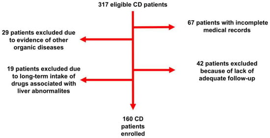
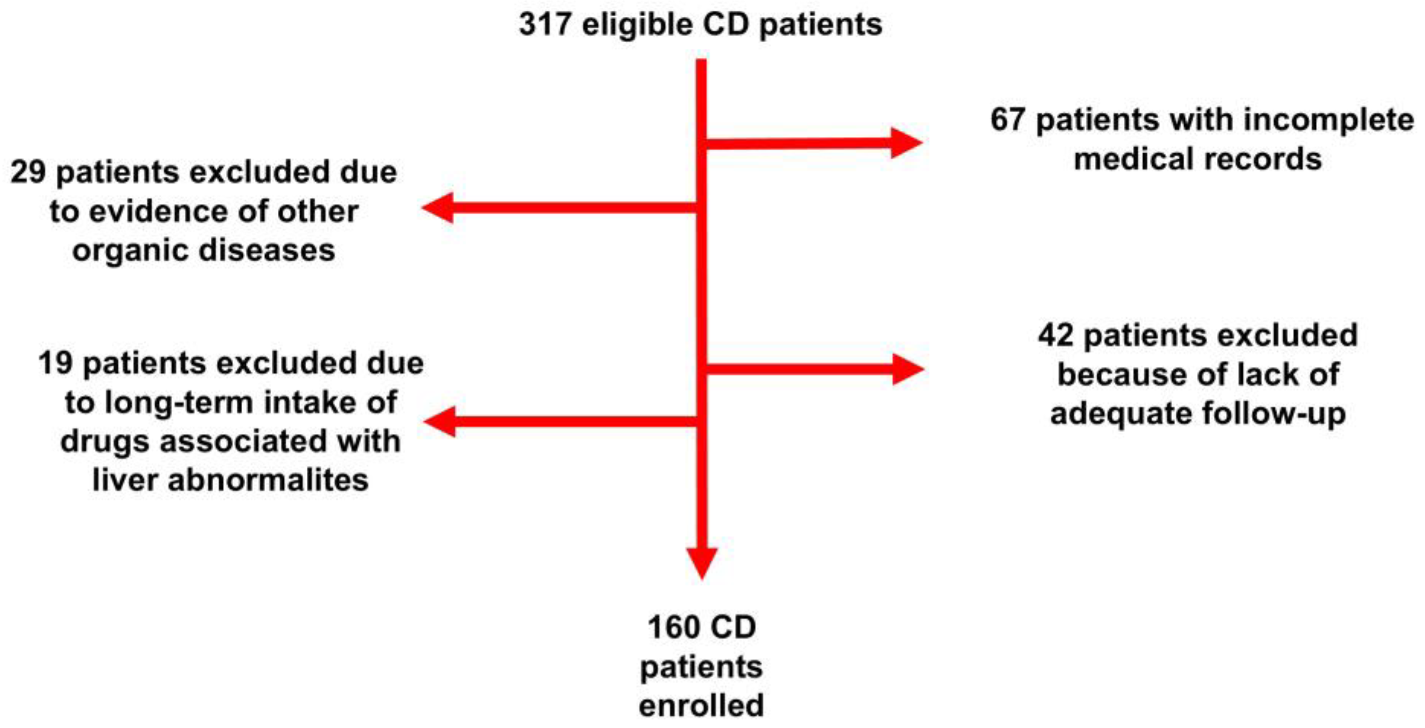
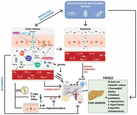
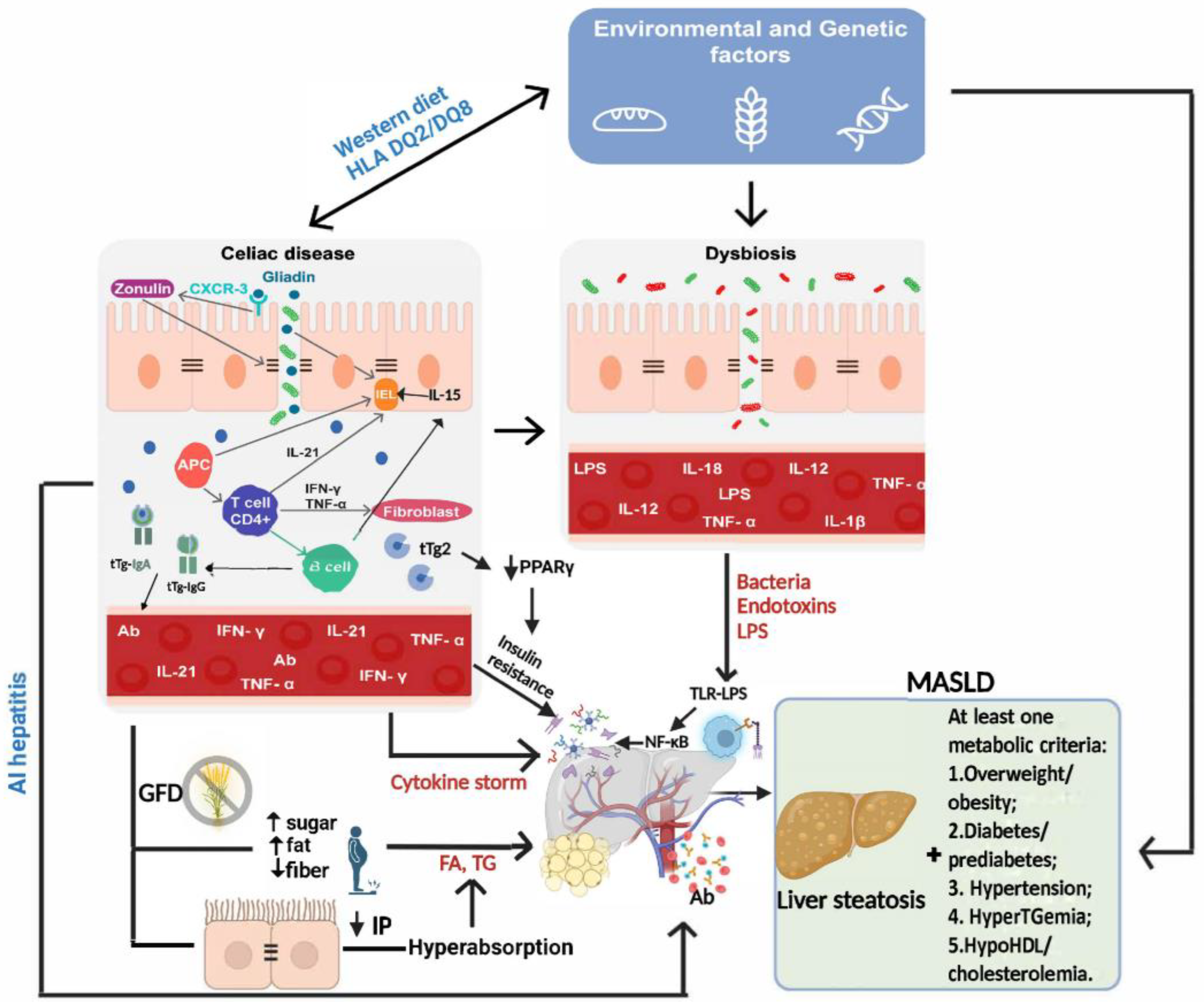

Disclaimer/Publisher’s Note: The statements, opinions and data contained in all publications are solely those of the individual author(s) and contributor(s) and not of MDPI and/or the editor(s). MDPI and/or the editor(s) disclaim responsibility for any injury to people or property resulting from any ideas, methods, instructions or products referred to in the content. |
© 2024 by the authors. Licensee MDPI, Basel, Switzerland. This article is an open access article distributed under the terms and conditions of the Creative Commons Attribution (CC BY) license (https://creativecommons.org/licenses/by/4.0/).