


Abstract
Background and Aims: Understanding regulatory interactions between hepatitis B virus (HBV) and host factors is essential for the development of next generation host-directed antiviral therapies and the achievement of a functional HBV cure. Here, we investigated HBV-induced alterations in host gene expression in primary human hepatocytes (PHH) to identify host factors exploited by the virus for replication and persistence. Whole-transcriptome sequencing (WTS) of HBV-infected PHH identified host pathways with potential roles in the HBV life cycle. RNA interference-based functional screening of dysregulated candidate genes identified cyclin L1 (CCNL1) as a key host factor. RNAi-mediated knockdown of CCNL1 reduced HBV gene expression, including hepatitis B surface antigen (HBsAg). Mechanistically, CCNL1 regulates phosphorylation of the C-terminal domain (CTD) of RNA polymerase II (RNAPII) at serine 2 (S2), consistent with a role in transcriptional regulation. CCNL1 knockdown further reduced the binding of total and phospho- (Ser2/Ser5) RNAPII, pan-acetylated histone H3 (H3ac), and H3K27ac to HBV covalently closed circular DNA (cccDNA), indicating impaired cccDNA-dependent transcription. In addition, CCNL1 expression was elevated in chronic hepatitis B patients compared with those with resolved infection. Collectively, these data demonstrate that CCNL1 promotes HBV transcription and replication through modulation of RNAPII phosphorylation and chromatin-associated transcriptional activity, identifying CCNL1 as a potential host susceptibility factor for HBV. Importance: Hepatitis B virus infection remains a major threat to human health in areas with high prevalence. There is need to fully understand the complex interactions between the virus and human host factors/processes to support ongoing efforts to develop anti-HBV therapies that can be used with existing therapies to achieve a better cure. HBV relies on host cellular factors and biological processes to establish and maintain efficient infection, making host–virus interactions attractive targets for therapeutic intervention. Thus, identifying host factors that support and/or restrict HBV infection is essential for understanding the molecular basis of chronic HBV infection and for developing host-targeting anti-HBV drugs. This study identifies cyclin L1 (CCNL1) as a host susceptibility factor that promotes HBV transcription and replication through regulation of RNA polymerase II activity and or post-transcriptional mechanisms.
1. Introduction
Chronic hepatitis B (CHB) infection affects over 250 million people worldwide and can lead to significant liver disease with long-term health consequences including liver damage, failure, and cancer [1,2]. Clinical management of CHB includes nucleos(t)ide analogs (NAs) and interferon (IFN)-based therapy [1,2]. While current therapeutic strategies suppress viral replication and reduce the risk of developing adverse liver disease, they cannot eliminate the virus or achieve a durable functional cure, as defined by the loss of HBV surface antigen (HBsAg) [1,3,4,5]. These limitations call for the implementation of novel strategies to identify the next generation of anti-HBV drug targets and drugs to boost ongoing efforts to eradicate HBV infection.
HBV is a small, partially double-stranded DNA virus belonging to the family Hepadnaviridae that exhibits high tropism for human hepatocytes. HBV encodes seven viral proteins and only one enzyme, DNA polymerase. Therefore, the virus relies heavily on host cell factors to infect and replicate in human hepatocytes. These host factors may either be intrinsic host factors that the virus coopts, or host restriction factors (HRFs), such as innate signaling pathways and defense mechanisms, which the virus must suppress to establish an efficient infection.
Although numerous host factors regulating HBV replication have been described, the importance and mechanism of action of various human cellular factors at different stages of the viral infection cycle remain unclear. For example, ectopic expression of sodium taurocholate (NTCP), the major receptor for HBV, in mouse primary hepatocytes does not render them susceptible to HBV infection [6], and different HepG2-NTCP clones display different degrees of susceptibility to HBV infection [7]. These observations suggest that other, as yet unknown, host factors are involved in establishing robust and efficient HBV infection. Thus, a deeper probing of host–pathogen interactions is needed to not only expand our knowledge of viral pathogenesis but may also result in the identification of novel anti-HBV therapy that can be used in combination with current therapies to achieve better, and more durable treatment outcomes.
In this study, we employed whole-transcriptome sequencing (WTS) to identify HBV-mediated gene perturbations in physiologically relevant primary human hepatocytes (PHH) to identify unique infection-driven host gene expression signatures. These genes were functionally investigated for their potential function as novel host susceptibility factors for HBV replication and transcription. Specifically, we report the identification of Cyclin L1 (CCNL1) as a possible HBV host factor that affects viral transcription. Cyclin L 1 is a non-canonical or transcriptional cyclin. It contains N-terminal cyclin box as well as the C-terminal arginine/serine-rich (SR) domain (SR) domain (reviewed) [8]. To date, CCNL1 has not been described as a viral host susceptibility or restriction factor; therefore, this study aimed to explore the role of CCNL1 in HBV replication. Loss of CCNL1 function results in a global decrease in HBV gene expression, whereas its ectopic expression leads to increased viral replication. Mechanistically, we demonstrate that CCNL1 regulates the transcription of nascent HBV RNA by modulating RNAPII activity and the subsequent epigenetic regulation of cccDNA. Finally, we show that CHB patients display a general increase in CCNL1 expression compared to those who have achieved a functional cure. Overall, these results corroborate the physiological role of CCNL1 in the control of HBV replication/transcription and expand our knowledge on the virus: human host interactions.
2. Materials and Methods
2.1. Cell Culture and Treatments
The HepG2-NTCP cells used in this study were a kind gift from Assoc Prof Tan Yee Joo, National University of Singapore, and have been reported to be susceptible to HBV infection [7]. HepG2-NTCP cells were maintained as described earlier [9]. HepAD38.7-Tet cells used in this study were cultured as previously described [10]. Tetracycline was removed from the HepAD38.7-Tet cells and the cells cultured in antibiotic free media for 3 days before seeding them for siRNA transfections. HEK293Tcells were maintained in antibiotic-free DMEM/F12 + Glutamax (Gibco) supplemented with 10% FBS. PHH were purchased from Lonza (Basel, Switzerland) and maintained according to the manufacturer’s instructions.
Lentiviral transduction of the cells was carried out in the presence of 5 μg/mL polybrene (sc-134220, Santa Cruz, Dallas TX, USA), followed by selection of positive clones in the presence of 10 μg/mL puromycin (ant-pr-1, InvivoGen, San Diego, CA, USA). All plasmid and siRNA transfections were performed using Lipofectamine 3000 (Invitrogen) according to the manufacturer’s protocol.
2.2. Production of HBV Stock Virus for Infection
The virus used for infection in this study was produced as described earlier [11]. Briefly, cell culture supernatant was harvested from tet-off-HepAD38.7 cells and filtered through a 0.22 µm polyethersulfone (PES) (Kent, WA, USA) membrane filter. This was followed by concentration using a heparin column (Cytiva, Marlborough, MA, USA) and elution with high salt, followed by dialysis in phosphate-buffered saline overnight.
2.3. Generation of Stable Cell Lines
ORF expressing CCNL1 (clone id: OHu03264) and empty vector control (pcDNA3.1+/C-(K)-DYK) were purchased from GenScript (Piscataway, NJ, USA). Subcloning and plasmid isolation was performed as described in the Supplementary Methods. Transfection was performed using Lipofectamine 3000 (Invitrogen, Waltham, MA, USA) with 9 μg of plasmid in 10 cm cell culture dishes. Cells expressing the plasmids were selected using DMEM supplemented with 10% FCS and 10 μg ml−1 blasticidin (Santa Cruz Biotechnology, Dallas, TX, USA).
2.4. Plasmids, Antibodies, Oligos, siRNA, and shRNA
The oligonucleotides used in this study were designed using Primer Plus V3 and synthesized by Integrated DNA Technologies, Inc. (IDT, Singapore, Singapore). Validated siRNAs targeting the three regions of the upregulated genes were ordered from Silencer Select (Applied Biosystems, Carlsbad, CA, USA). Control shRNAs and shRNAs targeting CCNL1 (TRCN0000062533, sequence: CCGGGCCTATCTACAGTGTGATGTACTCGAGTACATCACACTGTAGATAGGCTTTTTG) were obtained from Thermo Fisher Scientific (Thermo Fisher Scientific, Waltham, MA, USA). Access to these libraries was provided by high-throughput phenomics (EDDC). The following packaging vectors were used in this study: pMD2.G (Addgene plasmid #12259), pRSV-Rev, and pMDLg/pRRE, all gifts from Didier Trono [12]. A comprehensive list of the antibodies used in this study is shown in Table S1.
2.5. RNA-Seq of Infected Primary Human Hepatocytes
Fresh primary human hepatocytes (Invitrocue/Yecuris, Tualatin, OR USA) from FRG KO humanized mice were infected with HBV from a cell culture (HepAD38.7 cells) at an MOI of 3000. Cell lysates were collected 96 h post-infection. RNA isolation was performed using an RNeasy kit (Qiagen, Hilden, Germany). Quality was assessed using a Bioanalyzer, the library was prepared using SureSelect XT Human All Exon V6, and sequencing was performed using Novaseq 6000 (Theragen, Gyeonggi-do, Korea). Differentially expressed genes between mock-infected and HBV-infected samples were identified by DEGseq analysis in R V3.4.2. Gene set enrichment and pathway analyses were performed using DAVID Version 3042 software to determine the top upregulated and downregulated pathways, and the top genes were identified for further analysis. We intentionally used uncorrected p-values (p < 0.01) as a primary screening approach for discovery where false positives would be filtered during validation experiments.
2.6. Generation of Stable Knockdown Cells in HepG2-NTCP
HepG2-NTCP cells were seeded in 10 cm dishes and incubated until about 80% confluent. Then, the cells were transduced with lentivirus expressing respective shRNA targeting cyclin L1 and empty vector control in the presence of 5 µg/mL polybrene (Santa Cruz, Dallas TX, USA). The media was changed after 16 h and replaced with selection media supplemented with 5 µg/mL puromycin. The selection media was changed every 48 h, and RT-qPCR and immunoblotting were used to determine gene knockdown.
2.7. Immunofluorescence Staining and Confocal Microscopy
The cells were seeded in 96 well plates (PerkinElmer Ultra plates, Hayward, CA, USA) and treated as previously described. Cells were fixed in 4% paraformaldehyde at room temperature for 10 min, followed by permeabilization with 0.1% Triton-X for 5 min at room temperature. Fixed cells were blocked with 3% bovine serum albumin solution for 5 min at room temperature and incubated with primary antibodies overnight at 4 °C. The cells were washed thrice with 1X PBS to remove excess primary antibodies. The secondary antibody incubation was carried out for 1 h at room temperature before the cells were washed and counterstained with DAPI (Sigma Aldrich, Saint Louis, MO, USA). The plates were imaged using the Operetta CLS confocal microscope (PerkinElmer, Hayward, CA, USA). Infected cells were quantified using the Columbus Data Storage and Analysis System (PerkinElmer, Hayward, CA, USA). The data were normalized to the number of cells per well and the control wells were equated to 100%.
2.8. HBV Infection of HepG2-NTCP and PHH
HepG2-NTCP and PHH cells were seeded in type I collagen (R-011-K, Gibco)-coated plates. HepG2-NTCP cells were infected at an MOI of 3000 in the presence of 4% PEG8000 and 1% DMSO at 37 °C in a 5% CO2 incubator, as described previously [12]. Primary human hepatocytes were infected at an MOI of 2000 in the presence of 4% PEG8000 and 1% DMSO, as described previously [13]. The samples, cell lysate for RNA and DNA, as well as cell culture supernatants, were harvested at the indicated time points, as shown in the figure legends.
2.9. RNAi Screening in HepAD38.7 Cells
HepAD38.7 cells were maintained as previously described. A pool of three siRNAs per gene was spotted into 384 well plates at a final concentration of 20 nM. Gene silencing was performed by reverse transfection using the Lipofectamine 3000 reagent protocol (0.1 μL Lipofectamine 3000 per well in Opti-MEM medium) (Thermo Fisher Scientific, Waltham, MA, USA). The cells were washed, and the culture medium was changed one day post-transfection. Transfected cells were incubated at 37 °C in a 5% CO2 incubator for 72 h. For reproducibility, the screen was performed independently three times, each consisting of four technical replicates.
2.10. Quantification of HBsAg, HBeAg, and Extracellular HBV DNA
The cell culture supernatant was collected at the end of the experiment. For HBsAg ELISA, 384 well polystyrene plate Maxisorp Cat# P6366-1CS (Sigma-Aldrich, Saint Louis, MO, USA) was coated with 6 μg/mL mouse monoclonal anti-HBsAg (ab20758 at 6 μg/mL) in 0.05 M carbonate-bicarbonate buffer pH 9.6 (Cat# C3041-100, Sigma Aldrich, Saint Louis, MO, USA) and incubated overnight at 4 °C. The plates were thawed and washed three times with wash buffer (0.05% Tween 20 in 1X PBS) before blocking in assay diluent (10% FBS in 1X PBS), followed by incubation for 2 h at room temperature. The plates were washed three times before adding 25 μL of sample and standard (HBsAg subtype adw2) full-length protein, ab91276) and incubated for 2 h at room temperature. Plates were washed five times before adding rabbit polyclonal anti-biotin (ab68520 1:1000 dilution) and streptavidin-HRP (cat#554066, BD 1:1000) followed by 1-h incubation at room temperature. The plates were washed 7 times to remove any excess working detector, followed by adding TMB substrate solution (Pierce, ThermoScientific) and incubated at room temperature for 30 min. The reaction was stopped by adding 1N HCl stop solution before reading the absorbance at 450 nm using a Tecan 100 spectrophotometer. HBsAg levels are expressed as a percentage of the control set at 100%. HBeAg was quantified following a one-step sandwich CLIA protocol, according to the manufacturer’s instructions (Autobio Diagnostics, Zhengzhou, China). Wild-type and defective HBV DNA was amplified from DNA extracted from 200 μL of cell culture supernatant from HBV-infected HepG2-NTCP, PHH, and HepAD38.7, as previously described [14,15].
2.11. Quantitative Real-Time PCR (qPCR)
RNA was extracted from the cell lysates according to the Total RNeasy kit protocol (Qiagen, Hilden, Germany). cDNA was synthesized from 1 μg of extracted RNA using Invitrogen SuperScript IV VILO Master Mix (Cat. 11766500, Life Technologies, Waltham, MA, USA). HBV pre-genomic RNA and gene mRNA levels for knockdown were determined using the SYBR Fast-based qPCR master mix kit (KAPPA BIOSYSTEMS, Wilmington, MA, USA) in the Quant studio 7 PCR system (Applied Biosystems, Carlsbad, CA, USA). The pgRNA primers used were as follows: forward, CGTTTTTGCCTTCTGACTTCTTTC and reverse, ACAGAGCTGAGGCGGTGTCTA [16]. qPCR data were analyzed using the Livack method as described previously [17]. All primers used in this study for quantitative polymerase chain reaction (qPCR) were purchased from Integrated DNA Technologies (Singapore, Singapore).
2.12. Quantification of cccDNA Using TaqMan Probe-Based qPCR
HBV cccDNA was quantified as described earlier [18,19]. Briefly, DNA was extracted at the end of the experiment according to the DNA mini kit (Qiagen, Hilden, Germany) protocol with proteinase K digestion for 1 h at 37 °C. A total of 1 μg of the extracted intracellular DNA was digested with T5 exonuclease (New England Biolabs) to remove the relaxed circular and single-stranded HBV DNA according to the manufacturer’s protocol [20]. Briefly, digestion was performed at 37 °C in the presence of 10 units of T5 exonuclease and NEBuffer 4 for 30 min. The reaction was stopped by adding 11 mM EDTA. Quantification of cccDNA was performed using the specific primer Fwd-primer: 5′-GGGGCGCACCTCTCTTTA-3′ ccDNA Rev-primer: 5′-AGGCACAGCTTGGAGGC-3’ TaqMan probe: 5′-FAM-TCACCTCTGCCTAATCATCTC-TAMRA-3’ [19]. To enhance the specificity of the PCR, 100 nmol/L forward and 400 nmol/L reverse primers were used. Denaturation was conducted at 95 °C for 10 min, followed by 40 cycles of step 1 (95 °C for 1 s) and step 2 (65 °C for 1 min), as previously reported by Allweiss et al. [21].
2.13. Western Blot Analysis
Protein samples were separated by SDS–PAGE, and then transferred to PVDF membrane, transferred to PVDF membranes, blocked with InterceptTM (TBS) buffer (Bio-Rad, Hercules, CA, USA, P/N: 927-70001) or 5% skim milk for 1 h at room temperature and incubated with primary antibodies at 4 °C overnight. Following incubation with the appropriate secondary antibodies (LI-COR IRDye® 800 CW anti-rabbit 92,632,211 (LiCor, Nebraska, USA 1:5000) or 680 CW anti-mouse 9,268,070 (LiCor, 1:5000)), the blots were visualized using the ChemiDoc XRS + system (Bio-Rad).
2.14. ChIP Assays
Chromatin immunoprecipitation (ChIP) was performed using the MAGnify Chromatin Immunoprecipitation System (492024; Applied Biosystems, Carlsbad, CA, USA). The cells were fixed with 1% paraformaldehyde (PFA) for 10 min at room temperature. Fixation was stopped by adding a 1.25 M glycine solution for 5 min at room temperature. Cells were subsequently washed in chilled PBS (Eurobio, Les Ulis, France), resuspended in ChIP lysis buffer supplemented with protease inhibitors at 1 million cells/50 mL, and incubated for 5 min on ice. The chromatin solution was then sonicated for 15 cycles of 30 s ON and 30 s OFF using a Picoruptor Sonicator (Diagenode, Seraing, Belgium) to generate 200 to 500 bp DNA fragments. The debris was pelleted by centrifugation at 20,000× g and 4 °C for 5 min. The sheared chromatin was diluted to a concentration of 200,000 cells/100 mL and incubated overnight at 4 °C with the previously coupled antibody–Dynabeads protein A/G under end-over-end rotation. Ten microliters of mouse anti-RNAPII, RNAPII Ser2P, RNAPII Ser5P, and rabbit anti-H3K27ac antibodies (Active Motif, Carlsbad, CA, USA) were used (Table S1). Immunoprecipitation with non-specific immunoglobulins (Applied Biosystems, Carlsbad, CA, USA) was included in each experiment as a negative control. After washing and reverse crosslinking, immunoprecipitated chromatin was purified using DNA purification magnetic beads (Applied Biosystems, Carlsbad, CA, USA), according to the manufacturer’s protocol. Purified DNA was analyzed by real-time quantitative PCR using specific primers and fluorescent probes for HBV cccDNA (primers CCGTGTGCACTTCGCTTCA/GCACAGCTTGGAGGCTTGA; probes CATGGAGACCACCGTGAACGCCC).
2.15. Viability/Cytotoxicity Assays
Cell viability and cytotoxicity were evaluated using the CellTiter-Glo Luminescent assay (Promega, Madison, WI, USA) and cell-count kit-8 (Dojindo, Kumamoto, Japan), respectively, following the manufacturer’s instructions.
2.16. Cell Painting Assay
Stable knockdown and control HepG2-NTCP cells were seeded in 384 well plates and infected with HBV as described previously. The detailed staining protocol is described in the Supplementary File. The cells were fixed and stained with cell painting dyes followed by imaging with a Perkin Elmer Operetta CLS, and image segmentation and feature extraction were performed using CellProfiler 3.0 pipelines adapted from Bray et al. [22]. The detailed cell painting protocol is described in the Supplementary Methods. Cell painting analysis pipelines using pycytominer version 1.1 and R V3.4.2 scripts were developed at the High Throughput Phenomics Lab at EDDC.
2.17. Statistical Analysis
Data analysis was performed using GraphPad Prism 8 using different tests, as indicated in the figure legends. Network analysis of the upregulated genes was performed using the STRING database, as previously described [23]. Differentially expressed genes from the RNA-seq data were analyzed in R studio using the DEGseq package, as reported previously [24].
3. Results
3.1. Identification of CCNL1 as a Host Susceptibility Factor in HBV Infection
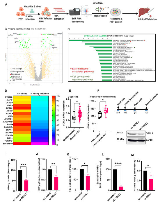
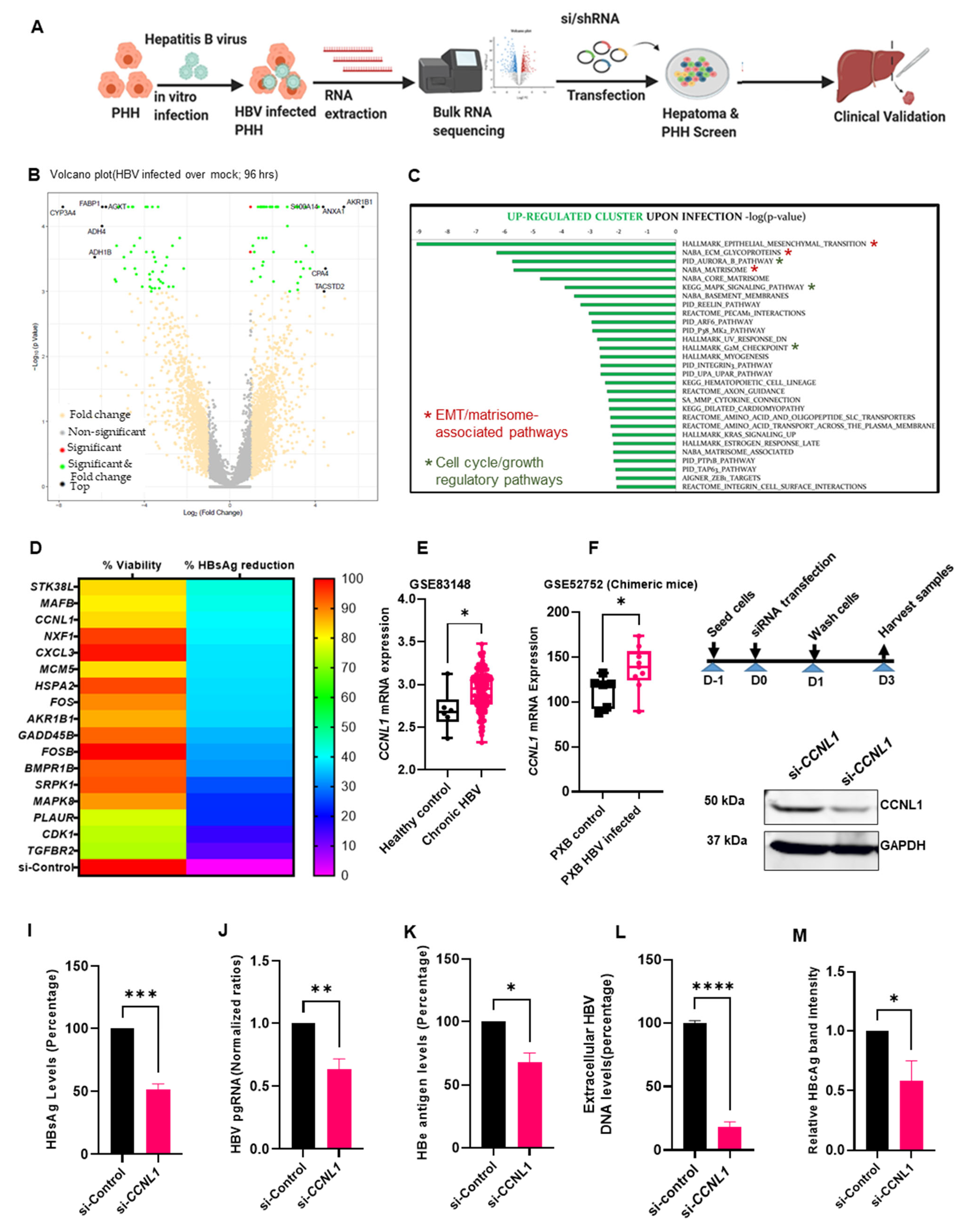
Primary human hepatocytes (PHH) offer a physiologically relevant model for identifying and validating human host factors that regulate HBV infection [22,25,26]. We hypothesized that HBV infection alters the host transcriptome to support its replication and transcription. Therefore, we performed an integrated transcriptomic analysis of HBV-infected PHH combined with RNAi-mediated loss-of-function studies to validate the potential role(s) in the regulation of various aspects of the HBV life cycle (Figure 1A). First, we performed bulk RNA-seq analysis of HBV-mediated transcriptomic changes in PHH to identify host genes that were either up- or down-regulated during HBV infection, compared to mock-infected controls (see volcano plot in Figure 1B). Gene set enrichment analysis (GSEA) of differentially expressed genes 96 h post HBV infection revealed 69 significantly upregulated genes based on a log2 fold change >1.5 and p-value < 0.01 (Figure 1B,C). Pathway analysis of these genes revealed enrichment of previously described cellular processes associated with HBV infection, including the G2/M checkpoint, solute carriers, and p38 mitogen-activated protein kinase (MAPK) (Figure 1C) [27,28]. Intriguingly, our study also identified the enrichment of unique pathways that bear the hallmarks of cancer, including EMT, the core matrisome, Aurora B kinase, and VEGFR signaling (Figure 1C). In contrast, pathways that are known to restrict HBV infection, such as hypoxia, were downregulated in our analysis (Figure S1A) [29]. However, we note that hypoxia-inducible factors have also been reported to enhance HBV replication [30]. Additionally, consistent with existing evidence linking HBV infection and reprogramming and/or alterations in host cell metabolism [31,32], we identified several pathways involved in various metabolic processes that were deregulated upon HBV infection in PHH (Figure 1C and Figure S1B). Together, these results enabled the characterization of HBV-induced alterations in the host transcriptome of PHH upon infection.
Figure 1.
Identification of CCNL1 as a host susceptibility factor in HBV infection. (A) Schematic methodology used to identify and validate the role of CCNL1 during HBV infection. (B) Volcano plot displays upregulated and downregulated genes upon HBV infection compared to mock infected PHH. (C) Top upregulated pathways upon HBV infection. (D) siRNA targeting each of the top 69 upregulated host factors were used in a HepAD38.7 RNAi screen to assess their role in HBV replication. Non-targeting control siRNA was included as a control. Hepatitis B surface antigen (HBsAg) ELISA was conducted on the cell culture supernatant, and cell viability was determined by CCK-8. Representative HBsAg reduction and cell viability are shown in the heat map. (E) Expression of Cyclin L1 in healthy control and chronic HBV patient from micro-array dataset GSE83148. (F) CCNL1 expression in chronic HBV model of Phoenix Bio chimeric mice (GSE52572) is shown for the control and CHB. (G) Experimental outline for the subsequent RNAi screen in HepAD38.7 cells. (H) Knockdown of CCNL1 is validated by Western blot. (I–M) Levels of HBsAg, HBV pgRNA, HBe antigen extracellular HBV DNA, and relative band intensity for the HBc antigen are shown. Statistical significance shown as p-value was analyzed by Student’s t-test in GraphPad prism 8. Data are represented as mean ± SEM (n = 3) from 3 independent experiments each containing 3 technical replicates (I–M), **** p < 0.0001, *** p < 0.001, ** p < 0.01, * p < 0.05, ns; non-significant. siRNA screen was performed independently 3 times, each containing 3 technical replicates.
Although the gene expression data were consistent with some of the key pathways previously identified in the context of HBV infection, we also identified previously unsuspected players that have not been described. Therefore, we analyzed the function of the newly identified virally induced host genes in HBV replication and transcription. We designed a siRNA-based screen targeting the upregulated genes identified from WTS analysis. We used the HepAD38.7 cell line, which is easy to maintain in culture and contains HBV pgRNA under the control of a tetracycline-inducible on/off switch that can support HBV replication [10,26,33]. The siRNA library consisted of three siRNAs per target gene, and a non-targeting siRNA was used as a negative control. From the HBsAg ELISA, we focused on candidate host factors whose knockdown resulted in a marked reduction in HBsAg levels without any appreciable effect on cell viability. This resulted in the identification of cyclin L1, whose knockdown resulted in a ~40% reduction in HBsAg (Figure 1D). Notably, the effect of CCNL1 on quantitative HBsAg levels was comparable with some of the previously described HBV host factors (also identified in our transcriptomic studies), including nuclear export transport factor (NXF1), which has been linked to the transport of pgRNA from the nucleus to the cytoplasm [34,35], and CDK1 and cellular kinase R protein-specific kinase 1 (SRPK1), both of which have been linked to the phosphorylation of HBV core protein [36,37,38,39]. To validate the clinical relevance of CCNL1 as a putative prognostic factor in CHB patients, we analyzed the expression of CCNL1 in liver tissues retrieved from the Gene Expression Omnibus database (GSE83148, and GSE52752). Remarkably, we observed consistent upregulation of CCNL1 during chronic HBV infection (Figure 1E,F). Furthermore, we observed enhanced expression of CCNL1 in a cohort of local patients with unresolved CHB compared to those with resolved infection, as assessed by the loss of HBsAg (Figure S1B,B’). Notably, analysis of the expression profiles in HBV e antigen-positive (HBeAg+) and -negative (HBeAg-) patients showed high CCNL1 expression in CHB patients irrespective of their HBeAg status (Figure S1C). In contrast, patients who achieved a functional cure (loss of HBsAg) displayed significantly reduced expression of CCNL1 (Figure S1C). Next, we evaluated the effects of cyclin L1 knockdown on HBV replication and gene expression in HepAD38.7 cells (Figure 1G). Knockdown of Cyclin L1 was first validated by Western blotting and rt-qPCR (Figure 1H and Figure S2A), and, as shown in Figure 2B, did not affect cell viability. Remarkably, CCNL1 knockdown significantly reduced multiple viral parameters, including HBsAg, pgRNA, HBeAg, extracellular HBV DNA, and HBcAg (Figure 1I–M and Figure S2C). These results suggest that CCNL1 may be regulating HBV replication and transcription.
Figure 2.
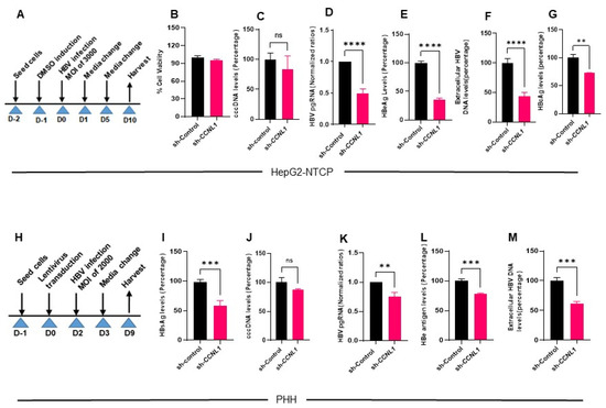
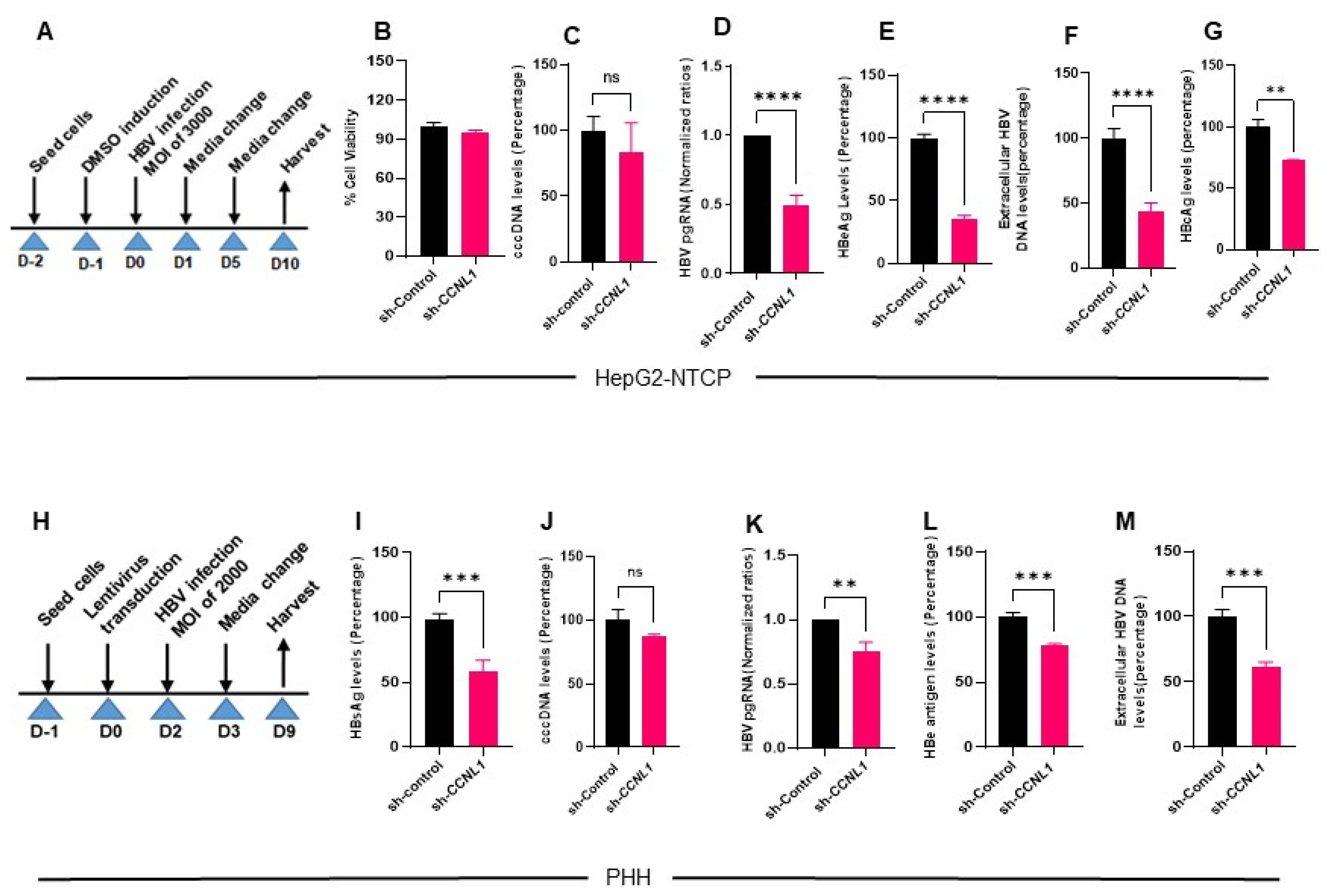
Functional validation of CCNL1 in live infection models of HBV reveals its effect on distinct viral markers. (A) Experimental outline in HepG2-NTCP is shown. (B) Knockdown of CCNL1 did not have any effect on the cell viability. (C) No significant effect on the cccDNA is observed upon knockdown of Cyclin L1 in HepG2-NTCP. (D–F) There was a significant reduction in pgRNA, HBeAg, and extracellular HBV DNA levels in CCNL1 KD cells. (G) HB core antigen (HBcAg) immunofluorescence analysis (IFA) shows the levels of infection in control and CCNL1 knockdown cells. (G) Quantified HBcAg normalized to the number of cells expressed as a percentage of the infection in control cells. (H) Validation of the role of CCNL1 during HBV replication in primary human hepatocytes. (I) Knockdown of Cyclin L1 showed a remarkable reduction in surface antigen levels. (J) Consistently, the knockdown of CCNL1 did not have significant effect on cccDNA. (K–M) Knockdown of CCNL1 resulted in reduced levels of pgRNA, HBe antigen and extracellular HBV DNA. Statistical significance shown as p-value was analyzed by Student’s t-test in GraphPad prism 8. Data are represented as mean ± SEM (n = 3), **** p < 0.0001, *** p < 0.001, ** p < 0.01, ns; non-significant. All the experiments shown above represent 3 biological replicates each consisting of 3 technical replicates.
3.2. Knockdown of Cyclin L1 Results in Reduced HBV Replication
To investigate the function of CCNL1 in a more physiological setting, we analyzed the effect of CCNL1 knockdown on HBV replication and RNA synthesis in HepG2-NTCP (Figure 2A) and PHH. CCNL1 knockdown in HepG2-NTCP cells was validated by WB (Figure S2D and quantified D’). We also observed enhanced CCNL1 expression in infected HepG2-NTCP cells compared to that in controls (Figure S2E,E’). Notably, the knockdown of cyclin L1 did not influence the viability of HepG2-NTCP cells (Figure 2B), ruling out any non-specific or adverse effects. Interestingly, we did not observe any significant effect on HBV cccDNA levels upon CCNL1 knockdown in HepG2-NTCP cells (Figure 2C), suggesting that CCNL1 likely functions downstream of cccDNA. Moreover, like the observations in HepAD38.7 cells, we found that loss-of-function of CCNL1 in HepG2-NTCP cells also resulted in a remarkable reduction in HBV pgRNA, HBeAg, and extracellular HBV DNA (Figure 2D–F). We also observed reduced levels of the hepatitis B core (HBcAg) protein in the knockdown cells compared to the controls (Figure 2G). In contrast, the ectopic expression of CCNL1 in HBV-infected HepG2-NTCP robustly enhanced the expression of HBV pgRNA, HBeAg, and extracellular HBV DNA (Figure S4A–E), without any significant effect on cell viability or proliferation, suggesting a specific role in HBV control.
To further bolster these findings, we assessed the role of CCNL1 in the HBV infection cycle in a live PHH infection model (Figure 2H–M). The shRNA-mediated knockdown of Cyclin L1 was validated at the protein and mRNA levels (Figure S3A and Figure 3B). Like observations in other cell lines, knockdown of CCNL1 did not influence PHH viability (Figure S3C). Notably, we observed reduction in the levels of HBsAg (Figure 2I), concomitant with a marked reduction in the levels of pgRNA (Figure 2K), HBe antigen (Figure 2L), and extracellular HBV DNA (Figure 2M) in PHH transduced with CCNL1 shRNA. However, the effect of sh-CCNL1 on cccDNA levels remained insignificant, further corroborating the notion that CCNL1 is likely to play a role in HBV control downstream of cccDNA formation. Thus far, we have shown that cyclin L1 regulates HBV replication and gene expression without affecting the levels of cccDNA, possibly after the formation of cccDNA.
Figure 3.
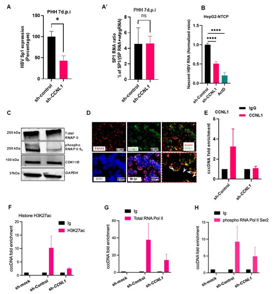
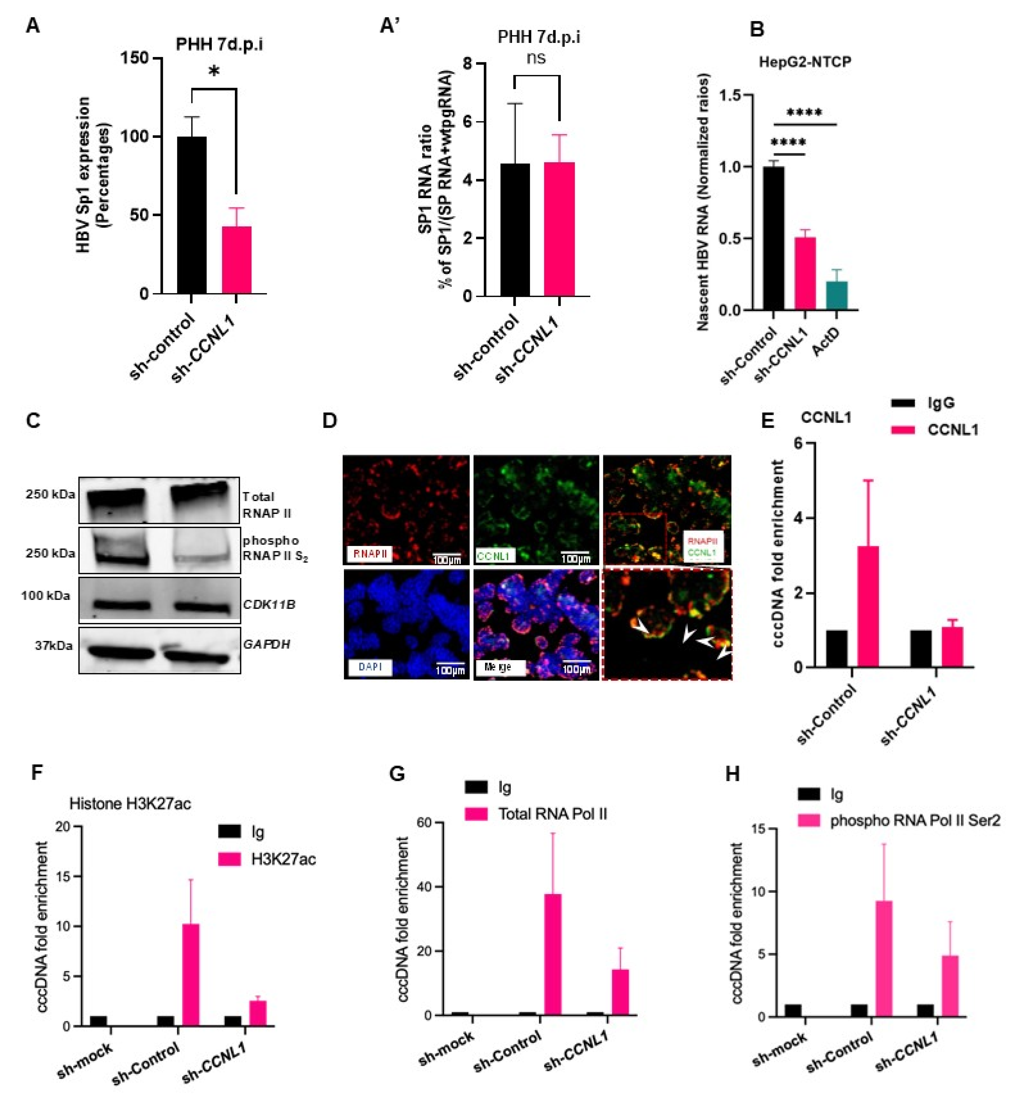
CCNL1 controls expression levels of HBV RNA by modulating cccDNA-dependent transcription. (A,B) Knockdown of Cyclin L1 significantly reduced the production of nascent HBV RNA in HepG2-NTCP cells. Statistical significance shown as p-value was analyzed by Student’s t-test for two groups in GraphPad prism 8. Data are represented as mean ± SEM (n = 3), * p < 0.05, **** p < 0.0001, ns; non-significant. (C) The effect of loss-of-function of CCNL1 on total RNA polymerase II, phosphorylated S2, and CDK11B is shown by Western blot. (D) Co-localization of Cyclin L1 and total RNAPII in HepG2-NTCP cells (the zoomed-out region is shown at the bottom right of the figure). The scale bar is 100 μm. (E) CCNL1 binds to cccDNA. (F–H) The recruitment of RNA polymerase II and transcription mark on HBV cccDNA were detected by ChIP assay in HBV-infected HepG2-NTCP cells. cccDNA-ChIP was conducted in CCNL1 knockdown and control HepG2-NTCP cells infected with HBV (F) The recruitment of H3K27ac on HBV cccDNA was significantly decreased after knockdown of CCNL1. As examined by ChIP-qPCR assay, CCNL1 knockdown remarkably reduced total RNA polymerase II recruitment on HBV cccDNA. RNAPII (G) and phosphorylated C-terminal domain serine 2 associated cccDNA were reduced in CCNL1-deficient cells (H). Shown data are representative of 3 independent experiments. Data are presented as the means ± SD. All the experiments shown above represent 3 biological replicates each consisting of 3 technical replicates.
3.3. CCNL1 Modulates the Production of HBV RNAs by Regulating the Activity of the Total RNAPII
Given that CCNL1 contains arginine-serine-rich dipeptide repeats, which are associated with pre-mRNA splicing [40], we evaluated the effect of CCNL1 knockdown and overexpression on HBV splicing. We observed that the knockdown of CCNL1 resulted in an overall decrease in the levels of HBV-spliced RNA variants, including singly spliced HBV RNA (Sp1) (Figure S5A,B). The splice variants were analysed as described recently [41]. Intriguingly, analysis of the singly spliced Sp1 RNA in the HBeAg± cohort of patients showed differential expression, with high levels of splice variants observed in HBeAg+, moderate levels in HBeAg−, and an almost complete absence in patients with HBsAg loss (Figure S5D). Altogether, these results highlight the physiological relevance and association of CCNL1 expression with the clinical features of patients with CHB.
However, given that our findings in live infection models suggest a role for CCNL1 in controlling pgRNA levels, we wondered whether the alterations in the levels of splice variants resulted from alterations in the levels of pgRNA upon CCNL1 knockdown. Indeed, normalization of the levels of splice variant Sp1 against pgRNA, CCNL1 knockdown cells, abrogated any of the observed absolute effects on splicing (Figure S6A,A’). These results suggest that the impact of CCNL1 on the control of HBV splicing is likely to be indirect due to the regulation of pgRNA expression.
Next, we assessed whether Cyclin L1 influenced the production of HBV RNA. Indeed, siRNA- or shRNA-mediated knockdown of CCNL1 in HepAD38.7 (Figure S6B) and HepG2-NTCP (Figure 3A) cells resulted in a significant reduction in the levels of nascent HBV RNA. These results indicate that CCNL1 most likely plays a role in activating HBV RNA transcription. Previous studies have reported that CCNL1 interacts with CDK11B to phosphorylate the C-terminal domain (CTD) of RNA polymerase II (RNAPII) [41]. Indeed, siRNA-mediated knockdown of CCNL1 resulted in reduced phosphorylation of CTD-RNAPII at the Ser 2 (S2) residue (Figure 3B), while overexpression resulted in increased S2 phosphorylation of CTD-RNAPII (Figure S6C). Knockdown or ectopic expression of CCNL1 did not significantly affect the expression of CDK11B, highlighting that the interaction did not affect the expression/stability of CDK11B (Figure 3B and Figure S6C). Additionally, immunofluorescence analysis revealed co-localization of CCNL1 and total RNAPII (Figure 3C), further corroborating the putative function of CCNL1 complexes in the regulation of RNAPII activity, which has been implicated in cccDNA-dependent viral transcription [42].
Next, we used a chromatin immunoprecipitation (ChIP) assay to assess whether CCNL1 can bind to cccDNA. Indeed, immunoprecipitation with an anti-CCNL1 antibody followed by PCR (ChIP-PCR) revealed that cyclin L1 can bind to HBV cccDNA, and the binding is diminished upon its knockdown (Figure 3D). To assess the molecular consequences of CCNL1 knockdown on cccDNA-based transcription, we performed ChIP experiments using anti-H3K27ac, total RNAPII, phosphorylated RNAPII Ser2 and Ser5, and pan-acetylated histone H3ac. Interestingly, we observed a reduction in the binding of H3K27ac (a marker of active cccDNA-based transcription) to cccDNA upon CCNL1 knockdown (Figure 3E). Additionally, consistent with the observed regulation of RNAPII phosphorylation, which was recently shown to regulate cccDNA-based viral transcription [43], our ChIP assays revealed that knockdown of CCNL1 also resulted in reduced binding of both total RNAPII and CTD-phosphorylated RNAPII to Ser2 (Figure 3F,G) and Ser5 (Figure S6D), to cccDNA. Furthermore, we observed a reduced interaction of pan-acetylated H3ac with cccDNA upon knockdown of CCNL1 (Figure S6E,F). Next, we hypothesized that the observed effects could be the result of a direct interaction between CCNL1 and viral RNAs. We evaluated the enrichment of HBV S, core, and X RNAs in the RNA-pull-down assay with CCNL1. Indeed, we observed a significant enrichment for the HBV core, S, and X RNAs in the CCNL1 pulldown samples compared to the IgG control (Figure S6G,H). Altogether, these results suggest that CCNL1 may regulate HBV transcription by modulating the activity and recruitment of total RNAPII to cccDNA, and thereby, the subsequent downstream consequences of rewiring the epigenetic landscape of cccDNA by altering histone marks.
3.4. Unbiased Morphometric Analysis of Cellular Processes Altered by CCNL1 Knockdown in HepG2-NTCP Cells
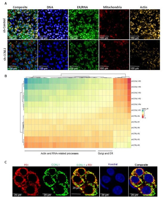
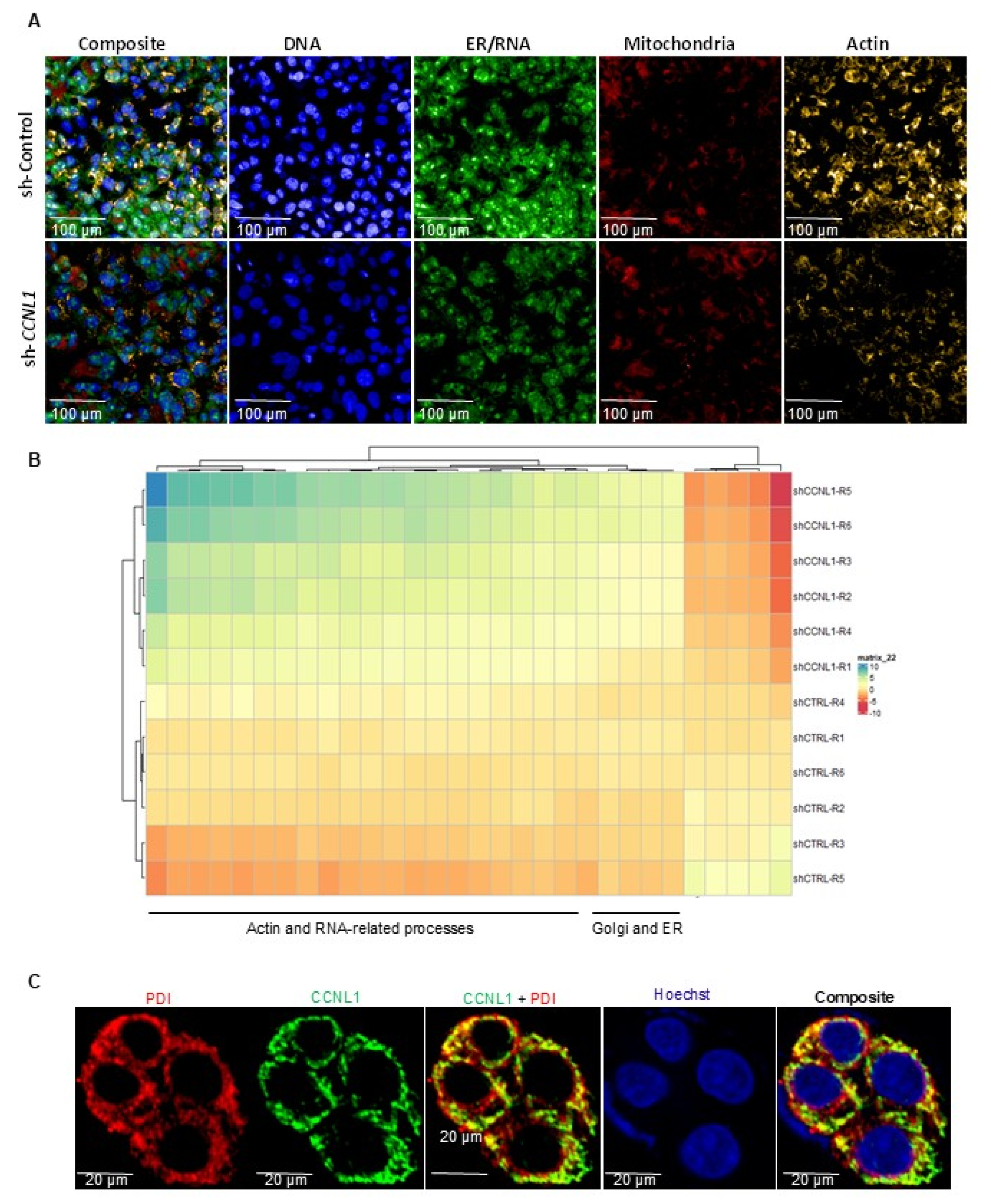
Thus far, our data suggests that CCNL1 could serve as an HBV host susceptibility factor regulating various aspects of the viral infection cycle, including viral transcription, and key cellular features vital to HBV replication. To further assess the phenotypic impact of CCNL1 knockdown on cellular features and cell-biological processes in an unbiased, holistic manner, we performed morphometric analysis with a cell painting assay using multiplexed fluorescent dyes [44] (Figure 4). Principal component analysis (PCA) revealed two distinct cell clusters between sh-Control and shCCNL1 HBV-infected HepG2-NTCP cells (Figure S7). Images from different channels were segmented into primary objects. Morphological, textural, and intensity features were extracted (Figure 4A) using CellProfiler. The Pycytominer package was used to identify critical morphometric changes upon CCNL1. Notably, CCNL1 knockdown appeared to be associated with actin- and RNA-related processes, as well as intracellular transport, as suggested by the enrichment of Golgi-ER-related processes (Figure 4B). To confirm these observations, we performed IFA to co-stain for the ER marker, protein disulfide isomerase (PDI), and CCNL1. Interestingly, we observed strong co-localization between the CCNL1 protein and PDI (Figure 4C). These observations indicated possible perturbations in cellular processes upon CCNL1 knockdown. We speculated that some of the affected pathways may be required for the HBV infection cycle. Further studies are required to validate these findings. Altogether, the data from our study suggests a working model whereby CCNL1 interacts with CDKs, such as CDK11B, to promote the phosphorylation of RNAPII. Consequently, phosphorylated RNAPII is recruited to cccDNA, where it regulates viral transcription.
Figure 4.
Morphometric analysis upon CCNL1 knockdown reveals phenotypic alterations in ER-related features that may associate with host pathways regulating the viral life cycle. (A) Sample images from cell painting assay in HepG2-NTCP cells transfected with shControl or shCCNL1 and infected with HBV showing DNA, RNA/ER, mitochondria and Actin and composite are shown for the control and CCNL1 knockdown cells. Scale bar is 100 μm. (B) The top 30 differential cell features between control and knockdown cells are shown in the heatmap. (C) Immunofluorescence analysis (IFA) image showing co-localization of ER marker, PDI, and CCNL1 in HepG2-NTCP cells. The scale bar is 20 μm. All the experiments shown above represent 3 biological replicates each consisting of 3 technical replicates.
4. Discussion
The HBV infection cycle largely depends on host cell factors. Given that HBV has a high tropism to the human liver and mainly infects hepatocytes, the lack of optimal physiologically relevant, humanized models to study HBV–host cell interactions has remained a significant hurdle in the quest for identification of host-directed therapeutics (reviewed in [26]). Here, we performed RNA-seq analysis of HBV-infected PHH, which serves as a physiologically relevant model to identify putative host factors that are modulated and utilized by HBV to promote replication. Numerous candidate host factors identified from this screen have been described in the literature to regulate HBV infection, such as SRPK1, CDK1, and NXF1 [37,38], thereby providing a proof-of-concept for our approach.
Among the candidate host genes that were upregulated in PHH upon HBV infection, we focused on the downstream functional characterization of CCNL1. The role of CCNL1 during HBV and other viral infections have not been elucidated. This study demonstrated that CCNL1 may serve as a host factor that supports efficient HBV replication and transcription. Specifically, siRNA/shRNA-mediated knockdown of CCNL1 results in a marked reduction in multiple viral parameters in heterologous cell models of HBV replication and live infection. Notably, we observed reduced extracellular HBV DNA and inhibition of HBV gene expression, including pgRNA, HBsAg, HBcAg, and HBeAg (Figure 1 and Figure 2). Given that CCNL1 has been shown to interact with CDK11B to phosphorylate the CTD of RNAPII to modulate gene expression [8,41,45], we also investigated the effect of CCNL1 on phosphorylation, and therefore on the activity of RNAPII. Indeed, the data from knockdown studies indicated that CCNL1 regulates phosphorylation of RNAPII Ser2 (Figure 3 and Figure S6). In this context, it is important to note that total and phospho-RNAPII have been shown to play crucial roles in the transcription of HBV from cccDNA [43].
Along the same lines of reasoning, we hypothesized that by influencing RNAPII activity, CCNL1 could modulate the transcription of HBV RNA. Indeed, we found that CCNL1 knockdown resulted in reduced expression of the nascent HBV transcript, highlighting its importance in controlling viral transcription. Furthermore, ChIP-PCR assays showed that knockdown of CCNL1 resulted in reduced H3K27 acetylation, a known marker of active cccDNA transcription [46,47] as well as the binding of total RNAPII and RNAPII S2 to cccDNA [43]. Previous studies have shown that CCNL1 forms a complex with SC35/SRSF2, CDK11, and CDK12 to regulate pre-mRNA splicing [8,40,48]. Interestingly, spliced HBV RNA variants have been identified in the serum and liver tissues of patients with chronic HBV [49,50,51,52]. These spliced variants and products have been linked to play a role as immune decoys, associated with liver damage, fibrosis, and the development of HCC [14,15,53,54,55,56,57]. Thus, we analyzed the HBV splice variants in our models and found that knockdown of CCNL1 resulted in a global decrease in HBV variants in hepatoma cells. However, given our observation that CCNL1 controls HBV transcription, normalization of the levels of spliced HBV RNA against total HBV RNA or pgRNA failed to reveal any appreciable difference in the levels of splicing upon loss-of-function of CCNL1. Altogether, our data suggests that while CCNL1 may have an indirect role in modulating the levels of HBV RNA splice variants by regulating the overall transcription of HBV, it is less likely to play a direct role in pre-mRNA splicing of nascent HBV RNA (Figure 3). In addition, knockdown of CCNL1 resulted in a decreased association of total and Ser2-phosphor RNAPII with cccDNA, further highlighting its role in regulating cccDNA transcription. These observations corroborate recent reports suggesting that the regulation of total RNAPII phosphorylation affects cccDNA-based viral transcription [43]. Our studies showed that CCNL1 co-localizes with total RNAPII and is likely recruited to the cccDNA mini-chromosome in a phosphorylation-dependent manner (Figure 3). Furthermore, knockdown of CCNL1 resulted in reduced binding to HBV RNAs, suggesting its potential role in direct binding with viral RNAs to modulate their stability or expression. These observations remain to be further evaluated in future studies to directly link CCNL1 to HBV-RNA stability or expression.
Unbiased morphometric profiling of CCNL1 perturbation in HepG2-NTCP cells suggests that CCNL1 may modulate host cell structures required for viral replication including actin dynamics, the endoplasmic reticulum, and the Golgi architecture. These observations are notable in light of previous reports demonstrating that HBV proteins can modulate host actin polymerization and thereby cytoskeletal remodeling to promote viral replication and oncogenic transformation [58,59]. Specifically, HBx protein was shown to specifically interact with calmodulin (CaM), resulting in the displacement of HSP90, activating the cofilin pathway, thereby enhancing the polymerization of actin, and has been linked to HBV-mediated HCC metastasis [58,59,60]. The core protein was also reported to interact with the C-terminal domain of actin-binding proteins [61]. These observations highlight the diverse putative functions of CCNL1 in modulating key HBV-dependent cell biological phenotypes that may be relevant for CHB-driven chronic liver disease and cancer. The observed colocalization of CCNL1 with ER markers, including PDI, is also intriguing given the established role of ER-associated proteins such as ERp57 in HBV entry and membrane fusion [62]. While these associations remain correlative and need further validation, they indicate additional cellular pathways through which CCNL1 may indirectly regulate the HBV infection cycle.
Finally, the observed upregulation of CCNL1 in clinical samples from patients with chronic HBV infection underscores the clinical relevance of our findings. Notably reduced CCNL1 expression in patients exhibiting functional cure, as defined by HBsAg loss, suggests that CCNL1 expression correlates with active viral persistence. While these data do not directly link CCNL1 to chronic HBV, there is a possibility of validating CCNL1 as a biomarker for CHB. In summary, this study identifies CCNL1 as a host factor that promotes HBV transcription and replication, through modulation of RNAPII activity at cccDNA. A key limitation of this work is that we did not definitively uncouple CCNL1’s role in transcription versus that in modulating HBV-RNA stability. Future studies combining RNA decay measurements, and mechanistic study of HBV:CCNL1 interactions will be key to fully understanding the molecular role of CCNL1 in HBV infection. Overall, this study contributes to a better understanding of the role of host factors that regulate HBV infection biology.
Supplementary Materials
The following supporting information can be downloaded at: https://www.mdpi.com/article/10.3390/v18050545/s1, Figure S1: Analysis of HBV mediated transcriptomic changes; Figure S2: Evaluating the role of CCNL1 during HBV infection; Figure S3: Functional validation of Cyclin L1 in PHH; Figure S4: Ectopic expression of CCNL1 in HepG2-NTCP cells enhances HBV gene expression; Figure S5: Evaluating the role of Cyclin L1 on HBV RNA splicing; Figure S6: Cyclin L1 interacts with HBV RNA; Figure S7: Morphometric analysis of HepG2-NTCP cells after Cell Painting and hierarchical clustering display two different clusters of sh-CCNL1 and sh-control cells; Table S1: Patient details [16,41,44,50,63].
Author Contributions
Conceptualization, R.D., P.B., C.O.O., B.C.N., G.P., M.L. and S.G.L.; Methodology, R.D., P.B., C.O.O., B.C.N. and M.-L.P.; Investigation, C.O.O., B.C.N., M.-L.P., P.P.K.A., N.G., J.T.S., G.Y.L. and T.T.M.; Formal Analysis, B.C.N., C.O.O., N.S. and T.T.M.; Writing—Original Draft, C.O.O., B.C.N. and R.D.; Writing—Review and Editing, R.D., P.B., C.O.O., and B.C.N.; Funding Acquisition, R.D. and S.G.L.; Resources, S.G.L. and R.D.; Supervision, R.D., P.B., S.G.L., M.L. and B.C.N. All authors have read and agreed to the published version of the manuscript.
Funding
This work was supported by the National Medical Research Council (NMRC), Singapore, Open Fund-Large Collaborative Grant (OFLCG19May-0038) to SGL and RD, and the National Research Foundation (NRF), Competitive Research Programme (CRP26-2021-0062) to RD. The Agency for Science, Technology, and Research–Singapore International Graduate Award (A*STAR-SINGA)-funded COO.
Institutional Review Board Statement
Not applicable.
Informed Consent Statement
Not applicable.
Data Availability Statement
The original contributions presented in this study are included in the article/Supplementary Material. Further inquiries can be directed to the corresponding authors.
Acknowledgments
The authors thank Tan Yee Joo (Institute of Molecular and Cell Biology, Agency for Science Technology and Research, Singapore 138673, Singapore) for sharing reagents and viruses used for infection in this study. We thank David Durantel and Anna Salvetti of CIRI INSERM, Lyon, for sharing their protocols and for their support, insight, and guidance. We thank Koichi Watashi and Takaji Wakita (National Institute of Infectious Diseases, Japan) for providing reagents. We thank Adam Zlotnick for sharing the anti-HBc antibodies. We thank Ming Jie Lim for her assistance with the siRNA screening. We thank Tan Yee Joo for producing the virus stocks used in the infection experiments. We thank Weiyong Chua for providing administrative support. Lastly, we thank all patients whose samples were used for validation in this study.
Conflicts of Interest
Prof. Seng Gee Lim: Advisory Board: Gilead Sciences, Abbott, Roche, Janssen, GlaxoSmithKline, Grifols, Arbutus, Assembly. Speakers Bureau: Gilead Sciences, Abbott. Educational/research funding: Abbott, Merck Sharpe and Dohme, Gilead. The rest of the authors have nothing to report.
References
- Lampertico, P.; Agarwal, K.; Berg, T.; Buti, M.; Janssen, H.L.A.; Papatheodoridis, G.; Zoulim, F.; Tacke, F. EASL 2017 Clinical Practice Guidelines on the Management of Hepatitis B Virus Infection. J. Hepatol. 2017, 67, 370–398. [Google Scholar] [CrossRef] [Scilit] [PubMed]
- Narmada, B.C.; Khakpoor, A.; Shirgaonkar, N.; Narayanan, S.; Kim Aw, P.P.; Singh, M.; Ong, K.H.; Owino, C.O.; Ting Ng, J.W.; Yew, H.C.; et al. Single-Cell Landscape of Functionally Cured Chronic Hepatitis B Patients Reveals Activation of Innate and Altered CD4-CTL-Driven Adaptive Immunity. J. Hepatol. 2024, 81, 42–61. [Google Scholar] [CrossRef] [Scilit]
- Zeisel, M.B.; Lucifora, J.; Mason, W.S.; Sureau, C.; Beck, J.; Levrero, M.; Kann, M.; Knolle, P.A.; Benkirane, M.; Durantel, D.; et al. Towards an HBV Cure: State-of-the-Art and Unresolved Questions-Report of the ANRS Workshop on HBV Cure. Gut 2015, 64, 1314–1326. [Google Scholar] [CrossRef] [Scilit]
- Ghany, M.G. Current Treatment Guidelines of Chronic Hepatitis B: The Role of Nucleos(t)Ide Analogues and Peginterferon. Best Pract. Res. Clin. Gastroenterol. 2017, 31, 299–309. [Google Scholar] [CrossRef] [Scilit] [PubMed]
- Fanning, G.C.; Zoulim, F.; Hou, J.; Bertoletti, A. Therapeutic Strategies for Hepatitis B Virus Infection: Towards a Cure. Nat. Rev. Drug Discov. 2019, 18, 827–844. [Google Scholar] [CrossRef] [Scilit]
- Yan, H.; Peng, B.; He, W.; Zhong, G.; Qi, Y.; Ren, B.; Gao, Z.; Jing, Z.; Song, M.; Xu, G.; et al. Molecular Determinants of Hepatitis B and D Virus Entry Restriction in Mouse Sodium Taurocholate Cotransporting Polypeptide. J. Virol. 2013, 87, 7977–7991. [Google Scholar] [CrossRef] [Scilit]
- Iwamoto, M.; Watashi, K.; Tsukuda, S.; Aly, H.H.; Fukasawa, M.; Fujimoto, A.; Suzuki, R.; Aizaki, H.; Ito, T.; Koiwai, O.; et al. Evaluation and Identification of Hepatitis B Virus Entry Inhibitors Using HepG2 Cells Overexpressing a Membrane Transporter NTCP. Biochem. Biophys. Res. Commun. 2014, 443, 808–813. [Google Scholar] [CrossRef] [Scilit]
- Loyer, P.; Trembley, J.H. Roles of CDK/Cyclin Complexes in Transcription and Pre-MRNA Splicing: Cyclins L and CDK11 at the Cross-Roads of Cell Cycle and Regulation of Gene Expression. Semin. Cell Dev. Biol. 2020, 107, 36–45. [Google Scholar] [CrossRef] [Scilit] [PubMed]
- Makokha, G.N.; Abe-Chayama, H.; Chowdhury, S.; Hayes, C.N.; Tsuge, M.; Yoshima, T.; Ishida, Y.; Zhang, Y.; Uchida, T.; Tateno, C.; et al. Regulation of the Hepatitis B Virus Replication and Gene Expression by the Multi-Functional Protein TARDBP. Sci. Rep. 2019, 9, 8462. [Google Scholar] [CrossRef] [Scilit]
- Ogura, N.; Watashi, K.; Noguchi, T.; Wakita, T. Formation of Covalently Closed Circular DNA in Hep38.7-Tet Cells, a Tetracycline Inducible Hepatitis B Virus Expression Cell Line. Biochem. Biophys. Res. Commun. 2014, 452, 315–321. [Google Scholar] [CrossRef] [Scilit]
- Lim, Z.; Mohd-Ismail, N.K.B.; Png, E.; Sze, C.W.; Lin, Q.; Hong, W.; Lim, S.G.; Tan, Y.-J.; Gunaratne, J. Phosphoproteomics Unravel HBV Triggered Rewiring of Host Phosphosignaling Events. Int. J. Mol. Sci. 2022, 23, 5127. [Google Scholar] [CrossRef] [Scilit]
- Dull, T.; Zufferey, R.; Kelly, M.; Mandel, R.J.; Nguyen, M.; Trono, D.; Naldini, L. A Third-Generation Lentivirus Vector with a Conditional Packaging System. J. Virol. 1998, 72, 8463–8471. [Google Scholar] [CrossRef] [Scilit] [PubMed]
- Xiang, C.; Du, Y.; Meng, G.; Soon Yi, L.; Sun, S.; Song, N.; Zhang, X.; Xiao, Y.; Wang, J.; Yi, Z.; et al. Long-Term Functional Maintenance of Primary Human Hepatocytes in Vitro. Science 2019, 364, 399–402. [Google Scholar] [CrossRef] [Scilit] [PubMed]
- Preiss, S.; Littlejohn, M.; Angus, P.; Thompson, A.; Desmond, P.; Lewin, S.R.; Sasadeusz, J.; Matthews, G.; Dore, G.J.; Shaw, T.; et al. Defective Hepatitis B Virus DNA Is Not Associated with Disease Status but Is Reduced by Polymerase Mutations Associated with Drug Resistance. Hepatology 2008, 48, 741–749. [Google Scholar] [CrossRef] [Scilit]
- Bayliss, J.; Lim, L.; Thompson, A.J.V.; Desmond, P.; Angus, P.; Locarnini, S.; Revill, P.A. Hepatitis B Virus Splicing Is Enhanced Prior to Development of Hepatocellular Carcinoma. J. Hepatol. 2013, 59, 1022–1028. [Google Scholar] [CrossRef] [Scilit]
- Wang, H.; Liu, K.H.; Fang, B.A.M.; Wu, H.Q.; Li, F.D.; Xiang, X.G.; Tang, W.L.; Zhao, G.D.; Lin, L.Y.; Bao, S.; et al. Identification of Acetyltransferase Genes (HAT1 and KAT8) Regulating HBV Replication by RNAi Screening. Cell Biosci. 2015, 5, 66. [Google Scholar] [CrossRef] [Scilit]
- Livak, K.J.; Schmittgen, T.D. Analysis of Relative Gene Expression Data Using Real-Time Quantitative PCR and the 2−ΔΔCT Method. Methods 2001, 25, 402–408. [Google Scholar] [CrossRef] [Scilit]
- Ciechanover, A.; Elias, S.; Heller, H.; Hershko, A. “Covalent Affinity” Purification of Ubiquitin-Activating Enzyme. J. Biol. Chem. 1982, 257, 2537–2542. [Google Scholar] [CrossRef] [Scilit] [PubMed]
- Huang, J.T.; Yang, Y.; Hu, Y.M.; Liu, X.H.; Liao, M.Y.; Morgan, R.; Yuan, E.F.; Li, X.; Liu, S.M. A Highly Sensitive and Robust Method for Hepatitis B Virus Covalently Closed Circular DNA Detection in Single Cells and Serum. J. Mol. Diagn. 2018, 20, 334–343. [Google Scholar] [CrossRef] [Scilit]
- Qu, B.; Ni, Y.; Lempp, F.A.; Vondran, F.W.R.; Urban, S. T5 Exonuclease Hydrolysis of Hepatitis B Virus Replicative Intermediates Allows Reliable Quantification and Fast Drug Efficacy Testing of Covalently Closed Circular DNA by PCR. J. Virol. 2018, 92, e01117-18. [Google Scholar] [CrossRef] [Scilit]
- Allweiss, L.; Volz, T.; Giersch, K.; Kah, J.; Raffa, G.; Petersen, J.; Lohse, A.W.; Beninati, C.; Pollicino, T.; Urban, S.; et al. Proliferation of Primary Human Hepatocytes and Prevention of Hepatitis B Virus Reinfection Efficiently Deplete Nuclear CccDNA in Vivo. Gut 2018, 67, 542–552. [Google Scholar] [CrossRef] [Scilit]
- Galle, P.R.; Hagelstein, J.; Kommerell, B.; Volkmann, M.; Schranz, P.; Zentgraf, H. In Vitro Experimental Infection of Primary Human Hepatocytes with Hepatitis B Virus. Gastroenterology 1994, 106, 664–673. [Google Scholar] [CrossRef] [Scilit] [PubMed]
- Szklarczyk, D.; Morris, J.H.; Cook, H.; Kuhn, M.; Wyder, S.; Simonovic, M.; Santos, A.; Doncheva, N.T.; Roth, A.; Bork, P.; et al. The STRING Database in 2017: Quality-Controlled Protein-Protein Association Networks, Made Broadly Accessible. Nucleic Acids Res. 2017, 45, D362–D368. [Google Scholar] [CrossRef] [Scilit] [PubMed]
- Wang, L.; Feng, Z.; Wang, X.; Wang, X.; Zhang, X. DEGseq: An R Package for Identifying Differentially Expressed Genes from RNA-Seq Data. Bioinformatics 2009, 26, 136–138. [Google Scholar] [CrossRef] [Scilit] [PubMed]
- Schulze-Bergkamen, H.; Untergasser, A.; Dax, A.; Vogel, H.; Büchler, P.; Klar, E.; Lehnert, T.; Friess, H.; Büchler, M.W.; Kirschfink, M.; et al. Primary Human Hepatocytes—A Valuable Tool for Investigation of Apoptosis and Hepatitis B Virus Infection. J. Hepatol. 2003, 38, 736–744. [Google Scholar] [CrossRef] [Scilit]
- Hu, J.; Lin, Y.-Y.Y.; Chen, P.-J.J.; Watashi, K.; Wakita, T. Cell and Animal Models for Studying Hepatitis B Virus Infection and Drug Development. Gastroenterology 2019, 156, 338–354. [Google Scholar] [CrossRef] [Scilit]
- Chang, W.-W.; Su, I.-J.; Lai, M.-D.; Chang, W.-T.; Huang, W.; Lei, H.-Y. Suppression of P38 Mitogen-Activated Protein Kinase Inhibits Hepatitis B Virus Replication in Human Hepatoma Cell: The Antiviral Role of Nitric Oxide. J. Viral Hepat. 2008, 15, 490–497. [Google Scholar] [CrossRef]
- Lamontagne, J.; Mell, J.C.; Bouchard, M.J. Transcriptome-Wide Analysis of Hepatitis B Virus-Mediated Changes to Normal Hepatocyte Gene Expression. PLoS Pathog. 2016, 12, e1005438. [Google Scholar] [CrossRef] [Scilit]
- Hallez, C.; Li, X.; Suspène, R.; Thiers, V.; Bouzidi, M.S.; Dorobantu, C.M.; Lucansky, V.; Wain-Hobson, S.; Gaudin, R.; Vartanian, J.-P. Hypoxia-Induced Human Deoxyribonuclease I Is a Cellular Restriction Factor of Hepatitis B Virus. Nat. Microbiol. 2019, 4, 1196–1207. [Google Scholar] [CrossRef] [Scilit]
- Wing, P.A.C.; Liu, P.J.; Harris, J.M.; Magri, A.; Michler, T.; Zhuang, X.; Borrmann, H.; Minisini, R.; Frampton, N.R.; Wettengel, J.M.; et al. Hypoxia Inducible Factors Regulate Hepatitis B Virus Replication by Activating the Basal Core Promoter. J. Hepatol. 2021, 75, 64–73. [Google Scholar] [CrossRef] [Scilit]
- Masson, J.J.R.; Billings, H.W.W.; Palmer, C.S. Metabolic Reprogramming during Hepatitis B Disease Progression Offers Novel Diagnostic and Therapeutic Opportunities. Antivir. Chem. Chemother. 2017, 25, 53–57. [Google Scholar] [CrossRef] [Scilit]
- Li, H.; Zhu, W.; Zhang, L.; Lei, H.; Wu, X.; Guo, L.; Chen, X.; Wang, Y.; Tang, H. The Metabolic Responses to Hepatitis B Virus Infection Shed New Light on Pathogenesis and Targets for Treatment. Sci. Rep. 2015, 5, 8421. [Google Scholar] [CrossRef] [Scilit]
- Ladner, S.K.; Otto, M.J.; Barker, C.S.; Zaifert, K.; Wang, G.H.; Guo, J.U.T.; Seeger, C.; King, R.W. Inducible Expression of Human Hepatitis B Virus (HBV) in Stably Transfected Hepatoblastoma Cells: A Novel System for Screening Potential Inhibitors of HBV Replication. Antimicrob. Agents Chemother. 1997, 41, 1715–1720. [Google Scholar] [CrossRef] [Scilit]
- Yang, C.-C.; Huang, E.-Y.; Li, H.-C.; Su, P.-Y.; Shih, C. Nuclear Export of Human Hepatitis B Virus Core Protein and Pregenomic RNA Depends on the Cellular NXF1-P15 Machinery. PLoS ONE 2014, 9, e106683. [Google Scholar] [CrossRef] [Scilit] [PubMed]
- Li, H.-C.; Huang, E.-Y.; Su, P.-Y.; Wu, S.-Y.; Yang, C.-C.; Lin, Y.-S.; Chang, W.-C.; Shih, C. Nuclear Export and Import of Human Hepatitis B Virus Capsid Protein and Particles. PLoS Pathog. 2010, 6, e1001162. [Google Scholar] [CrossRef] [Scilit]
- Macovei, A.; Petrareanu, C.; Lazar, C.; Florian, P.; Branza-Nichita, N. Regulation of Hepatitis B Virus Infection by Rab5, Rab7, and the Endolysosomal Compartment. J. Virol. 2013, 87, 6415–6427. [Google Scholar] [CrossRef] [Scilit]
- Ludgate, L.; Ning, X.; Nguyen, D.H.; Adams, C.; Mentzer, L.; Hu, J. Cyclin-Dependent Kinase 2 Phosphorylates S/T-P Sites in the Hepadnavirus Core Protein C-Terminal Domain and Is Incorporated into Viral Capsids. J. Virol. 2012, 86, 12237–12250. [Google Scholar] [CrossRef] [Scilit] [PubMed]
- Daub, H.; Blencke, S.; Habenberger, P.; Kurtenbach, A.; Dennenmoser, J.; Wissing, J.; Ullrich, A.; Cotten, M. Identification of SRPK1 and SRPK2 as the Major Cellular Protein Kinases Phosphorylating Hepatitis B Virus Core Protein. J. Virol. 2002, 76, 8124–8137. [Google Scholar] [CrossRef] [Scilit]
- Heger-Stevic, J.; Zimmermann, P.; Lecoq, L.; Böttcher, B.; Nassal, M. Hepatitis B Virus Core Protein Phosphorylation: Identification of the SRPK1 Target Sites and Impact of Their Occupancy on RNA Binding and Capsid Structure. PLoS Pathog. 2018, 14, e1007488. [Google Scholar] [CrossRef] [Scilit]
- Dickinson, L.A.; Edgar, A.J.; Ehley, J.; Gottesfeld, J.M. Cyclin L Is an RS Domain Protein Involved in Pre-MRNA Splicing. J. Biol. Chem. 2002, 277, 25465–25473. [Google Scholar] [CrossRef] [Scilit] [PubMed]
- Chabrolles, H.; Auclair, H.; Vegna, S.; Lahlali, T.; Pons, C.; Michelet, M.; Couté, Y.; Belmudes, L.; Chadeuf, G.; Kim, Y.; et al. Hepatitis B Virus Core Protein Nuclear Interactome Identifies SRSF10 as a Host RNA-Binding Protein Restricting HBV RNA Production. PLoS Pathog. 2020, 16, e1008593. [Google Scholar] [CrossRef] [Scilit]
- Rall, L.B.; Standring, D.N.; Laub, O.; Rutter, W.J. Transcription of Hepatitis B Virus by RNA Polymerase II. Mol. Cell. Biol. 1983, 3, 1766–1773. [Google Scholar] [CrossRef] [Scilit]
- Fan, Y.; Liang, Y.; Liu, Y.; Fan, H. PRKDC Promotes Hepatitis B Virus Transcription through Enhancing the Binding of RNA Pol II to CccDNA. Cell Death Dis. 2022, 13, 404. [Google Scholar] [CrossRef] [Scilit] [PubMed]
- Bray, M.A.; Singh, S.; Han, H.; Davis, C.T.; Borgeson, B.; Hartland, C.; Kost-Alimova, M.; Gustafsdottir, S.M.; Gibson, C.C.; Carpenter, A.E. Cell Painting, a High-Content Image-Based Assay for Morphological Profiling Using Multiplexed Fluorescent Dyes. Nat. Protoc. 2016, 11, 1757–1774. [Google Scholar] [CrossRef] [Scilit] [PubMed]
- Maita, H.; Nakagawa, S. What Is the Switch for Coupling Transcription and Splicing? RNA Polymerase II C-Terminal Domain Phosphorylation, Phase Separation and Beyond. Wiley Interdiscip. Rev. RNA 2020, 11, e1574. [Google Scholar] [CrossRef] [Scilit]
- Pollicino, T.; Belloni, L.; Raffa, G.; Pediconi, N.; Squadrito, G.; Raimondo, G.; Levrero, M. Hepatitis B Virus Replication Is Regulated by the Acetylation Status of Hepatitis B Virus CccDNA-Bound H3 and H4 Histones. Gastroenterology 2006, 130, 823–837. [Google Scholar] [CrossRef] [Scilit]
- Yang, B.; Li, B.; Jia, L.; Jiang, Y.; Wang, X.; Jiang, S.; Du, S.; Ji, X.; Yang, P. 3D Landscape of Hepatitis B Virus Interactions with Human Chromatins. Cell Discov. 2020, 6, 95. [Google Scholar] [CrossRef] [Scilit] [PubMed]
- Chen, H.-H.; Wang, Y.-C.; Fann, M.-J. Identification and Characterization of the CDK12/Cyclin L1 Complex Involved in Alternative Splicing Regulation. Mol. Cell. Biol. 2006, 26, 2736–2745. [Google Scholar] [CrossRef] [Scilit]
- Soussan, P.; Garreau, F.; Zylberberg, H.; Ferray, C.; Brechot, C.; Kremsdorf, D. In Vivo Expression of a New Hepatitis B Virus Protein Encoded by a Spliced RNA. J. Clin. Investig. 2000, 105, 55–60. [Google Scholar] [CrossRef] [Scilit]
- Sommer, G.; Heise, T. Posttranscriptional Control of HBV Gene Expression. Front. Biosci. 2008, 13, 5533–5547. [Google Scholar] [CrossRef] [Scilit]
- Candotti, D.; Allain, J.-P. Biological and Clinical Significance of Hepatitis B Virus RNA Splicing: An Update. Ann. Blood 2016, 2, 1–14. [Google Scholar] [CrossRef] [Scilit]
- Lee, G.H.; Wasser, S.; Lim, S.G. Hepatitis B Pregenomic RNA Splicing-The Products, the Regulatory Mechanisms and Its Biological Significance. Virus Res. 2008, 136, 1–7. [Google Scholar] [CrossRef] [Scilit]
- Duriez, M.; Mandouri, Y.; Lekbaby, B.; Wang, H.; Schnuriger, A.; Redelsperger, F.; Guerrera, C.I.; Lefevre, M.; Fauveau, V.; Ahodantin, J.; et al. Alternative Splicing of Hepatitis B Virus: A Novel Virus/Host Interaction Altering Liver Immunity. J. Hepatol. 2017, 67, 687–699. [Google Scholar] [CrossRef] [Scilit] [PubMed]
- Soussan, P.; Tuveri, R.; Nalpas, B.; Garreau, F.; Zavala, F.; Masson, A.; Pol, S.; Brechot, C.; Kremsdorf, D. The Expression of Hepatitis B Spliced Protein (HBSP) Encoded by a Spliced Hepatitis B Virus RNA Is Associated with Viral Replication and Liver Fibrosis. J. Hepatol. 2003, 38, 343–348. [Google Scholar] [CrossRef] [Scilit] [PubMed]
- Pol, J.G.; Lekbaby, B.; Redelsperger, F.; Klamer, S.; Mandouri, Y.; Ahodantin, J.; Bieche, I.; Lefevre, M.; Souque, P.; Charneau, P.; et al. Alternative Splicing-Regulated Protein of Hepatitis B Virus Hacks the TNF-α-Stimulated Signaling Pathways and Limits the Extent of Liver Inflammation. FASEB J. 2015, 29, 1879–1889. [Google Scholar] [CrossRef] [Scilit] [PubMed]
- Wu, S.-X.; Chen, W.-N.; Jing, Z.-T.; Liu, W.; Lin, X.-J.; Lin, X. Hepatitis B Spliced Protein (HBSP) Suppresses Fas-Mediated Hepatocyte Apoptosis via Activation of PI3K/Akt Signaling. J. Virol. 2018, 92, e01273-18. [Google Scholar] [CrossRef] [Scilit]
- Chen, J.; Wu, M.; Wang, F.; Zhang, W.; Wang, W.; Zhang, X.; Zhang, J.; Liu, Y.; Liu, Y.; Feng, Y.; et al. Hepatitis B Virus Spliced Variants Are Associated with an Impaired Response to Interferon Therapy. Sci. Rep. 2015, 5, 16459. [Google Scholar] [CrossRef] [Scilit]
- Kim, M.J.; Kim, J.; Im, J.S.; Kang, I.; Ahn, J.K. Hepatitis B Virus X Protein Enhances Liver Cancer Cell Migration by Regulating Calmodulin-Associated Actin Polymerization. BMB Rep. 2021, 54, 614–619. [Google Scholar] [CrossRef] [Scilit]
- Kong, F.; You, H.; Tang, R.; Zheng, K. The Regulation of Proteins Associated with the Cytoskeleton by Hepatitis B Virus X Protein during Hepatocarcinogenesis (Review). Oncol. Lett. 2017, 13, 2514–2520. [Google Scholar] [CrossRef] [Scilit]
- Lara-Pezzi, E. The Hepatitis B Virus X Protein (HBx) Induces a Migratory Phenotype in a CD44-Dependent Manner: Possible Role of HBx in Invasion and Metastasis. Hepatology 2001, 33, 1270–1281. [Google Scholar] [CrossRef] [Scilit]
- Huang, C.-J.; Chen, Y.-H.; Ting, L.-P. Hepatitis B Virus Core Protein Interacts with the C-Terminal Region of Actin-Binding Protein. J. Biomed. Sci. 2000, 7, 160–168. [Google Scholar] [CrossRef]
- Pérez-Vargas, J.; Teppa, E.; Amirache, F.; Boson, B.; de Oliveira, R.P.; Combet, C.; Böckmann, A.; Fusil, F.; Freitas, N.; Carbone, A.; et al. A Fusion Peptide in Pres1 and the Human Protein-Disulfide Isomerase Erp57 Are Involved in Hepatitis b Virus Membrane Fusion Process. Elife 2021, 10, e64507. [Google Scholar] [CrossRef] [Scilit]
- Sekiba, K.; Otsuka, M.; Ohno, M.; Yamagami, M.; Kishikawa, T.; Suzuki, T.; Ishibashi, R.; Seimiya, T.; Tanaka, E.; Koike, K. Inhibition of HBV Transcription From CccDNA With Nitazoxanide by Targeting the HBx–DDB1 Interaction. Cell Mol. Gastroenterol. Hepatol. 2019, 7, 297–312. [Google Scholar] [CrossRef] [Scilit]
Disclaimer/Publisher’s Note: The statements, opinions and data contained in all publications are solely those of the individual author(s) and contributor(s) and not of MDPI and/or the editor(s). MDPI and/or the editor(s) disclaim responsibility for any injury to people or property resulting from any ideas, methods, instructions or products referred to in the content. |
© 2026 by the authors. Licensee MDPI, Basel, Switzerland. This article is an open access article distributed under the terms and conditions of the Creative Commons Attribution (CC BY) license.