


Abstract
To elucidate the effect of cold stress on Dendroctonus valens larvae, a study was conducted under controlled laboratory conditions to examine the physiological and biochemical mechanisms associated with cold stress, coupled with transcriptome sequencing. Physiological and biochemical assessments indicated stable water content in larvae during cold stress initiation, with triglycerides and fats serving as primary energy reserves that decreased over cold stress progression. Glycogen and trehalose were identified as energy sources for larval energy metabolism, with their levels increasing as cold stress duration extended. Superoxide dismutase (SOD) activity exhibited an initial decline followed by an increase, while peroxidase (POD) activity initially rose before decreasing over induction time, and catalase (CAT) activity decreased during cold stress induction. Transcriptome sequencing at various time points revealed 4630 upregulated and 1554 downregulated genes, predominantly involved in metabolic pathways such as carbohydrate, amino acid, and lipid metabolism. Quantitative polymerase chain reaction (qPCR) results validated the transcriptome data accuracy. This investigation delineated the physiological, biochemical, and transcriptome alterations during cold stress, offering a theoretical framework for the rational prediction of Dendroctonus valens outbreaks.
1. Introduction
Dendroctonus valens (Coleoptera: Curculionidae) is a species of bark beetle known for infesting tree trunks and roots [1,2,3,4]. While it can affect various tree species such as pine, Picea asperata, Pseudotsuga sinensis, Abies fabri, and Larix gmelinii, its primary impact is observed in pine trees [5]. In its native range in North America, D. valens typically acts as a secondary pest, targeting weakened or injured trees without causing widespread levels of tree mortality [6]. However, since its introduction to China in 1998, D. valens populations have rapidly proliferated in China, leading to extensive tree mortality and significant economic losses in the country [7,8,9,10,11,12].
Insects are temperature-sensitive organisms that adjust their physiological and behavioral traits in response to environmental temperature changes [13,14,15]. Temperature is a crucial factor affecting their geographical distribution, population dynamics, and seasonal behaviors [16,17,18,19,20,21]. To combat low-temperature stress, insects have developed complex physiological and biochemical adaptations that enhance their resilience to cold [20,21,22,23]. Trehalose, the main sugar in insect hemolymph, forms a protective layer on cell surfaces under extreme conditions, effectively supporting physiological functions [24]. High trehalose levels significantly boost insect cold tolerance [25,26]. Defensive enzymes like superoxide dismutase (SOD), catalase (CAT), and peroxidase (POD) are vital in detoxifying oxygen free radicals; SOD detoxifies these radicals, while CAT and POD convert them into water and oxygen [27].
Transcriptomics plays a crucial role in evaluating gene expression differences, protein evolutionary rates, genetic–phenotypic relationships, phylogenetic connections, chromosomal structures, and regulatory mechanisms [4,28,29,30]. It is a powerful tool for systematically uncovering the molecular mechanisms behind low-temperature adaptation in insects [31]. When insects face low-temperature stress, their cells initiate a comprehensive defense and adaptation program by altering the transcription levels of numerous genes. High-throughput sequencing studies have shown that low-temperature exposure triggers a significant number of differentially expressed genes (DEGs) in insects [32,33]. For example, Huawei Ren et al. [32] studied the transcriptomic changes in Spodoptera frugiperda under low-temperature conditions and identified 7476 DEGs. The main gene families involved include heat shock proteins (HSPs), chitinase/chitin-binding proteins, cytochrome P450, and carboxylate esterases. These DEGs are mainly enriched in pathways related to carbon metabolism, fatty acid metabolism, amino acid biosynthesis, and drug metabolism. Similarly, Yun Duan et al. [33] explored the transcriptomic changes in Leucania loreyi across three developmental stages under low temperatures using comparative transcriptomics. They identified 1740 DEGs with significant differences in response across the developmental stages, involving various metabolic enzymes, epidermal proteins (CPs), and heat shock protein (HSP) genes. These DEGs are primarily enriched in pathways associated with energy and hormone metabolism.
Current investigations on D. valens have primarily focused on geographical distribution, occurrence patterns, and biological characteristics [4,34,35]. Currently, there is a conspicuous lack of research integrating the physiological and biochemical responses of D. valens with transcriptomic profiles under low-temperature conditions. This knowledge gap significantly impedes a comprehensive understanding of the adaptive mechanisms employed by D. valens in response to cold stress. The scarcity of studies on this topic limits the accuracy of assessments regarding its overwintering survival capacity, projections of its range expansion into colder regions under climate change scenarios, and the formulation of targeted prevention and control strategies based on its physiological weaknesses. To address this, the present study constitutes the first integrative cross-level analysis linking phenotypic and molecular data, as well as individual metrics to systemic networks in the context of D. valens, a major invasive forestry pest. The findings are expected to provide essential data for elucidating the overwintering ecology of this species, improve population dynamic models through the incorporation of key physiological parameters, and ultimately lay a theoretical foundation for developing more scientifically robust and forward-looking climate adaptive forest pest management strategies.
2. Materials and Methods
2.1. Test Insect Source
Larvae were obtained from the habitat of D. valens in Liaoning Province, China, specifically in Chaoyang Lingyuan City. These larvae were then housed in feeding boxes and provided with Pinus tabulaeformis phloem sawdust as their food source. The progression of larval development was meticulously monitored and documented for subsequent experimental analysis. The environmental conditions within the artificial climate chamber (Shanghai Yiheng, Shanghai, China) were maintained at 25 °C, darkness, and a humidity level of 65%.
2.2. Determination of Cold Press Temperature
During the experiment, 10 larvae were set as a group and the process was repeated three times. In this experiment, healthy larvae with body lengths ranging from 7.5 to 8.2 mm were selected and placed in centrifuge tubes containing 2 mL of feed. Each tube housed one larva, positioned 1 cm away from the feed. The experimental conditions included six temperature gradients (−8 °C, −4 °C, 0 °C, 4 °C, 10 °C, 25 °C) and four durations of diastolic induction (6 h, 8 h, 12 h, 24 h).
2.3. Sample Collection and Preservation
In this experiment, 15 larvae were set as a control group and the process was repeated three times. Non-cold stress larvae reared at 25 °C for 1 h were for physiological and biochemical analyses. Larvae induced for 1, 24, 120, and 240 h, as well as larvae released from cold stress 3 h after cold stress induction for 240 h, were selected from the experimental groups for further investigation.
The control group comprised transcription group sequencing samples from larvae induced for 1 h (D1h), 24 h (D24h), and 120 h (D120h) under cold stress conditions (−4 °C), as well as non-cold stress larvae developed for 1 h (ND1h), 24 h (ND24h), and 120 h (ND120h) at 25 °C. Additionally, control samples included larvae induced at −4 °C for 120 h and subsequently released from cold stress (DTJC), larvae induced at −4 °C for 120 h and then released from cold stress for 3 h (DTWJC), and larvae reared at 25 °C for 120 h.
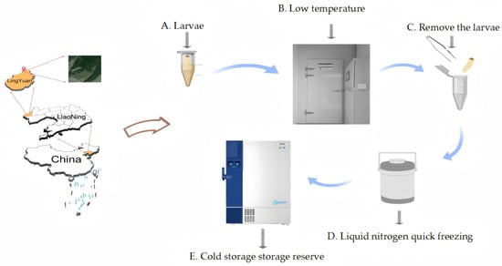
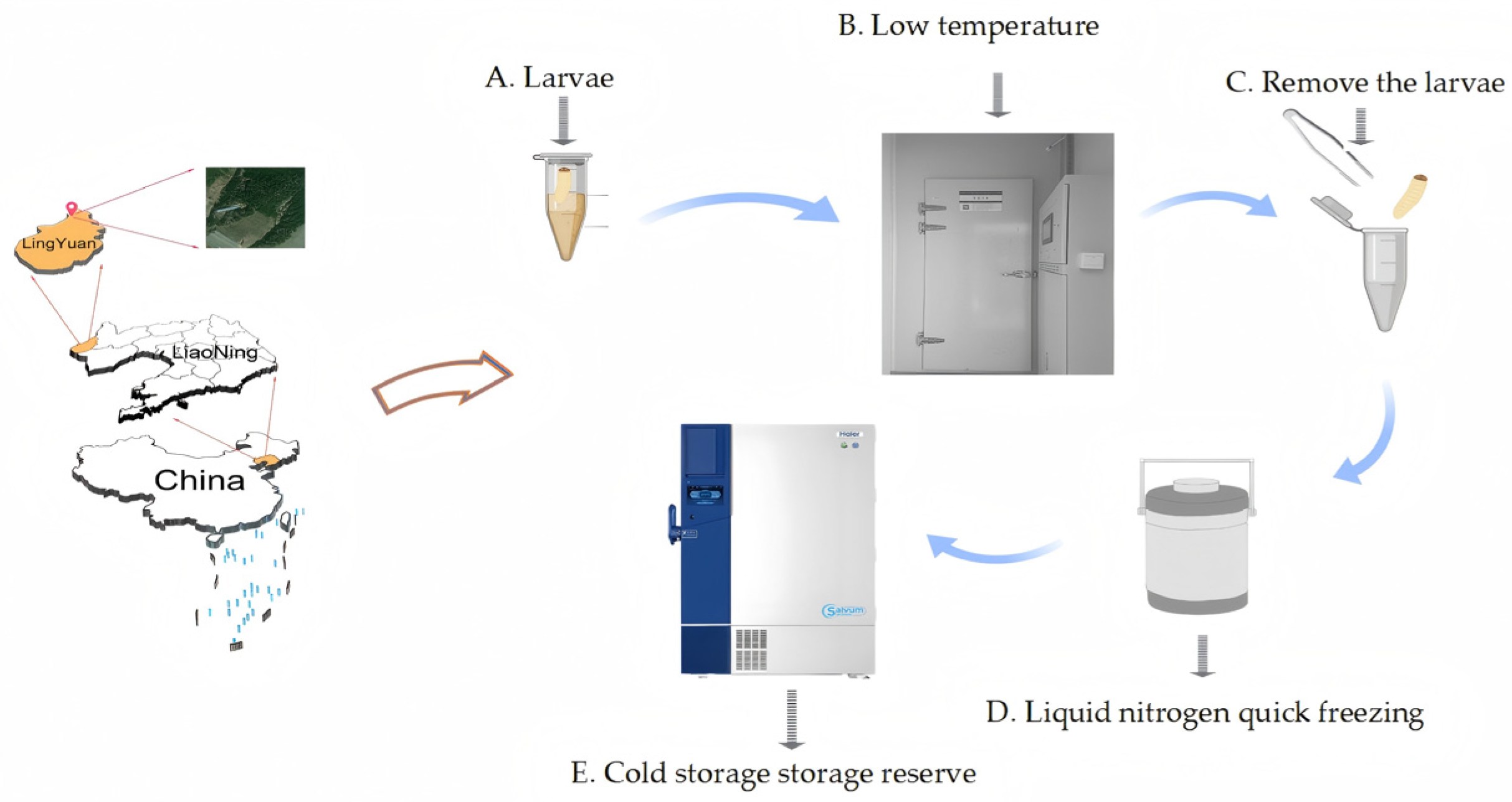
To mitigate the impact of individual variability on test outcomes, three larvae were pooled per sample at each time point in this investigation, with a total of three replicates. Subsequently, all samples were promptly frozen in liquid nitrogen and preserved at −80 °C for future analysis. The process of sample collection and processing for the experiment is presented (Figure 1).
Figure 1.
Sample collection and treatment.
2.4. Determination of Physiological and Biochemical Substances
The study examined the physiological adaptations and cold tolerance of cold stress and non-cold stress larvae at −4 °C. Water content and fat content were assessed using the drying method and vanillin phosphate chromogenic method, respectively. Additional physiological parameters were analyzed using the reagent kit (Sangon Biotech, Shanghai, China).
2.5. Transcriptome Analysis
Total RNA was extracted using the Qubit 2 RNA detection kit (Sangon Biotech, Shanghai, China), and 30 cDNA libraries were prepared, comprising 15 samples of non-cold stress larvae, 9 samples of cold stress larvae, 3 samples of cold stress-terminated larvae, and 3 samples of fully cold stress-terminated larvae. Subsequently, the libraries were sequenced on the MGISEQ-200 high-throughput sequencing platform from MGI Tech Co (Shenzhen, China). Data analysis was conducted by aligning the sequences to the genome, following the methodology outlined in the study by Liu Zhudong et al. [36].
2.6. Quantitative Real-Time PCR (qRT-PCR) Validation
Ten upregulated genes associated with larval cold stress were chosen for RT-qPCR validation, with TUB serving as the internal reference gene. The sequences and lengths of the genes and internal references can be found in Table 1. Reverse transcription of RNA was conducted utilizing the M-MuLV first strand cDNA synthesis kit (Sangon Biotech, Shanghai, China), and primers were designed using TBtools-II (Toolbox for Biologists) v2.357 software to confirm the fidelity of transcriptome data from cold stress and non-cold stress larvae of D. valens.
Table 1.
List of primers used for qRT-PCR.
2.7. Data Analysis
Physiological and biochemical measurements underwent statistical analysis using Excel 2018 and IBM SPSS Statistics 27. The analysis was conducted using the one-way ANOVA method. Graphs were generated using GraphPad Prism 9.1. Transcriptome analyses involved Excel 2018, DESeq2, KEGG Pathway, R (V3.6.2) ggplot2. Visualization was conducted using Wei Sheng Xin (www.bioinformations.com.cn) and GraphPad Prism 9.1. Use of differentially expressed genes DESeq2 (p < 0.05, |FC (foldchange)| ≥ 2) (https://www.omicshare.com/tools/, accessed on 13 January 2026). The log 2-fold change values in the graphs represent the expression levels of distinct differential genes.
3. Results
3.1. Determination of Cold Stress Temperature
Morphological analysis revealed significant differences in the characteristics of the larvae of the D. valens larvae when subjected to varying temperatures and processing durations. At 25 °C, the larvae exhibit heightened activity, characterized by vigorous wriggling. Their bodies appear milky white, adorned with prominent spots. In contrast, at 10 °C, 4 °C, the spots gradually diminish as the temperature decreases and the treatment duration increases, resulting in larvae that enter a semi-active state and remain stationary after 24 h. At temperatures of 0 °C and −4 °C, the larvae become stiff and quiescent, with their body color shifting to milky yellow. The spots on their surface are notably lighter than those of larvae at 25 °C. At −8 °C, the larvae succumb to death, and their body surface becomes extensively blackened (as shown in Figure 2).
Figure 2.
Morphological photos of D. valens under different temperature treatments in the laboratory. (A) larval morphology at 25 °C; (B) larval morphology at 10 °C, 4 °C; (C) larval morphology at 0 °C; (D) larval morphology at −4 °C; (E) larva morphology at −8 °C.
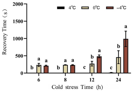
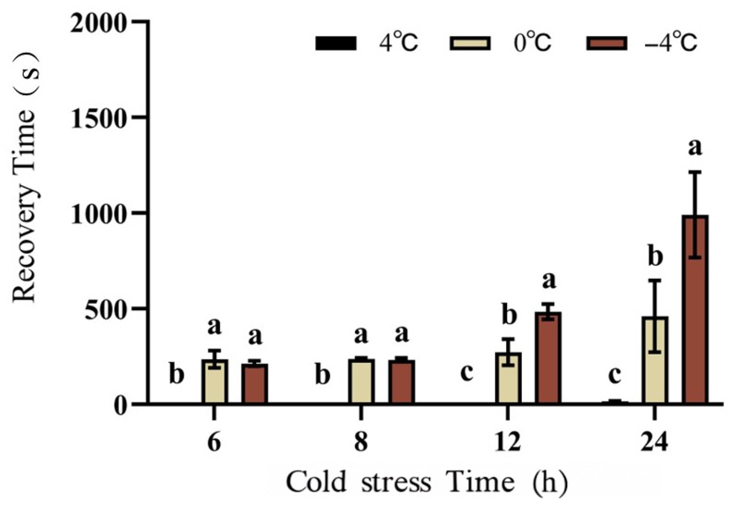
The induction effect of cold stress on the larvae of D. valens is most pronounced at −4 °C, as showen in Figure 3. At temperatures of 25 °C and 10 °C, the larvae are unable to enter the cold stress state. At 4 °C and 0 °C, the larvae remain stationary but exhibit a shorter recovery time when returned to a warmer environment. Consequently, these temperatures are not optimal for the larvae to enter cold stress. Under varying treatment conditions, only the larvae at 4 °C, 0 °C, and −4 °C exhibited a static state, with recovery times to an active feeding state of a maximum of 3.7 s, 460 s, and 1191 s, respectively (Figure 3). The findings of this study suggest that temperature plays a critical role in determining both the duration of cold stress and its subsequent release in the larvae of D. valens. As temperature decreases, the duration of cold stress in these larvae is significantly extended. Thus, temperature influences both the rate of cold stress development and the overall duration of cold stress in the larvae of D. valens. Cold stress favor the maintenance of cold stress, whereas higher temperatures facilitate its release.
Figure 3.
The recovery time of D. valens larvae under different temperature treatments in the laboratory, “h” represents hours, and “s” represents seconds. a, b, c indicate statistically significant differences. The same letter indicates no significant difference between two or more sets of data, while different letters indicate a significant difference between two sets of data.
3.2. Changes in Water Content in Larvae
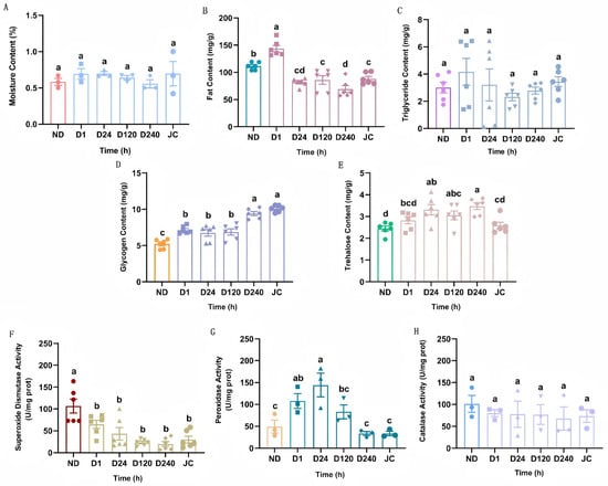
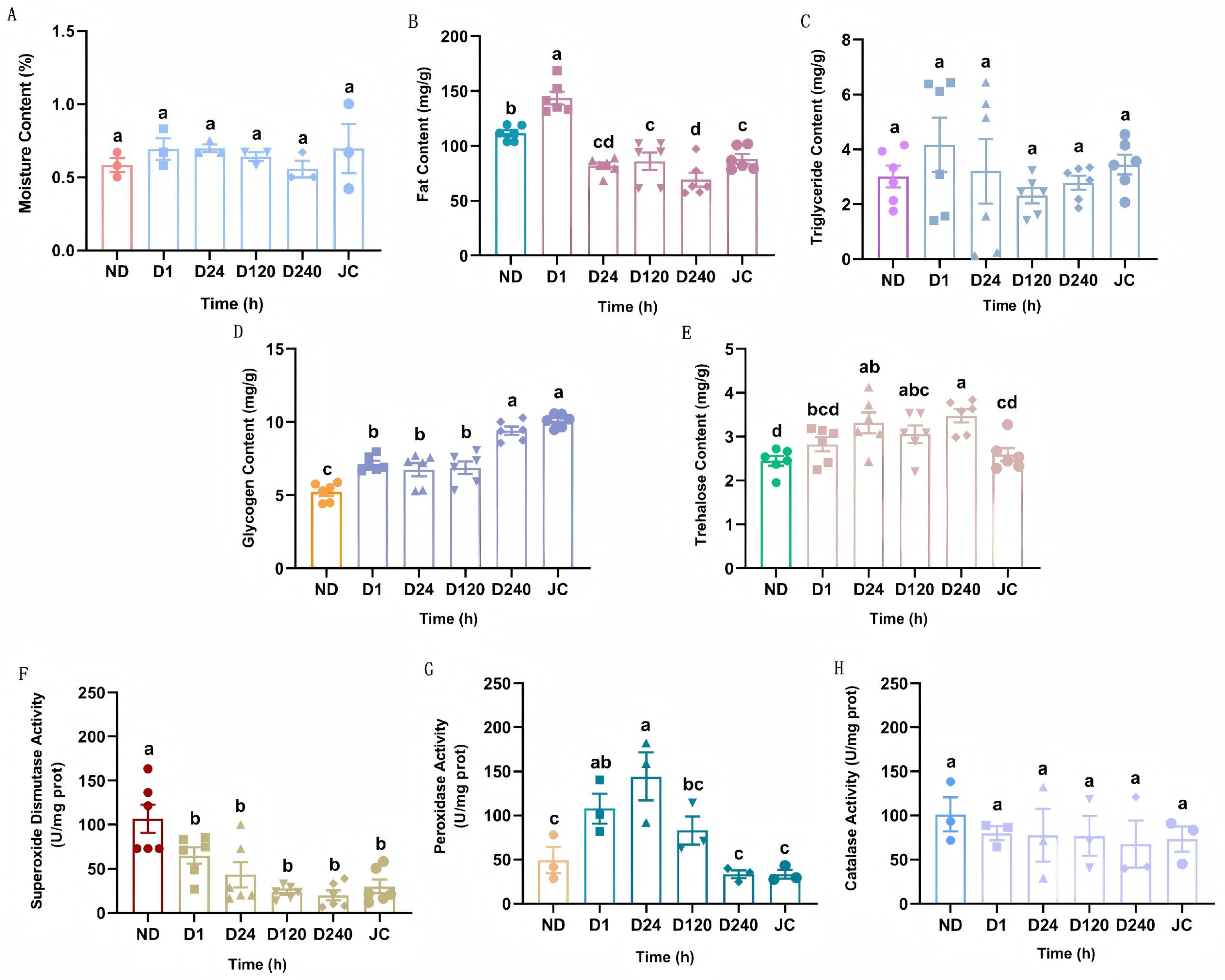
Figure 4A displays the fluctuations in larval water content during cold stress induction, showing no significant differences between the control group and each time point (p > 0.05). Larval water content ranged from 56% to 70% throughout cold stress induction. Despite surpassing the control group’s water content at the 3 h post-cold stress mark, this disparity was not statistically significant (p > 0.05). Examination of water content dynamics in D. valens larvae revealed that non-cold stress larvae predominantly obtained water through feeding. The stable water content in cold stress larvae played a critical role in increasing body fluid concentration and enhancing cold tolerance, facilitating a seamless progression through the entire cold stress period.
Figure 4.
Changes in physiological and biochemical indicators of. D. valens larvae under cold stress induction conditions. (A): Molsture Content; (B): Fat Content; (C): Triglyceride Content; (D): Glycogen Content; (E): Trehalose Content; (F): Superoxide Dismutase Activity; (G): Peroxidase Activity; (H): Catalase Activity. “h” represents hours. Reference for the precise value can be found in Supplementary Material Table S1. a, b, c indicate statistically significant differences. The same letter indicates no significant difference between two or more sets of data, while different letters indicate a significant difference between two sets of data.
3.3. Changes in Energy Substances in Larvae
The lipid levels in D. valens larvae exhibited a gradual decline during cold stress initiation (Figure 4B). Initially, at 1 h of cold stress induction, the lipid content was notably higher compared to the control group; however, it significantly decreased by 24 h. Subsequently, following cold stress termination, the lipid content in the larvae significantly rose to 71.35 mg/g at 3 h, surpassing the levels observed at 240 h post-cold stress induction (p < 0.05).
In D. valens larvae, the induction of cold stress led to an initial decline followed by a rise in triglyceride levels (Figure 4C). One hour into cold stress induction, triglyceride levels were elevated in larvae compared to the control group, albeit without statistical significance. Following this, triglyceride levels marginally rose to 3.44 mg/g after 3 h of cold stress termination, exceeding the levels in larvae induced for 240 h, with no significant distinction observed (p > 0.05).
During cold stress induction, larval glycogen content increased significantly compared to the control group (Figure 4D), reaching 9.41 mg/g at 240 h. Following cold stress termination, glycogen content further increased to 10.01 mg/g at 3 h, significantly surpassing levels observed in both the control group and other treatment groups (p < 0.05).
During cold stress induction, larvae showed a gradual rise in trehalose levels (Figure 4E). Trehalose levels at 24 h, 120 h, and 240 h were notably higher compared to the control group (p < 0.05). After 240 h of cold stress induction, protein levels reached a peak of 3.47 mg/g, decreasing to a minimum of 2.59 mg/g at 3 h post-cold stress termination, with no significant difference from the control group (p < 0.05).
3.4. Changes in Enzyme Activities in Larvae
During the initiation of cold stress, the activity of the SOD protective enzyme in larvae exhibited an initial decrease followed by an increase (Figure 4F). This activity was notably lower than that observed in the control group (p < 0.05). Specifically, after 240 h of cold stress induction, the SOD protective enzyme activity reached a minimum value of 19.95 U/mg, which subsequently rose to 29.98 U/mg after 3 h of cold stress termination. Notably, the activity at 3 h post cold stress termination was higher than that recorded at 240 h of cold stress induction (p > 0.05).
During cold stress induction, the activity of the protective enzyme peroxidase (POD) in larvae initially increased and then declined over time (Figure 4G). Specifically, the POD enzyme activity in larvae induced for 1 h and 24 h was significantly elevated compared to the control group. The enzyme activity peaked at 144.06 U/mg after 24 h of cold stress induction, surpassing that of the control group at 120 h, albeit without statistical significance (p > 0.05). Subsequently, at 240 h of cold stress induction, the POD enzyme activity commenced decreasing, reaching a nadir of 33.32 U/mg.
During the induction of cold stress, the activity of the CAT enzyme in larvae decreased, as depicted in Figure 4H. Although not statistically significant (p > 0.05), the enzyme activity in the treatment group was lower than that in the control group. Specifically, CAT enzyme activity decreased to 67.68 U/mg after 240 h of cold stress induction and rose to 73.14 U/mg 3 h after cold stress termination. While the enzyme activity at 3 h post-cold stress termination exceeded that of larvae induced at 240 h of cold stress induction, once again, the difference was not statistically significant (p > 0.05).
3.5. Transcriptome Data Quality Control
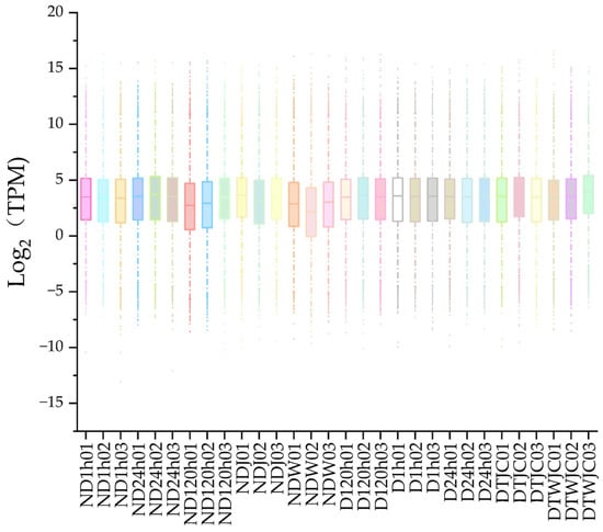
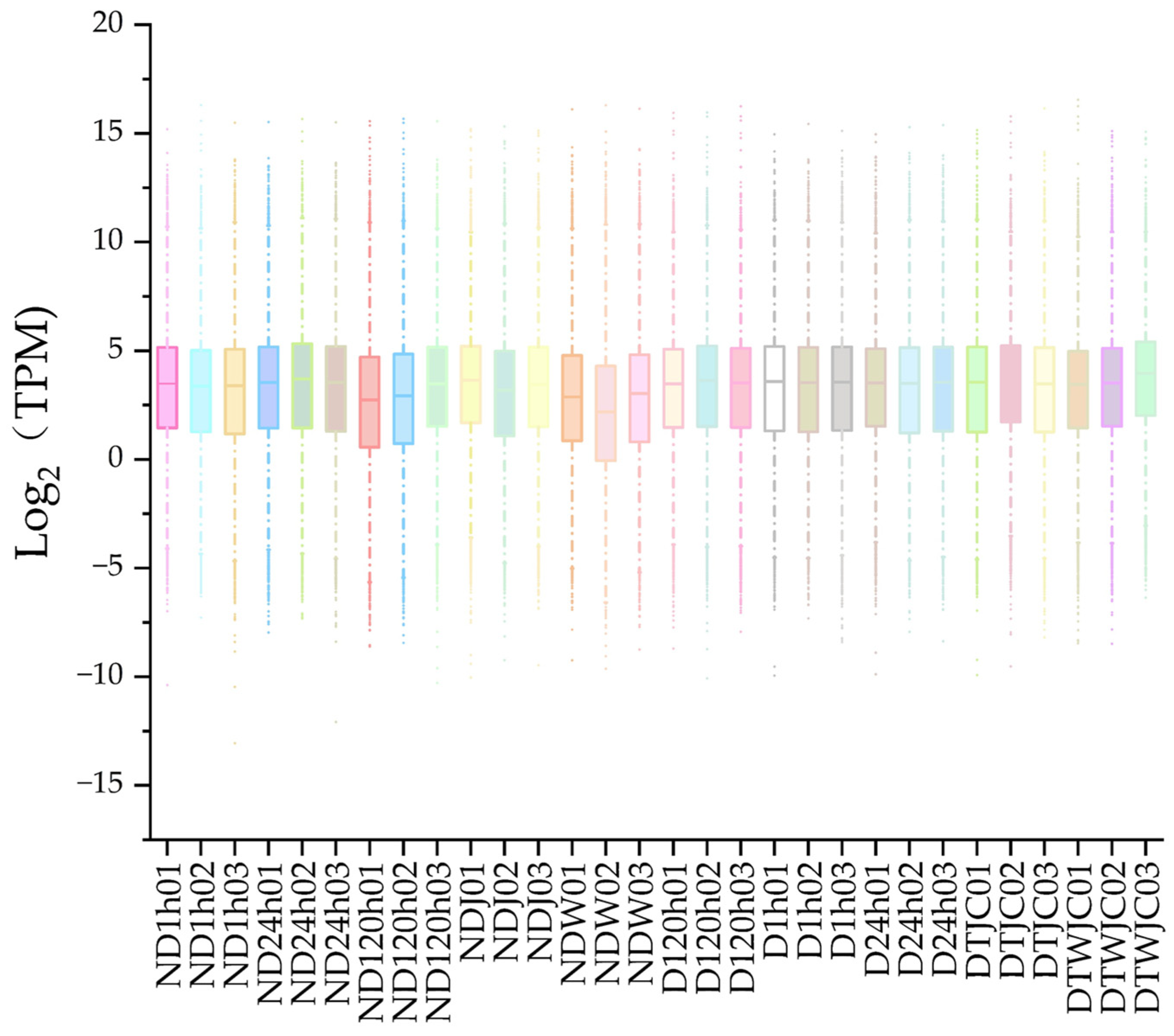
A total of 1,969,795,408 high-quality sequencing reads were acquired from 30 larval samples of D. valens following rigorous filtering and assembly. The Q30 base percentage surpassed 97% across all samples, with GC content varying from 45.25% to 49.98%. These parameters validate the reliability of the sequencing data, establishing its appropriateness for subsequent functional annotation and analysis. Visualization of gene expression levels among the samples was conducted using a boxplot (Figure 5). The notable similarity and strong correlation observed among biological replicates highlight the overall high quality of the transcriptome data, confirming its suitability for further analyses.
Figure 5.
Gene expression. Box plot of gene expression.
3.6. Analysis of Differentially Expressed Genes in Larvae
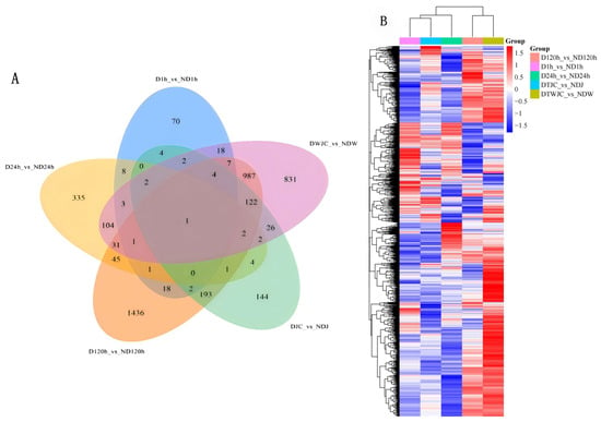
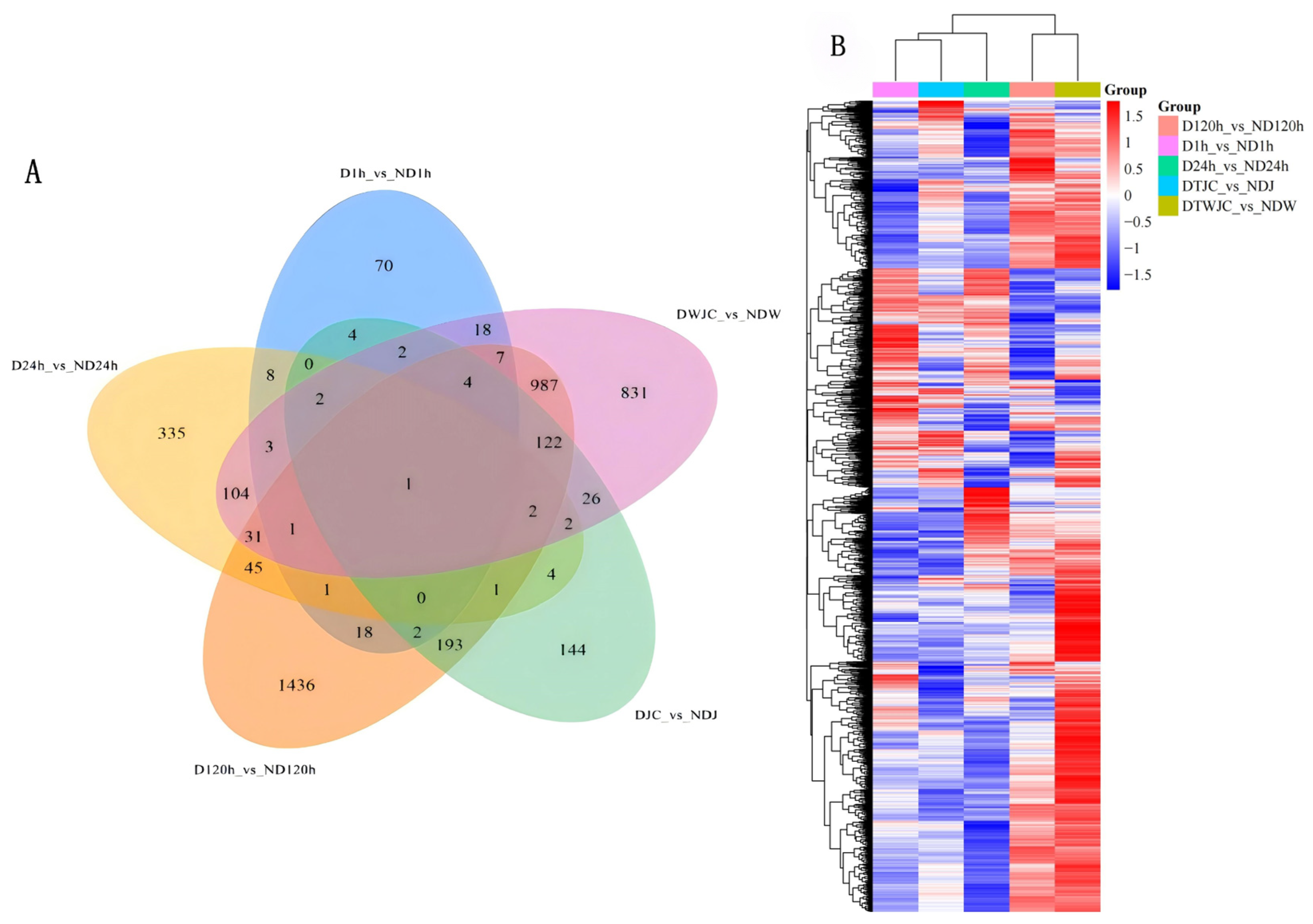
To investigate the cold stress-specific transcriptional alterations induced by cold stress in D. valens larvae, we conducted pairwise comparisons across distinct libraries to pinpoint differentially expressed genes. Figure 6 illustrates a total of 4404 differentially expressed genes identified across the four groups. Specifically, 141 genes exhibited differential expression between D1h and ND1h, with 70 genes being uniquely differentially expressed. DESeq analysis revealed 540 and 2851 differentially expressed genes at D24h and ND24h, as well as D120h and ND120h, respectively. Among these, 335 and 1436 genes were uniquely differentially expressed, respectively. Furthermore, 509 and 2143 genes displayed differential expression between DTJC and NDJ, and DTWJC and NDW, with 144 and 831 genes being unique to DTJC and NDJ, respectively. Through Venn diagram analysis, we observed that 1841 genes underwent changes during cold stress development without concurrent alterations in normal development.
Figure 6.
Differentially expressed genes in four treatments in the laboratory (A): Venn diagram; (B): Heat map. In the heat map, red indicates high expression. Blue indicates low expression.
3.7. Enrichment Analysis of Differentially Expressed Gene KEGG
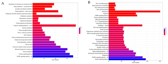
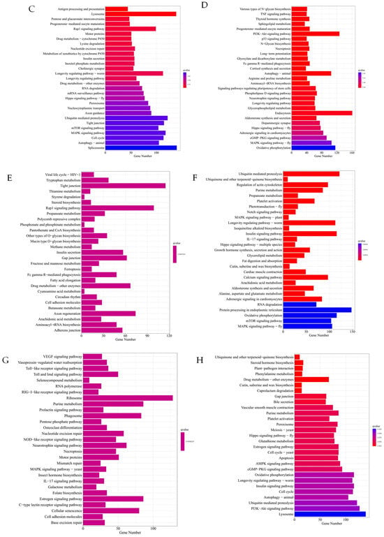
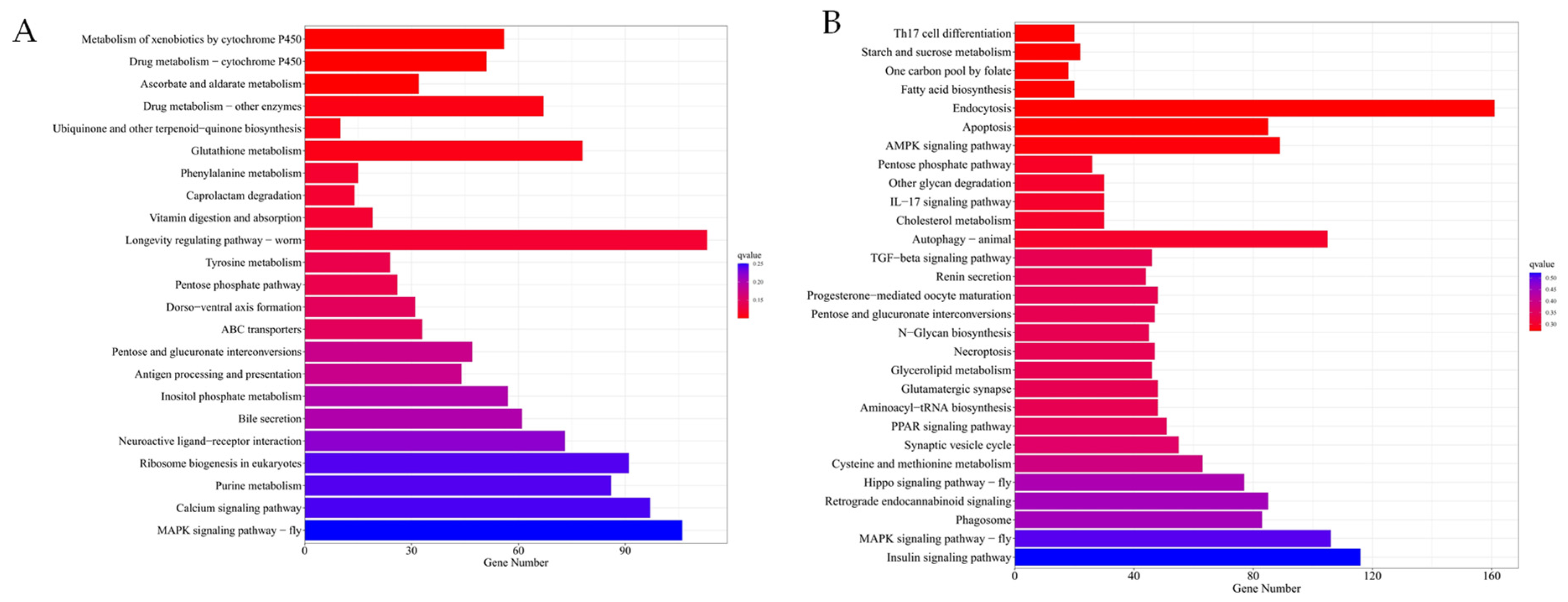
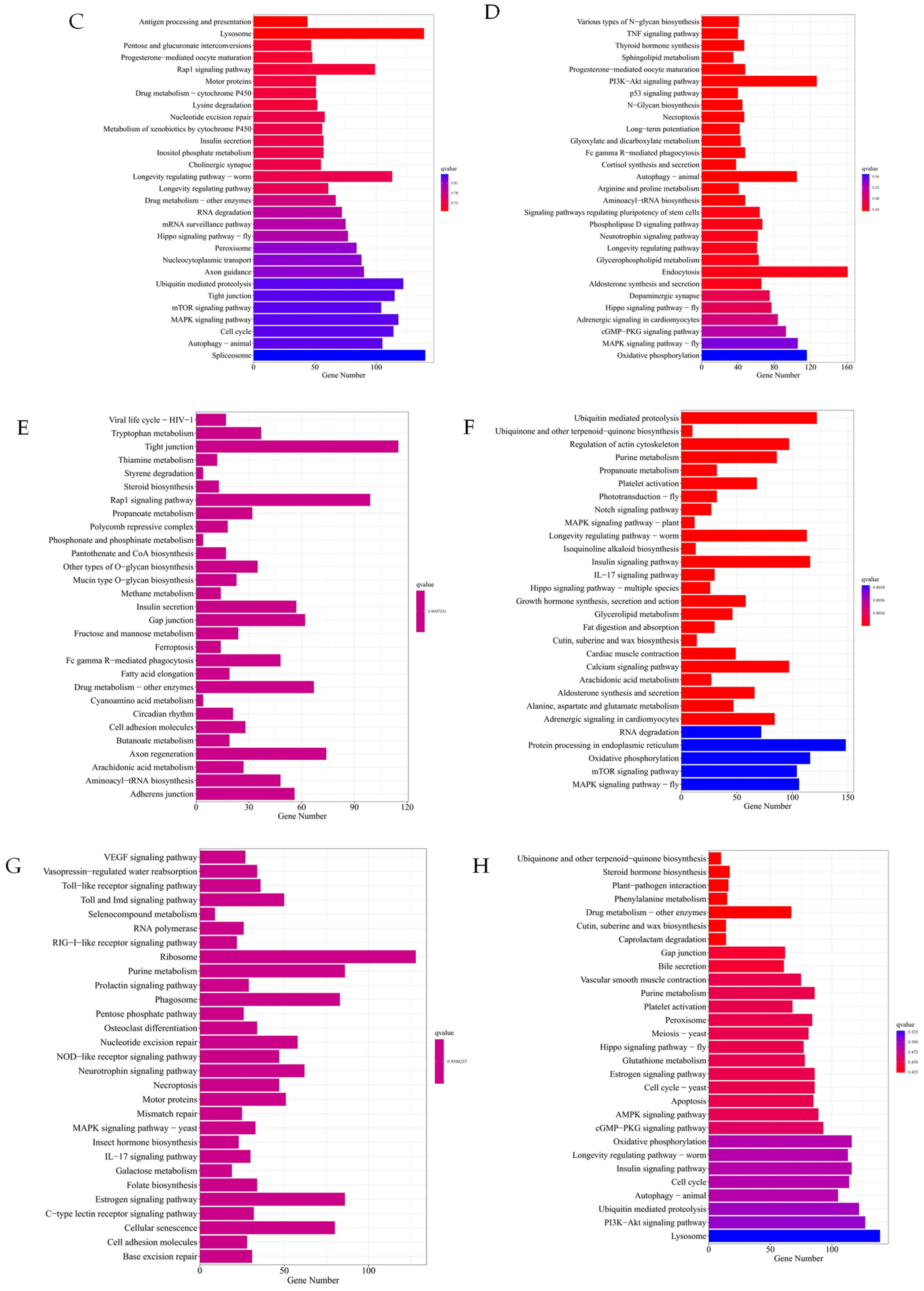
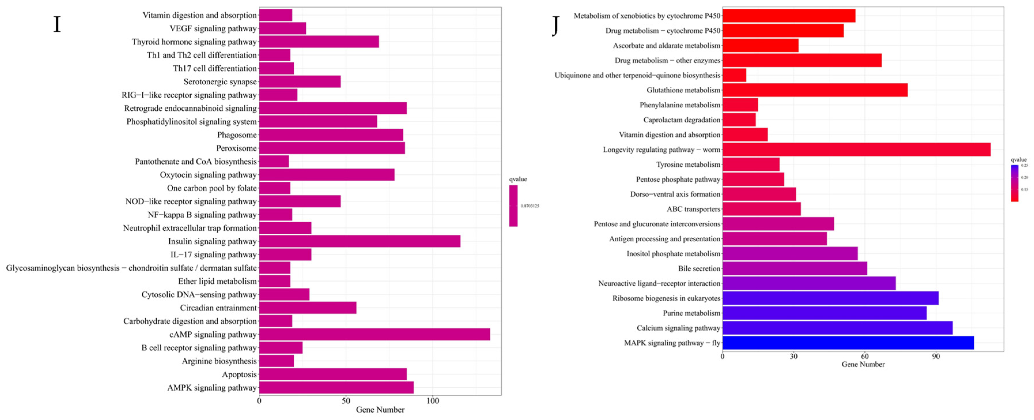
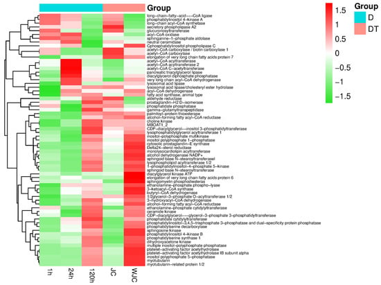
To determine the potential function of the identified differentially expressed gene (DEG), we performed KEGG enrichment analysis. With the exception of D1h and ND1h, up-regulated genes predominated over down-regulated genes in all comparisons. Notably, the difference in the number of up-regulated and down-regulated genes was minimal between the D120h and ND120h comparison groups. A significant proportion of differentially expressed genes in various groups were consistently associated with amino acid metabolism, lipid metabolism, and carbohydrate pathways, as illustrated in Figure 7. Subsequent analyses were based directly on these results.
Figure 7.
Differential expression analysis of KEGG. (A,B) The 1 h up-regulation metabolic pathway; (C,D) 24 h up-regulation metabolic pathway; (E,F) 120 h up-regulation metabolic pathway; (G,H) Up-regulated metabolic pathway of initial cold stress release, (I,J) Up-regulated metabolic pathway of complete cold stress release.
3.8. Comparative Analysis of Cold Stress Related Genes
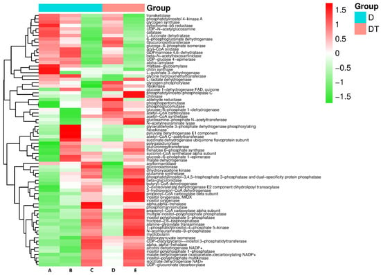
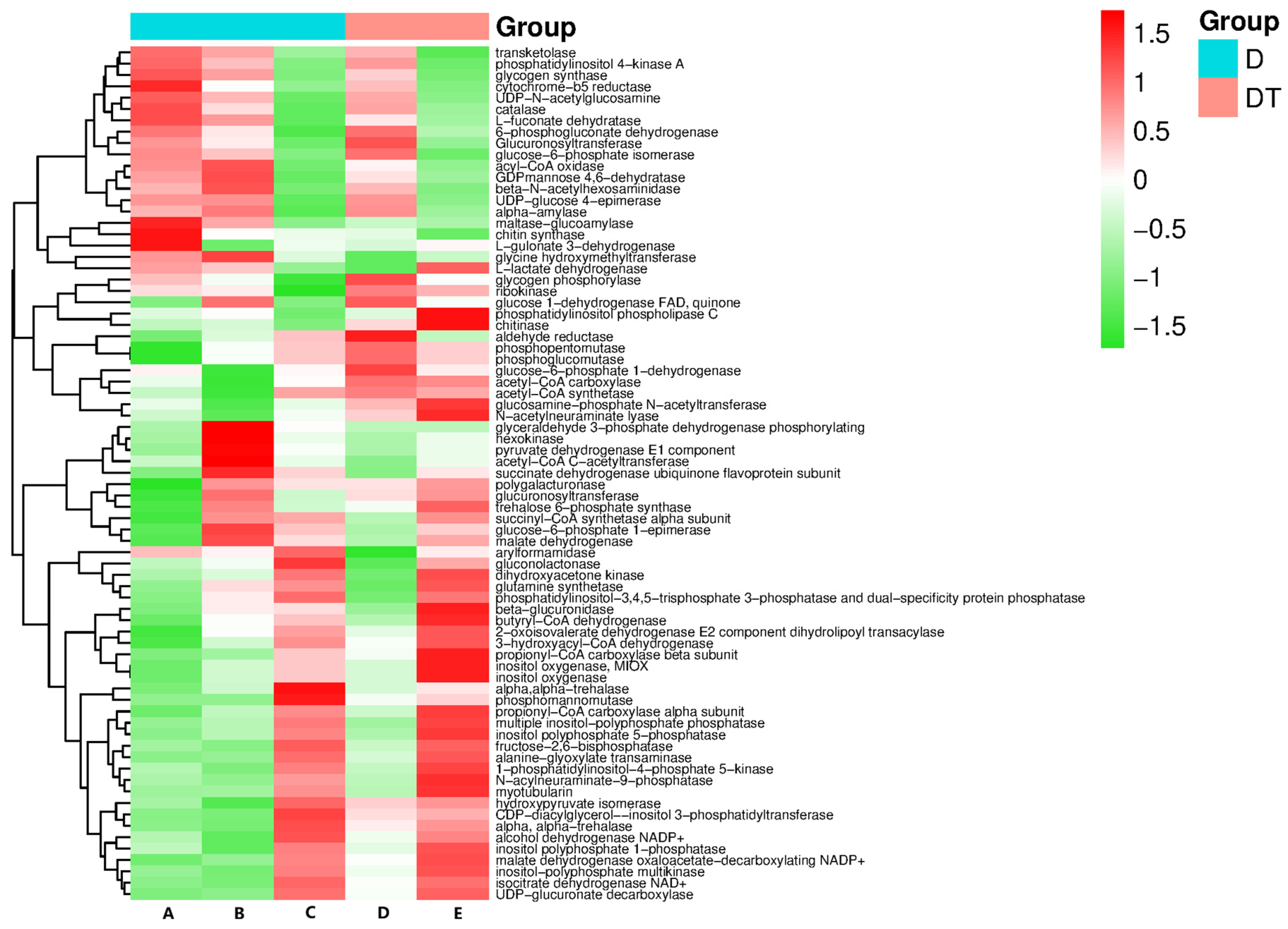
The KEGG enrichment analysis revealed pathways and enriched genes associated with carbohydrate metabolism, lipid metabolism, and amino acid metabolism. Figure 8 provides a detailed overview of the results for carbohydrate metabolism. In this pathway, 86 genes were up-regulated, while 64 genes were down-regulated. The up-regulated genes primarily included phosphomannose mutases (PMM), L-gulonate 3-dehydrogenase (CRYL1), and hexokinase (HK). Conversely, the down-regulated genes mainly consisted of polygalacturonase (E3.2.1.15), alcohol dehydrogenase NADP+ (adh), among others. Notably, the propionyl-CoA carboxylase β subunit (PCCB) exhibited significant upregulation during both initial and complete cold stress termination. Conversely, genes such as β-glucuronidase (GUSB), beta-N-acetylhexosaminidase (nagZ), and phosphatidylinositol 4-kinase A (PI4KA) were significantly down-regulated in this context.
Figure 8.
Carbohydrate metabolic pathway enrichment analysis. A. Differential gene expression at 1 h of cold stress; B. Differential gene expression at 24 h of cold stress; C. Differential gene expression at 120 h of cold stress; D. Differential gene expression at early cold stress termination; E. Differential gene expression at complete cold stress termination.
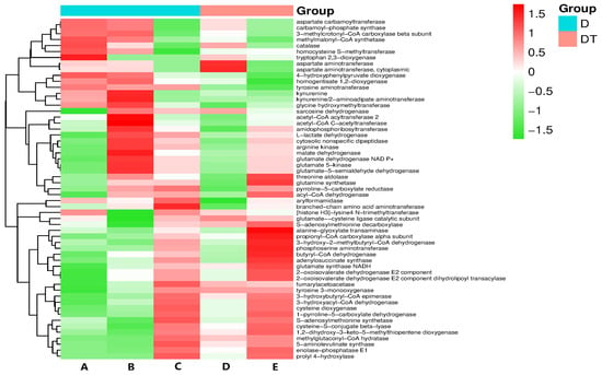
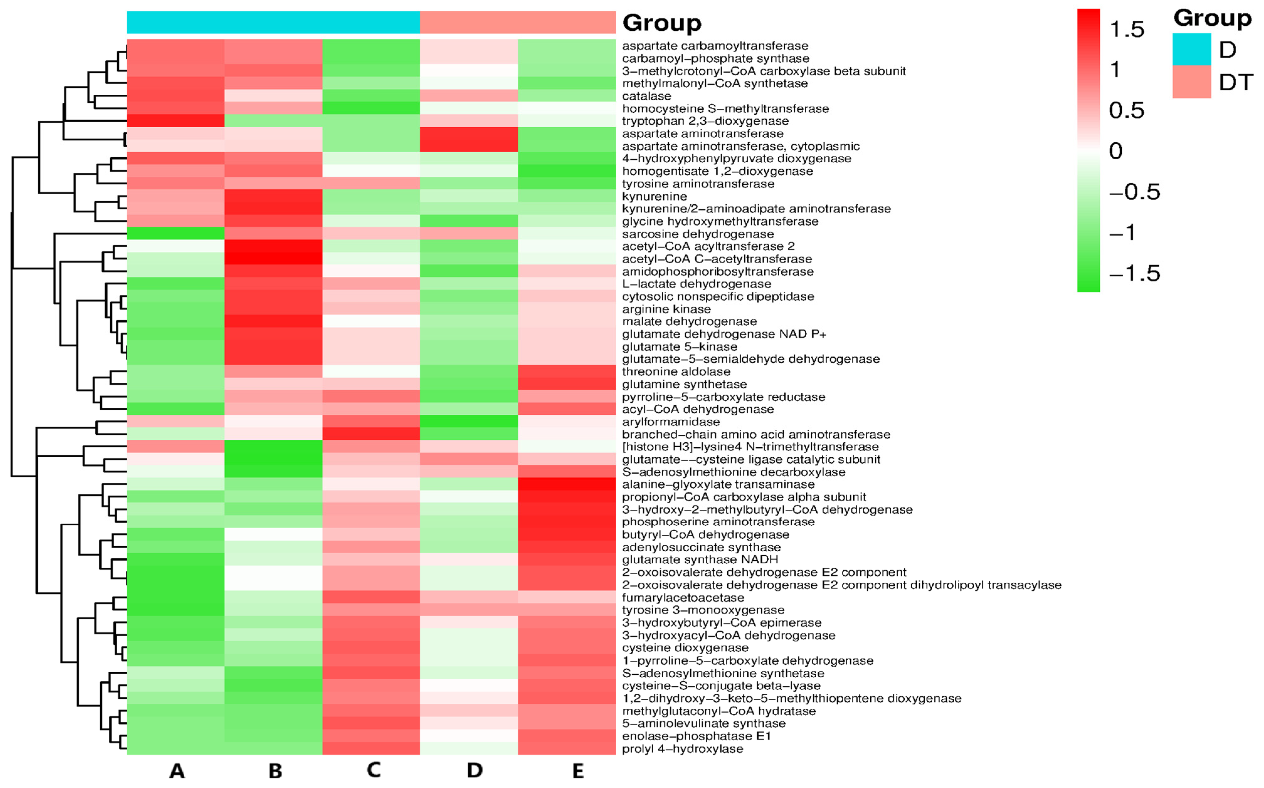
As depicted in Figure 9, in the context of amino acid metabolism, 63 genes exhibited up-regulation, whereas 26 genes displayed down-regulation. Particularly noteworthy was the significant up-regulation of malate dehydrogenase (mdh) during cold stress. Among the prominently up-regulated genes was 5-aminokonoglutarate synthase (5-aminolevulinate synthase, E2.3.1.37). Conversely, genes demonstrating notable downregulation included S-adenosylmethionine decarboxylase (speD) and 4-hydroxyphenylpyruvate dioxygenase (HPD). Isogenic tyrosine 3-monooxygenase (TH) showed significant up-regulation both at the onset and conclusion of cold stress, while 3-hydroxyacyl-CoA dehydrogenase (HADH) primarily contributed to the downregulation of HPD, homogentisate 1,2-dioxygenase (HGD), and other genes.
Figure 9.
Enrichment analysis of amino acid metabolic pathway. A. Differential gene expression at 1 h of cold stress; B. Differential gene expression at 24 h of cold stress; C. Differential gene expression at 120 h of cold stress; D. Differential gene expression at early cold stress termination; E. Differential gene expression at complete cold stress termination.
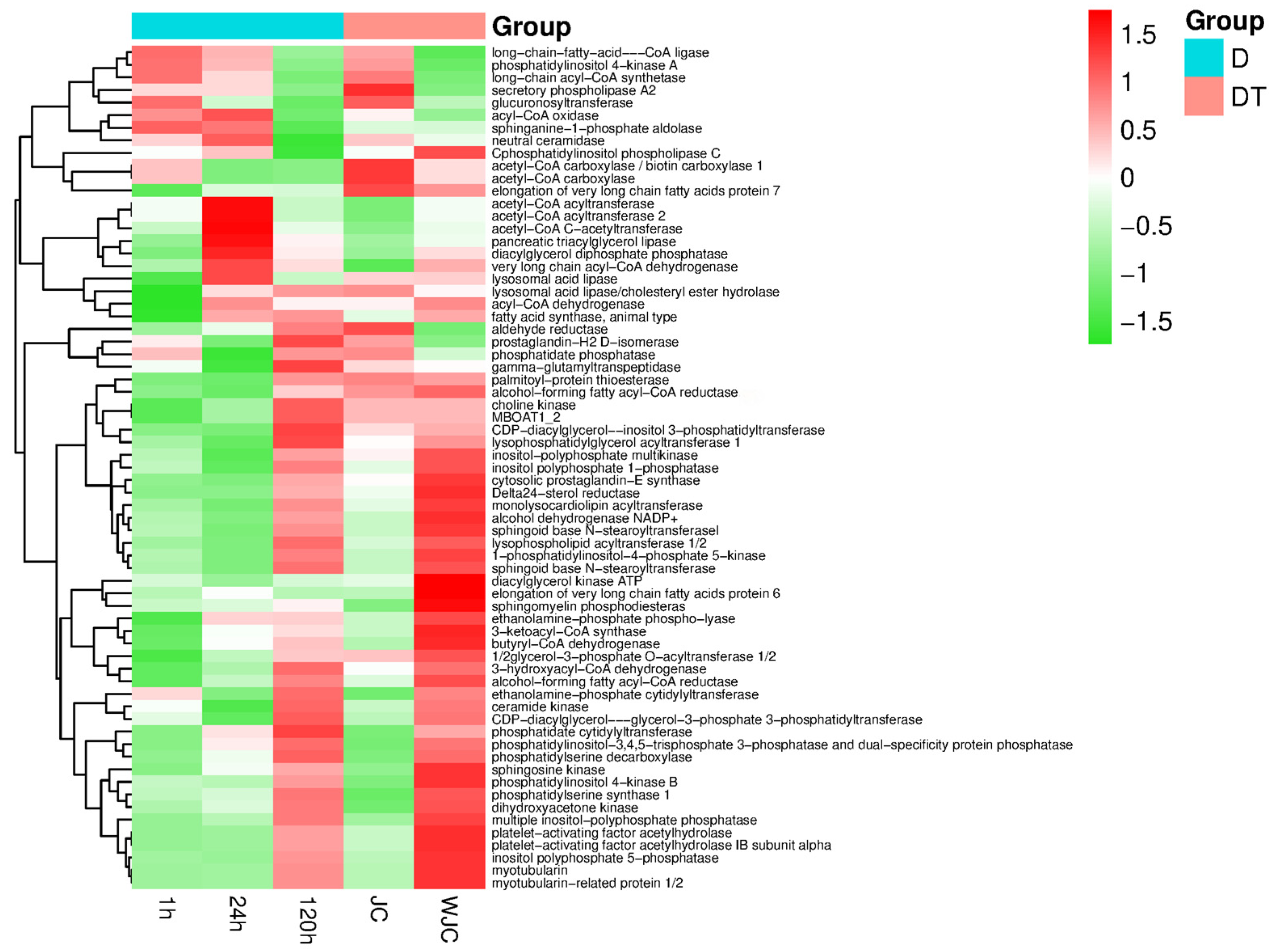
In Figure 10, 69 genes were up-regulated and 57 genes were down-regulated in lipid metabolism during cold stress. Specifically, CDP-diacylglycerol-inositol 3-phosphate transferase (CDIPT) exhibited significant up-regulation. Conversely, aldehyde reductase (AKR1B) and other genes were notably down-regulated, particularly lysosomal acid lipase (LIPA) and glycerol-3-phosphate O-acyltransferase 1/2 (GPAT 3_4). Sphingomyelin phosphodiesterase (SMPD1) showed significant upregulation at the onset and completion of cold stress termination. Conversely, butyryl-CoA dehydrogenase (ACADS) and other genes experienced significant downregulation, notably involving genes such as phosphatidylinositol 4-kinase A (PI4KA) and prostaglandin-H2 D-isomerase (PTGDS).
Figure 10.
Enrichment analysis of lipid metabolism pathway. 1 h—Differential gene expression at 1 h of cold stress; 24 h—Differential gene expression at 24 h of cold stress; 120 h—Differential gene expression at 120 h of cold stress; JC—Differential gene expression at early cold stress termination; WJC—Differential gene expression at complete cold stress termination.
3.9. Real-Time Quantitative PCR
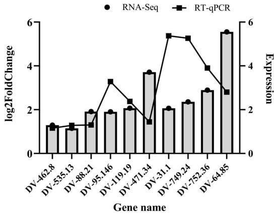
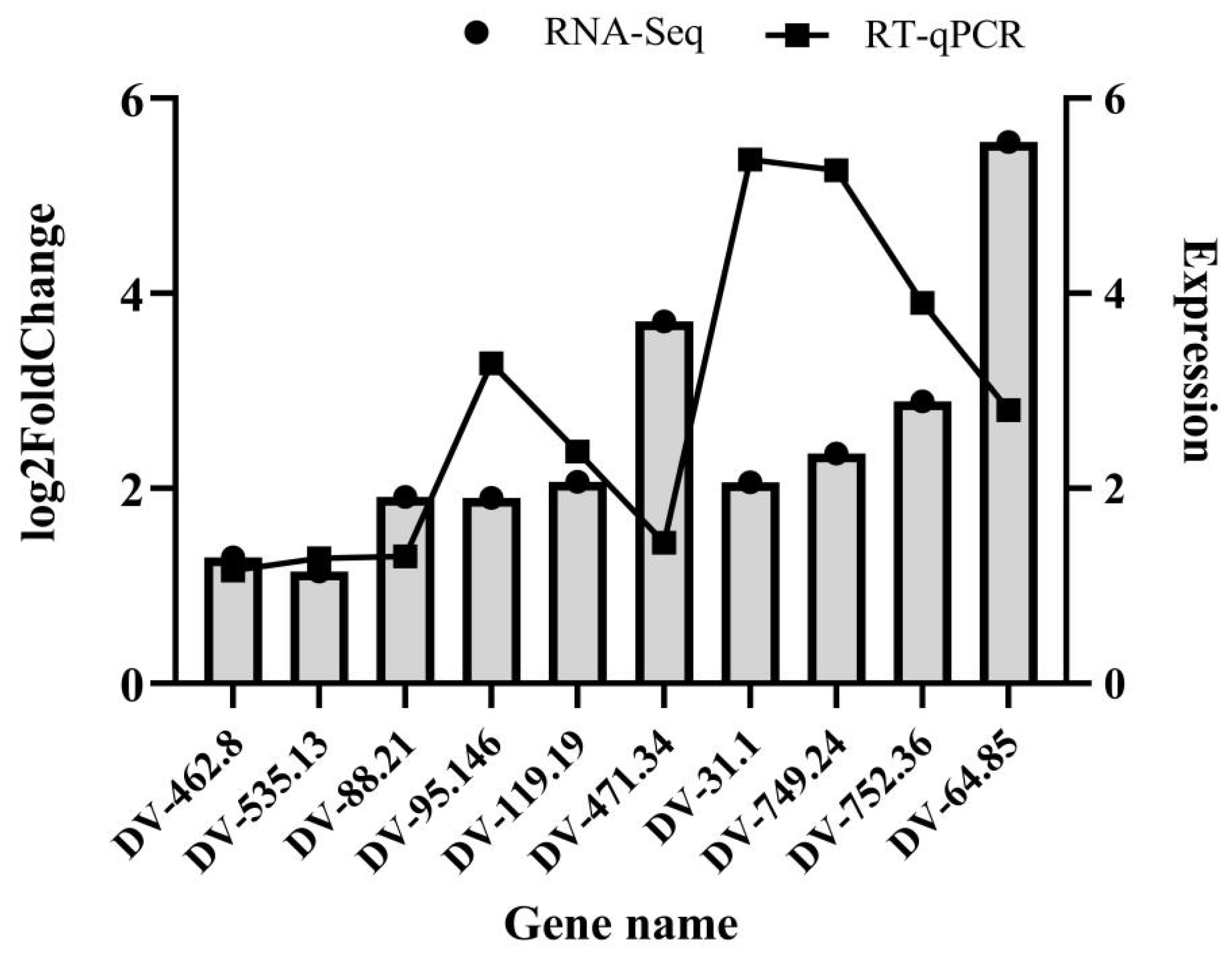
Genes crucial for carbohydrate, amino acid, and lipid metabolism were selected for validation using real-time quantitative PCR (qRT-PCR) following an analysis of differential gene expression (Figure 11). The qPCR results consistently aligned with the transcriptome data, confirming the accuracy of the transcriptome data. Specifically, DeoB, FadA, and DNM13 showed reduced expression levels during cold stress, in contrast to the heightened expression levels seen in other genes. Additionally, most genes exhibited a gradual decrease in expression levels with prolonged cold stress duration.
Figure 11.
qRT-PCR validation of RNA-seq data for differential genes.
4. Discussion and Conclusions
This study elucidated the physiological, biochemical, and molecular mechanisms underlying the regulation of cold stress in D. valens larvae under low-temperature stress. Our investigation not only delineated the dynamic patterns of energy metabolism and oxidative stress response during cold stress but also identified crucial metabolic pathways and genes that govern this intricate biological process through transcriptomic analysis. These findings offer a provides insights for comprehending the suggests potential mechanisms of insect cold stress and hold practical implications for the targeted management of D. valens.
4.1. Energy Metabolism Remodeling: Strategic Balance Between Cold Stress Maintenance and Release
During cold stress induction, triglycerides and fats were utilized as primary energy storage sources, while glycogen and trehalose levels increased. This observation contrasts with the traditional cold stress energetics model, where organisms accumulate lipids extensively in the pre-cold stress stage and gradually consume them during cold stress [37]. Lipids are typically favored for long-term cold stress energy storage due to their high energy density [38]. We postulated that the unconventional energy utilization observed in larvae of D. valens was influenced by the cold stress (−4 °C). Under freezing temperatures, trehalose, acting as an efficient cryoprotectant, serves a dual role beyond its energy provision. Its increased presence likely represents a direct adaptive response to cold stress, aiding in stabilizing cell membranes and proteins to mitigate ice crystal damage [39,40]. The metabolic shift reflects a strategic equilibrium between the larva’s requirements for energy supply and cold survival. While high-energy lipid reserves sustain basal metabolism, the synthesis of cryoprotectants like trehalose takes precedence. This discovery enhances our comprehension of the energy metabolism dynamics during extreme cold-induced cold stress.
Transcriptome data provides molecular evidence supporting the downregulation of key lipid metabolism genes like FASN and the upregulation of lipid catabolic genes such as ACADM, which collectively drive lipid consumption. Simultaneously, the downregulation of otsA, a crucial gene in trehalose synthesis in carbohydrate metabolism, may indicate that trehalose synthesis is completed early on or that its degradation pathway, rather than the synthetic pathway, is transcriptionally inhibited, leading to net accumulation. These intricate regulatory networks necessitate further analysis through advanced techniques like metabolic flow analysis.
4.2. Oxidative Stress Defense: Paradigm from Preadaptation to Dynamic Regulation
The fluctuating activities of three protective enzymes (SOD, POD, CAT) demonstrate a nuanced defense mechanism against oxidative stress [41,42]. Initially, SOD activity decreased, followed by an increase, indicating a sharp decline in metabolic rate and reduced ROS production during the early cold stress stage, leading to a temporary dormancy in the antioxidant defense system. As cold stress progressed, ROS accumulation due to cold stress and other stressors prompted a secondary activation of the defense system. The rise and subsequent fall in POD activity may correspond to the specific peroxide scavenging requirements at distinct stages. The consistent decline in CAT activity warrants further investigation. The contrasting findings in this study suggest that D. valens larvae may employ unique antioxidant mechanisms.
4.3. Multidimensional Metabolic Pathway Coordination: Molecular Hubs for Cold Stress
An essential innovative contribution of our study is the simultaneous integration of lipid, amino acid, and carbohydrate metabolic pathways within the cold stress regulatory network through KEGG enrichment analysis. While prior research has concentrated on individual metabolites [43,44,45,46,47], our study distinctly demonstrates that the effective initiation, sustenance, and termination of cold stress are contingent upon the synchronized functionality of these three pathways.
The significant role of amino acid metabolism is evident in the enrichment of differentially expressed genes related to it. For example, the upregulation of gdhA, which connects amino acid metabolism with the tricarboxylic acid cycle, indicates that amino acids may serve not only as substrates for protein synthesis but also as carbon skeletons for trehalose or glycogen synthesis via glycogenesis during cold stress [45,48]. Additionally, amino acids may directly act as alternative energy sources [44]. These findings offer valuable molecular insights into the mechanisms underlying the sustained yet limited energy supply during cold stress.
Metabolic flexibility plays a crucial role in terminating cold stress, as the rapid reversal in the expression of key genes occurs during this process. For instance, the upregulation of CAT and G6PD, which are pivotal enzymes in the pentose phosphate pathway, signifies the swift resumption of metabolic activities. This upregulation facilitates the production of NADPH, essential for supporting the reinitiation of biosynthetic processes and maintaining redox equilibrium.
Supplementary Materials
The following supporting information can be downloaded at: https://www.mdpi.com/article/10.3390/f17010136/s1.
Author Contributions
Conceptualization, D.L. and S.L.; methodology, D.L. and S.K.; software, H.C.; validation, D.L., S.L. and S.K.; formal analysis, Y.W.; investigation, B.D.; resources, S.J.; data curation, S.K.; writing—original draft preparation, B.D.; writing—review and editing, S.L.; visualization, B.D.; supervision, D.L.; project administration, S.J.; funding acquisition, S.J. All authors have read and agreed to the published version of the manuscript.
Funding
This research received no external funding.
Data Availability Statement
The original contributions presented in the study are included in the article and Supplementary Materials, further inquiries can be directed to the corresponding author.
Acknowledgments
We would like to express our sincere gratitude to the Liaoning Provincial Key Laboratory of Dangerous Forest Pest Management and Control for providing the experimental site and experimental equipment.
Conflicts of Interest
The authors declare that the research was conducted in the absence of any commercial or financial relationships that could be construed as a potential conflict of interest.
References
- Xu, D.; Xu, L.; Zhou, F.; Wang, B.; Wang, S.; Lu, M.; Sun, J. Gut Bacterial Communities of Dendroctonus valens and Monoterpenes and Carbohydrates of Pinus tabuliformis at Different Attack Densities to Host Pines. Front. Microbiol. 2018, 9, 1251. [Google Scholar] [CrossRef] [Scilit]
- Zheng, C.; Zhao, D.; Xu, Y.; Shi, F.; Zong, S.; Tao, J. Reference Gene Selection for Expression Analyses by qRT-PCR in Dendroctonus valens. Insects 2020, 11, 328. [Google Scholar] [CrossRef] [Scilit] [PubMed]
- Xu, L.; Sun, L.; Zhang, S.; Wang, S.; Lu, M. High-Resolution Profiling of Gut Bacterial Communities in an Invasive Beetle using PacBio SMRT Sequencing System. Insects 2019, 10, 248. [Google Scholar] [CrossRef] [Scilit] [PubMed]
- Zhao, D.; Zheng, C.; Shi, F.; Xu, Y.; Zong, S.; Tao, J. Expression analysis of genes related to cold tolerance in Dendroctonus valens. PeerJ 2021, 9, e10864. [Google Scholar] [CrossRef] [Scilit] [PubMed]
- Yan, Z.; Sun, J.; Don, O.; Zhang, Z. The red turpentine beetle, Dendroctonus valens LeConte (Scolytidae): An exotic invasive pest of pine in China. Biodivers. Conserv. 2005, 14, 1735–1760. [Google Scholar] [CrossRef] [Scilit]
- Taerum, S.J.; Duong, T.A.; de Beer, Z.W.; Gillette, N.; Sun, J.H.; Owen, D.R.; Wingfield, M.J. Large shift in symbiont assemblage in the invasive red turpentine beetle. PLoS ONE 2013, 8, e78126. [Google Scholar] [CrossRef] [Scilit]
- Sun, J.; Lu, M.; Gillette, N.E.; Wingfield, M.J. Red turpentine beetle: Innocuous native becomes invasive tree killer in China. Annu. Rev. Entomol. 2013, 58, 293–311. [Google Scholar] [CrossRef] [Scilit]
- Wang, Z.; Zhou, Q.; Zheng, G.; Fang, J.; Han, F.; Zhang, X.; Lu, Q. Abundance and Diversity of Ophiostomatoid Fungi Associated with the Great Spruce Bark Beetle (Dendroctonus micans) in the Northeastern Qinghai-Tibet Plateau. Front. Microbiol. 2021, 12, 721395. [Google Scholar] [CrossRef] [Scilit]
- Wang, B.; Salcedo, C.; Lu, M.; Sun, J. Mutual interactions between an invasive bark beetle and its associated fungi. Bull. Entomol. Res. 2012, 102, 71–77. [Google Scholar] [CrossRef] [Scilit]
- Lou, Q.Z.; Lu, M.; Sun, J.H. Yeast diversity associated with invasive Dendroctonus valens killing Pinus tabuliformis in China using culturing and molecular methods. Microb. Ecol. 2014, 68, 397–415. [Google Scholar] [CrossRef] [Scilit]
- Cheng, C.; Zhou, F.; Lu, M.; Sun, J. Inducible pine rosin defense mediates interactions between an invasive insect-fungal complex and newly acquired sympatric fungal associates. Integr. Zool. 2015, 10, 453–464. [Google Scholar] [CrossRef] [Scilit]
- Zhou, F.; Lou, Q.; Wang, B.; Xu, L.; Cheng, C.; Lu, M.; Sun, J. Altered Carbohydrates Allocation by Associated Bacteria-fungi Interactions in a Bark Beetle-microbe Symbiosis. Sci. Rep. 2016, 6, 20135. [Google Scholar] [CrossRef] [Scilit] [PubMed]
- Garrad, R.; Booth, D.T.; Furlong, M.J. The effect of rearing temperature on development, body size, energetics and fecundity of the diamondback moth. Bull. Entomol. Res. 2016, 106, 175–181. [Google Scholar] [CrossRef] [Scilit] [PubMed]
- Li, B.; Li, M.; Wu, J.; Xu, X. Transcriptomic analysis of differentially expressed genes in the oriental armyworm Mythimna separata Walker at different temperatures. Comp. Biochem. Physiol. Part D Genom. Proteom. 2019, 30, 186–195. [Google Scholar] [CrossRef] [Scilit] [PubMed]
- Iqbal, J.; Zhang, X.X.; Chang, Y.W.; Du, Y.Z. Differential Response of Leafminer Flies Liriomyza trifolii (Burgess) and Liriomyza sativae (Blanchard) to Rapid Cold Hardening. Insects 2021, 12, 1041. [Google Scholar] [CrossRef] [Scilit]
- Bale, J.S. Insects and low temperatures: From molecular biology to distributions and abundance. Philos. Trans. R. Soc. Lond. B Biol. Sci. 2002, 357, 849–862. [Google Scholar] [CrossRef] [Scilit]
- Li, C.; Liu, H.; Huang, F.; Cheng, D.F.; Wang, J.J.; Zhang, Y.H.; Sun, J.R.; Guo, W.C. Effect of temperature on the occurrence and distribution of colorado potato beetle (Coleoptera: Chrysomelidae) in China. Environ. Entomol. 2014, 43, 511–519. [Google Scholar] [CrossRef] [Scilit]
- Liao, J.; Liu, J.; Guan, Z.; Li, C. Duration of Low Temperature Exposure Affects Egg Hatching of the Colorado Potato Beetle and Emergence of Overwintering Adults. Insects 2021, 12, 609. [Google Scholar] [CrossRef] [Scilit]
- Izadi, H.; Mohammadzadeh, M.; Mehrabian, M. Cold Tolerance of the Tribolium castaneum (Coleoptera: Tenebrionidae), Under Different Thermal Regimes: Impact of Cold Acclimation. J. Econ. Entomol. 2019, 112, 1983–1988. [Google Scholar] [CrossRef] [Scilit]
- Pei, J.; Li, C.; Ren, L.; Zong, S. Factors Influencing Cold Hardiness during Overwintering of Streltzoviella insularis (Lepidoptera: Cossidae). J. Econ. Entomol. 2020, 113, 1254–1261. [Google Scholar] [CrossRef] [Scilit]
- Noor-Ul-Ane, M.; Jung, C. Characterization of Cold Tolerance of Immature Stages of Small Hive Beetle (SHB) Aethina tumida Murray (Coleoptera: Nitidulidae). Insects 2021, 12, 459. [Google Scholar] [CrossRef] [Scilit]
- Clark, M.S.; Worland, M.R. How insects survive the cold: Molecular mechanisms-a review. J. Comp. Physiol. B 2008, 178, 917–933. [Google Scholar] [CrossRef] [Scilit] [PubMed]
- Liu, H.; Wang, X.; Chen, Z.; Lu, Y. Characterization of Cold and Heat Tolerance of Bactrocera tau (Walker). Insects 2022, 13, 329. [Google Scholar] [CrossRef] [Scilit] [PubMed]
- Li, S.; Wang, J.; Tian, X.; Toufeeq, S.; Huang, W. Immunometabolic regulation during the presence of microorganisms and parasitoids in insects. Front. Immunol. 2023, 14, 905467. [Google Scholar] [CrossRef] [Scilit] [PubMed]
- Ding, L.; Li, Y.; Goto, M. Physiological and biochemical changes in summer and winter diapause and non-diapause pupae of the cabbage armyworm, Mamestra brassicae L. during long-term cold acclimation. J. Insect Physiol. 2003, 49, 1153–1159. [Google Scholar] [CrossRef] [Scilit]
- Lü, X.; Han, S.C.; Li, Z.G.; Li, L.Y.; Li, J. Gene Characterization and Enzymatic Activities Related to Trehalose Metabolism of In Vitro Reared Trichogramma dendrolimi Matsumura (Hymenoptera: Trichogrammatidae) under Sustained Cold Stress. Insects 2020, 11, 767. [Google Scholar] [CrossRef] [Scilit]
- Zhao, L.; Xu, X.; Xu, Z.; Liu, Y.; Sun, S. Diapause induction, color change, and cold tolerance physiology of the diapausing larvae of the Chouioia cunea (Hymenoptera: Eulophidae). J. Insect Sci. 2014, 14, 294. [Google Scholar] [CrossRef] [Scilit]
- Oppenheim, S.J.; Baker, R.H.; Simon, S.; DeSalle, R. We can’t all be supermodels: The value of comparative transcriptomics to the study of non-model insects. Insect Mol. Biol. 2015, 24, 139–154. [Google Scholar] [CrossRef] [Scilit]
- Alvarez, M.; Schrey, A.W.; Richards, C.L. Ten years of transcriptomics in wild populations: What have we learned about their ecology and evolution? Mol. Ecol. 2015, 24, 710–725. [Google Scholar] [CrossRef] [Scilit]
- Jiao, Y.; Feng, G.; Huang, L.; Nie, G.; Li, Z.; Peng, Y.; Li, D.; Xiong, Y.; Hu, Z.; Zhang, X. Complete Chloroplast Genomes of 14 Subspecies of D. glomerata: Phylogenetic and Comparative Genomic Analyses. Genes 2022, 13, 1621. [Google Scholar] [CrossRef] [Scilit]
- Dunning, L.T.; Dennis, A.B.; Park, D.; Sinclair, B.J.; Newcomb, R.D.; Buckley, T.R. Identification of cold-responsive genes in a New Zealand alpine stick insect using RNA-Seq. Comp. Biochem. Physiol. Part D Genom. Proteom. 2013, 8, 24–31. [Google Scholar] [CrossRef] [Scilit] [PubMed]
- Ren, H.; Zhi, J.; Li, D.; Yue, W.; Liu, L. Transcriptomic analysis of the response of Spodoptera frugiperda (Lepidoptera: Noctuidae) to short-term low-temperature stress. Comp. Biochem. Physiol. Part D Genom. Proteom. 2025, 54, 101394. [Google Scholar] [CrossRef] [Scilit] [PubMed]
- Duan, Y.; Chen, Q.; Bilal, M.; Wu, Y.; Gong, Z.; Wu, R.; Miao, J. Comparative Transcriptome Analysis Reveals Different Responses in Three Developmental Stages of Mythimna loreyi to Cold Stress. Insects 2024, 15, 554. [Google Scholar] [CrossRef] [Scilit] [PubMed]
- Liu, M.; Ding, J.; Lu, M. Influence of symbiotic bacteria on the susceptibility of Plagiodera versicolora to Beauveria bassiana infection. Front. Microbiol. 2023, 14, 1290925. [Google Scholar] [CrossRef] [Scilit]
- Ma, M.; Luo, J.; Li, C.; Eleftherianos, I.; Zhang, W.; Xu, L. A life-and-death struggle: Interaction of insects with entomopathogenic fungi across various infection stages. Front. Immunol. 2023, 14, 1329843. [Google Scholar] [CrossRef] [Scilit]
- Liu, Z.; Xing, L.; Huang, W.; Liu, B.; Wan, F.; Raffa, K.F.; Hofstetter, R.W.; Qian, W.; Sun, J. Chromosome-level genome assembly and population genomic analyses provide insights into adaptive evolution of the red turpentine beetle, Dendroctonus valens. BMC Biol. 2022, 20, 190. [Google Scholar] [CrossRef] [Scilit]
- Tan, Q.Q.; Liu, W.; Zhu, F.; Lei, C.L.; Wang, X.P. Fatty acid synthase 2 contributes to diapause preparation in a beetle by regulating lipid accumulation and stress tolerance genes expression. Sci. Rep. 2017, 7, 40509. [Google Scholar] [CrossRef] [Scilit]
- Yang, W.J.; Yuan, G.R.; Cong, L.; Xie, Y.F.; Wang, J.J. De novo cloning and annotation of genes associated with immunity, detoxification and energy metabolism from the fat body of the oriental fruit fly, Bactrocera dorsalis. PLoS ONE 2014, 9, e94470. [Google Scholar] [CrossRef] [Scilit]
- Zhou, H.; Lei, G.; Chen, Y.; You, M.; You, S. PxTret1-like Affects the Temperature Adaptability of a Cosmopolitan Pest by Altering Trehalose Tissue Distribution. Int. J. Mol. Sci. 2022, 23, 9019. [Google Scholar] [CrossRef] [Scilit]
- Zeng, B.; Wang, S.; Li, Y.; Xiao, Z.; Zhou, M.; Wang, S.; Zhang, D. Effect of long-term cold storage on trehalose metabolism of pre-wintering Harmonia axyridis adults and changes in morphological diversity before and after wintering. PLoS ONE 2020, 15, e0230435. [Google Scholar] [CrossRef] [Scilit]
- Zhan, X.; Liu, Y.; Liang, X.; Wu, C.; Liu, X.; Shui, J.; Zhang, Y.; Wang, Y.; Chen, Q. Methyl Jasmonate-Treated Pepper (Capsicum annuum L.) Depresses Performance and Alters Activities of Protective, Detoxification and Digestive Enzymes of Green Peach Aphid [Myzus persicae (Sulzer) (Hemiptera: Aphididae)]. J. Insect Sci. 2022, 22, 11. [Google Scholar] [CrossRef] [Scilit]
- Yuan, J.W.; Zheng, Y.; Chang, Y.W.; Bai, J.; Qin, J.; Du, Y.Z. Differential regulation of antioxidant enzymes in Frankliniella occidentalis (Thysanoptera: Thripidae) exposed to thermal stress. PeerJ 2021, 9, e12089. [Google Scholar] [CrossRef] [Scilit]
- Chowański, S.; Lubawy, J.; Paluch-Lubawa, E.; Spochacz, M.; Rosiński, G.; Słocińska, M. The physiological role of fat body and muscle tissues in response to cold stress in the tropical cockroach Gromphadorhina coquereliana. PLoS ONE 2017, 12, e0173100. [Google Scholar] [CrossRef] [Scilit] [PubMed]
- Durak, R.; Durak, T. Metabolic Response of Aphid Cinara tujafilina to Cold Stress. Biology 2021, 10, 1288. [Google Scholar] [CrossRef] [Scilit] [PubMed]
- Liu, Q.; Luo, D.; Wang, M.; Song, X.; Ye, X.; Jashenko, R.; Ji, R. Transcriptome analysis of the response to low temperature acclimation in Calliptamus italicus eggs. BMC Genom. 2022, 23, 482. [Google Scholar] [CrossRef] [Scilit] [PubMed]
- Adedeji, E.O.; Ogunlana, O.O.; Fatumo, S.; Beder, T.; Ajamma, Y.; Koenig, R.; Adebiyi, E. Anopheles metabolic proteins in malaria transmission, prevention and control: A review. Parasit. Vectors 2020, 13, 465. [Google Scholar] [CrossRef] [Scilit]
- Singh, K.; Arun Samant, M.; Prasad, N.G. Evolution of cross-tolerance in Drosophila melanogaster as a result of increased resistance to cold stress. Sci. Rep. 2022, 12, 19536. [Google Scholar] [CrossRef] [Scilit]
- Zhang, H.; Sun, F.; Zhang, W.; Gao, X.; Du, L.; Yun, X.; Li, Y.; Li, L.; Pang, B.; Tan, Y. Comparative Transcriptome Analysis of Galeruca daurica Reveals Cold Tolerance Mechanisms. Genes 2023, 14, 2177. [Google Scholar] [CrossRef] [Scilit]



Disclaimer/Publisher’s Note: The statements, opinions and data contained in all publications are solely those of the individual author(s) and contributor(s) and not of MDPI and/or the editor(s). MDPI and/or the editor(s) disclaim responsibility for any injury to people or property resulting from any ideas, methods, instructions or products referred to in the content. |
© 2026 by the authors. Licensee MDPI, Basel, Switzerland. This article is an open access article distributed under the terms and conditions of the Creative Commons Attribution (CC BY) license.