


Abstract
Pistacia lentiscus L. var. Chia is an endemic tree cultivated in the Southern part of Chios Greek Island. Chios mastiha, the aromatic resin secreted from this tree, has been used as traditional remedy since ancient times to cure many peptic system diseases and as a nutritional agent. Nowadays, Chios mastiha has been widely investigated for its biological activities and its chemical composition. A major part of Chios mastiha’s bioactive compounds are triterpenoids, which are proposed to interfere with glucocorticoid receptor (GR) signaling, acting as selective GR agonists. In this study a specific “neutral fraction” of Chios mastiha resin, a portion devoid of acidic triterpenoids, was investigated regarding its biological potential and chemical composition. The study aimed to determine if the neutral triterpenoids, the non-carboxylic ones, within this fraction drive Chios mastiha’s interference with GR signaling and whether it exhibits anti-inflammatory, apoptotic, and potential antilipidemic activities. The phytochemical characterization of this specific resin portion, applying 1H NMR and HPLC-QTOF-MS/MS analysis, identified novel unidentified Chios mastiha’s phenolic components (apigenin, astragalin, diosmetin, flavidin, genistein), a complex mixture of fatty acids (palmitic, stearic, oleic), non-carboxylic triterpenoids (lupeol, β-amyrin, keto-oleanolic aldehyde), and a trace of terpenoids. Biological assessment of DEX-induced GR transcriptional activation revealed that neutral triterpenoid fractions only minimally contribute to GR transcriptional activation while positively regulating GR and its target, phosphoenolpyruvate carboxykinase (PEPCK), protein levels. Additionally, negative regulation of the peroxisome proliferator-activated receptor alpha (PPARα) protein levels as well as inhibition of the TNFα-induced NF-κΒ activity and reduction in the p65 subunit of NF-κΒ protein levels, were observed, indicating potential antilipidemic and anti-inflammatory Chios mastiha’s neutral fraction activities, which were attributed to its composition in triterpenoids, fatty acids, and novel phenolic compounds. Moreover, mitochondrial-dependent induction of apoptosis accompanied by reduction in cell viability was observed in lupeol, β-amyrin, and fatty acids-enriched fractions. The plethora of bioactive compounds associated with a variety of Chios mastiha’s neutral fraction render Chios mastiha a valuable food additive and nutritional agent.
1. Introduction
Pistacia lentiscus L. var. Chia, the Chios mastiha tree is an endemic plant of the Anacardiaceae family, thriving exclusively on Chios Island of Greece. The aromatic mastiha resin is secreted from the trunk and the big branches during August and September, although mastiha tree cultivation lasts throughout the year. Chios mastiha resin has been utilized since ancient times as a Greek traditional remedy to treat gastrointestinal diseases [1]. Today, the resin is extensively used in the Mediterranean area as natural chewing gum, food flavoring, and sweet additive in beverages because of its unique aroma [1,2]. The phytochemical profile of Chios mastiha has been widely studied, allowing the identification of its metabolites. These are dominated by the insoluble polymer 1,4-poly-β-myrcene, which constitutes 25–30% of the entire resin. The remaining fraction consists of a triterpenoid component, subdivided into acidic triterpenoids (masticadienonic, betulinic, and oleanolic acid type) and neutral (non-carboxylic) triterpenoids (tirucallol, amyrin, dammaranone, and lupeol type). Additionally, the resin contains fatty acids, phenylpropanoids, polyketides, and other various terpenoid molecules [2,3,4,5,6,7]. These compounds, although partially masked by the polymer 1,4-poly-β-myrcene, are responsible for the Chios mastiha’s broad biological profile. This includes antimicrobial, antioxidant, and anti-inflammatory properties as well as anti-hyperglycemic, anti-hyperlipidemic, anti-proliferative, apoptotic [2,3,5,6,7], and neuroprotective activities [4].
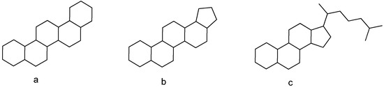
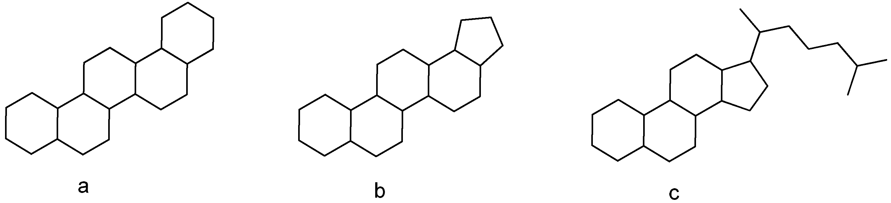
Triterpenoids exhibit structural similarity to glucocorticoids with their four- or five-ring scaffold that mimics the classic shape of steroids (Figure 1). The steroid hormones glucocorticoids play a critical role in many physiological processes, such as growth, development, metabolism, and apoptosis [8]. They exert their actions by binding to glucocorticoid receptor (GR), leading to transactivation or transrepression of nuclear-encoded GR target genes or target genes of other nuclear transcription factors that interact with GR in the nuclear environment [9,10]. Moreover, GR exerts non-genomic rapid actions and regulates mitochondrial gene expression and function by direct or indirect mechanisms of actions [11]. Glucocorticoid receptor activation exhibits profound anti-inflammatory effects via two distinct mechanisms, transcriptional repression of many pro-inflammatory genes and suppression of NF-κB activity. Dexamethasone (DEX), the synthetic glucocorticoid, is a widely prescribed drug for inflammation repression [12]. DEX is also widely applied for blood malignancies treatment, due to its cell-type-specific induction of apoptosis [13]. Nevertheless, DEX when applied in high doses and prolonged period of time for pharmaceutical purposes, results in GR transactivation that ends up in hyperglycemia, diabetes emergence, muscle atrophy, osteoporosis, hypertension, and peptic ulcer [12,13].
Figure 1.
General structure of triterpenoid: (a,b) five-member-ring scaffold, (c) four-member-ring scaffold.
In recent years, the plant metabolite terpenes have attracted research interest as potential selective glucocorticoid receptor modulators, with enhanced benefits and minimized side effects. Our previous studies identified many triterpenes, such as echinocystic acid, protopanaxadiol, protopanaxatriol, α- and β-boswellic acids, as regulators of glucocorticoid receptor signaling, inducing GR transrepression and suppressing or not significantly affecting its transactivation signaling, which is responsible for the adverse side effects of glucocorticoids [14,15,16]. We have also shown that different fractions from Pistacia lentiscus L. var. Chia resin and leaves induce suppression of GR transactivation and cause reduction in GR protein levels in a proteasomal-dependent activation manner [5]. Thus, crucial biological actions of Chios mastiha tree products are emerging, highlighting their potential as foods supplements or pharmaceutical agents with glucocorticoids’ beneficial actions and with minimized harmful side effects. The following questions remain to be addressed: Which class of triterpenoids, acidic or neutral, is involved in the regulation of GR signaling? Do the rest or novel unidentified compounds of Chios mastiha resin contribute to its biological activities, and how so?
Considering the above, the aim of this study was to investigate whether the neutral triterpenoids of Chios mastiha are responsible for Chios mastiha’s effects on glucocorticoid receptor (GR) signaling, to characterize the bioactive compounds associated with Chios mastiha’s biological activities, including anti-inflammatory, potential antilipidemic and apoptotic, and to identify novel potential bioactive compounds of Chios mastiha. For that purpose, a phytochemical protocol was employed to obtain a fraction devoid of acidic triterpenoids, followed by chromatographic subfractionation. These resulting subfractions were characterized and evaluated for their potential anti-inflammatory, antiglycemic, antilipidemic, cytotoxic, and apoptotic properties. The chemical characterization of Chios mastiha‘s fractions and evaluation of their biological activities contribute to the elucidation of the biochemical mechanisms of the therapeutic actions of Chios mastiha and strengthen the potential of its potential as a food additive, dietary supplement, or pharmaceutical agent.
2. Materials and Methods
2.1. Chemicals
Dulbecco’s modified Eagle medium (DMEM) (4.5 g/L glucose), L-glutamine, Penicillin/Streptomycin (Pen/Strep), and fetal bovine serum (FBS) were purchased from Gibco (ThermoFisher, Waltham, MA, USA). Dexamethasone (DEX) and Thiazolyl Blue Tetrazolium Bromide (MTT reagent) were purchased from Sigma-Aldrich (St. Louis, MO, USA). Recombinant Human Tumor Necrosis Factor alpha (TNFα) was purchased from Peprotech (ThermoFisher, MA, USA). Cocktail protease inhibitors were obtained from Roche (Mannheim, Germany). Prestained molecular weight protein markers were obtained from Proteintech (ThermoFisher, MA, USA). Reporter lysis buffer and luciferin were purchased from Promega Corporation (Madison, WI, USA). Silica gel 60 (70–230 mesh), reversed-phase (RP) C18 silica gel, and Celite® 545 particle size 0.02–0.1 mm, pH 10 (100 g/L, H2O, 20 °C), used for low-pressure chromatography and vacuum chromatography, were purchased from Macherey-Nagel (Düren, Germany). Isolation of compounds were monitored by TLC on Merck 60 F254 (0.25 mm) plates and visualized by staining with 5% H2SO4 in EtOH and heating. A Flash chromatography Isolera One with UV/DAD detection (Uppsala, Sweden) was used. 1H 400 MHz NMR spectra were measured on Bruker 400 spectrometers (Bruker®, Billerica, MA, USA). Chemical shifts were referenced to the residual solvent signal (CDCl3: δH = 7.26). Chemical reagents and solvents were from Aldrich (Darmstadt, Germany) and were used without any further purification, unless stated otherwise. For LC-QTOF-MS analysis, LC-MS grade methanol, water, and formic acid 98–100% were provided by Merck (Darmstadt, Germany). Resin from Pistacia lentiscus L. var. Chia (Chios mastiha) was kindly provided by the Chios Mastic Growers Association (https://www.gummastic.gr/en/, accessed on 27 March 2026), which is the certified organization for the production and identification of the Chios mastiha.
2.2. HPLC-QTOF-MS/MS Analysis
Chromatographic analysis was performed on an ExionAC LC system (SCIEX, MA) equipped with two pumps, a solvent degasser, an auto-sampler, and a controller. The X500R Q-TOF mass spectrometer (SCIEX, Framingham, MA, USA) was equipped with an electrospray ionization (ESI) turboVTM, according to Georgantopoulos et al. [4].
2.3. Extraction, Isolation and Phytochemical Analysis of Chios Mastiha’s Neutral Subfractions
Resin from Pistacia lentiscus L. var. Chia (10 g) was dissolved in EtOAc (100 mL) in a separating funnel. An aqueous basic solution (50 mL of water with 2% of NaOH) was added, obtaining a liquid/liquid partition between EtOAc and the basic water. The aqueous layer was collected separately, and the liquid/liquid partition was repeated to obtain a final volume of 100 mL of basic aqueous fraction containing the natrium salt of acidic components. This fraction was then acidified with 97% H2SO4 to pH3, causing the insolubility of acid molecules. The resulting mixture was extracted twice with 50 mL of CH2Cl2 to obtain the organic fraction containing the acid molecules, anhydrified with Na2SO4, and completely evaporated, affording 2.76 g of raw acid fraction. The EtOAc fraction, containing non-acid molecules, was evaporated at reduced pressure, affording 7.08 g of the neutral fraction. This latter portion was fractionated by low-pressure chromatography (LPC) on silica gel (250 g, petroleum ether-EtOAc gradient from 95:5 to 60:40 v/v) monitored by TLC on Merck 60 F254 (0.25 mm) plates and visualized by staining with 5% H2SO4 in EtOH and heating to afford five fractions (F1–F5). Fraction F1 was further purified with LPC on silica gel (12 g, petroleum ether-EtOAc gradient from 90:10 to 80:20 v/v) to afford 45.4 mg of subfraction 1a (SF 1a) as an oil which, after 1H NMR analysis, was revealed to be composed of a complex mixture of neutral triterpenoids and other aliphatic compounds. Fraction F2 was chromatographed with Isolera One on RP C-18 silica gel (12 g, solvent A: MeOH 0.03% formic acid, solvent B: H2O 0.03% formic acid gradient from 50:50 to 95:5) to obtain 73.5 mg of keto-oleanolic aldehyde (1) [17,18] (Figure S1 of the Supplementary Materials) and 356.2 mg of 1,4-poly-β-myrcene as an insoluble white powder. Fraction F3 was fractionated with low-pressure chromatography (LPC) on silica gel (60 g, petroleum ether-EtOAc gradient from 90:10 to 70:30 v/v) to afford subfraction 3a (SF 3a) (239 mg), further crystallized by MeOH to obtain 40.2 mg of keto-oleanolic aldehyde (1) [17] and subfraction 3b (SF 3b) (408 mg), subsequently purified with Isolera One on RP C-18 silica gel (12 g, solvent A: MeOH 0.03% formic acid, solvent B: H2O 0.03% formic acid gradient from 50:50 to 95:5), leading to β-amyrin (2), 376.4 mg [2] (Figure S2 of the Supplementary Materials). Fraction F4 was purified with low-pressure chromatography (LPC) on silica gel (60 g, petroleum ether-EtOAc gradient from 90:10 to 60:40 v/v) to afford subfraction 4a (SF 4a) (29.4 mg) as a mixture on neutral triterpenoid, subfraction 4b (SF 4b) (101.7 mg) as lupeol (3) [19] (Figure S3 of the Supplementary Materials), and subfraction 4c (SF 4c) (20.1 mg) as a mixture of lupeol (3) and keto-oleanolic aldehyde (1). The latter fraction F5 was purified by flash chromatography with Isolera One on RP C-18 silica gel (12 g, solvent A: MeOH 0.03% formic acid, solvent B: H2O 0.03% formic acid gradient from 50:50 to 95:5) to afford subfraction 5a (SF 5a) as lupeol (3, 75 mg) and β-amyrin (2, 224.6 mg). The process flow diagram of the isolation procedure is presented in Figure S4 in the Supplementary Materials. Fractions were stored under nitrogen in dark glass vials and sent immediately for biological evaluation. All the compounds were identified according to 1H NMR data present in the scientific literature.
To identify trace amounts of other resin compounds, HPLC-QTOF-MS/MS analysis was also conducted, as previously reported by Georgantopoulos et al. [4]. The identification of the compounds of the analysis was performed according to Karadimou et al. [20].
2.4. Antibodies
Mouse monoclonal antibodies against human GR, peroxisome proliferator-activated receptor alpha (PPARα) and glyceraldehyde-3-phosphate dehydrogenase (GAPDH), and rabbit polyclonal antibodies against the p65 subunit of NF-κB and phosphoenolpyruvate carboxykinase (PEPCK) were purchased from Santa Cruz Biotechnology (Dallas, TX, USA). Monoclonal antibodies against β-actin and procaspase-9 were provided by Proteintech (Planegg-Martinsried, Germany) and Cell Signaling Technology (Massachusetts, USA), respectively. Rabbit polyclonal antibodies against procaspase-3 and Bcl-2 (Cell Signaling Technology, Danvers, MA, USA) were also used.
2.5. Cell Culture
The human embryonic kidney HEK293 cells, characterized by high efficiency in transfections experiments and presence of GR protein, were obtained from the American type culture collection (ATCC) and cultured at 37 °C and 5% CO2 humidity in DMEM (4.5 g/L glucose) supplemented with 10% FBS, 2 mM L-glutamine, and 100 units/mL Pen/Strep. For luciferase assays, 24 h before transient transfection, cells were cultured in phenol red-free DMEM supplemented with 10% charcoal–dextran-stripped FBS (charcoal-stripped FBS, CSF), 2 mM L-glutamine, and 100 units/mL pen/strep.
2.6. MTT Cell Viability Assay
MTT assay was applied to assess the effect of neutral subfractions on cell growth. MTT is based on mitochondrial dehydrogenases activity assessment of metabolically active cells and, thus, is widely used for cell viability assessment and cytotoxic effect measurements [21]. MTT assay was performed in triplicates with three technical replicates. HEK293 cells were plated in 96-well plate, at a density of 1.5 × 10⁴ cells/well, for 24 h in DMEM (4.5 g/L glucose) supplemented with 10% FBS, 2 mM L-glutamine, and 100 units/mL pen/strep, as previously described. Then, cells were treated with resin fractions diluted in 100% EtOH, at a range concentration of 5–100 μg/mL. EtOH treatment (1:1000) was used as the control condition. After 48 h treatment, MTT reagent diluted in PBS 1X, was added at a final concentration of 0.5 mg/mL for 3–4 h. Finally, formazan crystals were diluted with 100% isopropanol, and after shaking, absorbance was measured at 570 nm, using a multimode plate reader (EnSpire, PerkinElmer, Wycombe, UK). Background absorbance was also measured at 690 nm as reference.
2.7. GR and NF-κB Transcriptional Activity Measurement
GR and nuclear factor-kappa B (NF-κB) transcriptional activity was measured by applying luciferase reporter gene assay, as previously described. Briefly, HEK293 cells were plated on 24-well plates, at a density of 5 × 10⁴ cells/well, in phenol red-free DMEM supplemented with 10% charcoal–dextran-stripped FBS (charcoal-stripped FBS, CSF), 2 mM L-glutamine, and 100 units/mL pen/strep. After 24 h, cells were transiently co-transfected, using calcium phosphate, with a glucocorticoid response element (GRE) or an NF-κΒ response element (NF-κB-RE) promoter-driven luciferase construct and a β-galactosidase reporter construct. At 14–16 h upon transfection, cells were washed in fresh medium for 24 h recovery and then they were triggered either by 1 μM DEX diluted in EtOH (stock solution 1 mM DEX diluted in EtOH was used) or 20 ng/mL TNFα diluted in ddH2O for assessment of GR or NF-κB activity, respectively, as previously described [22], in the presence or absence of the indicated amounts of Chios mastiha’s fractions for 6 h. EtOH treatment (1:1000) was used as the control condition. Then, cells were lysed in reporter lysis buffer, and the enzymatic activities of the expressed luciferase and β-galactosidase were measured using a chemiluminometer (LB 9508, Berthhold, Bad Wildbad, Germany). β-galactosidase activity measurement was used for normalization of luciferase activity (RLU).
2.8. Western Blot Analysis
HEK293 cells were plated on 6 well plates, at a density of 2 × 10⁵ cells/well, in DMEM (4.5 g/L glucose) supplemented with 10% charcoal–dextran-stripped FBS (charcoal-stripped FBS, CSF), 2 mM L-glutamine, and 100 units/mL pen/strep, for 48 h. Cells were then treated with the indicated amounts of Chios mastiha’s fractions for an additional 36 or 48 h. EtOH treatment (1:1000) was used as the control condition. Cells were then washed in PBS 1X, lysed in buffer contained 20 mM Tris pH:7.5, 250 mM NaCl, 0.5% Triton, 3 mM EDTA, and supplemented with cocktail protease inhibitors, DTT and PMSF. After Bradford protein measurement, cell extracts were electrophoresed in discontinuous SDS-PAGE and Western blotted with specific antibodies against GR, PEPCK, PPARα, p65, procaspase-3, procaspase-9, and Bcl-2, as previously described. β-actin or GAPDH protein levels were used for normalization. Enhanced chemiluminescence was used for the detection of protein bands. Quantification of protein band density was performed by the ImageJ program (v1.52). β-actin band intensity or GAPDH protein levels were used for the normalization of the results. Relative protein levels were expressed as band intensity normalized against the respective band’s intensity of β-actin or GAPDH. Relative protein levels in control cells were set to 1.
2.9. Statistical Analysis
All results are expressed as mean ± SD. Data were analyzed by independent t-test or by One-Way analysis of variance (ANOVA) or Two-Way ANOVA followed by Tukey’s post hoc test using Stat Plus LE software. Differences were considered significant at two-tailed p-value < 0.05.
3. Results
3.1. Phytochemical Analysis of Chios Mastiha Neutral Residue
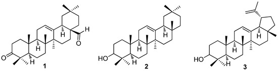
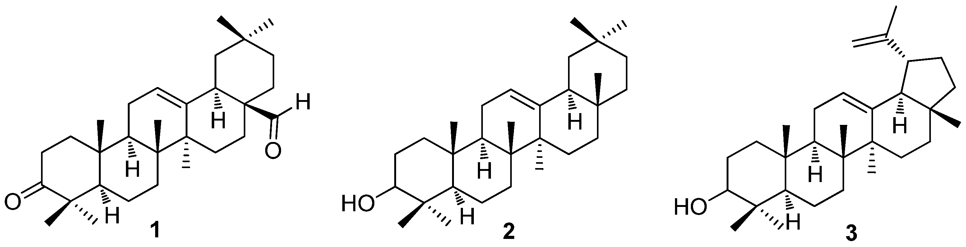
The liquid/liquid partition protocol applied to the P. lentiscus resin in this work aimed to obtain a neutral fraction devoid of acidic triterpenoids, capitalizing on their ability to be water soluble once salified in basic conditions. As a result of this procedure, the neutral residue was composed of a mixture of neutral triterpenoids and other molecules that were further divided with chromatographic techniques into fractions and subfractions until the isolation of the single main neutral triterpenoid, as reported in detail in Section 2.3. Extraction, isolation, and phytochemical analysis of Chios mastiha’s neutral subfractions are presented in Figure S4 of the Supplementary Materials. Results revealed α-amyrin (2) as the main compound being present with a yield of 6%, followed by lupeol (3, 1.77%) and keto-oleanolic aldehyde (1, 1.14%) (Figure 2). The phytochemical characterization was also completed by HPLC-QTOF-MF/MS analysis (Table 1).
Figure 2.
Structure of the main neutral triterpenoid identified by chromatography and 1H NMR analysis in the neutral fraction of Chios mastiha: keto-oleanolic aldehyde (1), β-amyrin (2), and lupeol (3).
Table 1.
Suspect compounds analyzed using HPLC-QTOF-MF/MS analysis in the P. lentiscus/Chios mastiha’s neutral residue and their normalized concentrations (μg/L).
The chemical analysis of Chios mastiha’s neutral subfractions applying HPLC-QTOF-MS/MS analysis resulted in the tentative identification of thirty compounds divided into fatty acids, polyketides, phenylpropanoids, and terpenoids. A characteristic phenolic fingerprint of the resin is presented in Figure S5 of the Supplementary Materials. Identification of fatty acids in Chios mastiha neutral residue was revealed in accordance with our previous studies [4]. Among them, palmitic, stearic, and oleic acids were the most abundant ones. Other fatty acids such as linoleic, caprylic, ricinoleic, crepenynic, nebraskanic, myristic, arachidonic, pentadecanoic, α and γ linoleic acids, and octyl formate were also detected in lower amounts. Flavidin was the major flavonoid, while coniferaldehyde was the most relevant phenylpropanoid. Interestingly, apigenin, astragalin, and diosmetin polyketides were detected exclusively in SF 4a and SF 1a, SF 5a, SF 4a fractions, respectively. Polyketides such as hesperidin, isoliquiritigenin di-glucoside, luteolin glucoside, and resveratrol were detected in all isolated subfractions. Phenylpropanoids such as fumaric acid and feroyl quinic acid were detected in F2, SF 3b and SF 5a fractions, whereas methoxycinnamic was detected in all the isolated subfractions. The monoterpenoid 6,7-dihydro-7-hydroxylinalool and the sesquiterpenoid α-irone were also detected (Table 1).
3.2. Chios Mastiha Neutral Residue Subfractions’ Effect on HEK293 Cell Proliferation
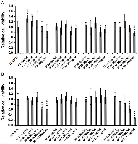
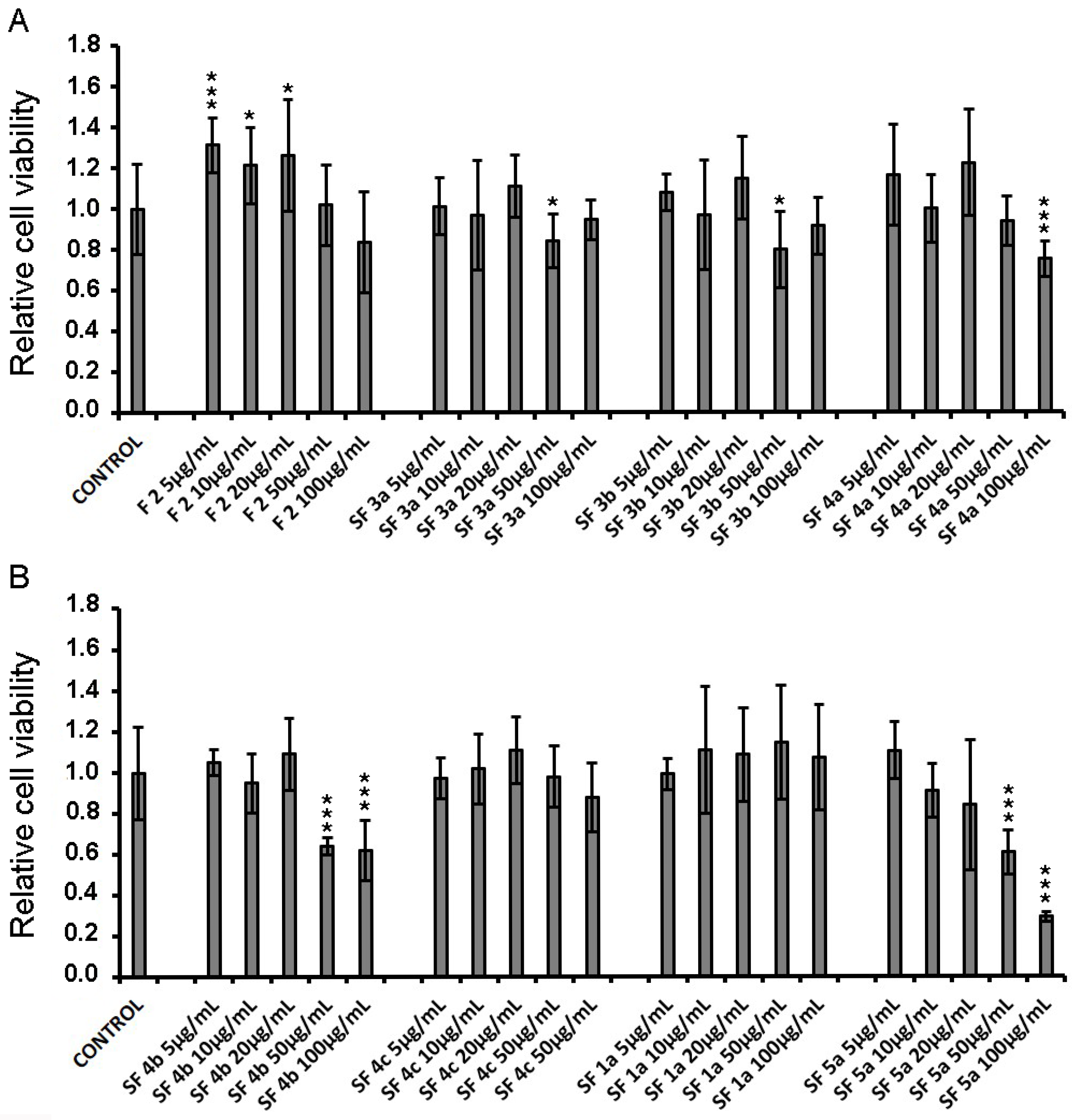
HEK293 cells were incubated with increasing concentrations of Chios mastiha neutral subfractions for 48 h (Figure 3). Then, cell viability was evaluated by applying MTT assay. As shown in Figure 3, F2 showed no reduction in HEK293 cell viability. Similarly, subfractions SF 4c and SF 1a (Figure 3B) did not cause any statistically significant alteration in HEK293 cell viability. Subfractions SF 3a, SF 3b, SF 4a, SF4b, and SF 5a reduced the viability of HEK293 cells up to 15, 20, 25, 35, and 70%, respectively, at higher concentrations of 50 or 100 μg/mL, with SF 5a being the most cytotoxic. Concentrations of 10 μg/mL, 30 μg/mL, and 50 μg/mL that showed no or low cytotoxicity in most isolated subfractions were further evaluated regarding their potential anti-inflammatory, antilipidemic, and GR-associated activities.
Figure 3.
Cell viability measurement of HEK293 cells, in the presence of Chios mastiha’s neutral subfractions F2, SF 3a, SF 3b, SF 4a (A) and SF 4b, SF 4c, SF 1a, SF 5a (B), upon 48 h treatment, assessed by MTT assay. Control cells were treated with EtOH (1:1000). Relative cell viability is expressed as cell viability of the Chios mastiha’s neutral subfractions at the indicated concentrations compared to control. Cell viability of control cells was set at 1. Data are expressed as the mean ± SD, (n = 4–9), * p < 0.05; *** p < 0.001.
3.3. Chios Mastiha’s Neutral Residue Subfractions Slightly Suppressed DEX-Induced GR Transcriptional Activation
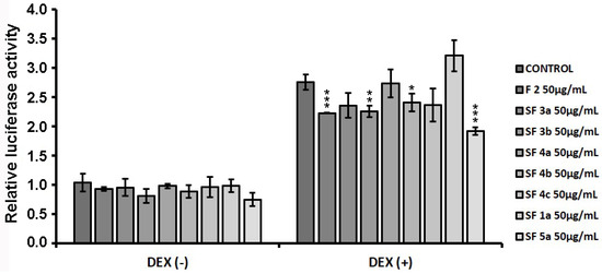
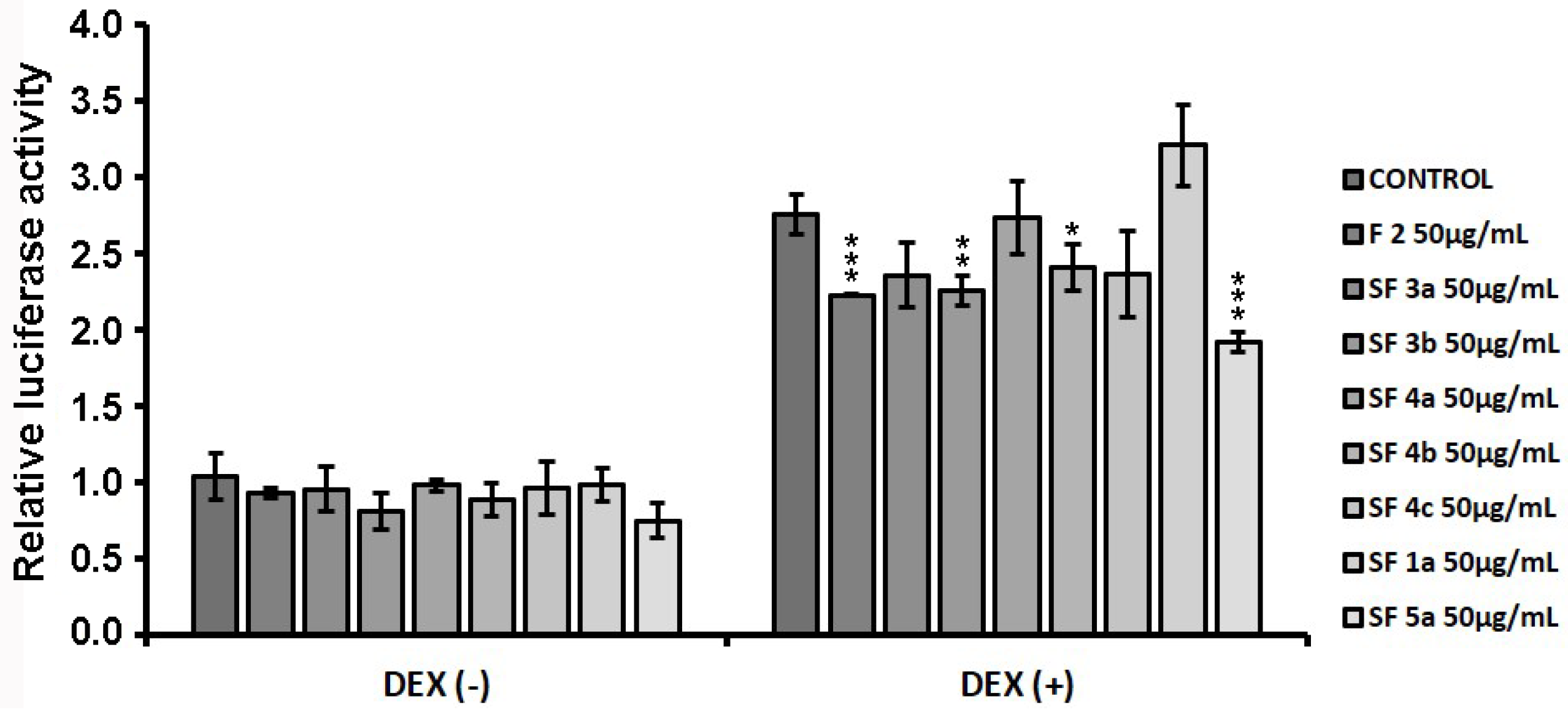
HEK293 cells were transiently co-transfected with GREs-luciferase and β-galactosidase constructs, as described in Materials and Methods, and treated with resin fractions at a concentration of 50 μg/mL, in the presence or absence of 1 µM DEX diluted in EtOH, for 6 h. EtOH (1:1000) treatment was used as the control condition. By applying luciferase reporter gene assay, effect of fractions on GR transcriptional activation was assessed. As shown in Figure 4, fraction F2 and subfraction SF 3b caused approximately 20% statistically significant reduction in DEX-induced GR transcriptional activation. Similarly, subfractions SF 4b and SF 5a caused approximately 10% and 30% statistically significant reduction, respectively, whereas in the presence of the rest of the isolated subfractions, no statistically significant effect was observed.
Figure 4.
Suppression of DEX-induced GR transcriptional activation by Chios mastiha’s neutral residue subfractions (F2, SF 3a, SF 3b, SF 4a, SF 4b, SF 4c, SF 1a, SF 5a) upon 6h treatment. Luciferase and β-galactosidase activity was measured in cell extracts from HEK293 cells cultured in hormone-depleted medium, transiently co-transfected with GREs-luciferase reporter gene construct and β-galactosidase reporter construct and subsequently treated with 50 µg/mL of neutral residue subfractions and/or 1 μΜ DEX, for 6 h. Control cells were treated with EtOH (1:1000). Relative luciferase activity was expressed as normalized luciferase activity against β-galactosidase activity. Relative luciferase activity of control cells was set as 1. Data are expressed as the mean ± SD, (n = 3), * p < 0.05; ** p < 0.01; *** p < 0.001.
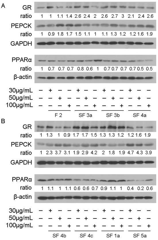
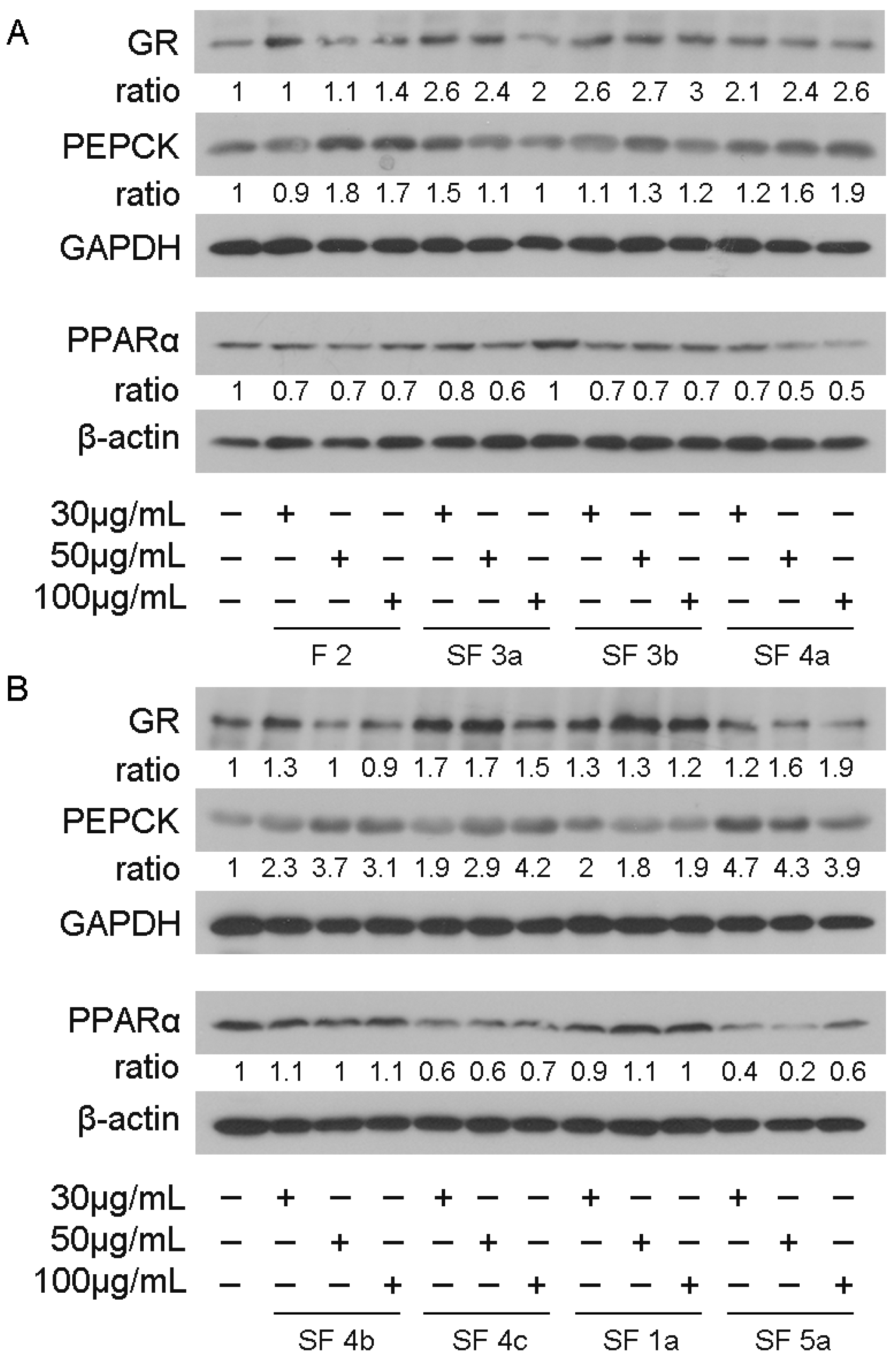
The suppressive effect of fraction F2 as well as subfractions SF 3b, SF 4b, and SF 5a of neutral residue on GR transcriptional activation was further investigated by assessing their effect on GR protein levels and its target genes, PEPCK [23] and PPARα [24,25], by applying Western blot analysis. Thus, HEK293 cells were treated with 30, 50, and 100 μg/mL of subfractions for 48 h (Figure 5). Treatment with EtOH (1:1000) was used as the control condition. As shown in Figure 5, upon 48 h treatment, the subfractions caused an increase in the GR protein levels, with subfraction SF 3b being the most active, causing a three-fold induction in GR protein levels compared to the controls, while subfraction SF 4b caused moderate increase, up to 30%. Notably, increased concentrations of SF 3a, SF 4b, SF 4c, and SF 5a slightly reversed this effect, having reached a plateau of the bioactivity of the fractions. Accordingly, all subfractions caused increase in PEPCK protein levels that constitute a GR target, with subfraction SF 5a being the most active, causing a 4.7-fold induction, while subfraction SF 3b, being the less active one, induced up to a 30% increase. Similarly, increased concentrations of SF 3a and SF 5a abrogated the increase in PEPCK protein levels. Regarding PPARα protein levels, upon 48 h treatment, fraction F2 as well as subfractions SF 3a, SF 3b, SF 4a, SF 4c, and SF 5a caused reductions of up to 30%, 40%, 30%, 50%, 40%, and 80%, respectively, with subfraction 5a being, again, the most active one. Subfractions SF 4b and SF 1a did not cause any significant changes (Figure 5). SF 4a exhibited a dose-dependent effect, while the rest of the subfractions did not cause any remarkable effect.
Figure 5.
Effect of Chios mastiha’s neutral subfractions F2, SF 3a, SF 3b, SF 4a (A) and SF 4b, SF 4c, SF 1a, SF 5a (Β) on GR, PEPCK and PPARα protein levels. Representative images from Western blot analysis of GR, PEPCK and PPARα in extracts from HEK293 cells treated with 30, 50, and 100 μg/mL of neutral residue subfractions for 48 h. Ratios express normalization of band intensity compared with the respective β-actin or GAPDH ones. Relative protein levels in control cells were set as 1.
3.4. Chios Mastiha’s Neutral Residue Subfractions Suppressed the TNFα-Induced NF-κΒ Transcriptional Activation
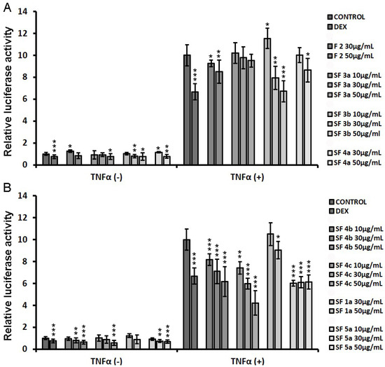
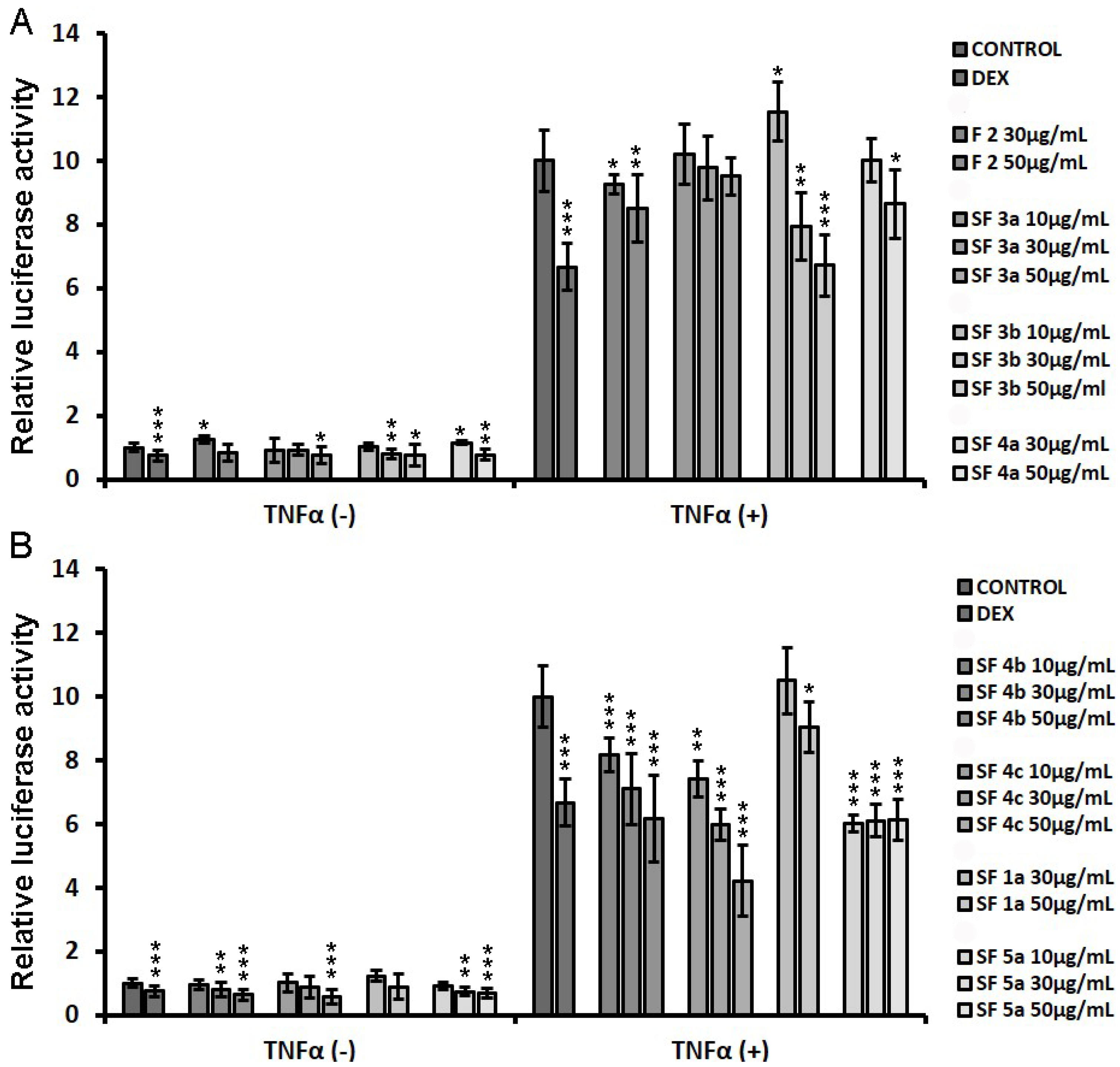
Applying luciferase reporter gene assay, effect of subfractions on NF-κΒ transcriptional activation was studied, evaluating their potential anti-inflammatory actions. Thus, HEK293 cells were transiently co-transfected with NF-κΒ-RE-luciferase and β-galactosidase constructs as described in Materials and Methods and subsequently treated with increasing concentrations of neutral residue subfractions (at a concentration range of 10-50 μg/mL), in the presence or absence of TNFα for 6 h. Luciferase and beta galactosidase activities were then assessed. Treatment with EtOH (1:1000) was used as the control condition. As shown in Figure 6, fraction F2 as well as subfractions SF 3b, SF 4a, SF 4b, SF 4c, SF 1a, and SF 5a caused statistically significant suppression of the TNFα-induced NF-κΒ transcriptional activation by up to approximately 15%, 30%, 15%, 40%, 60%, 10%, and 40%, respectively, in a dose-dependent manner. Subfraction SF 3a did not have any statistically significant effect.
Figure 6.
Suppression of TNFα-induced NF-κΒ transcriptional activation by the Chios mastiha’s neutral subfractions F2, SF 3a, SF 3b, SF 4a (A) and SF 4b, SF 4c, SF 1a, SF 5a (B). Luciferase and β-galactosidase activity was measured in cell extracts from HEK293 cells cultured in hormone-depleted medium, transiently co-transfected with an NF-κB-RE-luciferase reporter gene construct and a β-galactosidase reporter construct and subsequently treated with 10–50 µg/mL of neutral residue subfractions and/or 20 ng/mL TNFα for 6 h. Control cells were treated with EtOH (1:1000). Relative luciferase activity was expressed as normalized luciferase activity against β-galactosidase activity. Relative luciferase activity of control cells was set as 1. Data are expressed as the mean ± SD, (n = 3), * p < 0.05; ** p < 0.01; *** p < 0.001.
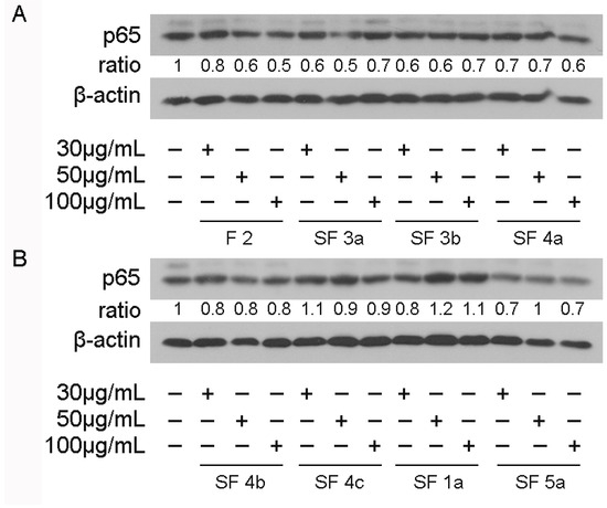
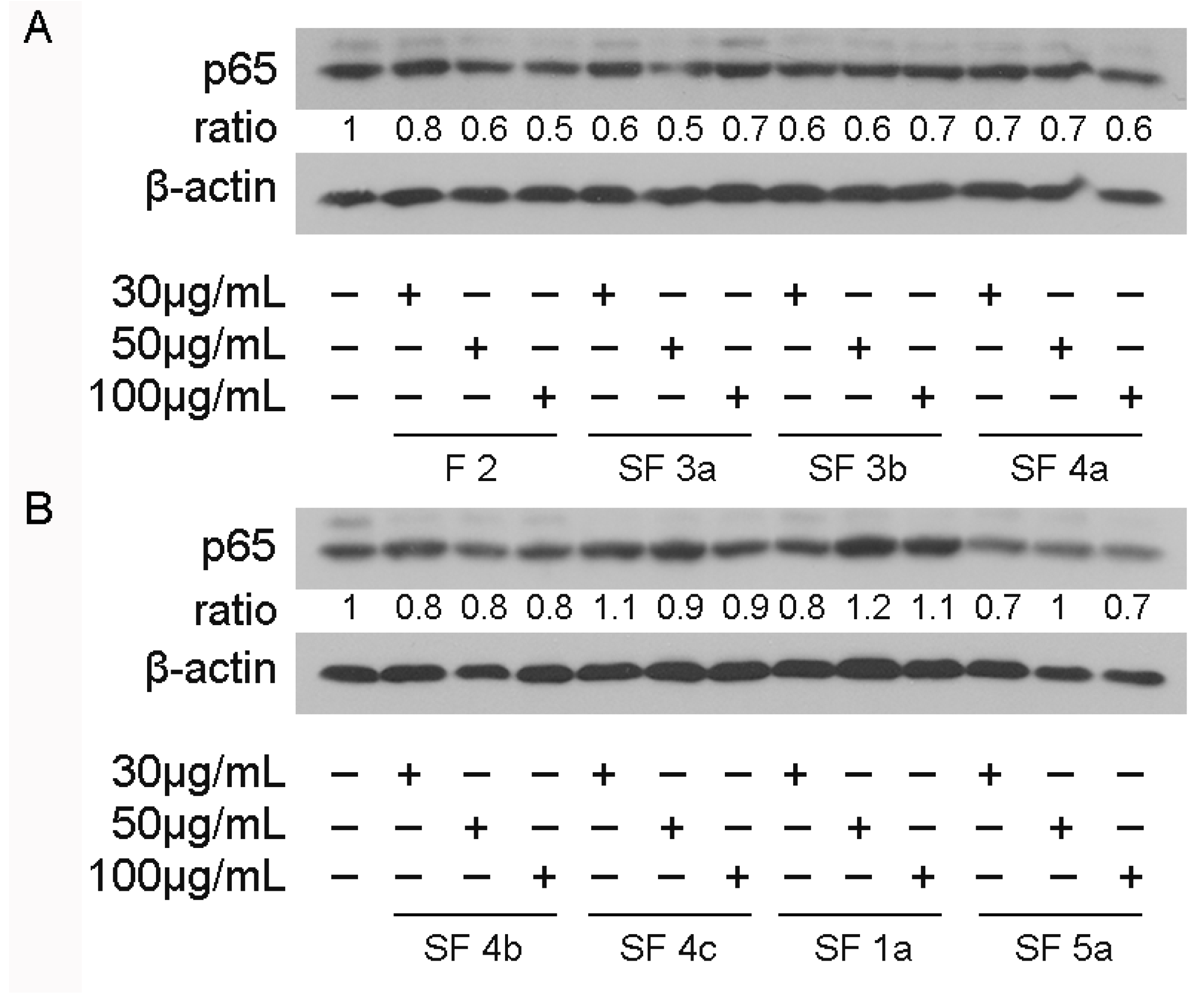
To investigate whether reduction in NF-κΒ transcriptional activation was associated with a reduction in its protein levels, Western blot analysis of the p65 subunit of NF-κΒ was applied in extracts from HEK293 cells treated with 30, 50, and 100 μg/mL of resin subfractions or EtOH (1:1000) for 48 h (Figure 7). As shown in Figure 7, upon 48 h treatment, fraction F2 as well as subfractions SF 3a, SF 3b, SF 4a, SF 4b, SF 4c, SF 1a, and SF 5a reduced p65 protein levels by up to 50%, 50%, 40%, 40%, 20%, 10%, 20%, and 30%, respectively, in a dose-dependent manner. Thus, the suppression of the TNFα-induced NF-κΒ transcriptional activation by Chios mastiha’s subfractions is accompanied by a reduction in p65 protein levels, indicating that the anti-inflammatory activity of neutral residue subfractions may be attributed to their effects on NF-κΒ transcriptional activity and/or regulation of NF-κΒ protein levels. Subfraction SF 4c exhibited the highest suppression of the TNFα-induced NF-κΒ transcriptional activation (Figure 6B) and negative regulation of p65 protein levels (Figure 7B).
Figure 7.
Effect of Chios mastiha’s neutral subfractions F2, SF 3a, SF 3b, SF 4a (A) and SF 4b, SF 4c, SF 1a, SF 5a (B) on p65 protein levels. Representative images from Western blot analysis of p65 in extracts from HEK293 cells treated with 30, 50, and 100 μg/mL of Chios mastiha’s neutral subfractions for 48 h. Ratios express normalization of band intensity compared with the respective β-actin one. Relative protein levels in control cells were set as 1.
3.5. Chios Mastiha’s Subfractions’ Effect on Apoptotic Pathways
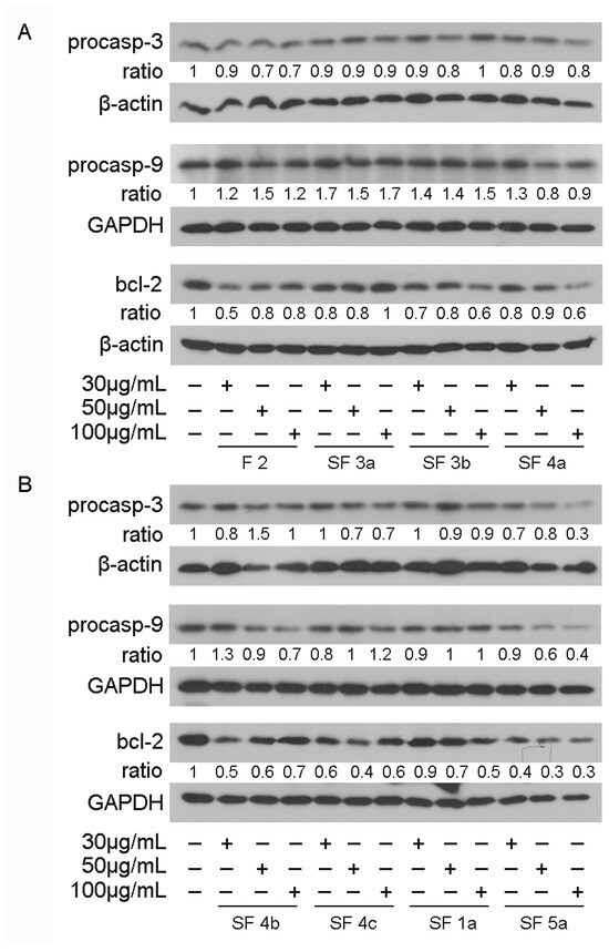
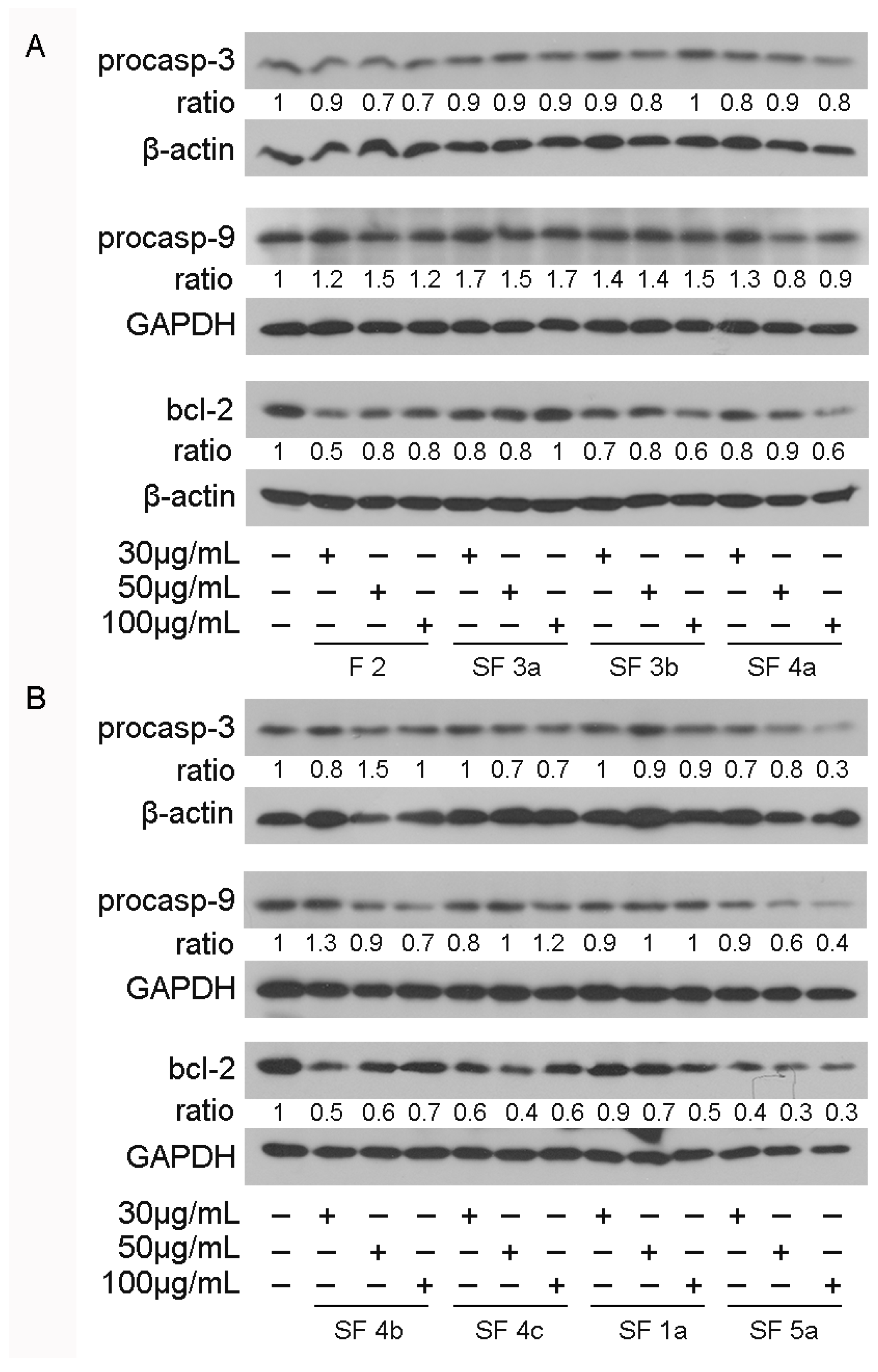
Effect of Chios mastiha’s subfractions on apoptosis was examined by applying Western blot analysis of apoptotic molecules, such as procaspase-3, procaspase-9, and bcl-2. For that purpose, HEK293 cells were treated with 30, 50, and 100 μg/mL of Chios mastiha’s neutral subfractions. Upon 48 h treatment (Figure 8), the resin subfractions SF 4a, SF 4b, SF 4c, SF 1a, and SF 5a caused reduction in procaspase-3 protein levels, with subfraction SF 5a being the most active, causing up to 70% reduction. Similarly, subfractions SF 4a, SF 4b, and SF 5a caused reduction in procaspase-9 protein levels by up to 20%, 30%, and 60%, respectively, at higher concentrations of 50 and 100 μg/mL, with subfraction SF 5a being the most active one. The reduced protein levels of procaspase-9 indicate activation of the intrinsic mitochondrial-dependent apoptotic pathway. Regarding bcl-2, all resin subfractions caused a reduction in its protein levels, with subfraction SF 5a being the most active, causing up to 70% reduction.
Figure 8.
Effect of Chios mastiha’s neutral subfractions F2, SF 3a, SF 3b, SF 4a (A) and SF 4b, SF 4c, SF 1a, SF 5a (B) on procaspase-3, procaspase-9, and bcl-2 protein levels. Representative images of Western blot analysis of procaspase-3, procaspase-9, and bcl-2 in extracts from HEK293 cells, treated with 30, 50, and 100 μg/mL of Chios mastiha’s subfractions for 48 h. Ratios express normalization of band intensity compared with the respective β-actin or GAPDH ones. Relative protein levels in control cells were set as 1.
4. Discussion
Chios mastiha, the Pistacia lentiscus L. var. Chia resin, has drawn researchers’ attention due to its constantly emerging therapeutic effects. Apart from being used as food additive and flavoring, Chios mastiha exhibits many therapeutic effects, due to its antimicrobial, antioxidant, anti-inflammatory, anti-diabetic, and anticancer properties. Triterpenoid compounds, which are plant secondary metabolites and the major chemical group in Chios mastiha extract, are suggested to be mainly responsible for Chios mastiha’s biological properties [2,3,7]. Results from our previous [4] and current work uncovered the presence of novel identified compounds in Chios mastiha’s fractions, such as fatty acids and polyketides compounds, which also contribute to Chios mastiha’s biological activities, based on their documented bioactivity. Regarding triterpenoid compounds, our studies and those from other research groups have shown their enrichment in resin and leaves from Pistacia lentiscus L. var. Chia [4,5,6,26,27]. The structural and functional similarity between triterpenoids and the steroid hormone glucocorticoids have triggered our interest to investigate their potential use as steroid hormones substitutes, with potent pharmaceutical application. Our studies and those from other research groups have recently unveiled triterpenoid compounds as potential glucocorticoid receptor modulators with increased transrepressional activity on NF-κΒ activation but with suppressed effect on transactivation of PEPCK gene expression that could give rise to glucose synthesis, with many adverse side effects, such as hyperglycemia, muscle atrophy, osteoporosis, hypertension, and glaucoma [5,14,15,16,28]. In this context, Chios mastiha appeared to be a potent source of such bioactive compounds.
Based on this, we have recently documented the interference of Chios mastiha’s different polarity fractions, enriched in terpenoids, with glucocorticoid signaling, highlighting the potential application of terpenoids from Chios mastiha as selective glucocorticoid receptor modulators [4]. In this study we aimed to identify which class of triterpenoids, neutral (lacking carboxylic groups) or acidic (containing carboxylic groups), within Chios mastiha, were responsible for the modulation of the glucocorticoid receptor signaling, and to characterize the potential regulatory effects of neutral Chios mastiha’s fraction on lipid metabolism, inflammation, and apoptosis, in relation to its chemical composition.
For this purpose, the resin was treated under basic conditions to obtain a neutral fraction devoid of acidic triterpenoids. Phytochemical characterization of the Chios mastiha neutral fraction revealed the abundance of major neutral triterpenoids, such as β-amyrin (2), lupeol (3), and keto-oleanolic aldehyde (1), in accordance with previous studies [6,26,29]. Moreover, the chromatographic process led to the obtainment of several fractions and subfractions of different compositions. In particular, the most relevant content of β-amyrin (2) was detected in SB 3b together with the maximum lipid pool, while keto-oleanolic aldehyde (1) was detected in F2 and SB 3a with a correspondingly low content of phenolic compounds. These latter metabolites, alongside lupeol, were mostly abundant in SF 5a.
Results from this study on the identification of Chios mastiha’s neutral subfractions’ interference with glucocorticoid receptor signaling showed no remarkable effect of the neutral fractions on DEX-induced GR transcriptional activation. In our previous studies, we have shown suppression of DEX-induced GR transcriptional activity by isolating Chios mastiha’s fractions containing both neutral and acids triterpenoids. In the present study, isolated neutral fractions were examined. Thus, the result from this study indicates that the approximately 50% reduction in DEX-induced GR transcriptional activation observed in our previous study by medium polar fraction, from Chios mastiha tree resin and leaves enriched in triterpenoids [5,6], could be attributed mainly to acidic terpenoids. F2 as well as subfractions SF 3b, SF 4b, and SF 5a caused moderate statistically significant suppression of DEX-induced GR transcriptional activation. Since lupeol (3) and β-amyrin (2) correspond to the main compounds of these fractions, it is concluded that these compounds could also contribute to the suppression of DEX-induced GR transactivation, although to a lower extent. Suppression of DEX-induced GR transcriptional activation was accompanied by an increase in GR and its target PEPCK protein level induction. This effect is also opposite to that observed previously by the medium polar fraction of Chios mastiha resin [6], further substantiating the notion that acidic terpenoids are the potential selective glucocorticoid receptor activators, exhibiting favorable negative effect both on GR transactivation and regulation of GR and its target PEPCK protein levels. The increase in GR protein levels, by neutral subfractions, is in line with the observed increase in its target PEPCK protein levels. This action may be induced by compounds other than terpenoids. Specifically, the increase in GR and PEPCK expression by neutral fractions could possibly be associated with the saturated fatty acid abundance in these fractions. Saturated fatty acid palmitate is reported to induce increase in intracellular cAMP synthesis through the palmitoylation of soluble adenylyl cyclase [30]. Subsequently, cAMP can activate GR and PEPCK protein synthesis via activation of the transcription of the human GR gene and increase in its mRNA stability [31] as well as via positive regulation of PEPCK synthesis at the transcriptional level [32] and stabilization of its mRNA [33]. In addition, our previous studies showed activation of GR proteasomal degradation by medium polar and polar fractions from Chios mastiha tree leaves [5]. This observation, in conjunction with the above-mentioned results from this study, showing increase in GR and PEPCK protein levels, indicates that subfractionation procedure may lead to abolishment or elimination of molecules that activate the proteolytic degradation of GR.
Chemical analysis from this study revealed the presence of phenolic compounds known for their substantial biological activities including anti-inflammatory, antioxidant, anticancer, and antilipidemic actions [34]. To our knowledge, it is the first time that apigenin, astragalin, diosmetin, flavidin, and genistein are identified as Chios mastiha’s compounds contributing to Chios mastiha’s biological activities.
Regarding the assessment of antilipidemic activities of Chios mastiha, we focused on neutral fractions’ effect on PPARα regulation. PPARα regulates lipid metabolism, especially, mitochondrial fatty acid import and β-oxidation, leading to ATP production [35]. Prolonged treatment with PPARα agonists can lead to lipid biosynthesis and hyperlipidemia [36]. PPARα also constitutes a direct glucocorticoid receptor target. Recent studies have shown that palmitic acid, stearic acid, oleic acid, and linoleic acid significantly activate PPARα/δ at physiologically relevant concentrations [37]. Ιn this study, Chios mastiha’s neutral subfractions exhibited a reduction in PPARα protein levels. SF 4a and SF 5a exhibited the highest activity. Palmitic acid is detected in almost the highest amounts in SF 4a subfraction. Since palmitic acid is considered a PPARα agonist and activator [38], it may be responsible for the documented Chios mastiha’s antilipidemic activities [6,39,40]. These activities may also be attributed to compounds such as β-amyrin (2) and lupeol (3) because of their known regulatory role in PPARα activity and/or expression [41,42,43,44,45,46,47]. Thus, enrichment of these compounds in SF 5a subfraction might be associated with the regulatory role of SF 5a fraction in PPARα protein level and Chios mastiha’s anti-hyperlipidemic effects. Moreover, polyketides such as flavonoid flavidin, genistein, luteolin glucosides, and stilbenoid resveratrol, identified in Chios mastiha’s subfractions and known for their antilipidemic action [48,49,50], may contribute to this action. Among them, genistein is reported to enhance the expression of genes involved in lipid metabolism through the activation of PPARα [51], and luteolin and genistein regulate PPARα expression as well [52,53].
Anti-inflammatory activities of Chios mastiha’s neutral subfractions were also confirmed in this study. Particularly, all neutral resin subfractions caused a dose-dependent statistically significant suppression of TNFα-induced NF-κΒ transcriptional activation, except for SF 3a, which did not cause any remarkable effect. The suppression of NF-κΒ transcriptional activation by Chios mastiha’s neutral subfractions is accompanied by reduction in p65 subunit protein levels, shedding more light on the anti-inflammatory biochemical mechanism of Chios mastiha’s resin action. This observation is in line with findings showing inhibition of p65 subunit phosphorylation by Chios mastiha’s neutral fraction [54]. Subfractions 3b, 4b, 4c, and 5a caused the highest suppression of the NF-κΒ transcriptional activation, with SF 5a being the most active one even at lower concentrations. Lupeol (3) which is present in SF 4b, SF 4c and SF 5a, was previously reported to be a strong anti-inflammatory compound, as it reduces prostaglandin E2, TNFα, and the production of many interleukins [54]. Previous studies also showed that lupeol (3) suppresses the phosphorylation and degradation of NF-κΒ inhibitor IκΒα, preventing NF-κΒ binding to DNA and subsequent expression of pro-inflammatory genes [43]. β-Amyrin (2), which was identified as a major compound in subfractions 3b and 5a, was reported to reduce TNFα serum levels [47,55] and to inhibit NF-κΒ nuclear translocation [56]. Thus, both our study and other previous studies support the notion that lupeol (3) and β-amyrin (2) are crucial bioactive compounds that could contribute to Chios mastiha’s anti-inflammatory activities, through interference with NF-κΒ signaling. Interestingly, our findings indicate that the coexistence of lupeol (3) and β-amyrin (2) in Chios mastiha’s neutral subfractions exhibit a synergistic effect on the suppression of NF-κΒ transcriptional activation. Moreover, compounds such as feroyl quinic and fumaric acids may also be responsible for the predominant anti-inflammatory activity of SF 5a fraction, since their anti-inflammatory activity is well-documented [57,58]. Fumaric acid has been found to inhibit NF-κΒ transcriptional activation in macrophage cells synergistically with dexamethasone [57], supporting our hypothesis of its interference with glucocorticoid receptor signaling. More interestingly, flavonoids such as apigenin, astragalin, diosmetin, and genistein may contribute to the strong anti-inflammatory activity of SF 4b, SF 4c, and SF 5a fractions, documented to be exerted, among others, via their suppressive effect on NF-κΒ signaling pathway, affecting both NF-κB protein levels [59] and activity [60,61,62,63,64]. Based on previous studies showing anti-inflammatory activity of terpenoids such as 6,7-dihydro-7-hydroxylinalool and α-irone, we assume that these compounds could also add to the neutral fractions’ anti-inflammatory activity [65]. Moreover, the considerable fatty acids presence in all the neutral fractions may also be associated with the neutral fractions’ suppressive effect on NF-κB transcriptional activation. Anti-inflammatory activity of oleic acid [66], α and γ linoleic acid [67,68], ricinoleic acid [69], myristic acid [70], and arachidonic acid [71], via regulation of NF-κΒ signaling and NF-κB protein expression [66], is well documented. Interestingly, the increased amount of oleic and linoleic acids in SF 3b is possibly related to the increased anti-inflammatory activity of SF 3b fraction. Moreover, although increased levels of palmitic acid in high fat diets and obese patients is associated with pro-inflammatory responses [72], recent pharmacological studies demonstrate that palmitic acid also exhibits potent anti-inflammatory and immune-enhancing effects [73].
Anti-inflammatory activities of Chios mastiha’s neutral subfractions were parallel to the anti-proliferative and anti-apoptotic ones. Among all subfractions, SF 4a, SF 4b, and SF 5a containing lupeol (3) and fatty acids, mainly palmitic acid, as common compounds, caused the highest statistically significant reduction in HEK293 cell viability, with SF 5a being the most cytotoxic one. Palmitic acid, in recent years has emerged as a promising anti-tumor agent with demonstrated efficacy against various malignancies including gastric cancer, liver cancer, cervical cancer, breast cancer, and colorectal cancer [73]. Also, subfraction 5a caused the highest reduction in procaspase-3 and bcl-2 protein levels, being the most apoptotic one. Interestingly, procaspase-9 protein levels decreased in the presence of SF 5a, indicating its apoptotic action was mediated through mitochondrial pathway. Previous studies had reported that lupeol (3) reduces proliferation of human prostate cancer cells and inhibits the growth of human melanoma, showing strong anticancer properties [54]. Also, lupeol (3) and palmitic acid were found to induce apoptosis through activation of mitochondrial pathway in many cancer cells [44,73]. The co-presence of lupeol (3), β-amyrin (2), and palmitic acid in SF 5a enhances its anti-proliferative and apoptotic activity. Apoptotic and anti-proliferative activities of β-amyrin (2) have also been observed before. More precisely, β-amyrin (2) was found to induce cell cycle arrest, apoptosis, and cytotoxicity in human hepatocarcinoma HepG2, HL-60 leukemia, colon carcinoma CaCo-2, and human embryonic kidney HEK293 cell lines [74,75,76]. β-Amyrin (2) was also found in SF 3b, which reduced HEK293 cell proliferation, but to a much lower extent, indicating for the first time that a synergistic effect on induction of anti-proliferative activity coexists with lupeol (3) and palmitic acid. Additionally, mitochondrial-dependent apoptosis is reported to be induced by compounds such as linoleic [77], arachidonic acid [78], and oleic acid [79], whereas compounds such as stearic acid [79] and pentadecanoic acid [80] are reported to exhibit protective effect on mitochondrial functionality. Thus, the relative concentration of these compounds is a crucial factor for the final biological outcome of the isolated subfractions.
5. Conclusions
In conclusion, our data reveal the potent anti-inflammatory, anti-proliferative activities of Chios mastiha’s neutral residue subfractions in HEK293 cells. Neutral subfractions were found to attenuate inflammation through reduction in TNFα-induced NF-κΒ transcriptional activity and regulation of the protein levels of the p65 subunit of NF-κΒ. These actions could be attributed to molecules such as lupeol (3) and β-amyrin (2), together with phenolic compounds and fatty acids known for their anti-inflammatory activity and enriched in the most active subfractions. Additionally, Chios mastiha subfractions induced apoptosis and attenuated cell viability by reducing pro-caspase-3, pro-caspase-9, and bcl-2 protein levels. Subfractions of fraction 4 and subfraction 5, with high levels of lupeol (3), β-amyrin (2), and palmitic acid, showed the highest cytotoxic and apoptotic activities, highlighting their potential as anticancer agents. It was revealed for the first time that the interference of Chios mastiha with GR signaling, leading to suppression of inflammation and glucose synthesis, is mainly exerted by the acidic terpenoids. Neutral subfractions induced moderate inhibition of DEX-induced GR transcriptional activity in luciferase assays and abolished negative regulation of Chios mastiha’s medium polar fraction on the synthesis of gluconeogenic enzymes. Nevertheless, neutral mastiha’s subfractions still preserved their regulatory effects on PPARα and the p65 subunit of ΝF-κΒ protein levels, preserving Chios mastiha’s antilipidemic and anti-inflammatory activities. Due to the high sensitivity of HPLC-QTOF-MS/MS analysis, we managed to detect fatty acids and identify for the first-time phenolic compounds, such as apigenin, astragalin, coniferaldehyde, diosmetin, flavidin, and genistein, with profound bioactivity, as Chios mastiha’s compounds. All these compounds are suggested to be responsible for every biological activity separately, but the common existence of these bioactive compounds could have a significant impact on the outcome of Chios mastiha’s fraction, as reported for other plant extracts. In summary, this study uncovered novel bioactive compounds in Chios mastiha and contributed to the characterization of the active compounds responsible for certain Chios mastiha’s therapeutic activities, highlighting the importance of Chios mastiha in the development of novel nutraceutical products. Moreover, the compartmentalization of Chios mastiha’s bioactive compounds, accompanied by dissociation of its biological activities, draws the path for the exploitation of Chios mastiha’s fractions in the development of novel nutraceutical or pharmaceutical products for specific medical purposes. The study provides insights into the association of Chios mastiha’s biological activities and the class of Chios mastiha’s bioactive compounds responsible for Chios mastiha’s biological activity. The exact biochemical pathway involved in these actions remains to be determined. Moreover, the bioactivity of the isolated compounds, alone and in combination, and their activity in in vitro and in vivo systems, in a broad range of concentrations, remains to be elucidated, which would help establish a precise Chios mastiha’s concentration, as a whole or as isolated fractions, in the development of novel nutraceutical or pharmaceutical products with targeted therapeutic applications.
Supplementary Materials
The following supporting information can be downloaded at: https://www.mdpi.com/article/10.3390/nutraceuticals6020033/s1, Figure S1: 1H NMR 400 MHz of keto-oleanolic aldehyde (1) in CDCl3; Figure S2: 1H NMR 400 MHz of β-amyrin (2) in CDCl3; Figure S3: 1H NMR 400 MHz of lupeol (3) in CDCl3; Figure S4: Chromatographic fractionation of Chios mastiha neutral fraction. Figure S5. Characteristic phenolic fingerprint representing (a) apigenin; (b) astragalin; (c) genistein.
Author Contributions
Conceptualization, A.-M.G.P.; methodology, A.-M.G.P., A.G., N.P.K. and F.P.; investigation, A.G., F.D.K., A.C., F.P. and N.P.K.; formal analysis, A.G., F.D.K., F.P., A.-M.G.P. and N.P.K.; resources, A.-M.G.P., A.C. and N.P.K.; writing—original draft preparation, A.-M.G.P. and F.D.K.; writing—review and editing, A.-M.G.P., F.D.K., F.P. and N.P.K.; visualization, F.D.K.; supervision, A.-M.G.P.; project administration, A.-M.G.P.; funding acquisition, A.-M.G.P. All authors have read and agreed to the published version of the manuscript.
Funding
This research was partially supported by the Postgraduate Programs “Application of Molecular Biology–Genetics–Diagnostic Biomarkers” and “Biotechnology—Quality assessment in Nutrition and the Environment”, Department of Biochemistry and Biotechnology, University of Thessaly (to A.-M.G.P.). The research was also supported by the Hellenic Foundation for Research and Innovation (HFRI) under the HFRI PhD Fellowship grant (Fellowship Number: 6247 to F.D.K.).
Data Availability Statement
All data, tables, and figures are original. Details on data analysis are available from the corresponding author upon reasonable request.
Acknowledgments
The authors would like to thank the Interdisciplinary Agri-Food Center, Aristotle University of Thessaloniki (KEAGRO-AUTH) for providing access to the equipment of the unit. This research was also supported by the “Chios Gum Mastic Growers Association” and the “mastihashop”.
Conflicts of Interest
The authors declare no conflicts of interest. The founders had no role in the design of the study; in the collection, analyses, or interpretation of data; in the writing of the manuscript, and in the decision to publish the result.
Abbreviations
The following abbreviations are used in this manuscript:
| NMR | Nuclear magnetic resonance |
| HPLC | High-performance liquid chromatography |
| QTOF | Quadrupole time-of-flight |
| MS | Mass spectrometry |
| PEPCK | Phosphoenolpyruvate carboxykinase |
| GR | Glucocorticoid receptor |
| PPARα | Peroxisome proliferator-activated receptor alpha |
| TNFα | Tumor necrosis factor alpha |
| NF-κB | Nuclear factor kappa beta |
| DEX | Dexamethasone |
| LPC | Low-pressure chromatography |
| HEK293 | Human embryonic kidney cells 293 |
| DMEM | Dulbecco’s modified eagle medium |
| GREs | Glucocorticoid response elements |
| EtOH | Ethanol |
References
- Paraschos, S.; Mitakou, S.; Skaltsounis, A.L. Chios gum mastic: A review of its biological activities. Curr. Med. Chem. 2012, 19, 2292–2302. [Google Scholar] [CrossRef] [Scilit]
- Pachi, V.K.; Mikropoulou, E.V.; Gkiouvetidis, P.; Siafakas, K.; Argyropoulou, A.; Angelis, A.; Mitakou, S.; Halabalaki, M. Traditional uses, phytochemistry and pharmacology of Chios mastic gum (Pistacia lentiscus var. Chia, Anacardiaceae): A review. J. Ethnopharmacol. 2020, 254, 112485. [Google Scholar] [CrossRef] [Scilit] [PubMed]
- Dimas, K.S.; Pantazis, P.; Ramanujam, R. Chios mastic gum: A plant-produced resin exhibiting numerous diverse pharmaceutical and biomedical properties. In Vivo 2012, 26, 777–785. [Google Scholar]
- Georgantopoulos, A.; Kalousi, F.D.; Pollastro, F.; Tsialtas, I.; Kalogiouri, N.P.; Psarra, A.G. Chemical Analysis and Antioxidant Activities of Resin Fractions from Pistacia lentiscus L. var. Chia in Neuroblastoma SH-SY5Y Cells. Molecules 2025, 30, 997. [Google Scholar] [CrossRef] [Scilit]
- Kalousi, F.D.; Pollastro, F.; Christodoulou, E.C.; Karra, A.G.; Tsialtas, I.; Georgantopoulos, A.; Salamone, S.; Psarra, A.G. Apoptotic, Anti-Inflammatory Activities and Interference with the Glucocorticoid Receptor Signaling of Fractions from Pistacia lentiscus L. var. chia Leaves. Plants 2022, 11, 934. [Google Scholar] [CrossRef] [Scilit]
- Kalousi, F.D.; Pollastro, F.; Karra, A.G.; Tsialtas, I.; Georgantopoulos, A.; Salamone, S.; Psarra, A.G. Regulation of Energy Metabolism and Anti-Inflammatory Activities of Mastiha Fractions from Pistacia lentiscus L. var. chia. Foods 2023, 12, 1390. [Google Scholar] [CrossRef] [Scilit]
- Papada, E.; Kaliora, A.C. Antioxidant and Anti-Inflammatory Properties of Mastiha: A Review of Preclinical and Clinical Studies. Antioxidants 2019, 8, 208. [Google Scholar] [CrossRef] [Scilit] [PubMed]
- Timmermans, S.; Souffriau, J.; Libert, C. A General Introduction to Glucocorticoid Biology. Front. Immunol. 2019, 10, 1545. [Google Scholar] [CrossRef] [Scilit]
- Liu, B.; Zhang, T.N.; Knight, J.K.; Goodwin, J.E. The Glucocorticoid Receptor in Cardiovascular Health and Disease. Cells 2019, 8, 1227. [Google Scholar] [CrossRef] [Scilit]
- Oakley, R.H.; Cidlowski, J.A. The biology of the glucocorticoid receptor: New signaling mechanisms in health and disease. J. Allergy Clin. Immunol. 2013, 132, 1033–1044. [Google Scholar] [CrossRef] [Scilit] [PubMed]
- Psarra, A.M.; Sekeris, C.E. Glucocorticoids induce mitochondrial gene transcription in HepG2 cells: Role of the mitochondrial glucocorticoid receptor. Biochim. Biophys. Acta 2011, 1813, 1814–1821. [Google Scholar] [CrossRef] [Scilit]
- Baschant, U.; Culemann, S.; Tuckermann, J. Molecular determinants of glucocorticoid actions in inflammatory joint diseases. Mol. Cell. Endocrinol. 2013, 380, 108–118. [Google Scholar] [CrossRef] [Scilit]
- Cain, D.W.; Cidlowski, J.A. Specificity and sensitivity of glucocorticoid signaling in health and disease. Best Pract. Res. Clin. Endocrinol. Metab. 2015, 29, 545–556. [Google Scholar] [CrossRef] [Scilit]
- Georgatza, D.; Gorgogietas, V.A.; Kylindri, P.; Charalambous, M.C.; Papadopoulou, K.K.; Hayes, J.M.; Psarra, A.G. The triterpene echinocystic acid and its 3-O-glucoside derivative are revealed as potent and selective glucocorticoid receptor agonists. Int. J. Biochem. Cell Biol. 2016, 79, 277–287. [Google Scholar] [CrossRef] [Scilit]
- Karra, A.G.; Konstantinou, M.; Tzortziou, M.; Tsialtas, I.; Kalousi, F.D.; Garagounis, C.; Hayes, J.M.; Psarra, A.G. Potential Dissociative Glucocorticoid Receptor Activity for Protopanaxadiol and Protopanaxatriol. Int. J. Mol. Sci. 2018, 20, 94. [Google Scholar] [CrossRef] [Scilit]
- Karra, A.G.; Tziortziou, M.; Kylindri, P.; Georgatza, D.; Gorgogietas, V.A.; Makiou, A.; Krokida, A.; Tsialtas, I.; Kalousi, F.D.; Papadopoulos, G.E.; et al. Boswellic acids and their derivatives as potent regulators of glucocorticoid receptor actions. Arch. Biochem. Biophys. 2020, 695, 108656. [Google Scholar] [CrossRef] [Scilit]
- Kumar, N.; Ravindranath, B.; Seshadri, T.R. Terpenoids of Pterocarpus santalinus heartwood. Phytochemistry 1974, 13, 633–636. [Google Scholar] [CrossRef] [Scilit]
- Newmark, R.A.; Majumdar, R.N. 13C-NMR spectra of cis-polymyrcene and cis-polyfarnesene. J. Polym. Sci. Part A Polym. Chem. 1988, 26, 71–77. [Google Scholar] [CrossRef] [Scilit]
- Sholichin, M.; Yamasaki, K.; Kasai, R.; Tanaka, O. 13C Nuclear Magnetic Resonance of Lupane-Type Triterpenes, Lupeol, Betulin and Betulinic Acid. Chem. Pharm. Bull. 1980, 28, 1006–1008. [Google Scholar] [CrossRef] [Scilit]
- Karadimou, C.; Petsa, E.; Ouroumi, N.A.; Papadakis, E.N.; Kontoudakis, N.; Theocharis, S.; Mourtzinos, I.; Menkissoglu-Spiroudi, U.; Kalogiouri, N.P.; Koundouras, S. Exploration of the anthocyanin and proanthocyanidin profile of Greek red grape skins belonging to Vradiano, Limnio, and Kotsifali cultivars, analyzed by a novel LC-QTOF-MS/MS method. Phytochem. Anal. PCA 2024, 35, 1781–1793. [Google Scholar] [CrossRef] [Scilit] [PubMed]
- Mosmann, T. Rapid colorimetric assay for cellular growth and survival: Application to proliferation and cytotoxicity assays. J. Immunol. Methods 1983, 65, 55–63. [Google Scholar] [CrossRef] [Scilit]
- Psarra, A.M.; Hermann, S.; Panayotou, G.; Spyrou, G. Interaction of mitochondrial thioredoxin with glucocorticoid receptor and NF-kappaB modulates glucocorticoid receptor and NF-kappaB signalling in HEK-293 cells. Biochem. J. 2009, 422, 521–531. [Google Scholar] [CrossRef] [Scilit]
- Friedman, J.E.; Yun, J.S.; Patel, Y.M.; McGrane, M.M.; Hanson, R.W. Glucocorticoids regulate the induction of phosphoenolpyruvate carboxykinase (GTP) gene transcription during diabetes. J. Biol. Chem. 1993, 268, 12952–12957. [Google Scholar] [CrossRef] [Scilit]
- Lemberger, T.; Saladin, R.; Vazquez, M.; Assimacopoulos, F.; Staels, B.; Desvergne, B.; Wahli, W.; Auwerx, J. Expression of the peroxisome proliferator-activated receptor alpha gene is stimulated by stress and follows a diurnal rhythm. J. Biol. Chem. 1996, 271, 1764–1769. [Google Scholar] [CrossRef] [Scilit]
- Lemberger, T.; Staels, B.; Saladin, R.; Desvergne, B.; Auwerx, J.; Wahli, W. Regulation of the peroxisome proliferator-activated receptor alpha gene by glucocorticoids. J. Biol. Chem. 1994, 269, 24527–24530. [Google Scholar] [CrossRef] [Scilit]
- Assimopoulou, A.N.; Papageorgiou, V.P. GC-MS analysis of penta- and tetra-cyclic triterpenes from resins of Pistacia species. Part I. Pistacia lentiscus var. Chia. Biomed. Chromatogr. BMC 2005, 19, 285–311. [Google Scholar] [CrossRef] [Scilit]
- Papageorgiou, V.P.; Bakola-Christianopoulou, M.N.; Apazidou, K.K.; Psarros, E.E. Gas chromatographic–mass spectroscopic analysis of the acidic triterpenic fraction of mastic gum. J. Chromatogr. A 1997, 769, 263–273. [Google Scholar] [CrossRef] [Scilit]
- Haridas, V.; Xu, Z.X.; Kitchen, D.; Jiang, A.; Michels, P.; Gutterman, J.U. The anticancer plant triterpenoid, avicin D, regulates glucocorticoid receptor signaling: Implications for cellular metabolism. PLoS ONE 2011, 6, e28037. [Google Scholar] [CrossRef] [Scilit]
- Paraschos, S.; Magiatis, P.; Mitakou, S.; Petraki, K.; Kalliaropoulos, A.; Maragkoudakis, P.; Mentis, A.; Sgouras, D.; Skaltsounis, A.L. In vitro and in vivo activities of Chios mastic gum extracts and constituents against Helicobacter pylori. Antimicrob. Agents Chemother. 2007, 51, 551–559. [Google Scholar] [CrossRef] [Scilit] [PubMed]
- Laudette, M.; Sainte-Marie, Y.; Cousin, G.; Bergonnier, D.; Belhabib, I.; Brun, S.; Formoso, K.; Laib, L.; Tortosa, F.; Bergoglio, C.; et al. Cyclic AMP-binding protein Epac1 acts as a metabolic sensor to promote cardiomyocyte lipotoxicity. Cell Death Dis. 2021, 12, 824. [Google Scholar] [CrossRef] [Scilit] [PubMed]
- Penuelas, I.; Encio, I.J.; Lopez-Moratalla, N.; Santiago, E. cAMP activates transcription of the human glucocorticoid receptor gene promoter. J. Steroid Biochem. Mol. Biol. 1998, 67, 89–94. [Google Scholar] [CrossRef] [Scilit]
- Yang, J.; Reshef, L.; Cassuto, H.; Aleman, G.; Hanson, R.W. Aspects of the control of phosphoenolpyruvate carboxykinase gene transcription. J. Biol. Chem. 2009, 284, 27031–27035. [Google Scholar] [CrossRef] [Scilit]
- Dhakras, P.S.; Hajarnis, S.; Taylor, L.; Curthoys, N.P. cAMP-dependent stabilization of phosphoenolpyruvate carboxykinase mRNA in LLC-PK1-F+ kidney cells. Am. J. Physiol. Ren. Physiol. 2006, 290, F313–F318. [Google Scholar] [CrossRef] [Scilit]
- Al-Khayri, J.M.; Sahana, G.R.; Nagella, P.; Joseph, B.V.; Alessa, F.M.; Al-Mssallem, M.Q. Flavonoids as Potential Anti-Inflammatory Molecules: A Review. Molecules 2022, 27, 2901. [Google Scholar] [CrossRef] [Scilit]
- Wang, L.; Cai, Y.; Jian, L.; Cheung, C.W.; Zhang, L.; Xia, Z. Impact of peroxisome proliferator-activated receptor-alpha on diabetic cardiomyopathy. Cardiovasc. Diabetol. 2021, 20, 2. [Google Scholar] [CrossRef] [Scilit]
- Rakhshandehroo, M.; Knoch, B.; Muller, M.; Kersten, S. Peroxisome proliferator-activated receptor alpha target genes. PPAR Res. 2010, 2010, 612089. [Google Scholar] [CrossRef] [Scilit]
- Honda, A.; Hosoda, A.; Kamichatani, W.; Ogasawara, M.; Miyazaki, S.; Kashiwagi, N.; Kamata, S.; Ishii, I. Peroxisome Proliferator-Activated Receptor alpha/delta/gamma Activation Profile by Endogenous Long-Chain Fatty Acids. Int. J. Mol. Sci. 2025, 26, 12020. [Google Scholar] [CrossRef] [Scilit]
- Kamata, S.; Oyama, T.; Saito, K.; Honda, A.; Yamamoto, Y.; Suda, K.; Ishikawa, R.; Itoh, T.; Watanabe, Y.; Shibata, T.; et al. PPARalpha Ligand-Binding Domain Structures with Endogenous Fatty Acids and Fibrates. iScience 2020, 23, 101727. [Google Scholar] [CrossRef] [Scilit] [PubMed]
- Kartalis, A.; Didagelos, M.; Georgiadis, I.; Benetos, G.; Smyrnioudis, N.; Marmaras, H.; Voutas, P.; Zotika, C.; Garoufalis, S.; Andrikopoulos, G. Effects of Chios mastic gum on cholesterol and glucose levels of healthy volunteers: A prospective, randomized, placebo-controlled, pilot study (CHIOS-MASTIHA). Eur. J. Prev. Cardiol. 2016, 23, 722–729. [Google Scholar] [CrossRef] [Scilit] [PubMed]
- Triantafyllou, A.; Chaviaras, N.; Sergentanis, T.N.; Protopapa, E.; Tsaknis, J. Chios mastic gum modulates serum biochemical parameters in a human population. J. Ethnopharmacol. 2007, 111, 43–49. [Google Scholar] [CrossRef] [Scilit] [PubMed]
- Edosuyi, O.; Choi, M.; Igbe, I.; Oyekan, A. Role of peroxisome proliferator-activated receptor (PPARα) in mediating renoprotective effects of fumarate. J. Pharm. Bioresour. 2025, 22, 37–44. [Google Scholar] [CrossRef] [Scilit]
- Georgiadis, I.; Karatzas, T.; Korou, L.M.; Agrogiannis, G.; Vlachos, I.S.; Pantopoulou, A.; Tzanetakou, I.P.; Katsilambros, N.; Perrea, D.N. Evaluation of Chios mastic gum on lipid and glucose metabolism in diabetic mice. J. Med. Food 2014, 17, 393–399. [Google Scholar] [CrossRef] [Scilit] [PubMed]
- Lee, C.; Lee, J.W.; Seo, J.Y.; Hwang, S.W.; Im, J.P.; Kim, J.S. Lupeol inhibits LPS-induced NF-kappa B signaling in intestinal epithelial cells and macrophages, and attenuates acute and chronic murine colitis. Life Sci. 2016, 146, 100–108. [Google Scholar] [CrossRef] [Scilit]
- Liu, K.; Zhang, X.; Xie, L.; Deng, M.; Chen, H.; Song, J.; Long, J.; Li, X.; Luo, J. Lupeol and its derivatives as anticancer and anti-inflammatory agents: Molecular mechanisms and therapeutic efficacy. Pharmacol. Res. 2021, 164, 105373. [Google Scholar] [CrossRef] [Scilit] [PubMed]
- Loza-Rodriguez, H.; Estrada-Soto, S.; Alarcon-Aguilar, F.J.; Huang, F.; Aquino-Jarquin, G.; Fortis-Barrera, A.; Giacoman-Martinez, A.; Almanza-Perez, J.C. Oleanolic acid induces a dual agonist action on PPARgamma/alpha and GLUT4 translocation: A pentacyclic triterpene for dyslipidemia and type 2 diabetes. Eur. J. Pharmacol. 2020, 883, 173252. [Google Scholar] [CrossRef] [Scilit]
- Popeijus, H.E.; van Otterdijk, S.D.; van der Krieken, S.E.; Konings, M.; Serbonij, K.; Plat, J.; Mensink, R.P. Fatty acid chain length and saturation influences PPARalpha transcriptional activation and repression in HepG2 cells. Mol. Nutr. Food Res. 2014, 58, 2342–2349. [Google Scholar] [CrossRef] [Scilit]
- Thirupathi, A.; Silveira, P.C.; Nesi, R.T.; Pinho, R.A. beta-Amyrin, a pentacyclic triterpene, exhibits anti-fibrotic, anti-inflammatory, and anti-apoptotic effects on dimethyl nitrosamine-induced hepatic fibrosis in male rats. Hum. Exp. Toxicol. 2017, 36, 113–122. [Google Scholar] [CrossRef] [Scilit]
- Qin, L.; Xiao, Q.; Deng, X. Research Progress on the Role and Mechanism of Flavonoids in Improving Metabolic Associated Fatty Liver Disease. Diabetes Metab. Syndr. Obes. Targets Ther. 2025, 18, 4597–4607. [Google Scholar] [CrossRef] [Scilit]
- Carrizzo, A.; Puca, A.; Damato, A.; Marino, M.; Franco, E.; Pompeo, F.; Traficante, A.; Civitillo, F.; Santini, L.; Trimarco, V.; et al. Resveratrol improves vascular function in patients with hypertension and dyslipidemia by modulating NO metabolism. Hypertension 2013, 62, 359–366. [Google Scholar] [CrossRef] [Scilit]
- Caporali, S.; De Stefano, A.; Calabrese, C.; Giovannelli, A.; Pieri, M.; Savini, I.; Tesauro, M.; Bernardini, S.; Minieri, M.; Terrinoni, A. Anti-Inflammatory and Active Biological Properties of the Plant-Derived Bioactive Compounds Luteolin and Luteolin 7-Glucoside. Nutrients 2022, 14, 1155. [Google Scholar] [CrossRef] [Scilit] [PubMed]
- Kim, S.; Shin, H.J.; Kim, S.Y.; Kim, J.H.; Lee, Y.S.; Kim, D.H.; Lee, M.O. Genistein enhances expression of genes involved in fatty acid catabolism through activation of PPARalpha. Mol. Cell. Endocrinol. 2004, 220, 51–58. [Google Scholar] [CrossRef] [Scilit]
- Liu, H.; Zhong, H.; Yin, Y.; Jiang, Z. Genistein has beneficial effects on hepatic steatosis in high fat-high sucrose diet-treated rats. Biomed. Pharmacother. 2017, 91, 964–969. [Google Scholar] [CrossRef] [Scilit] [PubMed]
- Shehnaz, S.I.; Roy, A.; Vijayaraghavan, R.; Sivanesan, S. Luteolin Mitigates Diabetic Dyslipidemia in Rats by Modulating ACAT-2, PPARalpha, SREBP-2 Proteins, and Oxidative Stress. Appl. Biochem. Biotechnol. 2023, 195, 4893–4914. [Google Scholar] [CrossRef] [Scilit]
- Saleem, M. Lupeol, a novel anti-inflammatory and anti-cancer dietary triterpene. Cancer Lett. 2009, 285, 109–115. [Google Scholar] [CrossRef] [Scilit]
- Holanda Pinto, S.A.; Pinto, L.M.; Cunha, G.M.; Chaves, M.H.; Santos, F.A.; Rao, V.S. Anti-inflammatory effect of alpha, beta-Amyrin, a pentacyclic triterpene from Protium heptaphyllum in rat model of acute periodontitis. Inflammopharmacology 2008, 16, 48–52. [Google Scholar] [CrossRef] [Scilit]
- Krishnan, K.; Mathew, L.E.; Vijayalakshmi, N.R.; Helen, A. Anti-inflammatory potential of beta-amyrin, a triterpenoid isolated from Costus igneus. Inflammopharmacology 2014, 22, 373–385. [Google Scholar] [CrossRef] [Scilit]
- Genito, C.J.; Eckshtain-Levi, M.; Piedra-Quintero, Z.L.; Krovi, S.A.; Kroboth, A.; Stiepel, R.T.; Guerau-de-Arellano, M.; Bachelder, E.M.; Ainslie, K.M. Dexamethasone and Fumaric Acid Ester Conjugate Synergistically Inhibits Inflammation and NF-kappaB in Macrophages. Bioconjugate Chem. 2021, 32, 1629–1640. [Google Scholar] [CrossRef] [Scilit]
- Jannus, F.; Sainz, J.; Reyes-Zurita, F.J. Principal Bioactive Properties of Oleanolic Acid, Its Derivatives, and Analogues. Molecules 2024, 29, 3291. [Google Scholar] [CrossRef] [Scilit] [PubMed]
- Baxa, D.M.; Yoshimura, F.K. Genistein reduces NF-kappa B in T lymphoma cells via a caspase-mediated cleavage of I kappa B alpha. Biochem. Pharmacol. 2003, 66, 1009–1018. [Google Scholar] [CrossRef] [Scilit]
- Barreca, M.M.; Alessandro, R.; Corrado, C. Effects of Flavonoids on Cancer, Cardiovascular and Neurodegenerative Diseases: Role of NF-kappaB Signaling Pathway. Int. J. Mol. Sci. 2023, 24, 9236. [Google Scholar] [CrossRef] [Scilit] [PubMed]
- Cardenas, H.; Arango, D.; Nicholas, C.; Duarte, S.; Nuovo, G.J.; He, W.; Voss, O.H.; Gonzalez-Mejia, M.E.; Guttridge, D.C.; Grotewold, E.; et al. Dietary Apigenin Exerts Immune-Regulatory Activity in Vivo by Reducing NF-kappaB Activity, Halting Leukocyte Infiltration and Restoring Normal Metabolic Function. Int. J. Mol. Sci. 2016, 17, 323. [Google Scholar] [CrossRef] [Scilit]
- Rizwan, A.; Shaikh, H.; Farooqi, H. Anticancer Potential of Polyphenols in Legumes: Mechanisms and Insights. Curr. Drug Metab. 2025, 26, 489–506. [Google Scholar] [CrossRef] [Scilit]
- Ruan, J.; Shi, Z.; Cao, X.; Dang, Z.; Zhang, Q.; Zhang, W.; Wu, L.; Zhang, Y.; Wang, T. Research Progress on Anti-Inflammatory Effects and Related Mechanisms of Astragalin. Int. J. Mol. Sci. 2024, 25, 4476. [Google Scholar] [CrossRef] [Scilit]
- Sun, Z.; Liu, K.; Liang, C.; Wen, L.; Wu, J.; Liu, X.; Li, X. Diosmetin as a promising natural therapeutic agent: In vivo, in vitro mechanisms, and clinical studies. Phytother. Res. PTR 2024, 38, 3660–3694. [Google Scholar] [CrossRef] [Scilit]
- Pereira, I.; Severino, P.; Santos, A.C.; Silva, A.M.; Souto, E.B. Linalool bioactive properties and potential applicability in drug delivery systems. Colloids Surf. B Biointerfaces 2018, 171, 566–578. [Google Scholar] [CrossRef] [Scilit]
- Lee, S.Y.; Le, D.D.; Bae, C.S.; Park, J.W.; Lee, M.; Cho, S.S.; Park, D.H. Oleic acid attenuates asthma pathogenesis via Th1/Th2 immune cell modulation, TLR3/4-NF-kappaB-related inflammation suppression, and intrinsic apoptotic pathway induction. Front. Immunol. 2024, 15, 1429591. [Google Scholar] [CrossRef] [Scilit]
- Ren, J.; Chung, S.H. Anti-inflammatory effect of alpha-linolenic acid and its mode of action through the inhibition of nitric oxide production and inducible nitric oxide synthase gene expression via NF-kappaB and mitogen-activated protein kinase pathways. J. Agric. Food Chem. 2007, 55, 5073–5080. [Google Scholar] [CrossRef] [Scilit] [PubMed]
- Chang, C.S.; Sun, H.L.; Lii, C.K.; Chen, H.W.; Chen, P.Y.; Liu, K.L. Gamma-linolenic acid inhibits inflammatory responses by regulating NF-kappaB and AP-1 activation in lipopolysaccharide-induced RAW 264.7 macrophages. Inflammation 2010, 33, 46–57. [Google Scholar] [CrossRef] [Scilit]
- Boddu, S.H.; Alsaab, H.; Umar, S.; Bonam, S.P.; Gupta, H.; Ahmed, S. Anti-inflammatory effects of a novel ricinoleic acid poloxamer gel system for transdermal delivery. Int. J. Pharm. 2015, 479, 207–211. [Google Scholar] [CrossRef] [Scilit] [PubMed]
- Huang, Q.; Chen, C.; Zhang, Z.; Xue, Q. Anti-inflammatory effects of myristic acid mediated by the NF-kappaB pathway in lipopolysaccharide-induced BV-2 microglial cells. Mol. Omics 2023, 19, 726–734. [Google Scholar] [CrossRef] [Scilit] [PubMed]
- Wang, B.; Wu, L.; Chen, J.; Dong, L.; Chen, C.; Wen, Z.; Hu, J.; Fleming, I.; Wang, D.W. Metabolism pathways of arachidonic acids: Mechanisms and potential therapeutic targets. Signal Transduct. Target. Ther. 2021, 6, 94. [Google Scholar] [CrossRef] [Scilit]
- Qiu, T.; Yang, X.; Wang, J.; Pan, C.; Chu, X.; Xiong, J.; Xie, J.; Chang, Y.; Wang, C.; Zhang, J. Obesity-induced elevated palmitic acid promotes inflammation and glucose metabolism disorders through GPRs/NF-kappaB/KLF7 pathway. Nutr. Diabetes 2022, 12, 23. [Google Scholar] [CrossRef] [Scilit]
- Wang, X.; Zhang, C.; Bao, N. Molecular mechanism of palmitic acid and its derivatives in tumor progression. Front. Oncol. 2023, 13, 1224125. [Google Scholar] [CrossRef] [Scilit]
- Barros, F.W.; Bandeira, P.N.; Lima, D.J.; Meira, A.S.; de Farias, S.S.; Albuquerque, M.R.; dos Santos, H.S.; Lemos, T.L.; de Morais, M.O.; Costa-Lotufo, L.V.; et al. Amyrin esters induce cell death by apoptosis in HL-60 leukemia cells. Bioorganic Med. Chem. 2011, 19, 1268–1276. [Google Scholar] [CrossRef] [Scilit]
- Maiyo, F.; Moodley, R.; Singh, M. Phytochemistry, cytotoxicity and apoptosis studies of Β-sitosterol-3-o-glucoside and beta-amyrin from prunus africana. Afr. J. Tradit. Complement. Altern. Med. AJTCAM 2016, 13, 105–112. [Google Scholar] [CrossRef] [Scilit]
- Wen, S.; Gu, D.; Zeng, H. Antitumor effects of beta-amyrin in Hep-G2 liver carcinoma cells are mediated via apoptosis induction, cell cycle disruption and activation of JNK and P38 signalling pathways. J. BUON Off. J. Balk. Union Oncol. 2018, 23, 965–970. [Google Scholar]
- Miglietta, A.; Bozzo, F.; Bocca, C.; Gabriel, L.; Trombetta, A.; Belotti, S.; Canuto, R.A. Conjugated linoleic acid induces apoptosis in MDA-MB-231 breast cancer cells through ERK/MAPK signalling and mitochondrial pathway. Cancer Lett. 2006, 234, 149–157. [Google Scholar] [CrossRef] [Scilit]
- Scorrano, L.; Penzo, D.; Petronilli, V.; Pagano, F.; Bernardi, P. Arachidonic acid causes cell death through the mitochondrial permeability transition. Implications for tumor necrosis factor-alpha aopototic signaling. J. Biol. Chem. 2001, 276, 12035–12040. [Google Scholar] [CrossRef] [Scilit] [PubMed]
- Wang, K.; Xie, X.; Hu, X.; Wang, Z.; Xia, J.; Wu, Q. Stearic acid alleviates aortic medial degeneration through maintaining mitochondrial dynamics homeostasis via inhibiting JNK/MAPK signaling. iScience 2024, 27, 110594. [Google Scholar] [CrossRef] [Scilit] [PubMed]
- Mercola, J. Molecular and cellular mechanisms of pentadecanoic acid. World J. Biol. Chem. 2025, 16, 111258. [Google Scholar] [CrossRef] [Scilit]

Disclaimer/Publisher’s Note: The statements, opinions and data contained in all publications are solely those of the individual author(s) and contributor(s) and not of MDPI and/or the editor(s). MDPI and/or the editor(s) disclaim responsibility for any injury to people or property resulting from any ideas, methods, instructions or products referred to in the content. |
© 2026 by the authors. Licensee MDPI, Basel, Switzerland. This article is an open access article distributed under the terms and conditions of the Creative Commons Attribution (CC BY) license.