Skip Content
You are currently on the new version of our website. Access the old version .

Journal of Functional Biomaterials

Journal of Functional Biomaterials is an international, interdisciplinary, peer-reviewed, open access journal on materials for biomedical use, published monthly online by MDPI. 

Indexed in PubMed | Quartile Ranking JCR - Q1 (Engineering, Biomedical)

All Articles (2,367)

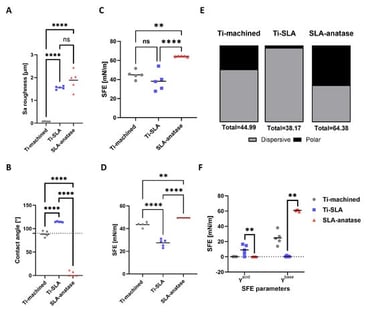
Physicochemical modification of titanium implants aims to enhance early osseointegration by improving bioactivity. This study deposited and evaluated an anatase TiO2 film on clinically relevant sandblasted, acid-etched titanium (Ti-SLA) to enhance in vitro bioactivity and osteogenic responses. An ~8 µm TiO2-anatase coating was deposited on Ti-SLA by reactive pulsed DC magnetron sputtering. Surface characterization included FE-SEM, helium ion microscopy, and XRD. Wettability and surface free energy (SFE) were evaluated by contact angle analysis. In vitro bioactivity was assessed by hydroxyapatite (HA) formation in twofold-concentrated simulated body fluid (2× SBF). Osteoblast responses were evaluated through cell adhesion, viability, alkaline phosphatase activity, gene expression, and mineralization. The coating produced hierarchical multi-globular microstructures decorated with faceted anatase nanocrystals. Ti-SLA’s initial hydrophobicity converted to a superhydrophilic, high-energy surface with increased polar SFE. Homogeneous HA crystallites deposited exclusively on SLA-anatase in 2× SBF. SAOS-2 cells showed enhanced metabolic activity, ALP activity, osteogenic gene upregulation, and improved mineralized matrix, while primary human osteoblasts exhibited increased metabolic activity and calcium deposition. The anatase coating produced a superhydrophilic, high-energy micro-nano surface that accelerates HA formation and enhances osteoblast function in vitro, warranting in vivo validation for early osseointegration.

6 February 2026

Physico-chemical characterization of the different surfaces. (A) Surface roughness (Sa) of sample discs determined by optical profilometry. (B) Quantitative results of static contact angle measurements of various surfaces. The grid line indicates the 90° threshold separating hydrophilicity from hydrophobicity. Surface free energy analysis of evaluated surfaces using Owens–Wendt (C) and Lifshitz–van der Waals/acid base (D) methods. The SFE was calculated by applying contact angle measurements to the OWRK and LW-AB equations. (E) Dispersive and polar component calculation using the Owens–Wendt method. (F) Comparison of acid and base components calculated by Lifshitz–van der Waals/acid between different surfaces. Each value represents the mean ± SD (n = 5). Statistical significance is indicated as follows: ** p < 0.01; **** p < 0.0001.

Over the past decade, endodontic biomaterials have shifted from being passive fillers to bioactive systems that can support repair and regeneration through validated physicochemical and biological mechanisms [...]

6 February 2026

  • Systematic Review
  • Open Access

Histological Tissue Response to Calcium Silicate-Based Cements Assessed in Human Tooth Culture Models: A Systematic Review

  • Alberto Cabrera-Fernández,
  • Hebertt Gonzaga dos Santos Chaves and
  • João Miguel Marques dos Santos
  • + 5 authors

Ex vivo human tooth culture models preserve the native dentine–pulp complex and offer a translational platform to study pulp-capping biomaterials. This systematic review aimed to synthesize the evidence on histological pulp tissue responses to calcium silicate-based cement (CSCs) used for direct pulp capping in human tooth culture models. The review followed PRISMA 2020 guidance. Eligible studies were ex vivo whole human tooth culture models with direct pulp exposure treated with commercial or experimental CSCs and reporting histological outcomes. Risk of bias was assessed using the QUIN tool. Thirteen studies were included. Most used immature human third molars (from 15- to 19-year-old patients) and culture periods up to 28 days, with a minority extending observation to 45–90 days. Across hydraulic CSCs, Biodentine was the most frequently evaluated material, followed by ProRoot MTA and several experimental hydraulic and resin-modified formulations. Overall, hydraulic CSCs were consistently associated with biocompatible pulp responses and a pro-mineralization pattern characterized by periexposure mineralized foci/osteodentin-like tissue; where assessed, immunohistochemistry supported odontoblast-like differentiation. In contrast, the resin-modified CSC TheraCal LC and other experimental resin-modified CSCs showed more heterogeneous findings, with reports of absent, delayed, or less prominent mineralization compared with reference hydraulic CSCs. In intact human tooth culture models, hydraulic CSCs show reproducible biocompatibility and early mineralization features consistent with reparative dentinogenesis, whereas resin-modified CSCs demonstrate more variable histological performance.

6 February 2026

Background: Autogenous tooth-derived grafts have been proposed as an alternative to xenografts for alveolar ridge preservation, offering biological similarity to bone and potentially more favorable remodeling. This study compared the healing outcomes of a collagenated xenograft, and a tooth-derived graft prepared with an automated processing device. Methods: Six Beagle dogs underwent bilateral extraction of the third and fourth mandibular premolars. Each animal contributed two sockets grafted with root-derived particulate prepared using an automated device for tooth cleaning, grinding, and demineralization, and two sockets grafted with a collagenated xenograft, all covered by a collagen membrane. After 3 months, histological sections were analyzed to assess crestal dimensions and the relative proportions of mature (lamellar) and immature bone (woven/parallel fibered), residual graft material, and soft tissues. Results: Lingual crest height did not differ between groups, whereas the buccal crest was slightly higher at xenograft sites compared with the tooth-graft sites. The tooth-graft group exhibited significantly fewer residual particles (0.5 ± 1.1%) and a higher proportion of total bone (65.6 ± 9.1%) compared with the xenograft group, which showed 19.7 ± 16.0% graft remnants (p = 0.032). Corticalization at the socket entrance was observed predominantly in the tooth-graft sites. No inflammatory infiltrates were detected in the examined section. Conclusions: Tooth-derived grafts promoted an almost complete replacement by vital bone with minimal residual material, whereas xenografts provided slightly better buccal contour preservation but resulted in regenerated tissues containing persistent graft particles. The biological differences observed may have implications for subsequent implant placement.

5 February 2026

News & Conferences

Issues

Open for Submission

Editor's Choice

Reprints of Collections

Functional Biomaterial for Bone Regeneration
Reprint

Functional Biomaterial for Bone Regeneration

Editors: Marija Čandrlić, Željka Perić Kačarević, Matej Tomas, Arun Kumar Rajendran
Feature Papers in Bone Biomaterials
Reprint

Feature Papers in Bone Biomaterials

Editors: Feng Chen, Zifei Zhou

Get Alerted

Add your email address to receive forthcoming issues of this journal.

XFacebookLinkedIn
J. Funct. Biomater. - ISSN 2079-4983