You are currently on the new version of our website. Access the old version .

International Journal of Molecular Sciences

International Journal of Molecular Sciences is an international, peer-reviewed, open access journal providing an advanced forum for biochemistry, molecular and cell biology, molecular biophysics, molecular medicine, and all aspects of molecular research in chemistry, and published semimonthly online by MDPI.
The Epigenetics Society, European Chitin Society (EUCHIS), Spanish Society for Cell Biology (SEBC) and others are affiliated with IJMS and their members receive a discount on the article processing charges.
Indexed in PubMed | Quartile Ranking JCR - Q1 (Biochemistry and Molecular Biology)

All Articles (107,352)

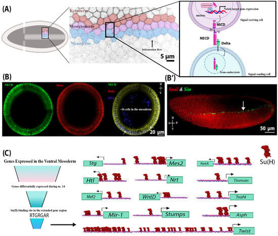
Notch signalling is a critical regulator of multiple developmental processes through its ability to control gene expression and thereby influence cell fate specification and cell proliferation through direct cell–cell communication. Although Notch signalling has been implicated in myogenesis during late embryogenesis, its role in early mesoderm development has been largely unexplored. Endocytosis of the Notch ligand Delta and the Notch receptor extracellular domain, a critical step in Notch pathway activation, has been extensively observed in the ventral mesoderm of the early Drosophila embryo, indicating a potential for Notch signalling activity in this early germ layer. Here, we present evidence that genes critical to mesoderm development require and are responsive to Notch signalling activity. Using a novel light-inducible Optogenetic variant of the Notch intracellular domain (OptoNotch), which affords precise spatial and temporal control over ectopic activation of Notch signalling, in combination with high-resolution fluorescent RNA in situ hybridization and qPCR, we identified a set of mesodermal genes whose expression is directly regulated by Notch signalling. We also provide evidence that Notch signalling indirectly regulates the dorsal–ventral patterning program mediated by the Toll signalling pathway through the Dorsal/Twist/Snail gene network. Our findings demonstrate that Notch signalling regulates ventral mesoderm patterning and is critical for establishing the mesoderm–mesectoderm–ectoderm boundary by regulating gene expression patterns and providing negative feedback on the upstream patterning network.

27 January 2026

NECD and Delta endocytosis in the mesoderm indicate that Notch signalling is active in the mesoderm. (A) A schematic of Notch signalling activation during early embryogenesis in the presumptive ventral mesoderm, neighbouring mesectoderm, and ectoderm cells in cellularized embryos. Notch signalling is activated via the binding of the ligand (located on the outermost mesoderm cell), Delta, to the extracellular domain of the Notch receptor (NECD) on a neighbouring mesectoderm cell (signal-receiving cell). This interaction leads to a series of Notch receptor cleavage events, which separate the Notch extracellular and intracellular domains. NECD and Delta are endocytosed into the signal-sending mesoderm cell, while the intracellular domain of Notch (NICD) is liberated from the plasma membrane and translocates to the nucleus, where it regulates gene expression. (B) Cross-sections of different wild-type embryos at stage 4 (cellularized blastoderm) showing the internalized Delta and NECD in mesodermal and mesectodermal (indicated by arrows) cells, indicating endocytosis and therefore Notch signalling activation. (B’) A lateral view of a stage 4 wild-type embryo showing the RNA expression of a Notch target gene, Sim (green), and its repressor, Snail (red), in the mesectoderm and mesoderm, respectively. (C) A summary of in silico analysis showing candidate Notch target genes that were differentially expressed in the mesoderm at nuclear cycle 14 and contained a Su(H) binding site in their proximal promoter. These include Asph, Tinman, Traf4, Twist, WntD, Stumps, String (Stg), Heartless (Htl), NetrinA (NetA), Neurotactin (Nrt), mir-1, Mes2, and Mef2. Only the binding sites preceding the 5′UTR are shown here. Su(H) sites are found upstream of the 5′UTR of all the transcripts of the genes above, except for Asph and Twist.

bFGF Oligomeric Stability Drives Functional Performance in Human Pluripotent Stem Cells

  • Dylan E. Iannitelli,
  • Naryeong Kim and
  • Julien Camperi
  • + 5 authors

Basic fibroblast growth factor (bFGF) and Transforming growth factor-beta (TGF-β) are key regulators of human pluripotent stem cell (hPSC) maintenance, supporting pluripotency and self-renewal. bFGF is particularly critical for sustaining the undifferentiated state and is commonly supplied through feeder-derived conditioned media. Similarly, TGF-β promotes hPSC expansion by modulating signaling pathways and contributing to a supportive stem cell niche. In this study, we investigated how the quality and variability of these growth factors influence hPSC culture performance. To address this, we developed and applied multiple physicochemical characterization methods—including size exclusion and reverse-phase chromatography—to assess growth factor purity and identify impurities across different material sources. Our findings show that certain post-translational modifications in TGF-β (e.g., oxidized variants) did not measurably affect hPSC culture. However, high temperature-dependent instability of bFGF preparations significantly altered hPSC morphology and growth. These findings underscore the need for improved quality control of growth factor components in culture media to ensure consistent hPSC maintenance, thus decreasing variability across experiments. This study highlights the value of correlating analytical physicochemical data with process performance, thereby advancing material understanding, enabling more efficient process development, and facilitating the identification of critical material attributes that affect the quality of cell therapy products.

27 January 2026

Assessment of Recombinant Human bFGF Oligomeric State from Different Vendors (A, B, C) using SEC-HPLC. (A) Purity profiles of bFGF materials obtained from Vendors A–C immediately after thaw at 4 °C and direct injection. Short-term stability profiles were determined by SEC-HPLC for bFGF-A (B) and for bFGF-B and bFGF-C (C). After thawing and holding aliquots at 4 °C, they were subsequently incubated at 37 °C for periods of 0, 0.25, and 4 h before analysis. Monomer (Mono., %) and aggregate (Agg., %) are quantified by peak-area integration and summarized.

Sympathetic nervous system (SNS) imbalance is a common pathological basis for cardiovascular diseases, non-alcoholic fatty liver disease, and diabetes. This review focuses on these diseases, analyzing two core mechanisms: excessive sympathetic excitation induced by endoplasmic reticulum stress (ERS) or autophagy dysfunction in key central nuclei (e.g., hypothalamus, rostral ventrolateral medulla); and ERS/autophagy abnormalities in peripheral target organs caused by chronic SNS overactivation. Existing studies confirm that chronic SNS overactivation promotes peripheral metabolic overload via sustained catecholamine release, inducing persistent ERS and disrupting the protective unfolded protein response (UPR)–autophagy network, ultimately leading to cell apoptosis, inflammation, and fibrosis. Notably, central ERS or autophagy dysfunction further perturbs autonomic homeostasis, exacerbating sympathetic overexcitation. This review systematically elaborates on SNS overactivation as a critical bridge mediating UPR–autophagy network dysregulation in central and peripheral tissues, and explores therapeutic prospects of targeting key nodes (e.g., chemical chaperones, specific UPR modulators, nanomedicine), providing a theoretical basis for basic research and clinical translation.

27 January 2026

Brain ERS/autophagy initiates sympathetic nervous system (SNS) hyperactivity and drives peripheral lesions. Glucose-regulated protein 78 kDa (GRP78) expression reflects endoplasmic reticulum stress (ERS). GRP78 upregulation signals that cells are counteracting ERS, indicating activated ERS but preserved cellular regulatory capacity. Adequate upregulation reduces unfolded proteins to attenuates ERS, whereas insufficient upregulation drives the progression to decompensation. In contrast, GRP78 downregulation signals the loss of cellular ability to combat ERS, which is accompanied by persistent and aggravated ERS, endoplasmic reticulum (ER) dysfunction, upregulation of pro-apoptotic molecules such as C/EBP homologous protein (CHOP), and ultimately leads to cell injury or death. Low-frequency component, it is a classic experimental indicator reflecting sympathetic neurogenic vasomotor activity. Black dotted lines within the cell membrane are used to separate the two functional parts of the figure: the left part represents PVN-associated processes, while the right part represents RVLM-associated processes. In this figure, “↑” indicates an increase in expression or activity of the corresponding molecule/pathway; meanwhile, arrows of different colors represent distinct regulatory pathways (e.g., the orange dashed arrow corresponds to the high-fat diet pathway, etc.). Created in BioRender. Bo Xu, Renjun Wang. (2025) https://BioRender.com/w55g70q (accessed on 18 December 2025). Abbreviations: ABP (arterial blood pressure); CHOP (C/EBP homologous protein); eIF2α (eukaryotic translation initiation factor 2 subunit alpha); GRP78 (glucose-regulated protein 78); HMG B1 (high-mobility group box 1); IRE1 (inositol-requiring enzyme 1); LC3-II (microtubule-associated protein 1A/1B-light chain 3-II); LAMP-2 (lysosomal-associated membrane protein 2); MASLD (metabolic dysfunction-associated steatotic liver disease); NAFLD (non-alcoholic fatty liver disease); PERK (protein kinase RNA-like endoplasmic reticulum kinase); RAGE (receptor for advanced glycation end products); ROS (reactive oxygen species); RSNA (renal sympathetic nerve activity); SHR (spontaneously hypertensive rat); SIH (stress-induced hypertension); XBP1s (spliced X-box binding protein 1).

Pancreatic β-cell replacement represents a promising therapeutic avenue for insulin-dependent diabetes, yet clinical translation has been limited by donor scarcity, immune rejection, and incomplete engraftment. Three-dimensional (3D) pancreatic organoids derived from human pluripotent stem cells (hPSCs) or primary tissue offer a scalable and physiologically relevant platform, recapitulating native islet architecture, paracrine interactions, and glucose-responsive insulin secretion. Recent advances in differentiation protocols, vascularization strategies, and immune-protective approaches—including encapsulation and hypoimmunogenic engineering—have enhanced β-cell maturation, survival, and functional performance in vitro and in vivo. Despite these developments, challenges remain in achieving fully mature β-cells, durable graft function, and scalable, reproducible production that is suitable for clinical use. This review highlights the promise of pancreatic organoid engineering, emphasizing strategies to optimize β-cell maturation, vascular integration, and immune protection, and outlines key future directions to advance organoid-based β-cell replacement toward safe, effective, and personalized diabetes therapies.

27 January 2026

Stepwise reconstruction of pancreatic islet architecture and functional maturation through 3D organoid engineering. Schematic overview illustrating the developmental and engineering roadmap toward functional pancreatic islet organoids for β-cell replacement therapy. Native pancreatic islets, derived from the pancreas, serve as the structural and functional blueprint for bioengineering efforts. Stem cells undergo 2D differentiation followed by self-organization into 3D islet organoids, which are further enhanced by vascularization and extracellular matrix (ECM) support. Through stepwise bioengineered protocols, pancreatic islet organoids achieve higher-order molecular, metabolic, and functional maturation, supported by cell–cell and paracrine interactions, architectural cues, and scalability and reproducibility. The final stage involves engraftment of transplantable bioengineered pancreatic islet organoids into Type 1 diabetic patients, aiming to restore physiological insulin regulation.

News & Conferences

Issues

Open for Submission

Editor's Choice

Reprints of Collections

Advances in Research for Legume Genomics, Genetics, and Breeding
Reprint

Advances in Research for Legume Genomics, Genetics, and Breeding

Editors: Naoufal Lakhssassi, Adnane Boualem, Gunvant Patil

Get Alerted

Add your email address to receive forthcoming issues of this journal.

XFacebookLinkedIn
Int. J. Mol. Sci. - ISSN 1422-0067