Abstract
Notch signalling is a critical regulator of multiple developmental processes through its ability to control gene expression and thereby influence cell fate specification and cell proliferation through direct cell–cell communication. Although Notch signalling has been implicated in myogenesis during late embryogenesis, its role in early mesoderm development has been largely unexplored. Endocytosis of the Notch ligand Delta and the Notch receptor extracellular domain, a critical step in Notch pathway activation, has been extensively observed in the ventral mesoderm of the early Drosophila embryo, indicating a potential for Notch signalling activity in this early germ layer. Here, we present evidence that genes critical to mesoderm development require and are responsive to Notch signalling activity. Using a novel light-inducible Optogenetic variant of the Notch intracellular domain (OptoNotch), which affords precise spatial and temporal control over ectopic activation of Notch signalling, in combination with high-resolution fluorescent RNA in situ hybridization and qPCR, we identified a set of mesodermal genes whose expression is directly regulated by Notch signalling. We also provide evidence that Notch signalling indirectly regulates the dorsal–ventral patterning program mediated by the Toll signalling pathway through the Dorsal/Twist/Snail gene network. Our findings demonstrate that Notch signalling regulates ventral mesoderm patterning and is critical for establishing the mesoderm–mesectoderm–ectoderm boundary by regulating gene expression patterns and providing negative feedback on the upstream patterning network.
1. Introduction
Embryogenesis is a critical developmental process that is driven by patterns of gene expression, which collectively organize the coordinated cell proliferation and differentiation required for proper organismal development [1]. Drosophila melanogaster has been used as a model organism to study the critical mechanisms that direct embryogenesis [1] and has also served as a useful model system to study signalling pathways that coordinate body patterning and morphogenetic movements in the formation of tissues and organs, which collectively give rise to entire organisms [1,2].
Embryonic development can be divided into four main stages: cleavage, blastulation, gastrulation, and neurulation [3]. The first stage, cleavage, involves multiple rounds of cell division that drive the formation of the blastula from a single-cell zygote [3]. Blastulation subsequently results in the formation of the blastula through subsequent rounds of cell division, with some accompanying differentiation occurring at the end of blastulation [3]. Gastrulation involves the development of the three germ layers: the ectoderm, the mesoderm, and the endoderm [2]. The ectoderm forms the outermost layers of the organism: the surface ectoderm, neural crest, and the neural tube [4]. The mesoderm forms the mid-layer of the organism, which later develops into muscle, bone, cartilage, the circulatory and lymphatic systems, connective and adipose tissue, the notochord, the dermis, serous membranes, and the genitourinary system [4]. Finally, the last stage of embryogenesis, neurulation, involves the development of the nervous system [4]. During early embryogenesis in Drosophila melanogaster, 13 nuclear division cycles occur within a shared syncytial cytoplasm [3]. This early developmental stage is instructed by maternally deposited mRNAs and proteins that organize the anterior–posterior and dorsal–ventral body axes and define the segmental pattern of the embryo that later gives rise to different body segments of the larva and adult [5]. This early body plan is subsequently refined by functional products from the zygotic genome [5]. During the 14th nuclear cycle, individual cell membranes form to separate, encapsulate, and thus individuate nuclei during blastoderm cellularization [3]. Many maternally deposited materials are degraded during this stage, and the control of development becomes increasingly dependent upon zygotic gene expression [6,7]. One of the critical factors for establishing the dorsal–ventral axis is a maternally deposited transcription factor, Dorsal, which functions downstream of the Toll signalling pathway and forms a morphogen gradient across the dorsal–ventral axis [8]. Once Toll signalling is activated, Dorsal translocates to the nucleus, where it activates the expression of master regulators of gastrulation, Twist and Snail, which are also transcription factors that activate genes necessary for mesoderm specification [9,10]. At the ventral side of the blastoderm, a 21-cell-wide anterior–posterior strip of cells in the ventral-most region of the cellularized blastoderm make up the presumptive ventral mesoderm, which undergo a series of cell shape changes, namely apical flattening, apical constriction, and shortening along the apicobasal axis, followed by cell depolarization, dispersion, and migration along the ectoderm, to which they attach [2,4]. Cell shortening along the apicobasal axis contributes to ventral furrow invagination and mesoderm ingression [2]. Upon completion of mesoderm ingression, FGF signalling between mesodermal cells and the underlying ectoderm facilitates mesoderm collapse against the ectodermal cell layer [11]. These mesodermal cells subsequently lose polarity, transition to a mesenchymal cell fate, and undergo two rounds of cell division [11]. Following this, FGF signalling facilitates cell migration and spreading along the ectoderm to form a cellular monolayer [11].
Notch signalling is critical to cell proliferation, differentiation, and the development of virtually all tissues that give rise to the adult organism [1]. It is also essential to the development of both vertebrates and invertebrates [12]. For example, it has been previously shown that Drosophila melanogaster embryos homozygous for loss-of-function Notch alleles do not reach adulthood [13] as they exhibit neuralized defects and die during embryogenesis. The Notch receptor, a transmembrane protein, is activated by the binding of its ligand, Delta (Dl), on neighbouring signal-sending cells, followed by ligand internalization and Notch receptor activation through transcytosis of the labile Notch extracellular domain, and subsequent intramembrane cleavage by the gamma secretase complex [1]. Notch cleavage triggers the release of the Notch intracellular domain (NICD) from the plasma membrane and its concomitant transport to the nucleus, where it regulates gene expression through the removal of transcriptional co-repressors bound to Suppressor of Hairless (Su(H)) and the subsequent recruitment of transcriptional co-activators that cooperatively activate transcription of Notch target genes [1]. Ligand internalization through endocytosis has been previously shown to be necessary for Notch activation [1]. Intriguingly, although no role for Notch signalling has been established in ventral mesoderm patterning to date, endocytosis of Delta and the extracellular domain of the Notch receptor is observed in all mesodermal cells (Figure 1B), indicating that Notch signalling may be operative in ventral mesodermal cells [14]. Although the role of Notch signalling during embryogenesis has been extensively studied, little is known about its role in the development of the ventral mesoderm [1]. Likewise, the identity of potential Notch target genes in the ventral mesoderm is not yet known.
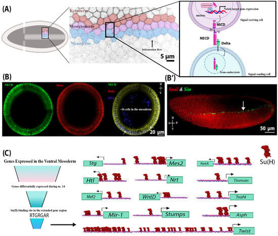
Figure 1.
NECD and Delta endocytosis in the mesoderm indicate that Notch signalling is active in the mesoderm. (A) A schematic of Notch signalling activation during early embryogenesis in the presumptive ventral mesoderm, neighbouring mesectoderm, and ectoderm cells in cellularized embryos. Notch signalling is activated via the binding of the ligand (located on the outermost mesoderm cell), Delta, to the extracellular domain of the Notch receptor (NECD) on a neighbouring mesectoderm cell (signal-receiving cell). This interaction leads to a series of Notch receptor cleavage events, which separate the Notch extracellular and intracellular domains. NECD and Delta are endocytosed into the signal-sending mesoderm cell, while the intracellular domain of Notch (NICD) is liberated from the plasma membrane and translocates to the nucleus, where it regulates gene expression. (B) Cross-sections of different wild-type embryos at stage 4 (cellularized blastoderm) showing the internalized Delta and NECD in mesodermal and mesectodermal (indicated by arrows) cells, indicating endocytosis and therefore Notch signalling activation. (B’) A lateral view of a stage 4 wild-type embryo showing the RNA expression of a Notch target gene, Sim (green), and its repressor, Snail (red), in the mesectoderm and mesoderm, respectively. (C) A summary of in silico analysis showing candidate Notch target genes that were differentially expressed in the mesoderm at nuclear cycle 14 and contained a Su(H) binding site in their proximal promoter. These include Asph, Tinman, Traf4, Twist, WntD, Stumps, String (Stg), Heartless (Htl), NetrinA (NetA), Neurotactin (Nrt), mir-1, Mes2, and Mef2. Only the binding sites preceding the 5′UTR are shown here. Su(H) sites are found upstream of the 5′UTR of all the transcripts of the genes above, except for Asph and Twist.
Here, we show that Notch signalling is active in the presumptive mesoderm and is required for proper progression through gastrulation. We identified a set of Notch target genes that are expressed in the ventral mesoderm and characterized their expression in embryos with either loss-of-function or gain-of-function mutations in the Notch pathway. In addition, we developed and characterized a novel tool, OptoNotch, that provides spatially and temporally precise light-gated activation of Notch signalling. Our findings support a mechanism whereby Notch signalling provides negative feedback control over the early patterning network established by the Toll pathway and thereby affects the Dorsal/Twist/Snail signalling network. Taken together, our results provide evidence that Notch signalling plays a critical role in the formation of the ventral mesoderm and in defining and positioning the boundary by distinguishing it from the ectoderm during early embryogenesis.
2. Results
2.1. Delta Endocytosis and NECD Trans-Endocytosis and Identification of Candidate Notch Target Genes in the Mesoderm
The role of Notch signalling during embryogenesis has been well characterized in the lateral ectoderm and the mesectoderm [15]. In the lateral ectoderm, Notch signalling works in concert with EGF and Toll signalling to coordinate neurogenesis [13,16]. In the mesectoderm, Notch signalling is required to form the boundary between the ectoderm and mesoderm through transcriptional activation of target genes, including single minded (sim) [17,18]. Several previous studies have suggested that Notch signalling is active in the ventral mesoderm, a hypothesis that is supported by the observation that the Drosophila Notch extracellular domain (NECD) is endocytosed in a Delta-dependent manner in the ventral mesoderm [14,19,20]. We confirmed the internalization of Delta and concomitant endocytosis of the NECD in ventral mesodermal cells (Figure 1B), which was previously reported [14,19,20]. This evidence suggests that Notch signalling may be operative in this tissue [14]. Traditionally, mesectodermal expression of sim, m8, bobble, bearded, and enhancer of split-related transgenes has been used as a marker of Notch signalling in the early embryo [18,21,22], where Delta endocytosis in the ventral mesoderm drives Notch activity in the mesectoderm, representing an intercellular and inter-cell-type flow of information (Figure 1A). However, since both sim and m8 are actively repressed in the ventral mesoderm by Snail, a transcriptional repressor that coordinates mesoderm formation, the activity of Notch signalling in the ventral mesoderm may have been overlooked [18]. When Snail repression was removed, as is the case in snail− mutants, sim RNA was detected in the mesoderm [16]. Thus, our study aimed to investigate the role of Notch signalling in the ventral mesoderm.
To address this question, we first generated a list of candidate transcriptional targets of Notch signalling using bioinformatic analysis of previously published Su(H) ChIP-seq data to identify genes that contain Su(H) binding sites proximal to their promoter and that exhibit differential expression during the nuclear cycle 14 (n.c. 14), which coincides with the increase in Notch signalling activity. This was performed in conjunction with available RNA in situ expression data from the Berkeley Drosophila genome project and with modENCODE RNA-seq expression data [17,23,24]. Using this approach, we identified Asph, Twist, Tinman, Traf4, WntD, Stumps, String, Heartless, NetrinA, Neurotactin, mir-1, Mes2, and Mef2 as candidate transcriptional targets of the Notch signalling pathway (Figure 1C).
2.2. Notch Signalling Is Required for the Expression of Critical Mesodermal Genes
To test the necessity of Notch signalling in ventral mesodermal development, we employed temperature-sensitive loss-of-function Delta mutant alleles (Dl−) to analyze the expression of the twelve candidate mesodermal Notch target genes we identified through in silico analysis. To accomplish this, we precisely quantified changes in transcript abundance and qualitatively assessed the localization of expression for each of the genes by using a combination of two orthogonal methods, quantitative real-time PCR (qRT-PCR) and fluorescent RNA in situ hybridization (FISH), respectively. We used sim as a readout of Notch signalling activity in both RNA FISH and qRT-PCR. In Dl−, sim expression in the mesectoderm was significantly reduced and was only detected in the posterior pole (Figure 2A–C). In addition, total sim expression was significantly reduced, as shown by qRT-PCR (Figure 2D).
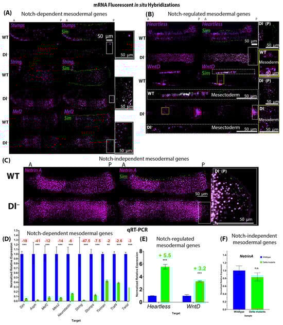
Figure 2.
Expression of mesodermal genes in wild-type and loss-of-function Delta mutant embryos shows that Notch signalling is necessary for the expression of many mesodermal genes. (A–C) Fluorescent mRNA in situ hybridizations against sim (green) and mesodermal genes (purple) in wild-type and Delta mutant embryos with a formed cephalic furrow (stage 5–6). (A) shows examples of genes whose expression was significantly reduced in Delta mutants (Dl−) compared to their wild-type (WT) counterparts (Stumps, String, and Mef2). (B) shows the two genes (Heartless and WntD) whose expression was upregulated in Dl−. The expression of WntD in the mesectoderm is shown below the corresponding embryo labelled “WT” or “Dl−” in white text, and expression in the mesoderm is shown in the insets to the right labelled “WT mesoderm” or “Dl− mesoderm”, corresponding to the yellow boxes in the images of the whole embryos. (C) Expression of NetrinA was unchanged in Dl−. N = 5 for each genotype per gene. (A–C) Insets to the right of each panel show the expression of sim (green) in the posterior pole of Dl− mutants {corresponding to the white boxes in the image of the whole embryo [‘Dl− (P)’]}, indicating that Notch signalling was effectively reduced. (D) qRT-PCR generated normalized relative expression of mesodermal genes and sim in wild-type and Delta mutant embryos with a cephalic furrow (stage 5–6). Expression of sim (p = 9.1711 × 10⁻⁶), Asph (p = 0.0007), Mef2 (p = 0.0007), Mes2 (p = 7.77419 × 10⁻⁵), Neurotactin (p = 0.0002), String (p = 0.0004), Stumps (p = 0.000149162), Tinman (p = 0.0067), Traf4 (p = 8.19966 × 10⁻⁵), and Twist (p = 5.80546 × 10⁻⁵) was significantly reduced in Delta mutants compared to stage-matched wild-type embryos. (E) Expression of Heartless (p = 1.39725 × 10⁻⁵) and WntD (p = 0.0022) was significantly increased in Delta mutants, while (F) the expression of NetrinA (p = 0.3359) was not significantly changed. Fold changes are reported, where decreases are indicated in red writing and increases are indicated in green writing. Independent two-tailed t-test were performed for each gene to compare the expression of each gene in Dl− to that in WT embryos. ‘**’ represents p < 0.01, and ‘***’ represents p < 0.005. Error bars represent the standard error of the mean. For each genotype, 30 embryos were used (N = 30).
Through this approach, we identified three classes of genes within this set of candidates based on changes in their expression levels in response to the loss of Notch signalling (Figure 2):
- Class 1—Genes whose expression requires Notch signalling. These include Asph, Mef2, Mes2, Neurotactin, String, Stumps, Tinman, Traf4, and Twist (Figure 2A,D);
- Class 2—Genes whose expression is repressed by Notch signalling, namely Heartless and WntD (Figure 2B,E);
- Class 3—Genes whose expression is independent of Notch signalling, namely NetrinA (Figure 2C,F).
All but one gene (NetrinA) showed significant changes in expression patterns between wild-type and Delta mutant embryos. However, WntD was expressed at higher levels in the mesoderm of Delta mutants, while its expression was significantly reduced in mesectodermal cells, compared to wild-type embryos (Figure 2B).
2.3. Development of OptoNotch Allows Titration of Ectopic Notch Signalling Activity and Incremental Expansion of Sim Expression
The above findings motivated us to test the sufficiency of Notch signalling to drive the expression of the ventral mesoderm target gene candidates, and thus, we developed a tool that allows us to ectopically activate Notch signalling with precise temporal control. We engineered a light-inducible variant of the NICD (OptoNotch) and expressed it in the early embryo using the GAL4/UAS system. We generated transgenic fly lines that contained two transgenes under the control of an upstream activating sequence (UAS) promoter. We then generated flies that contained maternally deposited GAL4, which drove the expression of the transgenes encoding the two components of our OptoNotch system. The detailed genetic crosses performed are described in the Materials and Methods section “Fly crosses and transgenic lines”.
We adapted the previously described system to develop a light-inducible variant of NICD [25]. Our system is composed of two distinct proteins. One is a cytosolic protein containing Cry2, mCherry, and the C-terminal portion of a split TEV protease (Cry2-mCherry-CTEV), while the other is a membrane-tethered protein containing mCD8, CIBN, the complementary N-terminal portion of TEV protease, an ASLOV domain that masks a TEV cleavage sequence in the dark and precedes NICD, and GFP (mCD8-CIBN-NTEV-NICD-GFP). Upon photoactivation with blue light (488 nm), Cry2 binds to CIBN, bringing the two components of the TEV protease together to reconstitute the functional protease, and ASLOV undergoes a conformational change, exposing the TEV cleavage sequence. The TEV protease then cleaves its cleavage sequence, liberating NICD from the membrane. NICD contains a nuclear localization sequence, as does the endogenous protein, so it translocates to the nucleus once it is liberated from the membrane.
Therefore, prior to photoactivation with blue light (488 nm), OptoNotch is tethered to the membrane. Upon photoactivation, NICD is cleaved through light-inducible reconstitution of TEV protease activity, promoting NICD translocation to the nucleus, where it can activate target gene expression (Figure 3A). We first tested OptoNotch in S2 cells. Prior to photoactivation, OptoNotch (tagged with GFP) was localized to the plasma membrane and internal membranous structures but was excluded from the nucleus. As expected, light exposure caused nuclear translocation of NICD; following one hour of continuous photoactivation, OptoNotch::GFP was detected in the nucleus (Figure 3B).
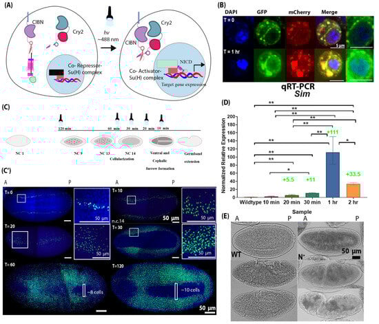
Figure 3.
Development of Optogenetic NICD (OptoNotch) revealed the competency of mesodermal cells to express a Notch target gene, sim. (A) Schematic of the mechanism of the Optogenetic variant of NICD (OptoNotch) that we developed, which is described in detail in the text. (B) S2 cells were transiently transfected with OptoNotch and imaged prior to photoactivation and after continuous photoactivation for 1 h. Prior to photoactivation (T = 0), NICD-GFP is excluded from the nucleus, while after 1 h (T = 1 h) of continuous photoactivation, NICD-GFP is observed in the nucleus (blue). mCherry (red) shows the localization of the second component of the OptoNotch system, Cry2-CTEV-mCherry. The two rightmost panels show nuclear GFP at T = 0 and T = 1 h. (C) Embryos expressing both components of the OptoNotch system were either not photoactivated (T = 0) or continuously photoactivated for 10 min (n = 3), 20 min (n = 3), 30 min (n = 10), 60 min (n = 10), or 120 min (n = 30) before fixation. Embryos photoactivated for longer durations were photoactivated at an earlier stage of embryogenesis. Embryos that were photoactivated for 10 min were undergoing cephalic furrow (CF) and ventral furrow (VF) invagination (during n.c. 14). Embryos photoactivated for 20 min were photoactivated during late n.c. 14, 5 min before VF invagination. Embryos photoactivated for 30 min were photoactivated during mid n.c. 14 or 15 min before the complete formation of the VF. Embryos photoactivated for 60 min were photoactivated 45 min before the completion of VF invagination, during early n.c. 14. Lastly, embryos photoactivated for 120 min were at n.c. 9 (~55 min before the beginning of cellularization) when photoactivated. (C’) The corresponding fluorescent in situ hybridizations against sim (green) in embryos expressing OptoNotch photoactivated for different durations. Nuclei are shown by DAPI nuclear counterstain (blue). Prior to photoactivation (T = 0), sim expression is identical to that in wild-type embryos (n = 10). Sim expression is incrementally expanded into lateral regions of OptoNotch embryos photoactivated for 10, 20, and 30 min (stage 4 embryos shown, prior to cephalic furrow formation). The regions of sim expression expansion are highlighted with white boxes T10 and T20. The adjacent insets were taken from the regions in the white boxes. Sim expression is observed in the mesoderm regions of OptoNotch embryos photoactivated for 60 and 120 min (stage 6 embryos shown). In embryos photoactivated for 60 min, the maximum number of cells (across the dorsoventral axis) lacking sim expression is ~8 cells, and in those photoactivated for 2 h, ~10 cells across the dorsal–ventral axis lack sim expression. These regions are highlighted with white boxes in the bottom two images. (D) qRT-PCR generated normalized relative expression of sim of stage 6 wild-type and OptoNotch embryos photoactivated for different durations, as indicated on the x-axis. N = 30 embryos for each group. A one-way ANOVA was performed (F(5,12) = 47.68; p = 0.0001). A post hoc Tukey HSD test was used to perform pairwise comparisons. Statistical significance is represented by stars, where ‘*’ indicates statistical significance determined by a Tukey HSD at p < 0.05 and ‘**’ represents p < 0.01. Fold changes in expression compared to wild-type embryos are indicated above the relevant bars. Error bars represent the standard error of the mean. (E) Brightfield images of wild-type (WT) and OptoNotch (N+) embryos (photoactivated for 2 h) at different stages of gastrulation: ventral furrow invagination or stage 6 (top), germband extension or stage 7 (middle), and stage 10 (bottom). Morphological defects along the anterior–posterior axis can be observed in N+ embryos during early gastrulation, and complete breakdown is observed during later stages; N = 30 for each genotype.
Next, we expressed our construct in the early embryo using a maternally deposited Gal4 driver to drive the expression of the two components of the OptoNotch system, which were under the control of a UAS promoter. We confirmed hyperactivation of Notch signalling by performing immunohistochemistry against NICD (Figure S6). Both the total and nuclear levels of NICD were significantly increased in OptoNotch blastoderm embryos compared to their wild-type counterparts (Figure S6). To assess the functional output of our OptoNotch system, we used sim expression as a readout for Notch signalling levels because it is a direct target of Notch signalling. Its expression domain has previously been shown to expand beyond the mesectoderm as a result of overexpression of the NICD [18]. As predicted, prior to photoactivation, the level of Notch signalling activity, shown by sim expression, was similar to wild-type embryos, as only endogenous Notch signalling was active (Figure 3C). We also showed that we could control the extent of Notch activation by varying the duration of photoactivation (Figure 3C,D). Embryos exposed to 10 min of light exhibited the smallest expansion in sim expression (which was only observed in embryos at mid n.c.14 embryos by in situ but not in older embryos with a cephalic furrow-represented in the qRT-PCR data) (Figure 3C,D). Incremental expansions were observed in sim expression that correlated with the duration of photoactivation, except that sim expression was higher in embryos photoactivated for 1 h than in those photoactivated for 2 h (Figure 3C,D). This could be due to negative feedback mechanisms, which downregulate sim expression. After 1 h of light exposure, the embryos expressed sim in the presumptive mesoderm, and the number of cells that did not contain sim mRNA was significantly decreased compared to wild-type embryos (~8 cells at most, 0 in some areas along the AP axis). This was also observed in embryos exposed to light for 2 h, but to a lesser extent (~10 cells at most, 0 around the cephalic furrow) (Figure 3C,D). These results demonstrate the successful development and application of a light-inducible NICD variant that provides titratable control over Notch signalling activity, which is sufficient to drive expression of the Notch target gene sim in the mesoderm without the removal of the repressor, Snail.
We also observed morphological defects in embryos exposed to light for 1 and 2 h (Figure 3E). These embryos (n = 30) exhibited ectopic invaginations along the AP axis during the early stages of gastrulation, where the ventral furrow was forming but the mesoderm was not fully internalized. Following internalization of the mesoderm, the cephalic furrow appeared closer to the middle of the mutant embryo along the AP axis compared to wild-type counterparts. There were also additional invaginations along the AP axis on the ventral side. At older stages that correspond to germband retraction, the mutant embryo was twisted, which was reminiscent of the phenotype of loss-of-function Twist mutant embryos (Figure S1). Embryos expressing OptoNotch that were not photoactivated developed normally, reached adulthood, and showed no morphological defects. Similarly, wild-type embryos that were photoactivated in the same manner as OptoNotch embryos also developed normally and reached adulthood with no morphological defects. We assessed sim expression in embryos expressing each single component and both components of the OptoNotch system together by FISH and observed a normal level of sim expression in embryos expressing each of the components alone and embryos that were not photoactivated (Figure S2).
2.4. Ectopic Notch Signalling Interferes with Mesoderm Formation and Decreases Mesodermal Target Gene Expression
To test whether Notch signalling activation is sufficient to drive the expression of the mesodermal genes, we activated Notch signalling using OptoNotch in the early embryo and examined mesodermal gene expression using FISH and qRT-PCR. We photoactivated n.c. 9 embryos expressing both components of the system by exposing them to light, using an LED light box, for 2 h continuously.
We found that there are two classes of genes within this set of candidates based on their expression levels when Notch signalling is overactivated (Figure 4):
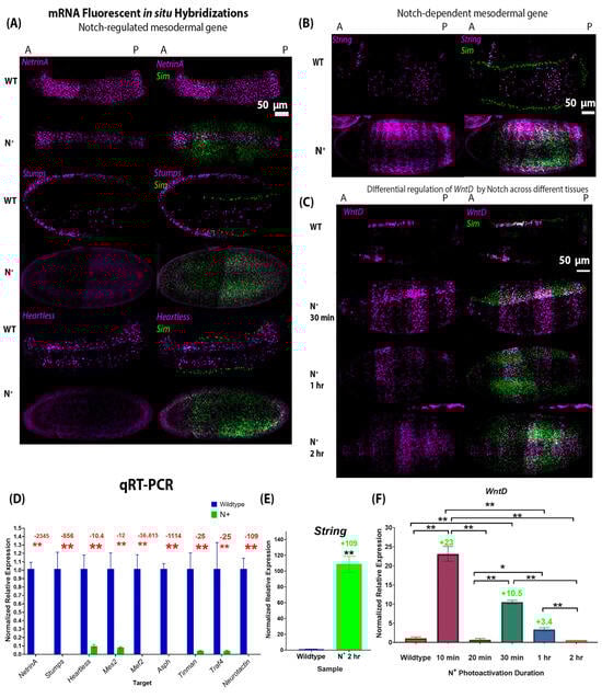
Figure 4.
Expression of mesodermal genes in wild-type and gain-of-function Notch mutant (OptoNotch) embryos shows that Notch hyperactivity suppresses most mesodermal genes and the sufficiency of Notch to drive overexpression of WntD and String. (A–C) Fluorescent mRNA in situ hybridizations against sim (green) and mesodermal genes (purple) in wild-type (WT) and N+ embryos (OptoNotch embryos photoactivated for 2 h) at the cephalic furrow (stage 6). (A) shows examples of genes whose expression was significantly reduced in N+ embryos compared to their WT counterparts, namely Stumps, NetrinA, and Heartless. (B,C) shows the expression of the two genes whose expression was upregulated in Notch loss-of-function mutants: String and WntD. Their expression expands laterally into the ectoderm of N+ embryos. (D) Expression of Asph, Mef2, Mes2, Neurotactin, NetrinA, Stumps, Tinman, Heartless, and Traf4 was significantly reduced in N+ compared to stage-matched wild-type embryos. The p-values from the t-test (described below) are Asph: p = 1.54988 × 10⁻⁶, Mef2: p = 5.32916 × 10⁻⁷, Mes2: p = 0.0002, Neurotactin: p = 8.46069 × 10⁻⁶, NetrinA: p = 2.1557 × 10⁻⁵, Stumps: p = 3.29648 × 10⁻⁶, Tinman: p = 7.16119 × 10⁻⁵, Heartless: p = 0.0006, and Traf4: p = 0.0006. (E) Expression of String is significantly increased in N+ embryos compared to wild-type embryos, p = 0.00028947. For (D,E), independent two-tailed t-test were performed for each gene to compare expression in N+ to that in wild-type embryos. ‘**’ indicates p < 0.01. Error bars represent the standard error of the mean. (F) Expression of WntD is significantly increased to different levels in OptoNotch embryos photoactivated for different durations of time compared to wild-type embryos, which is shown by increased in fold changes. N = 30 embryos for each group. A one-way ANOVA was performed (F(5,12) = 31.6664; p = 1.63 × 10⁻⁶). A post hoc Tukey HSD test was used to perform pairwise comparisons. Significant differences are denoted by ‘*’, which represents a p < 0.05, and ‘**’ represents p < 0.01. Error bars represent the standard error of the mean.
- Class 1—Genes whose expression is repressed by Notch signalling overactivation. These include Asph, Mef2, Mes2, Neurotactin, String, Stumps, Tinman, Traf4, Twist, and NetrinA (Figure 4A,D). Fold changes are denoted in Figure 4D, and p-values are reported in the figure caption. While the expression of these genes was downregulated in embryos where OptoNotch was photoactivated for 2 h, a subset of these genes were upregulated when Notch signalling was overactivated for shorter durations. This indicates that small to moderate increases in Notch signalling activity are sufficient to increase their expression, while very high levels of Notch signalling repress their expression (Figure S3).
In wild-type embryos, WntD is expressed at a high level in all mesectodermal cells and at a lower level in a heterogeneous segmented manner across the anterior–posterior axis of the ventral mesoderm, whereas it is not expressed in the ectoderm (Figure 4C) [26]. This unique expression pattern is a distinguishing feature of the mesoderm cells. In contrast, we observed that overactivation of Notch signalling in OptoNotch embryos alters WntD expression such that the mesectodermal cells exhibit lower expression levels, similar to those observed in neighbouring mesodermal cells. Strikingly, we also observed significant, titratable expansion of WntD expression in ectodermal cells. WntD expression was increased in OptoNotch embryos that were photoactivated for 30 min in the mesoderm, but was still segmented, and this pattern was distinct from that of neighbouring mesectoderm cells, where WntD was expressed in a continuous (non-segmented) 2–3-cell-wide stripe along the AP axis. In contrast, WntD was expressed in mesodermal, mesectodermal, and ectodermal cells in a segmented pattern across the AP axis of OptoNotch embryos photoactivated for 1 h, which exhibited the highest levels of Notch signalling, as measured by increases in sim expression (Figure 3). To confirm the association of OptoNotch with the extended gene regions of WntD, String, and Sim, we performed Chromatin Immunoprecipitation (ChIP) using magnetic GFP-Trap agarose beads and confirmed the presence of Su(H) consensus binding sites using PCR and amplicon sequencing (Figure S7G). We found that the GFP-trap beads eluted non-GFP-tagged proteins; however, using Western blot, we detected the presence of GFP-fused NICD (~37–150 kDa) only in photoactivated OptoNotch embryos (Figure S7B). In wild-type and the other control embryos (Cry2 alone, mCD8 alone, and the OptoNotch embryos that were not photoactivated), we detected the full-length Notch protein (~300 kDa) and a cryptic fragment below 75 kDa, but we did not detect any bands at 150 kDa or 110 kDa, the expected size of NICD-GFP and NICD, respectively (Figure S7A–F) [27,28]. We found that OptoNotch was associated with one region in the STRING gene locus, which contained one Su(H) binding site, and two regions in the WntD gene locus, which contained four sites in total (Figure S7G). We also detected a previously identified strong Su(H) binding site in SIM as a positive control (Figure S7G) [18]. Importantly, we did not detect any amplicons for the bound DNA eluted from wild-type or the other control embryos, indicating specific direct recruitment of NICD::GFP to Su(H) binding sites in the aforementioned identified regulatory regions. Ectopic Notch signalling alters the expression profile of mesodermal, mesectodermal, and ectodermal cells and, thus, these cell types are less distinguishable from one another. This emphasizes the important role that Notch signalling plays in boundary refinement.
Our observation that mesoderm and mesectodermal/ectodermal expression of WntD was differentially regulated by Notch signalling drove us to further investigate the regulatory relationship between Notch signalling and WntD expression levels. Given that WntD is a transcriptional target of the Dorsal/Twist/Snail network, we decided to investigate whether changes in the levels of Notch signalling activation affected the expression of Twist and Snail [26].
2.5. Notch Signalling Regulates Twist and Snail Expression
Twist and Snail are both transcription factors that play a critical role in mesoderm patterning and in gastrulation [2,4,29]. Twist is a transcription factor that is necessary for the expression of most mesodermal genes involved in gastrulation, while Snail is a transcriptional repressor that inhibits the expression of genes that specify other tissues, such as the mesectoderm and the ectoderm [4,29]. We analyzed embryos at cellularization to determine the number of cells in the mesoderm expressing Twist.
We found that the number of Twist-positive cells of loss-of-function Delta mutants (Dl−) and gain-of-function (N+) Notch mutants was significantly reduced, from 18 cells (±0.8, n = 5) in wild-type to 12 (±0.86, n = 4) in Dl− and 9 (±0.67, n = 3) in N+ mutants (Figure 5A,B). Ventral furrow invagination proceeds normally in Dl− mutants, but in N+ mutants, there were additional groups of cells that invaginate, giving rise to irregular, incomplete invagination in the embryos, and the ventral furrow appeared disorganized following ingression (Figure 5A,D). During later stages of gastrulation, both Dl− and N+ mutants show developmental defects, which resemble the Twist loss-of-function mutant phenotype (Figure S1). The reduced protein expression of Twist and Snail is supported by reduced gene expression of these mutants compared to their wild-type counterparts (Figure 5C).
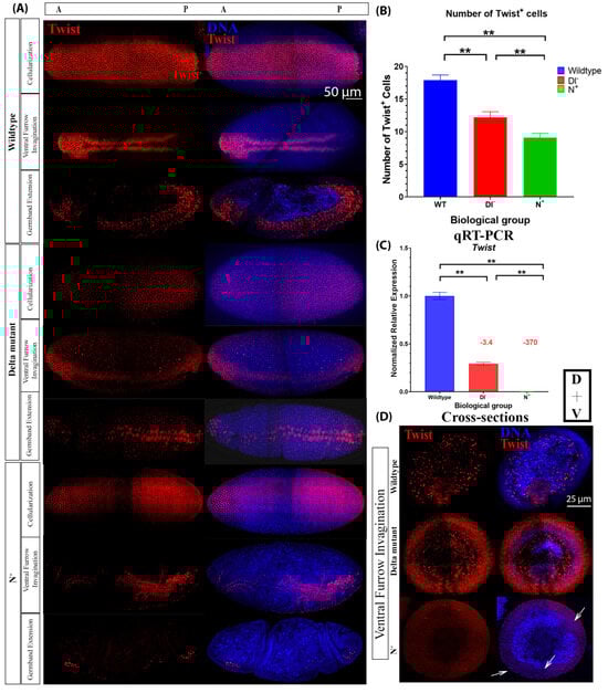
Figure 5.
Twist protein and mRNA expression vary between wild-type and Notch gain-of-function and loss-of-function mutant embryos. (A) Twist protein expression during cellularization, early gastrulation, and late gastrulation in wild-type (WT), loss-of-function (Delta mutant (Dl−)), and gain-of-function Notch mutants (N+, OptoNotch embryos photoactivated for 2 h prior to fixation). (B) Number of Twist-positive cells in cellular blastoderms (WT: n = 4, Dl−: n = 5, N+: n = 3). Error bars represent standard deviation. A one-way ANOVA was used to test for significance (F(2,9) = 126; p < 0.0001); then, a Tukey HSD test was performed, which showed that all groups were significantly different from one another (p = 0.01 for all pairwise comparisons). ‘**’ indicates p < 0.01. (C) qRT-PCR generated normalized relative mRNA expression of twist, n = 30 embryos at cephalic furrow formation in each genotype. Error bars represent the standard error of the mean. A one-way ANOVA was used to test for significance (F(3,8) = 436.7164; p = 3.33 × 10⁻⁹), then a Tukey HSD test was performed, which showed that all groups were significantly different from one another (p = 0.001 for all pairwise comparisons). ‘**’ denotes p < 0.01. Fold changes in expression compared to wild-type embryos are indicated above the relevant columns. (D) Twist protein expression in cross-sections of wild-type, Delta mutant and N+ embryos undergoing ventral furrow invagination. Twist is expressed in the invaginating cells of wild-type and Delta mutants; however, in N+ embryos, expression is reduced, and there are multiple groups of cells invaginating or beginning to invaginate (arrows).
We also analyzed Snail expression at the protein and mRNA levels. The number of Snail-positive cells during cellularization was significantly reduced in Dl− (12 cells ± 1.62, n = 5) and N+ mutants (9 cells ± 2.43, n = 3) compared to wild-type embryos (18 cells ± 0.79, n = 4) (Figure 6A,B). In Dl− mutants, snail mRNA expression was significantly increased, while in N+ mutants, snail mRNA expression was significantly decreased (Figure 6C,D). Altogether, this indicates that Notch signalling negatively regulates snail expression. Given that Twist and Snail are markers of mesodermal cell fate and that the number of Twist-positive cells approximately corresponds to the number of Snail-positive cells in wild-type and mutant embryos, these findings indicate that decreases and increases in Notch signalling beyond physiological levels lead to a reduction in the number of presumptive mesodermal cells in blastoderm embryos, which led to more drastic defects of gastrulation in older embryos (Figure 5A, Figure 6A and Figure S1). The discrepancy between Snail protein and mRNA expression in Dl− mutants indicates that there are two distinct inputs from Notch signalling, which will be discussed further below. Taken together, these data support the hypothesis that Notch signalling is active in the ventral mesoderm and is required for delineating the pattern of mesodermal gene expression and proper progression through gastrulation.
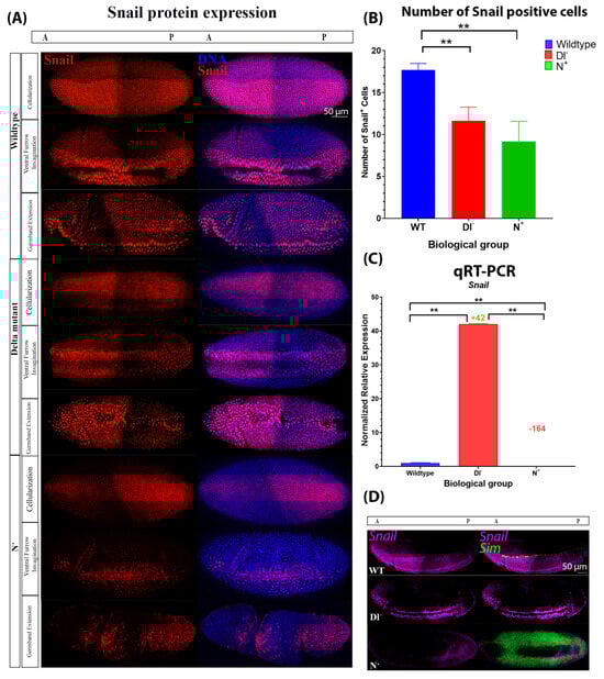
Figure 6.
Snail protein and mRNA expression varies between wild-type and Notch gain-of-function and loss-of-function mutant embryos. (A) Snail protein expression during cellularization, early gastrulation, and late gastrulation in wild-type, Dl−, and N+ mutant embryos. (B) Number of Snail-positive cells in cellular blastoderms (WT: n = 6, Dl−: n = 5, N+: n = 3). Error bars represent standard deviation. One-way ANOVA was used to determine whether there were significant differences (F(2,9) = 26.24; p = 0.0002), then a Tukey HSD test was performed, which showed that Dl− (p = 0.001) and N+ (p = 0.001) were significantly different from those of WT embryos but not from one another (p = 0.1504). ‘**’ indicates p < 0.01. (C) Normalized relative mRNA expression of snail, n = 30 embryos with a formed cephalic furrow of each genotype. Error bars represent SEM. Statistical significance is denoted by ‘**’ (p < 0.01) determined by ANOVA (F(3,8) = 443.6922; p = 3.13 × 10⁻⁹) and a Tukey HSD test, which showed that all groups were significantly different from one another (p = 0.001 for all pairwise comparisons). Fold changes in expression compared to wild-type embryos are reported above the relevant columns. (D) mRNA fluorescent in situ hybridizations against Snail (purple) and sim (green) in wild-type, Delta mutant, and N+ embryos.
Given that Twist and Snail are transcriptional targets of Dorsal, these results motivated us to investigate whether there is an input from Notch signalling on the Dorsal patterning gradient. We then analyzed Dorsal activity during cellularization in wild-type, Notch loss-of-function, and Notch gain-of-function embryos.
2.6. Notch Signalling Provides a Negative Feedback Signal on Dorsal Distribution by Regulating WntD Expression
As mentioned previously, Dorsal is a maternally deposited transcription factor that forms a morphogen gradient across the dorsal–ventral axis and is critical for mesoderm specification. In the absence of Toll signalling, Dorsal is bound to a protein named Cactus in the cytoplasm, which is responsible for restricting its entry into the nucleus [30]. Once Toll signalling is activated by Spätzle (Spz), Cactus is targeted for destruction, liberating Dorsal, which then translocates to the nucleus, where it activates gene expression [9,10].
As previously reported, we observed that the nuclear abundance of Dorsal is most concentrated in the ventral-most region of the blastoderm in wild-type embryos, which has been shown to activate transcription of two ‘master regulators’ of gastrulation: Twist and Snail [2,4,29,31]. In contrast, we observed that the number of cells that exhibit prominent nuclear localization of Dorsal is significantly reduced from (17 cells ± 2.82, n = 4) in wild-type embryos to (13 cells ± 1.46, n = 3) in Dl− mutants (Figure 7A,B). Interestingly, we observed two distinct classes of N+ mutant embryos:
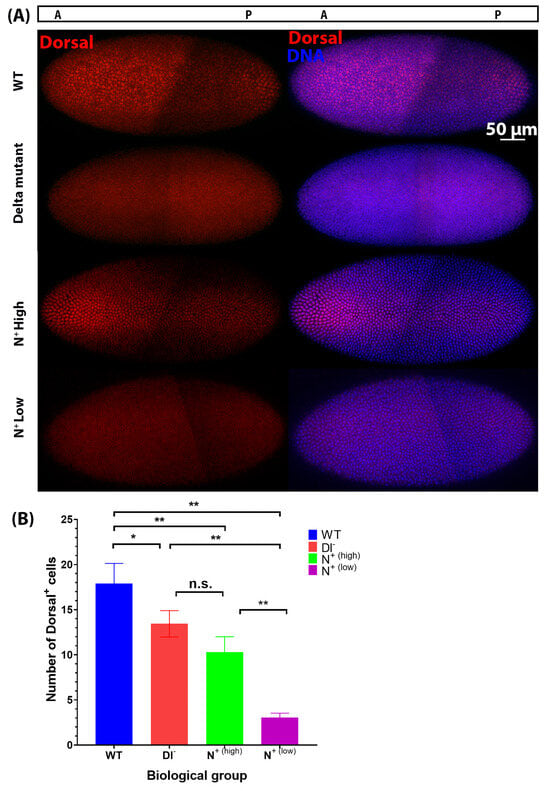
Figure 7.
Nuclear concentration of Dorsal on the ventral side of embryos decreases in Notch gain-of-function and loss-of-function mutants compared to wild-type embryos. (A) Dorsal protein expression (red) in cellular blastoderm embryos (stage 4). N+ are OptoNotch embryos photoactivated for 2 h prior to fixation. (B) The number of cells in the mesoderm was determined using the peak region from the line scan plots. One-way ANOVA was used to test for statistical significance between wild-type (WT) embryos (n = 4), Delta mutants (Dl−) (n = 3), N+(high) mutants that had a higher level of Dorsal expression (n = 3), and N+(low) mutants with a very low level of Dorsal signal (n = 3) (F(3,8) = 45.96; p < 0.0001). A Tukey HSD test was used to perform pairwise comparisons, which showed that Dl− and N+(high) were significantly different from WT and from N+(low), but not from one another, shown by ‘n.s’. N+(low) was significantly different from all other groups. ‘*’ indicates p < 0.05 and ‘**’ indicates p < 0.01. WT vs. Dl− p = 0.0365, WT vs. N+(high) p = 0.0017, WT vs. N+(low) p = 0.001, Dl− vs. N+(high) p = 0.1545, Dl− vs. N+(low), p = 0.0010053, N+(high) vs. N+(low) p = 0.0024. Error bars represent standard deviation.
This suggests that Notch signalling negatively regulates Dorsal nuclear distribution in the mesoderm, as when Notch signalling is increased, fewer cells contain a high nuclear concentration of Dorsal protein. These results are consistent with our observation of reduced expression of both Twist and Snail protein levels in Notch loss-of-function and gain-of-function mutants (Figure 5 and Figure 6). A reduction in the nuclear accumulation and activity of Dorsal in the ventral mesoderm is expected to result in reduced expression of its target genes, Snail and Twist. Considering that Dorsal activity is thought to precede the onset of Notch signalling during early embryogenesis, we propose that Notch is acting on the Dorsal/Twist/Snail network through an intermediate negative regulator, specifically WntD. Here, we provided evidence that Notch signalling upregulated WntD expression. As WntD inhibits Dorsal nuclear localization, the increase in WntD expression leads to decreases in the nuclear localization of Dorsal and transcriptional activation of its target genes, including Twist and Snail [26]. We therefore propose that ectopic Notch signalling is sufficient to alter ventral mesodermal patterning through the effects of its downstream target genes (i.e., WntD) on the Dorsal/Snail/Twist patterning network. Similarly, our findings indicate that decreases in Notch signalling result in reductions in Twist and Snail protein expression, leading to a reduction in mesodermal cell number in the early embryo, and result in a disorganized ventral furrow later in gastrulation, underscoring the necessity of Notch signalling for proper mesoderm formation and gastrulation.
3. Discussion
Disentangling the role of Notch signalling during early embryogenesis is critical to understanding the mechanisms involved in tissue formation, boundary establishment, and refinement. The processes involved in tissue and boundary formation are conserved in later stages of the organism’s life, as in stem cell renewal and differentiation. Unravelling the interactions between different signalling pathways, such as Notch signalling and Toll signalling, is also important for understanding disease pathogenesis, where the same processes are dysregulated and lead to disease [32].
The critical role of Notch signalling during embryogenesis has long been appreciated in the context of neurogenesis and myogenesis, which both occur after early mesoderm development [15,33]. However, while there is evidence to suggest that Notch signalling is active in the ventral mesoderm, its role in this early embryonic tissue has been largely unexplored. Previously, only a few Notch target genes expressed in the mesoderm were known, such as Twist, String, and Asph. While Notch signalling has been previously shown to regulate the expression of Twi, Asph, and String during later stages of embryogenesis and larval stages and in adult tissues, it remained unknown whether Notch signalling was required for mesodermal gene expression or mesoderm specification during early embryogenesis [34,35]. Here, for the first time, we present evidence for Notch activity in the ventral mesoderm and identify several target genes that are critical to mesoderm formation and gastrulation and are dependent on Notch signalling for their expression. These ventral mesoderm target genes include tinman, Mes2, and Mef2, all of which encode transcription factors that are necessary for myogenesis during embryonic and larval stages [36,37,38,39,40,41]. Some of these target genes, whose expression is dependent at least in part on Notch signalling, include factors that are involved in orchestrating or facilitating cell shape changes that are necessary for mesoderm invagination and internalization [42]. These include Traf4, which encodes a cytoplasmic protein involved in TNF signalling that plays a critical role in cellular apical constriction and is necessary for ventral furrow invagination [42]. Another affected gene, Neurotactin, encodes a cell adhesion molecule [43]. Decreases and increases in Notch signalling also affect the expression of htl and stumps, which encode key factors involved in Fibroblast Growth Factor (FGF) signalling that are required for mesoderm spreading [11]. Here we show that Notch signalling is required for the expression of Stumps but negatively regulates htl expression. Surprisingly, however, loss-of-function Notch mutants did not show defects in mesoderm spreading when cross-sections were examined (Figure S4). This indicates that the expression of these genes is only partially dependent on Notch signalling, which is consistent with previous studies that characterized them as targets of Twist [39,44,45]. Considering that Twist protein is expressed in these loss-of-function Delta mutants, we conclude that the reductions in Twist protein that we observed are not detrimental and are sufficient to support the role of Twist in regulating its downstream target genes and in promoting mesoderm spreading.
To test the sufficiency of Notch signalling to drive the expression of mesodermal genes, we developed a novel light-gated tool that allows precise temporal control. This system provides several advantages compared to traditional overexpression models that have been used previously: (1) OptoNotch is titratable, as shown by the incremental expansion of sim expression (Figure 3C,D); (2) the combination of OptoNotch with tissue-specific Gal4 drivers can provide more precise tissue-specific and temporal control; and (3) OptoNotch has the potential to provide more precise spatial control if combined with light sheet microscopy or 2-photon microscopy. In this study, OptoNotch provided titratable temporal control over Notch activation. There are a few cells in the embryo that did not express sim regardless of photoactivation time, which indicates that there are additional regulatory mechanisms that repress sim expression directly or act indirectly by inhibiting NICD translocation to the nucleus. In addition, we observed a decrease in ectopic expression after longer periods of continuous photoactivation, i.e., embryos photoactivated continuously for one hour had a higher total sim expression than those photoactivated continuously for 2 h. Ectopic sim expression was also detected in more mesodermal cells of embryos photoactivated for 1 h compared to those photoactivated for 2 h. This finding supports another report, which found that ectopic sim expression decreases after continuous overactivation of NICD, potentially due to nuclear desensitization to NICD [46].
In this study, we present evidence that critical mesodermal genes require Notch signalling for normal expression, shown by significant decreases in mesodermal gene expression in both loss-of-function and gain-of-function Notch mutant embryos. In the gain-of-function OptoNotch mutants that were photoactivated for a longer duration, expression of most of the mesodermal target genes was significantly reduced (Figure 4); however, expression in those photoactivated for shorter durations was significantly increased for many of the candidate genes, including Mes2, Tinman, Stumps, Twist, and Mef2 (Figure S3). This indicates that Notch is sufficient to increase the expression of these target genes, and that different genes require varying levels of Notch signalling. The decrease in expression we observed in these target genes following a long duration of photoactivation is likely caused by two mechanisms: (1) negative feedback inhibition inputs that follow increases in gene expression, some of which are Notch target genes encoding gene repressors, (E(spl)C and potentially emc) [34] and (2) decreases in Twist expression, as most of these genes are also transcriptional targets of Twist [39]. Our findings are supported by previous experiments performed later in gastrulation that showed that Notch signalling regulates mesodermal gene expression directly (i.e., Twist, Asph, and String) and indirectly by activating the expression of repressors of Twist [34].
Some of the candidate mesodermal Notch target genes we identified were downregulated following Notch overactivation, regardless of photoactivation duration, including Asph, Neurotactin, Traf4, and Heartless. Although these genes contain consensus Su(H) binding sites in their extended gene regions, our expression analysis indicates that Notch signalling is not sufficient to increase their expression in the mesoderm. This is likely due to their repression by other Notch target genes such as the E(spl)C genes, including emc, although this was not tested here. Another mechanism may involve negative feedback through post-translational modification regulation of endogenous NICD and/or OptoNotch proteins that promote their degradation [47,48]. In addition, post-translational modifications may also inhibit translation of particular transcripts or increase the rate of translation of others, which may explain some of the discrepancies observed between the level of RNA and protein (i.e., snail mRNA is increased in loss-of-function mutants, but protein expression is decreased). A potential role of post-transcriptional and post-translational regulation of transcriptional activation is supported by the decrease in the fold-change in sim expression in embryos that were photoactivated for 2 h compared to 1 h (Figure 3). Taken together, our results provide clear evidence that Notch signalling is normally active in the mesoderm during early embryogenesis and is required for normal levels of mesodermal gene expression. This requirement of Notch signalling is supported by our observation of defects in the ventral furrow during later stages of gastrulation (following the internalization of the mesoderm) in loss-of-function mutants (Figure 5). The ventral furrow of the loss-of-function mutants is internalized normally, and cross-sections show that mesoderm spreading appears normal. However, in some older mutant embryos, the ventral furrow appears disorganized (it is jagged, rather than straight, and has some gaps where the ventral closure is not complete) (Figure 5A and Figure 6A, ‘germband extension’ Dl− embryos). These observations indicate that the changes in mesodermal gene expression, while not catastrophic for initial mesoderm formation and internalization, lead to incomplete furrow zipping/closure, which then contributes to the disruption of subsequent steps of gastrulation.
Interestingly, we identified one gene, string, that behaves as a classic direct target of Notch signalling; its expression is significantly reduced in the absence of Notch signalling and is significantly increased upon overactivation of Notch signalling. We confirmed the physical association of OptoNotch to a region of the STRING locus containing a consensus Su(H) binding site using ChIP, PCR, and DNA sequencing. String encodes a phosphatase, cdc25/String, that activates a mitotic kinase, cdk1 [48,49]. Zygotic cell division is dependent on string expression [48,49]. Previous work showed that loss-of-function string mutants exhibit cell cycle arrest after nuclear cycle 14, which is the last cycle controlled by maternally deposited String; however, cell shape changes and morphogenetic movements are not affected, except that cell numbers in the mesoderm are reduced [48,49]. The precise temporal expression of string is critical for the coordination of cell division [49,50]. The morphological defects observed in the N+ mutants described here may also be due to premature accumulation of string, causing premature cell division prior to mesoderm internalization.
Interestingly, we identified a gene, WntD, that is differentially regulated by Notch signalling across neighbouring tissues, i.e., the mesoderm and the mesectoderm. Our expression analysis of loss-of-function mutants indicates that Notch signalling is required for WntD expression in mesectoderm cells that flank mesoderm cells on both sides, while it negatively regulates its expression in the mesoderm, as seen by the increase in expression in this region in the absence of Notch signalling activity (Figure 2). The presence of Su(H) consensus binding sites in the WntD genomic region indicates that Notch can activate its expression directly by binding to Su(H), which we confirmed using ChIP, PCR, and DNA sequencing (Figure S7G). This is likely one of the main inputs on WntD in the mesectoderm. Our results imply that Notch activates the expression of a repressor of WntD in the mesoderm, and in the absence of Notch signalling, this repression is relieved. The identity of this repressor remains unclear. In the gain-of-function mutant, expression of WntD was expanded laterally into the ectoderm, as was sim expression in those embryos, and the boundary between the mesoderm and ectoderm became less defined. This finding highlights the role of Notch signalling in defining the boundary between different tissues. Increases or decreases in Notch signalling resulted in changes in the expression profile of these boundary cells. In the loss-of-function mutants, the boundary cells lose expression of WntD and sim, while in GOF mutants, these cells were no longer distinct from neighbouring cells, as they expressed these two genes at similar levels to the neighbouring mesoderm and ectoderm cells. These changes in expression profile most likely contributed to the defects in the embryonic midline observed in the GOF mutants.
WntD encodes a secreted ligand of the Wnt family of proteins and has been identified as an inhibitor of Dorsal [26]. It was shown that WntD expression is regulated by the Dorsal/Twist/Snail genetic network and provides negative feedback inhibition by competitively binding to the Toll receptor instead of Spz, thereby preventing nuclear transport of Dorsal [51]. Our findings show that increases in WntD expression coincided with decreases in the size of the region with the most concentrated nuclear Dorsal, supporting the mechanism of negative feedback inhibition shown previously [51]. As expected, the decrease in nuclear Dorsal concentration resulted in decreases in the number of Snail- and Twist-positive cells, indicating that the mesoderm region is narrowed in both loss- and gain-of-function mutants, which emphasizes the role of Notch signalling in the development of the mesoderm. The discrepancy between Snail protein and mRNA expression levels, being that snail mRNA is increased but Snail protein levels are decreased in Dl− mutants, indicates that there are two distinct inputs from Notch signalling on Snail expression. Notch may directly downregulate snail expression but simultaneously upregulate a positive regulator of Snail protein expression, either on the transcriptional or post-transcriptional level, or vice versa. Put together, these findings provide evidence that in addition to functioning in a cell-autonomous manner to drive mesodermal gene expression, Notch signalling provides feedback inhibition signals on the upstream patterning programme, which is regulated by Toll signalling through Dorsal, indirectly by regulating WntD expression.
4. Materials and Methods
4.1. Analysis of ChIP-Seq Data for Su(H) Binding Sites in Mesodermal Genes
We generated a list of genes that were expressed in the mesoderm and were differentially expressed at nuclear cycle 14 of embryogenesis based on mRNA expression profiles available on BDGP and RNA-seq data from the modENCODE project [23].
To determine Su(H) binding sites in the extended regions of the genes of interest, we analyzed ChIP-seq data generated by Ozdemir et al. (2014) [17]. The study generated Su(H) ChIP-seq data for 2–4 h yw embryos using goat and rabbit antibodies. To predict Su(H) binding sites, we separately analyzed the data generated by the replicate experiments with the two antibodies and later combined the findings to obtain the non-redundant list of Su(H) binding sites for every gene.
The wig files were retrieved from Gene Expression Omnibus (GEO) (GEO accession: GSE59726). The wig files were converted to the bigwig format and then to the BEDgraph format using the tools wigToBigWig and bigWigToBedGraph, respectively. We further used the Model-based Analysis for ChIP-Seq v 3.0.3 (MACS3) peak caller to call peaks using BEDgraph files [52]. Specifically, the ‘bdgpeakcall’ command of the MACS3 suite of tools was used to call peaks using control-normalized signals in the BEDgraph file. The ‘bdgpeakcall’ command was used with the default parameters (signal threshold value: 5, minimum length of peak: 200). After obtaining the peak calls, the 20 bp region surrounding the point source of the peak was taken as the binding site. The predicted Su(H) binding sites were intersected with the extended regions of genes using BEDtools (v2.31.0) to obtain Su(H) binding sites on the genes’ extended regions [53].
4.2. Fly Crosses and Transgenic Lines
Flies were maintained at 25 °C and fed with semi-defined food made as described in [54]. Most of the stocks used here were obtained from the Bloomington Stock Center or were gifted by other labs as outlined in Table S1.
W1118 flies were used as wild-type controls. Delta-TS are heterozygous for two temperature-sensitive alleles of Delta, which are fully functional at room temperature (~21–23 °C) but non-functional at 32 °C (Figure S5) [55]. Resultant embryos from Delta-TS flies were collected for 1.5 h, then heat-shocked at 32 °C for 2 h and 20 min, and then fixed and stored in methanol at −20 °C.
We used the Best Gene injection services to generate our transgenic fly lines using Φ C31-mediated attp/attB transgenesis. The mCD8-CIBN-NTEV-NICD-GFP transgene was inserted using the attP40 insertion site on chromosome 2 and balanced over CyO, while the Cry2-CTEV-mCherry transgene was inserted using the attP2 insertion site on chromosome 3 and balanced over TM3.
To generate OptoNICD embryos, homozygous UASp>Cry2-CTEV-mCherry virgin females were crossed with homozygous UASp>mCD8-CIBN-NTEV-NICD-GFP males. Males and females from the resultant generation (heterozygous for both alleles) were crossed and their progeny was screened for double homozygote virgin females, which were crossed with male Mat-alpha-tubulin>Gal4 flies. The females from this progeny, whose genotype was {[y1] [w*]; UASp>mCD8-CIBN-NTEV-NICD-GFP/Mat-alpha- tubulin>Gal4; UASp>Cry2-CTEV-mCherry/Mat-alpha-tubulin>Gal4} were then crossed with any of their male siblings, and their resultant embryos, named OptoNICD were used for experiments. These embryos were collected in the dark and fixed in the dark (photoactivation time (T = 0)) or collected in the dark and photoactivated for 10, 20, 30, 60, or 120 min before fixation. We adjusted the collection times so the embryos would be fixed around the time they reached cephalic furrow formation or within 30 min of this developmental time point. In other words, the longer the exposure time, the shorter the collection time. We also collected embryos that were photoactivated for 120 min and then aged in the dark for another 1 h prior to fixation to study the effects of ectopic Notch activity on gastrulation progression. ‘N+’ embryos were photoactivated for 120 min before fixation or freezing for RNA collection.
4.3. Cloning
We generated the Cry2-mCherry-CTEV cassette using PCR and Gibson assembly, then subcloned it into a pUASP-attb plasmid (DGRC # 1358). The resultant plasmid was named pUASP-attb-Cry2-CTEV-mCherry. The sequences were amplified from the following plasmids: pCRY2PHR-mCherryN1 (addgene# 26866) and FRB_SCF2_LD6_CTEV (addgene# 58879). The primers used were purchased from Sigma (Oakville, ON, Canada) and are listed in Table S2. We also subcloned this cassette into a pAc5.1/V5-His A plasmid (Invitrogen #V4110-20, Carlsbad, CA, USA) by cutting the cassette out of pUASP-attb-Cry2-CTEV-mCherry using NotI and XbaI (NEB #R3189S and #R0145S, Whitby, ON, Canada) and ligating it to the linearized pAc5.1/V5-His A. The resultant plasmid was named pAC5.1-Cry2-mCherry-CTEV. Similarly, we subcloned the mCD8-CIBN-NTEV-NICD-GFP cassette, which was synthesized by Biobasic (Markham, ON, Canada), into pUASP-attb and pAC5.1/V5-His A. The resultant plasmids were named pUASP-attb-mCD8-CIBN-NTEV-NICD-GFP and pAC5.1-mCD8-CIBN-NTEV-NICD-GFP, respectively. We generated a pUASP-attb-mCD8-CIBN-NTEV-NICD without the GFP tag to allow us to perform experiments with other GFP-tagged proteins. We also inserted these cassettes into pUAST-attb (DGRC #1419, Bloomington, IN, USA) for future use. The resultant plasmids were named pUAST-attb-mCD8-CIBN-NTEV-NICD-GFP and pUAST-attb-Cry2-CTEV-mCherry.
4.4. S2 Cell Culture and Transient Transfection
We used S2-DRSC cells (DGRC # 181) to test our Optogenetic tool prior to generating transgenic fly lines. S2 cells were cultured at 25 °C in Schneider’s Insect Medium (Sigma-Aldrich Co. LLC #S0146) containing 10% heat-inactivated fetal bovine serum (FBS), 500 ng/mL of insulin, and 100 µg/mL of Penicillin and Streptomycin. To transiently express our Optogenetic variant of NICD, we transfected S2 cells with pAC5.1-mCD8-CIBN-NTEV-NICD-GFP and pAC5.1-Cry2-mCherry-CTEV using the TransIT-Insect Reagent (Mirus, MIR #6104, Madison, WI, USA) according to the manufacturer’s recommendation. The media was changed 24 h after transfection, and cells were imaged between 36 and 48 h after transfection. Cells were kept in the dark until imaging.
4.5. qRT-PCR
For each genotype, 30 embryos were staged and hand selected at the cephalic furrow prior to the complete invagination of the ventral furrow. We used this developmental stage for several reasons: (1) the genes of interest are all expressed zygotically and maternal material has been degraded, (2) Notch signalling is active, (3) this stage represents early gastrulation, and (4) this limits variability, since ventral furrow invagination occurs over 15 min in wild-type embryos. Embryos were flash-frozen in liquid nitrogen and stored at −80 °C for no longer than 1 week before RNA extraction. Total RNA was extracted using Trizol (Molecular Research Center Inc. #TR 118, Cincinnati, OH, USA) according to a previously published protocol [56]. To synthesize cDNA, 1 μg of RNA was loaded into a cDNA synthesis reaction. cDNA was generated using the Bio-rad iScript cDNA synthesis kit (#1708891, Mississauga, ON, Canada) according to manufacturer’s instructions; then, 1 μL of the synthesized cDNA was used in a 10 μL qPCR reaction, assembled using SYBR green (Bio-Rad, #1708880) according to manufacturer’s instructions. Bio-rad CFX Maestro1.1 (version 4.1.2433.1219) was used to read samples. Reactions were run in triplicate, and negative controls, a no-reverse transcriptase control, and no-template controls were included in each run. Gene expression in Delta-loss-of-function mutants was compared to gene expression in wild-type embryos. Gene expression in OptoNICD embryos expressing both components of the OptoNotch: UASp>mCD8-CIBN-NTEV-NICD-GFP and UASp>Cry2-CTEV-mCherry were compared to embryos expressing UASp>mCD8-CIBN-NTEV-NICD-GFP to account for any changes in global expression induced by the overexpression system. Data were analyzed using the relative quantification method, and normalization was performed using RPS20 and RPL32 as housekeeping genes. Primers used for each target gene were purchased from Sigma and are listed in Table S3. For experiments where only two genotypes were compared, significance was determined using a two-tailed t-test, performed using Excel; a p-value < 0.05 was considered significant and was denoted by “*”. For experiments where more than two genotypes were compared, a one-way ANOVA was performed to determine whether differences were significant (determined by a p-value < 0.05). If the p-value was <0.05, a post hoc Tukey HSD test was performed to determine which groups were significantly different from one another.
4.6. Immunohistochemistry
Embryos were collected on apple juice agar plates and fixed in 4% paraformaldehyde (Electron Microscopy Sciences, cat. 15710, Hatfield, PA, USA) and permeabilized in 1X PBT. Embryos were incubated in 5% blocking buffer (5% skim milk in 1X PBT) for 20 min. Primary antibodies were diluted in 1% blocking buffer as follows: α-Twist (1:100) (kindly gifted by Eric Wieschaus, Princeton University in Princeton, NJ, USA), α-snail (1:100) (kindly gifted by Eric Wieschaus), α-Dorsal (1:20, DSHB, anti-dorsal 7A4, Iowa City, IA, USA), α-NECD (1:500, DSHB, C458.2H), α-NICD (1:20, DSHB, C17.9C6), and α-Delta (1:20, DSHB, C594.9B) were added to the embryos and then incubated at 4 °C overnight. The next day, following three washes with 1% blocking buffer, the secondary antibodies were diluted and added to the embryos, which were rocked at room temperature for 2 h. The dilutions were as follows: α-mouse Alexa 568, α-mouse Alexa 488, and α-rat Alexa 568. Then the embryos were washed with 1% blocking buffer twice, followed by a wash with 1X PBT and one wash with 1X PBS, and then the DAPI was diluted in 1X PBS and added to the embryos, which were then rocked at room temperature for 20 min. Embryos were washed twice with 1X PBS and mounted in 50% glycerol/1X PBS. Cross-sections were cut by hand using a razor blade.
4.7. Fluorescent mRNA In Situ Hybridization
Anti-sense probes were synthesized using the DIG and biotin-RNA labelling kit (Roche, 11277073910 and 11685597910, respectively, Basel, Switzerland) according to the manufacturer’s recommendations. DNA templates were amplified using a forward primer and a reverse primer containing a T7 RNA polymerase promoter sequence (5′-GTAATACGACTCACTATAG-3′) from cDNA clones obtained from DGRC (Table S4). A probe against Sim was labelled with biotin, and the probes against the mesodermal genes were labelled with DIG to allow us to perform dual-probe in situ hybridizations. In situ hybridizations were performed according to the previously described protocol with the following modifications [57]. The hybridization buffer was supplemented with 5% dextran sulfate (Sigma #D6001). Rather than using milk to block, we used 5% BSA for the initial blocking step and 1% for the washes in between antibodies and diluted the antibodies in 1% BSA. We incubated embryos in TSA buffer (Tyramide Cy3 AATBioquest Sciences #11065, in 0.003% H2O2, Pleasanton, CA, USA) for 1 h, except when probing for wntD and Snail, where a 2 h incubation was necessary. The following antibodies were used in the following dilutions: monoclonal mouse α-biotin (1:300) (Jackson ImmunoResearch, 200-472-211, West Grove, PA, USA), anti-digoxigenin-POD Fab fragments (1:100) (Roche, 11207733910), and goat α-mouse Alexa 488 (1:300) (Invitrogen A32723TR).
4.8. Microscopy
All the imaging in this paper was performed using an inverted Zeiss Axio Observer spinning disc confocal microscope (Zeiss, Jena, Germany) equipped with a Yokogawa spinning disc head and a Prime BSI 16-bit camera fitted with 4 laser lines (350–400 nm, 450–490 nm, 545–575 nm, and 625–655 nm). Samples were acquired using a 20× objective (0.8 NA) for whole-mount embryos or a 40× oil immersion objective (1.4 NA) for S2 cells and embryo cross-sections. For experiments where more than two genotypes were compared, a one-way ANOVA was performed to determine if the differences were significant (determined by a p-value < 0.05). If the p-value was <0.05, a post hoc Tukey HSD test was performed to determine which groups were significantly different from one another.
4.9. Image Analysis
Image analysis was performed using ImageJ 1.54f (RRID:SCR_002798). To quantify the number of cells in the mesoderm expressing Dorsal, Snail, and Twist, we used line scans across the dorsoventral axis of embryos to generate a plot profile of the intensity across the embryo. We sampled 4 regions along the anterior–posterior axis per embryo and analyzed 3 or more embryos per genotype for each protein of interest. The specific numbers are stated in the corresponding figure captions. As expected, the plot profile generated a curve, where the peak region corresponds with the mesoderm, the area with the highest expression of these proteins. We then calculated the distance of the peak region of the plot profile and divided it by the average cell size (5 microns) to determine the number of cells that were in the peak region. We automated this process using a Python (3.10) script and used it to analyze the outputs generated by ImageJ from the plots. Statistical significance was determined using a one-way ANOVA, and differences were deemed significant when the p-value < 0.05. If the p-value was <0.05, a post hoc Tukey HSD test was performed to determine which groups were significantly different from one another.
4.10. Chromatin Immunoprecipitation and PCR Analysis
Embryos were collected on apple juice agar plates and fixed in 4% paraformaldehyde (Electron Microscopy Sciences, cat. 15710) and incubated in 2M Glycine in 1X PBT for 15 min to quench the fixation. The embryos were then washed with 1X PBT three times. Embryos were blocked with 5% BSA for 1 h then washed with 1X PBT three times. The washing solution was removed, and the embryos were flash frozen in liquid nitrogen and ground to a powder with a pestle. Chromatin was extracted from the embryos according to a previously described protocol using magnetic GFP-Trap agarose beads (ChromoTek cat. #gtma, Planegg, Germany) [58]. The beads were also blocked with 5% BSA for 1 h prior to being added to the lysates. Crosslinking was reversed using 5 M NaCl overnight at 65 °C. RNA and proteins were digested using RNAase and proteinaseK. DNA was purified using Trizol (Molecular Research Center Inc. #TR 118) and ethanol precipitation in the following manner. A total of 200 µL of Trizol was added to each sample. Samples were incubated at room temperature for 5 min. A total of 400 µL of chloroform was added to each sample, and the samples were then centrifuged at 10,000 rpm for 5 min. The lower phases were pipetted out without disrupting the interphase and discarded. DNA was then precipitated in 2 M sodium chloride and ethanol at −20 °C for 2 h. Samples were centrifuged for 10 min at 10,000 rpm. The DNA pellet was washed with 75% ethanol, dried, and dissolved in Tris-EDTA (TE) buffer (10 mM Tris-HCl (pH 8), 1 mM EDTA). A total of 20ng of DNA were used for each PCR reaction. For each gene, primers were designed to target a 200 bp region containing a Su(H) consensus binding site. All primer sequences are listed in Table S5. Amplicons were then sequenced by FlowGenomics (https://www.flowgenomics.com/).
4.11. SDS-PAGE and Western Blot Analysis
Embryos were fixed as described above, incubated in 2M Glycine, and blocked with 5% BSA as described above. Embryos were then lysed in ice-cold RIPA buffer containing a mixture of protease and phosphatase inhibitors—10 mM of phenylmethylsulfonyl fluoride, 1 mM of aprotinin, 1 mM of sodium orthovanadate and 1 mM of sodium fluoride—and homogenized by sonication. Embryo debris was removed by centrifugation at 13,000 rpm for 10 min. OptoNotch was isolated using magnetic GFP-Trap agarose beads (ChromoTek cat. #gtma) according to the manufacturer’s protocol. Crosslinking was reversed by incubating samples at 70 °C for 45 min in 10mM Dithiothreitol (DTT), 50mM Tris-Cl, 5 mM EDTA, and 1%SDS. Proteins were then eluted in 2X SDS buffer (120 mM Tris/Cl pH 6.8, 20% glycerol, 4% SDS, 0.04% bromophenol blue, and 10% βmercaptoethanol) at 95 °C for 5 min and used to SDS-PAGE. SDS-PAGE was performed on protein lysates using 8% resolving gels and 4% stacking gels run at 90 V for 2 h. For each sample, one SDS-PAGE gel was stained with Coomassie stain to visualize the total amount of protein and the other gel was used for Western blot analysis. Proteins were then transferred onto 0.2 μm nitrocellulose membranes at 100 V for 2 h and 20 min using wet transfer. The membranes were then blocked using 5% skim milk/PBT. The mouse α-NICD (DSHB, C17.9C6) primary antibody was used at 1:200 and diluted in blocking buffer. Membranes were incubated in antibodies overnight at 4 °C while rocking. The next day, membranes were washed once with 5% blocking buffer and with 1X PBT three times for 5 min each. An HRP-conjugated goat α-mouse secondary antibody goat anti-mouse IgG (Thermo Fisher Scientific #G-21040, Mississauga, ON, Canada) was added at a dilution of 1:2000 in blocking buffer and incubated for 1 h at room temperature with rocking. Membranes were then incubated in Clarity Max Western ECL substrate (Bio-Rad, cat. #1705062S) for 2 min and imaged with the Bio-Rad ChemiDoc Imaging System.
5. Conclusions
In conclusion, here we show that Notch signalling plays a dual role during early mesoderm development in embryogenesis. Notch activity drives the expression of critical mesoderm genes and simultaneously provides negative feedback regulation by activating the expression of at least one inhibitor of mesodermal gene expression, WntD, to maintain appropriate levels of mesodermal gene expression. In addition, we describe the generation of a novel light-gated tool, OptoNotch, that allows precise temporal activation of Notch signalling. Lastly, we present evidence of a negative feedback inhibition loop regulated by Notch signalling acting on Dorsal, as one of the key players specifying the dorsal–ventral axis and patterning of the ventral mesoderm through its genetic regulation of Twist and Snail, key regulators of gastrulation. We thus identified a novel role for Notch signalling in boundary refinement and mesoderm patterning.
Future Directions and Limitations
As a follow-up study, it would be interesting to identify the potential repressor that is activated in the mesoderm by Notch signalling to repress the expression of WntD, as well as to continue to characterize the morphological defects observed in the gain-of-function and loss-of-function mutant Notch embryos. The latter can be achieved by analyzing the expression of cell adhesion molecules and cytoskeletal proteins that are critical to mesoderm internalization and spreading.
Supplementary Materials
The following supporting information can be downloaded at https://www.mdpi.com/article/10.3390/ijms27031284/s1.
Author Contributions
Experiments were designed by M.M. and A.N. M.M. and A.N. wrote the original draft of the manuscript. M.M. prepared all figures. G.F. cloned pUASP-Cry2-CTEV-mCherry and advised on image analysis. M.M. cloned the other plasmids. A.A. and A.N. performed the in silico analysis of gene expression. A.A. wrote the methods section titled “Analysis of ChIP-seq data for Su(H) binding sites in mesodermal genes”. T.S. performed SDS-PAGE and Western blot analysis. S.B. assisted M.M. with embryo collections and performed some of the immunoprecipitation experiments. All other experiments were carried out by M.M. Data was interpreted by M.M., G.F., A.A. and A.N. P.L. provided resources for the in silico analysis and supervised A.A.’s work and manuscript editing. M.M., G.F., A.A., A.T., R.D.H., P.L. and A.N. contributed to the preparation of the final manuscript and approved revisions. This work was supervised by A.N. Funding for this work was acquired by A.N. A.A.’s bioinformatics work was performed on the high-performance computing servers provided by SHARCNET (sharcnet.ca/) and the Digital Research Alliance of Canada (alliancecan.ca). All authors have read and agreed to the published version of the manuscript.
Funding
This research was funded by the National Science and Engineering Research Council of Canada (Grant #: RGPIN-2018-06781 NSERC).
Institutional Review Board Statement
For all experiments performed in this study, Research Ethics Board Approval was obtained from Brock University. No vertebrates were used in this study. All methods were reported in accordance with ARRIVE guidelines.
Informed Consent Statement
Not applicable.
Data Availability Statement
The original contributions presented in this study are included in the article/Supplementary Materials. Further inquiries can be directed to the corresponding authors.
Conflicts of Interest
The authors declare no conflicts of interest.
Abbreviations
The following abbreviations are used in this manuscript:
| FGF | Fibroblast Growth Factor |
| Dl | Delta |
| NICD | Notch intracellular domain |
| Su(H) | Suppressor of Hairless |
| sim | Single minded |
| nc | Nuclear cycle |
| FISH | Fluorescent in situ hybridization |
| RT-qPCR | Reverse Transcriptase-qPCR |
| OptoNotch | Optogenetic Notch |
| GFP | Green Fluorescent Protein |
| AP | Anterior–posterior |
| Dl− | Delta loss-of-function mutant |
| WT | Wild-type |
| N+ | Notch gain-of-function (OptoNotch) |
| Twi | Twist |
| Sna | Snail |
| LOF | Loss-of-function |
| GOF | Gain-of-function |
| ChIP-seq | Chromatin Immunoprecipitation sequencing |
References
- Bray, S.J. Notch signalling in context. Nat. Rev. Mol. Cell Biol. 2016, 17, 722–735. [Google Scholar] [CrossRef]
- Leptin, M. Gastrulation in Drosophila: The logic and the cellular mechanisms. EMBO J. 1999, 18, 3187–3192. [Google Scholar] [CrossRef]
- Farrell, J.A.; O’Farrell, P.H. From egg to gastrula: How the cell cycle is remodeled during the Drosophila mid-blastula transition. Annu. Rev. Genet. 2014, 48, 269–294. [Google Scholar] [CrossRef]
- Leptin, M. Drosophila gastrulation: From pattern formation to morphogenesis. Annu. Rev. Cell Dev. Biol. 1995, 11, 189–212. [Google Scholar] [CrossRef]
- St Johnston, D.; Nüsslein-Volhard, C. The origin of pattern and polarity in the Drosophila embryo. Cell 1992, 68, 201–219. [Google Scholar] [CrossRef]
- Tautz, D. Regulation of the Drosophila segmentation gene hunchback by two maternal morphogenetic centres. Nature 1988, 332, 281–284. [Google Scholar] [CrossRef]
- Tautz, D.; Pfeifle, C. A non-radioactive in situ hybridization method for the localization of specific RNAs in Drosophila embryos reveals translational control of the segmentation gene hunchback. Chromosoma 1989, 98, 81–85. [Google Scholar] [CrossRef]
- Roth, S.; Stein, D.; Nüsslein-Volhard, C. A gradient of nuclear localization of the dorsal protein determines dorsoventral pattern in the Drosophila embryo. Cell 1989, 59, 1189–1202. [Google Scholar] [CrossRef] [PubMed]
- Govind, S.; Brennan, L.; Steward, R. Homeostatic balance between dorsal and cactus proteins in the Drosophila embryo. Development 1993, 117, 135–148. [Google Scholar] [CrossRef] [PubMed]
- Lemaitre, B.; Meister, M.; Govind, S.; Georgel, P.; Steward, R.; Reichhart, J.M.; Hoffmann, J.A. Functional analysis and regulation of nuclear import of dorsal during the immune response in Drosophila. EMBO J. 1995, 14, 536–545. [Google Scholar] [CrossRef] [PubMed]
- Wilson, R.; Vogelsang, E.; Leptin, M. FGF signalling and the mechanism of mesoderm spreading in Drosophila embryos. Development 2005, 132, 491–501. [Google Scholar] [CrossRef]
- Joutel, A.; Tournier-Lasserve, E. Notch signalling pathway and human diseases. Semin. Cell Dev. Biol. 1998, 9, 619–625. [Google Scholar] [CrossRef] [PubMed]
- Lehmann, R.; Jiménez, F.; Dietrich, U.; Campos-Ortega, J.A. On the phenotype and development of mutants of early neurogenesis inDrosophila melanogaster. Wilhelm Rouxs Arch. Dev. Biol. 1983, 192, 62–74. [Google Scholar] [CrossRef] [PubMed]
- De Renzis, S.; Yu, J.; Zinzen, R.; Wieschaus, E. Dorsal-ventral pattern of Delta trafficking is established by a Snail-Tom-Neuralized pathway. Dev. Cell 2006, 10, 257–264. [Google Scholar] [CrossRef]
- Campos-Ortega, J.A. Mechanisms of early neurogenesis in Drosophila melanogaster. J. Neurobiol. 1993, 24, 1305–1327. [Google Scholar] [CrossRef]
- Hemavathy, K.; Meng, X.; Ip, Y.T. Differential regulation of gastrulation and neuroectodermal gene expression by Snail in the Drosophila embryo. Development 1997, 124, 3683–3691. [Google Scholar] [CrossRef]
- Ozdemir, A.; Ma, L.; White, K.P.; Stathopoulos, A. Su(H)-mediated repression positions gene boundaries along the dorsal-ventral axis of Drosophila embryos. Dev. Cell 2014, 31, 100–113. [Google Scholar] [CrossRef]
- Morel, V.; Schweisguth, F. Repression by suppressor of hairless and activation by Notch are required to define a single row of single-minded expressing cells in the Drosophila embryo. Genes Dev. 2000, 14, 377–388. [Google Scholar] [CrossRef]
- Pavlopoulos, E.; Pitsouli, C.; Klueg, K.M.; Muskavitch, M.A.; Moschonas, N.K.; Delidakis, C. neuralized Encodes a peripheral membrane protein involved in delta signaling and endocytosis. Dev. Cell 2001, 1, 807–816. [Google Scholar] [CrossRef] [PubMed]
- Lai, E.C.; Deblandre, G.A.; Kintner, C.; Rubin, G.M. Drosophila neuralized is a ubiquitin ligase that promotes the internalization and degradation of delta. Dev. Cell 2001, 1, 783–794. [Google Scholar] [CrossRef]
- Lai, E.C.; Tam, B.; Rubin, G.M. Pervasive regulation of Drosophila Notch target genes by GY-box-, Brd-box-, and K-box-class microRNAs. Genes Dev. 2005, 19, 1067–1080. [Google Scholar] [CrossRef] [PubMed]
- Lai, E.C.; Bodner, R.; Posakony, J.W. The Enhancer of split Complex of Drosophila includes four Notch-regulated members of the Bearded gene family. Development 2000, 127, 3441–3455. [Google Scholar] [CrossRef] [PubMed]
- Brown, J.B.; Boley, N.; Eisman, R.; May, G.E.; Stoiber, M.H.; Duff, M.O.; Booth, B.W.; Wen, J.; Park, S.; Suzuki, A.M.; et al. modENCODE RNA-Seq Data Remapped to BDGP Release 6 Genome Assembly. (No. 2014.8.27) [Dataset]. Available online: https://flybase.org/reports/FBrf0226107.html (accessed on 10 January 2020).
- Stathopoulos, A.; Van Drenth, M.; Erives, A.; Markstein, M.; Levine, M. Whole-genome analysis of dorsal-ventral patterning in the Drosophila embryo. Cell 2002, 111, 687–701. [Google Scholar] [CrossRef]
- Lee, D.; Creed, M.; Jung, K.; Stefanelli, T.; Wendler, D.J.; Oh, W.C.; Mignocchi, N.L.; Lüscher, C.; Kwon, H.-B. Temporally precise labeling and control of neuromodulatory circuits in the mammalian brain. Nat. Methods 2017, 14, 495–503. [Google Scholar] [CrossRef]
- Ganguly, A.; Jiang, J.; Ip, Y.T. Drosophila WntD is a target and an inhibitor of the Dorsal/Twist/Snail network in the gastrulating embryo. Development 2005, 132, 3419–3429. [Google Scholar] [CrossRef]
- Kelly, D.F.; Lake, R.J.; Walz, T.; Artavanis-Tsakonas, S. Conformational variability of the intracellular domain of Drosophila Notch and its interaction with Suppressor of Hairless. Proc. Natl. Acad. Sci. USA 2007, 104, 9591–9596. [Google Scholar] [CrossRef]
- Blaumueller, C.M.; Qi, H.; Zagouras, P.; Artavanis-Tsakonas, S. Intracellular Cleavage of Notch Leads to a Heterodimeric Receptor on the Plasma Membrane. Cell 1997, 90, 281–291. [Google Scholar] [CrossRef]
- Leptin, M. Twist and snail as positive and negative regulators during Drosophila mesoderm development. Genes Dev. 1991, 5, 1568–1576. [Google Scholar] [CrossRef]
- Steward, R. Relocalization of the dorsal protein from the cytoplasm to the nucleus correlates with its function. Cell 1989, 59, 1179–1188. [Google Scholar] [CrossRef] [PubMed]
- Roth, S.; Hiromi, Y.; Godt, D.; Nüsslein-Volhard, C. Cactus, a maternal gene required for proper formation of the dorsoventral morphogen gradient in Drosophila embryos. Dev. Camb. Engl. 1991, 112, 371–388. [Google Scholar] [CrossRef]
- Espinosa, L.; Cathelin, S.; D’Altri, T.; Trimarchi, T.; Statnikov, A.; Guiu, J.; Rodilla, V.; Inglés-Esteve, J.; Nomdedeu, J.; Bellosillo, B.; et al. The Notch/Hes1 pathway sustains NF-κB activation through CYLD repression in T cell leukemia. Cancer Cell 2010, 18, 268–281. [Google Scholar] [CrossRef]
- Park, M.; Yaich, L.E.; Bodmer, R. Mesodermal cell fate decisions in Drosophila are under the control of the lineage genes numb, Notch, and sanpodo. Mech. Dev. 1998, 75, 117–126. [Google Scholar] [CrossRef]
- Tapanes-Castillo, A.; Baylies, M.K. Notch signaling patterns Drosophila mesodermal segments by regulating the bHLH transcription factor twist. Development 2004, 131, 2359–2372. [Google Scholar] [CrossRef] [PubMed]
- Krejcí, A.; Bernard, F.; Housden, B.E.; Collins, S.; Bray, S.J. Direct response to Notch activation: Signaling crosstalk and incoherent logic. Sci. Signal. 2009, 2, ra1. [Google Scholar] [CrossRef]
- Sandmann, T.; Girardot, C.; Brehme, M.; Tongprasit, W.; Stolc, V.; Furlong, E.E.M. A core transcriptional network for early mesoderm development in Drosophila melanogaster. Genes Dev. 2007, 21, 436–449. [Google Scholar] [CrossRef] [PubMed]
- Yin, Z.; Xu, X.L.; Frasch, M. Regulation of the twist target gene tinman by modular cis-regulatory elements during early mesoderm development. Development 1997, 124, 4971–4982. [Google Scholar] [CrossRef]
- Zimmermann, G.; Furlong, E.E.; Suyama, K.; Scott, M.P. Mes2, a MADF-containing transcription factor essential for Drosophila development. Dev. Dyn. Off. Publ. Am. Assoc. Anat. 2006, 235, 3387–3395. [Google Scholar] [CrossRef] [PubMed]
- Furlong, E.E.; Andersen, E.C.; Null, B.; White, K.P.; Scott, M.P. Patterns of gene expression during Drosophila mesoderm development. Science 2001, 293, 1629–1633. [Google Scholar] [CrossRef]
- Taylor, M.V.; Beatty, K.E.; Hunter, H.K.; Baylies, M.K. Drosophila MEF2 is regulated by twist and is expressed in both the primordia and differentiated cells of the embryonic somatic, visceral and heart musculature. Mech. Dev. 1995, 50, 29–41, Erratum in Mech. Dev. 1995, 51, 139–141. [Google Scholar] [CrossRef]
- Gajewski, K.; Kim, Y.; Choi, C.Y.; Schulz, R.A. Combinatorial control of Drosophila mef2 gene expression in cardiac and somatic muscle cell lineages. Dev. Genes Evol. 1998, 208, 382–392. [Google Scholar] [CrossRef]
- Mathew, S.J.; Rembold, M.; Leptin, M. Role for Traf4 in Polarizing Adherens Junctions as a Prerequisite for Efficient Cell Shape Changes. Mol. Cell. Biol. 2011, 31, 4978–4993. [Google Scholar] [CrossRef] [PubMed]
- de la Escalera, S.; Bockamp, E.O.; Moya, F.; Piovant, M.; Jiménez, F. Characterization and gene cloning of neurotactin, a Drosophila transmembrane protein related to cholinesterases. EMBO J. 1990, 9, 3593–3601. [Google Scholar] [CrossRef]
- Sun, J.; Macabenta, F.; Akos, Z.; Stathopoulos, A. Collective Migrations of Drosophila Embryonic Trunk and Caudal Mesoderm-Derived Muscle Precursor Cells. Genetics 2020, 215, 297–322. [Google Scholar] [CrossRef]
- Dawes-Hoang, R.E.; Parmar, K.M.; Christiansen, A.E.; Phelps, C.B.; Brand, A.H.; Wieschaus, E.F. folded gastrulation, cell shape change and the control of myosin localization. Development 2005, 132, 4165–4178. [Google Scholar] [CrossRef]
- Viswanathan, R.; Hartmann, J.; Pallares Cartes, C.; De Renzis, S. Desensitisation of Notch signalling through dynamic adaptation in the nucleus. EMBO J. 2021, 40, e107245. [Google Scholar] [CrossRef]
- Mortimer, N.T.; Moberg, K.H. The archipelago ubiquitin ligase subunit acts in target tissue to restrict tracheal terminal cell branching and hypoxic-induced gene expression. PLoS Genet. 2013, 9, e1003314. [Google Scholar] [CrossRef]
- Kopan, R.; Ilagan, M.X.G. The canonical Notch signaling pathway: Unfolding the activation mechanism. Cell 2009, 137, 216–233. [Google Scholar] [CrossRef]
- Edgar, B.A.; O’Farrell, P.H. Genetic Control of Cell Division Patterns in the Drosophila Embryo. Cell 1989, 57, 177–187. [Google Scholar] [CrossRef] [PubMed]
- Lehman, D.A.; Patterson, B.; Johnston, L.A.; Balzer, T.; Britton, J.S.; Saint, R.; Edgar, B.A. Cis-regulatory elements of the mitotic regulator, string/Cdc25. Development 1999, 126, 1793–1803. [Google Scholar] [CrossRef]
- Rahimi, N.; Averbukh, I.; Haskel-Ittah, M.; Degani, N.; Schejter, E.D.; Barkai, N.; Shilo, B.-Z. A WntD-Dependent Integral Feedback Loop Attenuates Variability in Drosophila Toll Signaling. Dev. Cell 2016, 36, 401–414. [Google Scholar] [CrossRef] [PubMed]
- Feng, J.; Liu, T.; Zhang, Y. Using MACS to identify peaks from ChIP-Seq data. Curr. Protoc. Bioinform. 2011, 34, 2.14.1–2.14.14. [Google Scholar] [CrossRef]
- Quinlan, A.R.; Hall, I.M. BEDTools: A flexible suite of utilities for comparing genomic features. Bioinformatics 2010, 26, 841–842. [Google Scholar] [CrossRef]
- Backhaus, B.; Sulkowski, E.; Schlote, F.W. A semi-synthetic, general-purpose medium for Drosophila melanogaster. Dros. Inf. Serv. 1984, 60, 210–212. [Google Scholar]
- Parody, T.R.; Muskavitch, M.A. The pleiotropic function of Delta during postembryonic development of Drosophila melanogaster. Genetics 1993, 135, 527–539. [Google Scholar] [CrossRef] [PubMed]
- Bogart, K.; Andrews, J. Extraction of Total RNA from Drosophila; CGB Technical Report; Center for Genomics and Bioinformatics: Bloomington, Indiana, 2006. [Google Scholar]
- Wilk, R.; Murthy, S.U.M.; Yan, H.; Krause, H.M. In Situ Hybridization: Fruit Fly Embryos and Tissues. Curr. Protoc. Essent. Lab. Tech. 2010, 4, 9.3.1–9.3.24. [Google Scholar] [CrossRef]
- Profiling Histone Modification Patterns in Plants Using Genomic Tiling Microarrays.—Document—Gale Academic OneFile. Available online: https://go.gale.com/ps/i.do?id=GALE%7CA183472902&sid=googleScholar&v=2.1&it=r&linkaccess=fulltext&issn=15487091&p=AONE&sw=w&casa_token=-jYPOv9jUG0AAAAA:lzbOp-huFgmxDLAK2mz0Hj10HpLAWJJlv5IjazTLJ447KJr_iXz9l2399XpZYqUkEwZBlumbeqkIsw (accessed on 11 January 2026).
Disclaimer/Publisher’s Note: The statements, opinions and data contained in all publications are solely those of the individual author(s) and contributor(s) and not of MDPI and/or the editor(s). MDPI and/or the editor(s) disclaim responsibility for any injury to people or property resulting from any ideas, methods, instructions or products referred to in the content. |
© 2026 by the authors. Licensee MDPI, Basel, Switzerland. This article is an open access article distributed under the terms and conditions of the Creative Commons Attribution (CC BY) license.