Nanobiophotonics: From Cell Imaging to Clinical Applications

A special issue of Cells (ISSN 2073-4409). This special issue belongs to the section "Cell Methods".

Deadline for manuscript submissions: closed (31 August 2023) | Viewed by 6875

Special Issue Editor


E-Mail Website
Guest Editor
Faculty of Physics, Lomonosov Moscow State University, Leninskie Gory 1, 119991 Moscow, Russia
Interests: nanomedicine; biophotonics; biosensorics, porous silicon, silicon nanowires; photoluminescence; Raman microspectroscopy; SERS; nanomaterials; nanoparticles

Special Issue Information

Dear Colleagues,

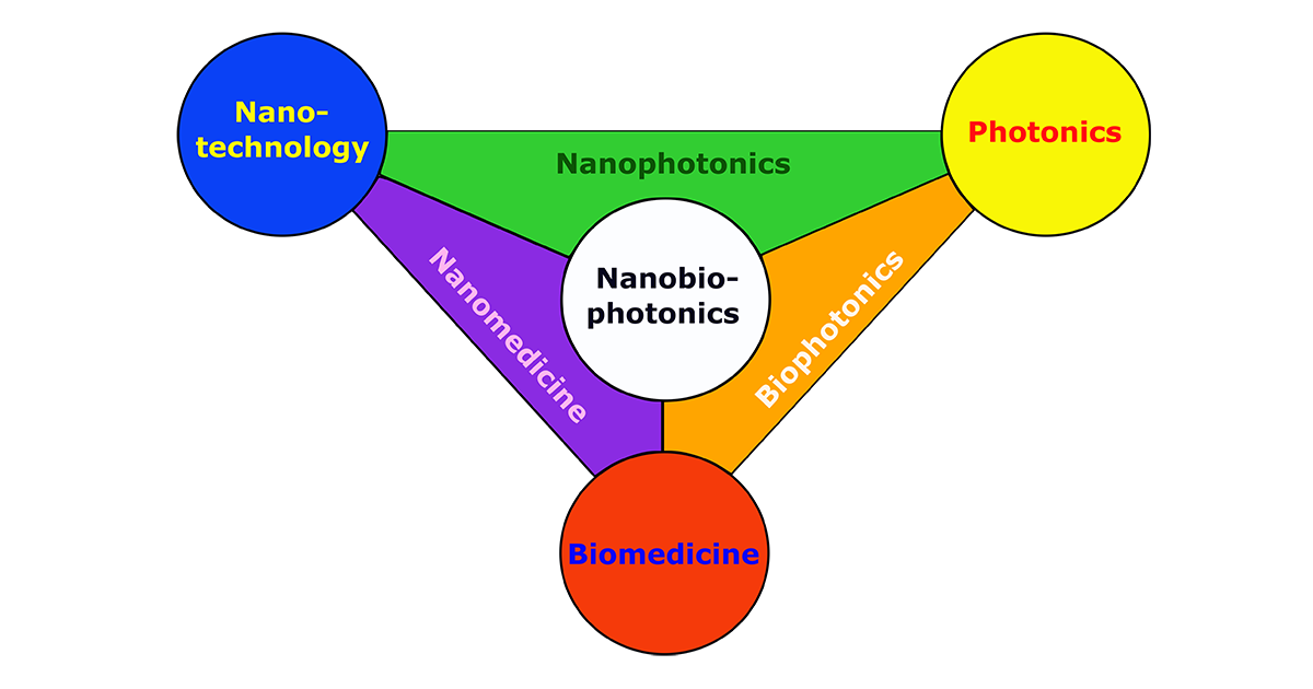
Nanobiophotonics is a rapidly developing field of science that combines nanotechnology, biomedicine, and photonics. It involves interdisciplinary research and development in areas from the fundamental mechanisms of the interaction of light and nanomaterials with cells and tissues to clinical diagnosis and therapy. While nanophotonics focuses on the interactions between light and matter at the nanoscale, biophotonics deals with the interactions between light with biological materials and nanomedicine applies nanotechnology to the diagnosis, prevention, and treatment of diseases. Nanobiotechnology is at the interface of all these disciplines.

This Special Issue will cover advanced optical imaging techniques such as high-resolution fluorescence microscopy, Raman microspectroscopy, coherent anti-Stokes Raman spectroscopy (CARS), time-resolved fluorescence (FLIM), etc., as well as optical sensing techniques using various nanoparticles, quantum dots, and plasmonic nanostructures for intracellular diagnostics and light-induced therapy.

Dr. Liubov A. Osminkina
Guest Editor

Manuscript Submission Information

Manuscripts should be submitted online at www.mdpi.com by registering and logging in to this website. Once you are registered, click here to go to the submission form. Manuscripts can be submitted until the deadline. All submissions that pass pre-check are peer-reviewed. Accepted papers will be published continuously in the journal (as soon as accepted) and will be listed together on the special issue website. Research articles, review articles as well as short communications are invited. For planned papers, a title and short abstract (about 100 words) can be sent to the Editorial Office for announcement on this website.

Submitted manuscripts should not have been published previously, nor be under consideration for publication elsewhere (except conference proceedings papers). All manuscripts are thoroughly refereed through a single-blind peer-review process. A guide for authors and other relevant information for submission of manuscripts is available on the Instructions for Authors page. Cells is an international peer-reviewed open access semimonthly journal published by MDPI.

Please visit the Instructions for Authors page before submitting a manuscript. The Article Processing Charge (APC) for publication in this open access journal is 2700 CHF (Swiss Francs). Submitted papers should be well formatted and use good English. Authors may use MDPI's English editing service prior to publication or during author revisions.

Keywords

  • nanobiophotonics
  • nanoparticles
  • quantum dots
  • plasmonic nanostructures
  • optical imaging
  • luminescent microscopy
  • Raman
  • optical biosensors
  • photothermal therapy
  • photodynamic therapy

Published Papers (3 papers)

Order results
Result details
Select all
Export citation of selected articles as:

Research

17 pages, 7863 KiB  
Article
Calcein-Modified CeO2 for Intracellular ROS Detection: Mechanisms of Action and Cytotoxicity Analysis In Vitro
by Nikita N. Chukavin, Vladimir K. Ivanov and Anton L. Popov
Cells 2023, 12(19), 2416; https://doi.org/10.3390/cells12192416 - 7 Oct 2023
Cited by 2 | Viewed by 1287
Abstract
Cerium oxide nanoparticles (CeO2 NPs) are metal-oxide-based nanozymes with unique reactive oxygen species (ROS) scavenging abilities. Here, we studied new CeO2 NPs modified with calcein (CeO2-calcein) as an intracellular ROS inactivation/visualization theranostic agent. The molecular mechanisms of the CeO [...] Read more.
Cerium oxide nanoparticles (CeO2 NPs) are metal-oxide-based nanozymes with unique reactive oxygen species (ROS) scavenging abilities. Here, we studied new CeO2 NPs modified with calcein (CeO2-calcein) as an intracellular ROS inactivation/visualization theranostic agent. The molecular mechanisms of the CeO2-calcein intracellular activity, allowing for the direct monitoring of ROS inactivation in living cells, were studied. CeO2-calcein was taken up by both normal (human mesenchymal stem cells, hMSc) and cancer (human osteosarcoma, MNNG/Hos cell line) cells, and was easily decomposed via endogenous or exogenous ROS, releasing brightly fluorescent calcein, which could be quantitatively detected using fluorescence microscopy. It was shown that the CeO2-calcein has selective cytotoxicity, inducing the death of human osteosarcoma cells and modulating the expression of key genes responsible for cell redox status as well as proliferative and migration activity. Such cerium-based theranostic agents can be used in various biomedical applications. Full article
(This article belongs to the Special Issue Nanobiophotonics: From Cell Imaging to Clinical Applications)
Show Figures

Figure 1

11 pages, 4295 KiB  
Article
Sensing Cells-Peptide Hydrogel Interaction In Situ via Scanning Ion Conductance Microscopy
by Tatiana N. Tikhonova, Vasilii S. Kolmogorov, Roman V. Timoshenko, Alexander N. Vaneev, Dana Cohen-Gerassi, Liubov A. Osminkina, Petr V. Gorelkin, Alexander S. Erofeev, Nikolay N. Sysoev, Lihi Adler-Abramovich and Evgeny A. Shirshin
Cells 2022, 11(24), 4137; https://doi.org/10.3390/cells11244137 - 19 Dec 2022
Cited by 2 | Viewed by 1693
Abstract
Peptide-based hydrogels were shown to serve as good matrices for 3D cell culture and to be applied in the field of regenerative medicine. The study of the cell-matrix interaction is important for the understanding of cell attachment, proliferation, and migration, as well as [...] Read more.
Peptide-based hydrogels were shown to serve as good matrices for 3D cell culture and to be applied in the field of regenerative medicine. The study of the cell-matrix interaction is important for the understanding of cell attachment, proliferation, and migration, as well as for the improvement of the matrix. Here, we used scanning ion conductance microscopy (SICM) to study the growth of cells on self-assembled peptide-based hydrogels. The hydrogel surface topography, which changes during its formation in an aqueous solution, were studied at nanoscale resolution and compared with fluorescence lifetime imaging microscopy (FLIM). Moreover, SICM demonstrated the ability to map living cells inside the hydrogel. A zwitterionic label-free pH nanoprobe with a sensitivity > 0.01 units was applied for the investigation of pH mapping in the hydrogel to estimate the hydrogel applicability for cell growth. The SICM technique that was applied here to evaluate the cell growth on the peptide-based hydrogel can be used as a tool to study functional living cells. Full article
(This article belongs to the Special Issue Nanobiophotonics: From Cell Imaging to Clinical Applications)
Show Figures

Figure 1

20 pages, 2006 KiB  
Article
The Surface Charge of Polymer-Coated Upconversion Nanoparticles Determines Protein Corona Properties and Cell Recognition in Serum Solutions
by Liuen Liang, Arun V. Everest-Dass, Alexey B. Kostyuk, Zahra Khabir, Run Zhang, Daria B. Trushina and Andrei V. Zvyagin
Cells 2022, 11(22), 3644; https://doi.org/10.3390/cells11223644 - 17 Nov 2022
Cited by 2 | Viewed by 2275
Abstract
Applications of nanoparticles (NPs) in the life sciences require control over their properties in protein-rich biological fluids, as an NP quickly acquires a layer of proteins on the surface, forming the so-called “protein corona” (PC). Understanding the composition and kinetics of the PC [...] Read more.
Applications of nanoparticles (NPs) in the life sciences require control over their properties in protein-rich biological fluids, as an NP quickly acquires a layer of proteins on the surface, forming the so-called “protein corona” (PC). Understanding the composition and kinetics of the PC at the molecular level is of considerable importance for controlling NP interaction with cells. Here, we present a systematic study of hard PC formation on the surface of upconversion nanoparticles (UCNPs) coated with positively-charged polyethyleneimine (PEI) and negatively-charged poly (acrylic acid) (PAA) polymers in serum-supplemented cell culture medium. The rationale behind the choice of UCNP is two-fold: UCNP represents a convenient model of NP with a size ranging from 5 nm to >200 nm, while the unique photoluminescent properties of UCNP enable direct observation of the PC formation, which may provide new insight into this complex process. The non-linear optical properties of UCNP were utilised for direct observation of PC formation by means of fluorescence correlation spectroscopy. Our findings indicated that the charge of the surface polymer coating was the key factor for the formation of PC on UCNPs, with an ensuing effect on the NP–cell interactions. Full article
(This article belongs to the Special Issue Nanobiophotonics: From Cell Imaging to Clinical Applications)
Show Figures

Graphical abstract

Back to TopTop