


Abstract
Spinal cord injury (SCI) constitutes an inestimable public health issue. The most crucial phase in the pathophysiological process of SCI concerns the well-known secondary injury, which is the uncontrolled and destructive cascade occurring later with aberrant molecular signaling, inflammation, vascular changes, and secondary cellular dysfunctions. The use of mesenchymal stem cells (MSCs) represents one of the most important and promising tested strategies. Their appeal, among the other sources and types of stem cells, increased because of their ease of isolation/preservation and their properties. Nevertheless, encouraging promise from preclinical studies was followed by weak and conflicting results in clinical trials. In this review, the therapeutic role of MSCs is discussed, together with their properties, application, limitations, and future perspectives.
1. Introduction
Spinal cord injury (SCI) constitutes an inestimable public health issue, with an incidence of 40–80 per million people per year [1]. Generally, young adults are involved, where the burden of permanent neurological damage is unbearable for patients, for their caregivers, and for the health system. Prevention plays of course a key role, such as in cases of road accidents, criminal acts, or secondary causes (tumors, degenerative diseases); however, the real challenge involving scientists is about therapy, given the absence of a gold standard or effective treatment. Most of the post-traumatic degeneration of the nervous system is caused by multifactorial secondary damage including different molecular processes such as inflammation, neuronal death, ionic dysregulation, free radicals and lipid peroxidation, disconnection of normal nerve pathways, blood–brain barrier dysfunction, apoptosis, and necrosis, followed by cavitation processes and retrograde degeneration. In traumatic SCI, an early surgical decompression seems to be important in preventing secondary damage, in the range between 8 and 24 h after injury [2,3,4,5], together with spinal fixation to allow correct nursing and rehabilitation.
One of the most important and promising tested strategies involves the use of stem cells. Among them, mesenchymal stem cells (MSCs) are particularly appealing and showed hopeful promise in preclinical research, followed by weak and conflicting results in clinical trials. In this review, the therapeutic role of MSCs is discussed, together with their properties, application, limitations, and future perspectives.
2. Spinal Cord Injury
The spinal cord of mammals is organized in ten laminae of neurons, named dorsoventrally, according to the Rexed description (1952 and 1954) [6,7]. The neurons are mostly multipolar and vary in size. In the dorsal laminae, sensory neurons are found which receive inputs from the dorsal root ganglion cells and project to other spinal levels or to the upper centers of the sensory pathways. In the ventral laminae, cholinergic large motoneurons are devoted to the control of muscle contraction with motor axons. Somewhat in between, interneurons of different morphologies receive the descending projections and recurrent axonal fibers from spinal motoneurons, and influence motoneuron activity (Mai and Paxinos, 2011) [8]. Spinal cord neurons form intraspinal circuits which are controlled by descending pathways. The reflex arc is the most elementary intraspinal circuit.
The acute phase of SCI depends on the mechanism of trauma, which could be caused by contusion, laceration, stretch, compression, or direct massive destruction. The events related to the trauma constitute the primary injury, with disruption of neuronal pathways [9]. During the immediate phase (occurring within the first two hours) (Rowland et al., 2008) [10], neurons and glial cells at the lesion site die either by necrosis or by apoptosis (Zhang et al., 2012) [11]. Therefore, spinal cord repair should aim first to restore intraspinal circuits, and then to obtain regrowth of descending pathways to regain voluntary control of these intraspinal circuits.
It is well known that the most crucial phase in the pathophysiological process of SCI concerns the secondary injury, which is the uncontrolled and destructive cascade occurring later with aberrant molecular signaling, inflammation, vascular changes, and secondary cellular dysfunctions [12,13,14,15].
2.1. Secondary Injury
In the injured spinal cord, to a greater or lesser extent depending on the primary injury, a large amount of destructive processes upset the environment. Taking into account the vascular scenario, a global reduction of blood flow is observed, as a result of vasospasm, together with focal microhemorrhages or thrombosis, causing a global disfunction of the blood–spinal cord barrier [15,16]. The cascade of events also affects electrolytic homeostasis around cellular membranes and their ion pumps/transporters. Potassium (K+) increases its extracellular concentration, while sodium (Na+) and calcium (Ca2+) concentrations increase intracellularly [17,18]. This leads to the blockage of neuronal transmission. The influx of water caused by acidosis promotes cytotoxic edema followed by cellular death [18,19,20]. Many molecules are released, such as free radicals and neurotransmitters. The inflammatory process involves an immune response mediated by cellular invasion after disruption of the blood–spinal cord barrier. T cells, macrophages, microglia, and neutrophils infiltrate the neuronal tissue, acquiring a proinflammatory phenotype. The environment develops with the production of cytokines such as interleukin-1 beta (IL-1β), interleukin-1 alpha (IL-1α), tumor necrosis factor alpha (TNF-α), and interleukin-6 (IL-6), recruiting more cells in loco promoting neurodegeneration [21,22,23,24].
2.2. Chronic Phase and Neurodegeneration
The chronic phase is characterized by scar formation after gliosis and deposit of the extracellular matrix. Molecules with growth-inhibitory effects are released and target neuronal receptors. Oligodendrocyte death in the primary injury seems to be a crucial point in the SCI, because myelin debris contains inhibitory molecules preventing axonal growth in animal models, such as Nogo-A protein or myelin associated glycoprotein (MAG) [25,26,27,28]. Proteoglycans are also involved in the chronic phase and show a different pattern of functions in the pathophysiological process; while most of them constitute a limitation for axonal regrowth with their inhibitory features, others seem to border and limit the scar, preventing further amplification of tissue damage [29,30].
In this scenario, the removal of cellular debris and the cell environment is a key point for neuroregeneration; the modulation of macrophages, with their different phenotypes (M1 and M2) and effects in supporting neuroprotection or boosting inflammation, is then a multifactorial and crucial step in determining final outcomes [21,22,23,24].
Noteworthy, the regeneration of neurons within the injured spinal cord seems a pipe dream in mammals, but it is innate in the axolotl (salamander) where specific molecules may regulate glial reaction after SCI and promote the proliferation and migration of glial cells to replace the missing neural tube and stimulate axonal growth [31]. The identification of the cellular mechanisms which control neural regeneration is fundamental to promoting spinal cord repair after injury. The modulation of intraneuronal signaling networks and of the extracellular milieu is pivotal to enhance axonal regeneration, thus stimulating the regrowth of intraspinal circuits and of the descending and ascending pathways of the spinal cord [32]. The modulation of glial scar formation and of the alterations in the perineuronal nets, and the control of neuroinflammation following SCI are mandatory for spinal cord repair, even though far from being achieved [33]. Finally, axonal sprouting, synapse plasticity, and remodeling, in part cell-autonomous, may be differently regulated by many cells and molecules in the different compartments of the lesioned spinal cord [34].
3. Stem Cell Therapy and Appeal of MSCs
Stem cell division gives birth to an asymmetrical offspring with an additional progenitor cell and a daughter stem cell. A stem cell is able to differentiate into different phenotypes, thereby determining its potency. Totipotency is defined in the case where all terminal cell populations could be achieved, while multipotency describes the possibility to pursue a more restricted pattern of phenotypes.
The promotion of synapse formation or axon elongation by transplanted neuronal progenitors after damage was described in animal models [35]. Direct modulation in the differentiation of stem cells into terminal phenotypes expanded the focus of research, while promising studies showed encouraging recovery of neurological deficits after transplantation of derived cellular populations from embryonic stem cells (ESCs) in rodents after SCI [36,37,38,39,40].
Since then, researchers multiplied their fields of interest, ranging from the modulation of phenotypic pathways and optimization of transplant techniques, to imaging techniques in order to obtain spatial and temporal information on the grafts [41,42], up to clinical studies starting with the Geron clinical trial which promoted the use of human ESC-derived oligodendrocyte progenitor cells (OPCs) in the site of injury [43]. Although mechanisms are far from being elucidated, stem cell functions seem linked mostly to their paracrine effects and trophic support as shown in other neurological degenerative diseases [16,44,45,46]. Given by a relatively high number of studies focusing on SCI and neuronal repair, both in vivo and in vitro, evidence shows that a combinatory strategy involving not only stem cells, but also gene therapy, biomolecular targets and drugs, and biomaterials as scaffolds could dramatically improve the functional outcomes after SCI [16].
In this charming landscape, mesenchymal stem cells (MSCs) gained attention because of their easy isolation (from different sources) and preservation, raising no ethical concern [47,48,49], and of the limited risk of developing tumors [49]. In the case of ESCs, indeed many ethical controversies limited their application because of the problems related to the violation of a human embryo [50]. Additionally, MSCs maintain their regenerative potential even after cryopreservation at 80° C [51]. Their proliferation is very rapid, and a high multilineage differentiation can be obtained [47]. Immunoreactivity or a reaction versus hosts is minimal or absent.
Finally, MSCs show properties of “homing”, being able to migrate toward the site of lesion (Figure 1). According to other authors, we previously observed this phenomenon in SCI experimental models both by immunofluorescence reactions [52] and in MRI experiments [42]. Many authors demonstrated that this cellular behavior is mediated by several inflammatory or chemotactic factors [53]; for example, the vascular endothelial growth factor and hepatocyte growth factor, released at the injury level, can actually attract MSCs [53,54]. Additionally, the SDF-1α/CXCR4 (stromal cell-derived factor-1α/C–X–C chemokine receptor 4) axis plays an important role in these mechanisms [55]; the impairment or the upregulation of this axis can respectively affect or increase the MSC homing ability [56,57]. Other factors able to positively influence MSC migration include substance P [58] and the granulocyte colony-stimulating factor [59]. However, the precise mechanisms justifying the homing MSC ability are still largely unknown [54].
Figure 1.
The main factors and mechanisms influencing mesenchymal stem cell (MSC) homing process are illustrated. (A) When injected either intravenously or intraspinally, MSCs show remarkable properties of “homing”. (B) At the injury site, some molecules (such as VEGF, HGF, cytokines, etc.) are secreted; when transplanted into the spinal parenchyma, MSCs are attracted by chemotactic stimuli and migrate toward the lesion site. (C) Moreover, when injected intravenously, MSCs can interact with endothelial cells through the VLA-4−VCAM-1 interaction; then, the extravasation is mediated by the interaction between the C–X–C chemokine receptor 4 and stromal cell-derived factor-1α (SDF-1), a chemotactic cytokine induced by proinflammatory stimuli. Created with BioRender software.
4. Secretome of MSCs
Although some differences were reported depending on the source, MSCs show a remarkable autocrine and paracrine activity [60,61] (Figure 2).
Figure 2.
The main MSC sources, including bone marrow, umbilical cord, adipose tissue, and amnion. MSCs can exert both autocrine and paracrine effects. Among the molecules secreted, we can include several immunomodulatory and trophic factors, and anti-inflammatory cytokines; when transplanted in an injured spinal cord, the grafted cells can positively influence the host environment. Created with BioRender software.
Through their secretome, MSCs can stimulate proliferation and differentiation of different cell types, including themselves. Notably, it was demonstrated that the release of growth factors, cytokines, and interleukins can also influence MSC migration (see also “homing” mechanism above), via an autocrine loop; indeed, when exposed to conditioned medium (i.e., the medium where MSCs are cultured), the MSC expression of Aquaporin 1 and CXCR4 (two membrane proteins involved in cell migration) significantly increased, by activating Akt and Erk intracellular signal pathways, and caused an enhancement of MSC migration [55].
Moreover, the MSC secretome can also exert immunomodulatory, anti-inflammatory, neurotrophic/neuroprotective and angiogenetic effects on the host microenvironment (as necessary in case of SCI).
The immunomodulation is realized thanks to the expression of the major histocompatibility complex-I on the MSC surface, in this way preventing T-cell recognition and induction of a host immune response [62]. Moreover, MSCs are able to inhibit the proliferation, the activation, and differentiation of T cells [63,64].
Concerning their anti-inflammatory potential, MSCs can secrete a variety of soluble molecules; among the anti-inflammatory cytokines, we can include tumor necrosis factor (TNF) β1, interleukin (IL)-13, IL-18 binding protein, ciliary neurotrophic factor (CNTF), neurotrophin 3 factor (NT-3), IL-10, IL-12p70, IL-17E, IL-27; moreover, MSCs can also modulate cytokine production of the host, for example, by inhibiting the release of pro-inflammatory cytokines (as interferon-γ and tumor necrosis factor α) or increasing the release of anti-inflammatory IL-10 [44,65].
To exert neuroprotection, MSCs secrete a number of neurotrophic factors, as brain-derived growth factor (BDNF), glial-derived growth factor (GDNF), nerve growth factor (NGF), NT-1, NT-3, CNTF, and basic fibroblast growth factor (bFGF) [44,65,66,67,68,69]; through these factors, MSCs can, on one side, prevent nerve degeneration and apoptosis, and, on the other, support neurogenesis, axonal growth, re-myelination, and cell metabolism [70,71,72,73,74,75,76].
MSCs can also induce angiogenesis, an important process by which new vasculature sprouts from pre-existing blood vessels; to this aim, MSCs secrete the tissue inhibitor of metalloproteinase-1, vascular endothelial growth factor, hepatocyte growth factor (HGF), platelet-derived growth factor (PDGF), IL-6, and IL-8. The production of these factors is particularly important for supporting the wound healing processes [77,78].
5. MSCs
MSCs can be obtained from different sources, each of which bears intrinsic characteristics differences, as shown below (Figure 2; Table 1) [52,79,80,81,82,83,84,85,86,87,88,89,90,91].
Table 1.
Mesenchymal stem cell (MSC) characteristics.
5.1. Bone Marrow Mesenchymal Stem Cells (BM-MSCs)
These cells are found within the adult bone marrow, where they contribute to hematopoiesis and bone regeneration. BM-MSCs can not only be obtained from humans, rodents, or primates, but also from several animal species such as sheep, dogs, cats, and bovines (Figure 3) [88,92,93,94,95,96,97,98].
Figure 3.
In preclinical experiments, bone marrow (BM)-MSCs can be isolated from femurs of adult mice and expanded in vitro; when cultured, the cells display the typical fibroblast-like shape. Created with BioRender software. Scale bar = 40 μm.
The possibility to differentiate into cells of mesodermal origin and to adhere to plastic distinguishes BM-MSCs from hematopoietic cells. Their range of differentiation is larger than expected, including not only mesenchymal cells such as osteocytes, chondrocytes, and adipocytes, but also a broad range of lineages expressing non-mesenchymal markers [99,100]. Pre-clinical studies collected promising results (Table 2) Wislet-Gendebien et al. addressed the question of differentiation of MSCs in vitro trying to identify neuronal phenotypes. A series of markers were evaluated [100]. Authors found Nestin expression in some groups of cells, a marker for the responsive characteristic of MSCs to extrinsic signals. In Nestin-positive cells, they also registered an overexpression of proteins like sox2, sox10, pax6, fed, erbB2, and erb4. These cells showed a neuron-like conduction, responding to several neurotransmitters (GABA, glycine, glutamate). Compared to neurons, however, trains of action potentials or synaptic activities in co-cultured Nestin-positive MSCs were not observed.
Table 2.
Main preclinical studies regarding MSCs.
BM-MSCs can not only be transplanted directly into the damaged spinal cord, but also infused with intravenous injections because of the aforementioned homing properties [101,102,103,104,105,106]. Deng et al. transplanted BM-MSCs two weeks after dorsal SCI in monkeys [104]. A partial functional improvement was noticed in terms of a slight motor recovery (active joints movements in the study group) and in electrophysiological studies with evoked potentials. Monkeys were then studied after three months with characterization of the scar. No neuronal cells were found. The analysis revealed the presence of markers such as neuron-specific enolase (NSE), the neurofilament (NF), and the glial fibrillary acidic protein (GFAP) in approximately 10% of the cells. The true blue, originally injected at the caudal side of injuries, was at the end traceable in the rostral thoracic spinal cord, red nucleus, and sensorymotor cortex. Zurita et al. transplanted BM-MSCs three months after dorsal SCI in pigs [105]. Authors used a motor score (0–10, with 10 considered as animals without deficits). Twelve weeks after transplantation, pigs that underwent stem cell therapy showed a mean score of 6.2 on the motor function scale. Some of the treated animals were even able to get up spontaneously. Recovery of evoked potentials was also noticed.
Many authors investigated BM-MSC transplantation in dogs [88,92,93,94,95]. Among them, Ryu et al. recorded improved neurological outcomes in MCSs groups after acute transplantation (one week after trauma), since all dogs had purposeful hind limb motion. He also showed that some MSCs expressed markers for neurons (NF160), neuronal nuclei (NeuN) and astrocytes (GFAP). NF160- and NeuN-positive neurons were found, and GFAP-positive reactive astrocytes were observed more often in the control group than in MSCs groups [88].
Hofstetter et al. in 2002 studied stem cell therapy after both iperacute and acute (one week) transplantation. Populations of neuron-like cells with the presence of neural markers were found, but they were not able to depolarize their membrane-like mature neurons. No clinical benefits were recorded. In the acute SCI group, a population of neuronal progenitors and astrocytes of the host were found in tissues after introduction of BM-MSCs in the lesion site [107].
Jeon SR et al. described one of the first applications of these cells in patients with cervical SCI (Table 3). In this case, cells were isolated from iliac bones and then subjected to intramedullary or intradural introduction after expansion in a subacute and chronic state. After six months, most patients showed a slight improvement of motor function in the upper limbs, while magnetic resonance imaging (MRI) showed changes at the level of treatment in terms of the disappearance of the cavity margin and the presence of fiber-like streaks [116]. No evidence of neoplasm growth was observed even at three years follow-up [117] This study and others showed promising but very limited results (Table 3) [118,119,120,121,122,123,124,125,126]. Dai et al. [118] tested BM-MSCs in a randomized study with complete and chronic SCI patients. Neurological functions were evaluated with AIS grading, ASIA score, residual urine volume, and neurophysiological examination. In the treatment group (N = 20), 10 had clinical improvement. Mean motor improvement with AIS grading was 0.9 ± 1.07, that with the ASIA score was 11.5 ± 17.07, that with the sensory prick score was 5.2 ± 7.78, and that with the sensory light touch score was 5.4 ± 8.22. Residual urine volume (mL) was decreased with a mean of 61.55 ± 77.43. Patients were followed up for six months after an interval between the injury and stem cell therapy of 51.9 ± 18.3 months. No details about clinical improvements before stem cell therapy or other therapies were mentioned.
Table 3.
Main clinical studies on BM-MSC transplantation.
In a phase I/II controlled single-blind clinical trial, El-Kehir et al. [119] showed functional improvements over patients in the control group of stem cell therapy and physical therapy using AIS grading and ASIA scores in about half of the cases (46%), especially in patients with thoracic injuries with shorter durations of injury and smaller cord lesion. Motor recovery was recorded and promising but still qualitatively limited. Geffner et al. [120] described a partial efficacy of stem cell therapy with some improvements in ASIA, Barthel (quality of life), Frankel, and Ashworth scoring in eight cases (four acute, four chronic). Karamouzian et al. [121] described the results of a nonrandomized clinical trial of transplantation of BM-MSCs in 11 complete SCI patients against 20 in the control group. Results showed improvements of 45.5% of patients (a two-grade improvement from baseline, i.e., from ASIA A to ASIA C) in the study group vs. 15% in the control group, but were not statistically significant (p = 0.095). The heterogeneity and small number of the patients did not allow a reliable analysis. Mendonca et al. [122], Park et al. [123], and Cheng et al. (NCT01393977) described a slight functional improvement in small groups of patients treated with stem cells. Other papers and reviews questioned the extent of improvement and the correct timing of treatment [63,64]. Park et al. only described six cases with some improvements in motor function and changes in cord enhancement with the MRI [123]. Sykova et al. reported data from 20 patients with complete SCI who received transplants 10 to 467 days post-injury. Patients were then evaluated at three, six, and 12 months after implantation with ASIA protocol, the Frankel score, motor and somatosensory evoked potentials, and MRI evaluation of lesion size. Authors registered improvement in motor and/or sensory functions within three months in five of six patients with intra-arterial application, in five of seven acute patients, and in one of 13 chronic patients. Transplantation of cells appeared safe but there was no evidence that the observed beneficial effects were linked to cell therapy [124].
Therefore, new trials are needed, given the absence of protocols and the poor knowledge about mechanisms and outcomes of BM-MSC transplantation. In the ongoing trials (phase I and II), hundreds of patients should be enrolled, thus trying to improve quality of evidence.
Some studies are trying to explore benefits of different combinatory strategies involving not only BM-MSCs, but also technological tools such as virtual reality or exoskeletal stimulation to face the challenge with more holistic approaches [127].
BM-MSCs showed a very promising anti-inflammatory effect on cell environment. In animal models (rats), they promoted anti-inflammatory phenotypes of macrophages (M2) and suppressed lymphocyte proliferation before sustaining regeneration [128,129,130]. Furthermore, molecules such as vascular endothelial growth factor (VEGF), the glial cell-derived neurotrophic factor (GDNF), the nerve growth factor (NGF), and the brain-derived neurotrophic factor (BDNF) could be produced by MSCs and are currently related to the ability of MSCs to provide trophic support, studied in vivo with animal models [128,131,132]. This is probably why the genetically modified over-expression of these factors could improve clinical outcomes [133]. Finally, the homing properties of BM-MSCs could sustain targeted delivery of drugs acting like specific vectors [134].
In clinical trials involving SCI patients, BM-MSCs were injected with an intrathecal approach in about half of the cases, while, in the remaining studies, other routes of administration were used (in situ as grafts or with scaffold, intravenous or intramuscular) [16].
5.2. Umbilical Cord Mesenchymal Stem Cells (UC-MSCs)
These cells are obtained from cord blood or the umbilical cord [49], and grow in colonies with the support of growth factors [16,49]. The risk of graft rejection using these cells is very low as confirmed by studies demonstrating their hypoimmunogenicity [62]. In preclinical studies, using SCI animal models with rats or mouse, UC-MSCs showed a promising profile of neurotrophic, anti-apoptotic, and anti-inflammatory effects [135,136,137,138]. Molecular markers and neuron-like characteristics were observed after homogeneous maturation of UC-MSCs [139]. Despite the aforementioned results in pre-clinical studies, only few clinical trials were published describing minor improvements in some SCI patients [130,131,132]. Kang et al. described a case report of a young female patient with slight improvements after acute transplantation [140]. In the study of Yao et al., 25 patients with traumatic SCI (injury time >6 months) were treated with human umbilical cord blood stem cells via intravenous and intrathecal injection. The follow-up period was 12 months after transplantation. Results reported some autonomic restoration and changes in somatosensory evoked potentials [141]. The trial of Zhu et al. showed more promising, although limited results after transplantation in the chronic phase: 13 out of 20 patients improved their motor and sphincteric functions. Five out of 20 converted from complete to incomplete (two sensory, three motor; p = 0.038) SCI [142]. A phase II ongoing multicenter, randomized, sham-controlled trial (NCT03521336) recently started with patient enrollment, trying to evaluate efficacy of intrathecal transplantation of UC-MSCs. Completion of the study is expected in 2022.
5.3. Adipose-Derived Mesenchymal Stem Cells (AD-MSCs)
AD-MSCs represent an appealing source of transplantable MSCs, given the remarkable population of somatic stem cells and the availability of adipose tissue [143,144]. The ability of AD-MSCs to secrete growth factors, proteases, cytokines, extracellular matrix molecules, and immunomodulatory factors supports their potential of neuroregenerative, anti-apoptotic, angiogenetic, and wound healing actions [145]. Cellular survival pathways and repairing mechanisms in pre-clinical studies involved the upregulation of kinase proteins like ERK1/2 and Akt [40]. AD-MSC transplantation was studied in animal models showing no adverse effects but often unsatisfactory functional results [86,94,146]. Biomolecular and histological analysis revealed promising details. Kolar et al. [146] studied the effect of transplantation in rats with SCI. AD-MSCs were transplanted into the lateral funiculus 1 mm rostral and caudal to the C3–C4 lesion. In animals treated with cyclosporine, BDNF, vascular endothelial growth factor, and fibroblast growth factor-2 were expressed for about three weeks. An extensive ingrowth of 5HT-positive raphespinal axons was noticed in the trauma zone with some terminal arborizations reaching the caudal spinal cord. Furthermore, sprouting of raphespinal terminals in C2 contralateral ventral horn and C6 ventral horn on both sides was observed. Relative to the lesion scar, astrocytic processes extended into the middle of the trauma zone in association with regenerating axons. Menezes et al. described an abundant deposition of laminin at the lesion site and spinal midline, the appearance of cell clusters composed of neural-like precursors in the areas of laminin deposition, and the appearance of blood vessels [86]. Kim et al. showed a modification of the inflammatory environment after transplantation of AD-MSCs, with decreased astrogliosis-related signal molecules such as phosphorylated signal transducer and activator of transcription. Furthermore, markers like Tuj-1, Nestin, microtubule-associated protein 2, and neurofilament M were expressed as shown in other aforementioned studies [94]. In clinical studies, a slight sensory improvement was recorded in the majority of patients after intrathecal transplantation, but longitudinal clinical trials with concrete motor responses are still lacking [40]. Coadministration of other compounds, such as 17b-estradiol, and overexpression of Bcl-2 or chondroitinase ABC, were able to enhance therapeutic efficacy in dog models [147]. On this premis, a series of ongoing trials of AD-MSC transplantation is currently underway [40] (Table 3).
5.4. Amniotic Fetal Mesenchymal Stem Cells (AF-MSCs)
These cells can be obtained from amniotic membrane or amniotic fluid. Several features are attributed to AF-MSCs such as their multipotency, ease of isolation, and ability of proliferation, together with a low immunogenicity [148]. Despite this, only few pre-clinical studies in animal models were performed. They showed preliminary and limited results in terms of reduced inflammation and apoptosis, promoted angiogenesis, and provided trophic support [149,150,151]. No effective clinical trials followed pre-clinical investigations.
6. Biomaterials and Scaffolds for Stem Cell Therapy
Due to technological advances, researchers started to investigate biomaterials with the aim of promoting tissue repair, improving stem cell survival, and supporting their functions [152]. This strategy could be pursued using biomaterials as carriers, thereby ensuring stem cell biofactor delivery, or as a scaffold, offering a structural support for tissue regeneration [16].
Among synthetic polymers, biodegradable hydrogels (such as polylactic acid (PLA), polyglycol acid (PGA), and polyethylene glycol (PEG)) were developed to promote cellular survival and carry several advantages. They easily fill the lesion cavity after injection and show high flexibility, gas permeability, no toxicity, and a favorable mechanical profile [16]. Drugs, biomolecules, and biofactors can be loaded and released locally by hydrogels [153]. To improve proper micro-structure and ensure correct support, three-dimensional (3D) printing nano-architecture was developed to recreate a sustainable and attractive stem cell niche [16,154]. Usually, hydrogels are injected at the site of the lesion. The possibility to use them with a minimally invasive injection reduces the risk of a surgical procedure. Furthermore, they have the ability to load hydrophilic drugs and biomolecules with controlled release. Among the disadvantages, it is important to highlight that the kinetics and delivery of drugs could be inadequate because of uncontrolled diffusion or an unfavorable environment. For example, molecules with low steric hindrance cannot be controlled easily and might diffuse without reproducible control. Furthermore, the loading of hydrophobic drugs with a reduced affinity for the aqueous environment constitutes a real limitation. Bonds between drugs molecules could be built to increase or reduce the rate of release, depending on the ease of breakage of links, thereby offering controlled stem cell biofactor delivery [16].
Among natural scaffolds, a variety of materials were evaluated. Because of its biocompatibility, plasticity, and flexibility, fibrin was shown to promote regeneration and delay accumulation of astrocytes at the site of the injury. Enriched with stem cells or growth factors, fibrin improved survival and migration of transplanted cells, also increasing neural fiber density. Collagen and hyaluronic acid were proposed and used because of their elasticity, time of degradation, and ability to support cell adhesion and migration. Time of degradation plays a key role in letting the matrix produced by transplanted cells progressively replace the scaffold during the repairing process. An example of an innovative composite implant was described by Rochkind et al., cross-linked hyaluronic acid with growth factors and the adhesive molecule laminin (NVR-Gel), showing promising results. Natural scaffolds were able to reduce neuro-inflammation in the acute stage and support synaptic plasticity, as well as axonal outgrowth [106]. That said, despite this promising neuroengineering background, no concrete results were observed in clinical trials [16,155,156,157,158]. The field of polymeric scaffolds was developed to support stem cell survival and efficacy after transplantation, but favorable biomechanical properties did not translate encouraging pre-clinical studies into clinical success, highlighting the necessity for new comprehensive experimentations.
7. Limitations of Current Evidence and Future Directions
Stem cell technology is a growing and evolving field with an unquestionable appeal, as testified by many research papers and state-of-the-art reviews published in the English literature [16,40,80,158,159]. In our experience with MSCs or other stem cells [52,160], like in many other aforementioned papers, tests with animal models showed promising results. Despite this, many caveats arise and, thus, elicit caution against inordinate enthusiasm.
First of all, studies involving animal models are usually performed applying standardized protocols of lesions, treatments, and specific timings of transplantation in each group of investigation. These conditions are often inimitable in human patients with SCI, when timing and treatments are dependent on chance and emergency setting, or where lesions at the cord site could differ a lot from tailored laboratory damage. Most in vivo studies are necessarily performed with rodents, and, despite many anatomical or behavioral correspondences, human clinical trials should be the unavoidable aim of stem cell research.
Therefore, completed human trials showed only limited results. On the one hand, the use of MSCs in SCIs seems caused no harm; different trials [118,119,120,121,122,123,124,125,126,161] described, above all, the safety of stem cell therapy showing no adverse reactions or side effects. On the other hand, results in terms of clinical outcomes were poor compared to expectations. Among the others, few studies seemed particularly to encourage cell therapy.
Dai et al. [118] tested BM-MSCs in a randomized study with complete and chronic SCI patients. Neurological functions were evaluated with AIS grading, ASIA score, residual urine volume, and neurophysiological examination. In the treatment group (N = 20), 10 had clinical improvement. As already mentioned, patients were followed up for six months and details about clinical improvements before stem cell therapy or other therapies were not mentioned. Outcomes seemed limited, even if promising.
In the trial of El-Kehir et al. [119], functional improvements were noted but were confined and particularly involved patients with smaller and thoracic lesions. Geffner et al. [120] described partial efficacy of stem cell therapy with some improvements in ASIA, Barthel (quality of life), Frankel, and Ashworth scoring in eight cases (four acute, four chronic). The other clinical studies confirmed the trend of confined (Mendonca et al. [122], Park et al. [123], and Cheng et al. (NCT01393977)) or no significant improvements (Karamouzian et al. [121]). Many other trials failed to report satisfactory outcomes (NCT01186679, NCT02027246, NCT01769872, NCT01873547, NCT01624779, NCT01328860, NCT02237547, NCT01694927, and NCT01730183). There is a marked lack of large phase III trials of therapeutic efficacy, due to financial, ethical, and logistics reasons. [40] The phase III study of Oh et al. [162] showed weak efficacy in functional recovery, although some limitations could have compromised clinical results. For instance, only a single administration was given because of a restrictive government policy.
Finally, even if immunochemistry, molecular markers, and morphological tracts show that MSCs, once transplanted, present neuron-like characteristics, it is hard to consider them as such [16,129]; indeed, the expression of neuronal antigens can be simply due to the extremely immature nature of MSCs [163]. Moreover, cell fusion (between MSCs and neurons) was sometimes documented [164]; furthermore, when forced to transdifferentiate by chemical means (such as DMSO), MSCs showed evident morphological changes, which finally were simply attributed to cell shrinkage and changes in the cytoskeleton [165]. More sophisticated protocols are continuously being developed in order to differentiate MCSs into neurons [166,167]. However, currently, the efficacy of MSCs still seems in particular related to their paracrine activity, rather than to cellular replacement mechanisms [168].
Ongoing trials (Table 4) will probably help researchers improve knowledge about the clinical impact of stem cell therapy. Encouraging data from preclinical experiments were not concretely translated into clinical practice. This probably reflects the multifactorial and complex physiopathology of SCI, requiring a multimodal therapeutic approach. As a matter of fact, many points need to be further clarified and depicted, as listed below.
Table 4.
Ongoing trials about MSCs. IANR-SCIRFS = International Association of Neural Restoration Spinal Cord Injury Functional Rating Scale; NSC = neural stem cell; SCIM III = Spinal Cord Independence Measure III; UC = umbilical cord.
- The optimal therapeutic protocols regarding the preparation, type, and number of stem cells transplanted;
- The timing of transplantation and route of administration;
- The paracrine effects and their influence on functional recovery;
- The importance of biomaterials and scaffold;
- The importance of microenvironment;
- The plasticity and ability to recreate connections of neuronal cells.
- Additionally, logistics, ethical, and financial problems related to this field of research constitute a real challenge to face in order to channel basic science studies into clinical practice.
8. Conclusions
MSC therapy represents an intriguing field of research trying to face the burden of SCI. MSCs of different origin, together with scaffolds, can release immunomodulating and neuroprotective factors which may support neuron survival, axonal growth, and control of glial scarring in absence of significant side effects. Despite promising preclinical findings, clinical trials failed to keep their promises and are still far from obtaining functional recovery and restoring neural circuits. Further studies are needed to improve our knowledge on their mechanisms of action and on the cellular mechanisms preventing restoration of neural circuits after SCI, while combinatory strategies involving stem cells, biomaterials, and modifications of cell environment could be the key to translate fascinating premises into clinical practice. A better relationship between preclinical and clinical studies with a back-and-forth approach is mandatory to enhance the efficacy of cell therapy. Nevertheless, stem cell therapy in SCI injury remains an experimental therapy, possibly in association with others, and should be tested and provided at no cost for the patient. Moreover, patients should be aware of the poor clinical results obtained thus far in clinical trials to prevent exaggerated expectations and dramatic psychological consequences in the case of failure to obtain significant results.
Funding
This study was supported by Ministero dell’Istruzione, dell’Università e della Ricerca—MIUR project “Dipartimenti di eccellenza 2018-2022”.
Conflicts of Interest
The authors declare no conflicts of interest.
References
- International Perspectives on Spinal Cord Injury. Available online: http://apps.who.int/iris/bitstream/10665/94190/1/9789241564663_eng.pdf (accessed on 31 October 2014).
- Yılmaz, T.; Kaptanoğlu, E. Current and future medical therapeutic strategies for the functional repair of spinal cord injury. World J. Orthop. 2015, 6, 42–55. [Google Scholar] [CrossRef] [Scilit] [PubMed]
- Wilson, J.R.; Forgione, N.; Fehlings, M.G. Emerging therapies for acute traumatic spinal cord injury. CMAJ 2013, 185, 485–492. [Google Scholar] [CrossRef] [Scilit] [PubMed]
- Wilson, J.R.; Tetreault, L.A.; Kwon, B.K.; Arnold, P.M.; Mroz, T.E.; Shaffrey, C.; Harrop, J.S.; Chapman, J.R.; Casha, S.; Skelly, A.C.; et al. Timing of Decompression in Patients with Acute Spinal Cord Injury: A Systematic Review. Glob. Spine J. 2017, 7, 95S–115S. [Google Scholar] [CrossRef] [Scilit] [PubMed]
- Fehlings, M.G.; Perrin, R.G. The role and timing of early decompression for cervical spinal cord injury: Update with a review of recent clinical evidence. Injury 2005, 36, S13–S26. [Google Scholar] [CrossRef] [Scilit] [PubMed]
- Rexed, B. The cytoarchitectonic organization of the spinal cord in the cat. J. Comp. Neurol. 1952, 96, 414–495. [Google Scholar] [CrossRef] [Scilit] [PubMed]
- Rexed, B. A cytoarchitectonic atlas of the spinal cord in the cat. J. Comp. Neurol. 1954, 100, 297–379. [Google Scholar] [CrossRef] [Scilit] [PubMed]
- Mai, J.K.; Paxinos, G. The Human Nervous System, 3rd ed.; Academic Press: Cambridge, MA, USA, 2011; ISBN 9780123742360. [Google Scholar]
- Ackery, A.; Tator, C.; Krassioukov, A. A global perspective on spinal cord injury epidemiology. J. Neurotrauma 2004, 21, 1355–1370. [Google Scholar] [CrossRef] [Scilit]
- Rowland, J.W.; Hawryluk, G.W.; Kwon, B.; Fehlings, M.G. Current status of acute spinal cord injury pathophysiology and emerging therapies: Promise on the horizon. Neurosurg. Focus 2008, 25, E2. [Google Scholar] [CrossRef] [Scilit]
- Zhang, N.; Yin, Y.; Xu, S.J.; Wu, Y.P.; Chen, W.S. Inflammation & apoptosis in spinal cord injury. Indian J. Med. Res. 2012, 135, 287–296. [Google Scholar]
- Beattie, M.S.; Li, Q.; Bresnahan, J.C. Cell death and plasticity after experimental spinal cord injury. Prog. Brain Res. 2000, 128, 9–21. [Google Scholar]
- Blight, A.R. Spinal cord injury models: Neurophysiology. J. Neurotrauma 1992, 9, 147–150. [Google Scholar] [CrossRef] [Scilit] [PubMed]
- Grossman, S.D.; Rosenberg, L.J.; Wrathall, J.R. Relationship of altered glutamate receptor subunit mRNA expression to acute cell loss after spinal cord contusion. Exp. Neurol. 2001, 168, 283–289. [Google Scholar] [CrossRef] [Scilit] [PubMed]
- Silva, N.A.; Sousa, N.; Reis, R.L.; Salgado, A.J. From basics to clinical: A comprehensive review on spinal cord injury. Prog. Neurobiol. 2014, 114, 25–57. [Google Scholar] [CrossRef] [Scilit] [PubMed]
- Vismara, I.; Papa, S.; Rossi, F.; Forloni, G.; Veglianese, P. Current Options for Cell Therapy in Spinal Cord Injury. Trends Mol. Med. 2017, 23, 831–849. [Google Scholar] [CrossRef] [Scilit]
- Fehlings, M.G.; Nakashima, H.; Nagoshi, N.; Chow, D.S.; Grossman, R.G.; Kopjar, B. Rationale, design and critical end points for the Riluzole in Acute Spinal Cord Injury Study (RISCIS): A randomized, double-blinded, placebo-controlled parallel multi-center trial. Spinal Cord 2016, 54, 8–15. [Google Scholar] [CrossRef] [Scilit] [PubMed]
- Gazdic, M.; Volarevic, V.; Harrell, C.R.; Fellabaum, C.; Jovicic, N.; Arsenijević, N.; Stojkovic, M. Stem Cells Therapy for Spinal Cord Injury. Int. J. Mol. Sci. 2018, 19, 1039. [Google Scholar] [CrossRef] [Scilit] [PubMed]
- Garcia, E.; Aguilar-Cevallos, J.; Silva-Garcia, R.; Ibarra, A. Cytokine and Growth Factor Activation In Vivo and In Vitro after Spinal Cord Injury. Mediat. Inflamm. 2016, 2016, 9476020. [Google Scholar] [CrossRef] [Scilit] [PubMed]
- Hayta, E.; Elden, H. Acute spinal cord injury: A review of pathophysiology and potential of non-steroidal anti-inflammatory drugs for pharmacological intervention. J. Chem. Neuroanat. 2017, 87, 25–31. [Google Scholar] [CrossRef] [Scilit] [PubMed]
- David, S.; Lopez Vales, R.; Wee Yong, V. Harmful and beneficial effects of inflammation after spinal cord injury: Potential therapeutic implications. Handb. Clin. Neurol. 2012, 109, 485–502. [Google Scholar] [PubMed]
- Papa, S.; Caron, I.; Erba, E.; Panini, N.; De Paola, M.; Mariani, A.; Colombo, C.; Ferrari, R.; Pozzer, D.; Zanier, E.R.; et al. Early modulation of pro-inflammatory microglia by minocycline loaded nanoparticles confers long lasting protection after spinal cord injury. Biomaterials 2016, 75, 13–24. [Google Scholar] [CrossRef] [Scilit]
- Papa, S.; Caron, I.; Rossi, F.; Veglianese, P. Modulators of microglia: A patent review. Expert Opin. Ther. Pat. 2016, 26, 427–437. [Google Scholar] [CrossRef] [Scilit] [PubMed]
- Shechter, R.; Schwartz, M. Harnessing monocyte-derived macrophages to control central nervous system pathol-ogies: No longer ‘if’ but ‘how’. J. Pathol. 2013, 229, 332–346. [Google Scholar] [CrossRef] [Scilit] [PubMed]
- Fournier, A.E.; GrandPre, T.; Strittmatter, S.M. Identification of a receptor mediating Nogo-66 inhibition of axonal regeneration. Nature 2001, 409, 341–346. [Google Scholar] [CrossRef] [Scilit] [PubMed]
- GrandPre, T.; Nakamura, F.; Vartanian, T.; Strittmatter, S.M. Identification of the Nogo inhibitor of axon regeneration as a reticulon protein. Nature 2000, 403, 439–444. [Google Scholar] [CrossRef] [Scilit] [PubMed]
- Schwab, M.E.; Strittmatter, S.M. Nogo limits neural plasticity and recovery from injury. Curr. Opin. Neurobiol. 2014, 27, 53–60. [Google Scholar] [CrossRef] [Scilit] [PubMed]
- Schwab, M.E. Nogo and axon regeneration. Curr. Opin. Neurobiol. 2004, 14, 118–124. [Google Scholar] [CrossRef] [Scilit] [PubMed]
- Beller, J.A.; Snow, D.M. Proteoglycans: Road signs for neurite outgrowth. Neural Regen. Res. 2014, 9, 343–355. [Google Scholar]
- Yuan, Y.-M.; He, C. The glial scar in spinal cord injury and repair. Neurosci. Bull. 2013, 29, 421–435. [Google Scholar] [CrossRef] [Scilit]
- Sabin, K.Z.; Jiang, P.; Gearhart, M.D.; Stewart, R.; Echeverri, K. AP-1(cFos/JunB)/miR-200a regulate the pro-regenerative glial cell response during axolotl spinal cord regeneration. Commun. Biol. 2019, 2, 91. [Google Scholar] [CrossRef] [Scilit]
- Van Niekerk, E.A.; Tuszynski, M.H.; Lu, P.; Dulin, J.N. Molecular and Cellular Mechanisms of Axonal Regeneration After Spinal Cord Injury. Mol. Cell Proteom. 2016, 15, 394–408. [Google Scholar] [CrossRef] [Scilit]
- Tran, A.P.; Warren, P.M.; Silver, J. The Biology of Regeneration Failure and Success After Spinal Cord Injury. Physiol. Rev. 2018, 98, 881–917. [Google Scholar] [CrossRef] [Scilit] [PubMed]
- O’Shea, T.M.; Burda, J.E.; Sofroniew, M.V. Cell biology of spinal cord injury and repair. J. Clin. Investig 2017, 127, 3259–3270. [Google Scholar] [CrossRef] [Scilit] [PubMed]
- Lu, P.; Wang, Y.; Graham, L.; McHale, K.; Gao, M.; Wu, D.; Brock, J.; Blesch, A.; Rosenzweig, E.S.; Havton, L.A.; et al. Long-distance growth and connectivity of neural stem cells after severe spinal cord injury. Cell 2012, 150, 1264–1273. [Google Scholar] [CrossRef] [Scilit] [PubMed]
- Keirstead, H.S.; Nistor, G.; Bernal, G.; Totoiu, M.; Cloutier, F.; Sharp, K.; Steward, O. Human embryonic stem cell-derived oligodendrocyte progenitor cell transplants remyelinate and restore locomotion after spinal cord injury. J. Neurosci. 2005, 25, 4694–4705. [Google Scholar] [CrossRef] [Scilit] [PubMed]
- Deshpande, D.M.; Kim, Y.S.; Martinez, T.; Carmen, J.; Dike, S.; Shats, I.; Rubin, L.L.; Drummond, J.; Krishnan, C.; Hoke, A.; et al. Recovery from paralysis in adult rats using embryonic stem cells. Ann. Neurol. 2006, 60, 32–44. [Google Scholar] [CrossRef] [Scilit] [PubMed]
- Pera, M.F.; Andrade, J.; Houssami, S.; Reubinoff, B.; Trounson, A.; Stanley, E.G.; Oostwaard, D.W.-v.; Mummery, C. Regulation of human embryonic stem cell differentiation by BMP-2 and its antagonist noggin. J. Cell Sci. 2004, 117, 1269–1280. [Google Scholar] [CrossRef] [Scilit] [PubMed]
- McDonald, J.W.; Liu, X.Z.; Qu, Y.; Liu, S.; Mickey, S.K.; Turetsky, D.; Gottlieb, D.I.; Choi, D.W. Transplanted embryonic stem cells survive, differentiate and promote recovery in injured rat spinal cord. Nat. Med. 1999, 5, 1410–1412. [Google Scholar] [CrossRef] [Scilit]
- Jin, M.C.; Medress, Z.A.; Azad, T.D.; Doulames, V.M.; Veeravagu, A. Stem cell therapies for acute spinal cord injury in humans: A review. Neurosurg. Focus 2019, 46, E10. [Google Scholar] [CrossRef] [Scilit]
- Filippi, M.; Boido, M.; Terreno, E. Imaging of MSC transplantation in neuroscience. Oncotarget 2017, 8, 10781–10782. [Google Scholar] [CrossRef] [Scilit]
- Filippi, M.; Boido, M.; Pasquino, C.; Garello, F.; Boffa, C.; Terreno, E. Successful in vivo MRI tracking of MSCs labeled with Gadoteridol in a Spinal Cord Injury experimental model. Exp. Neurol. 2016, 282, 66–77. [Google Scholar] [CrossRef] [Scilit]
- Frantz, S. Embryonic stem cell pioneer Geron exits field, cuts losses. Nat. Biotechnol. 2012, 30, 12–13. [Google Scholar] [CrossRef] [Scilit]
- Boido, M.; Piras, A.; Valsecchi, V.; Spigolon, G.; Mareschi, K.; Ferrero, I.; Vizzini, A.; Temi, S.; Mazzini, L.; Fagioli, F.; et al. Human mesenchymal stromal cell transplantation modulates neuroinflammatory milieu in a mouse model of amyotrophic lateral sclerosis. Cytotherapy 2014, 16, 1059–1072. [Google Scholar] [CrossRef] [Scilit]
- Mazzini, L.; Vercelli, A.; Ferrero, I.; Boido, M.; Cantello, R.; Fagioli, F. Transplantation of mesenchymal stem cells in ALS. Prog. Brain Res. 2012, 201, 333–359. [Google Scholar] [PubMed]
- Gunetti, M.; Tomasi, S.; Giammò, A.; Boido, M.; Rustichelli, D.; Mareschi, K.; Errichiello, E.; Parola, M.; Ferrero, I.; Fagioli, F.; et al. Myogenic potential of whole bone marrow mesenchymal stem cells in vitro and in vivo for usage in urinary incontinence. PLoS ONE 2012, 7, e45538. [Google Scholar] [CrossRef] [Scilit]
- Lee, M.W.; Yang, M.S.; Park, J.S.; Kim, H.C.; Kim, Y.J.; Choi, J. Isolation of mesenchymal stem cells from cryopreserved human umbilical cord blood. Int. J. Hematol. 2005, 81, 126–130. [Google Scholar] [CrossRef] [Scilit]
- Dasari, V.R.; Veeravalli, K.K.; Dinh, D.H. Mesenchymal stem cells in the treatment of spinal cord injuries. World J. Stem Cells 2014, 6, 120–133. [Google Scholar] [CrossRef] [Scilit]
- Lu, L.L.; Liu, Y.J.; Yang, S.G.; Zhao, Q.J.; Wang, X.; Gong, W.; Han, Z.B.; Xu, Z.S.; Lu, Y.X.; Liu, D.; et al. Isolation and characterization of human umbilical cord mesenchymal stem cells with hematopoiesis-supportive function and other potentials. Haematologica 2006, 91, 1017–1026. [Google Scholar]
- Lo, B.; Parham, L. Ethical Issues in Stem Cell Research. Endocr. Rev. 2009, 30, 204–213. [Google Scholar] [CrossRef] [Scilit]
- Kotobuki, N.; Hirose, M.; Takakura, Y.; Ohgushi, H. Cultured autologous human cells for hard tissue regeneration: Prepa-ration and characterization of mesenchymal stem cells from bone marrow. Artif. Organs 2004, 28, 33–39. [Google Scholar] [CrossRef] [Scilit]
- Boido, M.; Rupa, R.; Garbossa, D.; Fontanella, M.; Ducati, A.; Vercelli, A. Embryonic and adult stem cells promote raphespinal axon outgrowth and improve functional outcome following spinal hemisection in mice. Eur. J. Neurosci. 2009, 30, 833–846. [Google Scholar] [CrossRef] [Scilit]
- Zachar, L.; Bačenková, D.; Rosocha, J. Activation, homing, and role of the mesenchymal stem cells in the inflammatory environment. J. Inflamm. Res. 2016, 9, 231–240. [Google Scholar] [CrossRef] [Scilit]
- Qu, J.; Zhang, H. Roles of Mesenchymal Stem Cells in Spinal Cord Injury. Stem Cells Int. 2017, 2017, 5251313. [Google Scholar] [CrossRef] [Scilit]
- Pelagalli, A.; Nardelli, A.; Lucarelli, E.; Zannetti, A.; Brunetti, A. Autocrine signals increase ovine mesenchymal stem cells migration through Aquaporin-1 and CXCR4 overexpression. J. Cell. Physiol. 2018, 233, 6241–6249. [Google Scholar] [CrossRef] [Scilit]
- Xu, Q.; Wang, J.; He, J.; Zhou, M.; Adi, J.; Webster, K.A.; Yu, H. Impaired CXCR4 expression and cell engraftment of bone marrow-derived cells from aged atherogenic mice. Atherosclerosis 2011, 219, 92–99. [Google Scholar] [CrossRef] [Scilit]
- Marquez-Curtis, L.A.; Gul-Uludag, H.; Xu, P.; Chen, J.; Janowska-Wieczorek, A. CXCR4 transfection of cord blood mesenchymal stromal cells with the use of cationic liposome enhances their migration toward stromal cell-derived factor-1. Cytotherapy 2013, 15, 840–849. [Google Scholar] [CrossRef] [Scilit]
- Hong, H.S.; Lee, J.; Lee, E.; Kwon, Y.S.; Lee, E.; Ahn, W.; Jiang, M.H.; Kim, J.C.; Son, Y. A new role of substance P as an injury-inducible messenger for mobilization of CD29(+) stromal-like cells. Nat. Med. 2009, 15, 425–435. [Google Scholar] [CrossRef] [Scilit]
- Petit, I.; Szyper-Kravitz, M.; Nagler, A.; Lahav, M.; Peled, A.; Habler, L.; Ponomaryov, T.; Taichman, R.S.; Arenzana-Seisdedos, F.; Fujii, N.; et al. G-CSF induces stem cell mobilization by decreasing bone marrow SDF-1 and up-regulating CXCR4. Nat. Immunol. 2002, 3, 687. [Google Scholar] [CrossRef] [Scilit]
- Sobacchi, C.; Palagano, E.; Villa, A.; Menale, C. Soluble Factors on Stage to Direct Mesenchymal Stem Cells Fate. Front. Bioeng. Biotechnol. 2017, 5, 32. [Google Scholar] [CrossRef] [Scilit]
- Baez-Jurado, E.; Hidalgo-Lanussa, O.; Barrera-Bailón, B.; Sahebkar, A.; Ashraf, G.M.; Echeverria, V.; Barreto, G.E. Secretome of Mesenchymal Stem Cells and Its Potential Protective Effects on Brain Pathologies. Mol. Neurobiol. 2019, 1–26. [Google Scholar] [CrossRef] [Scilit]
- Ryan, J.M.; Barry, F.P.; Murphy, J.M.; Mahon, B.P. Mesenchymal stem cells avoid allogeneic rejection. J. Inflamm. 2005, 2, 8. [Google Scholar] [CrossRef] [Scilit]
- Di Nicola, M.; Carlo-Stella, C.; Magni, M.; Milanesi, M.; Longoni, P.D.; Matteucci, P.; Grisanti, S.; Gianni, A.M. Human bone marrow stromal cells suppress T-lymphocyte proliferation induced by cellular or nonspecific mitogenic stimuli. Blood 2002, 99, 3838–3843. [Google Scholar] [CrossRef] [Scilit]
- Liang, X.; Ding, Y.; Zhang, Y.; Tse, H.F.; Lian, Q. Paracrine mechanisms of mesenchymal stem cell-based therapy: Current status and perspectives. Cell Transplant. 2014, 23, 1045–1059. [Google Scholar] [CrossRef] [Scilit]
- Vizoso, F.J.; Eiro, N.; Cid, S.; Schneider, J.; Perez-Fernandez, R. Mesenchymal Stem Cell Secretome: Toward Cell-Free Therapeutic Strategies in Regenerative Medicine. Int. J. Mol. Sci. 2017, 25, 1852. [Google Scholar] [CrossRef] [Scilit]
- Mead, B.; Logan, A.; Berry, M.; Leadbeater, W.; Scheven, B.A. Paracrine-mediated neuroprotection and neuritogenesis of axotomised retinal ganglion cells by human dental pulp stem cells: Comparison with human bone marrow and adipose-derived mesenchymal stem cells. PLoS ONE 2014, 9, e109305. [Google Scholar] [CrossRef] [Scilit]
- Kolar, M.K.; Itte, V.N.; Kingham, P.J.; Novikov, L.N.; Wiberg, M.; Kelk, P. The neurotrophic effects of different human dental mesenchymal stem cells. Sci. Rep. 2017, 7, 12605. [Google Scholar] [CrossRef] [Scilit]
- Teixeira, F.G.; Carvalho, M.M.; Neves-Carvalho, A.; Panchalingam, K.M.; Behie, L.A.; Pinto, L.; Sousa, N.; Salgado, A.J. Secretome of mesenchymal progenitors from the umbilical cord acts as modulator of neural/glial proliferation and differentiation. Stem Cell Rev. 2015, 11, 288–297. [Google Scholar] [CrossRef] [Scilit]
- Potapova, I.A.; Gaudette, G.R.; Brink, P.R.; Robinson, R.B.; Rosen, M.R.; Cohen, I.S.; Doronin, S.V. Mesenchymal stem cells support migration, extracellular matrix invasion, proliferation, and survival of endothelial cells in vitro. Stem Cells 2007, 25, 1761–1768. [Google Scholar] [CrossRef] [Scilit]
- Hofer, H.R.; Tuan, R.S. Secreted trophic factors of mesenchymal stem cells support neurovascular and musculoskeletal therapies. Stem Cell Res. Ther. 2016, 7, 131. [Google Scholar] [CrossRef] [Scilit]
- Razavi, S.; Ghasemi, N.; Mardani, M.; Salehi, H. Remyelination improvement after neurotrophic factors secreting cells transplantation in rat spinal cord injury. Iran. J. Basic Med. Sci. 2017, 20, 392–398. [Google Scholar]
- Lu, P.; Jones, L.L.; Tuszynski, M.H. BDNF-expressing marrow stromal cells support extensive axonal growth at sites of spinal cord injury. Exp. Neurol. 2005, 191, 344–360. [Google Scholar] [CrossRef] [Scilit]
- Neuhuber, B.; Timothy Hilmes, B.; Shumsky, J.S.; Gallo, G.; Fischer, I. Axon growth and recovery of function supported by human bone marrow stromal cells in the injured spinal cord exhibit donor variations. Brain Res. 2005, 1035, 73–85. [Google Scholar] [CrossRef] [Scilit]
- Crigler, L.; Robey, R.C.; Asawachaicharn, A.; Gaupp, D.; Phinney, D.G. Human mesenchymal stem cell subpopulations express a variety of neuro-regulatory molecules and promote neuronal cell survival and neuritogenesis. Exp. Neurol. 2006, 198, 54–64. [Google Scholar] [CrossRef] [Scilit]
- Wright, K.T.; Masri, W.E.; Osman, A.; Roberts, S.; Chamberlain, G.; Ashton, B.A.; Johnson, W.E. Bone marrow stromal cells stimulate neurite outgrowth over neural proteoglycans (CSPG), myelin associated glycoprotein and Nogo-A. Biochem. Biophys. Res. Commun. 2007, 354, 559–566. [Google Scholar] [CrossRef] [Scilit]
- Teixeira, F.G.; Carvalho, M.M.; Sousa, N.; Salgado, A.J. Mesenchymal stem cells secretome: A new paradigm for central nervous system regeneration? Cell. Mol. Life Sci. 2013, 70, 3871–3882. [Google Scholar] [CrossRef] [Scilit]
- Zanotti, L.; Angioni, R.; Calì, B.; Soldani, C.; Ploia, C.; Moalli, F.; Gargesha, M.; D’Amico, G.; Elliman, S.; Tedeschi, G.; et al. Mouse mesenchymal stem cells inhibit high endothelial cell activation and lymphocyte homing to lymph nodes by releasing TIMP-1. Leukemia 2016, 30, 1143–1154. [Google Scholar] [CrossRef] [Scilit]
- De Luca, A.; Gallo, M.; Aldinucci, D.; Ribatti, D.; Lamura, L.; D’Alessio, A.; De Filippi, R.; Pinto, A.; Normanno, N. Role of the EGFR ligand/receptor system in the secretion of angiogenic factors in mesenchymal stem cells. J. Cell. Physiol. 2011, 226, 2131–2138. [Google Scholar] [CrossRef] [Scilit]
- Arutyunyan, I.; Elchaninov, A.; Makarov, A.; Fatkhudinov, T. Umbilical Cord as Prospective Source for Mesenchymal Stem Cell-Based Therapy. Stem Cells Int. 2016, 2016, 6901286. [Google Scholar] [CrossRef] [Scilit]
- Boido, M.; Garbossa, D.; Fontanella, M.; Ducati, A.; Vercelli, A. Mesenchymal stem cell transplantation reduces glial cyst and improves functional outcome after spinal cord compression. World Neurosurg. 2014, 81, 183–190. [Google Scholar] [CrossRef] [Scilit]
- Choudhery, M.S.; Badowski, M.; Muise, A.; Harris, D.T. Comparison of human mesenchymal stem cells derived from adipose and cord tissue. Cytotherapy 2013, 15, 330–343. [Google Scholar] [CrossRef] [Scilit]
- Giampà, C.; Alvino, A.; Magatti, M.; Silini, A.R.; Cardinale, A.; Paldino, E.; Fusco, F.R.; Parolini, O. Conditioned medium from amniotic cells protects striatal degeneration and ameliorates motor deficits in the R6/2 mouse model of Huntington’s disease. J. Cell. Mol. Med. 2019, 23, 1581–1592. [Google Scholar] [CrossRef] [Scilit]
- Hass, R.; Kasper, C.; Böhm, S.; Jacobs, R. Different populations and sources of human mesenchymal stem cells (MSC): A comparison of adult and neonatal tissue-derived MSC. Cell Commun. Signal. 2011, 9, 12. [Google Scholar] [CrossRef] [Scilit]
- Leto Barone, A.A.; Khalifian, S.; Lee, W.P.; Brandacher, G. Immunomodulatory effects of adipose-derived stem cells: Fact or fiction? BioMed Res. Int. 2013, 2013, 383685. [Google Scholar] [CrossRef] [Scilit]
- Ma, J.; Wu, J.; Han, L.; Jiang, X.; Yan, L.; Hao, J.; Wang, H. Comparative analysis of mesenchymal stem cells derived from amniotic membrane, umbilical cord, and chorionic plate under serum-free condition. Stem Cell Res. Ther. 2019, 10, 19. [Google Scholar] [CrossRef] [Scilit]
- Menezes, K.; Nascimento, M.A.; Gonçalves, J.P.; Cruz, A.S.; Lopes, D.V.; Curzio, B.; Bonamino, M.; de Menezes, J.R.; Borojevic, R.; Rossi, M.I.; et al. Human mesenchymal cells from adipose tissue deposit laminin and promote regeneration of injured spinal cord in rats. PLoS ONE 2014, 9, e96020. [Google Scholar] [CrossRef] [Scilit]
- Pischiutta, F.; Brunelli, L.; Romele, P.; Silini, A.; Sammali, E.; Paracchini, L.; Marchini, S.; Talamini, L.; Bigini, P.; Boncoraglio, G.B.; et al. Protection of brain injury by amniotic mesenchymal stromal cell-secreted metabolites. Crit. Care Med. 2016, 44, e1118–e1131. [Google Scholar] [CrossRef] [Scilit]
- Ryu, H.H.; Kang, B.J.; Park, S.S.; Kim, Y.; Sung, G.J.; Woo, H.M.; Kim, W.H.; Kweon, O.K. Comparison of mesenchymal stem cells derived from fat, bone marrow, Wharton’s jelly, and umbilical cord blood for treating spinal cord injuries in dogs. J. Vet. Med. Sci. 2012, 74, 1617–1630. [Google Scholar] [CrossRef] [Scilit]
- Takahashi, A.; Nakajima, H.; Uchida, K.; Takeura, N.; Honjoh, K.; Watanabe, S.; Kitade, M.; Kokubo, Y.; Johnson, W.E.B.; Matsumine, A. Comparison of Mesenchymal Stromal Cells Isolated from Murine Adipose Tissue and Bone Marrow in the Treatment of Spinal Cord Injury. Cell Transplant. 2018, 27, 1126–1139. [Google Scholar] [CrossRef] [Scilit]
- Yang, C.; Wang, G.; Ma, F.; Yu, B.; Chen, F.; Yang, J.; Feng, J.; Wang, Q. Repeated injections of human umbilical cord blood-derived mesenchymal stem cells significantly promotes functional recovery in rabbits with spinal cord injury of two noncontinuous segments. Stem Cell Res. Ther. 2018, 9, 136. [Google Scholar] [CrossRef] [Scilit]
- Zhou, H.L.; Zhang, X.J.; Zhang, M.Y.; Yan, Z.J.; Xu, Z.M.; Xu, R.X. Transplantation of Human Amniotic Mesenchymal Stem Cells Promotes Functional Recovery in a Rat Model of Traumatic Spinal Cord Injury. Neurochem. Res. 2016, 41, 2708–2718. [Google Scholar] [CrossRef] [Scilit]
- Khan, I.U.; Yoon, Y.; Kim, A.; Jo, K.R.; Choi, K.U.; Jung, T.; Kim, N.; Son, Y.; Kim, W.H.; Kweon, O.K. Improved Healing after the Co-Transplantation of HO-1 and BDNF Overexpressed Mesenchymal Stem Cells in the Subacute Spinal Cord Injury of Dogs. Cell Transplant. 2018, 27, 1140–1153. [Google Scholar] [CrossRef] [Scilit]
- Penha, E.M.; Meira, C.S.; Guimaraes, E.T.; Mendonca, M.V.P.; Gravely, F.A.; Pinheiro, M.B.; Pinheiro, T.M.B.; Barrouin-Melo, S.M.; Ribeiro dos Santos, R.; Pereira Soares, M.B. Use of Autologous Mesenchymal Stem Cells Derived from Bone Marrow for the Treatment of Naturally Injured Spinal Cord in Dogs. Stem Cells Int. Vol. 2014, 2014, 437521. [Google Scholar] [CrossRef] [Scilit]
- Kim, Y.; Lee, S.H.; Kim, W.H.; Kweon, O.K. Transplantation of adipose derived mesenchymal stem cells for acute thoracolumbar disc disease with no deep pain perception in dogs. J. Vet. Sci. 2016, 17, 123–126. [Google Scholar] [CrossRef] [Scilit]
- Kim, Y.; Jo, S.H.; Kim, W.H.; Kweon, O.K. Antioxidant and anti-inflammatory effects of intravenously injected adipose derived mesenchymal stem cells in dogs with acute spinal cord injury. Stem Cell Res. Ther. 2015, 6, 229. [Google Scholar] [CrossRef] [Scilit]
- Zhu, X.; Liu, Z.; Deng, W.; Zhang, Z.; Liu, Y.; Wei, L.; Zhang, Y.; Zhou, L.; Wang, Y. Derivation and characterization of sheep bone marrow-derived mesenchymal stem cells induced with telomerase reverse transcriptase. Saudi J. Biol. Sci. 2017, 24, 519–525. [Google Scholar] [CrossRef] [Scilit]
- Martin, D.R.; Cox, N.R.; Hathcock, T.L.; Niemeyer, G.P.; Baker, H.J. Isolation and characterization of multipotential mesenchymal stem cells from feline bone marrow. Exp. Hematol. 2002, 30, 879–886. [Google Scholar] [CrossRef] [Scilit]
- Bosnakovski, D.; Mizuno, M.; Kim, G.; Takagi, S.; Okumura, M.; Fujinaga, T. Isolation and multilineage differentiation of bovine bone marrow mesenchymal stem cells. Cell Tissue Res. 2005, 319, 243–253. [Google Scholar] [CrossRef] [Scilit]
- Carrade, D.D.; Affolter, V.K.; Outerbridge, C.A.; Watson, J.L.; Ga-luppo, L.D.; Buerchler, S.; Kumar, V.; Walker, N.J.; Borjesson, D.L. Intradermal injections of equine allogeneic umbilical cord-derived mesenchymal stem cells are well tolerated and do not elicit immediate or delayed hypersensitivity reactions. Cytotherapy 2011, 13, 1180–1192. [Google Scholar] [CrossRef] [Scilit]
- Wislet-Gendebien, S.; Hans, G.; Leprince, P.; Rigo, J.M.; Moonen, G.; Rogister, B. Plasticity of cultured mesenchymal stem cells: Switch from nestin-positive to excitable neuron-like phenotype. Stem Cells 2005, 23, 392–402. [Google Scholar] [CrossRef] [Scilit]
- Mishra, P.J.; Banerjee, D. Activation and Differentiation of Mesenchymal Stem Cells. Methods Mol. Biol. 2017, 1554, 201–209. [Google Scholar]
- Mortada, I.; Mortada, R. Epigenetic changes in mesenchymal stem cells differentiation. Eur. J. Med. Genet. 2018, 61, 114–118. [Google Scholar] [CrossRef] [Scilit]
- Kozorovitskiy, Y.; Gould, E. Stem cell fusion in the brain. Nat. Cell Biol. 2003, 5, 952–954. [Google Scholar] [CrossRef] [Scilit]
- Deng, Y.B.; Liu, X.G.; Liu, Z.G.; Liu, X.L.; Liu, Y.; Zhou, G.Q. Implantation of BM mesenchymal stem cells into injured spinal cord elicits de novo neurogenesis and functional recovery. Cytotherapy 2006, 8, 210–214. [Google Scholar] [CrossRef] [Scilit]
- Zurita, M.; Vaquero, J.; Bonilla, C.; Santos, M.; De Haro, J.; Oya, S.; Aguayo, C. Functional recovery of chronic paraplegic pigs after autologous transplantation of bone marrow stromal cells. Transplantation 2008, 86, 845–853. [Google Scholar] [CrossRef] [Scilit]
- Tetzlaff, W.; Okon, E.B.; Karimi-Abdolrezaee, S.; Hill, C.E.; Sparling, J.S.; Plemel, J.R.; Plunet, W.T.; Tsai, E.C.; Baptiste, D.; Smithson, L.J.; et al. A systematic review of cellular transplantation therapies for spinal cord injury. J. Neurotrauma 2011, 28, 1611–1682. [Google Scholar] [CrossRef] [Scilit]
- Hofstetter, C.P.; Schwarz, E.J.; Hess, D.; Widenfalk, J.; El Manira, A.; Prockop, D.J.; Olson, L. Marrow stromal cells form guiding strands in the injured spinal cord and promote recovery. Proc. Natl. Acad. Sci. USA 2002, 99, 2199–2204. [Google Scholar] [CrossRef] [Scilit]
- Nishio, Y.; Koda, M.; Kamada, T.; Someya, Y.; Yoshinaga, K.; Okada, S.; Harada, H.; Okawa, A.; Moriya, H.; Yamazaki, M. The use of hemopoietic stem cells derived from human umbilical cord blood to promote restoration of spinal cord tissue and recovery of hindlimb function in adult rats. J. Neurosurg. Spine 2006, 5, 424–433. [Google Scholar] [CrossRef] [Scilit]
- Pal, R.; Chaitanya, G.; Rao, N.M.; Banerjee, P.; Krishnamororth, V.; Venkataramana, N.K. Functional recovery after transplantation of bone marrow-derived human mesenchymal stromal cells in a rat model of spinal cord injury. Cytotherapy 2010, 12, 792–806. [Google Scholar] [CrossRef] [Scilit]
- Nemati, S.N.; Jabbari, R.; Hajinasrollah, M.; Mehrjerdi, N.Z.; Azizi, H.; Hemmesi, K.; Moghiminasr, R.; Azhdari, Z.; Talebi, A.; Mohitmafi, S.; et al. Transplantation of Adult Monkey Neural Stem Cells into A Contusion Spinal Cord Injury Model in Rhesus Macaque Monkeys. Cell J. Yakhteh 2014, 16, 117–130. [Google Scholar]
- Gutierrez, J.; Lamanna, J.J.; Grin, N.; Hurtig, C.V.; Miller, J.H.; Riley, J.; Urquia, L.; Avalos, P.; Svendsen, C.N.; Federici, T.; Boulis, N.M. Preclinical Validation of Multilevel Intraparenchymal Stem Cell Therapy in the Porcine Spinal Cord. Neurosurgery 2015, 77, 604–612. [Google Scholar] [CrossRef] [Scilit]
- Hakim, R.; Covacu, R.; Zachariadis, V.; Frostell, A.; Sankavaram, S.R.; Brundin, L.; Svensson, M. Mesenchymal stem cells transplanted into spinal cord injury adopt immune cell-like characteristics. Stem Cell Res. Ther. 2019, 10, 115. [Google Scholar] [CrossRef] [Scilit]
- Cao, Q.; Zhang, P.; Howard, R.M.; Walters, W.M.; Tsoulfas, P.; Whittermore, S.R. Pluripotent stem cells engrafted into the normal or lesioned adulti rat spinal cord are restricted to glial lineage. Exp. Neurol. 2001, 167, 48–58. [Google Scholar] [CrossRef] [Scilit]
- Dasari, V.R.; Spomar, D.G.; Gondi, C.S.; Sloffer, C.A.; Saving, K.L.; Gujrati, M.; Rao, J.S.; Dinh, D.H. Axonal remyelination by cord blood stem cells after spinal cord injury. J. Neurotrauma 2007, 24, 391–410. [Google Scholar] [CrossRef] [Scilit]
- Cho, S.R.; Yang, M.S.; Yim, S.H.; Park, J.H.; Lee, J.E.; Eom, Y.W.; Jang, I.K.; Kim, H.E.; Park, J.S.; Kim, H.O.; et al. Neurally induced umbilical cord blood cells modesty repair injured spinal cords. Neuroreport 2008, 19, 1259–1263. [Google Scholar] [CrossRef] [Scilit]
- Jeon, S.R.; Park, J.H.; Lee, J.H.; Kim, D.Y.; Kim, H.S.; Sung, I.Y.; Choi, G.H.; Geon, M.H.; Kim, G.G. Treatment of spinal cord injury with bone marrow-derived, cultured autologous mesenchymal stem cells. Tissue Eng. Regen. Med. 2010, 7, 316–322. [Google Scholar]
- Park, J.H.; Kim, D.Y.; Sung, I.Y.; Choi, G.H.; Jeon, M.H.; Kim, K.K.; Jean, S.R. Long-term results of spinal cord injury therapy using mesenchymal stem cells derived from bone marrow in humans. Neurosurgery 2012, 70, 1238–1247. [Google Scholar] [CrossRef] [Scilit]
- Dai, G.; Liu, X.; Zhang, Z.; Yang, Z.; Dai, Y.; Xu, R. Transplantation of autologous bone marrow mesenchymal stem cells in the treatment of complete and chronic cervical spinal cord injury. Brain Res. 2013, 1533, 73–79. [Google Scholar] [CrossRef] [Scilit]
- El-Kheir, W.A.; Gabr, H.; Awad, M.R.; Ghannam, O.; Barakat, Y.; Farghali, H.A.; El Maadawi, Z.M.; Ewes, I.; Sabaawy, H.E. Autologous bone marrow-derived cell therapy combined with physical therapy induces functional improvement in chronic spinal cord injury patients. Cell Trans.-Plant. 2014, 23, 729–745. [Google Scholar] [CrossRef] [Scilit]
- Geffner, L.F.; Santacruz, P.; Izurieta, M.; Flor, L.; Maldonado, B.; Auad, A.H.; Montenegro, X.; Gonzalez, R.; Silva, F. Administration of autologous bone marrow stem cells into spinal cord injury patients via multiple routes is safe and improves their quality of life. Cell Transpl. 2008, 17, 1277–1293. [Google Scholar] [CrossRef] [Scilit]
- Karamouzian, S.; Nematollahi-Mahani, S.N.; Nakhaee, N.; Eskandary, H. Clinical safety and primary efficacy of bone marrow mesenchymal cell transplantation in subacute spinal cord injured patients. Clin. Neurol. Neurosurg. 2012, 114, 935–939. [Google Scholar] [CrossRef] [Scilit]
- Mendonça, M.V.; Larocca, T.F.; de Freitas Souza, B.S.; Villarreal, C.F.; Silva, L.F.; Matos, A.C.; Novaes, M.A.; Bahia, C.M.; de Oliveira Melo Martinez, A.C.; Kaneto, C.M.; et al. Safety and neurological assessments after autologous transplantation of bone marrow mesenchymal stem cells in subjects with chronic spinal cord injury. Stem Cell Res. Ther. 2014, 5, 126. [Google Scholar] [CrossRef] [Scilit]
- Park, H.C.; Shim, Y.S.; Ha, Y.; Yoon, S.H.; Park, S.R.; Choi, B.H.; Park, H.S. Treatment of complete spinal cord injury patients by autologous bone marrow cell transplantation and administration of granulocyte-macrophage colony stimulating factor. Tissue Eng. 2005, 11, 913–922. [Google Scholar] [CrossRef] [Scilit]
- Syková, E.; Homola, A.; Mazanec, R.; Lachmann, H.; Konrádová, S.L.; Kobylka, P.; Pádr, R.; Neuwirth, J.; Komrska, V.; Vávra, V.; et al. Autologous bone marrow transplantation in patients with subacute and chronic spinal cord injury. Cell Transpl. 2006, 15, 675–687. [Google Scholar] [CrossRef] [Scilit]
- Pal, R.; Venkataramana, N.K.; Bansal, A.; Balaraju, S.; Jan, M.; Chandra, R.; Dixit, A.; Rauthan, A.; Murgod, U.; Totey, S. Ex vivo-expanded autologous bone marrow-derived mesenchymal stromal cells in human spinal cord injury/paraplegia: A pilot clinical study. Cytotherapy 2009, 11, 897–911. [Google Scholar] [CrossRef] [Scilit]
- Moviglia, G.A.; Fernandez Viña, R.; Brizuela, J.A.; Saslavsky, J.; Vrsalovic, F.; Varela, G.; Bastos, F.; Farina, P.; Etchegaray, G.; Barbieri, M.; et al. Combined protocol of cell therapy for chronic spinal cord injury. Report on the electrical and functional recovery of two patients. Cytotherapy 2006, 8, 202–209. [Google Scholar]
- Stem Cell Spinal Cord Injury Exoskeleton and Virtual Reality Treatment Study. Identification No. NCT03225625. Available online: https://clinicaltrials.gov/ct2/showNCT03225625term=NCT03225625andrank=1 (accessed on 21 July 2014).
- Neirinckx, V.; Cantinieaux, D.; Coste, C.; Rogister, B.; Franzen, R.; Wislet-Gendebien, S. Concise review. Spinal cord injuries: How could adult mesenchymal and neural crest stem cells take up the challenge? Stem Cells 2014, 32, 829–843. [Google Scholar]
- Corcione, A.; Benvenuto, F.; Ferretti, E.; Giunti, D.; Cappiello, V.; Cazzanti, F.; Risso, M.; Gualandi, F.; Mancardi, G.L.; Pistoia, V.; et al. Human mesenchymal stem cells modulate B-cell functions. Blood 2006, 107, 367–372. [Google Scholar] [CrossRef] [Scilit]
- Nakajima, H.; Uchida, K.; Guerrero, A.R.; Watanabe, S.; Sugita, D.; Takeura, N.; Yoshida, A.; Long, G.; Wright, K.T.; Johnson, W.E.; et al. Transplantation of mesenchymal stem cells promotes an alternative pathway of macrophage activation and functional recovery after spinal cord injury. J. Neurotrauma 2012, 29, 1614–1625. [Google Scholar] [CrossRef] [Scilit]
- Kanekiyo, K.; Nakano, N.; Homma, T.; Yamada, Y.; Tamachi, M.; Suzuki, Y.; Fukushima, M.; Saito, F.; Ide, C. Effects of multiple injection of bone marrow mononuclear cells on spinal cord injury of rats. J. Neuro-Trauma 2017, 34, 3003–3011. [Google Scholar] [CrossRef] [Scilit]
- Vaquero, J.; Zurita, M. Functional recovery after severe CNS trauma: Current perspectives for cell therapy with bone marrow stromal cells. Prog. Neurobiol. 2011, 93, 341–349. [Google Scholar] [CrossRef] [Scilit]
- Sasaki, M.; Radtke, C.; Tan, A.M.; Zhao, P.; Hamada, H.; Houkin, K.; Honmou, O.; Kocsis, J.D. BDNF-hypersecreting human mesen-chymal stem cells promote functional recovery, axonal sprout-ing, and protection of corticospinal neurons after spinal cord injury. J. Neurosci. 2009, 29, 14932–14941. [Google Scholar]
- Wang, L.J.; Zhang, R.P.; Li, J.D. Transplantation of neurotrophin-3-expressing bone mesenchymal stem cells improves recovery in a rat model of spinal cord injury. Acta Neurochir. 2014, 156, 1409–1418. [Google Scholar] [CrossRef] [Scilit]
- Chua, S.J.; Bielecki, R.; Yamanaka, N.; Fehlings, M.G.; Rogers, I.M.; Casper, R.F. The effect of umbilical cord blood cells on outcomes after experimental traumatic spinal cord injury. Spine 2010, 35, 1520–1526. [Google Scholar] [CrossRef] [Scilit]
- Caron, I.; Rossi, F.; Papa, S.; Aloe, R.; Sculco, M.; Mauri, E.; Sacchetti, A.; Erba, E.; Panini, N.; Parazzi, V.; et al. A new three dimensional biomimetic hydrogel to deliver factors secreted by human mesenchymal stem cells in spinal cord injury. Biomaterials 2016, 75, 135–147. [Google Scholar] [CrossRef] [Scilit]
- Dasari, V.R.; Veeravalli, K.K.; Tsung, A.J.; Gondi, C.S.; Gujrati, M.; Dinh, D.H.; Rao, J.S. Neuronal apoptosis is inhibited by cord blood stem cells after spinal cord injury. J. Neurotrauma 2009, 26, 2057–2069. [Google Scholar] [CrossRef] [Scilit]
- Kao, C.H.; Chen, S.H.; Chio, C.C.; Lin, M.T. Human umbilical cord blood-derived CD34 cells may attenuate spinal cord injury by stimulating vascular endothelial and neurotrophic factors. Shock 2008, 29, 49–55. [Google Scholar]
- Kuh, S.U.; Cho, Y.E.; Yoon, D.H.; Kim, K.N.; Ha, Y. Functional recovery after human umbilical cord blood cells transplanta-tion with brain-derived neutrophic factor into the spinal cord injured rat. Acta Neurochir. Wien 2005, 147, 985–992. [Google Scholar] [CrossRef] [Scilit]
- Kang, K.S.; Kim, S.W.; Oh, Y.H.; Yu, J.W.; Kim, K.Y.; Park, H.K.; Song, C.H.; Han, H. A 37-year-old spinal cord-injured female patient, transplanted of multipotent stem cells from human UC blood, with improved sensory perception and mobility, both functionally and morphologically. Cytotherapy 2005, 7, 368–373. [Google Scholar]
- Yao, L.; He, C.; Zhao, Y.; Wang, J.; Tang, M.; Li, J.; Wu, Y.; Ao, L.; Hu, X. Human umbilical cord blood stem cell transplantation for the treatment of chronic spinal cord injury. Neural Regen. Res. 2013, 8, 397–403. [Google Scholar]
- Zhu, H.; Poon, W.; Liu, Y.; Leung, G.K.; Wong, Y.; Feng, Y.; Stephanie, C.P.N.g.; Tsang, K.S.; Sun, D.T.F.; Yeung, D.K.; et al. Phase I–II clinical trial assessing safety and efficacy of umbilical cord blood mononuclear cell transplant therapy of chronic complete spinal cord injury. Cell Transpl. 2016, 25, 1925–1943. [Google Scholar] [CrossRef] [Scilit]
- De Ugarte, D.A.; Morizono, K.; Elbarbary, A.; Alfonso, Z.; Zuk, P.A.; Zhu, M.; Dragoo, J.L.; Ashjian, P.; Thomas, B.; Benhaim, P.; et al. Comparison of multi-lineage cells from human adipose tissue and bone marrow. Cells Tissues Organs 2003, 174, 101–109. [Google Scholar] [CrossRef] [Scilit]
- Zuk, P.A.; Zhu, M.; Mizuno, H.; Huang, J.; Futrell, J.W.; Katz, A.J.; Benhaim, P.; Lorenz, H.P.; Hedrick, M.H. Multilineage cells from human adipose tissue: Implications for cell-based therapies. Tissue Eng. 2001, 7, 211–228. [Google Scholar] [CrossRef] [Scilit]
- Ohta, Y.; Hamaguchi, A.; Ootaki, M.; Watanabe, M.; Takeba, Y.; Iiri, T.; Matsumoto, N.; Takenaga, M. Intravenous infusion of adipose-derived stem/stromal cells improves functional recovery of rats with spinal cord injury. Cytotherapy 2017, 19, 839–848. [Google Scholar] [CrossRef] [Scilit]
- Kolar, M.K.; Kingham, P.J.; Novikova, L.N.; Wiberg, M.; Novikov, L.N. The therapeutic effects of human adipose-derived stem cells in a rat cervical spinal cord injury model. Stem Cells Dev. 2014, 23, 1659–1674. [Google Scholar] [CrossRef] [Scilit]
- Lee, S.H.; Kim, Y.; Rhew, D.; Kuk, M.; Kim, M.; Kim, W.H.; Kweon, O.K. Effect of the combination of mesenchymal stromal cells and chondroitinase ABC on chronic spinal cord injury. Cytotherapy 2015, 17, 1374–1383. [Google Scholar] [CrossRef] [Scilit]
- Kim, E.Y.; Lee, K.B.; Kim, M.K. The potential of mesenchymal stem cells derived from amniotic membrane and amniotic fluid for neuronal regenerative therapy. BMB Rep. 2014, 47, 135–140. [Google Scholar] [CrossRef] [Scilit]
- Bottai, D.; Scesa, G.; Cigognini, D.; Adami, R.; Nicora, E.; Abrignani, S.; Di Giulio, A.M.; Gorio, A. Third trimester NG2-positive amniotic fluid cells are effective in improving repair in spinal cord injury. Exp. Neurol. 2014, 254, 121–133. [Google Scholar] [CrossRef] [Scilit]
- Gao, S.; Ding, J.; Xiao, H.J.; Li, Z.Q.; Chen, Y.; Zhou, X.S.; Wang, J.E.; Wu, J.; Shi, W.Z. Anti-inflammatory and anti-apoptotic effect of combined treatment with methylprednisolone and amniotic membrane mesenchymal stem cells after spinal cord injury in rats. Neurochem. Res. 2014, 39, 1544–1552. [Google Scholar] [CrossRef] [Scilit]
- Sankar, V.; Muthusamy, R. Role of human amniotic epithelial cell transplantation in spinal cord injury repair research. Neuroscience 2003, 118, 11–17. [Google Scholar] [CrossRef] [Scilit]
- Rossi, F.; Perale, G.; Papa, S.; Forloni, G.; Veglianese, P. Current options for drug delivery to the spinal cord. Expert Opin. Drug Deliv. 2013, 10, 385–396. [Google Scholar] [CrossRef] [Scilit]
- Perale, G.; Rossi, F.; Santoro, M.; Peviani, M.; Papa, S.; Llupi, D.; Torriani, P.; Micotti, E.; Previdi, S.; Cervo, L.; et al. Multiple drug delivery hydrogel system for spinal cord injury repair strategies. J. Control. Release 2012, 159, 271–280. [Google Scholar] [CrossRef] [Scilit]
- Lee, J.M.; Yeong, W.Y. Design and printing strategies in 3D bioprinting of cell-hydrogels. Adv. Healthc. Mater. 2016, 5, 2856–2865. [Google Scholar] [CrossRef] [Scilit]
- Chen, B.K.; Knight, A.M.; de Ruiter, G.C.; Spinner, R.J.; Yaszemski, M.J.; Currier, B.L.; Windebank, A.J. Axon regeneration through scaffold into distal spinal cord after transection. J. Neurotrauma 2009, 26, 1759–1771. [Google Scholar] [CrossRef] [Scilit]
- Lin, X.Y.; Lai, B.Q.; Zeng, X.; Che, M.T.; Ling, E.A.; Wu, W.; Zeng, S.Y. Cell transplantation and neuroengineering approach for spinal cord injury treatment: A summary of current laboratory findings and review of literature. Cell Transplant. 2016, 25, 1425–1438. [Google Scholar] [CrossRef] [Scilit]
- Führmann, T.; Tam, R.Y.; Ballarin, B.; Coles, B.; Elliott Donaghue, I.; van der Kooy, D.; Nagy, A.; Tator, C.H.; Morshead, C.M.; Shoichet, M.S. Injectable hydrogel promotes early survival of induced pluripotent stem cell-derived oligoden-drocytes and attenuates longterm teratoma formation in a spinal cord injury model. Biomaterials 2016, 83, 23–36. [Google Scholar] [CrossRef] [Scilit]
- Garbossa, D.; Boido, M.; Fontanella, M.; Fronda, C.; Ducati, A.; Vercelli, A. Recent therapeutic strategies for spinal cord injury treatment: Possible role of stem cells. Neurosurg. Rev. 2012, 35, 293–311. [Google Scholar] [CrossRef] [Scilit]
- Garbossa, D.; Fontanella, M.; Fronda, C.; Benevello, C.; Muraca, G.; Ducati, A.; Vercelli, A. New strategies for repairing the injured spinal cord: The role of stem cells. Neurol. Res. 2006, 28, 500–504. [Google Scholar] [CrossRef] [Scilit]
- Boido, M.; Garbossa, D.; Vercelli, A. Early graft of neural precursors in spinal cord compression reduces glial cyst and improves function. J. Neurosurg. Spine 2011, 15, 97–106. [Google Scholar] [CrossRef] [Scilit]
- Satti, H.S.; Waheed, A.; Ahmed, P.; Ahmed, K.; Akram, Z.; Aziz, T.; Satti, T.M.; Shahbaz, N.; Khan, M.A.; Malik, S.A. Autologous mesenchymal stromal cell transplantation for spinal cord injury: A Phase I pilot study. Cytotherapy 2016, 18, 518–522. [Google Scholar] [CrossRef] [Scilit]
- Oh, S.K.; Choi, K.H.; Yoo, J.Y.; Kim, D.Y.; Kim, S.J.; Jeon, S.R. A phase III clinical trial showing limited ef cacy of autologous mesenchymal stem cell therapy for spinal cord injury. Neurosurgery 2016, 78, 436–447. [Google Scholar] [CrossRef] [Scilit]
- Deng, J.; Petersen, B.E.; Steindler, D.A.; Jorgensen, M.L.; Laywell, E.D. Mesenchymal stem cells spontaneously express neural proteins in culture and are neurogenic after transplantation. Stem Cells 2006, 4, 1054–1064. [Google Scholar] [CrossRef] [Scilit]
- Wurmser, A.E. Gage FH Stem cells: Cell fusion causes confusion. Nature 2002, 416, 485–487. [Google Scholar] [CrossRef] [Scilit]
- Lu, P.; Blesch, A.; Tuszynski, M.H. Induction of bone marrow stromal cells to neurons: Differentiation, transdifferentiation, or artifact? J. Neurosci. Res. 2004, 77, 174–191. [Google Scholar] [CrossRef] [Scilit]
- Takeda, Y.S.; Xu, Q. Neuronal Differentiation of Human Mesenchymal Stem Cells Using Exosomes Derived from Differentiating Neuronal Cells. PLoS ONE 2015, 10, e0135111. [Google Scholar] [CrossRef] [Scilit]
- Cortés-Medina, L.V.; Pasantes-Morales, H.; Aguilera-Castrejon, A.; Picones, A.; Lara-Figueroa, C.O.; Luis, E.; Montesinos, J.J.; Cortés-Morales, V.A.; De la Rosa Ruiz, M.P.; Hernández-Estévez, E.; et al. Neuronal Transdifferentiation Potential of Human Mesenchymal Stem Cells from Neonatal and Adult Sources by a Small Molecule Cocktail. Stem Cells Int. 2019, 2019, 7627148. [Google Scholar] [CrossRef] [Scilit]
- Park, W.S.; Ahn, S.Y.; Sung, S.I.; Ahn, J.Y.; Chang, Y.S. Strategies to enhance paracrine potency of transplanted mesenchymal stem cells in intractable neonatal disorders. Pediatr. Res. 2018, 83, 214–222. [Google Scholar] [CrossRef] [Scilit]
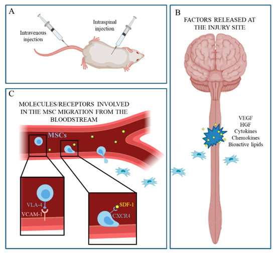
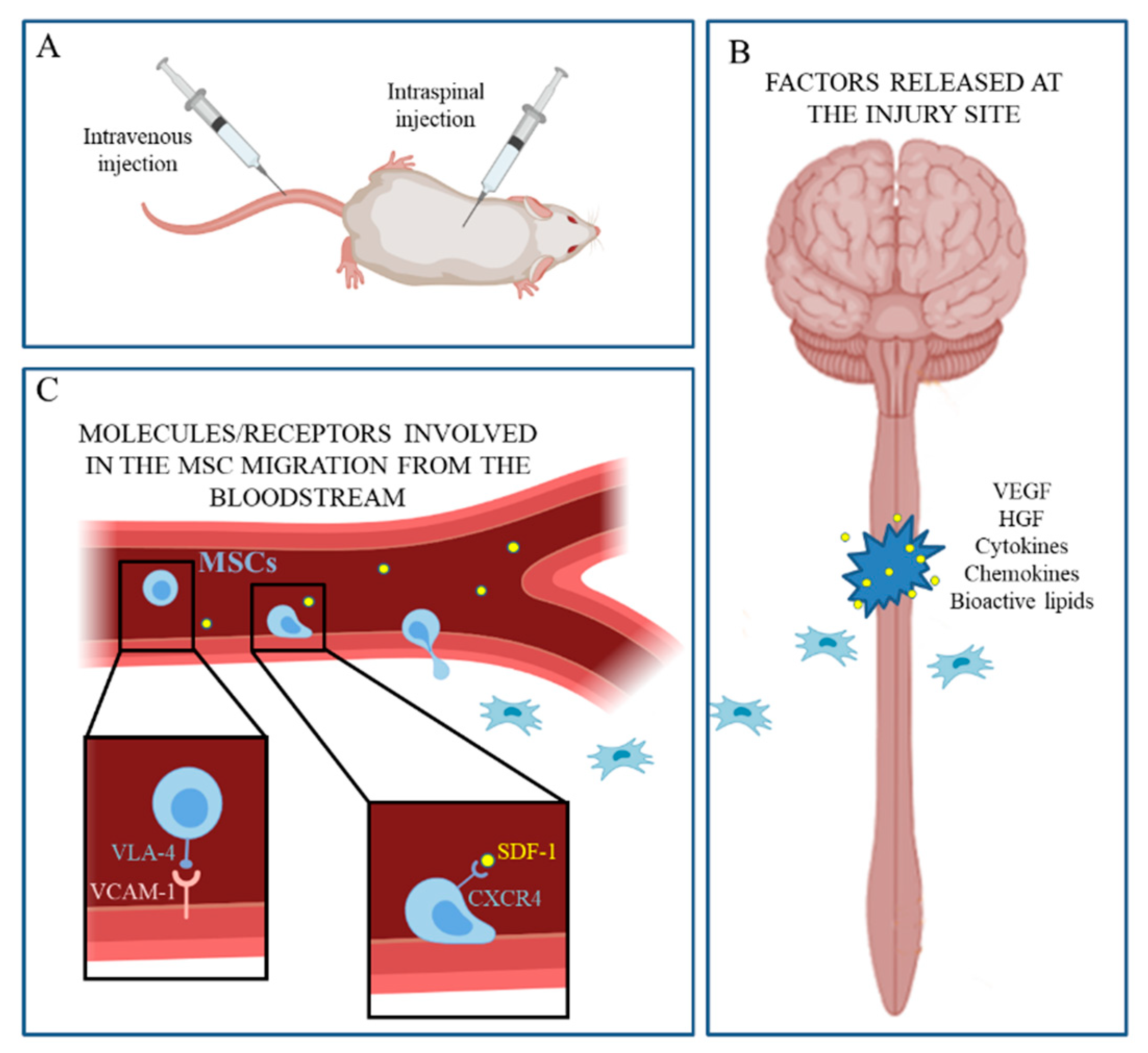
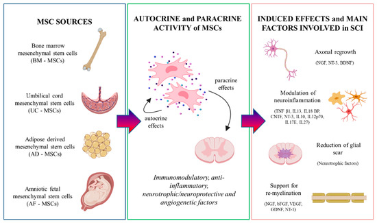
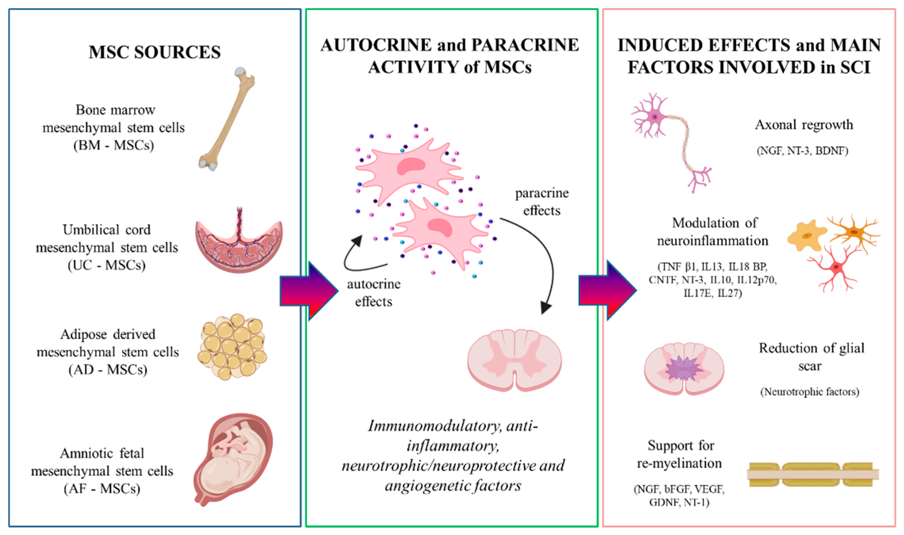
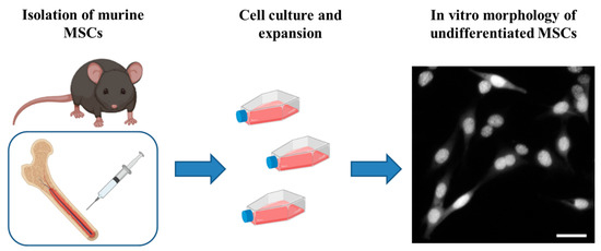
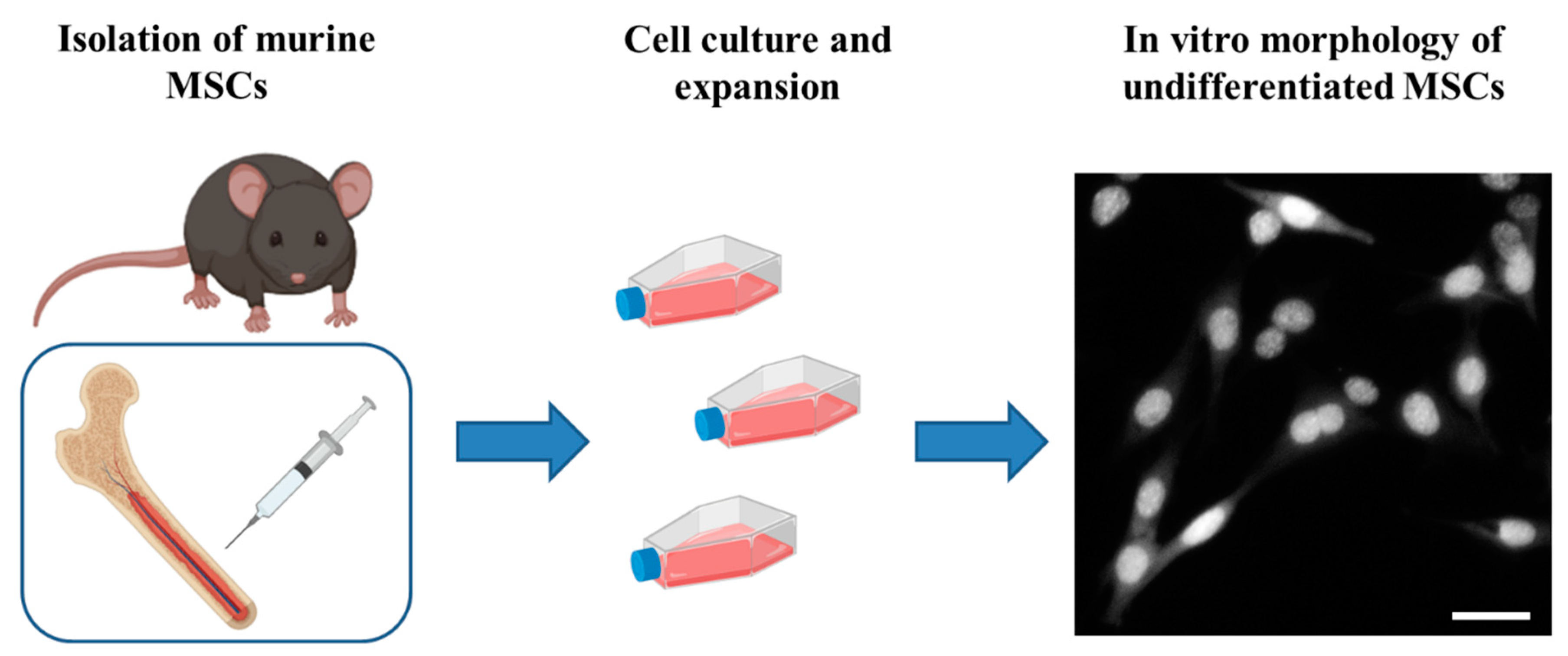

© 2019 by the authors. Licensee MDPI, Basel, Switzerland. This article is an open access article distributed under the terms and conditions of the Creative Commons Attribution (CC BY) license (http://creativecommons.org/licenses/by/4.0/).