


Abstract
Mushrooms are rich sources of polysaccharides, proteins, phenolic compounds, terpenoids, sterols, vitamins, minerals, and other biologically active constituents, and their extracts are increasingly used in functional foods, nutraceuticals, pharmaceuticals, cosmetics, and agricultural products. However, the chemical composition and biological activity of these bioactive extracts depend strongly on species, strain, cultivation substrate, developmental stage, processing, extraction solvent, and storage conditions. Therefore, suitable extraction procedures and reliable quality-control systems are essential to ensure the safety, efficacy, consistency, and reproducibility of mushroom-derived products. This review describes the main stages of mushroom processing, including raw-material authentication, drying, grinding, conventional and emerging extraction techniques, concentration, final drying, and storage. It also examines quality-control approaches, including physicochemical characterization, chromatographic and spectroscopic analysis, quantification of bioactive compounds, microbiological testing, contaminant analysis, and biological activity assays, together with the laboratory quality systems, regulatory scope boundaries, and stability requirements that underpin them. Finally, it highlights the importance of marker-based standardization, validated analytical methods, batch-to-batch consistency, and the combined use of chemical and biological quality attributes.
1. Introduction
Mushrooms have been consumed as food and used in traditional medicine across Asia, Europe, and the United States of America for centuries [1]. They are low in fat and calories and provide digestible protein, essential amino acids, dietary fiber, vitamins, and minerals [2]. In addition to their nutritional value, mushrooms are a rich source of diverse bioactive compounds, and advances in analytical methods have led to a marked increase in research on their nutraceutical potential [1]. A meta-analysis of 17 observational studies found that higher mushroom consumption was associated with a reduced risk of cancer when the highest intake category was compared with the lowest [3]. This has driven growth in the market for mushroom-based foods, supplements, and extracts. Preclinical studies have reported antioxidant, immunomodulatory, antimicrobial, anti-inflammatory, antidiabetic, hepatoprotective, and neuroprotective effects for mushroom preparations. Most of this work has concentrated on a small number of genera, including Ganoderma, Lentinula, Pleurotus, Hericium, Trametes, Grifola, Cordyceps, Inonotus, Tremella, Auricularia, and Agaricus [1]. Figure 1 shows eight of the most intensively studied species, each paired with a characteristic bioactive metabolite.
Figure 1.
Medicinal mushroom species and their characteristic bioactive compounds. Eight medicinal mushrooms (left) are paired, in the same reading order, with a representative bioactive metabolite of each (right). The species shown are Inonotus obliquus (chaga), Grifola frondosa (maitake), Ganoderma lucidum (reishi), Trametes versicolor (turkey tail), Hericium erinaceus (lion’s mane), Lentinula edodes (shiitake), Cordyceps militaris, and Tremella fuciformis (snow fungus).
These effects are linked to compounds from very different chemical classes. Polysaccharides, particularly β-(1→3)/(1→6)-D-glucans, are the most widely studied and are associated mainly with antioxidant, antitumor, and immunomodulatory activity [4,5]. Proteins form a second important group: more than 38 fungal immunomodulatory proteins (FIPs) have been described so far [6]. Lipophilic secondary metabolites are equally relevant. Phytochemical studies of Ganoderma alone have produced 431 compounds, most of them being lanostane-type triterpenoids such as ganoderic and lucidenic acids [7,8]. Other constituents of interest include phenolic compounds [9]; ergosterol, which is converted to vitamin D2 under UV exposure [10]; the antioxidants ergothioneine and glutathione [11]; nucleosides such as cordycepin in Cordyceps [1]; eight-carbon volatiles such as 1-octen-3-ol, which are responsible for the characteristic aroma of mushrooms [9]; and chitin and structural glucans, which act as dietary fiber [2]. Recovering these compounds from mushrooms presents considerable technical challenges. The fungal cell wall is built around an alkali-insoluble core of branched β-(1→3)-glucan, β-(1→6)-glucan, and chitin, to which other polysaccharides and proteins are attached (Figure 2) [12]. This structure restricts solvent penetration and must be disrupted before many intracellular compounds can be released. The target compounds also differ widely in polarity, molecular weight, and thermal stability. Water-soluble glucans, lipophilic triterpenoids and sterols, heat-labile proteins, and volatile compounds cannot all be recovered efficiently under one set of conditions. For this reason, both conventional and emerging extraction techniques are required for the selected compound [4,5].
Figure 2.
Structural organization of mushroom tissue and the fungal cell wall. Schematic representation of mushroom anatomy, including the pileus, gills, stipe, volva, mycelium, and hyphal organization, together with a magnified view of fungal hyphal ultrastructure on the left and the multilayered cell wall, composed primarily of glycoproteins, glucans, and chitin microfibrils and overlying the plasma membrane, on the right.
The chemical profile of a mushroom extract varies by species, growing conditions, and processing; therefore, batch-wise variation is expected unless these factors are precisely controlled and optimized [1,7]. Commercial products show the practical consequences. Of 19 G. lucidum dietary supplements purchased in the United States, only 5 had measured triterpene and polysaccharide contents consistent with their labels [13]. Drying, milling, and extraction also remove the morphological features used for species identification. As a result, DNA barcoding and chemometric fingerprinting are increasingly needed to authenticate powders and mycelial products [14,15,16].
Mushrooms readily accumulate heavy metals from their growth environment, and measurable lead and cadmium have been reported in edible mushrooms, with possible non-carcinogenic health risks for consumers in some countries [17]. Some species also concentrate radionuclides [18]. Microbial contamination, mycotoxins, pesticide residues, and residual solvents add to these risks (discussed in Section 11). A reliable quality-control program should therefore confirm identity, purity, potency, safety, and stability, rather than relying on extraction yield [1,13]. This review summarizes mushroom biomass pre-processing, the main conventional and emerging extraction techniques for bioactive compounds, and the analytical methods used to authenticate, standardize, and assess the safety of mushroom extracts. It also emphasizes marker-based standardization and integrating chemical, molecular, and biological assays into an effective, practical quality-control framework.
2. Biological and Chemical Characteristics of Mushrooms
2.1. Taxonomic Diversity
The chemical composition of mushrooms varies markedly among species. Closely related taxa can differ substantially in polysaccharide structure, triterpenoid composition, sterol content, and toxicological properties [7,8]. A good example is Ganoderma lucidum, a name applied almost universally, yet phylogenetic analysis shows that the Lingzhi mushroom cultivated in East Asia is not the same species as European G. lucidum and has been named G. lingzhi [19]. Accurate taxonomic identification is therefore the first requirement for any extraction or quality assessment. Classical identification relies on macroscopic morphology (cap shape, gill or pore attachment, and stipe characteristics), microscopic features (basidia, cystidia, and hyphal systems), spore characteristics (color, size, and ornamentation), and culture traits. These observations should be confirmed against authenticated herbarium specimens or reference strains from recognized culture collections [20].
2.2. Polymerase Chain Reaction (PCR)-Based DNA Barcoding
Morphological identification is less reliable when the mushroom has been powdered or processed, or when it belongs to cryptic species complexes whose members are visually indistinguishable [14]. In these cases, DNA barcoding provides a more reliable basis for species discrimination. Researchers isolate genomic DNA from fungal tissue, amplify a standardized barcode region by PCR, and then sequence it. The nuclear ribosomal internal transcribed spacer (ITS) region, located between the small-subunit (18S) and large-subunit (28S) rRNA genes, is the universal primary barcode for fungi. The resulting sequences are compared with reference databases such as GenBank or UNITE using the Basic Local Alignment Search Tool (BLAST) (Figure 3) [21]. Extensive thermal processing of mushrooms degrades DNA into short fragments, which limits PCR amplification from extracts and heavily processed products [14].
Figure 3.
Molecular workflow for mushroom species authentication. Genomic DNA is extracted from mushroom tissue followed by PCR amplification of the internal transcribed spacer (ITS) region. Species identity is subsequently assessed using agarose gel electrophoresis, Sanger sequencing, and comparison of the resulting sequence with reference databases using BLAST.
Standard ITS protocols use universal primers that anneal to conserved flanking regions. The most common pair is ITS1 (forward: 5′-TCCGTAGGTGAACCTGCGG-3′) and ITS4 (reverse: 5′-TCCTCCGCTTATTGATATGC-3′) [22]. Where greater specificity for fungi is needed, ITS1F (5′-CTTGGTCATTTAGAGGAAGTAA-3′) replaces ITS1, and where basidiomycete specificity is required, ITS4B replaces ITS4 [23]. A typical program includes initial denaturation at 94–95 °C for 3–5 min; 30–35 cycles of denaturation at 94 °C (30–60 s), annealing at 52–56 °C (30–60 s), and extension at 72 °C (1 min); and a final extension at 72 °C for 5–10 min. Amplicons of several hundred base pairs can be analyzed by agarose gel electrophoresis, purified, and sequenced by Sanger or next-generation sequencing (Figure 3) [14,21]. ITS does not resolve all species complexes; therefore, secondary markers such as the translation elongation factor 1-α (TEF1-α) or RNA polymerase II subunit (RPB2) genes may be required [19].
3. Factors Affecting Mushroom Extract Quality
The composition, bioactivity, and safety of a mushroom extract depend on a chain of interconnected factors. These act before extraction (biological and cultivation conditions), during extraction (matrix preparation and processing parameters), and after extraction (drying, storage, and preservation). Because β-glucans, triterpenoids, enzymes, and phenolics have different stability profiles, small changes at any processing stage can alter the potency and reproducibility of the final product [1,4]. Identifying and standardizing these factors is therefore essential to limit batch-to-batch variation and build robust industrial quality control (Figure 4).
Figure 4.
Critical process controls affecting mushroom extract integrity and bioactive potency. (A) Species and strain selection to ensure consistent, high-yielding bioactive profiles; (B) cultivation substrate selection, which can influence secondary metabolite biosynthesis; (C) developmental stage and harvest timing, as metabolite profiles vary throughout mushroom growth; (D) drying and storage conditions, including temperature, airflow, moisture, light, and oxygen exposure; (E) particle size and grinding conditions, which can affect filtration efficiency, thermal degradation, and loss of volatile compounds.
3.1. Species and Strain
A mushroom’s genotype establishes the baseline for the secondary metabolite profile and bioactive potential of its extracts. Species within the same genus, and even different strains of a species, can differ markedly in their metabolic output. Ganoderma spp., for example, produce distinct profiles of oxygenated triterpenoids such as ganoderic and lucidenic acids, which are known for their cytotoxic and anti-inflammatory activities [7,8]. Strain differences also influence polysaccharide and glucan content in cultivated Pleurotus mushrooms [9]. Commercial production should therefore rely on authenticated, genetically stable strains rather than wild-harvested or unverified biomass. Strain authentication by DNA barcoding [21], combined with chemical fingerprinting of production strains [16], supports predictable accumulation of target compounds and reduces the risk of propagating low-potency or misidentified strains [14].
3.2. Cultivation Substrate
The growth substrate controls the nutrients available to the fungus and, in turn, its biochemical composition. The carbon and nitrogen sources in the substrate, such as hardwood sawdust, cereal straw, agro-industrial residues, or liquid culture media, affect the synthesis of both primary and secondary metabolites. In Pleurotus and Agaricus species grown on different agro-industrial residues, substrate composition significantly changed polysaccharide, glucan, protein, lipid, and total phenolic contents, as well as antioxidant activity [9]. The same uptake capacity that enables mushrooms to accumulate beneficial compounds poses safety risks when the growth substrate is contaminated. Poorly stored or inadequately treated substrates can additionally introduce microbial contaminants and residues. Screening, sourcing, and standardizing substrate ingredients are therefore key quality-assurance steps for preventing contaminant carryover into the final extract [20].
3.3. Developmental Stage
Mushroom biochemistry changes throughout the life cycle, from vegetative mycelium and primordia to mature and post-sporulation fruiting bodies. Metabolite distribution also differs between tissues; in Pleurotus spp., for example, stipes contained more glucans, whereas pilei contained more protein and total phenolics [9]. In G. lucidum, triterpenoids, sterols, and phenolics were highest at the budding stage, whereas polysaccharides were higher at the mature stage, and key triterpenoid biosynthetic enzyme expression declined as the fruiting body developed [24]. Harvesting at inconsistent stages therefore introduces chemical variation. A reproducible chemical fingerprint depends on a well-defined harvest window, specified either by explicit morphological criteria, such as cap expansion or veil opening, or by fixed incubation times for mycelial products.
3.4. Drying and Storage
Post-harvest drying lowers water activity and slows enzymatic degradation and microbial growth. In general, water activity (aw) < 0.6 restricts microbial proliferation [25]. On the other hand, heat-induced post-harvest drying can damage heat-sensitive constituents such as proteins and enzymes, including heat-labile FIPs and the lipoxygenase and hydroperoxide lyase system that generates the characteristic eight-carbon volatiles, and promote phenolic oxidation [26,27]. Drying quality depends on key parameters such as temperature, duration, airflow, relative humidity, exposure to light and oxygen, and residual moisture content. Elevated temperatures promote thermal degradation, whereas prolonged drying at moderate temperatures increases oxidative exposure. Moreover, the choice of drying method strongly influences the volatile composition and sensory attributes of dried mushrooms [27]. Light and storage temperature have also been reported to influence ergosterol content in mushrooms [28]. Hot-air drying remains the most widely used method because of its low cost, while freeze-drying generally gives the best retention of structure and quality, but at a considerably higher cost [26].
3.5. Particle Size
Milling is the final stage of raw-material preparation and the interface between material handling and extraction, as it defines the physical state in which the biomass enters the extractant solvent. Reducing dried biomass to powder disrupts cell walls, increases the surface-area-to-volume ratio, and shortens diffusion paths; as a result, the solvent penetrates the matrix and dissolves target solutes more quickly [29]. Particle size should be optimized rather than minimized, as the gains in extraction efficiency from finer grinding are ultimately offset by processing difficulties and reduced stability. Very fine powders can swell and form viscous suspensions, especially in polysaccharide-rich materials, complicating filtration and solid–liquid separation. Frictional heat generated during intensive milling can also degrade heat-sensitive compounds and increase volatile losses [30]. For these reasons, the particle-size range should be defined experimentally for each material and extraction system, balancing mass transfer against downstream processability [29]. Sieved fractions of 180–425 µm (40–80 mesh) are often used in laboratory extraction studies.
4. Pre-Processing Protocols for Mushroom Biomass
Pre-processing strategies link harvesting and extraction and strongly determine input material quality, as post-harvest choices are irreversible. Fresh mushroom tissue is highly perishable and prone to microbial spoilage, autolytic enzymatic degradation, and structural heterogeneity. A standardized pretreatment sequence as described in subsequent sections is therefore needed to preserve bioactive constituents, reduce batch-to-batch variation, and meet pharmacopeial and good manufacturing practice requirements [31].
4.1. Authentication of Mushroom Material
Before physical processing, we taxonomically verify raw biomass. Macroscopic and microscopic evaluation, combined with DNA barcoding, confirms species identity and helps exclude adulterants, toxic look-alikes, and unintended strains (Figure 5) [14,20,21]. In the United States, manufacturers of dietary supplements are required to verify the identity of dietary ingredients [31]. Once identity is confirmed, manufacturers clean harvested fruiting bodies or cultivated mycelial blocks to remove substrate particles, soil, straw, and other debris. Dry cleaning with soft brushes, compressed air, or air-jet sorters is preferred because it removes coarse contaminants without adding water, which could accelerate tissue deterioration; raw-material testing should identify and limit foreign matter [20].
Figure 5.
Standardized pre-processing and preservation workflow for mushroom biomass. (1) Manual removal of mushrooms from grains; (2) rinsing, draining, and slicing to ensure uniform drying surface area; (3) controlled dehydration with monitored temperature and airflow; (4) milling into a defined particle-size range; (5) final packaging and sealing in airtight, light-protected poly bags with complete batch tracking labels.
When wet cleaning is unavoidable, keep washing brief and controlled. Water-soluble constituents such as low-molecular-weight polysaccharides, nucleosides (adenosine and cordycepin), and free amino acids can leach into wash water; therefore, avoid prolonged immersion. Short spray washing with clean, cold water, followed immediately by draining or air-knife dewatering, limits these losses. The cleaned mushrooms are then sliced to a uniform thickness (3–5 mm), promoting even heat and mass transfer, preventing local overheating, shortening drying time, and ensuring consistent moisture removal across the batch [26].
Slices are subsequently dried by validated methods such as convective hot-air drying, vacuum drying, microwave–vacuum drying, or freeze-drying. Convective drying of mushroom slices is carried out at 40–70 °C; for shiitake, convective drying has been compared with vacuum–microwave drying and freeze-drying [27]. Drying must be continued until the water activity of the product falls below 0.60 (Section 4.2). Control temperature, relative humidity, airflow, and light exposure to protect heat-labile proteins, volatile compounds, and phenolics, because the drying method directly affects the final composition [26,27]. The dried mushroom biomass is then milled in stainless-steel blade or hammer mills and sieved to a defined particle-size range (40–80 mesh, ~180–425 µm), which improves extraction efficiency while avoiding very fine dust that hinders filtration [29].
Frictional heating during milling could degrade sensitive constituents. Cryogenic grinding with liquid nitrogen (−196 °C) reduces these losses, particularly of volatile compounds; at low temperatures, the material becomes brittle and fractures more easily, which also limits oxidation and screen clogging [30]. Common configurations include cryogenic ball mills, in which the grinding jar and balls are cooled with liquid nitrogen; cryogenic pin or hammer mills, in which liquid nitrogen is injected into the feed or milling chamber; and jet mills, in which particle-on-particle collisions in a high-velocity gas stream achieve size reduction without mechanical contact surfaces [30]. These approaches are well established for spices and herbs, but their benefits for specific mushroom constituents still need experimental confirmation. Finally, pack the milled powder promptly in moisture-barrier, light-protective containers, such as high-density polyethylene drums with liners or aluminum-laminated pouches, preferably with desiccants. Storage conditions should be defined by stability testing; the International Council for Harmonization (ICH) long-term condition of 25 °C/60% relative humidity is a common reference point [32]. Cool, dark, and dry storage limits photo-oxidation, moisture uptake, and enzymatic degradation [33].
4.2. Industrial Validation, Moisture Content, and Water Activity
For commercial and pharmaceutical applications, pre-processing must follow a validated, documented procedure. Cleaning, slicing, and drying equipment should operate under current good manufacturing practice (cGMP) to prevent cross-contamination and to deliver reproducible microbial and chemical specifications across production lots [31]. An important distinction in process validation is the difference between moisture content and water activity. Moisture content is calculated as (Equation (1)):
Moisture content (%) = (Mass of water/Total mass of sample) × 100
Moisture content measures the total amount of water in the tissue, both bound and free, but it does not reliably predict microbial or chemical stability. Water activity (aw) describes the energy state of water in the matrix and reflects the fraction of water available for chemical reactions and microbial growth (Equation (2)):
where p is the vapor pressure of water in the sample, and p0 is the vapor pressure of pure water at the same temperature. Because microbial growth is generally prevented below aw ≈ 0.60 [25], maintaining pre-processed biomass below this value, together with a material-specific moisture limit [20], provides a stable and consistent feedstock for extraction.
aw = p/p0
5. Extraction Methods
Extraction transfers target constituents from the mushroom matrix into a suitable solvent. The choice of method depends on the target compound class, solvent polarity, thermal sensitivity, extraction time, solid-to-liquid ratio, particle size, pH, agitation, environmental and safety requirements, and the intended use of the final extract [29]. When compounds of different polarity are targeted, sequential extraction is useful. For example, a non-polar solvent can first remove lipids and sterols, aqueous ethanol can then recover phenolics and triterpenoids, and hot water can finally extract polysaccharides (Figure 6) [5,29].
Figure 6.
Overview of conventional and advanced extraction methodologies. Schematic illustrating (A) Maceration and Percolation (passive diffusion); (B) Soxhlet Extraction (vertical condenser with solvent reflux); (C) Ultrasound-Assisted Extraction (acoustic cavitation); (D) Microwave-Assisted Extraction (electromagnetic heating); (E) Enzyme-Assisted Extraction (enzymatic matrix degradation); (F) Pressurized Liquid/Accelerated Solvent Extraction (high temperature/pressure); (G) Supercritical Fluid Extraction (supercritical CO2); (H) Deep Eutectic Solvent Extraction (hydrogen-bond donor/acceptor systems).
Extraction conditions for mushroom constituents are best optimized with statistical experimental designs, such as response surface methodology (RSM) with Box–Behnken or Central Composite Design (CCD), rather than one-factor-at-a-time approaches [4]. These designs allow simultaneous modeling of key variables, including solvent composition, temperature, time, solid-to-liquid ratio, pH, ultrasound or microwave power, and number of extraction cycles, together with their interactions [4]. Optimization should balance recovery against extract quality by tracking responses such as dry yield, target compound content (β-glucans, total phenolics, or specific triterpenoids), bioactivity, and sustainability indicators such as solvent and energy use [29]. Conditions chosen only to maximize crude yield often reduce selectivity, degrade heat-labile compounds, or increase co-extraction of unwanted matrix components and pigments [29]. Table 1 summarizes the principles, typical operating conditions, main targets, advantages, and limitations of the techniques discussed below.
Table 1.
Main extraction techniques for mushroom bioactive compounds.
5.1. Hot-Water Extraction (HWE)
HWE is the conventional and most widely used method for recovering water-soluble polysaccharides, including β-glucans and protein-bound polysaccharides, from mushrooms [4,5]. In a representative laboratory procedure, dried and pre-processed mushroom powder (100 g, 40–80 mesh) is suspended in deionized water (2000 mL) at a solid-to-liquid ratio of 1:20 (w/v) and heated at 95 ± 2 °C under stirring (300–500 rpm) or reflux for 2–3 h, with repeated extraction to improve recovery [4,5]. The hot mixture is filtered through fine filter cloth or centrifuged (4000× g for 15 min) to remove insoluble cell-wall residues, and the clarified extract is concentrated under reduced pressure at 60–65 °C to 20% of its initial volume [29].
Crude polysaccharides are then precipitated by slowly adding cold ethanol (95–99%, about four volumes per volume of concentrate) to a final concentration of 75–80% (v/v) and holding the mixture at 4 °C for 12–24 h. The precipitate is recovered by centrifugation (6000× g for 20 min at 4 °C), redissolved in a minimal volume of water, dialyzed against distilled water for 48–72 h with frequent water changes (using a 3.5 kDa molecular weight cut-off) to remove salt and small molecules, and freeze-dried [5]. This product is a crude polysaccharide fraction that contains α-glucans, heteroglycans, and proteins in addition to β-glucans; further fractionation and specific assays are required before it can be described as a β-glucan preparation [5,42]. HWE is non-toxic, simple, scalable, and acceptable for food and nutraceutical products; however, it involves long extraction times, high energy demand, low selectivity, possible thermal degradation of proteins, and co-extraction of pigments and other impurities [4].
5.2. Alcoholic and Hydroalcoholic Extraction
Ethanol–water mixtures are used to extract moderately polar secondary metabolites, including phenolic acids and oxygenated triterpenoids such as ganoderic acids [8,29]. Food-grade ethanol is diluted to 50–80% (v/v), depending on the polarity of the target compounds, and mixed with mushroom powder at a solid-to-liquid ratio of 1:10 to 1:20 (w/v), corresponding to 10 g of powder in 100–200 mL of 60–70% ethanol. The mixture is stirred or refluxed at moderate temperature (50–65 °C) for 2–3 h, and the extraction is usually repeated for two or three cycles with fresh solvent [29]. The combined extracts are filtered (through Whatman No. 1 paper followed by a 0.45 µm membrane) and concentrated under vacuum below 50 °C (rotary evaporation at 40–45 °C) to limit degradation of thermolabile compounds, after which the concentrate is vacuum-dried or spray-dried, often with a carrier such as maltodextrin, to obtain a standardized powder [43]. Hydroalcoholic extraction provides good recovery of mid-polarity bioactive compounds, and food-grade ethanol is widely accepted by regulators; its main drawbacks are flammability and higher operating costs than water-only extraction [29].
5.3. Soxhlet Extraction
Soxhlet extraction continuously recycles freshly condensed solvent through the sample (about 1:12 to 1:25, w/v) and has been used for exhaustive recovery of non-polar to moderately polar constituents such as lipids, sterols, and triterpenes [29]. Place dried mushroom powder (10–20 g) in a porous cellulose thimble inside the extraction chamber and charge the boiling flask with 250 mL of a solvent such as ethanol, ethyl acetate, or n-hexane. The solvent is heated to reflux, and condensation and siphoning cycles continue for 6–12 h, at a rate of 4–6 siphoning cycles per hour, until extraction is complete [29]. The solvent is then removed under vacuum, and the residue is weighed to determine the exhaustive extraction yield. The method is simple, inexpensive, and efficient and remains a reference method for lipid and sterol determination. However, the method is intrinsically unsuited to polysaccharide recovery for three linked reasons. First, the solvents circulated in a Soxhlet apparatus are non-polar to moderately polar organic liquids in which β-glucans and heteroglycans are essentially insoluble; the aqueous ethanol that refluxes through the thimble is the same antisolvent used in Section 5.1 to precipitate crude polysaccharide, so the target polymer is driven out of solution rather than into it. Second, extraction depends on diffusion of the solute into the condensed solvent, and high-molecular-weight glucans do not diffuse appreciably out of a swollen cell-wall matrix into an organic phase, so extending the cycle count increases lipid and sterol yield without increasing polysaccharide yield. Third, the cellulose thimble retains particulate and colloidal material, so any solubilized polymer would largely be held back with the marc. Polysaccharide recovery instead requires a solvent in which the polymer is soluble and the wall swells, that is hot water or dilute alkali, followed by ethanol precipitation [5,29]. Soxhlet extraction should accordingly be regarded as a reference method for the lipophilic fraction of mushroom biomass rather than as a general-purpose exhaustive extraction. However, prolonged heating at the solvent boiling point can degrade thermolabile compounds, solvent consumption is high, and scale-up is difficult [29].
5.4. Maceration and Percolation
Maceration (static soaking) and percolation (continuous flow through a packed bed) are simple, low-energy methods operated at room temperature and are suitable for heat-sensitive compounds. In maceration, mushroom powder is kept in a closed vessel with water, aqueous ethanol, or an organic solvent (at 1:10 w/v) for 48–72 h at 20–25 °C with periodic agitation; the extract is then filtered, and the residue is pressed to recover retained solvent. In percolation, the powder is first moistened with solvent for 2 h to allow swelling, packed uniformly into a percolator, and eluted with fresh solvent at a slow, steady flow rate (1–3 mL/min at laboratory scale) until extraction is complete, after 5–10 bed volumes of solvent have passed. Both methods carry a low risk of thermal degradation and require little capital investment, but they are slow, consume large volumes of solvent, and generally give lower yields than dynamic or assisted methods [29].
5.5. Ultrasound-Assisted Extraction (UAE)
UAE relies on acoustic cavitation, the formation and violent collapse of microbubbles in the solvent, which disrupts cell walls and enhances solvent penetration and mass transfer [34]. Mushroom powder is mixed with water or 60% aqueous ethanol at a solid-to-liquid ratio of 1:10 to 1:20 (w/v) and sonicated with an ultrasonic bath or probe at 20–40 kHz. Because acoustic power strongly influences performance, it should be optimized and reported in comparable units, preferably as acoustic power density (W/mL) or intensity (W/cm2) determined calorimetrically [34]. Heat generated during sonication is removed by circulating coolant through a jacketed vessel, keeping the extraction temperature below 45 °C, and treatment times of 10–30 min, in pulsed mode (5 s on/2 s off) to limit local heating, are common before the extract is clarified by centrifugation (4000× g for 10–15 min) [34]. UAE shortens extraction from hours to minutes, reduces solvent use, and operates at moderate temperature. Limitations include free-radical formation during intense cavitation, which can oxidize sensitive compounds and depolymerize polysaccharides, and difficulty achieving uniform scale-up [4,34].
5.6. Microwave-Assisted Extraction (MAE)
MAE uses non-ionizing electromagnetic radiation at 2.45 GHz to heat polar solvents and intracellular water rapidly; the resulting internal pressure ruptures cell walls and releases intracellular compounds. The biomass is combined with a solvent of high dielectric loss, such as water or aqueous ethanol (at 1:15 w/v), and irradiated in either an open-vessel (atmospheric) or closed-vessel (pressurized) system. Microwave power of 200–800 W is applied to reach the target temperature (60–90 °C within 1–2 min), which is then held for 3–10 min, before the vessel is cooled and the extract filtered. MAE offers short extraction times, high yields, and low solvent consumption. However, it can cause local overheating, closed systems require pressure-rated equipment, and efficiency is poor with non-polar solvents that absorb little microwave energy [35].
5.7. Enzyme-Assisted Extraction (EAE)
Enzyme-assisted extraction (EAE) uses hydrolytic enzymes, such as cellulase, pectinase, and proteases including papain and dispase, individually or in combination, to recover mushroom-derived compounds under aqueous conditions [4,36]. Enzyme selection depends on the mushroom species, the target compound, and the preparation’s substrate specificity [36]. Chitinases and appropriate β-glucanases hydrolyze chitin and susceptible β-glucan linkages, respectively, whereas proteases hydrolyze proteins and may facilitate polysaccharide recovery through deproteinization [5]. Cellulase- and pectinase-containing preparations have also been used in mushroom extraction studies; however, their effectiveness should not be interpreted as evidence that cellulose or pectin is a major structural component of mushroom cell walls, which are built principally from chitin, β-glucans, and cell-wall proteins [12]. Commercial preparations are mixtures rather than single activities: Viscozyme L, derived from Aspergillus aculeatus, contains several carbohydrase activities, including arabanase, cellulase, β-glucanase, hemicellulase, and xylanase [36].
Select extraction conditions, including pH, temperature, enzyme loading, solid-to-liquid ratio, and incubation time, based on the enzyme preparation and target compound rather than using a universal protocol [36]. Enzyme loading should be reported relative to dry biomass and, where available, as activity units per gram of dry biomass, together with the supplier and the activity-unit definition. In an optimized ultrasound-assisted enzymatic extraction of Tricholoma matsutake polysaccharides, a complex enzyme preparation of 1.15% cellulase, 0.60% pectinase, and 0.95% dispase was applied at pH 5.0, with ultrasonication at 180 W for 24 min at 84.5 °C and a solid-to-liquid ratio of 1:30 (w/v) followed by enzymatic digestion at 50 °C for 30 min; the optimized process gave a polysaccharide yield of 19.74 ± 0.51%, more than four times that obtained by conventional hot-water extraction [44].
Following extraction, insoluble material is separated, and enzyme inactivation or removal is performed using a procedure compatible with the stability of the target compounds [4]. EAE can be combined with hot-water or ultrasound-assisted extraction to improve recovery [44]. However, enzyme cost, preparation-specific operating requirements, and unwanted hydrolysis of target polysaccharides or proteins can limit its application [36]. Therefore, researchers should evaluate preservation of molecular weight distribution, glycosidic linkage pattern, and biological activity experimentally rather than assume it [5].
5.8. Pressurized Liquid Extraction (PLE)/Accelerated Solvent Extraction
PLE uses elevated temperature and pressure to keep solvents liquid above their normal boiling points, which increases solubility and mass transfer. Operating conditions are 50–200 °C and 10–15 MPa [37], and the technique has been applied to sterol-enriched fractions obtained from mushrooms [38]. Mushroom powder is mixed with an inert dispersant such as diatomaceous earth or sand and packed into a stainless-steel extraction cell. The solvent (water, ethanol, or aqueous ethanol) is pumped into the heated cell (at 10.3 MPa, 1500 psi) and held for one or more static cycles of 5–10 min each, after which the cell is flushed with fresh solvent (60% of the cell volume) and purged with nitrogen (for 60–90 s), and the extract is collected directly for analysis or evaporation [37]. PLE is automated, fast (15–30 min per sample), and uses little solvent, but it requires costly pressure-rated equipment, and high temperatures can degrade thermolabile compounds if exposure is not optimized [37].
5.9. Supercritical Fluid Extraction (SFE)
SFE most often uses supercritical carbon dioxide (scCO2), obtained above its critical point (31.1 °C and 7.38 MPa), as a relatively non-polar extraction solvent for lipophilic mushroom constituents such as ergosterol, fatty acids, and less polar triterpenes; because carbon dioxide reverts to a gas on depressurization, it is readily removed and leaves extracts with minimal solvent residue [38,39]. Milled biomass is loaded into a high-pressure vessel, and liquid CO2 is pumped to the working pressure (10–40 MPa) and heated (40–60 °C) [39]. Ethanol can be added as a polar modifier, at 5–15% (v/v), to improve recovery of more polar compounds. In Agaricus bisporus, adding ethanol increased extraction yield; however, it reduced the extract’s sterol selectivity [38]. The fluid then passes through a pressure-reduction valve into a separator, where CO2 returns to the gas phase, leaving an extract essentially free of residual solvent. SFE uses a non-toxic, non-flammable solvent, applies low thermal stress, and is selective for lipophilic compounds, but it has high capital costs and poor efficiency for polar compounds; high-molecular-weight polysaccharides remain inaccessible even with a polar modifier, which extends the range only to moderately polar small molecules [39].
5.10. Deep Eutectic Solvents (DES) and Green Extraction
Deep eutectic solvents are formed by combining a hydrogen-bond acceptor, such as choline chloride, with a hydrogen-bond donor, such as urea, glycerol, or an organic acid, at a defined molar ratio, producing a mixture that melts at a much lower temperature than either component. Natural deep eutectic solvents (NADES), prepared from primary metabolites, are of particular interest for food and pharmaceutical applications. A common example is choline chloride and glycerol at a 1:2 molar ratio, heated with stirring (at 80 °C for 30–60 min) until a clear, homogeneous liquid forms. Water is usually added to reduce viscosity and improve mass transfer, and the optimal water content varies widely [40]. It has been reported that for Morchella importuna polysaccharides, a choline chloride–oxalic acid DES containing 90% (v/v) water combined with ultrasound gave a polysaccharide yield 4.5 times higher than HWE [41]. Separating the target compounds from the DES after extraction remains a key practical challenge. DES are tunable, low-volatility, and often biodegradable solvents that can improve extraction efficiency. Nevertheless, their high viscosity, the difficulty of separating the solvent from the product, and the need for toxicological and regulatory evaluation of any residual solvent remain important limitations [40].
6. Recovery, Concentration, and Drying of Extracts
A crude mushroom extract is a dilute and chemically heterogeneous liquid. Besides the target constituents, it usually contains fine cell-wall particles, proteins, pigments, free sugars, salts, and, in hydroalcoholic extracts, lipids and waxes [29,45]. Downstream processing has three main goals: (i) removing insoluble and unwanted soluble material, (ii) reducing solvent volume without damaging labile compounds, and (iii) turning the concentrate into a stable, well-defined solid. The best route depends on the solvent, the heat sensitivity and molecular size of the target compounds, the required purity, and the production scale. A typical sequence consists of clarification, concentration, optional purification or fractionation, drying, and formulation (Figure 7) [26,29].
Figure 7.
Downstream clarification, concentration, fractionation, and drying workflows for mushroom extracts. Key stages include: (A) Centrifugation: High-speed phase separation for primary liquid–solid clarification; (B) Filtration: Depth and stepped filtration to remove residual particulates; (C) Rotary Evaporation: Reduced-pressure solvent recovery at low temperatures; (D) Vacuum Concentration: Multi-sample evaporation for controlled liquid concentration; (E) Membrane Filtration: Size-exclusion fractionation using microfiltration (MF) and ultrafiltration (UF); (F) Lyophilization: Freeze-drying via sublimation to preserve heat-sensitive compounds; (G) Spray Drying: Continuous thermal atomization for rapid powder production.
Each step can alter the final product’s composition. Heating during evaporation can promote Maillard reactions and degrade phenolics and triterpenoids, whereas incomplete removal of proteins and pigments can interfere with later chemical and biological assays [45]. For this reason, the operating conditions of downstream steps should be documented as carefully as the extraction conditions, and marker compounds should be quantified before and after each major step to confirm that recovery is acceptable.
6.1. Extract Clarification
Clarification removes suspended solids that would otherwise foul membranes, block chromatographic columns, or produce turbid and unstable concentrates. At laboratory scale, crude extracts are centrifuged (4000–10,000× g at 4 °C for 15–20 min), and the supernatant is then filtered under vacuum through a Büchner funnel fitted with filter paper (Whatman No. 1) [29]. Because the relative centrifugal force (RCF) depends on the rotor radius, centrifugation conditions should be reported in × g rather than rpm; the two are related by RCF = 1.118 × 10⁻⁵ × r × N2, where r is the rotor radius (cm), and N is the rotor speed (rpm). For a rotor radius of 10 cm, 4000 × g corresponds to approximately 6000 rpm and 10,000× g to approximately 9500 rpm. At an industrial scale, decanter centrifuges, filter presses, or depth filters refine the product. Carry out clarification promptly after extraction, because aqueous extracts containing sugars and proteins are an excellent medium for microbial growth.
6.2. Rotary Evaporation Under Reduced Pressure
Rotary evaporation lowers the solvent’s boiling point by reducing the pressure above the liquid, allowing removal at temperatures well below its boiling point [29]. Rotating the flask spreads the liquid as a thin film on the heated wall, increasing the evaporation surface area and improving heat transfer. For ethanol and aqueous ethanol extracts, the water bath is set at 40–50 °C with a condenser cooled to 4–10 °C, and the flask is filled to no more than about half its volume. Set the vacuum close to the solvent’s vapor pressure at the desired boiling temperature (100–175 mbar for ethanol and 40–75 mbar for water at 30–40 °C) and rotate the flask at 100–150 rpm. The vacuum should be reduced gradually, because polysaccharide- and protein-rich extracts tend to foam and bump when pressure drops too quickly. Rotary evaporation is well suited to recovering ethanol for reuse and to concentrating extracts before precipitation or drying. However, concentrating aqueous extracts is slow because of water’s high latent heat, and prolonged heating of concentrated solutions increases the risk of browning and degradation of heat-labile compounds [29]. For large volumes, falling-film or multiple-effect evaporators, which operate on the same principle with much shorter residence times, are preferred at industrial scale.
6.3. Vacuum Concentration (Small-Volume Parallel Samples)
Centrifugal vacuum concentrators combine low pressure, mild heating, and centrifugal force to evaporate many small samples simultaneously. They are widely used in analytical workflows, for example, to dry fractions collected from solid-phase extraction or preparative chromatography before redissolution in the high-performance liquid chromatography (HPLC) mobile phase. Extracts are placed in tubes or deep-well plates with adequate headspace (filled to no more than 75% of their volume), and a moderate chamber temperature (30–40 °C) is applied under vacuum (1–20 mbar, depending on the solvent), with rotor speeds of 1000–1700 rpm. The centrifugal force keeps the liquid at the bottom of each tube, which prevents bumping and cross-contamination between samples. Because the process runs to dryness, losses of volatile compounds and adsorption of analytes to container walls can occur. Recovery should therefore be checked with spiked samples when vacuum concentration is part of a quantitative method, and the dried residue should be redissolved in a solvent that fully solubilizes the analytes. Matrix components that concentrate together with the analytes can also affect LC-MS measurements [46].
6.4. Membrane Filtration (Aqueous and Heat-Sensitive Concentrates)
Membrane processes separate components by size under a pressure gradient and operate at ambient temperature, making them attractive for heat-sensitive extracts. Ultrafiltration membranes, characterized by their nominal molecular weight cut-off (1–10 kDa for concentrating polysaccharide and protein fractions), retain polysaccharides and proteins while allowing water, salts, free sugars, and small molecules to pass [5,45]. Nanofiltration retains smaller solutes and can concentrate low-molecular-weight fractions. When a retentate is repeatedly diluted with water and re-concentrated (diafiltration), salts and small impurities are removed more completely, serving the same purpose as dialysis but on a larger scale. Ultrafiltration with membranes of different cut-offs has also been used to fractionate mushroom glucans by molecular size [5]. The main practical limitations are membrane fouling and concentration polarization, which reduce permeate flux over time, and the fact that nominal cut-off values do not give sharp separations for branched or highly hydrated polysaccharides. Therefore, verify membrane selection by measuring the molecular weight distribution of the retentate and permeate.
When structurally defined fractions are required, for example, for structure–activity studies or as reference materials, membrane processing is usually followed by further purification. Proteins are removed by the Sevag method (repeated shaking of the aqueous solution with chloroform/n-butanol, 4:1 or 5:1 v/v, for 20–30 min until no protein layer forms at the interface), trichloroacetic acid precipitation, or enzymatic hydrolysis; pigments are removed by adsorbent resins, activated carbon, or mild oxidation; and the material is then fractionated by anion-exchange chromatography (diethylaminoethyl, DEAE-cellulose), which separates neutral and acidic polysaccharides, eluted stepwise or with a linear NaCl gradient (0–0.5 M), and monitored by the phenol–sulfuric acid assay at 490 nm and by size-exclusion chromatography (Sephadex or Superdex media) [5,45]. Each purification step reduces yield, so the degree of purification should match the intended use of the extract.
6.5. Freeze-Drying/Lyophilization (Heat-Sensitive and High-Value Compounds)
Freeze-drying removes water by sublimation from the frozen state and is regarded as the method that best preserves the structure, color, aroma, and bioactive constituents of heat-sensitive materials, although its capital and operating costs are several times higher than those of hot-air drying [26]. During freezing, the concentrated extract is cooled completely (at −80 °C for at least 12 h or on refrigerated shelves), and most of the water forms ice crystals. During primary drying, the chamber pressure is lowered (below 0.1 mbar, with a condenser at −50 °C or lower), and heat is supplied so that the ice sublimes; the product temperature must remain below its collapse temperature to avoid loss of the porous structure. During secondary drying, the temperature is raised to desorb bound water and reach low residual moisture [26].
Freeze-dried extracts are porous and rehydrate rapidly but are also highly hygroscopic. Sugar-rich extracts may collapse or become sticky because their low glass transition temperatures allow the matrix to soften during drying or storage. Adding carriers such as maltodextrin or gum arabic before freezing raises the glass transition temperature and improves powder properties. In a study on freeze-dried hop extract, carrier choice affected moisture content, water activity, color, flowability, microstructure, and water-sorption behavior, and encapsulated powders were compared with the non-encapsulated extract during storage at different temperatures [47]. Freeze-drying cycles last 24–48 h for laboratory samples, limiting throughput and making the method best suited for high-value products and reference materials.
6.6. Spray-Drying (Large-Volume Heat-Tolerant Extracts)
Spray-drying converts a liquid feed into a powder in a single continuous operation and is the most used microencapsulation technique in the food industry [43]. The process involves atomizing the feed into fine droplets, contacting the droplets with hot air, rapidly evaporating water, and separating the dried particles, usually in a cyclone. Inlet air temperatures are 150–220 °C, with outlet temperatures of 50–80 °C [43]. Despite the high inlet temperature, evaporative cooling keeps the droplet temperature close to the wet-bulb temperature during most of the drying time, and residence times are only seconds, so thermal damage is limited.
Extracts rich in low-molecular-weight sugars and organic acids are difficult to spray-dry on their own because their low glass transition temperatures cause particles to stick to the chamber wall, which reduces yield and product quality. Carriers such as maltodextrin, which has high solubility, low viscosity, and a relatively high glass transition temperature, and gum arabic, which also provides emulsifying and film-forming properties, are therefore added to the feed [43]. Feed solids content, carrier type and ratio, inlet temperature, and feed rate together determine particle size, moisture content, bulk density, and retention of active compounds. Compared with freeze-dried powders, spray-dried powders have smaller, more uniform particles and are cheaper to produce, but they are less suitable for highly heat-sensitive or volatile compounds [26,43].
6.7. Encapsulation and Formulation
Encapsulation surrounds an extract’s active components with a protective wall material. It protects sensitive compounds from oxygen, light, moisture, and heat; masks unpleasant taste; improves handling and solubility; and can control release [43,48]. The choice of wall material depends on the properties of the core. For water-soluble extracts, maltodextrins of different dextrose equivalents are widely used because they are inexpensive, highly soluble, and form low-viscosity solutions, but they have poor emulsifying capacity; gum arabic is therefore often combined with maltodextrin when lipophilic components must be stabilized [43]. Other matrix (wall) materials include modified starches, proteins, sodium alginate, and chitosan. The core-to-wall ratio (1:1 to 1:4 w/w) and total solids content strongly influence encapsulation efficiency, which is usually defined as the proportion of the active compound retained inside the particles relative to the total amount present [48].
Poorly water-soluble fractions, such as triterpenoids and sterols, can be incorporated into lipid-based carriers, including liposomes, nanoemulsions, and solid lipid nanoparticles, which improve dispersibility in aqueous media and may enhance absorption [48]. These systems require additional characterization of particle size, polydispersity, surface charge, and physical stability, and their safety and regulatory status must be considered for food and supplement applications. For solid oral products, dried or encapsulated powders are blended with excipients such as microcrystalline cellulose (diluent), colloidal silica (glidant), and magnesium stearate (lubricant) and compressed into tablets or filled into hard gelatin or hydroxypropyl methylcellulose capsules. Because extract powders are hygroscopic, blending and filling should take place under controlled humidity (below 40% relative humidity, RH) [43]. Evaluate finished formulations for marker-compound content uniformity, disintegration, moisture content, water activity, and stability under long-term and accelerated storage conditions (40 °C/75% RH) [32].
7. Laboratory Management and Analytical Quality Systems
Every specification discussed in this review presumes that the underlying measurement is trustworthy, and for mushroom products that assumption is unusually fragile. The matrix is heterogeneous at the scale of a test portion, certified reference materials barely exist, and several of the most important methods are operationally defined rather than absolute, so the reported value depends on the procedure as much as on the sample. β-Glucan determined as total glucan minus α-glucan [42], total phenolics reported as gallic acid equivalents by the Folin–Ciocalteu reaction, and antioxidant capacity reported as a DPPH or FRAP value each return a number that is interpretable only alongside the protocol that produced it. Laboratory management is therefore not administrative overhead layered on top of the analytical work; it is what makes a specification mean the same thing in two laboratories, or in the same laboratory a year apart.
7.1. Competence and the Management System
ISO/IEC 17025 provides the framework for a testing laboratory: defined responsibilities, controlled procedures, impartiality, and demonstrated technical competence for each combination of method and matrix [49]. Accreditation is granted against a defined scope, and a scope covering inductively coupled plasma mass spectrometry for foodstuffs does not automatically extend to a concentrated fungal extract, so manufacturers commissioning contract testing should confirm that the accredited scope names both the technique and a comparable matrix. Where testing supports regulatory safety studies, the Principles of Good Laboratory Practice apply instead [50], and manufacturers of dietary supplements in the United States operate under the current good manufacturing practice requirements of 21 CFR Part 111, which oblige them to establish specifications and to verify the identity of each incoming dietary ingredient [31].
7.2. Sampling and Subsampling
Sampling is the dominant and most frequently neglected source of error for mushroom materials. Composition differs between pileus and stipe, between flushes of the same cultivation batch, and, above all, between fungal tissue and residual grain substrate in mycelial products, so a non-representative test portion yields a precise measurement of the wrong thing. A defined sampling plan should specify the number of increments taken across the lot and involve compositing, homogenization, milling to a stated particle size before subsampling (Section 3.5), and splitting by riffle or rotary divider rather than by scoop, and the mass of the final test portion be recorded. Reporting that mass matters: a trace-element result obtained on 0.5 g of an incompletely homogenized powder characterizes that 0.5 g and not the batch.
7.3. Method Validation and Verification
Analytical procedures used for release decisions should be validated in accordance with ICH Q2(R2), which, in its 2023 revision, organizes the work around specificity and selectivity, the reportable range together with the response or calibration model, validation of the lower range limits—which now subsume the detection and quantitation limits—as well as accuracy and precision, the latter two of which may be evaluated together against a combined performance criterion. Robustness is expected to be established during method development under ICH Q14, although Q2(R2) retains a short robustness section of its own [51,52]. Two points deserve emphasis for fungal matrices. Recovery must be established by spiking the mushroom matrix itself rather than the solvent, because co-extracted polysaccharides and pigments suppress or enhance electrospray ionization and shift apparent concentrations in LC-MS/MS, for which matrix-matched calibration or standard addition is the appropriate countermeasure [46]. As for operationally defined methods, validation must fix the conditions that define the result: the same Ganoderma lucidum sample returned 54 and 24 g/100 g β-glucan, depending only on whether sulfuric or hydrochloric acid was used for hydrolysis [42]. Where a laboratory adopts a pharmacopeial method unchanged, formal validation is replaced by verification that the method performs as intended in that laboratory on that matrix.
7.4. Reference Standards and Reference Materials
The scarcity of authentic standards is a constraint specific to this field. Few individual triterpenoids are available commercially in certified purity, which is why single-marker quantification with relative correction factors has been developed for Ganoderma triterpenes (Section 9.3) [53]. Laboratories that cannot buy a primary standard should qualify an in-house secondary standard against a characterized batch, record purity by mass balance, and set a defined re-qualification interval and storage condition. Identity work depends in parallel on authenticated reference material rather than on pure compounds: voucher specimens deposited in a recognized herbarium or culture collection, and authenticated reference extracts for comparison of thin-layer and liquid chromatographic fingerprints [13,15]. Because matrix-certified reference materials for mushroom products are effectively non-existent, participation in interlaboratory comparison is often the only available check on trueness [54].
7.5. Equipment, Environment, and Contamination Control
Instruments carrying a release decision require documented qualification and a calibration interval traceable to recognized standards: balances and reference weights, water activity meters verified against saturated salt solutions, drying ovens and freeze-dryers, chromatographic detectors, inductively coupled plasma mass spectrometers with internal standards and drift monitoring across the sequence, and high-purity germanium detectors with efficiency calibration at the counting geometry actually used. Contamination control needs attention that generic guidance does not supply. Dried mushroom powder is light and dusty, and cross-contamination between species during weighing is a realistic route to a false identity result, so dedicated weighing enclosures and cleaning verification between species are warranted. The same applies with greater force to molecular work, where amplicon carryover produces confident and incorrect barcoding calls unless pre-amplification and post-amplification activities are physically separated and negative controls are run with every batch (Section 2.2).
7.6. Documentation, Data Integrity, and Traceability
Records should be attributable, legible, contemporaneous, original and accurate. In addition, they should be complete, consistent, enduring and available throughout the retention period [55]. In practice, this means that audit trails enabled and reviewed in chromatography data systems the retention of raw data and processed reports, as well as a documented link from each result back through the test portion and the sample to the raw material lot, the cultivation batch, and the supplier. Retained samples of each raw material and finished extract lot should be held under the labeled storage conditions for the claimed shelf life along with a defined margin, so that a later complaint or stability deviation can be investigated against the original material [31].
7.7. Monitoring Laboratory Performance
Ongoing performance is demonstrated rather than assumed. System suitability criteria should be met before an analytical sequence is accepted, and a stable in-house control extract analyzed alongside routine samples and plotted on control charts will reveal drift that individually passing results conceal. A written out-of-specification procedure should distinguish assignable laboratory error from genuine product failure, with retesting permitted only under a defined protocol rather than until an acceptable value appears. Where proficiency testing schemes exist, they should be used [54], with one caveat specific to this field: because β-glucan and total triterpene values are method-dependent, performance scores are interpretable only within a method group, and a laboratory using a different procedure may be penalized for a difference that is procedural rather than analytical.
8. Quality-Control Framework
Apply quality control throughout the production process, not only to the finished product. A complete framework includes specifications for incoming raw materials, in-process controls at critical steps such as drying and extraction, and release testing of the finished extract, all supported by written procedures and batch records [20,31]. Its aim is to ensure each batch meets predefined criteria for identity, purity, strength or potency, and composition, and remains within these criteria throughout its shelf life. In the United States, dietary supplement manufacturers must establish specifications for these attributes and conduct at least one appropriate test or examination to verify the identity of each dietary ingredient before use [31].
For mushroom products, the framework must also address issues specific to fungal materials: the distinction between fruiting bodies and mycelium, the presence of residual grain substrate in mycelium-based products, and the resulting differences in glucan composition [13,42]. Product labels should state the species, the part used (fruiting body, mycelium, or spores), the substrate on which mycelium was grown, the extraction solvent, and, where applicable, the extract ratio and active-compound content. These distinctions are only meaningful if they can be verified analytically, and the tests below make that possible.
The discriminating measurement is not β-glucan content but the ratio of α-glucan to β-glucan, determined in a single procedure in which total glucan is measured after controlled sulfuric acid hydrolysis, α-glucan is measured separately after enzymatic hydrolysis of starch and glycogen, and β-glucan is obtained by difference [42]. Across a survey of authentic fruiting bodies, α-glucan did not exceed about 4 g/100 g and, in most species, remained below 1 g/100 g, while β-glucan ranged from approximately 6 to 74 g/100 g, depending on the species. In commercial products declared as mycelium, the relationship typically inverts, with α-glucan of 3 to 73 g/100 g against β-glucan of 0 to 11 g/100 g, because the starch of the cereal substrate remains essentially intact under cultivation and becomes the dominant glucan in the finished powder [42]. A workable specification for material labeled as fruiting body is therefore α-glucan not more than 5 g/100 g, together with β-glucan exceeding α-glucan. The inversion is typical rather than universal, and the exception is instructive: in the same survey, a deep-layer cultivated Cordyceps mycelial extract returned α-glucan of 3.0 and β-glucan of 10.9 g/100 g and thus satisfied both criteria, because an extraction step can remove the starch on which the test depends [42]. Glucan criteria therefore discriminate milled myceliated grain rather than mycelial extracts, and should be applied together with the orthogonal checks below. A low β-glucan value on its own is not evidence of adulteration, since Agaricus bisporus and Inonotus obliquus fruiting bodies contain only about 6 to 8 g/100 g [42]; it is the inversion of the ratio that identifies grain carryover.
Three caveats attach to this measurement and should be stated wherever it appears in a specification. The procedure is a published method rather than an officially collaboratively validated one, and it is operationally defined: the hydrolysis acid alters the result, with sulfuric and hydrochloric acid giving 54 and 24 g/100 g, respectively, for the same Ganoderma lucidum sample [42]. The assay also overestimates fungal β-glucan in grain-containing products, because cereal β-glucans and cellulose contribute to the measured value [56]. Confirmation by an orthogonal method is therefore advisable. Residual starch can be demonstrated directly by iodine staining, with the color abolished by α-amylase pretreatment, and the fatty acid profile of a grain-fermented product tracks the substrate rather than the fungus [56]. Ergosterol provides an independent estimate of fungal content, being a fungal membrane sterol absent from cereal grain itself. Though stored grain carries a mold-derived background, it should be read as total rather than living fungal biomass because it persists after cell death [57]. Authentic fruiting bodies contain roughly 20 to 110 mg ergosterol per 100 g fresh weight across individual retail composites, with means by mushroom type spanning 26 to 85 mg/100 g [58], whereas a survey of 19 commercial mushroom supplements found ergosterol spanning more than two orders of magnitude, from 1.8 to 562 µg/g dry weight, with only 5 of the 19 matching their labeled species by internal transcribed spacer sequencing, a figure the abstract of that study reports as 6 [59]. Two further approaches are chemically sound but not yet established in the published literature, and they are identified here as opportunities rather than as recommended practice: The first is sterol profiling, which exploits the fact that mushrooms contain essentially no stigmasterol or β-sitosterol while cereals are rich in both, subject to the analytical hazard that campesterol and ergosta-7,22-dienol nearly coelute on gas chromatography and require mass-spectrometric confirmation [58]. The second is cereal-specific plant DNA barcoding, since all published molecular authentications of mushroom supplements target fungal internal transcribed spacer regions and therefore cannot detect substrate carryover [59].
8.1. Authentication Procedure
Taxonomic identity is best confirmed by combining complementary approaches, because no single method is reliable for all material types [15,20]. For whole or sliced fruiting bodies, the morphological examination described in Section 2.1, compared with authenticated reference material, remains the first step [20]. Record color, odor, texture, and appearance during the initial screen, since deviations can indicate oxidation, microbial spoilage, thermal damage, or adulteration.
Powdered raw materials and mycelial products lack diagnostic morphology, so molecular or chemical methods must confirm their identity. ITS barcoding (Section 2.2) provides reliable species identification for most raw materials [14,21]. However, DNA in extracts and heavily heat-treated products is often degraded, and amplification may fail [14]. For such materials, chemical fingerprinting is more informative. High-performance thin-layer chromatography (HPTLC) or HPLC fingerprints compared with authenticated reference extracts can confirm the expected profile of marker compounds [13], and spectroscopic fingerprints evaluated with chemometric models can classify samples rapidly [16]. Keep voucher specimens and retained samples of each raw material lot, and document supplier qualification, cultivation substrate, harvest date, and processing history to ensure traceability [31].
8.2. Physicochemical Testing
Physicochemical tests define the baseline quality of raw materials and extracts and help detect early production deviations [20]. Loss on drying is measured (at 100–105 °C to constant mass) against a material-specific limit, and water activity is measured in addition (at 25 °C with a calibrated dewpoint or capacitance hygrometer), because water activity rather than total moisture determines microbial stability, and values below 0.60 both prevent microbial growth and slow deteriorative reactions (Section 4.2) [20,25]. Total ash, determined by gradual ignition to 500 to 600 °C until free of carbon, reflects the material’s inorganic content, while acid-insoluble ash, the residue remaining after boiling the total ash in dilute hydrochloric acid, specifically indicates contamination with soil or sand. Foreign matter, such as substrate residues, insect fragments, and other extraneous material, is determined by visual examination [20].
Extractable matter, determined as the proportion of material soluble in water or in a defined ethanol–water mixture (by cold maceration of 4 g of material in 100 mL of solvent for 24 h, with frequent shaking during the first 6 h), provides a simple measure of the consistency of raw materials and is useful for detecting exhausted or adulterated material [20]. For powders and extracts, pH, bulk and tapped density, particle size distribution, solubility, and dispersibility are also determined, because these properties affect extraction behavior, blending, capsule filling, and final product performance. Specifications for all these parameters should be established from data on multiple authentic batches rather than from a single reference sample.
9. Chemical Analysis of Mushroom Extracts
Chemical analysis profiles, quantifies, and authenticates the main bioactive constituents, primary metabolites, and secondary metabolites of mushroom extracts. It provides the basis for potency claims, chemical consistency, and batch-to-batch standardization [13,16]. Because no single method can describe the full composition of a mushroom extract, researchers typically use a combination of methods: rapid group assays for screening, chromatographic methods for individual marker compounds, structural methods for macromolecules, and fingerprinting methods for overall consistency. Table 2 summarizes the main analytical approaches, the information they provide, and their key limitations, and the following subsections discuss them.
Table 2.
Analytical methods for the chemical characterization of mushroom extracts.
9.1. Total Polysaccharide and Carbohydrate Profiling
Total carbohydrates in crude extracts are most often estimated with the phenol–sulfuric acid assay, in which concentrated sulfuric acid hydrolyzes glycosidic bonds and dehydrates the released monosaccharides to furfural derivatives that form colored products with phenol; absorbance is read at 490 nm against a glucose or dextran standard (1 mL of sample is mixed with 1 mL of 5% w/v phenol and 5 mL of concentrated sulfuric acid, and absorbance is read after cooling to room temperature) [45]. The assay is rapid and sensitive but non-specific. It responds to free mono- and oligosaccharides as well as polysaccharides; different monosaccharides give different color yields, and it cannot distinguish bioactive polysaccharides from starch or other glucans [42]. Results should therefore be reported as total carbohydrate rather than as polysaccharide or β-glucan content.
Detailed structural characterization requires purified fractions and a combination of methods. Molecular weight distribution is determined by high-performance size-exclusion chromatography (HPSEC), ideally with multi-angle laser light scattering and refractive index detection (eluted with an aqueous salt solution, such as 0.1 M NaNO3, at 0.5–1.0 mL/min), which gives absolute weight-average (Mw) and number-average (Mn) molecular weights and polydispersity without relying on dextran calibration standards [13,45]. Monosaccharide composition is determined after complete acid hydrolysis with trifluoroacetic acid (2 M at 110–120 °C for 2–4 h), by gas chromatography–mass spectrometry (GC-MS) of alditol acetates, or by HPLC or ion chromatography of the released sugars. Linkage positions are established by methylation analysis followed by GC-MS of partially methylated alditol acetates, and Fourier-transform infrared (FT-IR) spectra give supporting information on functional groups, pyranose ring features, possible anomeric characteristics, and the presence of uronic acids or protein, while definitive assignment of anomeric configuration and linkage requires nuclear magnetic resonance (NMR) spectroscopy and methylation analysis [5,45].
One- and two-dimensional NMR spectroscopy provides the most complete picture, confirming anomeric configurations, the sequence of residues, and the degree of branching. For example, it can distinguish a linear (1→3)-β-glucan from a (1→3)-β-glucan backbone with (1→6)-linked side chains [5]. Because molecular weight, branching, and chain conformation all influence the biological activity of mushroom polysaccharides, these structural data are needed to interpret bioactivity results and to compare extracts produced by different methods [13,45].
9.2. Specific β-Glucan Analysis
Specific methods are needed to quantify β-glucans and distinguish them from α-glucans and other carbohydrates. Products marketed for immunomodulatory activity should not be characterized by total polysaccharide content alone, because this value can include large amounts of starch, especially in mycelium products grown on grain substrates [13,42]. The enzymatic method developed for mushrooms and mycelial products addresses this problem. Total glucan is determined after controlled hydrolysis of the sample in concentrated and then dilute sulfuric acid, and the released glucose is measured with a specific glucose oxidase–peroxidase reagent. α-Glucan (mainly starch and glycogen) is determined separately after specific enzymatic hydrolysis, and β-glucan is calculated as the difference between the two values [42]. In this procedure, 100 mg of milled sample is treated with 12 M sulfuric acid in an ice-water bath for 2 h and then hydrolyzed in 2 M sulfuric acid at 100 °C for 2 h; α-glucan is measured after solubilization in 2 M KOH and hydrolysis with amyloglucosidase and invertase at 40 °C, and glucose is quantified with the glucose oxidase–peroxidase reagent at 510 nm [42]. This procedure is available as a commercial kit and has been applied to a wide range of mushroom and mycelial products [42].
The method measures β-glucan as glucose released, so it does not provide information on linkage type, branching, or molecular weight, and it may include non-bioactive structural glucans. Chromatographic analysis of hydrolysis products, FT-IR spectroscopy, and NMR can complement the enzymatic assay when structural confirmation is required [5,45]. Reporting β-glucan together with α-glucan content is particularly useful for detecting products in which starch from grain substrates has been declared as mushroom polysaccharide [42].
9.3. Triterpenoid Analysis
Triterpenoid profiling is central to species-specific quality control and potency assessment, particularly for Ganoderma products, in which hundreds of lanostane-type triterpenoids have been described [7,8]. Triterpenoids are usually extracted with ethanol, methanol, or chloroform, often with ultrasound assistance (1 g of powder in 20–50 mL of 95% ethanol or methanol for 30–60 min), and analyzed by reversed-phase HPLC on C18 columns with acetonitrile and acidified water (0.1% acetic or formic acid) as the mobile phase. Many ganoderic acids contain conjugated carbonyl groups and can be detected by UV absorption near 252 nm, the wavelength most commonly used in the literature, while the United States Pharmacopeia assay for Ganoderma lucidum fruiting body specifies 257 nm [62], whereas triterpenoids lacking a suitable chromophore require evaporative light scattering detection (ELSD) or mass spectrometry [16,53]. Liquid chromatography–tandem mass spectrometry (LC-MS/MS) provides structural information and allows tentative identification of compounds with no reference standards available. HPTLC fingerprinting, in which triterpenes appear as characteristic-colored bands after derivatization with 10% sulfuric acid in ethanol at 110 °C for 10 min and viewed in white light and at 365 nm, is a simpler option for routine identity screening [13].
The three platforms are better understood as occupying distinct roles than as alternatives, and Table 3 sets out the basis for choosing among them. HPTLC is a parallel and largely qualitative technique whose cost is fixed per plate rather than incurred per injection. A comprehensive HPTLC fingerprint was shown to cost about 37% of a combined HPTLC-identification and UHPLC-assay approach for a single sample and under 10% for a batch of thirteen, using 13 to 32 times less solvent [63], which is why it suits identity confirmation and the screening of many products for adulteration, and why the United States Pharmacopeia employs it as an identification test rather than as an assay [62]. HPLC-DAD, with detection limits of roughly 0.3 to 1.4 µg/mL and quantitation limits of 1 to 4 µg/mL across fourteen triterpenes [64], is sufficiently sensitive for batch-release potency testing, since declared triterpene content is typically of the order of 0.1 to 1%, and it is the basis of the pharmacopeial assay. Its weakness is selectivity of response rather than sensitivity: triterpene alcohols lacking the conjugated carbonyl system absorb only weakly near 250 nm and require a second wavelength, and relative response factors span roughly threefold between the two ganoderic acids calibrated and about sixfold across the fourteen compounds measured, so quantifying a panel against a single standard introduces systematic bias [62,64]. LC-MS/MS extends detection limits to the low nanogram-per-milliliter range, one to two orders of magnitude below HPLC-DAD [65], and uniquely allows compounds to be identified from fragmentation behavior without authentic standards, which matters when hundreds of lanostane triterpenoids are known and few are commercially available [66]. It is therefore an appropriate choice for structural work, low-abundance matrices such as spore powders, and biological samples, but it has the highest capital and per-sample cost and requires matrix-matched calibration to control ion suppression [46]. No published study has compared all three techniques on the same set of Ganoderma samples, so Table 3 is assembled from separate validated reports, and the figures should be read as representative rather than as a controlled comparison.
Table 3.
Comparison of analytical platforms for the determination of Ganoderma triterpenoids.
Marker panels should be chosen according to the source species. For G. lucidum and G. lingzhi, individual ganoderic acids and related triterpenes (ganoderic acids A, B, C2, D, and H) should be quantified rather than relying on a single colorimetric “total triterpene” value, which is non-specific and cannot distinguish species or detect substitution [13,53]. The limited commercial availability of pure triterpenoid standards has been addressed by quantitative analysis of multiple components using a single marker, in which relative correction factors are established so that many triterpenes can be quantified with one reference standard; this approach has been validated for 17 triterpenes in G. lingzhi and related species [53]. Because triterpenoid content changes with developmental stage [24], results should always be interpreted together with information on the part of the fungus used and its harvest stage.
9.4. Sterol Profiling and Authentication
Sterol analysis supports confirmation of fungal identity, characterizes the lipophilic fraction, and can reveal adulteration. Ergosterol occurs in mushrooms as both a free sterol and as fatty acid esters; therefore, samples are often saponified with ethanolic potassium hydroxide (10% w/v KOH in ethanol at 80 °C for 60 min) before extracting the unsaponifiable fraction into a non-polar solvent (three successive extractions with n-hexane). Ergosterol is then determined by reversed-phase HPLC with UV detection (C18 column, methanol or methanol-acetonitrile as mobile phase, detection at 280–282 nm), since its conjugated diene system absorbs strongly near 280 nm, or by GC with flame ionization or mass spectrometric detection after trimethylsilyl derivatization [28,38]. Because ergosterol is sensitive to light and oxidation, samples should be protected from light during preparation, and an antioxidant may be added to extraction solvents.
Ergosterol is the characteristic sterol of most fungi, whereas β-sitosterol, campesterol, and stigmasterol are typical plant sterols [61]. The relative proportions of fungal and plant sterols have therefore been proposed as a means of detecting dilution with plant-derived carriers or residual grain substrate, although no method validated for that purpose has been published (Section 8). Ergosterol is also a precursor of vitamin D2, which forms when mushrooms are exposed to UV light, so ergosterol and vitamin D2 are often analyzed together in UV-treated products [10]. Changes in ergosterol content during storage further make it a useful indicator of storage history [28].
9.5. Protein, Peptide, and Proteomic Analysis
Total protein is commonly estimated from total nitrogen determined by Kjeldahl digestion (concentrated sulfuric acid with copper catalyst at 420 °C) or combustion (Dumas) analysis. Mushrooms contain considerable non-protein nitrogen, mainly in chitin and free amino acids, so the conventional nitrogen-to-protein factor of 6.25 overestimates protein content, and a lower factor (4.38) has been widely used for mushrooms [2].
Beyond total protein, molecular weight distribution can be assessed by sodium dodecyl sulfate–polyacrylamide gel electrophoresis (SDS-PAGE) on 12–15% gels or size-exclusion chromatography. Researchers identify individual proteins and peptides after enzymatic digestion using reversed-phase HPLC, peptide mass fingerprinting, and high-resolution LC-MS/MS. These approaches are essential for characterizing functional proteins such as fungal immunomodulatory proteins, which have been classified into five structural sub-groups [6]. Because many bioactive proteins are denatured by heat, protein analysis is also useful for confirming processing conditions.
9.6. Fatty Acid Profiling
Fatty acid profiles provide lipophilic fingerprints and describe the nutritional quality of the lipid fraction. Lipids are extracted with solvent systems such as chloroform–methanol, converted to fatty acid methyl esters (FAMEs) by acid-catalyzed transesterification (14% boron trifluoride in methanol, or methanolic HCl, at 80–100 °C) or by base-catalyzed transesterification with methanolic KOH, and analyzed by capillary GC on a highly polar cyanopropyl column (60–100 m) with flame ionization or mass spectrometric detection. Individual FAMEs are identified by comparison with retention times of certified FAME standard mixtures. In cultivated Pleurotus and Agaricus species, polyunsaturated fatty acids predominated over saturated fatty acids; linoleic acid was the most abundant fatty acid, followed by oleic and palmitic acids, and the fatty acid profile varied with species and cultivation substrate. Because linoleic acid is the precursor of the characteristic eight-carbon mushroom volatiles, fatty acid composition is linked to aroma quality [9].
9.7. Volatile Compound and Aroma Analysis
Volatile organic compound (VOC) profiling assesses aroma, processing effects, and storage stability. Headspace solid-phase microextraction (HS-SPME) is the most common sample preparation technique because it is solvent-free and requires a small sample volume. The sample is equilibrated in a sealed vial at a controlled temperature (1–2 g in a 20 mL vial at 40–60 °C for 15–30 min), and a coated fiber is exposed to the headspace for 20–40 min to adsorb volatiles, which are then thermally desorbed in the GC injector (at 250 °C for 3–5 min) [27]. C-8 compounds are key contributors to mushroom aroma; 1-octen-3-ol is the prime aroma compound in most fungi [9]. Volatile analysis can therefore reveal differences in species, drying nature, and storage-related oxidation; however, results depend strongly on fiber type, equilibration conditions, and sample amount, which must be standardized for comparisons between batches.
9.8. Spectroscopic Fingerprinting and Chemometrics
Spectroscopic fingerprinting enables rapid, high-throughput authentication, process monitoring, and quality evaluation. Unlike chromatographic methods, which target selected compounds, spectroscopic methods capture a signal that reflects the sample’s overall chemical composition. Fourier-transform infrared (FT-IR) spectroscopy provides information on functional groups of carbohydrates, proteins, and lipids (spectra are recorded from 4000 to 400 cm−1 at 4 cm−1 resolution with 32–64 co-added scans); near-infrared (NIR) spectroscopy (780–2500 nm) is non-destructive and requires little or no sample preparation. Raman spectroscopy is less affected by water, and 1H-NMR provides highly reproducible profiles of both major/minor constituents [16].
Because spectra contain overlapping signals, they are interpreted with multivariate statistical methods. After pre-processing to correct baseline shifts and scattering effects, principal component analysis (PCA) is used to explore natural grouping and detect outliers, while supervised methods such as partial least squares-discriminant analysis (PLS-DA), support vector, and other machine learning models are trained to classify samples by species, geographical origin, or authenticity [15,16].
10. Biological Activity and Functional Testing
Biological assays evaluate functional activity when therapeutic or health-related claims are associated with an extract. They complement chemical profiling by showing whether the composition of an extract translates into measurable activity, and they can detect changes in bioactivity that chemical markers alone may miss [1,60]. Extracts are colored, turbid, and chemically complex, and they may contain reducing compounds, detergent-like substances, or microbial contaminants that interfere with assay chemistry. Reliable testing therefore requires appropriate vehicle and positive controls, dose–response designs covering a suitable concentration range, extract-only blanks, and replicate measurements, with IC50 or EC50 values calculated by non-linear regression. Table 4 summarizes the most common in vitro assays, the parameter measured in each, and the controls required.
Table 4.
In vitro assays commonly used to evaluate the biological activity of mushroom extracts.
10.1. Antioxidant Assays
Antioxidant assays measure an extract’s capacity to scavenge radicals, reduce oxidants, or protect substrates from oxidation, and they differ in their chemical mechanisms [60]. Assays based mainly on single electron transfer include the ferric reducing antioxidant power (FRAP) assay, which measures the reduction of a ferric-tripyridyltriazine complex to its intensely blue ferrous form at 593 nm, and the Folin–Ciocalteu assay, which is a non-specific measure of reducing capacity rather than a direct measure of biological antioxidant activity (Table 2). Hydrogen atom transfer assays include the oxygen radical absorbance capacity (ORAC) assay, which measures fluorescein protection from peroxyl radicals generated by an azo initiator at 37 °C. The 2,2-diphenyl-1-picrylhydrazyl (DPPH) and 2,2′-azino-bis(3-ethylbenzothiazoline-6-sulfonic acid) (ABTS) radical scavenging assays involve mixed mechanisms; absorbance decreases of the DPPH radical (515–517 nm) or the ABTS•+ radical cation (734 nm) are measured after a defined reaction time [60]. DPPH is used as a 0.1 mM methanolic solution and read after 30 min in the dark; the ABTS•+ radical is generated by reacting 7 mM ABTS with 2.45 mM potassium persulfate for 12–16 h in the dark and diluted to an absorbance of 0.70 ± 0.02 at 734 nm before use; the FRAP reagent is prepared by mixing 300 mM acetate buffer (pH 3.6), 10 mM TPTZ in 40 mM HCl, and 20 mM FeCl3 in a 10:1:1 ratio; and ORAC fluorescence is monitored at excitation and emission wavelengths of 485 and 520 nm [60].
Results are usually expressed as IC50 values or as Trolox equivalents per gram of dry extract, and they often correlate with total phenolic content [9]. Several precautions are needed. Correct for solvent effects and background absorbance from extract color or turbidity with sample blanks, and standardize reaction times, because slow-reacting antioxidants may not reach equilibrium. Since the assays measure different properties, their results are not interchangeable, and at least two assays with different mechanisms should be used [60]. Inhibition of lipid peroxidation, for example, measured with the thiobarbituric acid reactive substances (TBARS) assay, can complement radical-based assays. Most importantly, in vitro antioxidant capacity does not predict antioxidant effects in the body, where absorption, metabolism, and endogenous defense systems determine the outcome.
The divergence between these assays is not random scatter but follows from the chemistry of each endpoint acting on a compositionally mixed fungal matrix, and four mechanisms account for most of it. The first is solvent compatibility. Mushroom extracts contain both hydrophilic antioxidants, such as phenolic acids, ergothioneine, and protein-bound glucans, and lipophilic ones, such as ergosterol derivatives and triterpenoids; DPPH is read in methanol or ethanol, whereas the ABTS radical cation is water-soluble and tolerates aqueous and hydroalcoholic media, so a hot-water polysaccharide extract registers weakly by DPPH and appreciably by ABTS for reasons of partitioning alone. The second is steric accessibility. DPPH is a bulky, sterically hindered nitrogen-centered radical, and high-molecular-weight β-glucans and protein–polysaccharide complexes react with it slowly and incompletely because their reactive sites are not sterically available, whereas the smaller ABTS radical cation is less constrained; polysaccharide-rich extracts are therefore systematically underestimated by DPPH relative to ABTS. The third is pH. FRAP operates at pH 3.6, where phenolate ionization is suppressed and reducing power is understated, whereas ORAC operates near physiological pH; ergothioneine, one of the most abundant small-molecule antioxidants in mushrooms, exists predominantly as the thione at neutral pH and responds poorly in FRAP while quenching peroxyl radicals effectively [11]. The fourth is matrix interference specific to dried fungal material. Thermal drying generates Maillard reaction products with strong reducing power that register in FRAP and in the Folin–Ciocalteu assay without contributing proportionate radical-scavenging capacity, so a kiln-dried batch can outscore a freeze-dried one while containing fewer of the native antioxidants, and pigmented extracts from species such as Inonotus obliquus and Ganoderma absorb at the DPPH and ABTS reading wavelengths and bias the endpoint unless color-matched blanks are run [60]. For these reasons, a single assay value should not be reported as the antioxidant capacity of a mushroom extract. Where the attribute is used in a specification at all, it should be defined as a panel of at least one single-electron-transfer and one hydrogen-atom-transfer assay, run in a stated solvent on a stated extract type, with the drying history recorded, and interpreted as a batch-consistency indicator rather than as evidence of physiological benefit.
10.2. Antimicrobial Assays
Antimicrobial activity is first screened by agar diffusion, in which plates inoculated with a standardized suspension (0.5 McFarland standard, ~1.5 × 10⁸ colony-forming units (CFU)/mL for bacteria) receive extract-loaded paper disks (6 mm diameter) or wells, and inhibition zones are measured after incubation (35–37 °C for 18–24 h for bacteria and 24–48 h for yeasts) on Mueller–Hinton agar. This method is simple and inexpensive but only qualitative, because zone size also depends on the diffusion of extracted components through agar, which is poor for large or non-polar molecules. Quantitative activity is therefore determined by broth microdilution, using two-fold serial dilutions of the extract in 96-well plates with a final inoculum of 5 × 10⁵ CFU/mL in Mueller–Hinton broth. The minimum inhibitory concentration (MIC) is the lowest concentration that prevents visible growth, and the minimum bactericidal or fungicidal concentration (MBC/MFC) is the lowest concentration that kills at least 99.9%, determined by subculture onto agar [67].
Colored or turbid extracts can make visual reading of microdilution plates unreliable, so metabolic indicators such as resazurin (0.01–0.02% w/v, which changes from blue to pink in the presence of viable cells) or tetrazolium salts are often added to detect viable cells. Time-kill assays, in which viable counts are followed over time at selected concentrations, distinguish bacteriostatic from bactericidal effects [67]. Follow standardized protocols for test conditions, medium, inoculum density, incubation time, and solvent concentration, and include solvent controls and reference antimicrobials to ensure comparability across studies.
10.3. Immunomodulatory Assays
Immunomodulatory assays evaluate immune-cell activation by polysaccharides, β-glucans, and protein–polysaccharide complexes. Murine macrophages (RAW 264.7) or human THP-1 monocytes differentiated into macrophages are exposed to a range of extract concentrations (0.1–100 µg/mL, seeded at 1–5 × 10⁵ cells/mL), with lipopolysaccharide (LPS, 1 µg/mL) as a positive control, for 24 h. Nitric oxide production is quantified in the culture supernatant as nitrite using the Griess reaction (equal volumes of supernatant and Griess reagent, absorbance at 540 nm, sodium nitrite standard curve), and cytokines such as tumor necrosis factor-α (TNF-α), interleukin-6 (IL-6), and interleukin-1β (IL-1β) are measured by enzyme-linked immunosorbent assay (ELISA) or multiplex bead assays. Phagocytic activity and activation-marker expression can provide additional information. Cell viability should be measured in parallel, since cytotoxic concentrations can reduce or distort cytokine responses.
Endotoxin contamination is the most important source of false-positive results in these assays. Trace amounts of bacterial LPS, introduced during extraction or handling, can strongly activate macrophages through Toll-like receptor 4. Polysaccharide preparations should therefore be tested for endotoxin, for example with the Limulus amebocyte lysate (LAL) test [62]. Because LAL reagents can also react with (1→3)-β-D-glucans through an alternative activation pathway, glucan-blocking buffers or endotoxin-specific reagents are needed when fungal polysaccharides are tested. Polymyxin B, which binds LPS, is often used as a control, but it does not quench LPS effects completely and is insufficient on its own [68].
10.4. Cytotoxicity Assays
Cytotoxicity assays are used for preliminary in vitro safety screening and for anti-proliferative screening in cancer and non-cancer cell lines. Cells are seeded in 96-well plates (5 × 10³ to 1 × 10⁴ cells/well), exposed to a range of extract concentrations (0–500 µg/mL, with the vehicle, such as dimethyl sulfoxide (DMSO), kept at or below 0.1–0.5% v/v) for 24–72 h, and assessed with tetrazolium-based assays such as 3-(4,5-dimethylthiazol-2-yl)-2,5-diphenyltetrazolium bromide (MTT) or water-soluble tetrazolium salt-8 (WST-8), supplied as Cell Counting Kit-8 (CCK-8), in which metabolically active cells reduce the tetrazolium salt to a colored formazan (MTT at 0.5 mg/mL for 2–4 h, formazan dissolved in DMSO and read at 570 nm; CCK-8 read at 450 nm). Half-maximal inhibitory concentrations (IC50) are calculated from dose–response curves. Testing the same extract on non-cancer cells allows calculation of a selectivity index as the ratio of IC50 values in non-cancer and cancer cells. Tetrazolium assays measure metabolic activity rather than cell number, and extracts containing reducing compounds can reduce the reagent directly, producing false viability signals. Cell-free controls containing extract and reagent should therefore be included, and important results should be confirmed with an assay based on a different principle, such as membrane integrity or ATP content. Above all, in vitro viability data are exploratory and cannot be taken as evidence of therapeutic efficacy without appropriate in vivo studies and controlled clinical trials [1].
10.5. Enzyme-Inhibition Assays
Enzyme-inhibition assays evaluate the effect of extracts on enzymes linked to metabolic or inflammatory conditions, such as α-glucosidase and α-amylase (carbohydrate digestion), tyrosinase (melanin formation), cyclooxygenase-2 (inflammation), and acetylcholinesterase (neurotransmission). The enzyme is incubated with extract dilutions and a chromogenic or fluorogenic substrate at controlled pH and temperature, and product formation is followed spectrophotometrically (α-glucosidase with p-nitrophenyl-α-D-glucopyranoside at 37 °C and 405 nm; tyrosinase with L-DOPA at 475 nm; acetylcholinesterase by the Ellman method with acetylthiocholine and DTNB at 412 nm). A reference inhibitor, such as acarbose for α-glucosidase or kojic acid for tyrosinase, is included as a positive control. IC50 values are calculated from dose–response curves, and measurements at several substrate concentrations allow the mode of inhibition (competitive or non-competitive) to be determined from changes in the apparent kinetic constants.
Extract-only blanks are essential to correct for color, turbidity, and direct reactions between extract components and assay reagents. Polyphenols and other compounds can also inhibit enzymes non-specifically by binding or precipitating proteins, so strong inhibition in a purified-enzyme assay does not necessarily indicate a specific or physiologically relevant effect. As with other in vitro tests, activity should be confirmed in cell-based or in vivo models before functional claims are made [1].
11. Safety and Contaminant Testing
Safety testing confirms regulatory compliance and freedom from environmental, chemical, and biological hazards before product release. For mushroom extracts, relevant hazards arise from several sources, including the growth environment and substrate (heavy metals, pesticides, and radionuclides), post-harvest handling (microorganisms and mycotoxins), processing (residual solvents), and deliberate or accidental adulteration [17,20].
Three regulatory systems bear on these tests, and they are not interchangeable, so the first task is to establish which one applies. The European Pharmacopeia is a medicines standard and binds substances and preparations for pharmaceutical use; a mushroom extract sold as a food supplement falls outside it unless compliance is claimed voluntarily or the product is authorized as a herbal medicinal product [69]. The United States Pharmacopeia is enforceable for drugs but, for dietary supplements, applies only where the label claims conformance to USP or the National Formulary (NF), and it separates its requirements by product category: chapter <561> covers articles of botanical origin, <565> covers botanical extracts, and <2232> covers finished supplement dosage forms and states expressly that it is not intended to set limits for dietary ingredients [62]. European Union food law applies to mushrooms and mushroom preparations placed on the market as food, with Regulation (EC) No 396/2005 governing pesticide residues [70] and Regulation (EU) 2023/915 governing contaminants such as lead and cadmium [71]. Table 5 sets out the resulting scope boundaries by product category. Two features of that table deserve comment. The European maximum levels for fungi cover lead and cadmium but contain no entry for mercury, whose only relevant limit is the general 0.10 mg/kg for food supplements [71]; and the cultivated-mushroom lead entry names only Agaricus bisporus, Pleurotus ostreatus and Lentinula edodes, so a Ganoderma, Hericium or Cordyceps product is not covered by it, although the cadmium entry for cultivated fungi other than A. bisporus is open-ended and does cover such a product at 0.15 mg/kg. A further consequence of category is that chapters frequently cited together are not equivalent in kind: Ph. Eur. 2.6.12 and 2.6.13, like USP <61> and <62>, are harmonized method chapters that contain no acceptance criteria, which reside in Ph. Eur. 5.1.4 and 5.1.8 and in USP <2023> [62,69].
A further distinction is as important and more often overlooked. Extraction does not destroy non-volatile contaminants but redistributes them between the extract and the marc, so the enrichment observed in the extract results from the drug-to-extract ratio and the fraction of the contaminant that transfers into the solvent, bounded above by the ratio itself. Transfer is far from complete for metals in aqueous systems: across 23 herbal species and herb mixtures, mean transfer into a water infusion was approximately 14% for cadmium and 20% for lead, with species-dependent values exceeding 60% in some cases [72], so a tenfold aqueous extract enriches cadmium by roughly 1.4-fold rather than tenfold. Lipophilic solvents reverse the pattern, extracting little metal while efficiently carrying over alcohol-soluble mycotoxins such as the aflatoxins and ochratoxin A [73]. Compliance of the raw material is therefore not evidence of compliance of the extract whenever the product of transfer efficiency and extract ratio exceeds unity, and the relevant transfer efficiency has to be measured rather than assumed.
The three frameworks handle this inconsistently, and the inconsistency is itself worth stating. For pesticides, the European Pharmacopeia scales the permitted concentration explicitly: for a preparation with a drug-to-extract ratio of ten or less, the limit is the herbal drug limit multiplied by that ratio, and for a ratio above ten, the concentration limit is abandoned in favor of a derivation from the acceptable daily intake and the daily dose of the preparation [69]. The United States Pharmacopeia applies the same scaling in <565>, using an extraction factor defined as the ratio of the quantity of botanical article used to the quantity of extract obtained [62]. For heavy metals, by contrast, no scaling is permitted: the herbal drug limits apply unchanged to the extract, and European guidance states explicitly that a raw material may exceed those limits provided the resulting extract complies, placing the compliance point on the extract rather than the starting material [74]. A high-ratio extract is therefore held to a stricter effective standard than the material it came from and can fail even when that material passes. In European food law, neither dried fungi nor mushroom extracts carry a maximum level of their own, so the wet-weight levels for fungi are applied through a concentration or processing factor that the food business operator must provide and justify [71].
Setting limits on a daily-intake basis avoids much of this inconsistency, because the toxicologically relevant quantity is the amount consumed rather than its concentration in a carrier whose mass changes with the extract ratio. USP <2232> and USP <467> both derive their concentration limits from a permitted daily exposure under an explicit assumption about daily dose, and the European Pharmacopeia itself switches to an intake-based expression for pesticides once the extract ratio exceeds ten [62,69]. It should be noted, however, that ICH Q3D, the source of the elemental-impurity framework, explicitly excludes herbal products from its scope, as does the corresponding pharmacopoeial chapter, Ph. Eur. 5.20 [73,75]. The dose-based approach is therefore scientifically appropriate for mushroom extracts but not mandated, and this gap is among the clearest regulatory shortcomings affecting this product class. Selected reference limits relevant to mushroom extracts and supplements are summarized in Table 6. Test methods and limits should follow applicable pharmacopeias and regional regulations for the intended product category (e.g., foods, dietary supplements, or herbal medicinal products), and testing frequency can be set based on a documented risk assessment (Figure 8) [20,62].
Figure 8.
Environmental contaminants and safety risks threatening the integrity of commercial mushroom extract products. Schematic overview illustrating six key categories of hazardous contaminants absorbed during cultivation, processing, or storage: (1) Heavy metals (Pb, Cd, As, Hg), (2) microbiological pathogens and bacterial endospores, (3) intentional chemical or botanical adulterants, (4) mycotoxins (aflatoxins, ochratoxin A), (5) residual organic solvents, and (6) agricultural pesticide residues. The bottom figures show a comprehensive analytical testing framework for mushroom extract quality and safety. (A) Microbial Control: Screening for aerobic counts, yeasts/molds, and pathogens (Salmonella, E. coli); (B) Heavy Metals: ICP-MS quantification of toxic elements (Pb, Cd, Hg, As); (C) Residual Pesticides: GC-MS/MS and LC-MS/MS multi-residue screening; (D) Radionuclide Testing: Gamma spectroscopy (HPGe) for radioisotope contamination (137Cs, 40K); (E) Residual Solvents: Headspace GC-MS analysis per USP/ICH guidelines; (F) Mycotoxins: Quantification of toxic fungal metabolites (aflatoxins, ochratoxin A).
Table 5.
Scope of the principal regulatory frameworks for mushroom materials, by product category.
Table 6.
Selected contaminant tests and reference limits applicable to mushroom extracts and dietary supplements.
11.1. Microbiological Quality Control
Microbiological testing confirms compliance with microbial limits and the absence of specified pathogens [62,69]. Total aerobic microbial count (TAMC) is determined on soybean-casein digest agar and total combined yeast and mold count (TYMC) on Sabouraud dextrose agar, using plate-count, membrane filtration, or most-probable-number methods. Typically, 10 g of sample is suspended in 90 mL of buffered sodium chloride-peptone solution (1:10 dilution), and plates are incubated at 30–35 °C for 3–5 days for TAMC and at 20–25 °C for 5–7 days for TYMC. Before routine use, demonstrate the method’s suitability for each product by showing that added test microorganisms are recovered in the presence of the sample. This step is especially important for mushroom extracts, whose phenolics, triterpenoids, or other components may have antimicrobial activity that must be neutralized, for example by dilution or by adding neutralizing agents, to avoid falsely low counts [62,69].
The European Pharmacopeia generally assigns extracts to category B, that is, products in which the processing method reduces microbial levels. For category B, the acceptance criteria are 104 CFU/g for TAMC and 102 CFU/g for TYMC, with maximum acceptable counts of 20,000 and 200 CFU/g, respectively, because the Pharmacopeia interprets a prescribed criterion by a factor-of-two rule, whereas less stringent criteria (105 and 104 CFU/g) apply to category C products whose processing does not sufficiently reduce microbial load. For category B products, Escherichia coli must also be absent in 1 g, Salmonella must be absent in 25 g, and bile-tolerant Gram-negative bacteria must not exceed 102 CFU/g. Chapter 5.1.8 applies to oral use; for cutaneous and other non-oral routes the criteria of Ph. Eur. 5.1.4 apply instead, requiring 102 CFU/g for TAMC, 101 CFU/g for TYMC, and absence of Staphylococcus aureus and Pseudomonas aeruginosa in 1 g [69]. Comparable limits for dietary supplements are given in the United States Pharmacopeia (USP) [62]. Because aqueous extracts support microbial growth and some drying processes do not reach lethal temperatures, microbiological testing should include both raw materials and finished extracts.
11.2. Heavy Metals and Elemental Impurities
Mushrooms can accumulate toxic elements from substrate, soil, water, and air, and accumulation depends strongly on species, growing site, and the part of the fruiting body analyzed. A meta-analysis of 59 studies found that lead, cadmium, copper, and zinc tend to accumulate in caps, while manganese and nickel accumulate more in stipes, and that consumers in some countries may face non-carcinogenic risks [17]. In a survey of edible mushrooms from China, cadmium and arsenic were the main contributors to health risk [79]. Wild mushrooms from polluted areas are generally of greater concern than mushrooms cultivated on controlled substrates [18].
For analysis, representative samples (0.2–0.5 g) are digested with concentrated nitric acid and hydrogen peroxide (5–8 mL of 65% HNO3 and 1–2 mL of 30% H2O2, heated stepwise to 180–200 °C), in a closed-vessel microwave system, and elements are quantified by inductively coupled plasma–mass spectrometry (ICP-MS) or graphite furnace atomic absorption spectrometry [62,79]. Verify accuracy with certified reference materials and spike recoveries and control spectral interferences in ICP-MS with collision or reaction cells and internal standards (Sc, Ge, Rh, In, and Bi). USP General Chapter 2232, Elemental Contaminants in Dietary Supplements, sets permitted daily exposures for dietary supplements of 15 µg/day for inorganic arsenic, 5 µg/day for cadmium, 5 µg/day for lead, and 15 µg/day for total mercury (2 µg/day for methylmercury); for products taken at up to 10 g/day, the corresponding individual component limits are 1.5, 0.5, 0.5, and 1.5 µg/g (0.2 µg/g for methylmercury) [62]. Arsenic may first be measured as total arsenic on the assumption that all of it is inorganic, and speciation is required only if this limit is exceeded [62]. The general monograph on herbal drugs sets limits of 1.0 mg/kg for cadmium, 5.0 mg/kg for lead, and 0.1 mg/kg for mercury [69], and similar standards are applied to mushroom products.
11.3. Multi-Residue Pesticide Analysis
Pesticide residues can enter mushroom products through treated substrate materials, such as cereal straw or grain, and through fungicides and insecticides used during cultivation. Raw biomass should therefore be screened for a broad range of residues. Samples are extracted by the QuEChERS (quick, easy, cheap, effective, rugged, and safe) method, which involves extraction with acetonitrile, salt-induced phase separation with MgSO4 and NaCl (10 g of homogenized sample with 10 mL of acetonitrile, 4 g of MgSO4, and 1 g of NaCl), and clean-up by dispersive solid-phase extraction with sorbents such as primary-secondary amine, which removes sugars and organic acids, and C18, which removes lipids (150 mg of MgSO4 and 25 mg of primary-secondary amine per mL of extract) [76]. Dried mushrooms are usually rehydrated before extraction to improve partitioning.
The extracts are analyzed by GC-MS/MS for volatile and semi-volatile compounds, such as many organochlorines, organophosphates, and pyrethroids, and by LC-MS/MS for more polar compounds, such as carbamates, triazoles, and neonicotinoids, allowing hundreds of residues to be screened in one run, with limits of quantification of 0.01 mg/kg or lower. Because mushroom matrices cause significant ionization effects in mass spectrometry, matrix-matched calibration or isotope-labeled internal standards are needed for accurate quantification [46]. Detected residues must comply with applicable maximum residue levels; in the European Union, a default limit of 0.01 mg/kg applies where no specific limit has been set [70]. For dried products and concentrated extracts, consider processing factors when comparing results with limits set for fresh commodities.
11.4. Mycotoxin and Fungal Metabolite Screening
Mushrooms themselves are not major producers of regulated mycotoxins, but toxigenic molds such as Aspergillus, Penicillium, and Fusarium species can grow on inadequately dried or poorly stored mushrooms and on cereal substrates used for mycelium production [20]. Samples are extracted with aqueous methanol or acetonitrile (25 g of sample with 100 mL of methanol–water, 80:20 v/v), cleaned up on immunoaffinity columns specific for the target toxins, and analyzed by HPLC with fluorescence detection or by LC-MS/MS, which can measure several mycotoxins simultaneously. The European Pharmacopeia herbal drugs monograph specifies, where required, not more than 2 µg/kg of aflatoxin B1 and not more than 4 µg/kg of total aflatoxins (B1, B2, G1, and G2) [69].
The European Union sets maximum levels for ochratoxin A and other mycotoxins for specific food commodities, so the applicable limit depends on the product classification. For mycelium products grown on grain, fumonisins and trichothecenes such as deoxynivalenol and T-2 toxin should also be monitored on a risk basis. Prevention remains more effective than testing: rapid drying to low water activity, storage under dry and cool conditions, and control of substrate quality are the main measures that limit mycotoxin formation [25].
11.5. Residual Solvent Analysis
Residual solvent analysis confirms the removal of organic extraction solvents in accordance with ICH Q3C and pharmacopeial requirements [62,77]. Static headspace gas chromatography is the standard technique. A weighed sample, about 250 mg for a water-soluble article dissolved in water or about 500 mg for a water-insoluble article dissolved in a polar aprotic solvent such as dimethylformamide or dimethyl sulfoxide, is diluted to volume and an aliquot is transferred to a headspace vial, equilibrated at an elevated temperature (80 °C for 60 min), and the vapor phase is injected onto a GC column (a 30 m × 0.32 mm, 1.8 µm 6% cyanopropylphenyl-94% dimethylpolysiloxane column) with flame ionization or mass spectrometric detection [62]. Headspace sampling avoids injecting non-volatile extract components, protecting the GC system and reducing matrix interference.
ICH Q3C classifies solvents by toxicity. Class 1 solvents, which are known or suspected carcinogens or environmental hazards, should be avoided; limits include 2 ppm for benzene and 5 ppm for 1,2-dichloroethane. Class 2 solvents are restricted, with limits such as 3000 ppm for methanol, 290 ppm for hexane, and 600 ppm for dichloromethane. Class 3 solvents, such as ethanol, isopropanol, and ethyl acetate, have low toxic potential and are acceptable at up to 5000 ppm (0.5%) without further justification [77]. Supercritical CO2 and water leave no organic solvent residues, a practical advantage of these extraction methods.
11.6. Radionuclide Contaminant Testing
Radionuclide testing is relevant mainly for wild mushrooms collected in areas affected by nuclear fallout. Many wild species, particularly ectomycorrhizal fungi, accumulate radiocesium efficiently from forest soils, and contamination can persist for decades after deposition, whereas mushrooms cultivated on controlled substrates generally contain much lower levels [18]. Dried, ground samples are placed in standardized counting geometries, such as Marinelli beakers (0.5–1 L), and measured by gamma-ray spectrometry with a high-purity germanium (HPGe) detector, which identifies radionuclides by their characteristic gamma energies (661.7 keV for 137Cs; 604.7 and 795.9 keV for 134Cs), by counting times of several hours to about a day depending on activity [20].
The activity concentration of 137Cs is then compared with regulatory limits, which are expressed in terms of cesium-137. For imports into the European Union from regions affected by the Chornobyl accident, it must not exceed 600 Bq/kg for most products, or 370 Bq/kg for milk, milk products, and food for infants and young children; the list of controlled products was extended in 2024 to include processed products containing wild mushrooms [78]. Because drying concentrates radionuclides, report results on a clearly stated fresh- or dry-weight basis, and check the basis used for the applicable limit.
11.7. Adulteration, Mislabeling, and Authenticity Testing
Adulteration of mushroom products can take several forms, such as substituting a cheaper or related species; diluting with starch, maltodextrin, or other fillers; labeling mycelium grown on grain as fruiting body material; and spiking with undeclared synthetic or isolated compounds [13,15]. The extent of the problem is illustrated by the survey of commercial G. lucidum supplements described in Section 1, in which most products did not match their declared triterpene and polysaccharide contents [13].
Detection requires an orthogonal panel of methods. Species substitution is assessed by ITS barcoding of raw materials and by comparing chromatographic fingerprints with authenticated reference materials [13,14]; for multi-species products, DNA metabarcoding can identify several species in one sample [15]. High α-glucan and low β-glucan contents, along with HPSEC carbohydrate profiles, reveal undeclared starch and grain carriers [13,42], while the ergosterol-to-plant sterol ratio has been proposed as a further indicator of plant-derived material, though it has not yet been validated for that use [61]. High-resolution LC-MS/MS screening can detect spiked compounds, and NIR or FT-IR spectra evaluated with validated classification models can rapidly flag atypical batches for confirmatory testing [15,16].
12. Stability Monitoring and Packaging
Stability studies establish shelf-life and storage conditions and guide packaging choices that protect mushroom extracts from oxidative, hydrolytic, enzymatic, and thermal degradation. Relevant degradation processes include oxidation of phenolics and unsaturated lipids, degradation of ergosterol on exposure to light, Maillard browning in sugar-rich extracts, loss of volatile compounds, and physical changes such as caking of hygroscopic powders [28,32,43]. A stability program should use stability-indicating methods, meaning analytical methods that detect changes in the monitored attributes, and should cover marker-compound content, moisture and water activity, microbial quality, and physical properties such as color, odor, and flowability.
12.1. Stability Testing Parameters and Environmental Conditions
For products regulated as pharmaceuticals or herbal medicinal products, extracts are stored under long-term, accelerated, and stress conditions in controlled chambers in accordance with ICH Q1A(R2), a framework developed for drug substances and drug products [32]; for foods, dietary supplements, and cosmetics, the product-specific requirements of the target market apply, although the same conditions are widely used in practice. Long-term studies are conducted at 25 °C ± 2 °C/60% RH ± 5% RH, with testing every 3 months in the first year, every 6 months in the second year, and annually thereafter over the proposed shelf life. Accelerated studies are conducted at 40 °C ± 2 °C/75% RH ± 5% RH for 6 months, with at least three time points (0, 3, and 6 months). If a significant change, defined for a finished product as a 5% change in assay from the initial value or failure to meet other acceptance criteria, and for a bulk extract as failure to meet specification, occurs under accelerated conditions, additional testing at an intermediate condition (30 °C ± 2 °C/65% RH ± 5% RH) is required [32]. Studies should be performed on at least three batches packaged in the container intended for marketing.
Photostability is evaluated by exposing samples to not less than 1.2 million lux hours of overall illumination and not less than 200 W·h/m2 of near-ultraviolet energy according to ICH Q1B, with dark controls to separate light effects from thermal effects [32]. This is particularly relevant for extracts containing ergosterol or phenolics. Liquid or emulsified extracts that may encounter temperature fluctuations during distribution should also undergo freeze–thaw cycling (alternating −20 °C and 25 °C) to assess phase separation, precipitation, and viscosity changes. For multi-dose containers, in-use stability should be evaluated by simulating repeated opening and closing of the container over the intended period of use, exposing the product to moisture and oxygen.
12.2. Packaging and Container Closure Systems
Packaging must protect the product against the specific degradation pathways identified in stability studies. Key packaging material properties include water vapor transmission rate, oxygen transmission rate, and light transmission [33]. Solid extract powders are commonly packed in high-density polyethylene bottles with induction-sealed liners or in multilayer laminate pouches containing an aluminum foil layer, which provides a nearly complete barrier to moisture, oxygen, and light. Glass is impermeable to gases and moisture, and amber glass also blocks much of the ultraviolet and short-wavelength visible light that can degrade sensitive constituents [33].
Within the package, headspace oxygen can be reduced by flushing with nitrogen or argon before sealing, and oxygen absorbers can be included for highly oxidation-sensitive products. Desiccants such as silica gel or molecular sieves maintain low humidity in the headspace of containers with hygroscopic powders [33]. The need for moisture protection can be assessed from the extract’s water-sorption behavior. Amorphous powders produced by spray-drying or freeze-drying absorb moisture readily, and when their water content rises enough to lower the glass transition temperature below storage temperature, they become sticky and cake [43,47]. For the same reason, filling and packaging should be carried out under controlled temperature and low relative humidity (below 40% RH), and stability testing should confirm the extract’s compatibility with the packaging material, including possible sorption of volatile compounds into plastic.
13. Challenges and Future Perspectives
Despite considerable technical progress, the commercialization, standardization, and clinical translation of mushroom extracts still face several analytical and operational barriers, two of which are decisive and are set out separately below rather than subsumed under a general appeal to matrix complexity [1,45]. The lack of harmonized international testing protocols and certified reference materials adds to this problem, as do the frequent misidentification and mislabeling of commercial products, including the sale of grain-grown mycelium as fruiting body material [13,15,42]. Regulatory frameworks also differ widely, with mushroom extracts classified as foods, dietary supplements, or herbal medicines depending on the market, each with different quality requirements.
First, the structure–activity relationship of fungal polysaccharides remains unresolved in a way that makes rational marker selection ill-posed rather than merely difficult. Immunomodulatory potency depends on recognition of the β-(1→3) backbone by Dectin-1 and related receptors, on chain length, on the frequency and position of β-(1→6) branching, on solution conformation, and on covalently associated protein, yet no single descriptor reliably predicts activity. Branching ratios of roughly 0.2 to 0.33 are frequently among the most active, though exceptions are documented; high- and low-molecular-weight preparations of lentinan and schizophyllan have shown comparable antitumor activity; and the relative merits of single-helix and triple-helix conformations remain disputed [80]. The obstacle to marker selection is therefore not an absence of structural data but the absence of a structural descriptor that converges on activity. Because routine specifications quantify total β-glucan and none of these descriptors, a batch can meet its marker limit while being functionally unlike the material on which the supporting evidence was generated. Narrowing this gap requires molecular-weight-resolved and linkage-resolved methods, that is, size-exclusion chromatography with multi-angle laser light scattering together with methylation analysis and nuclear magnetic resonance (Section 9.1), to move out of characterization studies and into routine specifications and requires activity data to be reported against structurally defined material rather than against a total glucan figure [5,45].
The second is that no established quality framework accounts for interaction between extract components. Activity in fungal and botanical extracts frequently reflects the combined action of polysaccharides, triterpenoids, phenolics and proteins; isolated fractions can be less active than the whole extract from which they were separated, and the effect depends on the ratio in which components are present, so minor constituents may enable activity without being active themselves [81,82]. Specifications, however, are written one marker at a time and release decisions are made against individual thresholds, so a batch can satisfy every single-marker limit while losing a constituent that contributed disproportionately to the observed effect. Methods capable of detecting this already exist in outline. Similarity scoring of whole chromatographic fingerprints against a reference profile, fraction recombination experiments, and fingerprint-activity modeling, in which multiple chromatographic or spectroscopic features are regressed against a measured biological response across many batches, have been demonstrated for Ganoderma lucidum polysaccharides using gray relational analysis and partial least squares regression, yielding several hydrolysate peaks as candidate markers rather than one [83]. Adapting this class of approach to mushroom extracts is a concrete, tractable objective, and a more realistic route to meaningful specifications than continuing to search for a single representative marker.
Clinical evidence remains a major gap; strong in vitro antioxidant or cellular activity often does not translate into clinical benefit because of limited bioavailability and complex metabolism, and well-designed randomized clinical trials using chemically characterized extracts remain scarce [1]. Even when clinical studies are available, incomplete reporting of the species, the part used, the extraction method, and the chemical composition of the tested material makes it difficult to reproduce results or to relate them to commercial products. Addressing these limitations requires shifting from non-specific colorimetric assays to validated multi-marker panels, orthogonal analytical methods, and clinical studies that fully characterize products.
Progress will depend on combining emerging extraction technologies, advanced analytical platforms, and sustainable processing. Green extraction approaches, including pulsed electric fields, subcritical water, deep eutectic solvents, and membrane separation, can improve selectivity while reducing thermal damage and solvent use, although their scale-up, cost, and regulatory acceptance still need to be demonstrated [4,40]. At the same time, metabolomics, glycomics, and proteomics combined with chemometrics and machine learning are improving raw-material authentication, batch-consistency monitoring, and predictive quality modeling, provided models are trained on large, authenticated reference datasets [15,16]. Finally, the solid residues left after extraction, such as spent mushroom substrate, retain fiber, chitin, and residual bioactive compounds; valorizing them within a circular biorefinery, evaluated by life-cycle assessment, is a worthwhile longer-term objective but lies outside the analytical scope of this review [84,85].
14. Conclusions
Producing high-quality mushroom extracts requires coordinated control of raw-material authentication, cultivation conditions, pretreatment, extraction, concentration, drying, formulation, packaging, and storage. Water-based extraction generally suits polysaccharides and protein-bound polysaccharides, while free proteins require low-temperature aqueous methods, whereas hydroalcoholic and other organic solvent systems are better for phenolics, triterpenoids, sterols, and related compounds. Ultrasound-assisted, microwave-assisted, enzyme-assisted, pressurized liquid, supercritical fluid, and deep eutectic solvent extraction can improve efficiency but require careful optimization and validation, and downstream steps such as concentration, purification, and drying must be controlled with equal care because they strongly affect the final composition. Quality control should go beyond extraction yield and total polysaccharide content. A robust system includes species authentication by complementary morphological, molecular, and chemical methods; physicochemical testing; quantification of specific markers such as β-glucans and individual triterpenoids; chromatographic or spectroscopic fingerprinting; microbiological and contaminant testing against the limits of whichever framework applies to the product category, recognizing that none currently sets limits specific to mushroom extracts; stability testing; and, where justified, biological activity assessment with appropriate controls. Future work should focus on harmonized standards, certified reference materials, sustainable extraction technologies, a better understanding of structure–activity relationships, and stronger links between chemical composition, biological effects, and clinical outcomes.
Author Contributions
Conceptualization, V.B.; writing—original draft preparation, V.B. and S.J.; writing—review, and editing, M.A. and R.P.; visualization, M.A. All authors have read and agreed to the published version of the manuscript.
Funding
This work was supported by the U.S. Department of Agriculture, National Institute of Food and Agriculture (USDA-NIFA), grant no. 2021-67022-34889.
Institutional Review Board Statement
Not applicable.
Informed Consent Statement
Not applicable.
Data Availability Statement
No new data were created or analyzed in this study. Data sharing is not applicable to this article.
Acknowledgments
During the preparation of this manuscript, the authors used Claude (Anthropic, model Opus 5) for drafting and editing assistance. The authors have reviewed and edited the output and take full responsibility for the content of this publication.
Conflicts of Interest
Author Ruth Prashant is employed by R&P Biotics, Chennai, India. The remaining authors declare no conflicts of interest.
Abbreviations
| AAS | Atomic absorption spectrometry |
| ABTS | 2,2′-Azino-bis(3-ethylbenzothiazoline-6-sulfonic acid) |
| APCI | Atmospheric pressure chemical ionization |
| ASE | Accelerated solvent extraction |
| ATP | Adenosine triphosphate |
| aw | Water activity |
| BLAST | Basic Local Alignment Search Tool |
| CCD | Central composite design |
| CCK-8 | Cell Counting Kit-8 |
| CFR | Code of Federal Regulations |
| CFU | Colony-forming unit |
| cGMP | Current good manufacturing practice |
| DAD | Diode-array detection |
| DEAE | Diethylaminoethyl |
| DES | Deep eutectic solvent |
| DMSO | Dimethyl sulfoxide |
| DPPH | 2,2-Diphenyl-1-picrylhydrazyl |
| DTNB | 5,5′-Dithiobis(2-nitrobenzoic acid) |
| EAE | Enzyme-assisted extraction |
| EC50 | Half-maximal effective concentration |
| ELISA | Enzyme-linked immunosorbent assay |
| ELSD | Evaporative light scattering detection |
| ESI | Electrospray ionization |
| FAME | Fatty acid methyl ester |
| FID | Flame ionization detection |
| FIP | Fungal immunomodulatory protein |
| FRAP | Ferric reducing antioxidant power |
| FT-IR | Fourier-transform infrared spectroscopy |
| GC-MS | Gas chromatography–mass spectrometry |
| HPGe | High-purity germanium |
| HPLC | High-performance liquid chromatography |
| HPSEC | High-performance size-exclusion chromatography |
| HPTLC | High-performance thin-layer chromatography |
| HS-SPME | Headspace solid-phase microextraction |
| HWE | Hot-water extraction |
| IC50 | Half-maximal inhibitory concentration |
| ICH | International Council for Harmonization |
| ICP-MS | Inductively coupled plasma–mass spectrometry |
| IL | Interleukin |
| ITS | Internal transcribed spacer |
| LAL | Limulus amebocyte lysate |
| LC-MS/MS | Liquid chromatography–tandem mass spectrometry |
| L-DOPA | L-3,4-Dihydroxyphenylalanine |
| LOD | Limit of detection |
| LOQ | Limit of quantitation |
| LPS | Lipopolysaccharide |
| MAE | Microwave-assisted extraction |
| MALLS | Multi-angle laser light scattering |
| MBC | Minimum bactericidal concentration |
| MF | Microfiltration |
| MFC | Minimum fungicidal concentration |
| MIC | Minimum inhibitory concentration |
| MTT | 3-(4,5-Dimethylthiazol-2-yl)-2,5-diphenyltetrazolium bromide |
| NADES | Natural deep eutectic solvent |
| NIR | Near-infrared spectroscopy |
| NMR | Nuclear magnetic resonance |
| ORAC | Oxygen radical absorbance capacity |
| PCA | Principal component analysis |
| PCR | Polymerase chain reaction |
| Ph. Eur. | European Pharmacopeia |
| PLE | Pressurized liquid extraction |
| PLS-DA | Partial least squares-discriminant analysis |
| QuEChERS | Quick, easy, cheap, effective, rugged, and safe |
| RCF | Relative centrifugal force |
| RH | Relative humidity |
| RID | Refractive index detection |
| RPB2 | RNA polymerase II second largest subunit gene |
| RSM | Response surface methodology |
| SDS-PAGE | Sodium dodecyl sulfate–polyacrylamide gel electrophoresis |
| SFE | Supercritical fluid extraction |
| TAMC | Total aerobic microbial count |
| TBARS | Thiobarbituric acid reactive substances |
| TEF1-α | Translation elongation factor 1-α gene |
| TNF-α | Tumor necrosis factor-α |
| TPTZ | 2,4,6-Tripyridyl-s-triazine |
| TYMC | Total combined yeast and mold count |
| UAE | Ultrasound-assisted extraction |
| UF | Ultrafiltration |
| UHPLC | Ultra-high-performance liquid chromatography |
| USP | United States Pharmacopeia |
| UV | Ultraviolet |
| VOC | Volatile organic compound |
| WST-8 | Water-soluble tetrazolium salt-8 |
References
- Venturella, G.; Ferraro, V.; Cirlincione, F.; Gargano, M.L. Medicinal mushrooms: Bioactive compounds, use, and clinical trials. Int. J. Mol. Sci. 2021, 22, 634. [Google Scholar] [CrossRef] [Scilit]
- Kalač, P. A review of chemical composition and nutritional value of wild-growing and cultivated mushrooms. J. Sci. Food Agric. 2013, 93, 209–218. [Google Scholar] [CrossRef] [Scilit] [PubMed]
- Ba, D.M.; Ssentongo, P.; Beelman, R.B.; Muscat, J.; Gao, X.; Richie, J.P. Higher mushroom consumption is associated with lower risk of cancer: A systematic review and meta-analysis of observational studies. Adv. Nutr. 2021, 12, 1691–1704. [Google Scholar] [CrossRef] [Scilit] [PubMed]
- Leong, Y.K.; Yang, F.C.; Chang, J.S. Extraction of polysaccharides from edible mushrooms: Emerging technologies and recent advances. Carbohydr. Polym. 2021, 251, 117006. [Google Scholar] [CrossRef] [Scilit] [PubMed]
- Ruthes, A.C.; Smiderle, F.R.; Iacomini, M. D-Glucans from edible mushrooms: A review on the extraction, purification and chemical characterization approaches. Carbohydr. Polym. 2015, 117, 753–761. [Google Scholar] [CrossRef] [Scilit] [PubMed]
- Liu, Y.; Bastiaan-Net, S.; Wichers, H.J. Current understanding of the structure and function of fungal immunomodulatory proteins. Front. Nutr. 2020, 7, 132. [Google Scholar] [CrossRef] [Scilit] [PubMed]
- Baby, S.; Johnson, A.J.; Govindan, B. Secondary metabolites from Ganoderma. Phytochemistry 2015, 114, 66–101. [Google Scholar] [CrossRef] [Scilit] [PubMed]
- Galappaththi, M.C.A.; Patabendige, N.M.; Premarathne, B.M.; Hapuarachchi, K.K.; Tibpromma, S.; Dai, D.Q.; Suwannarach, N.; Rapior, S.; Karunarathna, S.C. A review of Ganoderma triterpenoids and their bioactivities. Biomolecules 2023, 13, 24. [Google Scholar] [CrossRef] [Scilit]
- Diamantopoulou, P.; Fourtaka, K.; Melanouri, E.M.; Dedousi, M.; Diamantis, I.; Gardeli, C.; Papanikolaou, S. Examining the impact of substrate composition on the biochemical properties and antioxidant activity of Pleurotus and Agaricus mushrooms. Fermentation 2023, 9, 689. [Google Scholar] [CrossRef] [Scilit]
- Cardwell, G.; Bornman, J.F.; James, A.P.; Black, L.J. A review of mushrooms as a potential source of dietary vitamin D. Nutrients 2018, 10, 1498. [Google Scholar] [CrossRef] [Scilit]
- Kalaras, M.D.; Richie, J.P.; Calcagnotto, A.; Beelman, R.B. Mushrooms: A rich source of the antioxidants ergothioneine and glutathione. Food Chem. 2017, 233, 429–433. [Google Scholar] [CrossRef] [Scilit] [PubMed]
- Gow, N.A.R.; Lenardon, M.D. Architecture of the dynamic fungal cell wall. Nat. Rev. Microbiol. 2023, 21, 248–259. [Google Scholar] [CrossRef] [Scilit] [PubMed]
- Wu, D.T.; Deng, Y.; Chen, L.X.; Zhao, J.; Bzhelyansky, A.; Li, S.P. Evaluation on quality consistency of Ganoderma lucidum dietary supplements collected in the United States. Sci. Rep. 2017, 7, 7792. [Google Scholar] [CrossRef] [Scilit] [PubMed]
- Raja, H.A.; Baker, T.R.; Little, J.G.; Oberlies, N.H. DNA barcoding for identification of consumer-relevant mushrooms: A partial solution for product certification? Food Chem. 2017, 214, 383–392. [Google Scholar] [CrossRef] [Scilit] [PubMed]
- Heryanto, C.M.; Phan, C.W.; Tan, Y.S.; Saw, S.N.; Seow, E.K. Current knowledge on mushroom and mushroom-based product authentication: From DNA barcoding, chemometrics, to artificial intelligence. Food Rev. Int. 2025, 41, 2372–2387. [Google Scholar] [CrossRef] [Scilit]
- Liu, H.; Liu, H.; Li, J.; Wang, Y. Review of recent modern analytical technology combined with chemometrics approach researches on mushroom discrimination and evaluation. Crit. Rev. Anal. Chem. 2024, 54, 1560–1583. [Google Scholar] [CrossRef] [Scilit] [PubMed]
- Dowlati, M.; Sobhi, H.R.; Esrafili, A.; FarzadKia, M.; Yeganeh, M. Heavy metals content in edible mushrooms: A systematic review, meta-analysis and health risk assessment. Trends Food Sci. Technol. 2021, 109, 527–535. [Google Scholar] [CrossRef] [Scilit]
- Falandysz, J.; Borovička, J. Macro and trace mineral constituents and radionuclides in mushrooms: Health benefits and risks. Appl. Microbiol. Biotechnol. 2013, 97, 477–501. [Google Scholar] [CrossRef] [Scilit] [PubMed]
- Cao, Y.; Wu, S.H.; Dai, Y.C. Species clarification of the prize medicinal Ganoderma mushroom “Lingzhi”. Fungal Divers. 2012, 56, 49–62. [Google Scholar] [CrossRef] [Scilit]
- World Health Organization. Quality Control Methods for Herbal Materials; WHO: Geneva, Switzerland, 2011; Available online: https://www.who.int/publications/i/item/9789241500739 (accessed on 8 October 2026).
- Schoch, C.L.; Seifert, K.A.; Huhndorf, S.; Robert, V.; Spouge, J.L.; Levesque, C.A.; Chen, W. Fungal Barcoding Consortium. Nuclear ribosomal internal transcribed spacer (ITS) region as a universal DNA barcode marker for Fungi. Proc. Natl. Acad. Sci. USA 2012, 109, 6241–6246. [Google Scholar] [CrossRef] [Scilit] [PubMed]
- White, T.J.; Bruns, T.; Lee, S.; Taylor, J. Amplification and direct sequencing of fungal ribosomal RNA genes for phylogenetics. In PCR Protocols: A Guide to Methods and Applications; Innis, M.A., Gelfand, D.H., Sninsky, J.J., White, T.J., Eds.; Academic Press: San Diego, CA, USA, 1990; pp. 315–322. [Google Scholar]
- Gardes, M.; Bruns, T.D. ITS primers with enhanced specificity for basidiomycetes: Application to the identification of mycorrhizae and rusts. Mol. Ecol. 1993, 2, 113–118. [Google Scholar] [CrossRef] [Scilit] [PubMed]
- Gao, X.; Huo, H.; Bao, H.; Wang, J.; Gao, D. Changes of active substances in Ganoderma lucidum during different growth periods and analysis of their molecular mechanism. Molecules 2024, 29, 2591. [Google Scholar] [CrossRef] [Scilit]
- Fontana, A.J. Appendix D: Minimum water activity limits for growth of microorganisms. In Water Activity in Foods: Fundamentals and Applications, 2nd ed.; Barbosa-Cánovas, G.V., Fontana, A.J., Schmidt, S.J., Labuza, T.P., Eds.; Wiley-Blackwell: Hoboken, NJ, USA, 2020; pp. 571–572. [Google Scholar] [CrossRef] [Scilit]
- Ratti, C. Hot air and freeze-drying of high-value foods: A review. J. Food Eng. 2001, 49, 311–319. [Google Scholar] [CrossRef] [Scilit]
- Politowicz, J.; Lech, K.; Lipan, L.; Figiel, A.; Carbonell-Barrachina, Á.A. Volatile composition and sensory profile of shiitake mushrooms as affected by drying method. J. Sci. Food Agric. 2018, 98, 1511–1521. [Google Scholar] [CrossRef] [Scilit] [PubMed]
- Villares, A.; Mateo-Vivaracho, L.; García-Lafuente, A.; Guillamón, E. Storage temperature and UV-irradiation influence on the ergosterol content in edible mushrooms. Food Chem. 2014, 147, 252–256. [Google Scholar] [CrossRef] [Scilit] [PubMed]
- Zhang, Q.W.; Lin, L.G.; Ye, W.C. Techniques for extraction and isolation of natural products: A comprehensive review. Chin. Med. 2018, 13, 20. [Google Scholar] [CrossRef] [Scilit] [PubMed]
- Balasubramanian, S.; Gupta, M.K.; Singh, K.K. Cryogenics and its application with reference to spice grinding: A review. Crit. Rev. Food Sci. Nutr. 2012, 52, 781–794. [Google Scholar] [CrossRef] [Scilit] [PubMed]
- U.S. Food and Drug Administration. Current Good Manufacturing Practice in Manufacturing, Packaging, Labeling, or Holding Operations for Dietary Supplements. Code of Federal Regulations, Title 21, Part 111. Available online: https://www.ecfr.gov/current/title-21/part-111 (accessed on 8 October 2026).
- International Council for Harmonisation. ICH Harmonised Guidelines Q1A(R2): Stability Testing of New Drug Substances and Products (2003) and Q1B: Photostability Testing of New Drug Substances and Products (1996); ICH: Geneva, Switzerland, 2003; Available online: https://www.ich.org/page/quality-guidelines (accessed on 8 October 2026).
- Robertson, G.L. Food Packaging: Principles and Practice, 3rd ed.; CRC Press: Boca Raton, FL, USA, 2013. [Google Scholar] [CrossRef] [Scilit]
- Chemat, F.; Rombaut, N.; Sicaire, A.G.; Meullemiestre, A.; Fabiano-Tixier, A.S.; Abert-Vian, M. Ultrasound assisted extraction of food and natural products. Mechanisms, techniques, combinations, protocols and applications. A review. Ultrason. Sonochem. 2017, 34, 540–560. [Google Scholar] [CrossRef] [Scilit] [PubMed]
- Chan, C.H.; Yusoff, R.; Ngoh, G.C.; Kung, F.W.L. Microwave-assisted extractions of active ingredients from plants. J. Chromatogr. A 2011, 1218, 6213–6225. [Google Scholar] [CrossRef] [Scilit] [PubMed]
- Puri, M.; Sharma, D.; Barrow, C.J. Enzyme-assisted extraction of bioactives from plants. Trends Biotechnol. 2012, 30, 37–44. [Google Scholar] [CrossRef] [Scilit] [PubMed]
- Mustafa, A.; Turner, C. Pressurized liquid extraction as a green approach in food and herbal plants extraction: A review. Anal. Chim. Acta 2011, 703, 8–18. [Google Scholar] [CrossRef] [Scilit] [PubMed]
- Gil-Ramírez, A.; Aldars-García, L.; Palanisamy, M.; Jiverdeanu, R.M.; Ruiz-Rodríguez, A.; Marín, F.R.; Reglero, G.; Soler-Rivas, C. Sterol enriched fractions obtained from Agaricus bisporus fruiting bodies and by-products by compressed fluid technologies (PLE and SFE). Innov. Food Sci. Emerg. Technol. 2013, 18, 101–107. [Google Scholar] [CrossRef] [Scilit]
- Herrero, M.; Mendiola, J.A.; Cifuentes, A.; Ibáñez, E. Supercritical fluid extraction: Recent advances and applications. J. Chromatogr. A 2010, 1217, 2495–2511. [Google Scholar] [CrossRef] [Scilit] [PubMed]
- Dai, Y.; van Spronsen, J.; Witkamp, G.J.; Verpoorte, R.; Choi, Y.H. Natural deep eutectic solvents as new potential media for green technology. Anal. Chim. Acta 2013, 766, 61–68. [Google Scholar] [CrossRef] [Scilit] [PubMed]
- Pan, X.; Xu, L.; Meng, J.; Chang, M.; Cheng, Y.; Geng, X.; Guo, D.; Liu, R. Ultrasound-assisted deep eutectic solvents extraction of polysaccharides from Morchella importuna: Optimization, physicochemical properties, and bioactivities. Front. Nutr. 2022, 9, 912014. [Google Scholar] [CrossRef] [Scilit]
- McCleary, B.V.; Draga, A. Measurement of β-glucan in mushrooms and mycelial products. J. AOAC Int. 2016, 99, 364–373. [Google Scholar] [CrossRef] [Scilit] [PubMed]
- Gharsallaoui, A.; Roudaut, G.; Chambin, O.; Voilley, A.; Saurel, R. Applications of spray-drying in microencapsulation of food ingredients: An overview. Food Res. Int. 2007, 40, 1107–1121. [Google Scholar] [CrossRef] [Scilit]
- Gao, W.; Wang, Y.; Lu, F.; Liu, F. Ultrasound-assisted enzymatic extraction of polysaccharides from Tricholoma matsutake: Optimization, structural characterization, and inhibition of α-synuclein aggregation. Foods 2024, 13, 4150. [Google Scholar] [CrossRef] [Scilit]
- Sun, Y.; He, H.; Wang, Q.; Yang, X.; Jiang, S.; Wang, D. A review of development and utilization for edible fungal polysaccharides: Extraction, chemical characteristics, and bioactivities. Polymers 2022, 14, 4454. [Google Scholar] [CrossRef] [Scilit]
- Trufelli, H.; Palma, P.; Famiglini, G.; Cappiello, A. An overview of matrix effects in liquid chromatography-mass spectrometry. Mass Spectrom. Rev. 2011, 30, 491–509. [Google Scholar] [CrossRef] [Scilit] [PubMed]
- Tatasciore, S.; Santarelli, V.; Neri, L.; González Ortega, R.; Faieta, M.; Di Mattia, C.D.; Di Michele, A.; Pittia, P. Freeze-drying microencapsulation of hop extract: Effect of carrier composition on physical, techno-functional, and stability properties. Antioxidants 2023, 12, 442. [Google Scholar] [CrossRef] [Scilit] [PubMed]
- Fang, Z.; Bhandari, B. Encapsulation of polyphenols: A review. Trends Food Sci. Technol. 2010, 21, 510–523. [Google Scholar] [CrossRef] [Scilit]
- ISO/IEC 17025:2017; General Requirements for the Competence of Testing and Calibration Laboratories, 3rd Edition. International Organization for Standardization/International Electrotechnical Commission: Geneva, Switzerland, 2017. Available online: https://www.iso.org/standard/66912.html (accessed on 27 October 2026).
- Organisation for Economic Co-operation and Development. OECD Principles of Good Laboratory Practice (as Revised in 1997); OECD Series on Principles of Good Laboratory Practice and Compliance Monitoring, No. 1, ENV/MC/CHEM(98)17; OECD: Paris, France, 1998. [Google Scholar] [CrossRef] [Scilit]
- International Council for Harmonisation. ICH Harmonised Guideline Q2(R2): Validation of Analytical Procedures; Final Version; ICH: Geneva, Switzerland, 2023; Available online: https://www.ich.org/page/quality-guidelines (accessed on 27 October 2026).
- International Council for Harmonisation. ICH Harmonised Guideline Q14: Analytical Procedure Development; Final Version; ICH: Geneva, Switzerland, 2023; Available online: https://www.ich.org/page/quality-guidelines (accessed on 27 October 2026).
- Liu, W.; Zhang, J.; Han, W.; Liu, Y.; Feng, J.; Tang, C.; Feng, N.; Tang, Q. One single standard substance for the simultaneous determination of 17 triterpenes in Ganoderma lingzhi and its related species using high-performance liquid chromatography. J. Chromatogr. B 2017, 1068–1069, 49–55. [Google Scholar] [CrossRef] [Scilit] [PubMed]
- ISO/IEC 17043:2023; Conformity Assessment—General Requirements for the Competence of Proficiency Testing Providers, 2nd Edition. International Organization for Standardization/International Electrotechnical Commission: Geneva, Switzerland, 2023. Available online: https://www.iso.org/standard/80864.html (accessed on 27 October 2026).
- World Health Organization. Guideline on data integrity. In WHO Expert Committee on Specifications for Pharmaceutical Preparations: Fifty-Fifth Report; WHO Technical Report Series No. 1033, Annex 4; WHO: Geneva, Switzerland, 2021; Available online: https://www.who.int/publications/m/item/annex-4-trs-1033 (accessed on 27 October 2026).
- Windsor, C.; Kreynes, A.E.; Chilton, J.S.; Chioffi, W.A.; Krishnamurthy, A.; Ishii, M. Comparative study of chaga (Inonotus obliquus) dietary supplements using complementary analytical techniques. Int. J. Mol. Sci. 2025, 26, 2970. [Google Scholar] [CrossRef] [Scilit]
- Ng, H.E.; Raj, S.S.A.; Wong, S.H.; Tey, D.; Tan, H.M. Estimation of fungal growth using the ergosterol assay: A rapid tool in assessing the microbiological status of grains and feeds. Lett. Appl. Microbiol. 2008, 46, 113–118. [Google Scholar] [CrossRef] [Scilit] [PubMed]
- Phillips, K.M.; Ruggio, D.M.; Horst, R.L.; Minor, B.; Simon, R.R.; Feeney, M.J.; Byrdwell, W.C.; Haytowitz, D.B. Vitamin D and sterol composition of 10 types of mushrooms from retail suppliers in the United States. J. Agric. Food Chem. 2011, 59, 7841–7853. [Google Scholar] [CrossRef] [Scilit] [PubMed]
- Risoli, S.; Nali, C.; Sarrocco, S.; Cicero, A.F.G.; Colletti, A.; Bosco, F.; Venturella, G.; Gadaleta, A.; Gargano, M.L.; Marcotuli, I. Mushroom-based supplements in Italy: Let’s open Pandora’s box. Nutrients 2023, 15, 776. [Google Scholar] [CrossRef] [Scilit]
- Munteanu, I.G.; Apetrei, C. Analytical methods used in determining antioxidant activity: A review. Int. J. Mol. Sci. 2021, 22, 3380. [Google Scholar] [CrossRef] [Scilit]
- Weete, J.D.; Abril, M.; Blackwell, M. Phylogenetic distribution of fungal sterols. PLoS ONE 2010, 5, e10899. [Google Scholar] [CrossRef] [Scilit] [PubMed]
- United States Pharmacopeial Convention. United States Pharmacopeia and National Formulary (USP–NF); General Chapters <61>, <62>, <467>, <561>, <565>, <2023>, and <2232>, and the Monograph Ganoderma Lucidum Fruiting Body; USP: Rockville, MD, USA, 2024; Available online: https://www.uspnf.com (accessed on 8 October 2026).
- Frommenwiler, D.A.; Trefzer, D.; Schmid, M.; Cañigueral, S.; Reich, E. Comprehensive HPTLC fingerprinting: A novel economic approach to evaluating the quality of Ganoderma lucidum fruiting body. J. Liq. Chromatogr. Relat. Technol. 2020, 43, 414–423. [Google Scholar] [CrossRef] [Scilit]
- Ha, D.T.; Loan, L.T.; Hung, T.M.; Han, L.V.N.; Khoi, N.M.; Dung, L.V.; Min, B.S.; Nguyen, N.P.D. An improved HPLC-DAD method for quantitative comparisons of triterpenes in Ganoderma lucidum and its five related species originating from Vietnam. Molecules 2015, 20, 1059–1077. [Google Scholar] [CrossRef] [Scilit] [PubMed]
- Liu, Y.; Liu, Y.; Qiu, F.; Di, X. Sensitive and selective liquid chromatography-tandem mass spectrometry method for the determination of five ganoderic acids in Ganoderma lucidum and its related species. J. Pharm. Biomed. Anal. 2011, 54, 717–721. [Google Scholar] [CrossRef] [Scilit] [PubMed]
- Qi, Y.; Zhao, L.; Sun, H.H. Development of a rapid and confirmatory method to identify ganoderic acids in Ganoderma mushrooms. Front. Pharmacol. 2012, 3, 85. [Google Scholar] [CrossRef] [Scilit] [PubMed]
- Balouiri, M.; Sadiki, M.; Ibnsouda, S.K. Methods for in vitro evaluating antimicrobial activity: A review. J. Pharm. Anal. 2016, 6, 71–79. [Google Scholar] [CrossRef] [Scilit] [PubMed]
- Tynan, G.A.; McNaughton, A.; Jarnicki, A.; Tsuji, T.; Lavelle, E.C. Polymyxin B inadequately quenches the effects of contaminating lipopolysaccharide on murine dendritic cells. PLoS ONE 2012, 7, e37261. [Google Scholar] [CrossRef] [Scilit] [PubMed]
- European Directorate for the Quality of Medicines and HealthCare. European Pharmacopoeia, 11th ed.; Chapters 2.4.27, 2.6.12, 2.6.13, 2.6.31, 2.8.13, 2.8.18, 5.1.4, 5.1.8, and 5.20, and Monographs 1433 (Herbal Drugs) and 0765 (Herbal Drug Extracts); EDQM: Strasbourg, France, 2022; Available online: https://pheur.edqm.eu (accessed on 8 October 2026).
- Regulation (EC) No 396/2005 of the European Parliament and of the Council of 23 February 2005 on maximum residue levels of pesticides in or on food and feed of plant and animal origin and amending Council Directive 91/414/EEC. Off. J. Eur. Union 2005, L 70, 1–16. Available online: https://eur-lex.europa.eu/eli/reg/2005/396/oj (accessed on 8 October 2026).
- Commission Regulation (EU) 2023/915 of 25 April 2023 on maximum levels for certain contaminants in food and repealing Regulation (EC) No 1881/2006. Off. J. Eur. Union 2023, L 119, 103–157. Available online: https://eur-lex.europa.eu/eli/reg/2023/915/oj (accessed on 27 October 2026).
- Ababneh, F.A. The hazard content of cadmium, lead, and other trace elements in some medicinal herbs and their water infusions. Int. J. Anal. Chem. 2017, 2017, 6971916. [Google Scholar] [CrossRef] [Scilit] [PubMed]
- European Medicines Agency, Committee on Herbal Medicinal Products. Questions and Answers on Quality of Herbal Medicinal Products/Traditional Herbal Medicinal Products; Revision 7; EMA/HMPC/41500/2010 Rev. 7; EMA: Amsterdam, The Netherlands, 2023; Available online: https://www.ema.europa.eu/en/documents/other/questions-answers-quality-herbal-medicinal-products-traditional-herbal-medicinal-products-revision-7_en.pdf (accessed on 27 October 2026).
- European Medicines Agency, Committee on Herbal Medicinal Products. Guideline on Specifications: Test Procedures and Acceptance Criteria for Herbal Substances, Herbal Preparations and Herbal Medicinal Products/Traditional Herbal Medicinal Products; Revision 3; EMA/HMPC/CHMP/CVMP/162241/2005 Rev. 3; EMA: Amsterdam, The Netherlands, 2022; Available online: https://www.ema.europa.eu/en/documents/scientific-guideline/guideline-specifications-test-procedures-and-acceptance-criteria-herbal-substances-herbal-preparations-and-herbal-medicinal-productstraditional-herbal-medicinal-products-revision-3_en.pdf (accessed on 27 October 2026).
- International Council for Harmonisation. ICH Harmonised Guideline Q3D(R2): Guideline for Elemental Impurities; ICH: Geneva, Switzerland, 2022; Available online: https://www.ich.org/page/quality-guidelines (accessed on 27 October 2026).
- Anastassiades, M.; Lehotay, S.J.; Štajnbaher, D.; Schenck, F.J. Fast and easy multiresidue method employing acetonitrile extraction/partitioning and “dispersive solid-phase extraction” for the determination of pesticide residues in produce. J. AOAC Int. 2003, 86, 412–431. [Google Scholar] [CrossRef] [Scilit]
- International Council for Harmonisation. ICH Guideline Q3C(R8): Impurities: Guideline for Residual Solvents; ICH: Geneva, Switzerland, 2021; Available online: https://www.ich.org/page/quality-guidelines (accessed on 8 October 2026).
- Commission Implementing Regulation (EU) 2020/1158 of 5 August 2020 on the conditions governing imports of food and feed originating in third countries following the accident at the Chernobyl nuclear power station, as amended by Commission Implementing Regulation (EU) 2024/256 of 17 January 2024. Off. J. Eur. Union 2020, L 257, 1–13. Available online: https://eur-lex.europa.eu/eli/reg_impl/2020/1158/oj (accessed on 8 October 2026).
- Qin, G.; Liu, J.; Zou, K.; He, F.; Li, Y.; Liu, R.; Zhang, P.; Zhao, G.; Wang, T.; Chen, B. Analysis of heavy metal characteristics and health risks of edible mushrooms in the mid-western region of China. Sci. Rep. 2024, 14, 26960. [Google Scholar] [CrossRef] [Scilit] [PubMed]
- Han, B.; Baruah, K.; Cox, E.; Vanrompay, D.; Bossier, P. Structure-functional activity relationship of β-glucans from the perspective of immunomodulation: A mini-review. Front. Immunol. 2020, 11, 658. [Google Scholar] [CrossRef] [Scilit] [PubMed]
- Rajčević, N.; Bukvički, D.; Dodoš, T.; Marin, P.D. Interactions between natural products—A review. Metabolites 2022, 12, 1256. [Google Scholar] [CrossRef] [Scilit]
- Caesar, L.K.; Cech, N.B. Synergy and antagonism in natural product extracts: When 1 + 1 does not equal 2. Nat. Prod. Rep. 2019, 36, 869–888. [Google Scholar] [CrossRef] [Scilit] [PubMed]
- Liu, J.; Zhang, J.; Feng, J.; Tang, C.; Yan, M.; Zhou, S.; Chen, W.; Wang, W.; Liu, Y. Multiple fingerprint–activity relationship assessment of immunomodulatory polysaccharides from Ganoderma lucidum based on chemometric methods. Molecules 2023, 28, 2913. [Google Scholar] [CrossRef] [Scilit]
- Leong, Y.K.; Ma, T.W.; Chang, J.S.; Yang, F.C. Recent advances and future directions on the valorization of spent mushroom substrate (SMS): A review. Bioresour. Technol. 2022, 344, 126157. [Google Scholar] [CrossRef] [Scilit] [PubMed]
- Gao, X.; Tang, X.; Zhao, K.; Balan, V.; Zhu, Q. Biogas production from anaerobic co-digestion of spent mushroom substrate with different livestock manure. Energies 2021, 14, 570. [Google Scholar] [CrossRef] [Scilit]
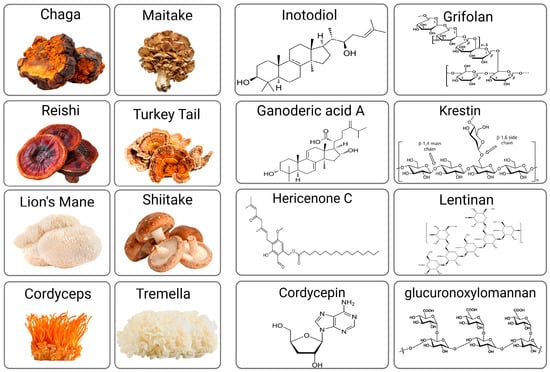
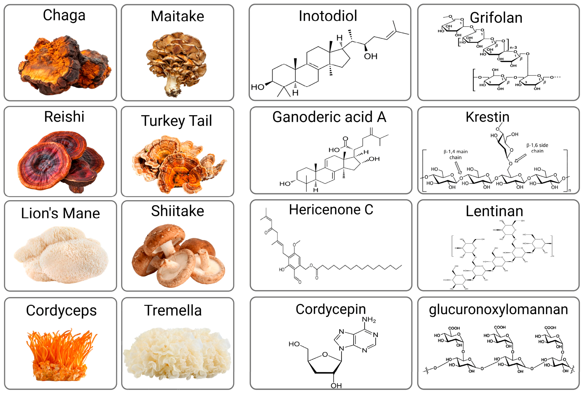
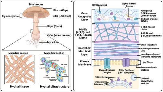
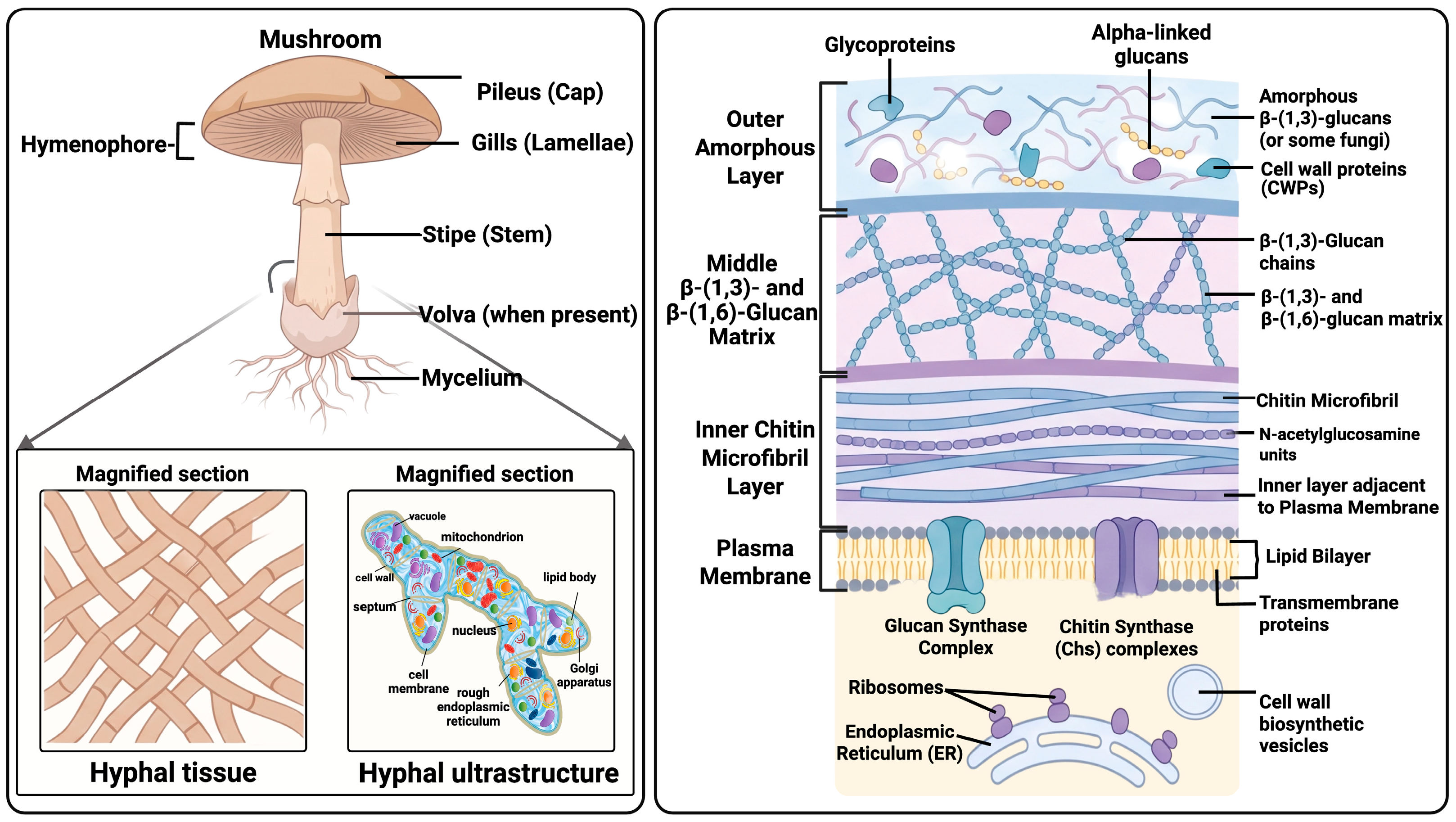
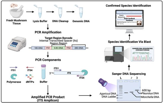
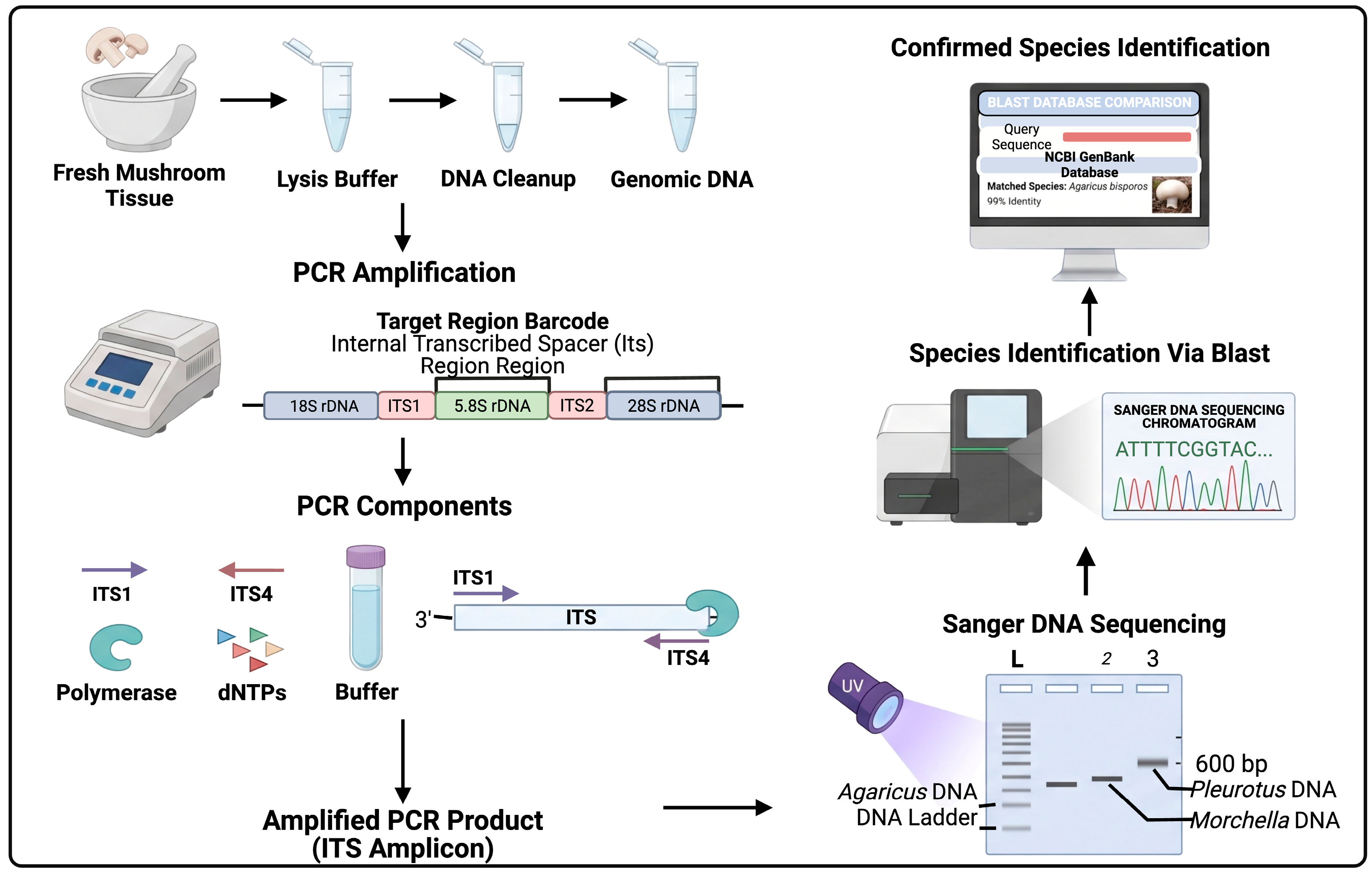
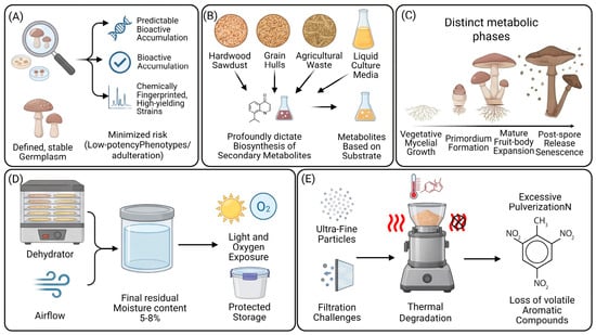
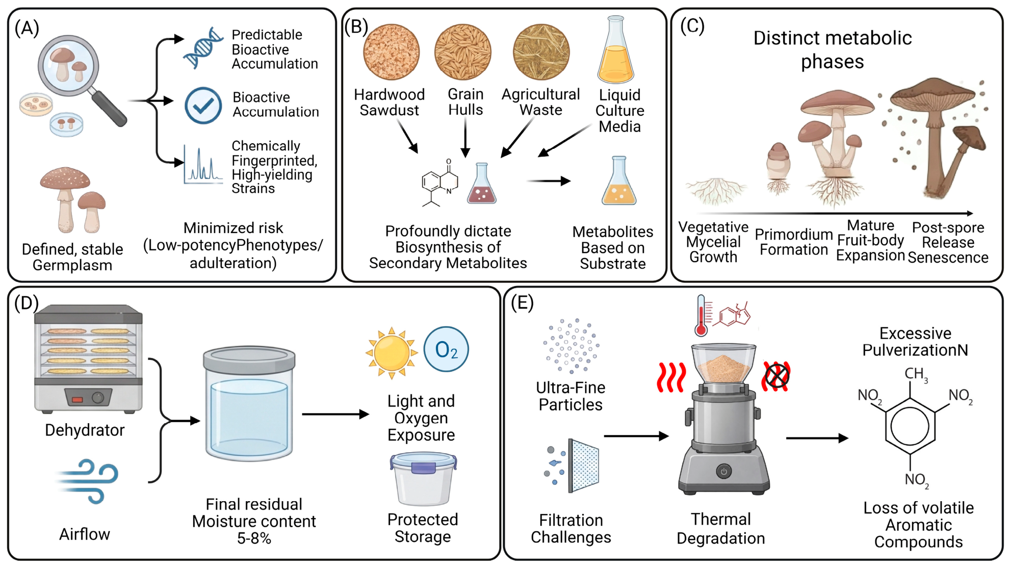
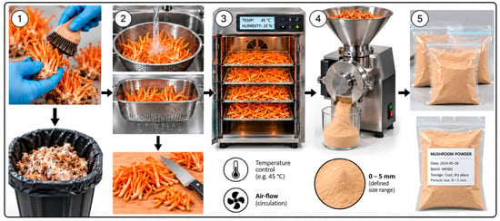
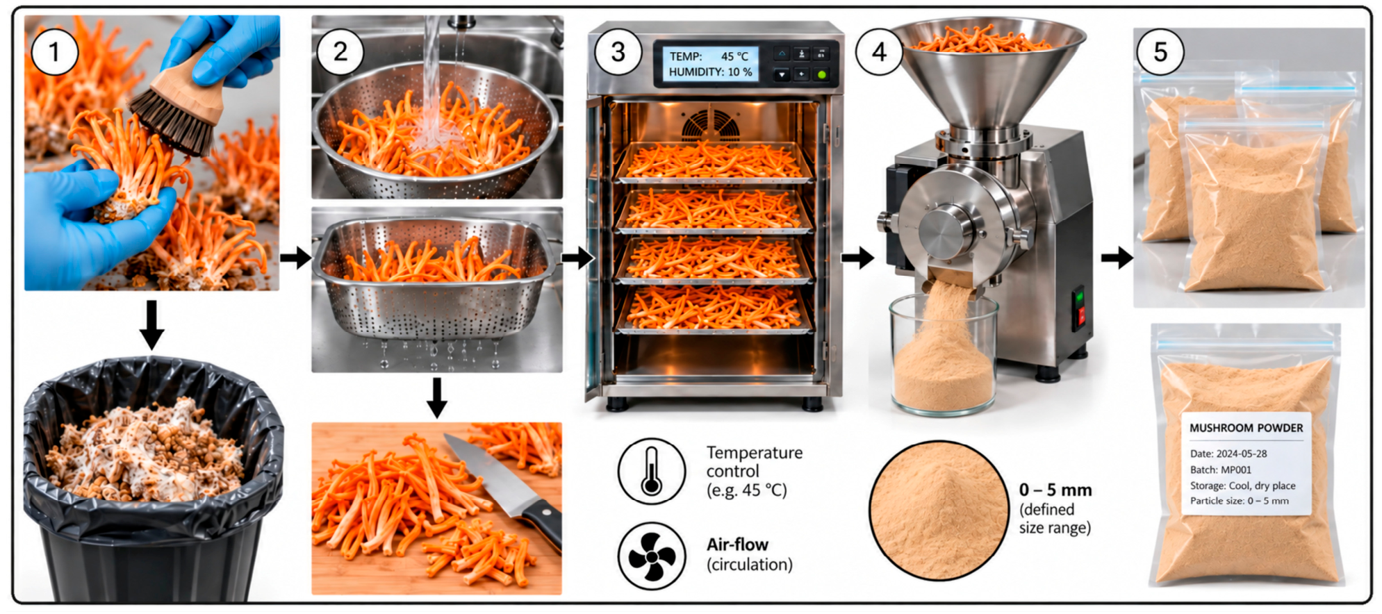
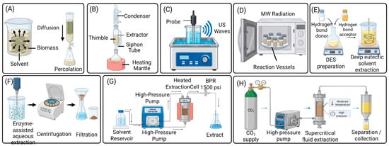
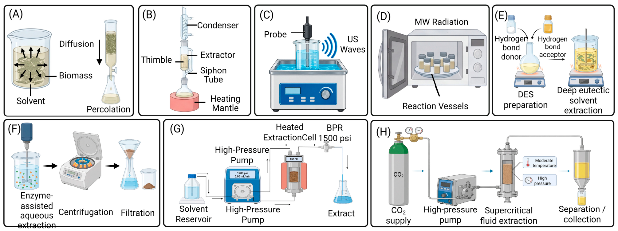
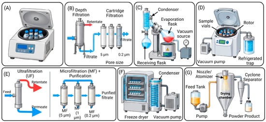
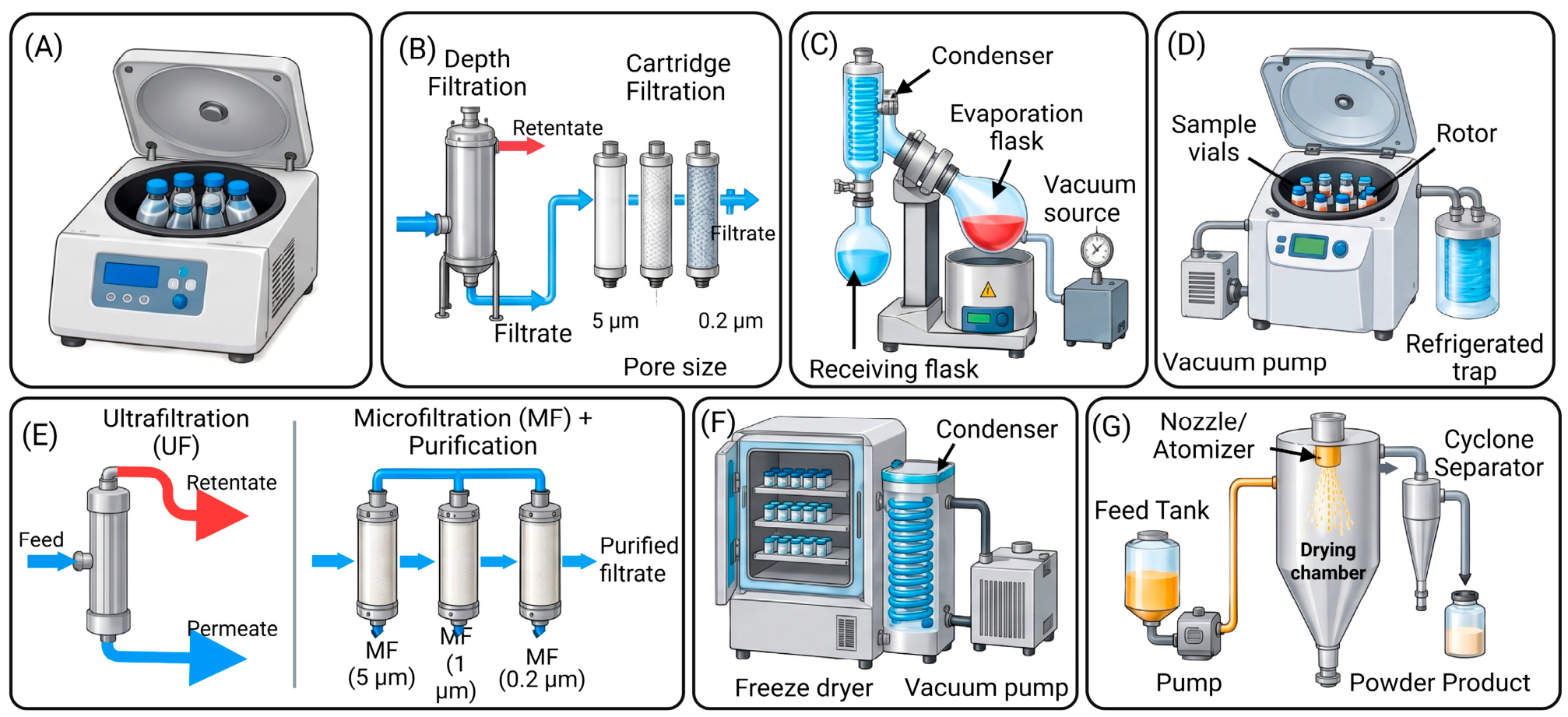
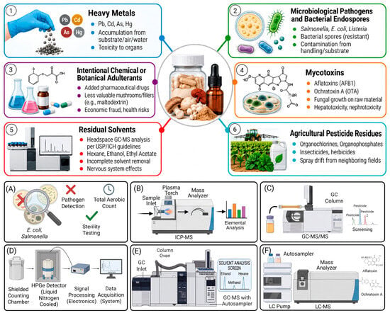
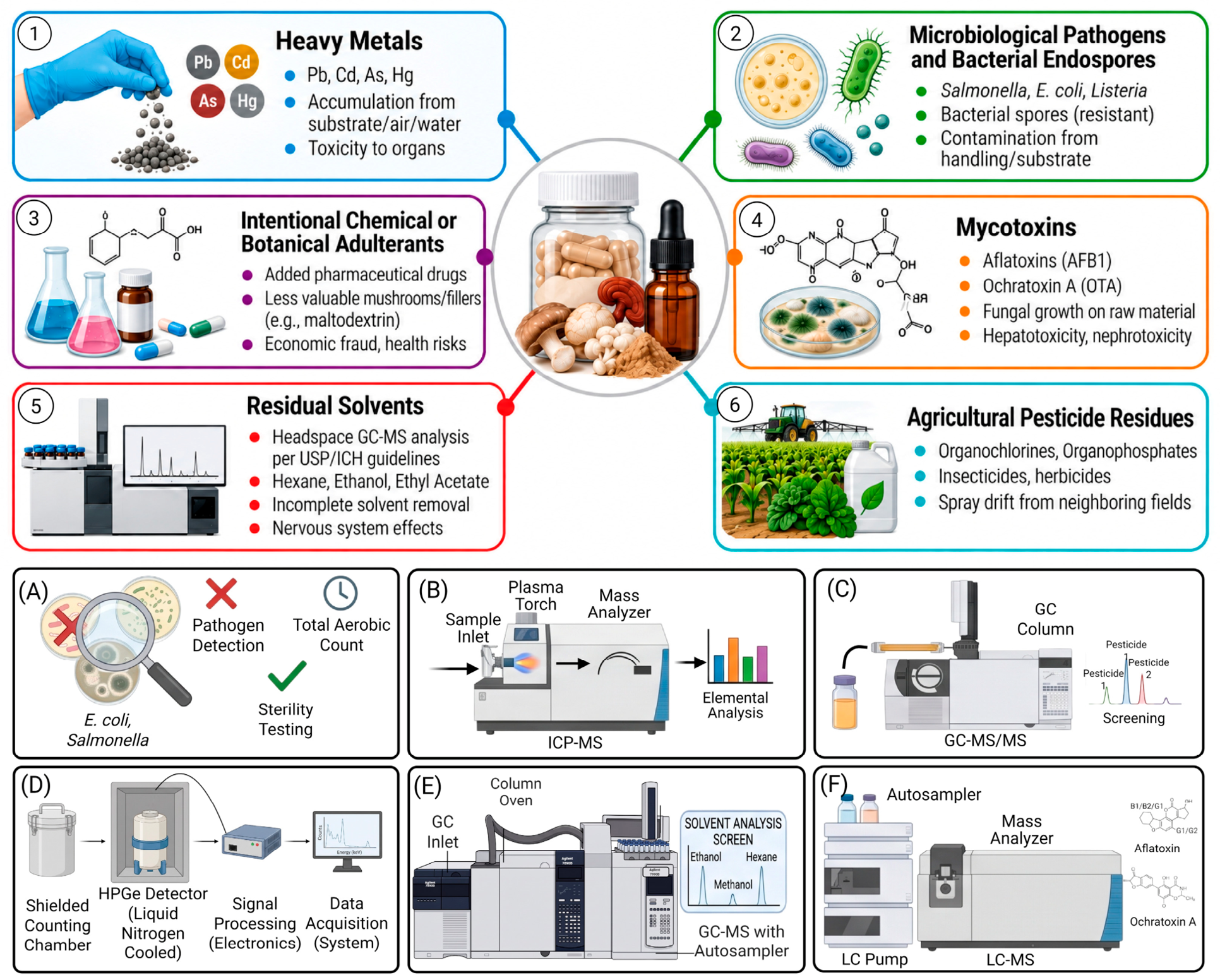

Disclaimer/Publisher’s Note: The statements, opinions and data contained in all publications are solely those of the individual author(s) and contributor(s) and not of MDPI and/or the editor(s). MDPI and/or the editor(s) disclaim responsibility for any injury to people or property resulting from any ideas, methods, instructions or products referred to in the content. |
© 2026 by the authors. Licensee MDPI, Basel, Switzerland. This article is an open access article distributed under the terms and conditions of the Creative Commons Attribution (CC BY) license.