


Abstract
Anatomical museums face growing challenges in maintaining their educational relevance, institutional visibility, and scientific identity. While many historical collections survive as heritage assets, fewer remain integrated into active teaching and research environments. The Veterinary Anatomical Museum of Madrid (VAMM) is a distinctive example among university anatomical collections because it combines documented historical continuity since 1792 with sustained educational, scientific, conservation, and outreach activities. This commentary argues that continuity itself constitutes a form of anatomical heritage. Using the Madrid collection as a case study, we examine how historical collections, plastinated specimens, congenital malformations collections, digital resources, educational innovations, and public engagement can coexist within a single institutional framework. Particular attention is given to the fact that, despite its documented continuity and active museological functions, the collection is not formally recognized as a museum within the institutional structure of Complutense University of Madrid. We propose the concept of the Living Anatomical Museum as an interpretative framework through which historical collections and contemporary anatomical practices interact to preserve existing heritage while simultaneously generating new anatomical heritage for future generations. Although the proposed framework remains conceptual, it offers opportunities for future evaluation of the educational, scientific, and societal contributions of anatomical collections within contemporary universities. The Madrid experience suggests that the long-term preservation of anatomical heritage depends not only on conserving specimens but also on sustaining the academic practices that give them meaning.
1. Introduction
This study adopts the definition of a museum proposed by the International Council of Museums (ICOM): a not-for-profit, permanent institution serving society by researching, collecting, conserving, interpreting and exhibiting tangible and intangible heritage, and providing opportunities for education, enjoyment, reflection and knowledge sharing [1,2]. This contemporary understanding emphasizes accessibility, inclusivity, participation, sustainability and community engagement as core dimensions of museum practice [3]. University museums occupy a unique position at the intersection of teaching, research, and public engagement. They combine the traditional functions of a museum with the educational and scientific missions of higher education institutions [4,5]. Their collections are important academic resources for the research, preservation and interpretation of cultural, historical and scientific heritage [6]. Scientific collections preserve evidence of research practices and teaching traditions, as well as knowledge production. This contributes to our understanding of the history of science and technology [7,8]. They have evolved alongside academic disciplines and have become a distinct field of museological research [9,10,11,12,13]. Contemporary university museums are increasingly operating as dynamic environments that combine heritage preservation, research, education, digital engagement and public outreach. However, there are important differences among institutions regarding governance, recognition, sustainability and the integration of collections into current academic activities [4,10,11].
Historically, anatomical museums have played an important role in preserving, interpreting, and transmitting anatomical knowledge. These museums originated from the teaching collections established in medical and veterinary schools during the 18th and 19th centuries. They were created to support anatomical instruction by displaying and studying specimens, models, and preparations [14,15,16,17,18,19,20,21]. Over time, many of these institutions acquired additional scientific, historical, and cultural value, becoming recognized as important repositories of anatomical heritage [14,15,16,17,18,19,20,21,22,23].
Several veterinary anatomical museums combine historical significance with active educational, scientific and outreach functions. Examples include the Musée Fragonard de l’École vétérinaire de Maisons-Alfort in France [24], the Museo Anatomico Veterinario at the University of Pisa [25], MAVet at the University of Naples Federico II [26], the Royal Veterinary College Anatomy Museum [27], and the Museo de Anatomía Veterinaria de Córdoba [23]. Together, these institutions illustrate the diverse ways in which veterinary anatomical collections continue to contribute to teaching, research, heritage preservation and public engagement.
In recent decades, anatomical museums have evolved from primarily collection-centered institutions into multidisciplinary environments that integrate heritage preservation, education, research, digital technologies and science communication [17,20,21,28,29,30]. Instead of functioning solely as repositories of historical objects, anatomical museums are now often considered multidisciplinary environments where heritage preservation, research, education, and science communication converge [17,21,28,29,30]. Despite this evolution, many university anatomical collections face challenges related to visibility, institutional recognition, and long-term sustainability [11,13]. In some cases, collections have survived while becoming disconnected from the teaching and research activities that originally justified their creation. This situation raises fundamental museological question: what enables an anatomical museum to remain relevant as a museum over time?
The Veterinary Anatomical Museum of Madrid (VAMM) provides a valuable case study through which this question can be explored. Originating from the anatomical cabinet of the Royal Veterinary School of Madrid established in 1792, the collection has remained linked to veterinary anatomical teaching despite multiple institutional transformations and relocations [31,32,33,34,35]. Historical documentation indicates a continuity between the original cabinet and the collections currently maintained by the Departmental Section of Anatomy and Embryology at Complutense University of Madrid and the Veterinary Museum [31,32,33,35]. For clarity, the term VAMM is used throughout this commentary to refer to the historical anatomical collections currently curated by the Departmental Section of Anatomy and Embryology and the Complutense Veterinary Museum, which together support their conservation and heritage functions.
While many contemporary anatomical museums successfully combine conservation, teaching, research and public engagement, the VAMM offers an additional perspective. Its significance derives not only from the preservation of historical collections, but also from the sustained interaction between inherited anatomical resources and newly generated educational and scientific materials. Historical specimens, plastinated preparations, digital resources, educational innovations and research activities coexist within a single institutional framework, allowing anatomical heritage to function as an active component of contemporary academic practice rather than solely as a record of the past.
This commentary examines the significance of functional continuity from a museological perspective. We contend that the VAMM’s value transcends the historical significance of its holdings, also residing in its continuous contributions to education, research, conservation and public engagement. Using the VAMM as a documented case study, we introduce the concept of the Living Anatomical Museum as an interpretative framework through which to understand how anatomical collections can connect historical heritage with contemporary academic practice. Rather than establishing or validating a formal museological model, this commentary aims to stimulate discussion about the role of anatomical collections in modern universities and veterinary schools and to lay the groundwork for future empirical evaluation in various institutional settings.
2. Continuity as Heritage
The history of the VAMM is closely tied to the development of veterinary education in Spain. The collection originated from the anatomical cabinet of the Royal Veterinary School of Madrid, which was established in 1792 [31,32,33,34,35]. Since then, the collection has undergone successive institutional transformations, relocations, and educational reforms while remaining closely associated with anatomical teaching and veterinary training. The anatomical collection has had a complex history, involving successive relocations which resulted in the loss and deterioration of many specimens. Originally housed at the Royal Veterinary School in Madrid following its establishment in 1793, the collection was transferred several times as the institution relocated to different sites throughout the 19th and 20th centuries [34,35]. Significant losses occurred during the relocation from the Embajadores Street facilities in 1958. Following this, the remaining specimens were temporarily accommodated in temporary buildings before being transferred to the current Departmental Section of Anatomy in 1968, where an anatomical museum was established [35]. Until the 1980s, the collection was displayed in the VAMM showcases. Subsequently, as these spaces were converted into staff offices, the specimens were redistributed among display cabinets in the department’s classrooms and corridors. Since then, the Departmental Section of Anatomy has played a central role in housing, safeguarding and preserving the collection, ensuring its continuity during a period when dedicated museum facilities were unavailable [34,35], a role that it continues to fulfill to this day.
Later, as part of a broader heritage recovery initiative launched by the Complutense University of Madrid, a restoration project for the historical collection was started. The project aimed to preserve, document and enhance the collection’s scientific and educational value. In 2005, the collection was incorporated into the Complutense Veterinary Museum, where it has been preserved and curated alongside the Departmental Section of Anatomy and Embryology ever since [35].
The historical continuity discussed in this commentary is supported by archival documentation, including historical inventories, institutional records, and published historical accounts describing the evolution of veterinary education in Madrid throughout the nineteenth and twentieth centuries.
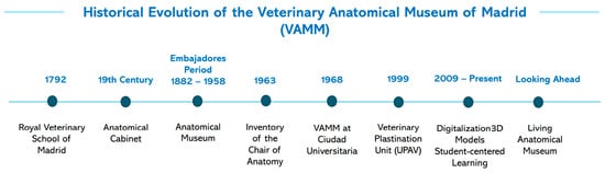
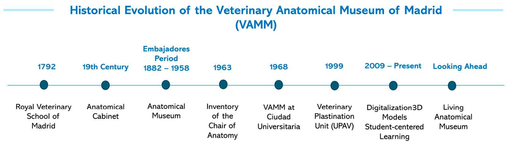
Figure 1 summarizes the historical trajectory of the collection, illustrating the continuity between the Royal Veterinary School, subsequent institutional reorganizations, and the current Departmental Section of Anatomy and Embryology.
Figure 1.
The evolution of the historical anatomical museum, from the Royal Veterinary School of Madrid in 1792, through to its current status as the Departmental Section of Anatomy and Embryology at the University of Complutense, Madrid.
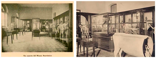
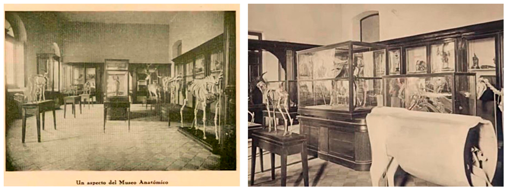
Additional historical photographs document the museum’s presence during the period when the School of Veterinary Medicine was located at the Glorieta de Embajadores (Figure 2).
Figure 2.
Historical view of the Veterinary Anatomical Museum at the School of Veterinary Medicine in Embajadores.
An important source that documents this continuity is the 1963 inventory of the Chair of Anatomy. This was compiled during a period of institutional transition and the reorganization of the collection [36]. The inventory documents the composition, organization, and educational purpose of the anatomical collections, providing evidence of their preservation during a period of institutional change.
Table 1 summarizes the evolution of anatomical teaching resources, from traditional dissections and anatomical cabinets to contemporary museum and digital resources.
Table 1.
Evolution of anatomical teaching resources from traditional anatomical cabinets to modern museum and digital resources.
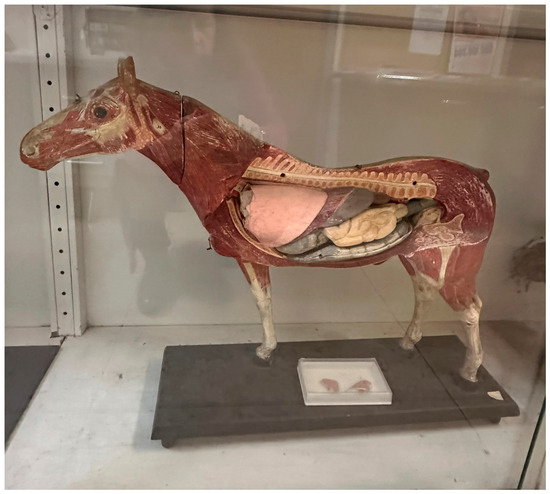
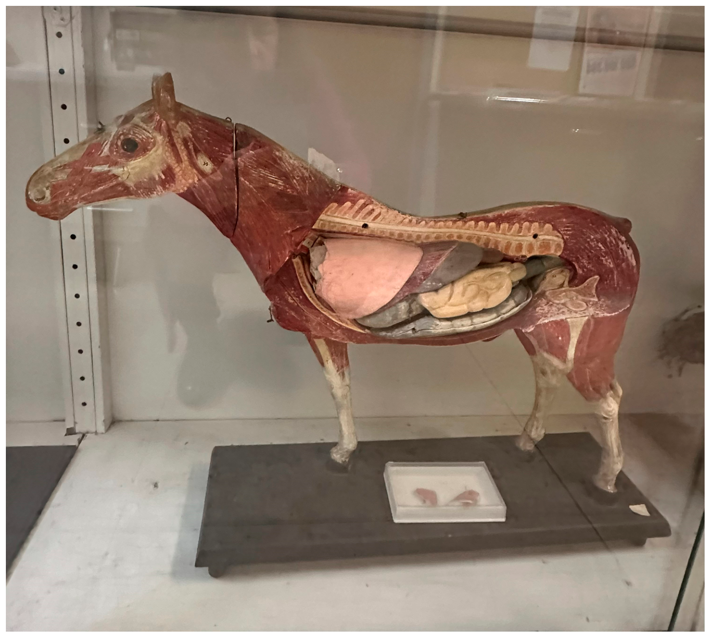
The 1963 inventory documents a diverse range of anatomical teaching resources representing different periods in the history of veterinary education, including wax models, plaster reproductions, papier-mâché preparations, articulated skeletons, embryological specimens and preserved anatomical preparations [35,36]. Many of these materials remain preserved within the collection and continue to support veterinary anatomy education through practical instruction, comparative demonstrations, museum visits and independent study activities. Together with more recent plastinated specimens, digital resources and three-dimensional anatomical models, they provide a tangible record of the evolution of anatomical pedagogy while demonstrating the continued educational relevance of the collection. Figure 3 shows a representative example: a historical papier-mâché equid model with removable viscera.
Figure 3.
Historical papier-mâché anatomical model of an equid with viscera from the anatomical collection of the Departmental Section of Anatomy and Embryology. Such models were widely used in the teaching of veterinary anatomy before modern preservation techniques were introduced.
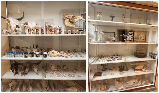
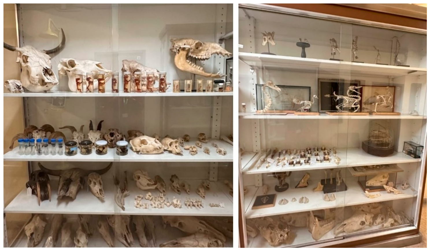
These materials provide valuable evidence of the evolution of anatomical teaching methods and specimen-preparation techniques throughout the history of veterinary education (Table 1). The collection also contains numerous osteological and anatomical preparations that have played a central role in educating veterinarians (Figure 4).
Figure 4.
Historical osteological and anatomical specimens preserved in the collections of the Departmental Section of Anatomy and Embryology. The display cases contain a variety of anatomical specimens from different animal species, including mounted bird skeletons, cleared and stained embryos, stained brain sections, malformed vertebrae and comparative skulls. Most of the anatomical specimens date from the second half of the 20th century to the present day, and were prepared using various anatomical techniques, such as skeletal preparation, clearing and staining, dissection, fixation and mounting. These materials continue to serve important educational purposes in the teaching of veterinary anatomy, and all anatomical specimens contribute to the communication of scientific knowledge and public outreach.
From a museological perspective, the significance of these collections extends beyond their age or rarity. They preserve evidence of how anatomy has been taught, interpreted, and communicated across generations of veterinary students and anatomists while continuing to support contemporary educational activities. Osteological specimens, anatomical preparations and historical teaching models are still incorporated into practical sessions, comparative anatomy demonstrations, museum visits and independent learning activities. Their continued use strengthens the connection between historical heritage and current educational practice, illustrating how anatomical collections can preserve not only objects but also educational traditions, scientific knowledge and institutional memory.
Within the proposed Living Anatomical Museum framework, continuity is understood not only as the survival of specimens, but also as the persistence of the educational, scientific and institutional practices that give those specimens meaning. From this perspective, anatomical heritage is not solely inherited from the past; it is actively sustained through continued academic use and reinterpretation.
3. Generating Future Anatomical Heritage
The Madrid experience raises an important museological question: when does a contemporary anatomical specimen become part of anatomical heritage? Although no universally accepted criterion exists, heritage value appears to emerge through the interaction of long-term preservation, scientific significance, educational relevance, rarity, documentation and sustained institutional use. From this perspective, anatomical heritage is not determined exclusively by age. Instead, it develops over time as specimens become integrated into teaching traditions, support scientific discoveries, contribute to professional training or document unique biological phenomena. Consequently, anatomical heritage should be understood as a dynamic process rather than a fixed category. The contemporary collections preserved within the VAMM illustrate how educational and research materials may gradually acquire historical, scientific and heritage value while remaining actively used in academic environments. The conservation of scientific heritage is increasingly being recognized as a strategic activity for universities seeking to document and communicate their intellectual legacy [37].
A significant milestone in this process was the establishment of the Veterinary Anatomy Plastination Unit (UPAV) in 1999. This introduced new possibilities for preparing, preserving, studying and disseminating anatomical specimens, while also contributing to their long-term conservation [38,39,40,41,42].
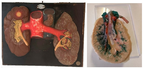
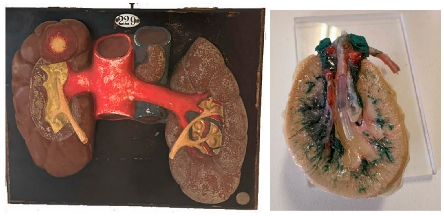
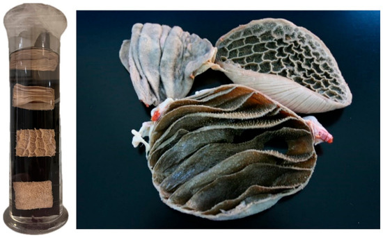
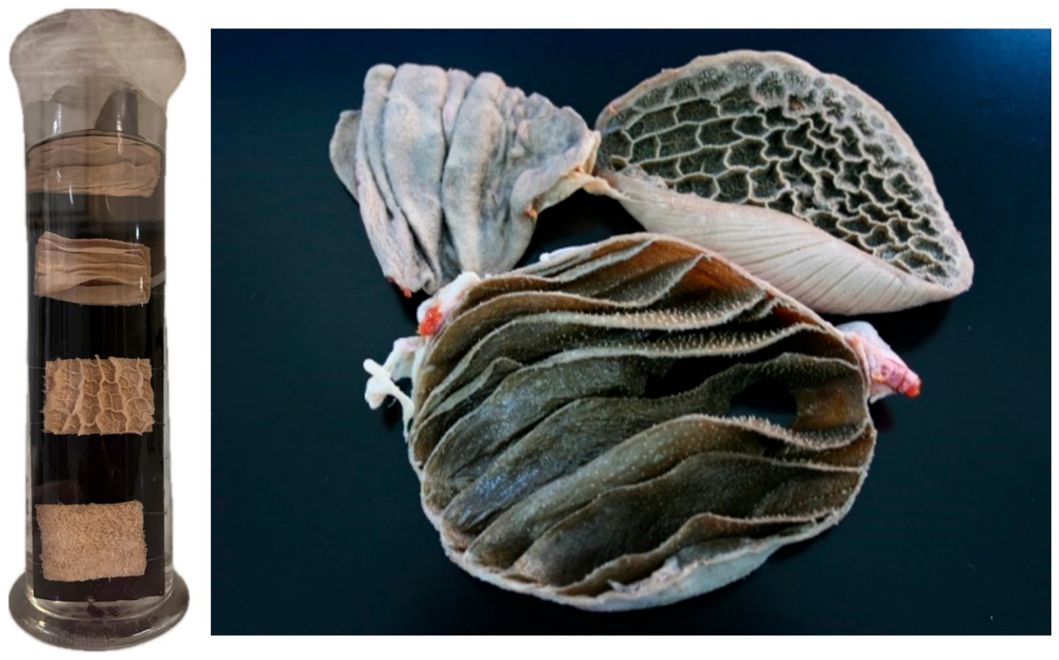
The relationship between historical and contemporary educational resources is evident when traditional anatomical models are compared with plastinated preparations designed to fulfill similar educational objectives. Figure 5 and Figure 6 illustrate this comparison. Figure 5 shows continuity between two anatomical models of the kidneys.
Figure 5.
The image on the left is a historical model of a bovine’s kidneys and major and renal vessels, bearing the inventory number from the anatomical museum’s catalog. The image on the right is a plastinated section of a goat’s kidney with colored latex-filled vessels.
Figure 6.
Ruminant stomach mucosa preserved in formalin from the historical collection. On the right is a natural plastinated preparation of a goat’s stomach mucosa.
Together, these examples illustrate how different generations of anatomical resources may serve equivalent educational purposes despite employing distinct preservation technologies. The comparison highlights continuity in anatomical teaching objectives while simultaneously demonstrating the evolution of anatomical representation and specimen preparation techniques.
The presence of historical anatomical models alongside contemporary plastinated specimens creates opportunities for comparative learning. Students can explore how anatomical representation, preservation techniques and teaching methodologies have evolved over time. Rather than replacing historical collections, plastination has expanded the range of resources available for anatomical education and contributed to anatomical specimen preservation [38,39,40,41,42].
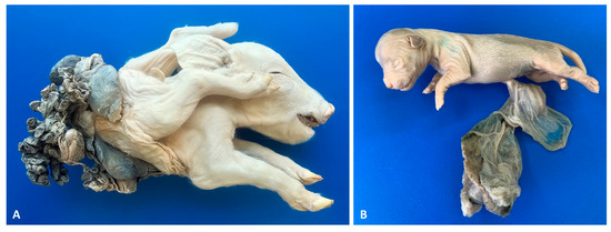
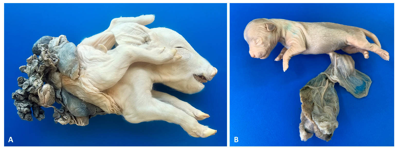
Another significant development is the GIMCAD’s (Congenital Malformations and Developmental Anatomy Research Group) creation of a plastinated collection of congenital malformations. The collection comprises specimens from pigs, dogs, cats and cattle, which have been accumulated over several decades through diagnostic, clinical and research activities. These materials support the study of developmental anatomy and comparative pathology, as well as veterinary education, while preserving uncommon anatomical conditions that are rarely represented in conventional teaching collections. Some of these specimens are shown in Figure 7.
Figure 7.
Specimens preserved using the plastination technique (A) A newborn piglet with a fetal body stalk anomaly (BSA), historically classified as Schistosoma reflexum. (B) A newborn puppy with an omphalocele connected to its intact placenta.
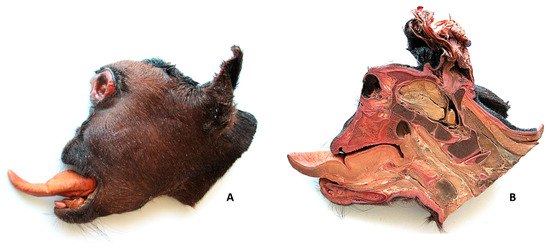
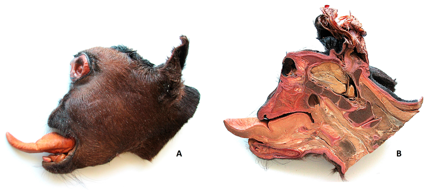
The collection includes rare and complex developmental anomalies that would be difficult to preserve and study using conventional methods. Figure 8 shows a plastinated anatomical specimen of a calf with cyclopia, demonstrating plastination’s ability to preserve external morphology and internal anatomical structures in cases of severe congenital malformations.
Figure 8.
Plastinated anatomical specimen of a newborn calf with cyclopia, the most severe form of holoprosencephaly (A). Left lateral view of the calf’s head. (B) Medial view of the same specimen, showing a sagittal section that allows for detailed examination of the head’s internal anatomical structures.
One distinctive feature of this collection is its origin. Most of the specimens originated from a professional collection that Professor Luis Avedillo assembled independently during his clinical career, before joining the Complutense University of Madrid. The collection was maintained and preserved under his ownership because of the specimens’ scientific and diagnostic value. Following the establishment of UPAV, a few selected specimens were plastinated to ensure their long-term preservation for educational purposes. While the original collection remains distinct, the plastinated specimens currently used for teaching, research and outreach activities are included in the broader framework proposed here.
From a heritage perspective, these plastinated specimens serve as research materials, educational resources, and potential future heritage objects. Their value stems not only from their rarity or scientific significance, but also from their ongoing integration into research, teaching, and public dissemination activities. The Madrid experience therefore suggests that anatomical heritage is not only inherited from previous generations but can also be actively created through modern scientific and educational practices.
Research derived from malformation specimens has contributed to the anatomical description and classification of body wall defects, VACTERL association, Cantrell syndrome, and prune belly syndrome, among other congenital malformations in domestic species [43,44,45,46,47,48,49,50,51,52]. These specimens function as research materials, educational resources, and heritage objects simultaneously.
The examples presented here do not validate a formal museological model. Instead, they demonstrate how contemporary anatomical resources can gradually gain scientific, educational and historical importance through preservation and ongoing academic use. The Madrid collection is a useful case study in this regard, demonstrating how anatomical heritage can be preserved and generated within the same institutional environment.
4. Towards a Living Anatomical Museum
4.1. Conceptual Framework
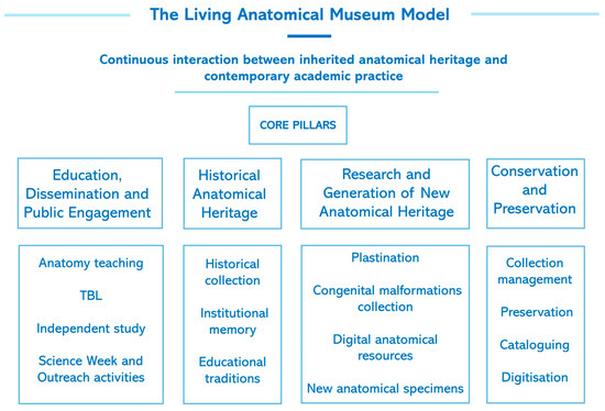
The preceding sections have described two complementary processes: the preservation of historical anatomical heritage, and the creation of new heritage through modern scientific and educational activities. Together, these processes form the basis of the Living Anatomical Museum. Within this framework, four interconnected dimensions sustain the Living Anatomical Museum:
- Historical anatomical heritage;
- Conservation and preservation;
- Research and generation of new anatomical resources;
- Education, dissemination, and public engagement.
Figure 9 illustrates the proposed conceptual framework of the Living Anatomical Museum.
Figure 9.
Proposed conceptual framework of the Living Anatomical Museum. The Living Anatomical Museum is presented as an interpretative framework based on the continuous interaction between inherited anatomical heritage and contemporary academic practice. Four interconnected dimensions sustain this framework: Historical Anatomical Heritage, Conservation and Preservation, Research and Generation of New Anatomical Resources, and Education, Dissemination and Public Engagement. Together, these dimensions contribute to the preservation, utilization, and ongoing creation of anatomical heritage.
The VAMM is an example of an anatomical collection that goes beyond the traditional role of a repository of specimens. Historical collections, plastinated preparations, digital resources, educational innovations, scientific research, and public engagement activities all coexist within the same institution and interact continuously.
Rather than proposing a new museum category, the Living Anatomical Museum is presented here as an interpretative framework. Its defining feature is the dynamic relationship between inherited anatomical heritage and contemporary anatomical practice. Historical collections continue to support teaching, research and outreach, while ongoing academic activities simultaneously generate the anatomical heritage of future generations.
4.2. Research and Heritage Creation
Within the Living Anatomical Museum framework, research is one of the mechanisms through which new anatomical heritage is generated. The GIMCAD group is a prime example of this process, linking scientific investigation, specimen preservation, and the creation of future heritage. Through its work in developmental anatomy, teratology, congenital anomaly research and comparative pathology, the group generates scientific knowledge while also producing distinctive anatomical specimens that enrich the museum environment. Unlike traditional collections assembled through historical acquisition, these materials originate directly from contemporary diagnostic and research activities [43,44,45,46,47,48,49,50,51,52]. Some of these specimens subsequently acquire educational, scientific, and heritage value through their incorporation into teaching, research, and outreach activities [53]. Thus, contemporary research contributes to both knowledge production and the ongoing creation of anatomical heritage.
4.3. Educational Innovation
Educational innovation constitutes another essential dimension of the Living Anatomical Museum framework. Contemporary student-centered approaches, including flipped classroom methodologies, Team-Based Learning (TBL), digital learning resources, and technology-enhanced instruction, increasingly incorporate museum-derived anatomical resources into active learning environments [54,55,56,57,58].
In recent years, historical osteological specimens and plastinated anatomical preparations have been incorporated into veterinary anatomy courses via TBL practical sessions, guided independent study activities, and preparatory learning exercises. These resources expand the educational scope of collections, moving beyond mere exhibition to facilitate direct engagement with authentic anatomical material.
Preliminary educational evaluations conducted during the 2024/25 and 2025/26 academic years indicate that incorporating VAMM-derived anatomical resources into active-learning strategies improves student preparation and engagement. These findings will be reported in detail elsewhere, but they demonstrate how historical collections and newly generated anatomical resources can contribute to modern educational outcomes, while also reinforcing the academic importance of anatomical heritage. Therefore, independent study using real anatomical models could complement the collaborative benefits of TBL by improving individual readiness and supporting the development of deeper conceptual understanding and application skills [53,57,58].
These observations are consistent with the wider literature emphasizing the importance of object- and collection-based learning in higher education. Direct engagement with material objects has been linked to improved observation skills, critical thinking, student engagement, and reflective learning [59,60,61]. Within the Living Anatomical Museum framework, anatomical collections serve as both preserved heritage and active educational resources that support experiential learning.
4.4. Digital Resources and Public Engagement
Digital technologies further enhance the educational and conservation value of anatomical collections. Three-dimensional reconstructions, virtual resources, and online repositories offer an alternative way of accessing anatomical knowledge, while reducing the need to handle fragile historical specimens [28]. These developments demonstrate how historical and contemporary anatomical resources can be integrated into a single educational ecosystem.
Public engagement is the fourth pillar of the Living Anatomical Museum. Activities such as Madrid Science Week, plastination workshops, and anatomy outreach programmes enable the dissemination of veterinary and anatomical knowledge beyond the university environment. Such initiatives strengthen the societal relevance of anatomical collections, reinforcing their educational and cultural value. Examples of these activities are provided in Supplementary Videos S1 and S2, which document plastinated anatomical specimen presented during the 2024 and 2025 editions of Madrid Science Week, respectively. These activities have made it possible to share veterinary and anatomical sciences outside of a university setting.
The Living Anatomical Museum should therefore be understood as being about more than just preserving anatomical heritage. It is a dynamic environment in which historical collections, contemporary scientific activities, educational innovation and public engagement interact to generate scientific, educational and cultural value. By linking preservation with active academic use, the framework demonstrates how anatomical heritage can remain relevant in contemporary universities.
5. Recognition and Future Perspectives
The proposed Living Anatomical Museum framework should be understood as an interpretative tool rather than a universally applicable model. Anatomical collections operate within diverse institutional, cultural, and administrative contexts, and their development is shaped by local histories, resources, and governance structures. Further comparative studies are therefore required to determine whether similar patterns of interaction between historical heritage and contemporary academic activity can be identified in other university anatomical collections.
The VAMM is a particularly informative case study because its documented historical continuity extends from the anatomical cabinet established at the Royal Veterinary School of Madrid in 1792 to the collections currently maintained by the Departmental Section of Anatomy and Embryology and the Complutense Veterinary Museum. Throughout this period, the collections have continued to contribute to anatomical teaching, specimen preservation, research, plastination, the development of digital resources, and public dissemination [32,33,35].
Currently, the collections are divided between the Departmental Section of Anatomy and Embryology and the Complutense Veterinary Museum. Together, these institutions support the conservation, cataloguing and educational use of the collections, as well as research activities and public outreach. While numerous specimens remain actively incorporated into anatomy teaching and research, others are preserved within the Veterinary Museum as part of the university’s scientific and historical heritage. Despite carrying out many of the functions typically associated with museums, such as conservation, collection management, documentation, research, education and dissemination, the VAMM has not been formally recognized as a museum within Complutense University of Madrid’s institutional structure. This highlights a broader museological question concerning the relationship between administrative designation and functional reality.
This situation is not unique. Many university collections occupy a position that blurs the boundaries between teaching infrastructure, scientific collections, heritage assets, and formally recognized museums [9,10,11,12,13,19,20,21,22,23,24,25,26,27]. Consequently, institutional classification may not always reflect their complete educational, scientific, and cultural contributions.
Nevertheless, formal recognition could provide important benefits, including increased visibility, improved conservation planning, enhanced cataloguing efforts, access to heritage networks, and stronger institutional support for long-term sustainability. It may also help to preserve the scientific identity of anatomical collections by emphasizing their ongoing contribution to knowledge and education, rather than viewing them solely as historical assets.
Long-term sustainability depends on conservation activities, governance structures, institutional commitment, staffing, funding strategies, cataloguing policies, and accreditation mechanisms [62,63]. While these issues are beyond the scope of this commentary, they are still important factors to consider when planning the future development of university anatomical collections.
The Madrid experience suggests that, when evaluating the significance of university anatomical collections, continuity of function should be considered alongside formal designation. From this perspective, a collection’s ability to support teaching, research, conservation and public engagement over time may be as important as its administrative classification. Comparing similar institutions in future studies may help determine whether functional continuity is a useful criterion for understanding the long-term relevance of anatomical museums within contemporary universities.
The long-term relevance of anatomical heritage hinges not only on preserving specimens, but also on maintaining the academic practices that imbue them with meaning.
6. Conclusions
The VAMM is a valuable case study, demonstrating how historical collections can remain relevant by continuing to play an active role in teaching, research, conservation, and public engagement. The VAMM’s collections document over two centuries of veterinary anatomical education, remaining integrated into contemporary academic practice.
The historical collections provide evidence of past teaching traditions and specimen-preparation techniques. In contrast, plastinated specimens, congenital malformation collections, and digital resources demonstrate contemporary anatomy departments’ capacity to generate new forms of scientific and educational heritage. Together, these resources establish a connection between inherited collections and newly created anatomical materials.
The Living Anatomical Museum is proposed as an interpretative framework through which this relationship can be understood. Within this framework, anatomical heritage is preserved through conservation, active educational use, scientific production, and public dissemination. Historical collections remain relevant because they continue to inform contemporary academic activities. Meanwhile, ongoing research and educational innovation generate the anatomical heritage of the future.
Although the Living Anatomical Museum remains a conceptual framework requiring further evaluation, experience in Madrid suggests that the long-term preservation of anatomical heritage hinges not only on safeguarding specimens, but also on sustaining the scientific, educational and cultural practices that give them meaning. From this perspective, ensuring the continuity of these practices may be as important as formal recognition in ensuring the future relevance of university anatomical collections.
A museum does not simply remain alive because its specimens survive; it remains alive because the scientific, educational and cultural practices that give those specimens meaning continue to evolve across generations.
Supplementary Materials
The following supporting information can be downloaded at https://www.mdpi.com/article/10.3390/anatomia5040029/s1, Video S1: Exhibition of plastinated specimens 2024, and Video S2: Exhibition of plastinated specimens 2025.
Author Contributions
Conceptualization, N.M.-A.; methodology, N.M.-A. and L.A.; formal analysis, N.M.-A. and L.A.; investigation, N.M.-A. and L.A.; writing—original draft preparation, N.M.-A.; writing—review and editing, N.M.-A. and L.A.; visualization, N.M.-A.; supervision, N.M.-A. All authors have read and agreed to the published version of the manuscript.
Funding
This research received no external funding.
Institutional Review Board Statement
Not applicable.
Informed Consent Statement
Not applicable.
Data Availability Statement
No new data was generated or analyzed for this commentary. References to ongoing educational research are provided for contextual purposes only and are not included in the data presented in this manuscript.
Conflicts of Interest
The authors declare no conflicts of interest.
Abbreviations
The following abbreviations are used in this manuscript:
| TBL | Team-based Learning |
| UPAV | Veterinary Anatomy Plastination Unit |
| VAMM | Veterinary Anatomical Museum of Madrid |
References
- International Council of Museums. ICOM Museum Definition. 2022. Available online: https://icom.museum/en/resources/standards-guidelines/museum-definition/ (accessed on 10 September 2026).
- Brulon Soares, B.; Bonilla-Merchav, L. The Museum Definition Handbook: Words Inspiring Action, 1st ed.; ICOM: Paris, France, 2025; Available online: https://icom.museum/en/news/publication-of-the-museum-definition-handbook-words-inspiring-action/ (accessed on 10 September 2026).
- International Council of Museums. ICOM Code of Ethics for Museums. 2026. Available online: https://icom.museum/wp-content/uploads/2026/06/Code-of-Ethics_2026_EN-1.pdf (accessed on 10 September 2026).
- Lourenço, M.C. Between Two Worlds: The Distinct Nature and Contemporary Significance of University Museums and Collections in Europe. Ph.D. Thesis, Université Paris I Panthéon-Sorbonne, Paris, France, 2005. [Google Scholar]
- Lourenço, M.C. Are university collections and museums still meaningful? Outline of a research project. Museologia 2010, 2, 51–60. [Google Scholar]
- Mouliou, M. Museum representations of the classical past in post-war Greece: A critical analysis. Μουσείο Μπενάκη 2008, 3, 83–109. [Google Scholar] [CrossRef] [Scilit]
- Alberti, S.J.M. Objects and the museums. Isis 2005, 96, 559–571. [Google Scholar] [CrossRef] [Scilit]
- Macdonald, S. Collecting Practices. In A Companion to Museum Studies; Macdonald, S.E., Ed.; Blackwell Publishing Ltd.: Oxford, UK, 2006. [Google Scholar]
- Tucci, P. Role of university museums and collections in disseminating scientific culture. Museologia 2010, 2, 17–22. [Google Scholar]
- Erbay, F. The Role of University Museums. UNIMUSEUM 2018, 1, 1–4. [Google Scholar]
- Plaza, C. Museums in Universities: Predicaments and Potentialities. Mus. Int. 2022, 74, 74–85. [Google Scholar] [CrossRef] [Scilit]
- Plaza Salgado, C.B.; Narbona Medina, L.M. Archives in University Science Museums: Proposals for Their Museological Transformation. Collections 2025, 21, 305–320. [Google Scholar] [CrossRef] [Scilit]
- Jardine, N. Reflections on the preservation of recent scientific heritage in dispersed university collections. Stud. Hist. Philos. Sci. Part A 2013, 44, 735–743. [Google Scholar] [CrossRef] [Scilit]
- Alexander, E.P.; Alexander, M. Museums in Motion: An Introduction to the History and Functions of Museums, 2nd ed.; Rowman & Littlefield: Lanham, MD, USA, 2007. [Google Scholar]
- Kamath, V.G.; Ray, B.; Pai, S.R.; Avadhani, R. The origin of anatomy museums. Eur. J. Anat. 2024, 18, 63–67. [Google Scholar]
- Shilkova, T.V.; Efimova, N.V.; Sokolova, T.L. The role of the anatomy museum in university education. Samara J. Sci. 2020, 9, 300–307. [Google Scholar] [CrossRef] [Scilit]
- Kongtueng, P.; Yotanyamaneewong, S. A lesson from our institute; Why do veterinary schools need an anatomy museum? Vet. Integr. Sci. 2021, 19, 567–579. [Google Scholar] [CrossRef] [Scilit]
- Orsini, D.; Martini, M.; Ottoboni, S.; Aglianó, M.; Franci, D.; Lorenzoni, P.; Saverino, D. Drawings, artifacts and anatomical preparations: The collections of the Senese Museums for teaching Anatomy yesterday as today. Ital. J. Anat. Embryol. 2024, 128, 75–83. [Google Scholar] [CrossRef] [Scilit]
- Reddy, M.; Rajesh, S.; Annamraju, M.R.; Rani, T.; Manasa, K. Anatomy Museums in Medical Education: Utilization patterns and perceived learning benefits among Indian medical graduates. Eur. J. Anat. 2026, 30, 395–400. [Google Scholar] [CrossRef] [Scilit]
- Sharma, B.; Alhazmi, A.; Nasir, N.; Bridget, B. Innovative Learning in Anatomy Education: Assessing the Impact of Low-Cost 3D Deep Learning Anatomical Models in Museum-Based Instruction. J. Surg. Educ. 2025, 83, 103748. [Google Scholar] [CrossRef] [Scilit] [PubMed]
- Ekim, O.; Güneröz, C. What Makes a Museum Interactive? An Overview of Education Activities in the Ankara Veterinary Anatomy Museum. J. Mus. Educ. 2024, 49, 348–366. [Google Scholar] [CrossRef] [Scilit]
- Doom, M.; Casteleyn, C.; Simoens, P. Museum of Morphology. In Proceedings of the Academic Culture of Remembrance Conference, Ghent, Belgium, 16–17 March 2011. [Google Scholar]
- Jiménez Hernández, A.; Jordano Barbudo, M.A. El patrimonio anatómico y zootécnico de la Facultad de Veterinaria de la Universidad de Córdoba (España) y su función pedagógica. Boletín Arte-UMA 2019, 40, 167–177. [Google Scholar] [CrossRef] [Scilit]
- École Nationale Vétérinaire d’Alfort. Musée Fragonard. Available online: https://www.vet-alfort.fr/domaine-d-alfort/musee-fragonard/ (accessed on 17 September 2026).
- University of Pisa. Museo Anatomico Veterinario: Collection. Available online: https://mav.sma.unipi.it/en/collection/ (accessed on 17 September 2026).
- Università degli Studi di Napoli Federico II. MAVet Museum of Veterinary Anatomy. Available online: https://mavet.org/en/collection (accessed on 17 September 2026).
- Royal Veterinary College, University of London. Anatomy Online. Available online: https://www.rvc.ac.uk/e-anatomy (accessed on 17 September 2026).
- Daniela, L. Virtual Museums as Learning Agents. Sustainability 2020, 12, 2698. [Google Scholar] [CrossRef] [Scilit]
- Hadavibavili, P.; Ilçioğlu, K. Artwork in anatomy education: A way to improve undergraduate students’ self-efficacy and attitude. Anat. Sci. Educ. 2023, 17, 66–76. [Google Scholar] [CrossRef] [Scilit] [PubMed]
- Magno, G.; Zanatta, A. Teaching with human remains: Curatorship, technological innovation and ethical engagement at the Morgagni Museum of Human Anatomy. Pathologica 2026, 118, 126–133. [Google Scholar] [CrossRef] [Scilit] [PubMed]
- Poza Tejedor, P. El gremio de traperos y la escuela veterinaria de Madrid. Inf. Vet. 2018, 3, 48–51. Available online: https://www.historiaveterinaria.org/update/paraaehv2018-1518116361.pdf (accessed on 10 November 2025).
- Casas de Mendoza, N. Historia de la Escuela de Veterinaria de Madrid. Bol. Vet. Madr. 1848, 80, 177–182. [Google Scholar]
- Sanz Egaña, C. Historia de la Veterinaria Española; Espasa Calpe: Madrid, Spain, 1941. [Google Scholar]
- Suárez Fernández, G. (Ed.) Libro Conmemorativo del Bicentenario de la Facultad de Veterinaria, 1793–1993; Editorial Complutense: Madrid, Spain, 1993; pp. 34–55. [Google Scholar]
- Sánchez de Lollano Prieto, J.; Sánchez Ortiz, A. The ceroplastic collection of the Royal Veterinary School in Madrid: A history waiting to be recovered. J. Hist. Collect. 2019, 31, 291–308. [Google Scholar] [CrossRef] [Scilit]
- Facultad de Veterinaria de Madrid UCM. Inventario de la Cátedra de Anatomía de la Facultad de Veterinaria de Madrid UCM; Archivo de la Unidad de Historia de la Veterinaria, Universidad Complutense de Madrid (UCM): Madrid, Spain, 1963; Unpublished Archival Document. [Google Scholar]
- Wittje, R.; Lourenço, M.C. Universeum’s Working Group on Recent Heritage of Science. Literature on Recent Heritage of Science (Updated Version of the 2016 Bibliography). Available online: https://umac.icom.museum/wp-content/uploads/2018/08/References-recent-heritage-science-2018-08.pdf (accessed on 19 September 2026).
- Latorre, R.M.; García-Sanz, M.P.; Moreno, M.; Hernández, F.; Gil, F.; López, O.; Ayala, M.D.; Ramírez, G.; Vázquez, J.M.; Arencibia, A.; et al. How useful is plastination in learning anatomy? J. Vet. Med. Educ. 2007, 34, 172–176. [Google Scholar] [CrossRef] [Scilit] [PubMed]
- Latorre, R.; Bainbridge, D.; Tavernor, A.; López Albors, O. Plastination in Anatomy Learning: An Experience at Cambridge University. J. Vet. Med. Educ. 2016, 43, 226–234. [Google Scholar] [CrossRef] [Scilit] [PubMed]
- Sora, M.C.; Latorre, R.; Baptista, C.; López-Albors, O. Plastination-A scientific method for teaching and research. Anat. Histol. Embryol. 2019, 48, 526–531. [Google Scholar] [CrossRef] [Scilit] [PubMed]
- Godager, L.H.; Sudmann, S.M.; Vinje, H.; Rørtveit, R. Plastinated Prosections and Nomenclature Charts Are Valuable Supplementary Learning Resources for Veterinary Anatomy Students in Dissection Classes and for Self-Study. J. Vet. Med. Educ. 2026, 53, 236–245. [Google Scholar] [CrossRef] [Scilit] [PubMed]
- von Horst, C.; von Hagens, R.; Sora, C.M.; Henry, R.W. History and development of plastination techniques. Anat. Histol. Embryol. 2019, 48, 512–517. [Google Scholar] [CrossRef] [Scilit] [PubMed]
- Martín-Alguacil, N.; Avedillo, L. Body stalk anomalies in the pig- Definition and classification. Mol. Gen. Genom. Med. 2020, 8, e1227. [Google Scholar] [CrossRef] [Scilit] [PubMed]
- Martín-Alguacil, N.; Cozar, J.M.; Avedillo, L. Body Stalk Anomalies in Pigs: Current Trends and Future Directions in Classification. Animals 2025, 15, 460. [Google Scholar] [CrossRef] [Scilit] [PubMed]
- Cozar, J.M.; Avedillo, L.; Martín-Alguacil, N. Clinical Presentation and Diagnostic Challenges of Congenital Thoracoabdominal Wall Defects in Dogs: Insights from a Case Series and Literature Synthesis. Animals 2026, 16, 701. [Google Scholar] [CrossRef] [Scilit] [PubMed]
- Martín-Alguacil, N.; Avedillo, L. VACTERL Association in a female pig (Sus scrofa domesticus). J. Comp. Pathol. 2019, 173, 8–12. [Google Scholar] [CrossRef] [Scilit] [PubMed]
- Martín-Alguacil, N.; Avedillo, L. Cantrell Syndrome (Thoracoabdominal Ectopia Cordis; Anomalous Umbilical Cord; Diaphragmatic, Pericardial and Intracardiac Defects) in the Pig (Sus scrofa domesticus). J. Comp. Pathol. 2020, 174, 99–103. [Google Scholar] [CrossRef] [Scilit] [PubMed]
- Martín-Alguacil, N.; Avedillo, L. Prune Belly syndrome in the Pig (Sus scrofa domesticus). J. Comp. Pathol. 2020, 174, 81–85. [Google Scholar] [CrossRef] [Scilit] [PubMed]
- Martín-Alguacil, N.; Avedillo, L. Body wall defects and Amniotic band syndrome in pig (Sus scrofa domesticus). Anat. Histol. Embryol. 2020, 49, 144–149. [Google Scholar] [CrossRef] [Scilit] [PubMed]
- Martín-Alguacil, N.; Avedillo, L. Body wall defects: Gastroschisis and omphalocele in pigs (Sus scrofa domesticus). J. Comp. Pathol. 2020, 175, 69–74. [Google Scholar] [CrossRef] [Scilit] [PubMed]
- Martín-Alguacil, N.; Cozar, J.M.; Avedillo, L. Body stalk anomalies and their relationship to amniotic band disruption complex in six cats. J. Feline Med. Surg. 2025, 27, 1098612X251341068. [Google Scholar] [CrossRef] [Scilit] [PubMed]
- Martín-Alguacil, N.; Cozar, J.M.; Avedillo, L. Complex Body Wall Closure Defects in Seven Dog Fetuses: An Anatomic and CT Scan Study. Animals 2025, 15, 2030. [Google Scholar] [CrossRef] [Scilit] [PubMed]
- Martín-Alguacil, N. Elaboración de Prosecciones Plastificadas como Herramienta Docente para Facilitar el Estudio Independiente en Casos de Malformaciones Congénitas en Veterinaria, en un Entorno de PBL. Proyecto de Innovación Educativa nº80, Complutense University of Madrid. 2019. Available online: https://www.academia.edu/98625617/Elaboraci%C3%B3n_de_prosecciones_plastificadas_como_herramienta_docente_para_facilitar_el_estudio_independiente_en_casos_de_malformaciones_cong%C3%A9nitas_en_veterinaria_en_un_entorno_de_PBL (accessed on 21 September 2026).
- Martin-Alguacil, N.; Avedillo, L.; Gallego-Agúndez, M.; Mota-Blanco, R. Student-centered learning: Some issues and recommendations for its implementation in a traditional curriculum setting in health sciences. Educ. Sci. 2024, 14, 1179. [Google Scholar] [CrossRef] [Scilit]
- Martín-Alguacil, N.; Avedillo, L.; Mota-Blanco, R.A.; Marañón-Almendros, M.; Gallego-Agundez, M. ChatGPT as a Virtual Peer: Enhancing Critical Thinking in Flipped Veterinary Anatomy Education. Int. Med. Educ. 2025, 4, 34. [Google Scholar] [CrossRef] [Scilit]
- Martín-Alguacil, N.; Mota-Blanco, R.; Avedillo, L.; Marañón-Almendros, M.; Gallego-Agundez, M. Timing, Tools, and Thinking: H5P-Driven Engagement in Flipped Veterinary Education. Vet. Sci. 2025, 12, 1013. [Google Scholar] [CrossRef] [Scilit] [PubMed]
- Martín-Alguacil, N.; Avedillo, L. From illusion of competence to productive uncertainty: A longitudinal mixed-methods study of team-based learning in anatomy education. Educ. Sci. 2026, 16, 1265. [Google Scholar] [CrossRef] [Scilit]
- Azer, S.A.; Eizenberg, N. Do we need dissection in an integrated problem-based learning medical course? Perceptions of first- and second-year students. Surg. Radiol. Anat. 2007, 29, 173–180. [Google Scholar] [CrossRef] [Scilit] [PubMed]
- Chatterjee, H.J.; Hannan, L. Engaging the Senses: Object-Based Learning in Higher Education; Ashgate Publishing Ltd.: Farnham, UK, 2015. [Google Scholar]
- Romanek, D.; Lynch, B. Touch and the Value of Object Handling: Final Conclusions for a New Sensory Museology. In Touch in Museums: Policy and Practice in Object Handling; Chatterjee, J., Ed.; Routledge: Oxford, UK, 2008; pp. 275–286. [Google Scholar]
- Davies, J.P.; Pachler, N. (Eds.) Teaching and Learning in Higher Education: Perspectives from UCL; UCL IOE Press: London, UK, 2018. [Google Scholar]
- Lord, B.; Lord, G.D.; Martin, L. The Manual of Museum Planning: Sustainable Space, Facilities, and Operations, 3rd ed.; AltaMira Press: Lanham, MD, USA, 2012. [Google Scholar]
- Ambrose, T.; Paine, C. Museum Basics: The International Handbook, 4th ed.; Routledge: London, UK, 2018. [Google Scholar] [CrossRef] [Scilit]

Disclaimer/Publisher’s Note: The statements, opinions and data contained in all publications are solely those of the individual author(s) and contributor(s) and not of MDPI and/or the editor(s). MDPI and/or the editor(s) disclaim responsibility for any injury to people or property resulting from any ideas, methods, instructions or products referred to in the content. |
© 2026 by the authors. Licensee MDPI, Basel, Switzerland. This article is an open access article distributed under the terms and conditions of the Creative Commons Attribution (CC BY) license.