


Abstract
Background/Objectives: The deltopectoral approach is widely used in shoulder surgery. Safe exposure during this approach requires recognition of neurovascular structures within the operative field. A traversing nerve branch within the deltopectoral interval, anatomically consistent with a possible lateral pectoral nerve variant, remains poorly characterized. This study evaluated its observed frequency, course, and relationship to surgical landmarks. Methods: Bilateral deltopectoral dissections were performed in 6 cadavers (12 shoulders). The interval was identified using the cephalic vein and the plane between the deltoid and pectoralis major. Candidate branches were traced proximally and distally when feasible, and presence, laterality, and unilateral or bilateral occurrence were recorded. Distances from the coracoid process, proximal border of the pectoralis major insertion, and sternoclavicular joint were measured. Results: A traversing nerve branch was identified in 3 of 12 shoulders (25.0%; 95% CI, 5.5–57.2%) and 2 of 6 cadavers (33.3%; 95% CI, 4.3–77.7%). It was present in one left and two right shoulders; one cadaver demonstrated bilateral presence, and one demonstrated unilateral right-sided presence. In the three positive shoulders, the branch coursed inferiorly through the deltopectoral interval with variable positioning. The mean distance from the coracoid process was 7.0 cm (range, 6.5–8.1 cm), from the proximal border of the pectoralis major insertion was 2.7 cm (range, 1.6–3.9 cm), and from the sternoclavicular joint was 12.5 cm (range, 11.0–13.3 cm). Conclusions: A traversing nerve branch anatomically consistent with a possible lateral pectoral nerve variant was present in 3 of 12 shoulders in this cadaveric series. The observed location, approximately 6.5–8.1 cm distal to the coracoid process, may be relevant to anatomical understanding of the deltopectoral interval during surgical exposure; however, the small sample size precludes defining a reproducible surgical risk zone. Larger anatomical and clinical studies are required to establish the frequency, identity, and clinical significance of this branch. Given the limited sample size and incomplete anatomical confirmation, these findings should be considered exploratory and hypothesis-generating and should not be interpreted as estimates of population frequency or established clinical significance.
1. Introduction
The deltopectoral approach is one of the most commonly used approaches for shoulder surgery. It is frequently used in anatomic and reverse shoulder arthroplasty, hemiarthroplasty, proximal humerus and glenoid fracture fixation, management of glenohumeral infection, and open subscapularis repair. This approach uses the interval between the deltoid and pectoralis major muscles, providing broad access to the anterior shoulder without splitting the deltoid [1]. Despite this favorable exposure, several neurovascular structures remain at risk, particularly during deep dissection and medial or inferior retraction. The musculocutaneous nerve, axillary nerve, brachial plexus, anterior humeral circumflex vessels, and, during extended exposure, the radial nerve may encounter iatrogenic injury through direct trauma or traction [2,3,4]. Quantitative cadaveric studies have therefore emphasized the use of reproducible landmarks and safe zones to define the location of nearby neurovascular structures.
The lateral pectoral nerve classically arises from the lateral cord of the brachial plexus, most commonly receiving contributions from the C5-C7 nerve roots. It courses anterior to the axillary vessels, traverses the clavipectoral fascia near the pectoral branch of the thoracoacromial artery, and continues along the deep surface of the pectoralis major, where it provides motor innervation to the clavicular and upper sternocostal portions of the muscle. Injury to the pectoral nerves has been associated with pectoralis major denervation, atrophy, and functional impairment in breast and axillary surgery, although the clinical consequences of injury during shoulder surgery remain poorly defined [5,6].
Considerable variation has been reported in the spinal origins, number, branching pattern, and terminal distribution of the lateral pectoral nerve [7,8,9]. Boers et al. demonstrated variation in the number and location of pectoral nerve branches relative to surgically relevant bony landmarks, while Patel and Smith identified frequent branching variations within the infraclavicular brachial plexus, including variation in pectoral nerve origins [10,11]. In a cadaveric study of 40 pectoral regions, Madhavan et al. identified two lateral pectoral nerves in 77.5% of specimens and found that the superior nerve was closely related to the cephalic vein; one or both lateral pectoral nerves could be identified immediately inferior to the clavicle in 95% of specimens [12]. These findings demonstrate that pectoral nerve branches may lie near structures routinely used to identify and develop the deltopectoral interval. The cephalic vein itself also demonstrates variable branching, further complicating the relationship between neural and vascular structures in this exposure [13].
A discrete nerve branch traversing the deltopectoral interval in close association with the cephalic vein has not been systematically characterized. Based on its gross course and proximal connections, this structure is anatomically consistent with a possible variant branch of the lateral pectoral nerve, although its clinical and surgical significance remains uncertain. The purpose of this cadaveric study was to determine the observed frequency of this traversing nerve branch, describe its anatomical course, and quantify its position relative to reproducible surgical landmarks, including the coracoid process, proximal border of the pectoralis major insertion, and the sternoclavicular joint.
2. Materials and Methods
2.1. Specimens and Dissection
A cadaveric anatomical study was performed to evaluate the presence, course, and anatomical relationships of a nerve branch traversing the deltopectoral interval. Six cadavers, representing 12 shoulders, were included in the analysis. The donor cohort consisted of three female and three male cadavers, all of whom were documented as White.
Five of the six cadavers, representing 10 of 12 shoulders, had previously undergone axillary and breast dissections for educational purposes, whereas one cadaver, representing two shoulders, had not undergone prior dissection. Previously dissected specimens were included only when the deltopectoral interval and anatomical landmarks required for assessment remained sufficiently preserved to permit evaluation of the traversing branch. Because prior educational dissection could have altered neural structures, previous dissection status was recorded for each shoulder and considered in the interpretation of the observed frequency.
In the index specimen, the candidate branch was dissected proximally to establish continuity with a proximal neural structure and to characterize the anatomical pattern of the structure of interest; however, continuity to a specific spinal root or component of the brachial plexus was not established. Subsequently identified branches were classified as corresponding structures when they demonstrated concordant gross neural morphology, a similar course through the deltopectoral interval, and comparable relationships to the cephalic vein and surrounding anatomical landmarks. Classification as anatomically consistent with a possible lateral pectoral nerve variant was based on the combination of gross neural morphology, course through the deltopectoral interval, relationship to the cephalic vein and surrounding anatomical landmarks, and proximal neural continuity, rather than confirmation of a specific brachial plexus origin. Complete proximal tracing in the remaining positive shoulders was limited by prior educational dissection and disruption of the proximal neural anatomy, which prevented reliable continuous dissection to a specific component of the brachial plexus.
Dissection followed the layered deltopectoral approach described by Gadea et al., consisting of four anatomical layers separated by three tissue planes [1]. In specimens with prior educational dissection, the deltopectoral interval was already partially exposed. When additional exposure was required, a 10 to 15 cm longitudinal incision was made beginning at the level of the coracoid process and extending distally along the deltopectoral groove to the proximal border of the pectoralis major insertion. The interval between the deltoid and pectoralis major was identified, and the cephalic vein was followed proximally to confirm the appropriate tissue plane.
Shoulders were excluded if prior dissection significantly disrupted the deltopectoral interval or obscured key anatomical landmarks, including the coracoid process, cephalic vein, proximal border of the pectoralis major insertion, sternoclavicular joint, or course of the traversing nerve branch of interest. No shoulders met the exclusion criteria.
Following identification of the deltopectoral interval, careful soft tissue dissection was performed to identify neural structures located within or traversing the interval. When a candidate nerve branch was encountered, it was traced proximally and distally when feasible to characterize its course, anatomical relationships, and apparent origin. A structure was classified as a discrete nerve branch when it demonstrated gross neural morphology and could be followed along a reproducible anatomical course.
2.2. Preservation Method
Cadaveric specimens were preserved using a conventional formalin-based embalming solution prepared by the institutional anatomy laboratory. The embalming solution totaled 27.5 gallons and contained 5.45% formalin, 5.45% ethylene glycol, 18.18% ethyl alcohol, 3.64% phenol, 1.81% Permaflow (The Dodge Company, Billerica, MA, USA), and 65.45% water. Manufacturer information for the remaining components was unavailable because the embalming solution had been prepared as part of the institution’s routine donor-preservation process before initiation of the study.
2.3. Anatomical and Morphometric Assessment
For each shoulder, the presence or absence of the traversing nerve branch and its laterality were recorded. At the cadaver level, the finding was additionally classified as unilateral or bilateral. When a candidate branch was present, its location was measured in centimeters relative to three reproducible anatomical landmarks: the coracoid process, proximal border of the pectoralis major insertion, and sternoclavicular joint. The coracoid reference point was defined as the midpoint of the exposed coracoid process. The proximal border of the pectoralis major insertion was defined as the most superior margin of the tendon at its humeral attachment.
The distance from the coracoid process to the traversing nerve branch was measured along the proximal-to-distal axis. The distance from the nerve branch to the proximal border of the pectoralis major insertion was likewise measured along the proximal-to-distal axis. The distance from the sternoclavicular joint to the proximal or medial extent of the nerve branch was measured along the medial-to-lateral axis. The same anatomical landmarks and measurement orientations were used for all positive specimens.
During morphometric assessment, each cadaver was positioned supine with the arm resting at the side in a neutral position. Distances were measured as straight-line distances using a rigid centimeter ruler and were recorded to the nearest 0.1 cm. Measurements were not taken along the contour of the tissue surface. The coracoid-to-branch distance extended from the midpoint of the exposed coracoid process to the point at which the branch crossed the deltopectoral interval. The branch-to-pectoralis major insertion distance extended from this same crossing point to the most superior margin of the pectoralis major tendon at its humeral attachment. The sternoclavicular joint-to-branch distance extended from the center of the sternoclavicular joint to the medial/proximal extent of the visible branch. Measurements were obtained by two investigators by consensus using standardized anatomical endpoints.
The gross course of each identified branch through the deltopectoral interval and its relationship to surrounding structures, including the cephalic vein, were also documented. Candidate structures were considered nerve branches if they exhibited gross neural morphology and could be traced along a reproducible anatomical course. Because identification was based on gross morphology, anatomical course, and proximal relationships, no histologic confirmation, microscopic tracing, nerve stimulation, or complete terminal-distribution analysis was performed.
Descriptive statistics were used to characterize the findings. Data were organized and descriptive statistics were calculated using Microsoft Excel for Microsoft 365 (Microsoft Corporation, Redmond, WA, USA). The observed shoulder-level frequency was calculated as the number of shoulders containing the traversing branch divided by the total number of shoulders examined. The observed cadaver-level frequency was calculated as the number of donors demonstrating the branch on either side divided by the total number of cadavers examined. Exact 95% binomial confidence intervals were calculated for these proportions. Because bilateral shoulders from the same donor are not independent observations and the overall sample size was small, these frequencies were considered descriptive and hypothesis-generating rather than an estimate of population prevalence. Laterality and unilateral or bilateral occurrence were reported as counts and percentages. Morphometric measurements were reported individually for each positive shoulder and summarized using means and ranges.
3. Results
A total of six cadavers, representing 12 shoulders, were included in the analysis. Ten shoulders from five cadavers had undergone prior educational dissection, whereas two shoulders from one cadaver had not been previously dissected. The traversing nerve branch was identified in 3 of 12 shoulders, representing an observed shoulder-level frequency of 25% (95% CI, 5.5–57.2%). The branch was identified in 2 of 6 cadavers, representing an observed cadaver-level frequency of 33% (95% CI, 4.3–77.7%). Among the 10 previously dissected shoulders, the nerve branch was identified in 2 shoulders (20.0%); among the two previously undissected shoulders, it was identified in one shoulder (50.0%). Given the small and unequal groups, no inferential comparison was performed by prior dissection status.
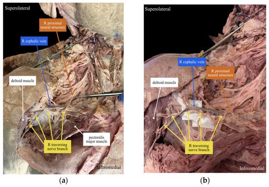
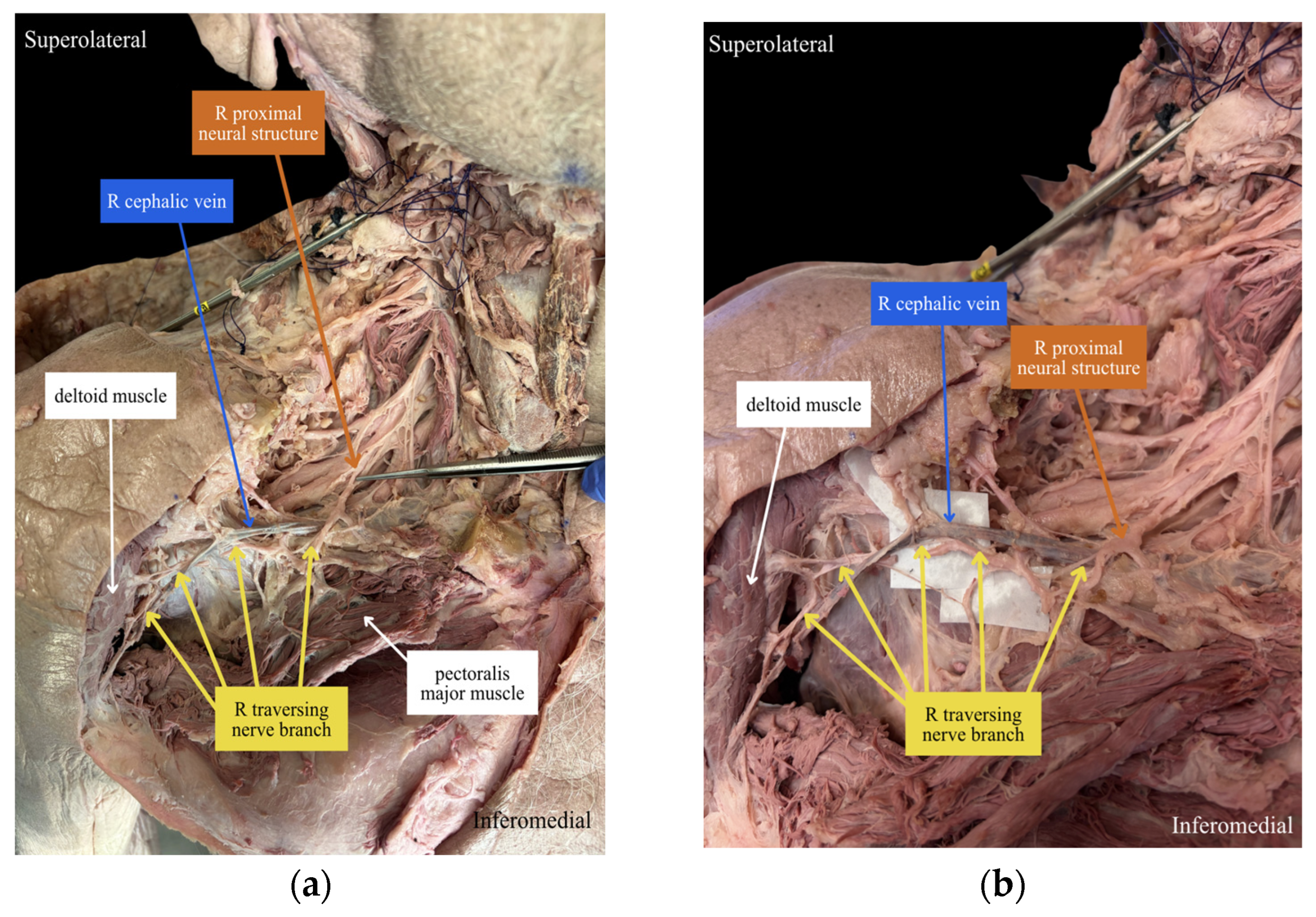
The branch in the index specimen was traced proximally to establish its neural continuity and characterize the anatomical pattern used during assessment of the remaining shoulders (Figure 1). The two additional branches exhibited similar gross morphology, course, and regional anatomical relationships but could not be independently traced to a specific spinal root or component of the brachial plexus. Their identification, therefore, represented an anatomical classification based on concordance with the index finding rather than confirmation of an identical proximal origin.
Figure 1.
Cadaveric dissection of the right deltopectoral interval in specimen 1 demonstrates a traversing nerve branch within the operative field. (a) Wide-field view demonstrating the course of the branch through the deltopectoral interval, with a probe positioned deep to the branch near its apparent origin. (b) Oblique view demonstrating the relationship of the branch to the adjacent soft tissue and neurovascular structures. Yellow arrows identify the traversing nerve branch; blue, orange, and white arrows identify the cephalic vein, proximal neural structure, and deltoid muscle, respectively.
The branch was identified in 1 of 6 left shoulders (16.7%) and 2 of 6 right shoulders (33.3%). One of 6 cadavers (16.7%) demonstrated bilateral presence of the branch, whereas one of six cadavers (16.7%) demonstrated unilateral right-sided presence. In all three positive shoulders, the nerve branch coursed inferiorly through the deltopectoral interval.
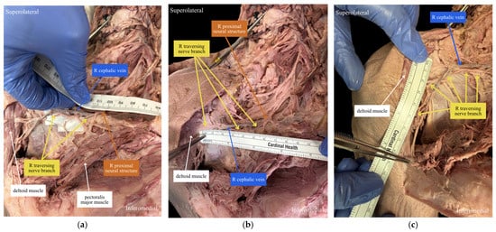
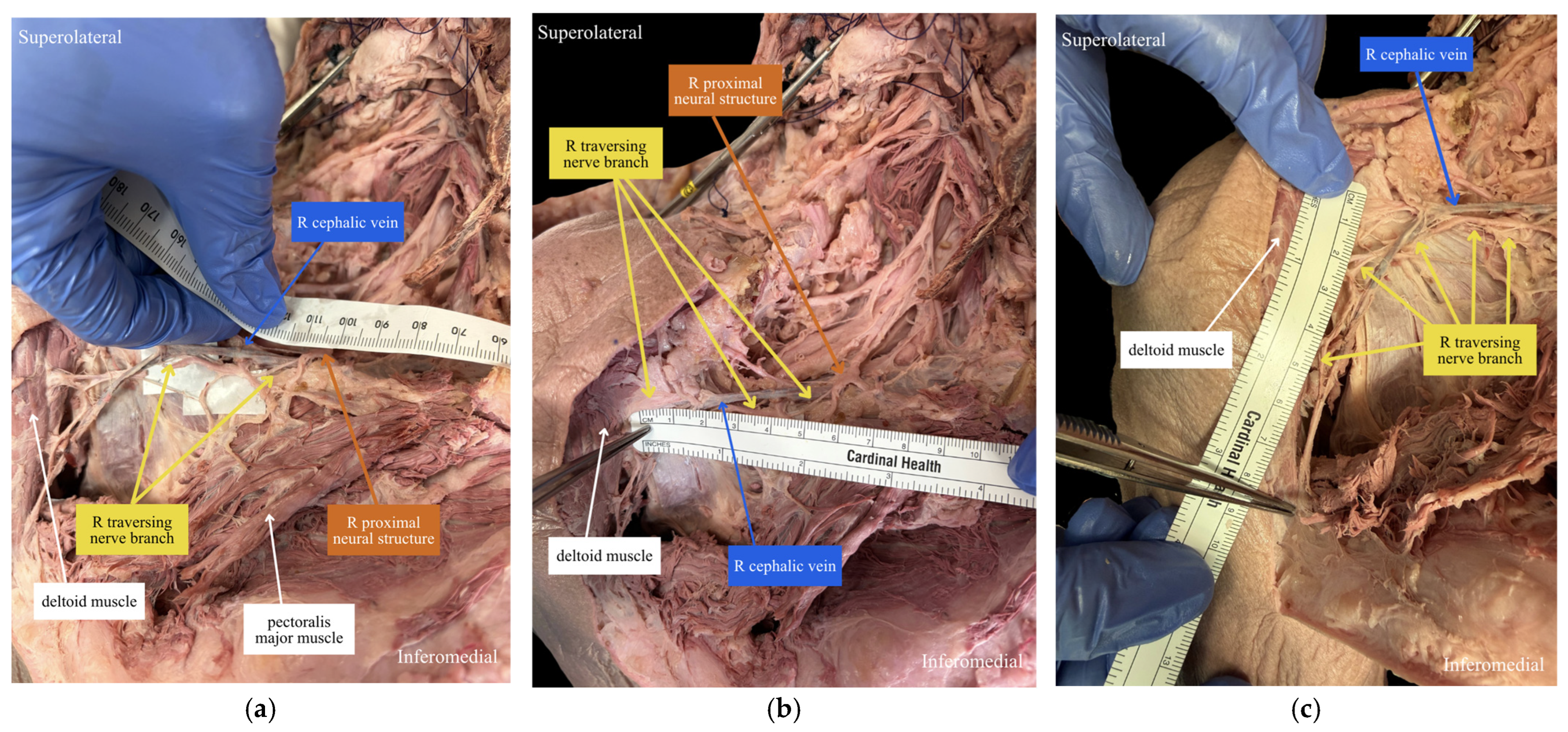
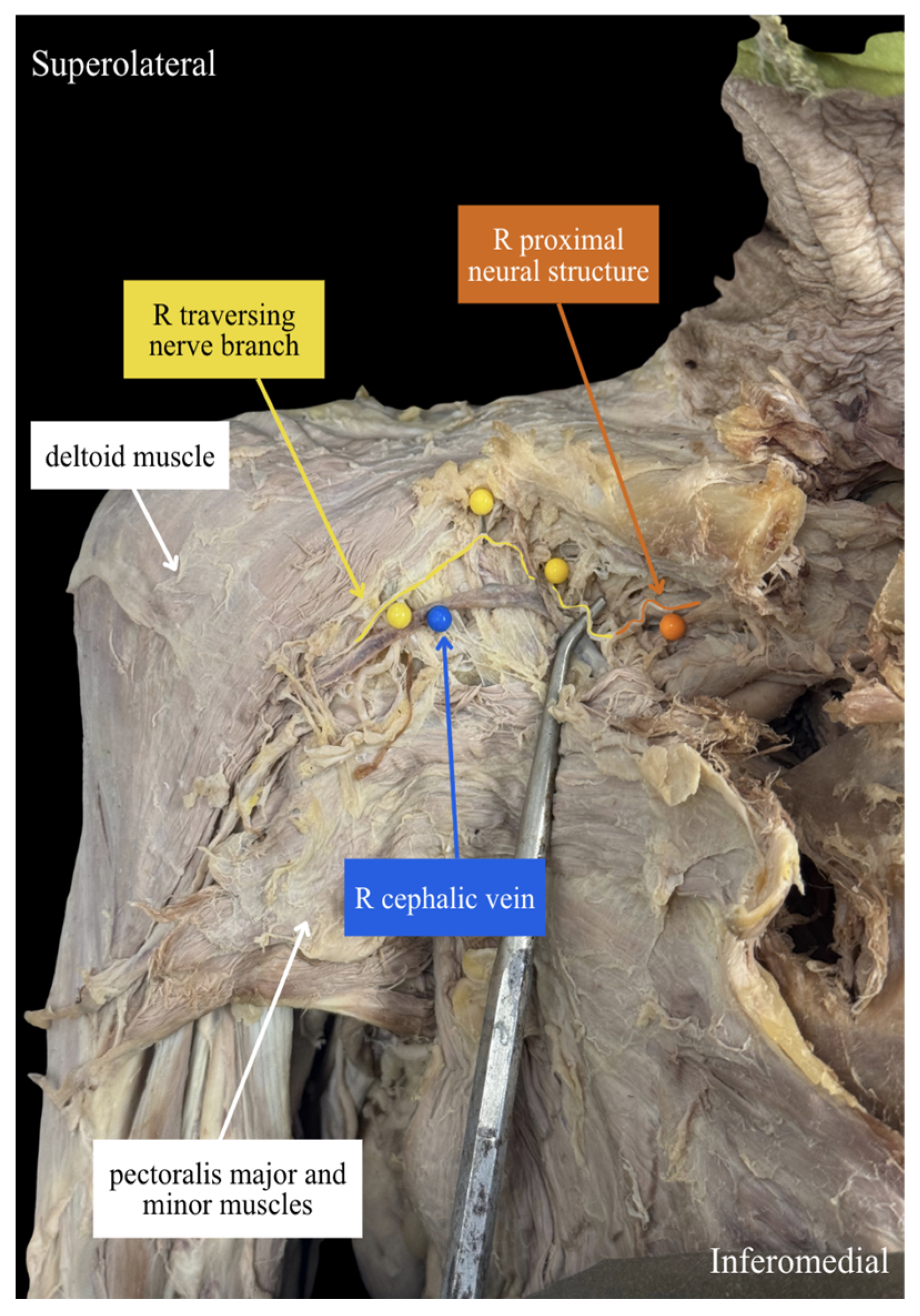
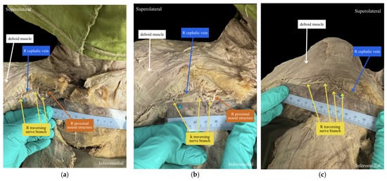
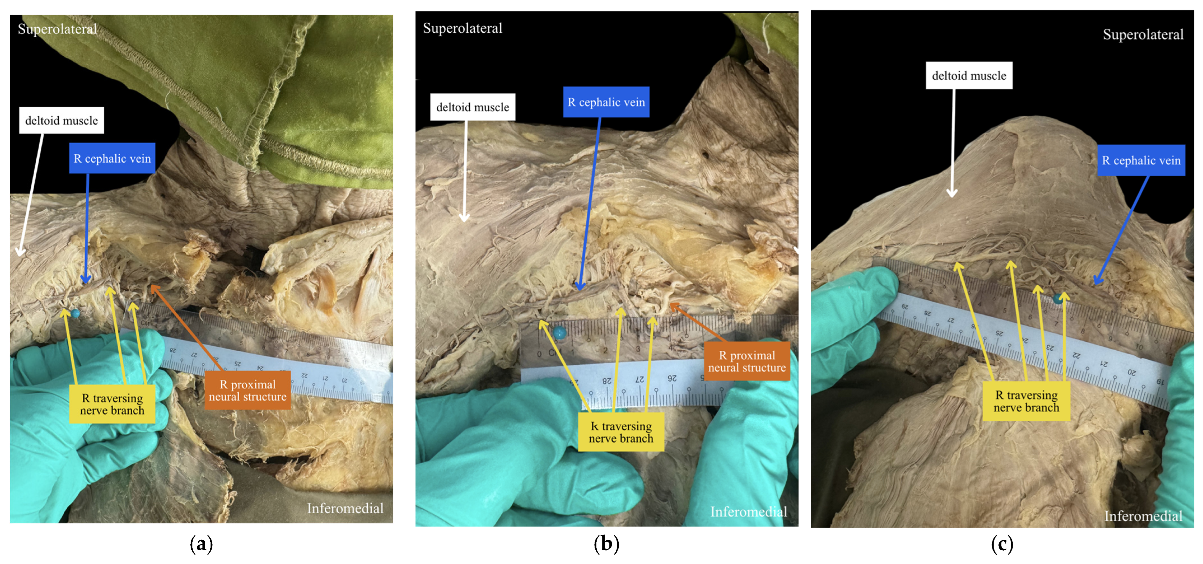
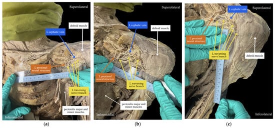
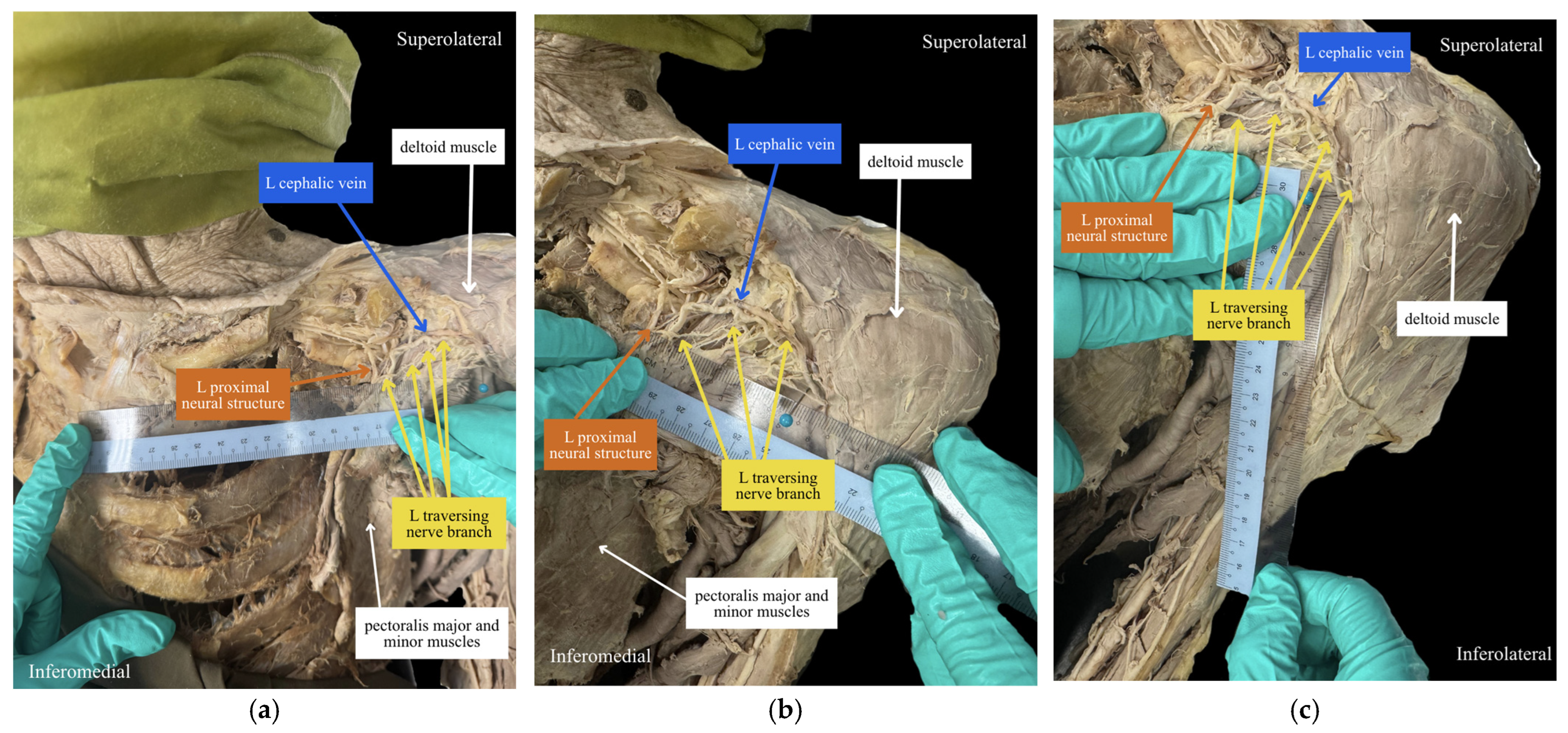
The mean distance from the coracoid process to the traversing nerve branch was 7.0 cm (range, 6.5–8.1 cm). The mean distance from the nerve branch to the proximal border of the pectoralis major insertion was 2.7 cm (range, 1.6–3.9 cm), and the mean distance from the sternoclavicular joint to the proximal/medial extent of the nerve branch was 12.5 cm (range, 11.0–13.3 cm). Representative morphometric measurements from the index specimen are shown in Figure 2. The corresponding traversing branch and its relationship to the adjacent cephalic vein and proximal neural structure in Specimen 2 are shown in Figure 3. Quantitative measurements for the right shoulder of Specimen 2 are shown in Figure 4, with corresponding measurements for the left shoulder shown in Figure 5. Individual measurements are reported in Table 1.
Figure 2.
Gross anatomic measurements of the traversing nerve branch in Specimen 1, right shoulder. Cadaveric dissection demonstrating the course of the traversing branch within the deltopectoral interval and its relationship to reproducible anatomic landmarks. (a) Proximal-to-distal measurement from the coracoid process to the traversing nerve branch. (b) Medial-to-lateral measurement from the sternoclavicular joint to the proximal/medial extent of the traversing nerve branch. (c) Proximal-to-distal measurement from the traversing nerve branch to the proximal border of the pectoralis major insertion. Yellow arrows identify the traversing nerve branch; blue, orange, and white arrows identify the cephalic vein, proximal neural structure, and deltoid muscle, respectively.
Figure 3.
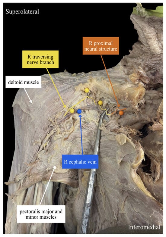
Cadaveric dissection of the right deltopectoral interval in specimen 2 demonstrating a traversing nerve branch within the operative field. Yellow pins and the yellow arrow identify the branch of interest. The orange pin and arrow identify an associated proximal neural structure that was not continuously traced to a specific spinal root or component of the brachial plexus. The blue pin and arrow identify the cephalic vein.
Figure 4.
Gross anatomic measurements of the traversing nerve branch in Specimen 2, right shoulder. Cadaveric dissection demonstrating the location of the traversing branch within the deltopectoral interval relative to reproducible anatomic landmarks. (a) Medial-to-lateral measurement from the sternoclavicular joint to the proximal/medial extent of the traversing nerve branch. (b) Proximal-to-distal measurement from the coracoid process to the traversing nerve branch. (c) Proximal-to-distal measurement from the traversing nerve branch to the proximal border of the pectoralis major insertion. Yellow arrows identify the traversing nerve branch; blue, orange, and white arrows identify the cephalic vein, proximal neural structure, and deltoid muscle, respectively.
Figure 5.
Gross anatomic measurements of the traversing nerve branch in Specimen 2, left shoulder. Cadaveric dissection demonstrating the location of the traversing branch within the deltopectoral interval relative to reproducible anatomic landmarks. (a) Medial-to-lateral measurement from the sternoclavicular joint to the proximal/medial extent of the traversing nerve branch. (b) Proximal-to-distal measurement from the coracoid process to the traversing nerve branch. (c) Proximal-to-distal measurement from the traversing nerve branch to the proximal border of the pectoralis major insertion. Yellow arrows identify the traversing nerve branch; blue, orange, and white arrows identify the cephalic vein, proximal neural structure, and adjacent musculature.
Table 1.
Quantitative measurements of the traversing nerve branch within the deltopectoral interval. Measurements of the traversing nerve branch relative to reproducible anatomic landmarks in the three shoulders in which the nerve was identified. Coracoid-to-branch and branch-to-proximal border of the pectoralis major insertion distances were measured in the proximal-to-distal axis. Sternoclavicular joint-to-medial/proximal branch extent was measured along the medial-to-lateral axis.
4. Discussion
The principal finding of this cadaveric study was the identification of a discrete nerve branch traversing the deltopectoral interval in 3 of 12 shoulders (25.0%) and 2 of 6 cadavers (33.3%). Based on its gross morphology, anatomical course, and proximal relationships, the structure was considered anatomically consistent with a possible variant branch of the lateral pectoral nerve. The branch followed a generally inferior course through the interval, although its precise position varied among the three positive shoulders. The mean distance from the coracoid process was 7.0 cm (range, 6.5–8.1 cm), from the proximal border of the pectoralis major insertion was 2.7 cm (range, 1.6–3.9 cm), and from the sternoclavicular joint was 12.5 cm (range, 11.0–13.3 cm). These findings indicate that the crossing neural tissue may be encountered within the deltopectoral interval; however, the small sample size and clustering of three positive shoulders within only two donors preclude a reliable estimate of population prevalence, a laterality association, or a reproducible surgical risk zone. Accordingly, the observed frequency should be regarded as a descriptive finding from this series rather than an estimate of the frequency of this anatomy in the general population.
Previous anatomical studies have demonstrated substantial variability in the number and course of the lateral pectoral nerve; however, these studies generally describe the nerve in the proximal pectoral region or along the deep surface of the pectoralis major rather than traversing the exposed deltopectoral interval. Macchi et al. reported that the lateral pectoral nerve coursed approximately 55 ± 7 mm inferomedially beneath the pectoralis major fascia and parallel to the thoracoacromial vessels [8]. In contrast, the branch identified in the present study was located within the deltopectoral interval itself and extended more distally through the operative field. This distinction suggests that the observed branch represents a previously under-described course rather than the typical lateral pectoral nerve in a different position.
Madhavan et al. further demonstrated substantial variability in lateral pectoral nerve anatomy, identifying two lateral pectoral nerves in 77.5% of 40 pectoral regions, and one or both nerves located immediately inferior to the clavicle in 95% of specimens [12]. The superior lateral pectoral nerve was also closely related to the cephalic vein. These findings support the possibility that the lateral pectoral nerve branches may lie near structures routinely encountered during development of the deltopectoral interval. However, the branch observed in the present study extended farther distally and traversed the operative interval rather than remaining confined to the proximal pectoral region. Accordingly, the observed frequency in the present series should not be directly compared with the frequencies reported for lateral pectoral nerve variants generally, because the present study evaluated branches traversing the deltopectoral interval [12].
An additional consideration is the variability in nomenclature used to describe the pectoral nerves. Although the pectoral nerves are conventionally described as the lateral and medial pectoral nerves, alternative classification systems have described three distinct pectoral nerves, superior, middle, and inferior, based on their origins, courses, and muscular distributions [9,13]. Substantial variation in the origins and branching patterns of these nerves has also been recognized in anatomical reviews and compendia of human anatomical variation [9,14]. In the present study, the term lateral pectoral nerve was retained for consistency with the terminology used in the comparative surgical–anatomical literature and because the gross course and proximal relationships of the observed branches were most compatible with this nerve. However, because complete proximal and distal tracing was not achieved in all specimens, definitive assignment of the observed branch within either nomenclature cannot be made.
From a surgical–anatomical perspective, these findings can also be considered within the framework described by Gadea et al. [1]. The authors characterized the deltopectoral approach as a sequence of four anatomical layers separated by three tissue planes and emphasized careful, atraumatic development of the interval. More recently, Farias et al. quantified the relationships of the axillary, musculocutaneous, and subscapular nerves to landmarks encountered through the deltopectoral approach, further demonstrating the close proximity of neural structures to the operative field [15]. The present findings suggest that, although the deltopectoral interval is traditionally considered an internervous plane, small neural branches may still traverse the operative field. Therefore, identification of the muscular interval does not necessarily indicate that all crossing tissue can be safely divided without further inspection.
The relationship of the observed branch to the cephalic vein may be particularly relevant during the development of this interval. Radkowski et al., in an anatomical study of 40 fresh-frozen shoulders, identified more lateral than medial tributaries to the cephalic vein and a greater number of branches in the proximal than distal portion of the deltopectoral incision, findings that supported lateral retraction of the vein in most specimens [16]. The present study identifies an additional structure that may be encountered in this same operative region: a traversing nerve branch in proximity to the cephalic vein. Although the distance between the branch and the cephalic vein was not quantitatively measured, the observed relationship suggests that small neural structures may lie adjacent to tissues encountered during vein mobilization. The present findings do not support one direction of cephalic vein retraction over another and do not establish a basis for specific recommendations regarding cephalic vein mobilization or management.
Clinical observations further demonstrate that unexpected neural structures may be encountered within the deltopectoral interval. Pizzo et al. reported an unusual axillary nerve variant coursing deep to the cephalic vein during reverse shoulder arthroplasty, requiring identification of the nerve and modification of the surgical exposure [17]. Although the nerve observed by Pizzo et al. was the axillary nerve rather than a pectoral nerve branch, the report similarly illustrates that neural structures not typically expected within the deltopectoral interval may be encountered during surgical exposure. The present findings extend this observation by identifying a smaller traversing branch whose gross morphology, anatomical course, and proximal relationships were consistent with a possible lateral pectoral nerve variant. Together, these findings support careful identification of neural-appearing structures encountered adjacent to the cephalic vein rather than assuming they represent fibrous or vascular tissue.
The functional consequences of injury to this branch during shoulder surgery remain unknown. The anatomical and surgical literature from breast and axillary surgery has emphasized preservation of the pectoral nerves to avoid denervation and atrophy of the pectoralis major [5,6]. Conversely, overlapping innervation and communications between the medial and lateral pectoral nerves may reduce the effect of injury to a small accessory or terminal branch [8,9]. Comparative studies of deltopectoral and anterosuperior shoulder approaches generally report overall neurologic events rather than lateral pectoral nerve-specific outcomes [18,19]. Accordingly, the present study supports recognition of the branch but does not establish that transection results in clinically meaningful weakness, pain, atrophy, or neuroma formation.
This study has several strengths. Bilateral dissections permitted reporting of both shoulder-level and cadaver-level frequency and allowed assessment of unilateral and bilateral occurrence. Morphometric measurements were obtained relative to osseous and muscular landmarks that are readily identifiable during the deltopectoral approach. The study also provides an initial quantitative description of a traversing branch that has not previously been systematically characterized within this interval.
The findings should be interpreted in the context of several important limitations. First, the study included only six cadavers, and the three positive shoulders arose from only two donors. All donors were documented as White, further limiting generalizability of the observed anatomical variation. Accordingly, although the gross morphology, course, and regional relationships of these branches were concordant with the index specimen, their classification as the same anatomical structure remains presumptive.
Second, five of the six cadavers, representing 10 of 12 shoulders, had undergone prior educational axillary or breast dissection. Previous dissection may have disrupted or removed small neural branches, potentially resulting in underdetection. Conversely, alteration of normal tissue may have made remaining branches more conspicuous and increased the likelihood of identification. The branch was identified in 2 of 10 previously dissected shoulders and in 1 of 2 previously undissected shoulders; however, the small and unequal group sizes preclude meaningful comparison. Accordingly, prior educational dissection could have biased the observed frequency in either direction, resulting in either overestimation or underestimation of the true frequency of this anatomical finding. Formalin-based preservation may also alter tissue compliance and spatial relations compared with those encountered in living patients.
Third, although proximal dissection in the index specimen supported the neural nature and likely pectoral nerve association of the traversing branch, complete proximal tracing was not performed in the other two positive shoulders. Those branches were classified based on concordant gross morphology, course, and anatomical relationships. Histologic confirmation, microscopic tracing, nerve stimulation, and complete delineation of the proximal origin and terminal distribution were not performed. Therefore, classification as a possible lateral pectoral nerve variant remains presumptive, and the three branches cannot be confirmed to have shared an identical spinal or plexus origin.
Finally, morphometric measurements were obtained using a rigid centimeter ruler and were not normalized to donor dimensions. Measurements were obtained by two investigators rather than recorded independently. Consequently, interobserver agreement could not be quantified, and no formal assessment of intraobserver or interobserver reliability was performed. Therefore, the reported distances should be interpreted as descriptive measurements of the three positive shoulders rather than precise estimates of branch location in the broader population.
Future studies should include larger, demographically diverse cohorts of fresh-frozen, previously undissected specimens using standardized bilateral exposure. Microscopic tracing to the brachial plexus and pectoralis major, histologic confirmation, nerve diameter measurement, and quantitative mapping relative to the cephalic vein and thoracoacromial vessels would help clarify the identity and operative relationships of this branch. Repeated measurements by multiple observers and normalization to donor dimensions would improve morphometric reliability. If the branch is subsequently identified in living patients, prospective assessment of pectoralis major strength, muscle volume, electromyography, pain, and neurologic symptoms would be necessary to determine the clinical consequences of injury and the value of routine preservation.
5. Conclusions
A discrete nerve branch anatomically consistent with a possible lateral pectoral nerve variant was observed traversing the deltopectoral interval in 3 of 12 shoulders in this cadaveric series. In the three positive shoulders, the branch was located 6.5–8.1 cm distal to the coracoid process and demonstrated variable positioning within the operative field. These findings indicate that neural tissue may cross the deltopectoral interval despite its traditional characterization as an internervous plane. The region in which these branches were observed may warrant particular attention during development of the interval; however, the limited sample is insufficient to define a reproducible surgical risk zone. These preliminary, hypothesis-generating observations require confirmation in larger anatomical and clinical studies to confirm the identity, distribution, frequency, and clinical significance of this observed branch.
Author Contributions
Conceptualization, C.N.W., T.J.M. and J.F.; methodology, C.N.W., T.J.M. and J.F.; validation, C.N.W. and T.J.M.; formal analysis, C.N.W.; investigation, C.N.W., T.J.M. and N.R.Z.; resources, T.J.M., C.M. and J.F.; data curation, C.N.W.; writing—original draft preparation, C.N.W.; writing—review and editing, C.N.W., T.J.M., N.R.Z., J.T., C.M. and J.F.; visualization, C.N.W.; supervision, T.J.M. and J.F.; project administration, C.N.W. and J.F. All authors have read and agreed to the published version of the manuscript.
Funding
This research received no external funding.
Institutional Review Board Statement
Ethical review and approval were not required for this cadaveric anatomical study under the policies of the Ohio University Heritage College of Osteopathic Medicine. The study used de-identified donor specimens obtained through the Ohio University Heritage College of Osteopathic Medicine Body Donor Program. The specimens were donated for medical education and/or research in accordance with applicable donor consent procedures and institutional policies. No living human participants were involved, and no identifiable donor information was collected, analyzed, or reported.
Informed Consent Statement
Not applicable. This study did not involve living human participants. De-identified cadaveric specimens were obtained through the institutional anatomical donor program and used in accordance with donor consent procedures and institutional policies.
Data Availability Statement
The data supporting the findings of this study are included within the article. Additional raw morphometric data are not publicly available due to ethical and privacy considerations related to the cadaveric donor specimens but may be made available from the corresponding author upon reasonable request.
Acknowledgments
The authors gratefully acknowledge the donors and their families for their generous contribution to medical education and anatomical research. The authors also thank the body donor program for its support in facilitating cadaveric study and anatomical dissection.
Conflicts of Interest
The authors declare no conflicts of interest.
References
- Gadea, F.; Bouju, Y.; Berhouet, J.; Bacle, G.; Favard, L. Deltopectoral approach for shoulder arthroplasty: Anatomic basis. Int. Orthop. 2015, 39, 215–225. [Google Scholar] [CrossRef] [Scilit] [PubMed]
- Klepps, S.; Auerbach, J.; Calhon, O.; Lin, J.; Cleeman, E.; Flatow, E. A cadaveric study on the anatomy of the deltoid insertion and its relationship to the deltopectoral approach to the proximal humerus. J. Shoulder Elb. Surg. 2004, 13, 322–327. [Google Scholar] [CrossRef] [Scilit] [PubMed]
- Vajapey, S.; Contreras, E.; Cvetanovich, G.; Neviaser, A. Neurologic complications in primary anatomic and reverse total shoulder arthroplasty: A review. J. Clin. Orthop. Trauma 2021, 20, 101475. [Google Scholar] [CrossRef] [Scilit] [PubMed]
- Fu, M.; Hendel, M.; Chen, X.; Warren, R.; Dines, D.; Gulotta, L. Surgical anatomy of the radial nerve in the deltopectoral approach for revision shoulder arthroplasty and periprosthetic fracture fixation: A cadaveric study. J. Shoulder Elb. Surg. 2017, 26, 2173–2176. [Google Scholar] [CrossRef] [Scilit] [PubMed]
- Moosman, D. Anatomy of the pectoral nerves and their preservation in modified mastectomy. Am. J. Surg. 1980, 139, 883–886. [Google Scholar] [CrossRef] [Scilit] [PubMed]
- Hoffman, G.; Elliott, L. The anatomy of the pectoral nerves and its significance to the general and plastic surgeon. Ann. Surg. 1987, 205, 504–506. [Google Scholar] [CrossRef] [Scilit] [PubMed]
- Lee, K. Anatomic variation of the spinal origins of lateral and medial pectoral nerves. Clin. Anat. 2007, 20, 915–918. [Google Scholar] [CrossRef] [Scilit] [PubMed]
- Macchi, V.; Tiengo, C.; Porzionato, A.; Stecco, C.; Mazzoleni, F.; De Caro, R. Medial and lateral pectoral nerves: Course and branches. Clin. Anat. 2007, 20, 157–162. [Google Scholar] [CrossRef] [Scilit] [PubMed]
- Porzionato, A.; Macchi, V.; Stecco, C.; Loukas, M.; Tubbs, R.; De Caro, R. Surgical anatomy of the pectoral nerves and the pectoral musculature. Clin. Anat. 2012, 25, 559–575. [Google Scholar] [CrossRef] [Scilit] [PubMed]
- Boers, N.; Bleys, R.L.; Schellekens, P.P. The nerve supply to the pectoralis major: An anatomical study and clinical application of the denervation in subpectoral breast implant surgery. J. Plast. Reconstr. Aesthetic Surg. 2022, 75, 415–423. [Google Scholar] [CrossRef] [Scilit] [PubMed]
- Patel, N.T.; Smith, H.F. Clinically Relevant Anatomical Variations in the Brachial Plexus. Diagnostics 2023, 13, 830. [Google Scholar] [CrossRef] [Scilit] [PubMed]
- Madhavan, C.; Rajasekharan, S.; Varghese, S. Anatomy of lateral pectoral nerve and its clinic significance. Natl. J. Clin. Anat. 2022, 11, 79–83. [Google Scholar] [CrossRef] [Scilit] [PubMed]
- Aszmann, O.; Rab, M.; Kamolz, L.; Frey, M. The anatomy of the pectoral nerves and their significance in brachial plexus reconstruction. J. Hand Surg. 2000, 25, 942–947. [Google Scholar] [CrossRef] [Scilit] [PubMed]
- Mahan, M.; Spinner, R. Nerves of the Upper Extremity. In Bergman’s Comprehensive Encyclopedia of Human Anatomic Variation; Tubbs, R., Shoja, M., Loukas, M., Eds.; Wiley-Blackwell: Hoboken, NJ, USA, 2016. [Google Scholar] [CrossRef] [Scilit]
- Farias, A.L.N.; Tanaka, L.Y.; de Castro, L.V.; da Costa, M.P.; Filho, R.B.; Tenor, A.C., Jr. Neurological Anatomy Applied to the Deltopectoral Surgical Approach: Safety Parameters in the Latarjet Procedure. Rev. Bras. Ortop. 2025, 60, s00441800921. [Google Scholar] [CrossRef] [Scilit] [PubMed]
- Radkowski, C.; Richards, R.; Pietrobon, R.; Moorman, C., 3rd. An anatomic study of the cephalic vein in the deltopectoral shoulder approach. Clin. Orthop. 2006, 442, 139–142. [Google Scholar] [CrossRef] [Scilit] [PubMed]
- Pizzo, R.; Lynch, J.; Adams, D.; Yoon, R.; Liporace, F. Unusual anatomic variant of the axillary nerve challenging the deltopectoral approach to the shoulder: A case report. Patient Saf. Surg. 2019, 13, 9. [Google Scholar] [CrossRef] [Scilit] [PubMed]
- Georgoulas, P.; Fiska, A.; Ververidis, A.; Drosos, G.; Perikleous, E.; Tilkeridis, K. Reverse shoulder arthroplasty, deltopectoral approach vs. anterosuperior approach: An overview of the literature. Front. Surg. 2021, 8, 721054. [Google Scholar] [CrossRef] [Scilit] [PubMed]
- Seok, H.; Park, J.; Park, S. Anterosuperior approach versus deltopectoral approach for reverse total shoulder arthroplasty: A systematic review and meta-analysis. J. Orthop. Surg. 2022, 17, 527. [Google Scholar] [CrossRef] [Scilit] [PubMed]

Disclaimer/Publisher’s Note: The statements, opinions and data contained in all publications are solely those of the individual author(s) and contributor(s) and not of MDPI and/or the editor(s). MDPI and/or the editor(s) disclaim responsibility for any injury to people or property resulting from any ideas, methods, instructions or products referred to in the content. |
© 2026 by the authors. Licensee MDPI, Basel, Switzerland. This article is an open access article distributed under the terms and conditions of the Creative Commons Attribution (CC BY) license.