


Abstract
This work evaluates agarose (AG)- and chitosan (CS)-based hydrogels incorporating a dexamethasone–cyclodextrin complex (Dex) as potential bioink candidates for wound healing applications. For this purpose, biopolymer hydrogels were formulated and tested to determine their structure, morphology, and rheological behavior under different strain, frequency, and temperature conditions, as well as their swelling behavior, surface functionality, protein adhesion capability, and antibacterial activity. Scanning electron microscopy revealed an interconnected membrane-like porous structure in the agarose/chitosan (BAC) hydrogels, which also showed a fibril-like microstructure when Dex was added. X-ray diffraction showed that these structures had a semicrystalline nature, with a main broad peak from approximately 15 to 20°. Rheological tests confirmed the viscoelastic behavior of the developed hydrogels. Chitosan incorporation reduced the rigidity of the formulations, whereas dexamethasone did not significantly affect the viscoelastic response of the BAC hydrogel. Additionally, the temperature sweep showed that Dex did not substantially alter the viscoelastic response of the BAC hydrogel while reducing mass loss after immersion in phosphate-buffered saline and not significantly affecting the swelling behavior. Further, Coomassie blue staining tests demonstrated favorable protein interaction with the BAC-based hydrogels, indicating that Dex incorporation did not compromise their adsorption capacity. Regarding their antibacterial activity, the hydrogels showed no major effects on Escherichia coli and Staphylococcus aureus in disk diffusion tests. However, liquid diffusion tests showed almost no bacterial growth for hydrogels incorporating CS. Thus, the present results demonstrate the potential of the BAC hydrogel system for biomedical and cosmetic applications.
1. Introduction
Natural polysaccharides (NPSs) have been widely used in various fields, especially in biomaterials and tissue engineering, due to their biocompatibility, biodegradability, abundant availability, and structural versatility [1,2]. NPS-based matrices such as starch, cellulose, pectin, agar, agarose, alginate (alginic acid and its salts), carrageenan, xanthan gum, and chitin are frequently reinforced with nanoparticles or other functional components to enhance their physicochemical and mechanical properties, enabling the development of nanocomposite scaffolds with controlled biodegradability and improved structural performance [3,4]. Among them, agarose (AG), which is extracted by dissolving the polysaccharides from dried seaweed in hot water, deserves special attention as it is a biocompatible and biodegradable natural polymer composed of repeating agarobiose units, with the ability to form three-dimensional (3D) gel-like networks. Agarose forms physically crosslinked hydrogel through a thermoreversible gelation process. At high temperatures (90–100 °C), hydrogen bonds between agarose chains are disrupted, allowing the polymer to dissolve in water as random coils and form a homogeneous solution. Upon cooling (30–40 °C), agarose chains undergo a conformational transition and reassociate through hydrogen bonding, forming double-helical structures that aggregate into junction zones and generate a stable three-dimensional hydrogel network [5]. This interconnected structure provides mechanical stability, retains water, and facilitates the diffusion of oxygen and nutrients within the scaffold, making agarose an attractive material for tissue engineering applications. Despite its natural biocompatibility, AG is frequently combined with other polymers to improve its biological interaction and functionality [5].
Chitosan (CS), in turn, is one of the most widely used biopolymers in the design of scaffolds for bone and tissue regeneration. It can be obtained from seafood processing waste (mainly shrimp and crab shells), and it is a renewable, biodegradable, and highly versatile polysaccharide for hydrogel fabrication. It is composed of deacetylated D-glucosamine and N-acetyl-D-glucosamine units. It is also notable for its high biocompatibility and biodegradability, as well as strong bactericidal and bacteriostatic activity. These properties make CS a highly attractive material for biomedical applications [6]. However, both polymers exhibit structural and functional limitations when used individually. For this reason, their combination or chemical functionalization with other natural polymers or bioactive agents has been explored with the aim of improving their performance as matrices in various medical applications.
The use of natural polymers in wound treatment development has been particularly researched in recent decades. Indeed, traditional wound care methods, such as topical drug application or skin grafting, do not always achieve optimal results due to limitations related to tissue integration, infection, or incomplete healing. In this context, recent advances in additive manufacturing have opened new opportunities to transform wound regeneration strategies. Natural bioinks have attracted considerable attention as skin substitutes for bioprinting applications in tissue regeneration and wound healing [7]. By definition, a bioink combines cells, biomaterials, and/or biologically active components capable of providing a three-dimensional microenvironment to support cell growth, differentiation, and function [8]. In tissue engineering applications, bioinks are typically composed of hydrogels based on biocompatible polymers that act as three-dimensional support matrices. These systems provide a hydrated and structurally stable environment that promotes cell encapsulation, nutrient exchange, and new tissue formation [7,8]. In this context, bioink design should not only focus on providing structural support and biocompatibility but also on incorporating additional therapeutic functionalities.
The integration of bioactive agents within hydrogel matrices allows for the modulation of key biological processes, such as inflammation, cell proliferation, and angiogenesis, thereby promoting suitable regenerative microenvironments. Among these agents, dexamethasone, a synthetic corticosteroid widely used in clinical practice, has garnered considerable interest due to its ability to induce the proliferation and osteogenic differentiation of mesenchymal stem cells. For this reason, it has been incorporated into scaffolds designed for bone tissue engineering applications [9]. Recently, Xu et al. reported that dexamethasone concentrations in the 10–100 nM range promote osteogenic differentiation, with more pronounced effects observed at higher concentrations within this range.
In addition, dexamethasone exhibits anti-inflammatory and immunomodulatory properties that contribute to the regulation of the inflammatory phase of wound healing by modulating the local immune response, reducing leukocyte infiltration, and promoting a favorable microenvironment for tissue repair [10,11]. Tu et al. reported that dexamethasone improved the healing of frostbite injuries by reducing inflammation and stimulating vasculogenesis, highlighting its potential to promote skin regeneration [12]. Similarly, Chen et al. evaluated the dose-dependent effect of dexamethasone on wound healing in septic and non-septic models, finding that a single dose of dexamethasone (1 mg/kg) enhanced wound repair in septic models by controlling excessive inflammation and maintaining the M1/M2 macrophage balance; however, it delayed healing in healthy animals [13]. Furthermore, dexamethasone-loaded hydrogels have emerged as promising platforms for wound healing applications by enabling localized and sustained drug delivery. Yang et al. developed a bioadhesive hydrogel incorporating dexamethasone sodium phosphate, where localized dexamethasone release contributed to the regulation of inflammatory responses and improved cutaneous wound healing [14]. Thus, the functionalization of dexamethasone within polymer matrices must be carefully designed to ensure localized and controlled release at the injury site. This approach maximizes its anti-inflammatory and osteogenic effects while minimizing the adverse effects associated with systemic absorption [15].
On the other hand, CS-based bioinks have shown promise in regenerating tissues such as skin, cartilage, bone, and blood vessels. These bioinks can be tailored to exhibit adjustable biological, antibacterial, mechanical, and drug delivery properties to meet the specific requirements of different wound types, further expanding their potential [16]. For example, Choi et al. developed a CS hydrogel composed of type II collagen and chondroitin sulfate. The results demonstrated improved mechanical and cellular properties, supporting its use as an interconnected 3D structure that promotes cartilage regeneration [17]. Also, Madhumathi et al. developed chitin/nanoplate composite scaffolds for wound healing applications. These scaffolds exhibited excellent antibacterial activity against S. aureus and E. coli, as well as good blood coagulation capacity, demonstrating their potential for wound healing [18]. Another study developed a thermosensitive bioink based on CS hydrogels saturated with carbon dioxide and AG. The CS/AG scaffolds demonstrated a high capacity for cell proliferation and survival. According to the in vivo assays in mice with skin wounds, the authors confirmed the biocompatibility of the dressing and the beneficial effect of chitosan on wound healing [3].
Despite these advances, significant challenges remain. One of the main limitations of current bioink formulations for bioprinting is their limited versatility, both in terms of biological performance and rheological behavior. In particular, the physicochemical properties of bioinks, including their rheological behavior, are critical parameters that influence printability, shape fidelity, and mechanical stability during and after extrusion. These properties are also recognized as important factors that may influence subsequent cell responses and tissue maturation. However, many commercially available and studied systems still predominantly rely on three polymers, namely alginate, collagen, and gelatin, which, although supporting cell viability post-printing, are largely confined to providing structural and basic biological support [19]. As reported in the literature, several studies have explored the chemical modification of these polymers to enhance their functionality, yet such strategies frequently require toxic solvents, time-consuming multi-step syntheses, costly equipment, and complex purification protocols, which limit their translational potential [20,21]. Further, it might lead to the release of cytotoxic degradation byproducts over time, thereby compromising long-term biocompatibility. Based on these considerations, this study investigates whether the physicochemical, rheological, and biological properties of AG and CS hydrogels support their consideration as potential bioink candidates for tissue engineering and wound healing applications. Thus, this study presents the development and characterization of a biocompatible dexamethasone-functionalized bioink composed of only two components.
2. Materials and Methods
2.1. Materials
Agarose (CAS: 9012-36-6), chitosan (CAS: 9012-76-4), and a water-soluble dexamethasone–methyl-β-cyclodextrin (Dex) complex with a 6.5 wt.% dexamethasone content (CAS: 50-02-2) were purchased from Sigma-Aldrich. All reagents were used as received.
2.2. Bioink Preparation
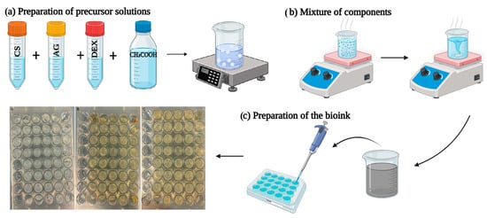
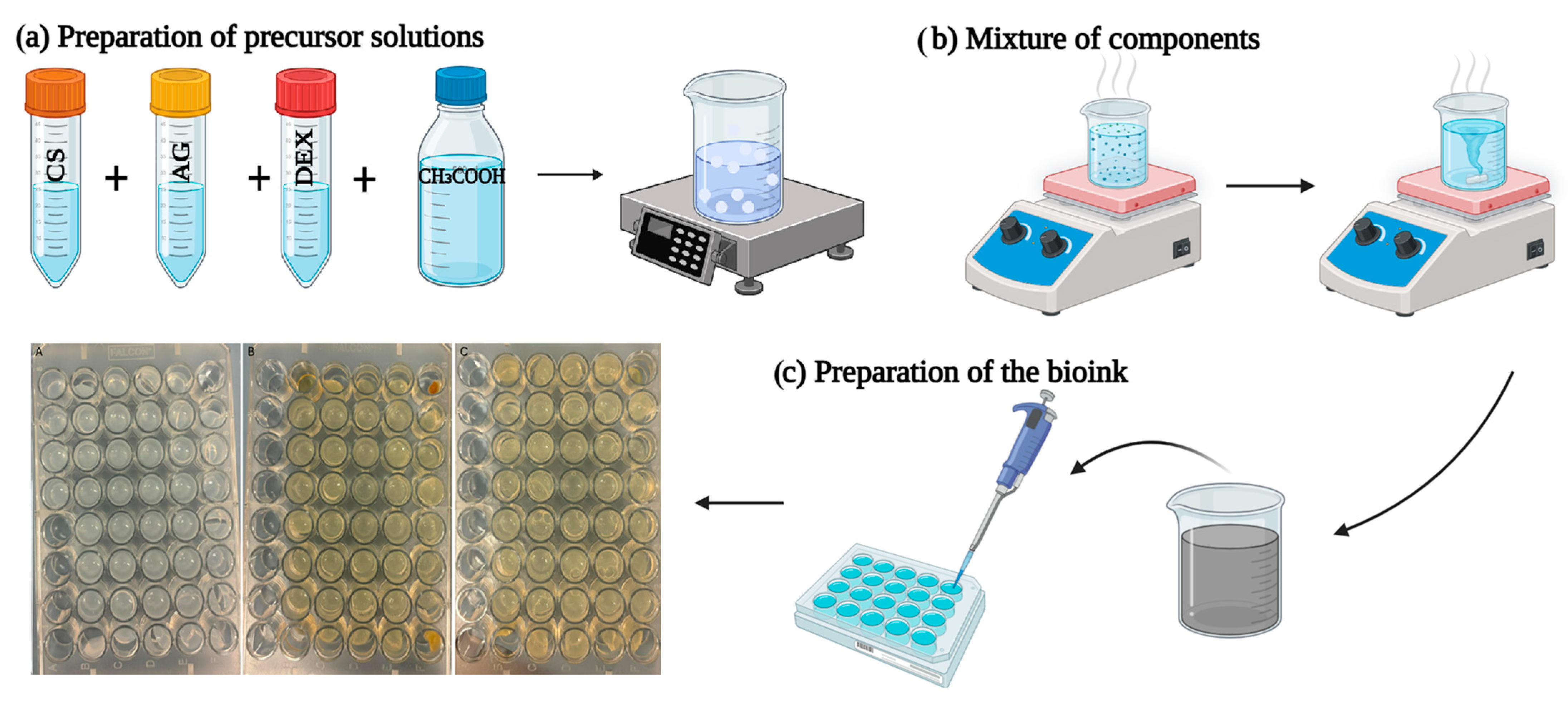
The hydrogel-based bioink formulations were prepared from CS and AG solutions functionalized with Dex. Three formulations were designed, as detailed in Table 1, to evaluate the effect of the polymer combination and drug incorporation on the properties of the bioinks. First, AG was dissolved in acetic acid previously heated in a water bath at 80 °C under constant stirring to ensure complete dissolution. Then, CS was gradually added to the AG solution. The resulting BAC solution was then allowed to stand for 30 min at a controlled temperature to ensure homogeneity. The bioink incorporating the dexamethasone–cyclodextrin complex (BACD) was obtained following a similar procedure, with a modification in the order of component addition. In this case, AG was first dissolved, followed by Dex and, finally, CS. The mixture was maintained at 80 °C in a water bath and constantly stirred for 30 min until a homogeneous formulation was obtained. Right after stopping stirring, 500 μL of the formulation was poured into 48-well plates. For physicochemical characterization, all the BA, BAC and BACD bioinks prepared were lyophilized. Figure 1 shows a schematic illustration of the preparation process of the developed formulations.
Table 1.
Compositions and proportions used for the preparation of bioinks.
Figure 1.
Schematic illustration of the fabrication process of BA, BAC, and BACD. Created with BioRender.com.
2.3. Physicochemical Characterization of Bioinks
2.3.1. Scanning Electron Microscopy
The bioink structure and porous morphology were studied by scanning electron microscopy (SEM) using a JEOL-6100 instrument from JEOL Ltd. (Tokyo, Japan) Prior to observation, a drop of carbon ink was applied to ensure adequate electrical conductivity of the samples before sputter coating. Subsequently, a conductive layer approximately 5 nm thick with 99.99% purity was deposited using a BIORAD-POLARON sputter coater (Bio-Rad/Polaron, Uckfield, UK).
2.3.2. X-Ray Diffraction (XRD)
XRD measurements were performed using a Rigaku Miniflex-600 diffractometer (Rigaku Corporation, Tokyo, Japan) equipped with a D/tex Ultra2 detector to assess the crystalline structure of AG, CS, and Dex. Diffraction patterns were recorded using Cu Kα radiation at 40 kV and 15 mA. Data were acquired in θ–2θ geometry over a 2θ range of 5–90°, with a step size of 0.02° and a scanning rate of 20°/min.
2.3.3. Raman Spectroscopy
Raman spectroscopy was performed using a Horiba Jobin-Yvon LabRam HR800 spectrometer (HORIBA Jobin Yvon S.A.S., Longjumeau, France) equipped with a CCD detector. Measurements were carried out using an optical microscope with a 100× objective. A Nd:YAG laser with an excitation wavelength of 532 nm was employed. The nominal spectral resolution was 0.35 cm−1. The instrument was calibrated using the 520 cm−1 reference band of a silicon wafer. Raman mapping was performed with an acquisition time of 3 s per scan, accumulating 40 scans, over a spectral range of 100–3000 cm−1. All measurements were conducted at room temperature.
2.3.4. Fourier Transform Infrared (FTIR) Spectroscopy
FTIR spectroscopy was carried out using a Cary 630 spectrometer (Agilent Technologies, Santa Clara, CA, USA) to confirm the structure and functional groups of CS, AG, Dex and the bioink formulations. Scanning was performed in the range from 500 to 4000 cm−1, with a resolution of 4 cm−1.
2.3.5. Swelling
For the swelling tests, the bioink samples were placed in 12-well plates and immersed in a sufficient volume of PBS to ensure complete immersion throughout the experiment. Swelling measurements were performed after 6, 24, and 96 h. The weight of the sample was recorded before soaking (W0) and after soaking at time t (Wₜ). The swelling ratio (Sᵣ) was calculated as follows [22]:
Sr (%) = [(Wt − W0)/W0] 100%
The results are expressed as the mean ± standard deviation (n = 4).
2.3.6. In Vitro Degradation
In vitro degradation tests were performed by immersing the samples in PBS at room temperature using an aluminum support system within a well plate. Measurements were carried out at 48 h and at 7, 15, and 29 days. At each interval, the samples were removed from the PBS, briefly drained, weighed, and then dried in an incubator at 37 °C for 48 h. In vitro degradation was obtained using Equation (2). The weight loss percentage (WL%) was calculated using Equation (2):
WL% = ((W0 − Wd)/W0) 100%
2.3.7. Protein Adsorption Assay
The protein adhesion capacity of the bioinks was evaluated using Coomassie Brilliant Blue staining tests. For each bioink formulation, two samples were treated with 10 mL of fetal bovine serum (FBS) for 30 min, while one sample was kept as an untreated control. After incubation, the samples were washed with PBS to remove non-adhered proteins and fixed with 1.5 mL of 4% paraformaldehyde for 15 min. The control sample was subjected to the same fixation procedure. Subsequently, all samples were rinsed with PBS and stained with Coomassie Brilliant Blue for 30 min. Excess dye was removed using a solution of ethanol, acetic acid, and water. Finally, macroscopic images were acquired to qualitatively evaluated protein adsorption on the hydrogel surface.
2.4. Release of Dexamethasone
The samples were weighed before the release experiments. The release of dexamethasone was studied for 7 days. For this purpose, each sample was immersed in one of the wells of a six-well untreated-culture plate with 10 mL of a 1× PBS solution, which simulates a physiological environment. The samples were then incubated under stirring at 100 rpm and 37 °C. Aliquots of 20 μL were extracted from the PBS medium in which dexamethasone was released after 2, 4, 6, 8, 25, 48, 72, and 168 h. The concentration of dexamethasone in the PBS was determined using a reverse High-Performance Liquid Chromatograph, Jasco PU-4180 RHLC (JASCO Corporation, Tokyo, Japan). A C18 column was used and detection was performed at 240 nm with a UV-4075 UV/VIS detector (JASCO Corporation, Tokyo, Japan). A mixture of water and acetonitrile (55:45) was used as the mobile phase and the measurement protocol was set to observe the dexamethasone peak at a retention time of 2.82 min over 5 min analysis intervals. The aliquots were diluted to 50 mL using the mobile phase before being introduced into the system. The injection volume was 20 µL and the workflow rate was 1 mL/min. An external calibration curve was prepared using dexamethasone standard solutions in the concentration range of 10–250 µg/mL. The calibration curve showed good linearity, with the regression equation y = 39,221x − 146,225 and a coefficient of determination (R2) of 0.9965. Dexamethasone concentrations in the release samples were calculated from this calibration curve.
2.5. Rheology
Rheological characterization was carried out using an MCR 702e rotational rheometer (Anton Paar, Graz, Austria) equipped with a parallel plate with a diameter of 25 mm. For rheological measurements, the plate gap was fixed at 0.5 mm, and excess material was carefully trimmed from the sample edges before each test to ensure a uniform geometry. The samples were placed on the lower plate of the rheometer using a syringe, thereby avoiding the formation of air bubbles, before adjusting the separation between the plates. Frequency sweep measurements within a frequency range of 0.1 to 100 Hz were conducted at 25 °C using a constant shear strain of 0.5%. The strain sweep was performed from 0.01 to 90% at a frequency of 1 Hz and 25 °C. The storage modulus (G′) and loss modulus (G″) were recorded as functions of frequency, strain or temperature (from 10 to 50 °C) to evaluate the viscoelastic behavior. One measurement was performed for each sample.
2.6. Microbiological Characterization
The antimicrobial performance of the materials and their effect on bacterial growth were evaluated using two complementary methods: (i) a disk diffusion test, in which hydrogel disks were placed on previously inoculated Mueller–Hinton agar plates to detect potential zones of growth inhibition, and (ii) growth curve analysis in liquid culture, where bacterial proliferation in the presence of the hydrogels was followed by optical density measurements. Antimicrobial activity was tested against standard reference strains, namely the Gram-positive Staphylococcus aureus (ATCC 338662) and the Gram-negative Escherichia coli (ATCC 25922).
For the agar diffusion assay, Mueller–Hinton agar plates were prepared and inoculated with bacterial suspensions of E. coli and S. aureus, which were adjusted to a turbidity value equivalent to 0.25 McFarland. Aliquots of 100 µL of each suspension were pipetted onto the agar surface and spread uniformly using a sterile spreader [23]. Pre-hydrated hydrogel specimens were then gently placed on the inoculated agar, ensuring complete contact between the hydrogel and the surface of the medium. Before placement, hydrogels were equilibrated in sterile Luria–Bertani (LB) broth to maintain their hydration state and promote diffusion of the active compound throughout the network. Ciprofloxacin and ampicillin disks served as reference controls. The plates were incubated at 37 °C for 24 h, after which inhibition halos were recorded by macroscopic photography and used to quantify antimicrobial activity.
In parallel, bacterial growth in liquid culture was assessed by monitoring optical density. The same 0.25 McFarland bacterial suspensions used for the agar plates were inoculated into tubes containing the different hydrogel formulations, together with a positive control (LB broth without material) and negative controls (cultures containing ciprofloxacin or ampicillin). Cultures were incubated at 37 °C with constant shaking for 24 h. Bacterial growth kinetics were followed by measuring the optical density at 600 nm (OD600) at defined time intervals (1, 2, 4, 6, 8, and 24 h) using a Varioskan Lux multimode microplate reader (Thermo Fisher Scientific, Vantaa, Finland).
3. Results and Discussion
3.1. Scanning Electron Microscopy
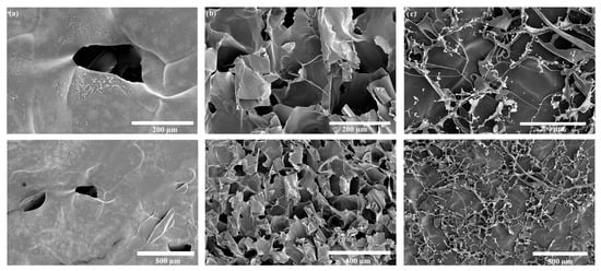
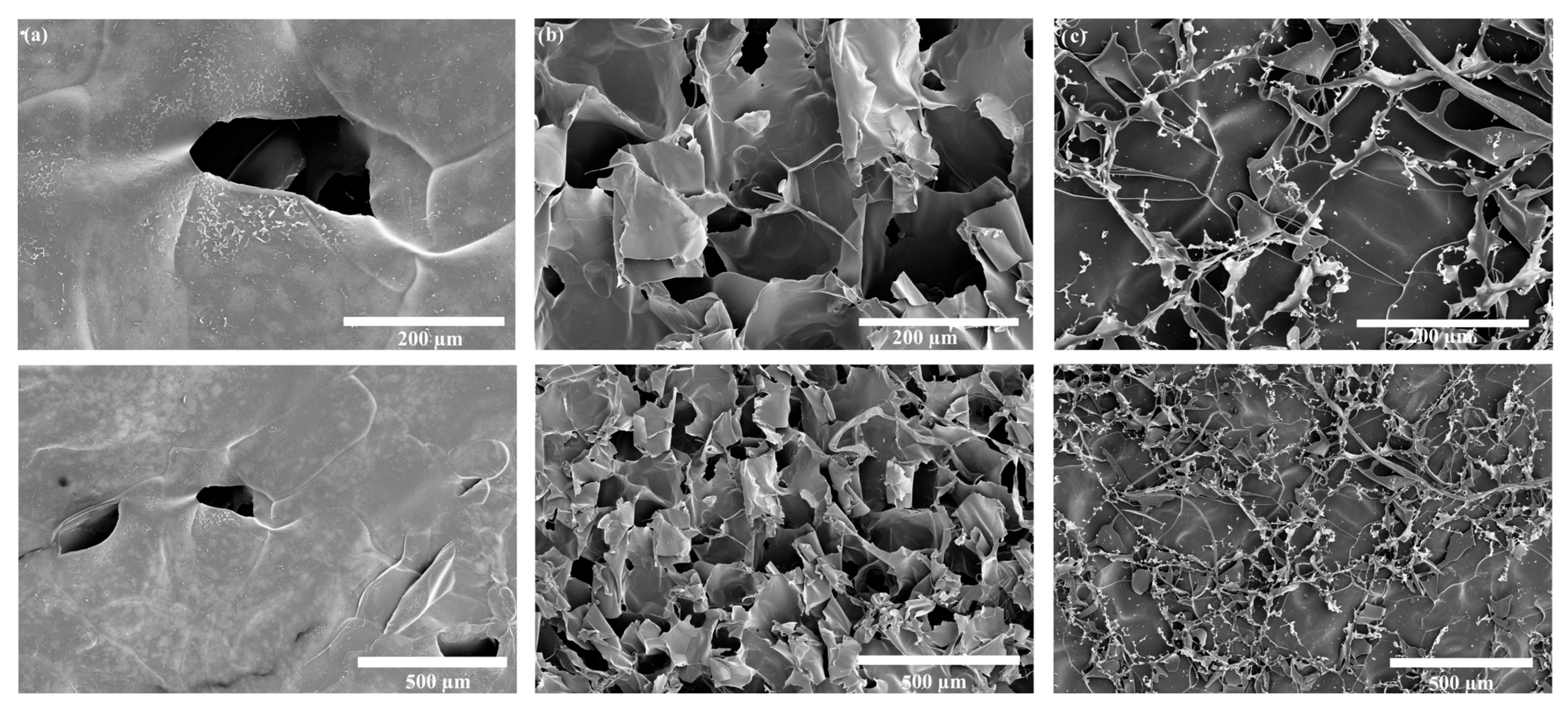
Figure 2 presents the SEM micrographs of the formulated bioinks. The BA bioink (Figure 2a) displays a relatively compact microstructure with low pore density and limited pore interconnectivity. Although some pores are observed, their distribution is sparse, suggesting a restricted porous architecture. This morphology may be associated with the formation of a dense three-dimensional agarose network during gelation, as well as with the agarose concentration and processing conditions used, resulting in a compact structure under the formulation conditions of this study [24,25]. In contrast, incorporation of chitosan in the BAC formulation (Figure 2b) induces marked microstructural rearrangements, yielding a rougher surface and a clearly more porous matrix, where smaller pores in the tens-of-micrometers range coexist with larger macropores approaching a few hundred micrometers, as observed at 200–500 µm scales. This combined micro-/macroporous organization is consistent with porous architecture commonly reported for hydrogel-based wound dressings, where interconnected porosity has been associated with efficient fluid transport and may facilitate nutrient transport, cell migration, and tissue ingrowth [26]. For its part, the BACD bioink (Figure 2c) further evolves toward a highly interconnected porous network with a membrane-like, fibrillar architecture and a more heterogeneous pore-size distribution, characteristics indicative of increased structural complexity and an open framework that may be advantageous for localized drug delivery applications [27]. These morphological differences highlight the strong influence of bioink composition and, more specifically, the inclusion of the chitosan and dexamethasone complex on the resulting multiscale porosity, which is expected to influence the mechanical integrity, degradation behavior, and cell–material interactions of the hydrogels [28]. Overall, the SEM observations reveal a well-developed porous architecture, supporting the potential of the BAC-based formulations, particularly the Dex-functionalized formulation, as bioink candidates for wound healing applications.
Figure 2.
SEM images of the bioinks: (a) BA bioink, (b) BAC bioink, and (c) BACD bioink (scale bars: 200 and 500 µm, as indicated).
3.2. Crystallinity
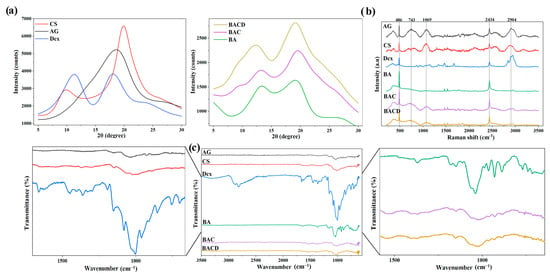
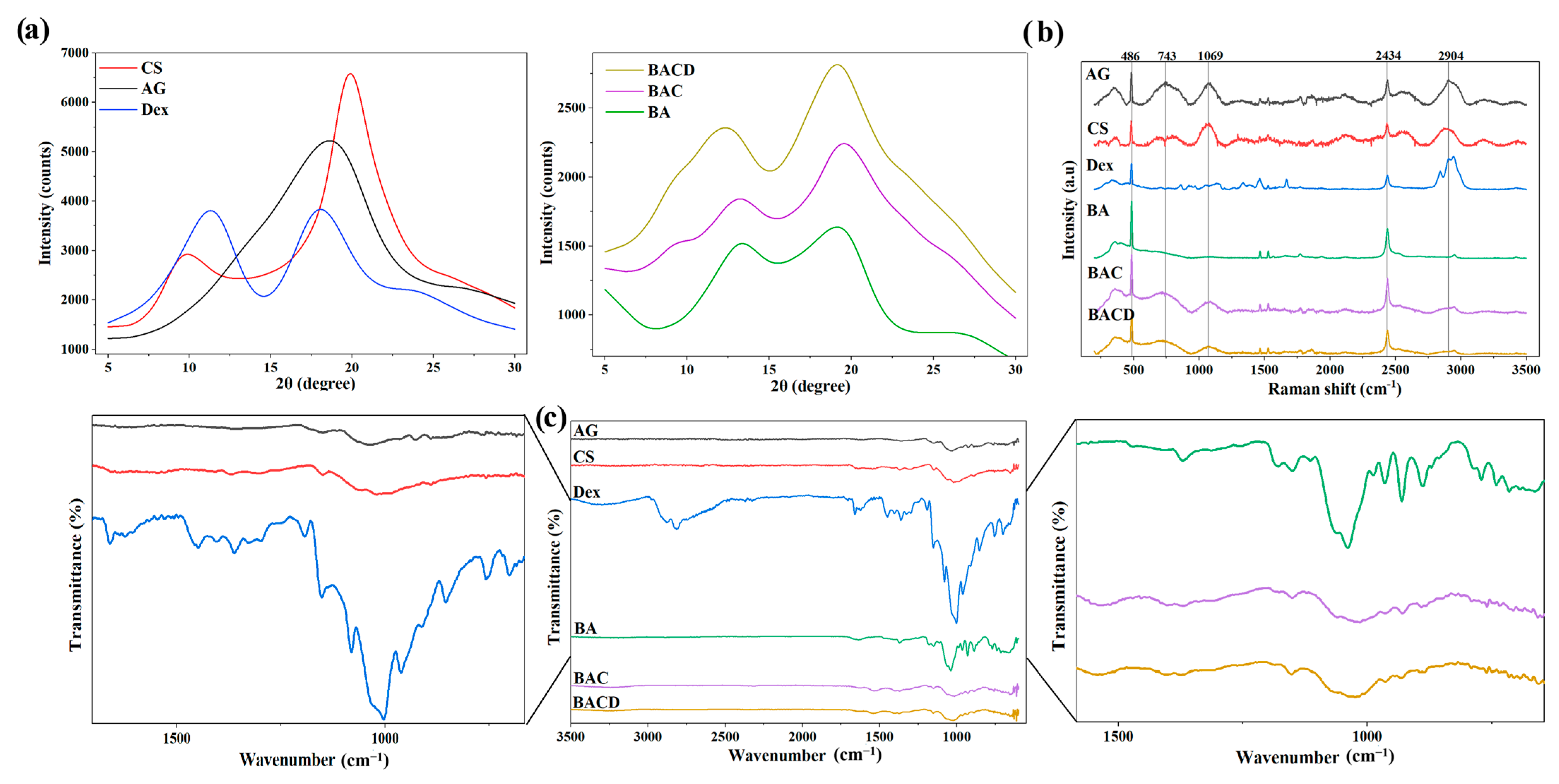
XRD analysis was performed to evaluate the crystalline structure of the precursors and the formulated bioinks (BA, BAC, and BACD). As shown in Figure 3a (left), the XRD pattern of the pure AG precursor exhibits a broad diffraction peak centered at approximately 2θ = 18°, which is characteristic of its semicrystalline structure and is consistent with previous reports in the literature [29]. Pure chitosan displays the characteristic broad reflections at approximately 2θ = 10° and 20°, corresponding to its semicrystalline structure [30], whereas the Dex complex exhibits several sharp diffraction peaks, confirming its crystalline nature [31]. In contrast, the BA bioink (Figure 3a right) presents two broad diffraction peaks in the 2θ regions of approximately 9–13° and 18°, indicating a predominantly amorphous structure compared to pure AG. This behavior can be attributed to the reorganization of polysaccharide chains during the gelation process, which alters the crystalline arrangement of the native polymer [32]. The incorporation of chitosan (BAC) produced slight variations in the intensity and width of these diffraction peaks, suggesting intermolecular interactions between AG and CS that promote partial structural rearrangement [33]. Finally, the BACD bioink exhibits a similar diffractogram to that of BAC, with only minor changes in peak intensity, indicating that the drug was incorporated into the hydrogel matrix without substantially altering its structural organization [31]. Overall, these results indicate that the formulated bioink candidates possess a predominantly amorphous structure, which may be advantageous for biomedical applications, as amorphous matrices can promote swelling and facilitate biological interactions.
Figure 3.
Physicochemical characterization of precursor controls (AG, CS, and Dex) and bioinks (BA, BAC, and BACD): (a) X-ray diffractograms; (b) Raman spectra, normalized and shifted along the Y-axis for clarity; and (c) FTIR spectra, shifted along the Y-axis for clarity.
3.3. Raman Spectra
Figure 3b presents the Raman spectra of the initial solid materials and the formulated bioinks. The AG spectrum exhibits an intense band at 486 cm−1, together with characteristic signals at 743 and 1069 cm−1, which are associated with vibrational modes of the polysaccharide structure (C-O-C ring formation and C-O stretching, respectively). CS displays well-defined bands in the 800–1500 cm−1 region, associated with characteristic vibrations of its polysaccharide backbone. The Dex complex exhibit characteristic bands in the 2800–3000 cm−1 regions, attributed to C-H stretching vibrations of the dexamethasone structure. Additionally, contributions from methyl-β-cyclodextrin, mainly associated with C-O-C- and carbohydrate-related vibrations, may overlap with the observed signals. The Raman spectra of the bioink formulations are also shown. All formulations exhibit characteristic vibrational signals of the polymeric components within the 400–1800 cm−1 region. The BA bioink exhibits spectral features similar to those of the native polymer, indicating that the main structural motifs are preserved after gel formation. For the BAC bioink, an increase in signal intensity and the emergence of bands in the 1000–1200 cm−1 region are observed. This can be attributed to the contribution of CS. Upon incorporation of Dex, subtle modifications appear in the Raman profile, particularly in the high-wavenumber region around 2904 cm−1. This region may reflect contributions from the dexamethasone–methyl-β-cyclodextrin complex as well as from the polymeric components and methyl-β-cyclodextrin [34].
3.4. FTIR Analyses
The FTIR spectrum of agarose (Figure 3c), both in its powder and bioink forms, exhibits a characteristic band at 1046 cm−1 attributed to C–O axial stretching, as well as a band at 1371 cm−1 associated with C–C bending vibrations. Additionally, a peak around 890 cm−1 is observed, corresponding to C–H deformation of the anomeric carbon, and a band at 931 cm−1 is assigned to 3,6-anhydrogalactose [35]. For CS powder, the FTIR spectrum displays a band at 1579 cm−1 corresponding to N–H bending vibrations overlapping with the amide II band, as well as a band at 1649 cm−1 associated with the amide I vibration. The absorption band at 1153 cm−1 is attributed to the antisymmetric stretching of the C–O–C bridge, while the bands at 1089 and 1033 cm−1 are related to skeletal vibrations involving C–O stretching. The spectrum of the BAC bioink reveals the characteristic bands of both polymers. However, a shift in the chitosan band from 1575 cm−1 to 1560 cm−1 (Figure 3c) is observed, suggesting intermolecular interactions between agarose and chitosan. Similar band shifts have been reported in agarose–chitosan systems and have been associated with strengthened hydrogen-bonding interactions between the amino groups of CS and the hydroxyl groups of AG. Furthermore, band broadening in this region supports the presence of polymer–polymer interactions [36,37,38]. In the BACD bioink, most of the characteristic absorption bands are preserved, indicating that the incorporation of Dex does not significantly alter the chemical structure of the polymeric matrix.
3.5. Swelling and In Vitro Degradation
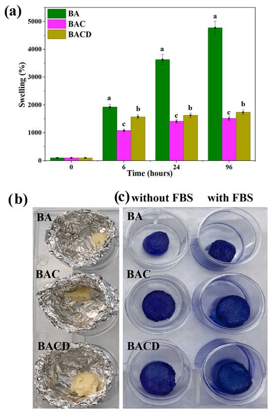
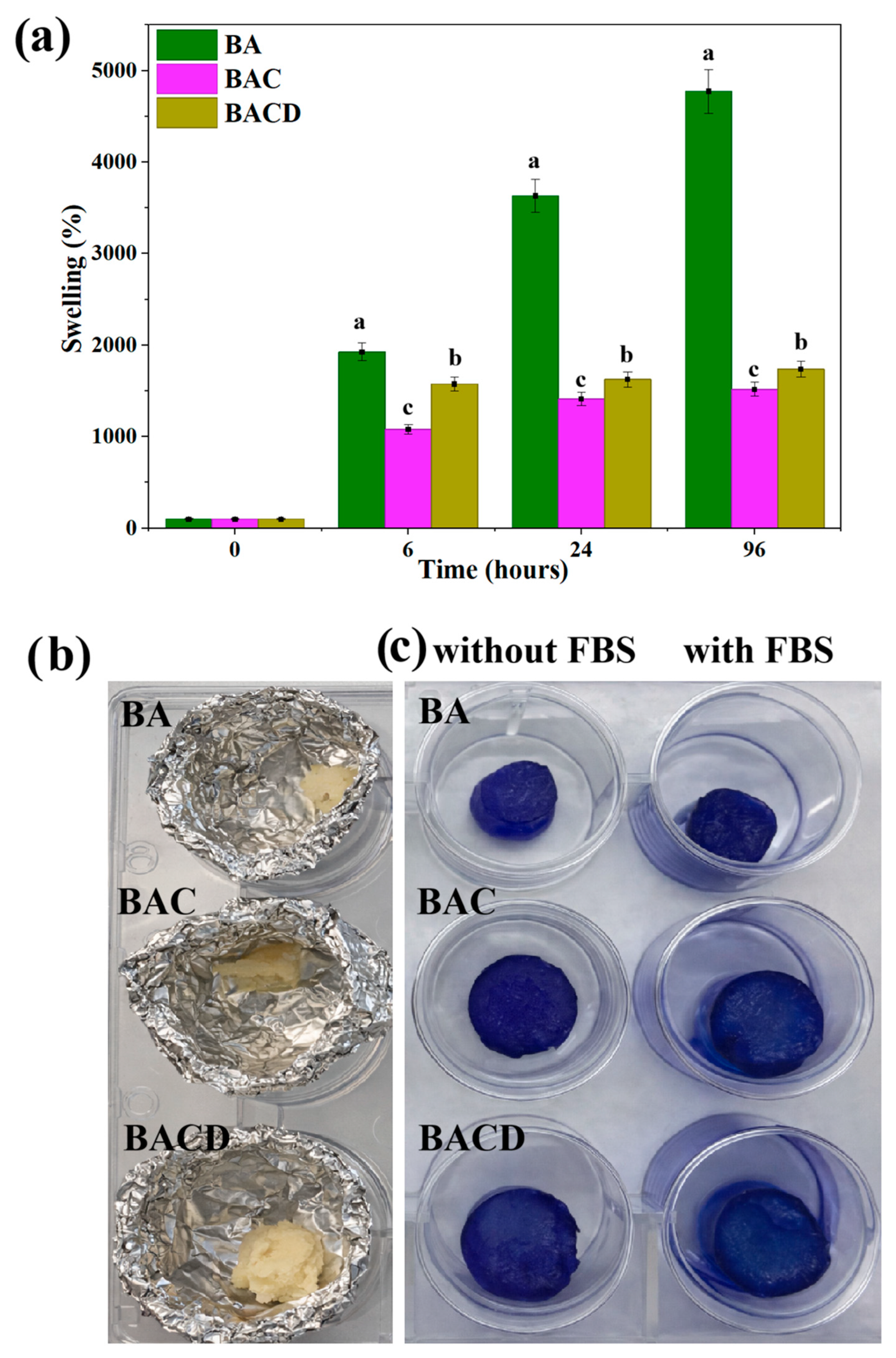
The swelling behavior of the bioink formulations was evaluated in PBS over 96 h. Figure 4a indicates that all developed formulations absorbed substantial amounts of PBS, with swelling ratios ranging from approximately 1500 to 4700%. This high swelling capacity can be attributed to the hydrophilic nature of agarose and chitosan, as well as to the presence of hydroxyl and amino functional groups (–OH and –NH2), which were previously identified by FTIR analysis [39]. To evaluate the effects of formulation and incubation time on swelling behavior, a two-way ANOVA was performed, revealing significant effects of formulation, exposure time, and their interaction (p < 0.0001 for all factors). The formulation accounted for the highest proportion of the total variability (64.76%), followed by time (16.77%) and the formulation × time interaction (18.46%). Since the interaction between factors was significant, Tukey’s multiple comparisons test was performed to identify differences among formulations at each time point. All pairwise comparisons between formulations were statistically significant (adjusted p < 0.0001). Different letters in Figure 4a indicate statistically significant differences among formulations according to Tukey’s multiple comparisons test. BA exhibited significantly higher swelling ratios compared with BAC and BACD at 6, 24, and 96 h, whereas BAC consistently showed the lowest swelling capacity throughout the experiment.
Figure 4.
(a) Swelling percentage of the BA, BAC, and BACD bioinks after incubation in PBS for 6, 24, and 96 h. Data are presented as the mean ± SD (n = 4). Statistical analysis was performed using two-way ANOVA followed by Tukey’s multiple comparisons test. Different lowercase letters indicate statistically significant differences among formulations according to Tukey’s multiple comparisons test. (b) Representative macroscopic images of the bioink samples after the degradation assay and subsequent drying, highlighting the slight shrinkage in shape but the absence of apparent structural disintegration. (c) Protein adsorption on hydrogels. Representative macroscopic images of hydrogels stained with Coomassie Brilliant Blue after incubation with or without FBS.
The BA bioink exhibited the highest swelling value (4775% ± 6.201 at 96 h), which can be associated with its highly hydrophilic and less densely crosslinked network structure. In contrast, the BAC hydrogel exhibited the lowest swelling ratio (1517% ± 2.732 at 96 h). Generally, an increase in crosslinking density leads to a decrease in swelling capacity and an increase in hydrogel rigidity, as the polymer network has a reduced ability to expand during water uptake [40]. Previous studies on agarose/chitosan systems have reported that the incorporation of chitosan promotes the formation of hydrogen bonds between both polysaccharides, reducing the number of exposed hydroxyl groups and modifying the structural organization of the polymer network [41]. In the present study, these interactions may have contributed to the formation of a more compact polymer network, thereby limiting hydrogel expansion during water uptake and resulting in the lower swelling ratio observed for the BAC formulation. The BACD bioink displayed a slightly higher swelling ratio (1739% ± 2.352 at 96 h) compared to BAC. This behavior may be related to the contribution of Dex, which slightly modifies the polymer network’s organization without substantially affecting the mechanical integrity of the structure. This difference suggests greater structural heterogeneity and a more open porous network, consistent with the SEM observations, while maintaining the structural integrity of the polymeric matrix [37].
For its part, Figure 4b shows the degradation behavior of the bioink formulations. Degradation was evaluated by monitoring both structural changes and dry weight variations over 29 days of incubation in PBS. Throughout the study, all scaffolds maintained their overall structural integrity, indicating that degradation was negligible within the evaluated timeframe. The dry weight of the bioinks changed only slightly, with no statistically significant differences among the different formulations, confirming that mass loss was minimal or absent. As illustrated by the macroscopic images, the bioinks exhibit visible shrinkage after drying, but this contraction is mainly associated with water removal rather than actual material degradation. These findings are in good agreement with previous reports on chitosan-based composites, which typically show very limited degradation in PBS or aqueous environments over comparable periods [42,43]. These findings demonstrate the stability of the hydrogels under the evaluated in vitro conditions; however, further studies are required to determine their degradation behavior under in vivo conditions.
3.6. Protein Adsorption Assay
The protein interaction capacity of the bioinks was qualitatively evaluated through protein adsorption assays using a Coomassie Brilliant Blue assay (Figure 4c). After FBS exposure, the BA bioink exhibited positive blue staining compared with the untreated sample, confirming the presence of adsorbed proteins on the AG surface. The BAC bioink also showed positive protein staining following FBS incubation, demonstrating that the incorporation of chitosan preserved the protein adsorption capability of the hydrogel. This behavior is expected because protein adsorption is primarily governed by hydrophobic and electrostatic interactions [44]. Therefore, the presence of CS, which introduces positively charged amino groups, may influence electrostatic interactions at the bioink surface, as well as protein binding capability [45]. Similarly, the BACD bioink exhibited positive protein staining after FBS treatment, suggesting that the incorporation of Dex did not compromise the ability of the hydrogel to adsorb serum proteins. These results demonstrate that all bioink formulations are capable of adsorbing proteins, an essential property for tissue engineering applications because the adsorbed protein layer promotes subsequent cell adhesion and biological performance [46,47]. Taken together, the Coomassie Brilliant Blue staining assays qualitatively confirmed that all bioink formulations were able to promote serum protein adsorption on their surface. In line with these observations, no marked differences in macroscopic staining intensity were observed among BA, BAC and BACD bioinks after FBS exposure, which is consistent with their common polysaccharide-based composition and the relatively modest Dex loading, both of which are expected to support comparable levels of nonspecific protein adsorption. In addition, the interconnected porous architecture of the scaffolds may influence dye penetration and retention by capillarity, as well as the potential trapping of dye molecules within the pore network, making subtle differences in protein uptake difficult to discern visually. Nevertheless, the overall protein-positive staining across all samples indicates that these bioinks provide protein-adsorbing surfaces, a desirable feature for wound healing and tissue engineering applications, where the formation of an adsorbed protein layer is known to facilitate subsequent cell adhesion and modulate biological responses [46,47].
3.7. Release of Dexamethasone
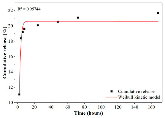
Figure 5 shows the cumulative dexamethasone release profile of the hydrogel over 168 h. For each time point, the mean cumulative release and standard deviation were calculated from the three independent samples, which were then represented as a single averaged release curve. The release behavior was time-dependent, with an initial phase of relatively rapid dexamethasone release during the first few hours of the assay, followed by a slower and more sustained stage that gradually approached a quasi-equilibrium region. At the end of the experiment (168 h), the cumulative amount of dexamethasone released into 10 mL of PBS ranged between approximately 0.62 and 0.67 mg, corresponding to about 18–25% of the theoretical dexamethasone content in the hydrogels. This calculation was based on the theoretical dexamethasone content provided by the water-soluble dexamethasone–methyl-β-cyclodextrin complex, which contains 6.5 wt.% dexamethasone. However, the actual dexamethasone loading in the final hydrogel formulations was not experimentally determined. Therefore, the non-released fraction cannot be directly attributed to dexamethasone retention within the hydrogel matrix. The incomplete release may be related to possible losses of dexamethasone during the fabrication process, incomplete incorporation of the dexamethasone-containing complex, and local heterogeneities in drug distribution within the hydrogel network [48,49]. Nevertheless, the SEM, Raman, and FTIR results provided complementary evidence supporting the presence of the dexamethasone–methyl-β-cyclodextrin complex within the bioink matrix.
Figure 5.
Cumulative release profile of dexamethasone from BAC-based hydrogels in PBS over 168 h, obtained by HPLC. Each point represents the mean cumulative release from three independent samples (n = 3) ± SD. The solid line corresponds to the fit obtained with the Weibull kinetic model, illustrating the initial burst followed by a sustained release phase that gradually approaches a plateau.
Although a relatively rapid initial burst was observed, dexamethasone release continued progressively over the entire 168 h, yielding an overall gradual release profile. This behavior may be associated with the diffusion of dexamethasone through the hydrated polymer network and its interactions with the components of the agarose–chitosan matrix [48,49]. Since the actual dexamethasone loading was not experimentally determined, this release profile should be interpreted as descriptive of the release behavior rather than as evidence of a specific release mechanism.
To gain further insight into the release mechanism and the factors controlling the dexamethasone release rate, the averaged experimental data were fitted to several kinetic models, including zero-order, first-order, Higuchi, Hixson–Crowell, Korsmeyer–Peppas, and Weibull models, as well as a hyperbolic Michaelis–Menten model [50]. Diffusion-based models such as the Higuchi and Korsmeyer–Peppas models provided only limited agreement with the experimental curves, suggesting that dexamethasone release was not governed exclusively by a simple diffusional process [51]. The hyperbolic Michaelis–Menten model yielded a better fit than the conventional linear models, capturing the initial rapid release followed by a progressive decrease in the release rate, but the best overall description was obtained with the Weibull model, with determination coefficients (R2) in the range 0.945–0.968. In this framework, the cumulative fraction of dexamethasone released, F, as a function of time, t, is described by
where a is a characteristic time parameter of the release process and b (2.39) is the shape parameter that describes the profile of the release curve. A b value greater than one is commonly associated with a release profile characterized by an initial acceleration followed by a decrease in the release rate [52]. The good agreement with the Weibull model indicates that this empirical model adequately describes the dexamethasone release profile of the lyophilized BAC hydrogels. The observed release behavior may be associated with a complex, multifactorial process involving contributions from diffusion, structural relaxation of the polysaccharide network, and interactions between dexamethasone and the hydrogel matrix, as previously reported for similar hydrogel-based delivery systems [53,54]. This release behavior may be associated with the structural characteristics of the BAC hydrogel network, where the hydrated polysaccharide matrix and porous architecture may regulate dexamethasone diffusion and contribute to the sustained release profile observed over time. The lower swelling capacity observed for BAC suggests a more compact polymer network, which may limit water uptake and consequently reduce dexamethasone mobility within the hydrogel matrix. Therefore, the swelling behavior and structural organization of the agarose/chitosan network may contribute to the release kinetics described by the Weibull model.
3.8. Rheology Analyses
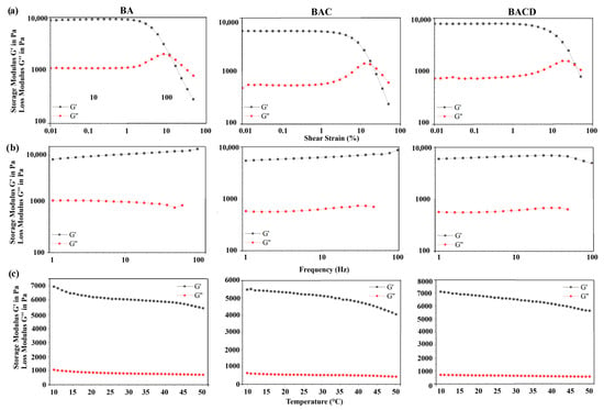
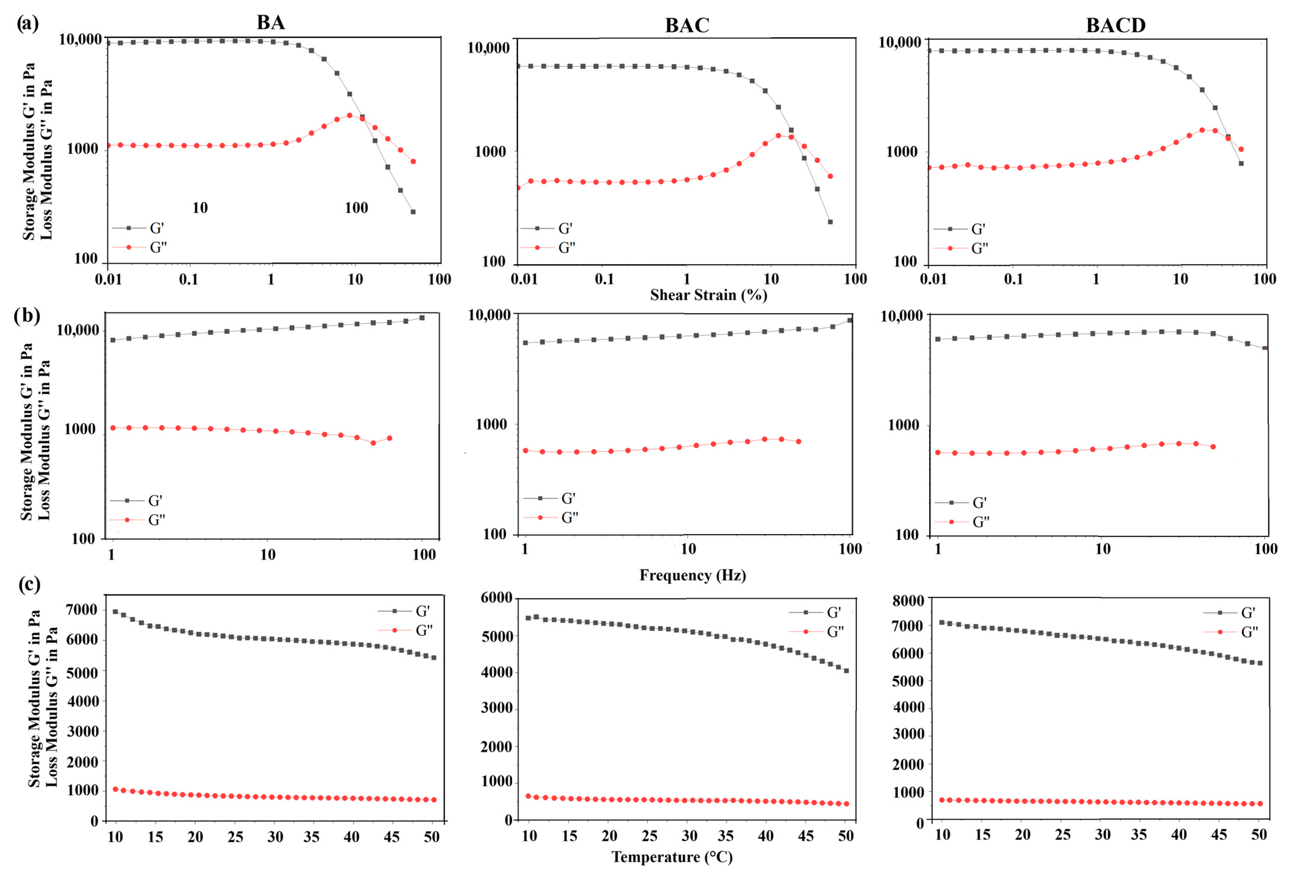
Rheological analysis of the bioinks developed revealed the typical viscoelastic behavior of gel-like materials. Figure 6a presents the amplitude sweep results, showing a well-defined linear viscoelastic region (LVR) for all formulations, characterized by the predominance of the storage modulus (G′) over the loss modulus (G″). The BA bioink exhibited the highest G′ values, indicating a more rigid and structurally organized network. The incorporation of CS reduced the rigidity of the bioinks compared with the agarose formulation, whereas the subsequent incorporation of Dex did not produce significant changes in the viscoelastic response. These observations may be associated with modifications in intermolecular interactions and a decrease in physical crosslinking density. All three bioinks displayed predominantly elastic behavior up to strain values close to 10%, beyond which crossover between G′ and G″ was observed, indicating structural breakdown.
Figure 6.
Rheological properties of BA, BAC and BACD: (a) G′ and G″ as a function of shear strain; (b) G′ and G″ as a function of frequency; and (c) G′ and G″ as a function of temperature variation over time.
Figure 6b illustrates the frequency dependence of the storage and loss moduli. A strain amplitude of 0.5% was selected within the LVR, where the moduli remain independent of deformation. G′ represents the elastic contribution of the material and showed minimal variation with frequency across all formulations, confirming the formation of stable polymeric networks. The highest G′ values were again observed for the agarose-based bioink, reflecting its greater structural rigidity compared to the chitosan-containing systems. The frequency dependence of the loss modulus (G″) exhibited moderate differences among formulations, suggesting that chitosan incorporation altered the relaxation dynamics of the polymer network. In contrast, Dex incorporation did not produce significant changes in the viscoelastic response. Additionally, the complex’s viscosity decreased progressively with increasing frequency, displaying a linear trend on a logarithmic scale, which confirms the pseudoplastic (shear-thinning) behavior of the system [55]. Finally, Figure 6c presents the temperature sweep analysis. A gradual decrease in G’ was observed with increasing temperature, which is attributable to the weakening of intermolecular interactions. Nevertheless, G′ remained higher than G″ throughout the evaluated temperature range (10–50 °C), indicating that the bioinks retain their structural integrity at physiological temperatures. This thermal stability is essential for potential tissue engineering applications [20,56].
3.9. Assessment of Antibacterial Activity
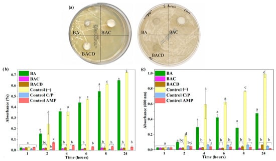
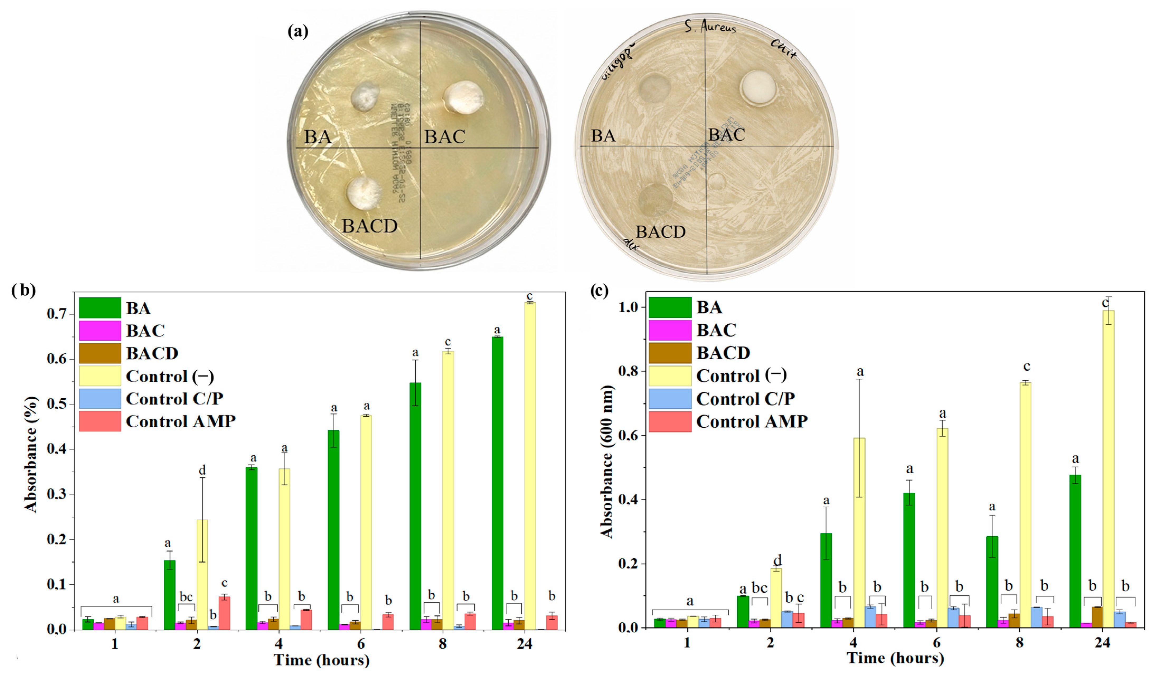
The antibacterial activity of the bioinks was evaluated against Escherichia coli and Staphylococcus aureus. The BA, BAC, and BACD formulations were compared with a negative control and commercial antibiotics (ciprofloxacin and ampicillin). In the agar disk diffusion assay (Figure 7a), only very small inhibition halos were observed around the BAC and BACD samples, indicating a weak but detectable antibacterial effect in this solid-medium test. As expected, the BA bioink behaved similarly to the negative control and did not generate inhibition zones, confirming the absence of intrinsic antibacterial activity.
Figure 7.
(a) Evaluation of antibacterial activity by agar disk diffusion, showing the inhibition halos formed around the bioink samples and antibiotic controls. (b) Bacterial growth curves (OD600) of E. coli and (c) S. aureus in liquid culture in the presence of the different bioinks, compared with negative and antibiotic controls. Data are presented as the mean ± SD (n = 2). Different lowercase letters indicate statistically significant differences among formulations within each time point according to Tukey’s multiple comparisons test following two-way ANOVA (α = 0.05). Groups sharing at least one letter are not significantly different (p > 0.05).
In contrast, the bacterial growth curves in liquid culture (Figure 7b,c) revealed a more pronounced antibacterial response for the chitosan-containing bioinks. Both BAC and BACD produced a marked reduction in optical density compared with the negative control, suggesting an inhibitory effect of chitosan on bacterial proliferation in the suspension. This antibacterial effect is commonly attributed to electrostatic interactions between the protonated amino groups (–NH3+) of chitosan and the negatively charged microbial cell envelope, which may alter membrane permeability and affect bacterial growth [57,58]. Two-way ANOVA revealed significant effects of formulation, time, and their interaction for both E. coli and S. aureus (p < 0.0001 for all effects), indicating that bacterial growth varied significantly according to the formulation and incubation time. Tukey’s multiple comparisons revealed significant differences among formulations at several time points; however, no significant differences were observed between BAC and BACD at any evaluated time point for either bacterial strain (p > 0.05). Notably, BAC and BACD showed stronger inhibitory effects against S. aureus than against E. coli, indicating a more pronounced antibacterial response against Gram-positive bacteria. This difference is consistent with the structural features of bacterial cell envelopes. Indeed, Gram-positive bacteria possess a thick peptidoglycan layer rich in teichoic and lipoteichoic acids and lack an outer membrane, which facilitates direct interaction between chitosan and the cell surface. By contrast, Gram-negative bacteria such as E. coli have an outer membrane containing lipopolysaccharides (LPSs) that acts as an additional permeability barrier, limiting the access and effectiveness of chitosan [37,59]. Overall, these findings demonstrate that the chitosan-containing formulations exhibited measurable antibacterial activity in liquid culture, while the incorporation of the dexamethasone–methyl-β-cyclodextrin complex did not significantly alter the antibacterial response of BAC under the conditions tested.
Thus, according to these findings, the incorporation of chitosan imparts the bioinks with measurable antibacterial properties, characterized by reduced bacterial growth in liquid culture and a limited inhibitory effect in disk diffusion assays, particularly against Gram-positive microorganisms, thereby highlighting their potential for biomedical applications in infection-prone environments.
4. Conclusions
In this study, three bioinks were formulated based on agarose (BA), agarose/chitosan (BAC), and the agarose/chitosan/dexamethasone complex (BACD) at different proportions. The physicochemical and biological properties of the composite bioinks were systematically characterized. SEM, FTIR and Raman spectroscopies and XRD analyses confirmed the formation of polymeric networks with predominantly amorphous characteristics and demonstrated intermolecular interactions between agarose and chitosan. The incorporation of Dex did not significantly alter the chemical structure of the matrix, indicating its successful integration without compromising the integrity of the polymer network. Rheological analysis revealed that all formulations exhibited typical viscoelastic gel behavior, with a well-defined linear viscoelastic region and a dominant elastic response (G′ > G″). In the BACD system, a slight reduction in network stiffness was observed; however, structural stability was maintained across the evaluated frequency and temperature ranges, including physiological conditions. These rheological features suggest that the developed formulations may be considered potential bioink candidates for future 3D bioprinting applications, although further studies addressing printability are required. For their part, swelling and degradation studies demonstrated that bioink composition directly influenced water absorption capacity and structural stability. Agarose-based formulations exhibited high swelling ratios; however, the incorporation of chitosan enhanced network stability. Finally, antibacterial activity assays indicated that chitosan-containing bioinks exhibited pronounced antibacterial effects, particularly against Gram-positive bacteria. These findings demonstrate that chitosan is the main contributor to the antibacterial properties of the bioinks, while dexamethasone incorporation does not significantly modify this response. Overall, the incorporation of Dex into the agarose–chitosan matrix was compatible with the physicochemical, rheological, and biological properties of the bioinks, supporting their potential application in wound healing and related biomedical fields.
Author Contributions
C.Z.-L.: conceptualization, methodology, supervision, project administration, and funding acquisition; D.S.-O.: investigation; J.J.L.: investigation and original draft preparation; J.G.-M.: methodology, resources, data curation, and visualization; J.P.F.-B.: methodology and resources; M.B.-G.: methodology and resources; J.R.P.: methodology and resources; M.R.B.: methodology and resources; C.L.-A.: investigation and visualization; J.M.M.-H.: methodology and resources; D.N.-B.: formal analysis, original draft preparation, and visualization; V.H.G.: formal analysis, visualization, and original draft preparation. All the coauthors: writing—review and editing. All authors have read and agreed to the published version of the manuscript.
Funding
This research received financial support from Project BIOCONTROL, which was coordinated by OVIS INVESTIGACIONES, AIE, together with its partner organizations, within the framework of the “Financiación estructurada de proyectos de I + D por Agrupaciones de Interés Económico (A.I.E.)” and in collaboration with INVENTIUM. Additional funding was provided, in part, by the Era-Min Cofund 2023 initiative COOL&SMARTTIT, MCIU/AEI/10.13039/501100011033/UE.
Institutional Review Board Statement
Not applicable.
Informed Consent Statement
Not applicable.
Data Availability Statement
The data supporting this study’s findings are available from the corresponding authors upon reasonable request.
Conflicts of Interest
The authors declare that this study received funding from Project BIOCONTROL, coordinated by OVIS INVESTIGACIONES, AIE, together with its partner organizations, within the framework of the “Finan-ciación estructurada de proyectos de I+D por Agrupaciones de Interés Económico (A.I.E.)” and in collaboration with INVENTIUM. Additional funding was provided, in part, by the Era-Min Co-fund 2023 initiative COOL&SMARTTIT, MCIU/AEI/10.13039/501100011033/UE. The funder was not involved in the study design, collection, analysis, interpretation of data, the writing of this article or the decision to submit it for publication.
References
- Elango, J.; Zamora-Ledezma, C.; Maté-Sánchez de Val, J. Natural vs Synthetic Polymers: How Do They Communicate with Cells for Skin Regeneration—A Review. J. Compos. Sci. 2023, 7, 385. [Google Scholar] [CrossRef] [Scilit]
- Zamora-Mendoza, L.; Valdiviezo-Cuenca, Y.J.; Agurto-Rodríguez, G.; Guillén, P.O.; López, F.; Guanoliquin, L.V.; Zambrano, C.; Zamora-Ledezma, C.; Rodríguez, J.; Gómez-Guerra, G.; et al. Ascidians as a Sustainable Source of Cellulose: Physicochemical Characterization, Degradability, and Relevance for Bioplastic Applications. Biopolymers 2026, 117, e70099. [Google Scholar] [CrossRef] [Scilit] [PubMed]
- Mania, S.; Banach-Kopeć, A.; Maciejewska, N.; Czerwiec, K.; Słonimska, P.; Deptuła, M.; Baczyński-Keller, J.; Pikuła, M.; Sachadyn, P.; Tylingo, R. From Bioink to Tissue: Exploring Chitosan-Agarose Composite in the Context of Printability and Cellular Behaviour. Molecules 2024, 29, 4648. [Google Scholar] [CrossRef] [Scilit] [PubMed]
- Das, A.; Ringu, T.; Ghosh, S.; Pramanik, N. A Comprehensive Review on Recent Advances in Preparation, Physicochemical Characterization, and Bioengineering Applications of Biopolymers. Polym. Bull. 2023, 80, 7247–7312. [Google Scholar] [CrossRef] [Scilit] [PubMed]
- Jiang, F.; Xu, X.-W.; Chen, F.-Q.; Weng, H.-F.; Chen, J.; Ru, Y.; Xiao, Q.; Xiao, A.-F. Extraction, Modification and Biomedical Application of Agarose Hydrogels: A Review. Mar. Drugs 2023, 21, 299. [Google Scholar] [CrossRef] [Scilit] [PubMed]
- Saeedi Garakani, S.; Khanmohammadi, M.; Atoufi, Z.; Kamrava, S.K.; Setayeshmehr, M.; Alizadeh, R.; Faghihi, F.; Bagher, Z.; Davachi, S.M.; Abbaspourrad, A. Fabrication of Chitosan/Agarose Scaffolds Containing Extracellular Matrix for Tissue Engineering Applications. Int. J. Biol. Macromol. 2020, 143, 533–545. [Google Scholar] [CrossRef] [Scilit] [PubMed]
- Biswal, T. Biopolymers for Tissue Engineering Applications: A Review. Mater. Today Proc. 2021, 41, 397–402. [Google Scholar] [CrossRef] [Scilit]
- Gungor-Ozkerim, P.S.; Inci, I.; Zhang, Y.S.; Khademhosseini, A.; Dokmeci, M.R. Bioinks for 3D Bioprinting: An Overview. Biomater. Sci. 2018, 6, 915–946. [Google Scholar] [CrossRef] [Scilit] [PubMed]
- Noh, J.H.; Lee, Y.; Kim, M.S. 3D-Printed Scaffolds: Incorporating Dexamethasone Microspheres and BMP2 for Enhanced Osteogenic Differentiation of Human Mesenchymal Stem Cells. Colloids Surf. B Biointerfaces 2025, 253, 114705. [Google Scholar] [CrossRef] [Scilit] [PubMed]
- Neelon, J.; Yau, I.; Carlsson, A.H.; Smithson, S.B.; Varon, D.E.; Chan, C.K.; Chan, R.K.; Nuutila, K. Topical Application of Anti-Inflammatory Agents on Burn Wounds and Their Effect on Healing. Burns 2024, 50, 107290. [Google Scholar] [CrossRef] [Scilit] [PubMed]
- Tang, Q.; Chen, Y.; Liu, M.; Ning, X.; Chen, Q.; He, S. Enhancing the Therapeutic Efficacy of Dexamethasone for Oral Ulcers through Adhesive Gelatin Hydrogel-Based Nano-Delivery System. Mater. Today Bio 2026, 39, 103341. [Google Scholar] [CrossRef] [Scilit] [PubMed]
- Tu, H.; Zhang, D.; Barksdale, A.N.; Wadman, M.C.; Muelleman, R.L.; Li, Y.L. Dexamethasone Improves Wound Healing by Decreased Inflammation and Increased Vasculogenesis in Mouse Skin Frostbite Model. Wilderness Environ. Med. 2020, 31, 407–417. [Google Scholar] [CrossRef] [Scilit] [PubMed]
- Chen, Y.; Chen, X.; Zhou, Q. Different Effects of a Perioperative Single Dose of Dexamethasone on Wound Healing in Mice with or without Sepsis. Front. Surg. 2023, 10, 927168. [Google Scholar] [CrossRef] [Scilit] [PubMed]
- Yang, F.; Lu, D.; Chen, Y.; Qi, F.; Wang, X.; Li, J.; Fu, Q.; Li, R.; Wu, D.; Wang, J.; et al. Bioadhesive Supramolecular Polymer/Hyaluronic Acid Hydrogel with Sustained Release of Zinc Ions and Dexamethasone for Diabetic Wound Healing. Int. J. Biol. Macromol. 2025, 286, 137752. [Google Scholar] [CrossRef] [Scilit] [PubMed]
- Xu, W.; Huang, W.; Cai, X.; Dang, Z.; Hao, L.; Wang, L. Dexamethasone Long-Term Controlled Release from Injectable Dual-Network Hydrogels with Porous Microspheres Immunomodulation Promotes Bone Regeneration. ACS Appl. Mater. Interfaces 2024, 16, 40581–40601. [Google Scholar] [CrossRef] [Scilit] [PubMed]
- Ghobadi, F.; Kalantarzadeh, R.; Ashrafnia Menarbazari, A.; Salehi, G.; Fatahi, Y.; Simorgh, S.; Orive, G.; Dolatshahi-Pirouz, A.; Gholipourmalekabadi, M. Innovating Chitosan-Based Bioinks for Dermal Wound Healing: Current Progress and Future Prospects. Int. J. Biol. Macromol. 2025, 298, 140013. [Google Scholar] [CrossRef] [Scilit] [PubMed]
- Choi, B.; Kim, S.; Lin, B.; Wu, B.M.; Lee, M. Cartilaginous Extracellular Matrix-Modified Chitosan Hydrogels for Cartilage Tissue Engineering. ACS Appl. Mater. Interfaces 2014, 6, 20110–20121. [Google Scholar] [CrossRef] [Scilit] [PubMed]
- Madhumathi, K.; Sudheesh Kumar, P.T.; Abhilash, S.; Sreeja, V.; Tamura, H.; Manzoor, K.; Nair, S.V.; Jayakumar, R. Development of Novel Chitin/Nanosilver Composite Scaffolds for Wound Dressing Applications. J. Mater. Sci. Mater. Med. 2010, 21, 807–813. [Google Scholar] [CrossRef] [Scilit] [PubMed]
- Elango, J.; Zamora-Ledezma, C. Rheological, Structural, and Biological Trade-Offs in Bioink Design for 3D Bioprinting. Gels 2025, 11, 659. [Google Scholar] [CrossRef] [Scilit] [PubMed]
- Banach-Kopeć, A.; Mania, S.; Tylingo, R.; Wawrzynowicz, A.; Pawłowska, M.; Czerwiec, K.; Deptuła, M.; Pikuła, M. Thermosensitive Composite Based on Agarose and Chitosan Saturated with Carbon Dioxide. Preliminary Study of Requirements for Production of New CSAG Bioink. Carbohydr. Polym. 2024, 336, 122120. [Google Scholar] [CrossRef] [Scilit] [PubMed]
- Smandri, A.; Nordin, A.; Hwei, N.M.; Chin, K.-Y.Y.; Abd Aziz, I.; Fauzi, M.B. Natural 3D-Printed Bioinks for Skin Regeneration and Wound Healing: A Systematic Review. Polymers 2020, 12, 1782. [Google Scholar] [CrossRef] [Scilit] [PubMed]
- Miguel, S.P.; Ribeiro, M.P.; Brancal, H.; Coutinho, P.; Correia, I.J. Thermoresponsive Chitosan-Agarose Hydrogel for Skin Regeneration. Carbohydr. Polym. 2014, 111, 366–373. [Google Scholar] [CrossRef] [Scilit] [PubMed]
- Giacopello, C.; Foti, M.; Passantino, A.; Fisichella, V.; Aleo, A.; Mammina, C. Serotypes and Antibiotic Susceptibility Patterns of Salmonella Spp. Isolates from Spur-Thighed Tortoise, Testudo Graeca Illegally Introduced in Italy. Hum. Vet. Med. 2012, 4, 76–81. [Google Scholar]
- Roach, B.L.; Nover, A.B.; Ateshian, G.A.; Hung, C.T. Agarose Hydrogel Characterization for Regenerative Medicine Applications: Focus on Engineering Cartilage. In Biomaterials from Nature for Advanced Devices and Therapies; Wiley: Hoboken, NJ, USA, 2016; pp. 258–273. [Google Scholar] [CrossRef] [Scilit]
- Jayawardena, I.; Turunen, P.; Garms, B.C.; Rowan, A.; Corrie, S.; Grøndahl, L. Evaluation of Techniques Used for Visualisation of Hydrogel Morphology and Determination of Pore Size Distributions. Mater. Adv. 2023, 4, 669–682. [Google Scholar] [CrossRef] [Scilit]
- Zhang, W.; Liu, L.; Cheng, H.; Zhu, J.; Li, X.; Ye, S.; Li, X. Hydrogel-Based Dressings Designed to Facilitate Wound Healing. Mater. Adv. 2024, 5, 1364–1394. [Google Scholar] [CrossRef] [Scilit]
- Adhikari, J.; Perwez, M.S.; Das, A.; Saha, P. Development of Hydroxyapatite Reinforced Alginate–Chitosan Based Printable Biomaterial-Ink. Nano-Struct. Nano-Objects 2021, 25, 100630. [Google Scholar] [CrossRef] [Scilit]
- Elango, J.; Zamora-Ledezma, C.; Alexis, F.; Wu, W.; Maté-Sánchez de Val, J.E. Protein Adsorption, Calcium-Binding Ability, and Biocompatibility of Silver Nanoparticle-Loaded Polyvinyl Alcohol (PVA) Hydrogels Using Bone Marrow-Derived Mesenchymal Stem Cells. Pharmaceutics 2023, 15, 1843. [Google Scholar] [CrossRef] [Scilit] [PubMed]
- Hu, Y.; Kim, Y.; Hong, I.; Kim, M.; Jung, S. Fabrication of Flexible PH-Responsive Agarose/Succinoglycan Hydrogels for Controlled Drug Release. Polymers 2021, 13, 2049. [Google Scholar] [CrossRef] [Scilit] [PubMed]
- Cicek Ozkan, B. Cellulose and Chitosan Biopolymer Composites Reinforced with Graphene and Their Adsorption Properties for Basic Blue 41. Cellulose 2022, 29, 9637–9655. [Google Scholar] [CrossRef] [Scilit]
- Miño, K.; Pérez, A.; Iza-García, T.X.; Cabascango, D.G.; Gordillo, C.; Gordillo, A.; Vispo, N.; Machado, A.; Zamora-Ledezma, C.; Alexis, F. Antimicrobial Cellulose Hydrogels against Gram-Positive and Gram-Negative Bacteria. Front. Soft Matter 2024, 4, 1362677. [Google Scholar] [CrossRef] [Scilit]
- Su, T.; Zhang, M.; Zeng, Q.; Pan, W.; Huang, Y.; Qian, Y.; Dong, W.; Qi, X.; Shen, J. Mussel-Inspired Agarose Hydrogel Scaffolds for Skin Tissue Engineering. Bioact. Mater. 2021, 6, 579–588. [Google Scholar] [CrossRef] [Scilit] [PubMed]
- Heydarifard, S.; Gao, W.; Fatehi, P. Impact of Counter Ions of Cationic Monomers on the Production and Characteristics of Chitosan-Based Hydrogel. ACS Omega 2019, 4, 15087–15096. [Google Scholar] [CrossRef] [Scilit] [PubMed]
- Esquivias, L.; Rivero-Antúnez, P.; Zamora-Ledezma, C.; Domínguez-Rodríguez, A.; Morales-Flórez, V. Intragranular Carbon Nanotubes in Alumina-Based Composites for Reinforced Ceramics. J. Sol-Gel Sci. Technol. 2019, 90, 162–171. [Google Scholar] [CrossRef] [Scilit]
- Jarosz, A.; Kapusta, O.; Gugała-Fekner, D.; Barczak, M. Synthesis and Characterization of Agarose Hydrogels for Release of Diclofenac Sodium. Materials 2023, 16, 6042. [Google Scholar] [CrossRef] [Scilit] [PubMed]
- Zamora-Mora, V.; Velasco, D.; Hernández, R.; Mijangos, C.; Kumacheva, E. Chitosan/Agarose Hydrogels: Cooperative Properties and Microfluidic Preparation. Carbohydr. Polym. 2014, 111, 348–355. [Google Scholar] [CrossRef] [Scilit] [PubMed]
- Hu, Z.; Hong, P.; Liao, M.; Kong, S.; Huang, N.; Ou, C.; Li, S. Preparation and Characterization of Chitosan-Agarose Composite Films. Materials 2016, 9, 816. [Google Scholar] [CrossRef] [Scilit] [PubMed]
- El-Hefian, E.A.; Nasef, M.M.; Yahaya, A.H. Preparation and Characterization of Chitosan/Agar Blended Films: Part 1. Chemical Structure and Morphology. E-J. Chem. 2012, 9, 1431–1439. [Google Scholar] [CrossRef] [Scilit]
- Felfel, R.M.; Gideon-Adeniyi, M.J.; Zakir Hossain, K.M.; Roberts, G.A.F.; Grant, D.M. Structural, Mechanical and Swelling Characteristics of 3D Scaffolds from Chitosan-Agarose Blends. Carbohydr. Polym. 2019, 204, 59–67. [Google Scholar] [CrossRef] [Scilit] [PubMed]
- Córdoba, E.A.; Agudelo, N.A.; Giraldo, L.F.; Echeverri-Cuartas, C.E. Exploring the Potential of Carboxymethyl Chitosan and Oxidized Agarose to Form Self-Healing Injectable Hydrogels. Polysaccharides 2025, 6, 49. [Google Scholar] [CrossRef] [Scilit]
- Cao, Q.; Zhang, Y.; Chen, W.; Meng, X.; Liu, B. Hydrophobicity and Physicochemical Properties of Agarose Film as Affected by Chitosan Addition. Int. J. Biol. Macromol. 2018, 106, 1307–1313. [Google Scholar] [CrossRef] [Scilit] [PubMed]
- Sadeghianmaryan, A.; Naghieh, S.; Yazdanpanah, Z.; Alizadeh Sardroud, H.; Sharma, N.K.; Wilson, L.D.; Chen, X. Fabrication of Chitosan/Alginate/Hydroxyapatite Hybrid Scaffolds Using 3D Printing and Impregnating Techniques for Potential Cartilage Regeneration. Int. J. Biol. Macromol. 2022, 204, 62–75. [Google Scholar] [CrossRef] [Scilit] [PubMed]
- Kumar, N.; Desagani, D.; Chandran, G.; Ghosh, N.N.; Karthikeyan, G.; Waigaonkar, S.; Ganguly, A. Biocompatible Agarose-Chitosan Coated Silver Nanoparticle Composite for Soft Tissue Engineering Applications. Artif. Cells Nanomed. Biotechnol. 2018, 46, 637–649. [Google Scholar] [CrossRef] [Scilit] [PubMed]
- Ma, D.; Zheng, Z.; Qian, S.; Chen, W.; Bian, D.; Ni, Z. Study on the Protein Adsorption and Its Properties of Self-Assembled Chitosan Coating. Friction 2025, 13, 9441074. [Google Scholar] [CrossRef] [Scilit]
- Arias-Andrade, Y.V.; Veloza, L.A.; Sepúlveda-Arias, J.C. Nanocompuestos de Quitosano Aplicados Al Campo de La Medicina Regenerativa. Una Revisión Sistemática. Sci. Tech. 2020, 25, 604–615. [Google Scholar] [CrossRef] [Scilit]
- Ghasemi-Mobarakeh, L.; Kolahreez, D.; Ramakrishna, S.; Williams, D. Key Terminology in Biomaterials and Biocompatibility. Curr. Opin. Biomed. Eng. 2019, 10, 45–50. [Google Scholar] [CrossRef] [Scilit]
- Jayakumar, R.; Menon, D.; Manzoor, K.; Nair, S.V.; Tamura, H. Biomedical Applications of Chitin and Chitosan Based Nanomaterials—A Short Review. Carbohydr. Polym. 2010, 82, 227–232. [Google Scholar] [CrossRef] [Scilit]
- Thang, N.H.; Chien, T.B.; Cuong, D.X. Polymer-Based Hydrogels Applied in Drug Delivery: An Overview. Gels 2023, 9, 523. [Google Scholar] [CrossRef] [Scilit] [PubMed]
- Costa, P.; Sousa Lobo, J.M. Modeling and Comparison of Dissolution Profiles. Eur. J. Pharm. Sci. 2001, 13, 123–133. [Google Scholar] [CrossRef] [Scilit] [PubMed]
- Long, J.; Nand, A.V.; Bunt, C.; Seyfoddin, A. Controlled Release of Dexamethasone from Poly(Vinyl Alcohol) Hydrogel. Pharm. Dev. Technol. 2019, 24, 839–848. [Google Scholar] [CrossRef] [Scilit] [PubMed]
- Zamora-Ledezma, C.; Hernández, A.B.; López-González, I.; Elango, J.; Paindépice, J.; Alexis, F.; González-Sánchez, M.; Morales-Flórez, V.; Mowbray, D.J.; Meseguer-Olmo, L. Fabrication, Physical–Chemical and Biological Characterization of Retinol-Loaded Poly(Vinyl Alcohol) Electrospun Fiber Mats for Wound Healing Applications. Polymers 2023, 15, 2705. [Google Scholar] [CrossRef] [Scilit] [PubMed]
- Ignacio, M.; Chubynsky, M.V.; Slater, G.W. Interpreting the Weibull Fitting Parameters for Diffusion-Controlled Release Data. Phys. A Stat. Mech. Its Appl. 2017, 486, 486–496. [Google Scholar] [CrossRef] [Scilit]
- Pourtalebi Jahromi, L.; Ghazali, M.; Ashrafi, H.; Azadi, A. A Comparison of Models for the Analysis of the Kinetics of Drug Release from PLGA-Based Nanoparticles. Heliyon 2020, 6, e03451. [Google Scholar] [CrossRef] [Scilit] [PubMed]
- de Jesús Martín-Camacho, U.; Rodríguez-Barajas, N.; Sánchez-Burgos, J.A.; Pérez-Larios, A. Weibull β Value for the Discernment of Drug Release Mechanism of PLGA Particles. Int. J. Pharm. 2023, 640, 123017. [Google Scholar] [CrossRef] [Scilit] [PubMed]
- Klučáková, M. How the Addition of Chitosan Affects the Transport and Rheological Properties of Agarose Hydrogels. Gels 2023, 9, 99. [Google Scholar] [CrossRef] [Scilit] [PubMed]
- García Martínez, V. Materiales Multifuncionales de Fibra de Carbono y Matriz Benzoxacina Dopada Con Nanopartículas de Grafeno. Doctoral Dissertation, Universidad Rey Juan Carlos, Móstoles, Spain, 2017. [Google Scholar]
- Guarnieri, A.; Triunfo, M.; Scieuzo, C.; Ianniciello, D.; Tafi, E.; Hahn, T.; Zibek, S.; Salvia, R.; De Bonis, A.; Falabella, P. Antimicrobial Properties of Chitosan from Different Developmental Stages of the Bioconverter Insect Hermetia Illucens. Sci. Rep. 2022, 12, 8084. [Google Scholar] [CrossRef] [Scilit] [PubMed]
- Lijnev, A.; Elango, J.; Gómez-López, V.M.; Pérez-Albacete Martínez, C.; Granero Marín, J.M.; Maté Sánchez De Val, J.E. Antibacterial and Proliferative Effects of NaOH-Coated Titanium, Zirconia, and Ceramic-Reinforced PEEK Dental Composites on Bone Marrow Mesenchymal Stem Cells. Pharmaceutics 2022, 15, 98. [Google Scholar] [CrossRef] [Scilit] [PubMed]
- Yan, D.; Li, Y.; Liu, Y.; Li, N.; Zhang, X.; Yan, C. Antimicrobial Properties of Chitosan and Chitosan Derivatives in the Treatment of Enteric Infections. Molecules 2021, 26, 7136. [Google Scholar] [CrossRef] [Scilit] [PubMed]

Disclaimer/Publisher’s Note: The statements, opinions and data contained in all publications are solely those of the individual author(s) and contributor(s) and not of MDPI and/or the editor(s). MDPI and/or the editor(s) disclaim responsibility for any injury to people or property resulting from any ideas, methods, instructions or products referred to in the content. |
© 2026 by the authors. Licensee MDPI, Basel, Switzerland. This article is an open access article distributed under the terms and conditions of the Creative Commons Attribution (CC BY) license.