


Abstract
As a three-dimensional in vitro model, organoid technology represents a revolutionary breakthrough in precision medicine. By harnessing the self-organizing capabilities of stem cells within biomimetic extracellular matrices, it enables the generation of miniature tissues that recapitulate key structural and functional characteristics of their source organs. Conventional two-dimensional cell cultures lack tissue architecture and microenvironmental cues, whereas animal models are hindered by interspecies differences and inadequate representation of human pathological heterogeneity. By effectively addressing these limitations, organoids have emerged as powerful platforms that are highly representative of human physiology and disease processes in oncology, genetic disorders, and infectious diseases. They demonstrate significant potential for use in drug screening, toxicity assessment, and the development of personalized treatment strategies. Although challenges such as limited vascularization, lack of standardized culture protocols, and ethical considerations remain, the integration of multidisciplinary approaches such as AI-assisted analysis, organ-on-a-chip systems, and 3D bioprinting, together with increasing policy support and industrial advancement, is accelerating the clinical translation of organoid technology. In this review, the construction strategies for and applications of organoid models are systematically summarized, and their value and limitations in disease modeling, precision medicine, and preclinical research are highlighted. Finally, future development pathways driven by multidisciplinary collaboration and standardization are outlined.
1. Introduction
Preclinical research serves as the cornerstone of drug development and disease mechanism elucidation. However, traditional two-dimensional (2D) cell culture and animal models have long been relied upon and exhibit increasingly evident limitations in translational medical research [1]. The 2D culture systems lack the support of an extracellular matrix and three-dimensional spatial organization and fail to recapitulate the physiological microenvironment and cell–cell interactions of native tissues [2]. Owing to fundamental interspecies differences in drug metabolism, immune responses, and pathological progression, animal models have significantly compromised abilities to predict human physiology and disease processes [3]. In the study of highly heterogeneous diseases such as cancer, traditional models struggle to maintain the genomic diversity and cellular subpopulation architecture of primary tumors [4], whereas animal models cannot fully replicate immune regulatory networks and stromal signaling exchanges within the human tumor microenvironment [5]. These inherent shortcomings of conventional models are considered major contributing factors to the eventual failure of approximately 90% of drugs that enter clinical trials.
To overcome these bottlenecks, organoid technology has emerged as a groundbreaking three-dimensional in vitro model system. Organoids are derived from adult stem cells (ASCs) or pluripotent stem cells and, through self-organization within biomimetic materials mimicking the native extracellular matrix, they form miniature tissues that possess the key cell types, spatial architecture, and physiological functions of the source organ [6]. Since pioneering work on intestinal organoids in 2009, this technology has achieved significant breakthroughs across multiple organ systems and disease models [7]. In particular, the successful establishment of patient-derived tumor organoid (PDTO) models, which have high retention of the histological features, mutational spectra, and drug response heterogeneity of primary tumors, has substantially increased the physiological relevance and predictive accuracy of preclinical studies [8,9]. Organoids not only demonstrate unique advantages in modeling organ development, disease mechanisms, and drug metabolism but also provide a novel platform for high-throughput drug screening and the development of personalized treatment strategies [10].
This review systematically outlines the comprehensive applications and developmental trajectory of organoid technology in biomedical research. It first delves into the role of organoids in disease modeling, covering key areas such as genetic disorders, cancer biology, and infectious disease mechanisms. It subsequently focuses on analyzing the application prospects of organoids to translational medicine, including drug toxicity assessment, prediction of individualized therapeutic efficacy, and tissue engineering strategies in regenerative medicine (Figure 1). Lastly, it examines the core challenges currently faced by this technology, such as limited vascularization and model standardization issues, and offers perspectives on the role of emerging technologies such as organ-on-a-chip systems and 3D bioprinting—in the context of multidisciplinary integration—in advancing the functionalization and clinical translation of organoids. By synthesizing recent advances from basic biology, engineering, and clinical medicine, this review aims to provide a systematic reference for researchers, facilitating the transition of organoid technology from basic research to clinical application.
Figure 1.
Schematic of an integrated organoid-based platform for disease modeling and translational medicine. This figure illustrates the core applications of organoid technology across four functional modules. Arrows indicate the flow of samples, data, or functional connections between modules. (A,B) Disease Modeling Module. (A) Patient-derived organoids (PDOs) are established from biopsy samples through tissue digestion and 3D culture in Matrigel. (B) Established organoids undergo multi-omics profiling (e.g., DNA/RNA sequencing) to dissect disease heterogeneity and clonal evolution. (C,D) Drug Testing Module. (C) Organoids are employed for high-throughput drug sensitivity screening to evaluate therapeutic efficacy. (D) Integration with organ-on-a-chip technology enables dynamic perfusion culture for pharmacokinetic and toxicity assessment. (E) Regenerative Medicine & Clinical Translation Module. AI-predictive models combined with clinical diagnostics facilitate the translation of organoid research into regenerative therapies (e.g., organoid transplantation) and personalized treatment strategies. (F) Biobank Support Module. Serving as a foundational resource, the biobank is connected to all upstream modules via dashed lines, providing standardized organoid lines and associated data to support both basic research and clinical translation.
2. Organoids and Organ-on-a-Chip Systems
2.1. Organoids Systems
The evolution of tumor models from traditional two-dimensional (2D) cell culture systems to more physiologically relevant three-dimensional (3D) models, such as tumor spheroids and organoids, represents a significant advance [11]. Among these, patient-derived organoids (PDOs) have emerged as a particularly powerful model system. Established via 3D culture of tumor cells, PDOs retain the molecular heterogeneity, structure, and function of the primary tumor. This preserved biological fidelity is the foundation of their utility [12,13]. Beyond patient-specific systems, organoids more broadly are generated from Induced Pluripotent Stem Cells (iPSCs) or ASCs and self-organize into structures that mimic native tissue architecture and function [14,15]. Compared with conventional 2D cultures, organoids exhibit organ-specific cell types and spatial organization, retain a full genetic background, and support long-term in vitro culture, thereby establishing themselves as powerful tools for disease modeling and therapy development [16,17,18]. The fidelity of PDOs is exemplified by their maintenance of inter- and intratumor heterogeneity and their capacity to accurately reconstruct 3D tissue structures through cell–cell and cell–matrix interactions [19]. This inherent complexity enables PDOs to retain critical tumor biological traits and reconstruct the tumor microenvironment (TME) through co-culture systems [20,21].
This fidelity grants PDOs remarkable clinical predictive power in translational medicine. For example, in gastroesophageal and colorectal cancers, PDOs have demonstrated clinical prediction accuracies of 91.7% and 84.4%, respectively, with corresponding high specificities that are critical for reliably identifying non-responders [22,23]. This functional accuracy is underpinned by a high degree of genomic concordance (e.g., >82% shared mutations in breast cancer PDOs) and the retention of patient-specific drug response phenotypes [24]. Furthermore, by integrating 3D printing and organ-on-a-chip technologies, organoid models can aid in surgical planning and lesion localization. The biofabrication strategy for surgical planning follows a dual pathway. First, imaging-based 3D bioprinting enables the creation of patient-specific anatomic models. Utilizing biomaterials that mimic tissue biomechanics, these models accurately replicate the three-dimensional spatial relationships between the tumor and critical surrounding structures. They provide surgeons with a tangible, sectionable, high-fidelity platform for preoperative surgical approach planning and resection margin simulation, which is particularly valuable in complex oncologic surgeries of the liver, pancreas, or gastrointestinal tract [25,26,27]. Second, patient-derived tumor organoids (PDOs) form a parallel pillar for biological decision-making. These in vitro-cultured microtissues retain the heterogeneity of the primary tumor and can be used to test sensitivity to chemotherapy, targeted agents, and radiation regimens. The resulting drug sensitivity profiles can guide the selection of neoadjuvant therapies or inform adjuvant treatment strategies post-resection, enabling comprehensive planning that integrates anatomic resection with systemic therapeutic strategy [28]. Looking forward, such high-fidelity models are also amenable to integration with augmented reality (AR) navigation systems, offering potential for real-time, intraoperative lesion localization and guidance.
2.2. Organ-on-a-Chip Systems
Organ-on-a-chip technology is an advanced, microfluidic-based in vitro model designed to overcome the limitations of conventional organoids in mimicking the in vivo microenvironment. Through the culture of cells and tissues within microscale channels, this technology enables precise control over physiological conditions such as fluid flow, mechanical forces, and tissue-tissue interactions, thereby more accurately recapitulating organ-level physiological and pathological states than other models [29]. Organ-on-a-chip systems not only retain the genetic properties of the parent tissues but also integrate multiple tissue interactions and dynamic microenvironments, providing more reliable outcomes for drug evaluation. These systems have been successfully established for various tissues, including the liver, intestines, skin, and reproductive system, demonstrating broad prospects as critical tools for drug screening and biological analysis [30,31,32].
In terms of pathophysiological simulation, organ-on-a-chip platforms use microfluidic perfusion systems to emulate biophysical and biochemical stimuli such as vascular perfusion, mechanical stress, and concentration gradients. These capabilities support gas exchange, metabolic activity, and tissue-specific functions. For instance, lung chips can be used to model targeted viral infection processes and evaluate drug effects on alveolar-capillary barrier function [33], whereas cardiovascular chips can be used to study cellular communication disorders and oxidative stress responses in ischemia–reperfusion injury [34,35]. Moreover, by incorporating patient-specific cells and 3D-bioprinted anatomical structures, such chips can be used to establish pathology models closely linked to clinical phenotypes, significantly increasing their translational value [36,37].
A key frontier in the development of organ-on-a-chip technology is the engineering of functional vascular and immune microenvironments, which are essential for advancing physiological relevance. Since most tissues require vascular networks to maintain nutrient supply and waste removal when they exceed 400 μm in thickness in vitro, researchers are developing integrated strategies to achieve perfusable vascularization within chips [38,39]. A representative strategy involves co-culturing tumor cells with fibroblasts to engineer a pro-angiogenic niche. In this system, tumor-derived factors polarize resident fibroblasts into a cancer-associated fibroblast (CAF) phenotype. These activated CAFs orchestrate a permissive microenvironment by secreting a robust cocktail of pro-angiogenic factors (e.g., VEGF, FGF2, IL-8) and extensively remodeling the extracellular matrix to provide biochemical and topographical guidance. When endothelial cells are introduced into this primed niche, they respond by proliferating, migrating, and undergoing tubulogenesis, ultimately forming an infiltrating, perfusable capillary network. This results in a vascularized micro-tumor that recapitulates critical in vivo stroma-vessel interactions for drug delivery and metastasis studies [40,41]. Moreover, progress has been made in simulating immune responses—for instance, using microfluidic devices to investigate interactions between tumor biopsy tissues and infiltrating lymphocytes dynamically, providing new platforms for immunotherapy research [42].
To improve resource utilization and model reproducibility, novel technologies such as droplet microfluidics have been introduced into the organ-on-a-chip field. These tools enable high-throughput and standardized organoid culture through the generation of uniform droplets, significantly increasing sample utilization efficiency and experimental throughput [43]. Furthermore, gradient generators integrated into chip designs support the efficient evaluation of multi-concentration drugs and combination therapies. In the future, integrating chips into automated multi-well plate platforms is expected to achieve end-to-end standardization and automation from culture to detection [44].
The integration of biosensing and imaging technologies further increases the analytical power of organ-on-a-chip systems [45]. Electrochemical sensors enable real-time monitoring of key parameters such as pH [46], oxygen partial pressure [47], and glucose levels [48], dynamically reflecting the metabolic status of organoids. Advanced bioimaging techniques allow for high-resolution, non-destructive visualization of structure and function, revealing internal dynamic changes within organoids [49]. Combining multi-organ chip systems with bioprinting and sensing technologies can simulate whole-body drug metabolism processes, overcoming the limitations of single-organ models and advancing personalized medicine and new drug development [50]. With significant advantages in simulating human physiology and drug responses, organ-on-a-chip technology is poised to play an increasingly central role in precision medicine and translational research [51].
3D bioprinting has emerged not as a competitor to organoid technology but as a powerful complementary engineering paradigm, forging a distinct and synergistic research direction aimed at overcoming the inherent scale and complexity limitations of pure self-assembly [52,53]. While organoids excel in autonomously recapitulating the microscale micro-architecture and cell-fate decisions of tissues through innate biological self-organization, bioprinting provides unprecedented top-down control over macro-architecture and the precise spatial patterning of multiple cell types and matrices. This convergence is actively defining several key frontiers in biomedical research. Primarily, the focus has shifted from printing mature organoids to bioprinting organoid precursors or instructive microenvironments. By employing advanced bioinks—such as those incorporating decellularized extracellular matrix (dECM) or tunable synthetic hydrogels—researchers can create pre-defined, biologically active scaffolds that guide the subsequent growth, polarization, and maturation of organoids [54,55]. This approach directly addresses the critical challenge of scalability, enabling the fabrication of large, clinically relevant tissue constructs, such as centimetre-scale bone grafts, that retain essential physiological cell–cell interactions [56]. Furthermore, a paramount research thrust is the active engineering of vascularization and innervation. Strategies like sacrificial bioprinting to create perfusable channel networks or coaxial printing of endothelial cell-laden tubes are integrated with organoid cultures to ensure immediate nutrient perfusion [57]. The overarching goal of achieving functional anastomosis between these engineered networks and host circulation upon implantation remains a central translational objective, with parallel efforts targeting the integration of neuronal systems [58]. Additionally, this synergy is paving the way for personalized and high-throughput models. Bioprinting enables the spatially controlled arrangement of patient-derived cells—including tumor organoids, stromal components, and immune cells—to create bespoke models of the tumor microenvironment for drug testing [59]. Moreover, automated robotic bioprinting platforms are being developed for the standardized, high-throughput production of organoid arrays, offering a promising solution to the persistent issue of batch-to-batch variability in preclinical screening [60]. Therefore, the convergent research at the intersection of bioprinting and organoids is distinctly characterized by using bioprinting as a spatiotemporal orchestrator to guide, scale, and functionally augment the innate self-organizing potential of organoids. This paradigm is indispensable for building the next generation of complex, physiologically relevant in vitro models and for bridging the formidable gap toward creating implantable, hierarchical tissues in regenerative medicine. The trajectory of this field is being shaped by ongoing challenges, including the development of next-generation bioinks that seamlessly blend printability with enhanced bioactivity, the achievement of long-term stability and functional integration of printed vascular and neural systems, and the establishment of robust quality control metrics for these hybrid biological constructs.
2.3. Organoid Revolution
Organoid technology has emerged as a revolutionary breakthrough in biomedicine, providing a powerful tool to bridge the gap between traditional tumor models and clinical research [61]. Its impact is most evident in addressing the long-standing challenges of drug development. This direct recapitulation of human tumor biology translates into significantly improved predictive accuracy for drug sensitivity and disease mechanisms. A striking example is the use of liver cancer PDOs, which not only recapitulate histology but also replicate liver-specific metabolic functions—allowing for the study of drug resistance mechanisms, such as to sorafenib, in a human-relevant context [62]. This demonstrates organoids’ ability to overcome species-specific predictive biases.
Organoids offer unique solutions to species-specific dilemmas in biomedical research: First, functional substitutability—for instance, human liver organoids successfully simulate hepatitis C virus (HCV) infection-induced transcriptional reprogramming, a process that cannot be replicated in mouse models [63]. Second, dynamic monitoring capabilities—vascularized organoid-on-a-chip platforms—allow for real-time tracking of drug distribution kinetics, with vascular leakage features being significantly more strongly correlated with clinical imaging data than they are in animal models [64]. Third, immune interaction modeling—co-culture systems of organoids with tumor-infiltrating lymphocytes (TILs)—achieves an AUC value of 0.92 in predicting the response to PD-1 inhibitors, showcasing a unique capability beyond conventional xenograft models [65].
Technological innovations are further advancing organoid standardization and clinical translation. Novel micro-patterned scaffolds enabled by advanced fabrication techniques allow generation of up to 8000 homogeneous organoids in a single batch, increasing the high-throughput screening capacity by 20-fold [66]. Nanofiber hydrogel culture systems increase the expression of drug metabolism genes by 4.2-fold, more closely resembling human physiological conditions than other models [67]. Several companies are leveraging organoid technology for drug screening and toxicity testing. For instance, Crown Bioscience utilizes patient-derived tumor organoids for oncology drug discovery, while STEMCELL Technologies provides organoid culture systems for toxicology assessments [68,69]. These platforms enhance preclinical prediction of drug efficacy and safety.
These examples are part of a broader commercial landscape that validates the translational potential of organoids. The HUB Organoids platform, for instance, has pioneered the generation and biobanking of patient-derived organoids for high-throughput drug screening, with studies demonstrating strong correlations between organoid responses and clinical outcomes. Similarly, contract research organizations like Crown Bioscience offer extensive libraries of patient-derived organoid models for target validation and compound screening in oncology [70]. Beyond dedicated service providers, reagent companies such as STEMCELL Technologies supply standardized organoid culture systems that underpin reproducible model generation for toxicity testing across academia and industry [71]. Additionally, organ-on-a-chip companies like Emulate, Inc. integrate primary cells and organoid-derived tissues into microfluidic systems to create physiologically relevant models for drug safety and efficacy assessment [72]. The growth and success of these entities underscore the industrial adoption of organoid technology and reinforce its role in enhancing the predictive power of preclinical research.
The clinical translation pathway for organoids is now approaching a closed-loop process: biopsy samples can be used to establish PDO biobanks within 2–3 weeks, enabling simultaneous genomic sequencing and drug library screening [73]. A study utilizing patient-derived rectal cancer organoids to model combined chemoradiation demonstrated a predictive accuracy of 93.75% for treatment response, significantly outperforming predictions based on single-modality therapies. This model provides a valuable tool for therapeutically stratifying patients, enabling the pre-selection of effective regimens to avoid futile treatment in rectal cancer [74]. Although challenges remain in vascularization and immune microenvironment modeling, the integration of novel technologies such as organ-on-a-chip systems, single-cell sequencing, and CRISPR screening is continuously driving progress [75,76]. With the ongoing refinement and interdisciplinary integration of organoid culture technologies, it is anticipated that within three years, the use of organoids will enable the full-course prediction of individualized therapies, shorten drug development cycles from 12 years to 7 years, and ultimately usher in an “organoid-guided era” of precision medicine [77]. This integrative evolution, from foundational models to complex microphysiological systems, is schematically summarized in Figure 2. The figure depicts a strategic pipeline that begins with established cell lines and patient-derived xenografts (PDXs) for in vivo validation, and advances to the derivation of organoids within defined, three-dimensional microenvironments. This pipeline underscores how organoid technology synthesizes prior models, incorporating critical tumor microenvironment components—such as vasculature, immune cells, and stromal cells—to generate comprehensive, human-relevant cancer phenotypes. This holistic approach is central to the organoid revolution, bridging the gap between traditional research tools and clinically predictive models.
Figure 2.
Schematic of parallel derivation of pre-clinical models from patient tumors. The diagram illustrates how stable cell lines (for in vitro studies), patient-derived xenograft (PDX) models (for in vivo studies), and tumor organoids (for 3D in vitro culture) can all be independently established from a single tumor biopsy. The organoid culture is shown embedded within a 3D matrix droplet (e.g., Matrigel), representing a common, albeit undefined, stromal support. The top panel depicts the progression from normal epithelium to adenoma and adenocarcinoma, surrounded by key components of the tumor microenvironment. The bottom panel illustrates the evolution of experimental models from traditional 2D culture and animal models to 3D organoid systems, with arrows indicating increasing physiological relevance.
2.4. Consolidated Clinical Evidence and Translational Standing
To objectively gauge the predictive power of patient-derived tumor organoids (PDOs) and delineate their current translational standing, we consolidate the key clinical evidence from major solid malignancies in Table 1.
Table 1.
Clinical Predictive Accuracy of Patient-Derived Tumor Organoids Across Select Cancers.
The aggregated data reveal a robust consensus: PDOs demonstrate high predictive accuracy across diverse cancer types. Concordance rates with clinical response frequently exceed 85% for chemotherapy in gastrointestinal cancers, with specific studies in gastric cancer reaching 92% [78]. Notably, the predictive utility extends beyond cytotoxic agents to include targeted therapies and, critically, immunotherapies, where co-culture models have achieved an AUC of 0.92 for PD-1 inhibitor response prediction. This evolution marks a significant advance from initial binary sensitivity/resistance calls toward modeling more complex clinical endpoints, including correlations with progression-free survival.
Collectively, this evidence solidifies PDOs as a premier ex vivo platform for functional diagnostics. Their primary translational value lies in addressing clinical scenarios where actionable genomic biomarkers are absent or insufficient, such as predicting standard chemotherapy efficacy in pancreatic or colorectal cancers. They provide a biologically faithful, patient-specific assay that complements rather than replaces molecular profiling. However, this strong predictive validity, derived largely from retrospective and prospective correlative studies, defines the starting point, not the endpoint, for clinical integration. The imperative now shifts from validation to implementation. The path to routine clinical application is entirely contingent upon overcoming the interrelated challenges of standardization, scalability, assay turnaround time, and, ultimately, demonstration of improved patient outcomes in prospective interventional trials. These forthcoming hurdles constitute the critical next frontier, as discussed in the following sections.
3. Advancing Organoid Technology: Culture and Quality Control
3.1. Breakthroughs in the Core Culture Technology of Organoids
Owing to its undefined composition and batch-to-batch variability, conventional mouse-derived Matrigel has become a major limiting factor in organoid standardization [79]. In recent years, synthetic hydrogel systems have enabled biomimetic reconstruction of the spatiotemporal distribution of bioactive ligands [80]. These materials promote the progression of the extracellular matrix microenvironment through precise control of mechanical properties, viscoelasticity, and adaptable physicochemical characteristics and can incorporate growth factor sustained-release systems, significantly improving culture reproducibility and long-term stability. Notably, while decellularized extracellular matrix (dECM) hydrogels preserve tissue-specific ECM components and support the polarized growth and functional maturation of liver and kidney organoids—achieving gene expression profiles with >85% similarity to in vivo tissues—their inherent batch-to-batch variability and compositional complexity pose challenges for standardization. Therefore, the development of engineered ECM mimics that combine the biomimetic advantages of dECM with the reproducibility of synthetic hydrogels represents a promising direction for advancing organoid culture systems toward both physiological relevance and methodological consistency [81].
Advances in engineering the organoid microenvironment are pivotal for evolving these models from cellular aggregates into tissue-level systems. Microfluidic organ-on-a-chip technology addresses the core limitation of nutrient/oxygen gradient diffusion in static culture by constructing 3D microvascular networks for co-culture with organoids [82]. This is exemplified by the development of a vascularized kidney organoid-chip model, where kidney organoids and human umbilical vein endothelial cells (HUVECs) co-cultured in adjacent microfluidic channels form perfusable luminal connections under flow. This engineered system enables tracer delivery to organoid structures, validating the establishment of a functional microvascular network and providing a physiologically enhanced model for substance transport and toxicity studies [83]. Functionalized scaffolds—which can direct the extension of dorsal root ganglion neurons and promote synaptic formation—provide an engineering strategy toward innervated, complex organoids. Together, these advances help shift organoid models from a cellular scale toward integrated tissue-level systems, enabling more realistic studies of drug permeability and neurological disease mechanisms [84,85,86].
Microfluidic technology simulates in vivo physiological conditions through dynamic perfusion systems, significantly increasing organoid survival and functional maturation. Multi-organ chips achieve functional coupling of organoids via fluidic interconnection, modeling systemic drug pharmacokinetics and toxicity responses. For instance, intestinal absorption followed by hepatic metabolism and renal excretion of a drug can be continuously monitored on-chip, which provides a more physiologically integrated model for toxicity assessment [87].
To address issues of organoid size/morphology heterogeneity and low throughput associated with manual operation, droplet-based microfluidics enables large-scale standardized production by generating uniformly sized organoid precursor droplets. Bioreactor-based large-scale suspension culture under controlled agitation significantly improves yield and batch-to-batch consistency [88]. Automated platforms integrating robotic liquid handling and AI-driven image analysis achieve end-to-end automation from culture and drug dosing to phenotypic detection, greatly increasing experimental reproducibility and throughput [89]. Collectively, breakthroughs in matrix design, microfluidic integration, and scalable culture systems are transforming organoids from simple 3D aggregates into physiologically robust microtissues, setting the stage for reliable disease modeling and drug testing. These technological advancements collectively drive the transformation of organoids from foundational 3D aggregates into robust, scalable microphysiological systems suitable for advanced applications.
Single-cell multi-omics technologies allow for the systematic resolution of cellular heterogeneity and differentiation trajectories in organoids, providing molecular guidance for culture optimization [90]. CRISPR-Cas9 technology enables precise editing of the organoid genome, and genome-wide gRNA library screening identifies key genes involved in tumorigenesis and cell fate determination [91]. Integrating multi-omics data with CRISPR screening results in the establishment of a complete genotype-to-phenotype validation pipeline, offering functional evidence for precision medicine [92,93].
Label-free techniques such as optical coherence tomography (OCT) and Raman spectroscopic imaging enable real-time monitoring of organoid morphology, structure, and metabolic status [94]. The integration of microfluidic chips with biosensors allows for real-time detection of key metabolic indicators, including glucose levels, oxygen levels, and pH. For example, wide-field optical redox imaging measures the autofluorescence intensities of NADPH and FAD to assess the treatment response in organoids without labels [95].
Transplantation of human organoids under the renal capsule or subcutaneously into immunodeficient mice promotes vascularization and functional maturation through the host environment [96]. Xeno-symbiotic models, such as the Patient-Derived Xenograft (PDX) step integral to comprehensive research pipelines, provide a unique platform for studying interactions between human cells and the host microenvironment, although they require concomitant immunosuppression to mitigate rejection [97] (Figure 2). In summary, breakthroughs in organoid culture technology span matrix design, microenvironment construction, scalable production, functional analysis, and real-time monitoring. Together, these advances are transforming organoids from simple 3D aggregates into highly physiologically relevant microphysiological systems, providing powerful tools for disease modeling, drug screening, and personalized medicine [98]. While innovations in matrix design and microfluidic integration have vastly improved organoid fidelity and complexity, these advances simultaneously heighten the need for rigorous and standardized quality assessment—a prerequisite for confident clinical translation [99].
3.2. Quality Assessment and Standardization of Organoids
The reliability and reproducibility of organoid technology are contingent upon robust quality assessment frameworks and the implementation of standardized practices. A comprehensive evaluation must span three core dimensions—morphology, function, and genetic stability—to ensure that organoids accurately mimic the physiological and pathological characteristics of the source tissue, thereby providing a robust and reliable platform for scientific research and clinical translation [100]. A critical step toward this goal is the use of defined culture matrices. This is exemplified in the integrated pipeline shown in Figure 2, where organoids are cultured within a specified 3D support matrix (e.g., Matrigel). Replacing ill-defined matrices like conventional Matrigel with such characterized components is essential to reduce batch-to-batch variability and achieve the reproducibility required for clinical translation.
Morphological assessment is the primary step in evaluating organoid quality and focuses on the fidelity of three-dimensional structure, size uniformity, and cellular composition [101]. Histological staining and light microscopy can be used to determine whether organoids form organ-specific spatial structures, such as crypt-villus units in intestinal organoids [7], tubule-like structures in kidney organoids [102], or layered features in brain organoids [103]. Quantitative imaging techniques can be used to measure the diameter, volume, and luminal ratio systematically and calculate within-batch and between-batch coefficients of variation to assess culture stability. Immunohistochemistry or immunofluorescence is used to verify whether the spatial distribution of key cell types faithfully recapitulates the original tissue architecture [104]. Standardization efforts should focus on establishing organ-specific morphological scoring systems and minimum structural thresholds as well as promoting automation and uniformity in the imaging analysis pipeline [105].
Functional assessment is critical for evaluating the biological activity and predictive value of organoids. Organ-specific functional markers must be employed: liver organoids should be assessed for albumin secretion, urea synthesis, and cytochrome P450 enzyme activity [106], and intestinal organoids should be evaluated for mucus secretion, hormone response, and epithelial barrier function [107]. Additionally, drug response tests should demonstrate dose-dependent effects consistent with clinical patient responses to support personalized drug screening. Electrophysiological properties, such as rhythmic beating in cardiac organoids and synchronized neuronal firing in brain organoids, are also important evaluation criteria [108,109]. Standardization practices should involve defining organ-specific functional test panels and establishing activity thresholds for key indicators [110].
Genetic stability is the cornerstone of long-term culture and the reliable application of organoids, particularly for tumor organoids and long-term passaged models. Whole-exome sequencing can be used to monitor accumulated copy number variations and single-nucleotide variants during passaging, whereas single-cell DNA sequencing helps reveal subclonal evolution [111]. For tumor organoids, regular validation of the consistency of the driver gene mutation status with that of the primary tumor is essential [112]. Epigenetic stability, such as passaging-induced drift in DNA methylation profiles, should also be systematically evaluated [113]. Standardization efforts should include clearly defining maximum passage numbers, establishing intervals for regular genetic monitoring, and employing highly sensitive methods for the quantitative tracking of critical mutations.
To advance the standardization of organoid technology, an international consensus framework is needed. First, experimental protocols—including tissue dissociation, matrix selection, culture medium formulation, and passaging methods—should be standardized through unified standard operating procedures to increase reproducibility [114]. In data reporting, the FAIR principles (findable, accessible, interoperable, and reusable) should be adopted to ensure complete disclosure of culture conditions, passage history, and analytical workflows, thereby promoting data reusability and transparency [115]. Furthermore, a tiered quality certification system should be established to distinguish between “research-grade” and “clinical-grade” organoids, accommodating differential quality requirements across various application scenarios [116]. In summary, organoid quality assessment is a multidimensional and systematic endeavor that relies on the integration of morphological, functional, and genetic indicators [117]. The current core challenge lies in defining organ-specific “minimum essential indicator sets” and validating their effectiveness through multicenter collaboration [118]. Moving forward, there is an urgent need to develop automated, high-throughput quality control platforms to facilitate the transition of organoids from experimental tools to standardized biological reagents.
4. Clinical Translation of Organoids
4.1. Application of Organoids in Clinical Diagnostics
Organoids are transitioning from research tools to clinical aids. Their diagnostic value lies not only in preserving patient-specific pathology but also in enabling dynamic biomarker discovery and treatment guidance. As three-dimensional miniature organ models, organoids demonstrate two core values in clinical diagnostics: assisting pathological subtyping and developing early diagnostic biomarkers. In pathological subtyping, organoids serve as critical tools for personalized treatment decision-making by preserving the histological features and molecular heterogeneity of primary tumors (Figure 3). In colorectal cancer (CRC), PDOs exemplify this dual role. They faithfully maintain the histological and molecular subtypes of the original tumor. Critically, their in vitro sensitivity to chemotherapeutics like oxaliplatin/5-FU closely mirrors patient clinical response, enabling the identification of resistant subpopulations (e.g., TP53-mutant clones). This functional “drug sensitivity profile” moves beyond static genomic analysis to offer dynamic, personalized therapeutic guidance [119]. More cutting-edge explorations integrate artificial intelligence into organoid manufacturing processes and use deep learning to analyze correlations between organoid morphological features and pathological subtypes, enabling the automated classification of ovarian cancer organoids [120].
Figure 3.
Complementary strategies for in vitro tumor and disease modeling. Two parallel left-to-right workflows are presented, with the upper panel (purple) depicting patient-derived approaches and the lower panel (green) depicting pluripotent stem cell (PSC)-derived approaches. Solid arrows indicate key experimental steps. (Top) Patient-Derived Tumor Organoid (PDO) Establishment. Patient biopsy tissue is dissociated, and cancer cells are cultured in a 3D matrix to generate cancer organoids that retain tumor heterogeneity and microenvironmental features. The enlarged view illustrates the cellular diversity within PDOs, including cancer cells, drug-resistant subclones, and stromal components. (Bottom) PSC-Derived Disease Modeling via Genetic Engineering. ESCs/iPSCs are differentiated into normal organoids, then modified using CRISPR/Cas9 to introduce disease-associated mutations, yielding disease organoids. The enlarged view, using liver disease organoids as an example, depicts mutation-induced pathological phenotypes, such as hepatocyte dysfunction or abnormal biliary structures. Together, PDOs capture patient-specific tumor complexity for personalized drug testing, while PSC-derived models enable mechanistic studies of genetic diseases.
In the field of early diagnostic biomarker development, organoid-derived biomarkers demonstrate unique advantages. Brain organoid models reveal early abnormal tau protein phosphorylation patterns in Alzheimer’s disease, offering a novel in vitro validation platform for the ATN(I) classification system [121]. Notably, compared with traditional cell models, exosomes secreted by organoids carry organ-specific molecular signatures: the combination of miR-21-5p and miR-100-5p in intestinal organoid exosomes can differentiate Crohn’s disease from ulcerative colitis [122], while the EMT-related miRNA profile in thyroid cancer organoid exosomes can predict tumor invasiveness earlier than traditional cell models [123]. Kidney organoid studies have shown that injury markers dynamically induced by esculentoside A strongly correlate with those in preclinical animal models, confirming that these organoids can serve as reliable screening systems for nephrotoxicity biomarkers [124]. Through reconstructing biliary networks, liver organoids have for the first time captured the early biliary epithelial marker ANXA4, which is associated with primary sclerosing cholangitis, in an in vitro setting [125]. These findings support the use of organoids from mere disease models to “biomarker incubators,” as their three-dimensional microenvironments more authentically simulate in vivo intercellular communication and paracrine regulation.
Technological integration further expands diagnostic applications. CRISPR-edited knockout organoids can rapidly validate the pathological importance of candidate biomarkers, while microfluidic chip-cultured vascularized organoids enable dynamic monitoring of the secretion patterns in metastasis-related markers [126,127]. Through proteomic analysis, recent advances in brain injury organoids have revealed novel biomarker combinations, such as GFAP/IL-8, whose diagnostic efficacy surpasses that of traditional imaging-based classification [128]. Despite challenges such as insufficient vascularization, the diagnostic value of organoids in precision medicine is widely recognized—they not only serve as “living biobanks” that preserve patient-specific pathological features but also accelerate translational research through high-throughput screening [129]. With the integration of technologies such as single-cell sequencing, organoid-driven “spatiotemporal omics diagnostics” are expected to reshape clinical practice paradigms in the future.
4.2. Applications of Organoids in Disease Modeling
Organoid technology has emerged as a vital tool in disease modeling, demonstrating strong application potential across multiple medical fields. Its core value lies in its ability to simulate the structure and function of human organs strongly, providing a precise platform for mechanistic studies, drug screening, and personalized medicine. As schematically illustrated in Figure 3, tumor modeling can be pursued through two parallel yet complementary paths: direct derivation of PDOs from patient biopsies, or genetic engineering of PSCs to introduce disease-associated mutations. The practical implementation of this integrative modeling strategy is particularly evident in oncology. As outlined in Figure 2, patient-derived samples can be leveraged to establish organoids that recapitulate tumor heterogeneity and microenvironment. In oncology, patient-derived organoids (PDOs) preserve the heterogeneity, tissue architecture, and molecular characteristics of primary tumors through three-dimensional culture, making them key models in cancer research. In oncology, patient-derived organoids (PDOs) preserve the heterogeneity, tissue architecture, and molecular characteristics of primary tumors through three-dimensional culture, making them key models in cancer research. For example, living biobanks of breast cancer PDOs have been established that capture the profound heterogeneity of the disease. These models allow for the functional validation of therapeutic targets and the study of subtype-specific drug responses in a physiologically structured context [130]. Furthermore, the integration of organoids with microfluidic chips increases drug permeability and microenvironment controllability, significantly strengthening their utility in anticancer therapy development [131].
Organoids have shown outstanding performance in the study of genetic disease mechanisms and validating treatments and have become pivotal for modeling genetic diseases and validating therapeutic interventions. In cystic fibrosis (CF), iPSC-derived bronchial organoids enable functional assessment of CFTR gene repair [132]. In neurodevelopment, brain organoids not only model Zika virus-induced microcephaly by mimicking viral targeting of neural progenitors but also reveal synaptic abnormalities associated with SHANK3 mutations in autism [133]. These advances highlight the utility of organoids for elucidating disease mechanisms and evaluating candidate therapies.
Organoids provide physiologically relevant models for studying host–pathogen interactions in infectious diseases. The mechanism through which the rotavirus invades epithelial cells by disrupting the tight junction protein ZO-1 has been revealed in intestinal organoids [134], while the pathway through which the Helicobacter pylori CagA protein induces apoptosis has been confirmed in gastric organoids. In COVID-19 research, type II alveolar cells have been identified as primary targets of SARS-CoV-2 infection in alveolar organoids because of their high ACE2 receptor expression, and it has been demonstrated that viral replication can be inhibited by remdesivir [135]. Co-culture systems of lung organoids and immune cells have successfully simulated cytokine storms, providing a standardized platform for screening anti-inflammatory drugs [136]. Additionally, the mechanism of norovirus invasion via the CD300lf receptor has been elucidated in intestinal organoids [137], and the dynamic process of Zika virus transmission along synapses leading to microcephaly has been replicated in brain organoids [138].
Liver organoids have proven highly valuable in metabolic studies of non-alcoholic fatty liver disease (NAFLD) and non-alcoholic steatohepatitis (NASH). The induction of fatty acid overload recapitulates typical pathological features such as lipid accumulation, inflammatory responses, and the upregulation of the fibrosis marker α-SMA [139]. In diabetes research, through glucose-stimulated insulin secretion (GSIS) assays, the detrimental effects of SLC30A8 gene variants on β-cell function have been confirmed, and the proliferative effects of GLP-1 receptor agonists have been evaluated [140]. The development of vascularized islet organoids has further improved the physiological relevance of insulin secretion, increasing the predictive power of the model [141].
4.3. Application of Organoids in Therapeutic Development and Personalized Medicine
Organoid technology is rapidly emerging as a pivotal tool in therapeutic development and personalized medicine. By closely mimicking the physiological and pathological features of human organs, it provides a robust platform for drug screening, toxicity testing, and individualized treatment strategies. Owing to their three-dimensional architecture and retention of original tissue functions, In oncology, the use of patient-derived organoids (PDOs) for drug efficacy and safety assessment is particularly prominent due to their high physiological relevance and predictive power [142].
In the context of drug metabolism and toxicity evaluation, liver organoids accurately express drug-metabolizing enzymes such as those in the CYP450 family, substantially increasing the reliability of hepatotoxicity prediction [143]. Recent studies have shown that biliary organoids can be used to assess drug-induced bile duct injury, effectively addressing a gap in traditional toxicology models [144]. Similarly, by reconstituting tubule function, kidney organoids enable sensitive detection of nephrotoxic biomarkers such as KIM-1, with data showing a correlation of greater than 85% with clinical kidney injury [145]. These advances underscore the central role of organoids in drug safety assessment and hold promise for considerably reducing the use of animal experiments.
Patient-derived organoids (PDOs) retain the heterogeneity and microenvironmental features of primary tumors, offering direct evidence for personalized treatment selection. The clinical utility of this approach is powerfully demonstrated in gastric cancer. Studies show that PDOs can be used for high-throughput drug screening, with results showing up to 92% concordance between PDO sensitivity and actual patient response to chemotherapies like 5-FU [146]. This high concordance validates PDOs as “patient avatars” to guide individual treatment plans and avoid ineffective therapy. Moreover, co-culture systems combining organoids and immune cells provide a novel tool for evaluating immunotherapy efficacy, enabling the prediction of PD-1 inhibitor efficacy by simulating tumor-immune interactions—thereby supporting patient stratification and treatment optimization [147]. Beyond empirical sensitivity, PDOs can also model clinically relevant outcomes. For instance, the response of metastatic colorectal cancer PDOs to the FOLFOX regimen has been correlated with patient progression-free survival (PFS), and resistance in these models is associated with molecular features like ERCC1 overexpression, mirroring clinical findings [148].
Organoids provide a highly physiologically relevant platform for functional validation in gene editing and cell therapy. In genetic disease research, intestinal organoids are used to evaluate the efficacy of CFTR gene repair, in which the lumen formation capacity directly reflects the functional recovery of the channel [149]. In diabetes research, the CRISPR-Cas9-mediated knockout of the GLP-1R gene in pancreatic organoids revealed the critical role of the receptor in β-cell regeneration [140]. Furthermore, liver cancer organoids successfully model tumor evolution through targeted editing of driver genes such as TP53, offering an ideal system for developing targeted gene therapies [150].
Organoids also play important roles in evaluating the safety of cell therapies. For example, bone marrow organoids can simulate cytokine release syndrome (CRS) induced by CAR-T-cell therapy, with IL-6 secretion levels being significantly correlated with clinical toxicity grading in patients [151]. Brain organoids are used to assess the potential off-target effects of GD2-CAR-T cells on normal neural tissue, providing key insights for treatment safety and optimization [152]. These applications considerably shorten drug development timelines and increase the predictive power of preclinical evaluation.
4.4. Application of Organoids in Regenerative Medicine
Organoid technology is rapidly transitioning from a revolutionary in vitro modeling tool to a cornerstone of next-generation regenerative medicine. Its potential extends far beyond recapitulating disease pathology, positioning organoids as dynamic, architecturally sophisticated sources for functional tissue repair and replacement. Unlike conventional cell therapies that administer dissociated cells, organoids deliver pre-assembled, self-organized micro-tissues that preserve critical cell–cell and cell–matrix interactions intrinsic to native organ function. This inherent spatial organization provides a more physiologically relevant graft, significantly enhancing the potential for functional integration upon transplantation. Proof-of-concept studies have demonstrated this elegantly; for instance, patient-specific iPSC-derived liver organoids can engraft into murine models, showing not only structural amalgamation but also measurable functional repair of injured tissues [153]. A landmark 2025 study further demonstrated that metabolically functional human hepatocyte organoids could, upon transplantation, successfully repopulate a diseased mouse liver, functionally replacing host hepatocytes and restoring systemic metabolic parameters—a critical step toward bona fide organ replacement [154].
Concurrently, organoids serve as unparalleled in vitro platforms for deconstructing the fundamental mechanisms driving tissue regeneration. They enable high-resolution investigation of dynamic processes such as epithelial–mesenchymal transition, stem cell niche re-establishment, and coordinated extracellular matrix remodeling—processes often obscured in in vivo systems. For example, kidney organoids have been instrumental in modeling the early stages of nephron regeneration and have been deployed for high-throughput screening of compounds that potentiate tubule repair, offering direct translational pathways from mechanistic insight to therapeutic discovery [155]. Beyond mechanistic studies, the convergence of organoid science with advanced biofabrication is creating powerful synergies. The integration of organoids with 3D bioprinting and decellularized organ-derived scaffolds is enabling the construction of complex, multi-tissue architectures [156]. A pioneering 2025 study illustrated this by creating a modular, “ossification center-like” biphasic organoid via 3D bioprinting. This construct, featuring a core for osteogenesis and a shell for vasculogenesis, achieved rapid and high-quality bridging of critical-sized bone defects in animal models, effectively overcoming the historical challenge of scaling engineered tissues while maintaining a pro-regenerative microenvironment [157]. These examples underscore how 3D bioprinting transcends being a mere fabrication tool to become an integral component in the organoid regeneration pipeline, enabling the solution of long-standing challenges in tissue engineering. However, the translation of organoid-based grafts into routine clinical practice is contingent upon overcoming several persistent biological and engineering hurdles. The absence of robust, perfusable vascular networks and functional innervation limits graft size, survival, and functional maturation post-transplantation. While co-culture strategies and in vivo grafting under the renal capsule can induce host-derived vascularization, achieving rapid, controlled, and scalable vascular integration in vitro remains a primary focus. Furthermore, immune compatibility—whether through the use of patient-derived iPSCs or the development of universal “off-the-shelf” organoids derived from engineered cell lines—is essential for avoiding lifelong immunosuppression [158]. Future research must therefore pivot towards strategies that enhance organoid maturation to adult-like phenotypes, improve scalability and reproducibility through automated bioprocessing, and refine protocols for seamless surgical integration. The ongoing integration of microfluidic organ-on-a-chip systems to provide dynamic physiological cues, coupled with gene-editing tools to tailor immune responses, represents a promising multidisciplinary path forward. As these challenges are systematically addressed, organoids are poised to evolve from exploratory research tools into definitive, personalized therapeutic products, fundamentally reshaping the paradigms of tissue repair and organ regeneration.
4.5. Barriers in Translation
The physiological relevance of organoids is fundamentally constrained by two interrelated biological deficiencies: the absence of robust, hierarchical vascular networks and the lack of a fully integrated immune compartment. Most current organoid systems lack functional vasculature, leading to core hypoxia, nutrient deficits, and impaired functional maturation (e.g., in kidney organoid filtration). While co-culture or bioprinting strategies can initiate endothelial tube formation, these structures often lack the perfusion efficiency, hierarchical branching, and long-term stability of native vasculature, failing to recapitulate the dynamic reciprocity between blood flow and tissue function. Similarly, the inability to incorporate a diverse, spatially organized immune ecosystem—including tissue-resident and circulating immune cells—severely limits modeling of critical processes like tumor-immune evasion, chronic inflammation, and immunotherapy response. Current co-culture models are often simplified and cannot capture the spatiotemporal dynamics of immune recruitment and activation. These are not merely technical oversights but fundamental physiological gaps that hinder the predictive power of organoids for systemic drug effects and complex disease phenotype [159].
Second, the reproducibility of organoid cultures is affected by variability in cell sources, medium composition, matrix materials, and differentiation protocols, leading to significant differences between batches—even within the same laboratory [160]. For instance, regionalized assembloids of brain organoids still lack unified operational protocols and quality assessment criteria. Standardization challenges span multiple aspects, including culture systems, environmental control, and functional evaluation. Although artificial intelligence can provide data-driven optimization of culture protocols, the absence of international consensus standards severely limits data comparability and clinical translatability [161]. Technical fluctuations in automated large-scale culture—such as variations in 3D printing precision and fluid control stability—further exacerbate batch inconsistency.
Lastly, the specialized culture media, cytokines, and advanced equipment required for organoid construction and maintenance result in high costs, but extended culture periods limit their utility in acute disease research. From a clinical translation perspective, organoid technology faces the following dual hurdles: technically limited scalability and unvalidated systemic integration with host tissues. Scalability is constrained not only by the high cost and batch variability of culture components but also by the absence of standardized, automated bioprocessing platforms, which limits reproducible production at clinically relevant scales. Systemic integration remains a fundamental biological challenge, as the functional anastomosis of engineered vascular networks with host circulation, the establishment of appropriate neural connectivity, and the long-term immune acceptance of allogeneic grafts are all critical milestones yet to be robustly demonstrated in relevant in vivo models thorough validation in preclinical models before clinical trials can be confidently designed.
Beyond these technical and biological barriers, organoid translation is further complicated by regulatory and ethical uncertainties. From a regulatory standpoint, organoids lack unified good manufacturing practice (GMP) production standards, efficacy evaluation systems, and regulatory frameworks—whether intended as therapeutic products or screening tools. As the complexity of organoids increases, ethical concerns are gaining attention. For example, brain organoids exhibiting spontaneous neural electrical activity have sparked debate regarding the boundaries of “consciousness” and their moral status [162]. While the application of gene editing technologies in organoids facilitates disease mechanism research, potential misuse—such as germline editing or chimera generation—also raises ethical risks [163]. Furthermore, patient-derived organoids involve issues such as individual privacy protection, commercial use rights of samples, and species boundary considerations in animal transplantation. There is an urgent need for interdisciplinary dialog to establish the corresponding ethical guidelines and legal regulations [164].
On the regulatory front, organoid technology faces outdated frameworks and a lack of standards. Internationally, there is still no clear classification or approval pathway for organoid-based products, whether used as disease models, drug screening tools, or therapeutics. Clinical validation of companion diagnostic organoids is also progressing slowly because of the absence of unified analytical performance standards. Advancing the standardization and clinical translation of organoid technology requires the establishment of an internationally coordinated regulatory science system and the adoption of mechanisms such as “regulatory sandboxes” to accelerate safe and effective application [165]. In summary, realizing the full clinical potential of organoid technology will require further breakthroughs in vascularization and immune modeling, standardization, cost reduction, and ethical-regulatory alignment. Multidisciplinary collaboration and the integration of innovative technologies are essential for addressing these challenges.
5. Future Directions: Integration of AI and Sequencing Technologies
5.1. AI-Powered Organoid Analysis
The convergence of AI with organoid models is not merely an analytical upgrade but a transformative step toward predictive, personalized microphysiological systems. Although organoid technology has made significant strides in in vitro culture, physiological simulation, and drug evaluation—as driven by advanced platforms such as microfluidics—the complexity of growth and differentiation mechanisms, drug response pathways, and associated data analysis challenges remain major bottlenecks hindering clinical translation. The deep integration of artificial intelligence (AI) with organoid models is promising for substantially increasing the efficiency and reliability of organoid construction, phenotypic interpretation, and clinical application.
The convergence of AI with organoid models creates a uniquely powerful synergy for extracting insights from complex data. Organoid research often relies on techniques such as multiplex immunofluorescence imaging for functional assessment. However, the inherent multifocality, heterogeneity, and three-dimensional structural complexity of organoids make it difficult for traditional image analysis methods to achieve rapid and accurate spatial feature extraction and quantification. AI methods, particularly deep learning-based frameworks such as convolutional neural networks, provide effective tools for high-throughput, automated morphological quantification and pharmacodynamic evaluation of organoids [166]. For example, combined with bright-field or label-free optical coherence tomography (OCT) imaging, machine learning models enable non-invasive, real-time monitoring and quantitative analysis of dynamic changes in organoids, significantly improving the objectivity and reproducibility of experimental results. Generative adversarial networks (GANs) further support the reconstruction of fine 3D spatial structures from unlabeled images, avoiding sample damage caused by traditional staining methods and enabling long-term experiments [167].
Beyond morphological analysis, AI has demonstrated a strong ability to integrate multimodal data from organoids. The massive amount of multi-omics data generated during organoid experiments requires efficient and standardized analytical tools. AI methods can be used to combine these datasets to identify cellular subpopulation heterogeneity within organoids, to track functional evolution trajectories, and to correlate molecular and phenotypic responses following drug treatment [168]. Furthermore, AI-driven quality control modules help improve the stability and reproducibility of organoids, providing support for their standardization and clinical translation [169].
In the context of personalized medicine, the linkage between organoids and patient clinical data has opened new paradigms for disease modeling and treatment prediction. AI-predictive models integrate multi-omics features of organoids with patient clinical information to construct patient-specific drug response profiles [170]. Machine learning algorithms can identify biomarkers of drug sensitivity and predict the efficacy of chemotherapy or targeted therapy, which is particularly valuable in studies of highly heterogeneous cancers or rare diseases. Furthermore, reinforcement learning frameworks can simulate dynamic treatment scenarios and iteratively optimize dosing strategies on organoid platforms, thereby shortening drug screening cycles and reducing reliance on animal testing and costly clinical studies. Notably, the U.S. FDA has introduced new guidelines allowing certain drugs to bypass animal testing before entering clinical trials—a policy shift that further underscores the importance of organoids and organ-on-a-chip models as alternative systems. The integration of AI with organ-on-chip technology will enable real-time monitoring and closed-loop feedback, allowing for the dynamic adjustment of treatment strategies and advancing precision medicine from “static prediction” to “dynamic intervention”. In the future, as single-cell sequencing costs decline and edge computing technologies advance, distributed AI systems are expected to enable the real-time analysis of organoid data and clinical decision support [171]. Overall, the synergistic innovation of AI and organoid technology will profoundly transform existing paradigms in disease modeling, drug development, and personalized therapy, accelerating the translation from basic research to clinical applications.
5.2. Translational Advances in AI-Integrated Organoid Research
Building upon the theoretical foundations outlined above, the integration of artificial intelligence with organoid biology has rapidly progressed from conceptual promise to tangible, translational impact. Recent empirical studies have begun to deliver robust, clinically actionable insights by applying advanced computational frameworks to organoid models, thereby validating and extending the potential of this synergistic approach across key biomedical applications.
A primary translational thrust has been the use of AI to extract predictive biomarkers from organoid phenotypes [172]. For instance, deep learning analysis of high-content imaging data from patient-derived glioblastoma organoids has successfully identified complex morphological signatures correlating with drug response. A landmark 2023 study demonstrated that a machine learning model, trained on such phenotypic data from patient-derived glioblastoma organoids, could predict patient drug response and survival outcomes, effectively establishing AI-phenotyped organoids as a functional diagnostic platform for therapeutic stratification [173].
Beyond predictive analytics, AI is actively addressing the core challenge of reproducibility in organoid science. Automated, high-throughput culture systems using microcavity arrays now enable standardized production. A seminal 2020 study reported that such an automated platform significantly improved the uniformity and scalability of organoid generation, showcasing a direct path toward standardized, scalable production of high-fidelity models [174].
In drug discovery, AI-driven phenotyping is accelerating the screening pipeline for complex diseases. This is exemplified in neurodegenerative disease research, where a high-throughput platform analyzed Huntington’s disease cortical organoids derived from patient iPSCs. The AI quantified subtle, pre-degenerative neuronal dysfunction, enabling the rapid screening of a repurposed compound library. This approach led to the identification of compounds that ameliorated the disease phenotype in Huntington’s disease brain organoids, illustrating a direct route from AI-powered phenotypic screening to the discovery of novel therapeutic candidates [175].
Furthermore, generative AI models are expanding the experimental capabilities of organoid research. Techniques such as generative adversarial networks are being deployed to overcome technical limitations. For example, a generative AI framework can accurately reconstruct three-dimensional subcellular structures from limited two-dimensional imaging data. This capability not only preserves valuable samples but also enables detailed in silico studies, effectively creating a powerful computational adjunct to physical experimental models [176]. Collectively, these advances mark a significant evolution: AI is no longer merely an analytical tool but has become a generative and integrative engine within the organoid research workflow. By delivering validated predictive biomarkers, enhancing culture robustness, uncovering new drug leads, and creating in silico twins, AI-organoid integration is producing concrete outputs that are steadily bridging the gap between experimental models and clinical application.
5.3. Application of Organoid Technology in Single-Cell and Long-Read Sequencing
Organoid technology, when integrated with single-cell and long-read sequencing, demonstrates unprecedented spatiotemporal resolution, with its core value reflected in two key dimensions: dynamic monitoring and multi-omics integration. In terms of dynamic monitoring, real-time genomic analysis based on long-read sequencing technologies such as Nanopore is revolutionizing the application of organoid models in studying tumor evolution. For example, clonal dynamics in patient-derived tumor organoids under chemotherapeutic pressure can be captured using single-cell whole-genome sequencing to detect low-frequency drug-resistant mutations. The unique continuous reading capability of long-read sequencing enables the resolution of structural variations and the coordinated evolution of epigenetic modifications. Studies have confirmed that microfluidic chip systems can automate the entire process of organoid culture, digestion, and single-cell isolation. When combined with real-time Nanopore sequencing, this approach allows the accumulation of mutations in genes associated with chemotherapy resistance to be tracked, with single-base resolution even identifying subclonal-level chimeric mutations [177]. Furthermore, using combinatorial indexing strategies, dynamic single-cell multi-omics monitoring simultaneously captures transcriptomic and chromatin accessibility data, revealing spatiotemporal correlations between WNT/β-catenin pathway activation and open chromatin regions in drug-resistant cells [178]. This dynamic perspective provides a cellular molecular clock for understanding the evolution of tumor heterogeneity.
At the multi-omics integration level, the synergistic application of spatial transcriptomics and single-cell sequencing overcomes the spatial limitations associated with studying microenvironmental interactions within organoids. For instance, spatial transcriptomics can locate specific niches in intestinal organoids that interact with symbiotic microorganisms, while the combined use of single-cell immunomics and microbiome analysis deciphers the negative feedback mechanism between antimicrobial peptide secretion by goblet cells and the spatial distribution of the microbiota [179,180]. State-of-the-art algorithms, which integrate single-cell transcriptomics, chromatin accessibility, and spatial proteomics data through deep learning frameworks, have successfully mapped metabolic-immune interaction networks specific to liver lobule zones in hepatic organoids [181]. Notably, multi-omics integration has also revealed conservation differences in developmental regulation: single-cell multi-omics analysis of human kidney organoids revealed that, compared with those in adult kidneys, renal tubular cells in organoids exhibit broader open chromatin regions and fetal-like enhancer-promoter interaction patterns—findings functionally validated in proximal tubule cell lines via CRISPR interference experiments. These findings suggest that organoids may more closely resemble developing tissue states than mature tissue states [182].
Technical challenges and solutions coexist. In dynamic monitoring, distinguishing endogenous mutations in organoids from artificial variations introduced during in vitro culture requires paired normal tissue sequencing. Multi-omics integration faces issues such as data sparsity and batch effects, which emerging algorithms address by using generative adversarial networks to simulate real data distributions and optimize analytical workflows. In the future, single-cell multi-omics platforms combining synthetic biology and live-cell imaging are expected to enable four-dimensional reconstruction of organoid development and pathological processes. These advances will propel organoids from static models to dynamic systems, ultimately serving the personalized predictive needs of precision medicine. Together, AI-driven phenotyping and single-cell multi-omics are poised to evolve organoids from static models into dynamic, patient-avatars capable of guiding real-time therapeutic decisions.
6. Future Perspectives
Organoid technology is currently at a critical juncture, transitioning from basic research to clinical translation. Its future development will heavily rely on interdisciplinary innovation, the establishment of standardized systems, and deeper integration into clinical applications. Although challenges remain, this technology has the potential to reshape existing paradigms in disease research, drug development, and precision medicine. Further advancements in organoid technology will require deep integration with engineering, computational science, and biotechnology. Artificial intelligence and machine learning will significantly enhance the analysis of high-throughput imaging and single-cell multi-omics data from organoids, enabling automated phenotypic recognition, drug response prediction, and mechanistic exploration. The integration of organ-on-a-chip and biosensing technologies will allow for the simulation of interorgan interactions and complex microenvironments, while real-time monitoring of metabolic activity, oxygen consumption, and electrophysiological parameters will provide critical data for dynamic pharmacodynamic evaluation. Future efforts should be focused on developing more biocompatible biomimetic scaffold materials and addressing the integration of cross-scale, multi-modal data to improve both the physiological relevance of organoids and the quality of the data generated. Future advancements will likely emulate and extend integrative frameworks, such as the one depicted in Figure 2. The continued convergence of patient-derived samples, engineered microenvironments, and high-throughput functional validation across multiple model systems will be crucial. This iterative approach—from in vivo validation in PDXs to high-resolution analysis in organoids—will drive the creation of even more predictive and personalized microphysiological systems.
Future advancements will require convergent strategies that move beyond isolated improvements to achieve systemic integration. To overcome the vascularization challenge, a synergistic approach combining developmental biology principles (e.g., guided morphogenesis of vascular beds) with advanced biofabrication (e.g., 4D bioprinting of perfusable channels) and dynamic conditioning in organ-on-a-chip systems is essential. The goal is to create hierarchical, anastomosis-ready vascular networks within organoids prior to implantation. For immune reconstitution, the integration of patient-derived immune progenitors, synthetic immunology tools to engineer immune cell behavior, and spatially patterned co-culture systems on chips will be crucial to build functionally competent, patient-specific immune microenvironments. Ultimately, closing the gap between organoid models and human physiology depends on this purposeful fusion of engineering, stem cell biology, and immunology, transforming organoids into truly predictive, clinically actionable systems.
Regulatory agencies have begun recognizing the value of organoids in drug toxicity testing and personalized medicine—for instance, using standardized organoid protocols for hepatotoxicity assessment. Promoting industrialization will depend on addressing three core issues: the standardization of culture processes, the construction of quality control systems, and the development of large-scale production technologies. Collaboration among industry, academia, and research institutions is key to reducing costs, accelerating technology adoption, and establishing international consensus guidelines.
As foundational infrastructures for precision medicine, organoid biobanks are already playing important roles in various cancer fields. Optimizing cryopreservation and revival techniques, along with establishing sample-sharing platforms, will expand their coverage of diseases and populations. The combination of dynamic intervention systems and real-time monitoring technologies is expected to enable precise regulation and continuous monitoring of organoid function, providing closed-loop feedback for individualized therapies.
In the future, organoids are expected to serve as “living biological reagents,” fundamentally transforming research approaches to disease modeling, drug development, and regenerative medicine. Their value will be demonstrated in three main aspects: replacing animal models to enable human-specific disease studies; integrating multi-omics data to reveal disease mechanisms and predict therapeutic targets; and serving as cell sources for tissue repair in regenerative medicine. Ultimately, organoid technology will drive the transition of medicine toward an integrated precision system encompassing “patient-organoid-clinical” synergy. However, as organoid complexity increases, ethical concerns regarding the boundaries of “consciousness” and the implications of gene editing are becoming increasingly prominent, underscoring the urgent need for interdisciplinary collaboration to develop corresponding ethical guidelines and regulatory frameworks.
7. Conclusions
As three-dimensional physiologically relevant models, organoid technology has successfully bridged a critical gap between basic research and clinical translation by closely mimicking the structural complexity, cellular heterogeneity, and microenvironmental features of human organs. Organoids demonstrate exceptional capabilities in recapitulating tumor pathological behavior and patient-specific drug responses, providing a highly reliable platform for predicting drug efficacy and toxicity. For instance, colorectal cancer organoids can accurately replicate individual tumor biology, thereby accelerating the translation from mechanistic research to clinical intervention. Furthermore, studies involving organoid transplantation have preliminarily demonstrated their potential use in tissue repair and regenerative medicine.
Organoids have not only profoundly advanced our understanding of disease mechanisms but have also brought revolutionary changes to personalized medicine. Patient-derived organoids have been used to model a wide range of pathological mechanisms successfully, from genetic disorders and infectious processes to cancer progression. In personalized medicine, organoid platforms enable patient-specific prediction of drug responses, guiding the optimization of clinical chemotherapy, targeted therapy, and immunotherapy regimens. Moreover, by integrating iPSC-derived models and multi-omics technologies, organoids further support end-to-end integration from disease modeling to the development of individualized treatment strategies, offering tailored solutions for rare and complex diseases.
Despite their considerable translational potential, the widespread clinical adoption of organoid technology still faces several challenges. Technically, insufficient standardization of culture systems leads to batch-to-batch variability, limited functional maturity, and the absence of vascularization and innervation, underscoring the urgent need to establish unified quality assessment systems across laboratories. Further advancements in organoid technology will require deep interdisciplinary integration. Moreover, regulatory and industrial development remains lagging, necessitating the improvement of ethical frameworks, the promotion of clinical trial validation, and the formulation of international consensus guidelines. Moving forward, global collaboration and multidisciplinary cooperation—encompassing bioengineering, clinical medicine, and data science—will be essential to address these technical and administrative bottlenecks, ultimately enabling the scalable and standardized translation of organoids from experimental systems to clinical tools.
In summary, organoid technology bridges the gap between traditional preclinical models and human trials. Leveraging their high physiological relevance, organoids exhibit tremendous potential for elucidating disease mechanisms, enabling personalized drug screening, and optimizing regenerative treatment strategies. Future research should focus on multidisciplinary technology integration, the establishment of standardized systems, and the exploration of clinical translation pathways. Only by systematically addressing the core “3R” challenges—reproducibility, regulatory frameworks, and real-time monitoring—can the revolutionary role of organoids in precision medicine be fully realized, transforming them from research tools into clinical decision-support systems.
Author Contributions
Z.X. and R.Y.: conceptualization, investigation, writing—original draft. H.L. and Y.L.: supervision, validation, writing—review and editing. All authors have read and agreed to the published version of the manuscript.
Funding
This research received no external funding.
Data Availability Statement
No new data were created or analyzed in this study. Data sharing is not applicable to this article.
Acknowledgments
We thank our supervisors, [Han Luo and Yaling Liu], for their expert guidance and encouragement. Zhifeng Xue made substantial contributions to the literature review and writing of the manuscript. Runze Yang contributed significantly to the logical presentation and figure preparation. All authors have read and agreed to the published version of the manuscript.
Conflicts of Interest
The authors declare no competing interests.
Abbreviations
| 2D | Two-Dimensional |
| 3D | Three-Dimensional |
| PDTO | Patient-Derived Tumor Organoid |
| iPSCs | Induced Pluripotent Stem Cells |
| ASCs | Adult Stem Cells |
| PDOs | Patient-Derived Organoids |
| TME | Tumor Microenvironment |
| HUVECs | Human Umbilical Vein Endothelial Cells |
| NVUs | Neurovascular Units |
| dECM | Decellularized Extracellular Matrix |
| OCT | Optical Coherence Tomography |
| NADPH | Nicotinamide Adenine Dinucleotide Phosphate |
| FAD | Flavin Adenine Dinucleotide |
| CF | Cystic Fibrosis |
| CFTR | Cystic Fibrosis Transmembrane Conductance Regulator |
| TILs | Tumor-Infiltrating Lymphocytes |
| NAFLD | Non-Alcoholic Fatty Liver Disease |
| NASH | Non-Alcoholic Steatohepatitis |
| GSIS | Glucose-Stimulated Insulin Secretion |
| FDA | U.S. Food and Drug Administration |
| CRS | Cytokine Release Syndrome |
| CAR-T | Chimeric Antigen Receptor T-Cell |
| HIF-1α | Hypoxia-Inducible Factor-1α |
| GMP | Good Manufacturing Practice |
| PK-PD | Pharmacokinetic-Pharmacodynamic |
| AI | Artificial Intelligence |
| GANs | Generative Adversarial Networks |
| FAIR | Findable, Accessible, Interoperable, Reusable |
| AUC | Area Under the Curve |
| siRNA | Small Interfering RNA |
| mRNA | Messenger RNA |
| DNA | Deoxyribonucleic Acid |
References
- Hutchinson, L.; Kirk, R. High drug attrition rates—Where are we going wrong? Nat. Rev. Clin. Oncol. 2011, 8, 189–190. [Google Scholar] [CrossRef] [Scilit]
- Langhans, S.A. Three-Dimensional in Vitro Cell Culture Models in Drug Discovery and Drug Repositioning. Front. Pharmacol. 2018, 9, 6. [Google Scholar] [CrossRef] [Scilit]
- Mak, I.W.; Evaniew, N.; Ghert, M. Lost in translation: Animal models and clinical trials in cancer treatment. Am. J. Transl. Res. 2014, 6, 114–118. [Google Scholar] [PubMed]
- Gilbert, P.M.; Weaver, V.M. Cellular adaptation to biomechanical stress across length scales in tissue homeostasis and disease. Semin. Cell Dev. Biol. 2017, 67, 141–152. [Google Scholar] [CrossRef] [Scilit] [PubMed]
- Maman, S.; Witz, I.P. A history of exploring cancer in context. Nat. Rev. Cancer 2018, 18, 359–376. [Google Scholar] [CrossRef] [Scilit]
- Clevers, H. Modeling Development and Disease with Organoids. Cell 2016, 165, 1586–1597. [Google Scholar] [CrossRef] [Scilit]
- Sato, T.; Vries, R.G.; Snippert, H.J.; van de Wetering, M.; Barker, N.; Stange, D.E.; van Es, J.H.; Abo, A.; Kujala, P.; Peters, P.J.; et al. Single Lgr5 stem cells build crypt-villus structures in vitro without a mesenchymal niche. Nature 2009, 459, 262–265. [Google Scholar] [CrossRef] [Scilit]
- Drost, J.; Clevers, H. Organoids in cancer research. Nat. Rev. Cancer 2018, 18, 407–418. [Google Scholar] [CrossRef] [Scilit]
- Vlachogiannis, G.; Hedayat, S.; Vatsiou, A.; Jamin, Y.; Fernández-Mateos, J.; Khan, K.; Lampis, A.; Eason, K.; Huntingford, I.; Burke, R.; et al. Patient-derived organoids model treatment response of metastatic gastrointestinal cancers. Science 2018, 359, 920–926. [Google Scholar] [CrossRef] [Scilit]
- Kim, J.; Koo, B.; Knoblich, J.A. Human organoids: Model systems for human biology and medicine. Nat. Rev. Mol. Cell Biol. 2020, 21, 571–584. [Google Scholar] [CrossRef] [Scilit] [PubMed]
- Verduin, M.; Hoeben, A.; De Ruysscher, D.; Vooijs, M. Patient-Derived Cancer Organoids as Predictors of Treatment Response. Front. Oncol. 2021, 11, 641980. [Google Scholar] [CrossRef] [Scilit] [PubMed]
- Chen, K.; Li, Y.; Wang, B.; Yan, X.; Tao, Y.; Song, W.; Xi, Z.; He, K.; Xia, Q. Patient-derived models facilitate precision medicine in liver cancer by remodeling cell-matrix interaction. Front. Immunol. 2023, 14, 1101324. [Google Scholar] [CrossRef] [Scilit]
- Battistini, C.; Cavallaro, U. Patient-Derived In Vitro Models of Ovarian Cancer: Powerful Tools to Explore the Biology of the Disease and Develop Personalized Treatments. Cancers 2023, 15, 368. [Google Scholar] [CrossRef] [Scilit]
- Motoike, S.; Inada, Y.; Toguchida, J.; Kajiya, M.; Ikeya, M. Jawbone-like organoids generated from human pluripotent stem cells. Nat. Biomed. Eng. 2025, 9, 1816–1834. [Google Scholar] [CrossRef] [Scilit]
- Wu, J.; Li, T.; Guo, M.; Ji, J.; Meng, X.; Fu, T.; Nie, T.; Wei, T.; Zhou, Y.; Dong, W.; et al. Treating a type 2 diabetic patient with impaired pancreatic islet function by personalized endoderm stem cell-derived islet tissue. Cell Discov. 2024, 10, 45. [Google Scholar] [CrossRef] [Scilit]
- Jiang, X.; Oyang, L.; Peng, Q.; Liu, Q.; Xu, X.; Wu, N.; Tan, S.; Yang, W.; Han, Y.; Lin, J.; et al. Organoids: Opportunities and challenges of cancer therapy. Front. Cell Dev. Biol. 2023, 11, 1232528. [Google Scholar] [CrossRef] [Scilit]
- Choi, S.Y.; Kim, T.H.; Kim, M.J.; Mun, S.J.; Kim, T.S.; Jung, K.K.; Oh, I.U.; Oh, J.H.; Son, M.J.; Lee, J.H. Validating Well-Functioning Hepatic Organoids for Toxicity Evaluation. Toxics 2024, 12, 371. [Google Scholar] [CrossRef] [Scilit]
- Zhang, H.; Qu, P.; Liu, J.; Cheng, P.; Lei, Q. Application of human cardiac organoids in cardiovascular disease research. Front. Cell Dev. Biol. 2025, 13, 1564889. [Google Scholar] [CrossRef] [Scilit] [PubMed]
- Lee, D.; Kim, Y.; Kang, D.H.; Chung, C. Modeling Drug and Radiation Resistance with Patient-Derived Organoids: Recent Progress, Unmet Needs, and Future Directions for Lung Cancer. Cells 2025, 14, 1994. [Google Scholar] [CrossRef] [Scilit] [PubMed]
- Bittman-Soto, X.S.; Thomas, E.S.; Ganshert, M.E.; Mendez-Santacruz, L.L.; Harrell, J.C. The Transformative Role of 3D Culture Models in Triple-Negative Breast Cancer Research. Cancers 2024, 16, 1859. [Google Scholar] [CrossRef] [Scilit]
- Dionne, O.; Sabatie, S.; Laurent, B. Deciphering the physiopathology of neurodevelopmental disorders using brain organoids. Brain 2025, 148, 12–26. [Google Scholar] [CrossRef] [Scilit]
- Schmäche, T.; Fohgrub, J.; Klimova, A.; Laaber, K.; Drukewitz, S.; Merboth, F.; Hennig, A.; Seidlitz, T.; Herbst, F.; Baenke, F.; et al. Stratifying esophago-gastric cancer treatment using a patient-derived organoid-based threshold. Mol. Cancer 2024, 23, 10. [Google Scholar] [CrossRef] [Scilit] [PubMed]
- Ooft, S.N.; Weeber, F.; Dijkstra, K.K.; McLean, C.M.; Kaing, S.; Van Werkhoven, E.; Schipper, L.; Hoes, L.; Vis, D.J.; Van De Haar, J.; et al. Patient-derived organoids can predict response to chemotherapy in metastatic colorectal cancer patients. Sci. Transl. Med. 2019, 11, eaay2574. [Google Scholar] [CrossRef] [Scilit] [PubMed]
- Wu, H.; Wang, W.; Zhang, Y.; Chen, Y.; Shan, C.; Li, J.; Jia, Y.; Li, C.; Du, C.; Cai, Y.; et al. Establishment of patient-derived organoids for guiding personalized therapies in breast cancer patients. Int. J. Cancer 2024, 155, 324–338. [Google Scholar] [CrossRef] [Scilit]
- Li, G.; He, J.; Shi, J.; Li, X.; Liu, L.; Ge, X.; Chen, W.; Jia, J.; Wang, J.; Yin, M.; et al. Bioprinting functional hepatocyte organoids derived from human chemically induced pluripotent stem cells to treat liver failure. Gut 2025, 74, 1150–1164. [Google Scholar] [CrossRef] [Scilit]
- Wang, D.; Guo, Y.; Zhu, J.; Liu, F.; Xue, Y.; Huang, Y.; Zhu, B.; Wu, D.; Pan, H.; Gong, T.; et al. Hyaluronic acid methacrylate/pancreatic extracellular matrix as a potential 3D printing bioink for constructing islet organoids. Acta Biomater. 2022, 165, 86–101. [Google Scholar] [CrossRef] [Scilit] [PubMed]
- Gao, Z.; Du, H.; Yu, S.; Li, Q.; Shi, R.; Jiang, Z.; Yang, H.; Shen, L.; Zhou, H. 3D bioprinted human-scale intestine models for physiological and microbial insights through fluid-driven heterogeneity. Sci. Adv. 2025, 11, eady6562. [Google Scholar] [CrossRef] [Scilit]
- Cao, M.; Wang, R.; Cheng, X.; Yu, H.; Tong, Q.; Yao, Y. Patient-Derived Organoids for Guiding Neoadjuvant Chemotherapy in Bilateral Primary Breast Cancer: A Case Report. OncoTargets Ther. 2025, 18, 319–324. [Google Scholar] [CrossRef] [Scilit]
- Ingber, D.E. Human organs-on-chips for disease modelling, drug development and personalized medicine. Nat. Rev. Genet. 2022, 23, 467–491. [Google Scholar] [CrossRef] [Scilit]
- Juguilon, C.; Khosravi, R.; Radisic, M.; Wu, J.C. In Vitro Modeling of Interorgan Crosstalk: Multi-Organ-on-a-Chip for Studying Cardiovascular-Kidney-Metabolic Syndrome. Circ. Res. 2025, 136, 1476–1493. [Google Scholar] [CrossRef] [Scilit]
- Huh, D.; Matthews, B.D.; Mammoto, A.; Montoya-Zavala, M.; Hsin, H.Y.; Ingber, D.E. Reconstituting Organ-Level Lung Functions on a Chip. Science 2010, 328, 1662–1668. [Google Scholar] [CrossRef] [Scilit] [PubMed]
- Bhatia, S.N.; Ingber, D.E. Microfluidic organs-on-chips. Nat. Biotechnol. 2014, 32, 760–772. [Google Scholar] [CrossRef] [Scilit] [PubMed]
- Si, L.; Bai, H.; Rodas, M.; Cao, W.; Oh, C.Y.; Jiang, A.; Moller, R.; Hoagland, D.; Oishi, K.; Horiuchi, S.; et al. A human-airway-on-a-chip for the rapid identification of candidate antiviral therapeutics and prophylactics. Nat. Biomed. Eng. 2021, 5, 815–829. [Google Scholar] [CrossRef] [Scilit] [PubMed]
- Dominik, K.; Aleksandra, S.; Zuzanna, I.; Marcin, D.; Michał, W.; Elżbieta, J. A novel holder and microfluidic system for spatially controlled hypoxia induction, mechanical stimulation and cardiac regeneration research. Lab Chip 2025, 25, 5524–5537. [Google Scholar] [CrossRef] [Scilit]
- Peletier, M.; Zhang, X.; Klein, S.; Kroon, J. Multicellular 3D models to study myocardial ischemia-reperfusion injury. Front. Cell Dev. Biol. 2024, 12, 1494911. [Google Scholar] [CrossRef] [Scilit]
- Obeid, P.J.; Yammine, P.; El-Nakat, H.; Kassab, R.; Tannous, T.; Nasr, Z.; Maarawi, T.; Dahdah, N.; El Safadi, A.; Mansour, A.; et al. Organ-On-A-Chip Devices: Technology Progress and Challenges. ChemBioChem 2024, 25, e202400580. [Google Scholar] [CrossRef] [Scilit]
- Mathur, A.; Loskill, P.; Shao, K.; Huebsch, N.; Hong, S.; Marcus, S.G.; Marks, N.; Mandegar, M.; Conklin, B.R.; Lee, L.P.; et al. Human iPSC-based Cardiac Microphysiological System For Drug Screening Applications. Sci. Rep. 2015, 5, srep08883. [Google Scholar] [CrossRef] [Scilit]
- Rajan, S.A.P.; Aleman, J.; Wan, M.; Zarandi, N.P.; Nzou, G.; Murphy, S.; Bishop, C.E.; Sadri-Ardekani, H.; Shupe, T.; Atala, A.; et al. Probing prodrug metabolism and reciprocal toxicity with an integrated and humanized multi-tissue organ-on-a-chip platform. Acta Biomater. 2020, 106, 124–135. [Google Scholar] [CrossRef] [Scilit]
- Auger, F.A.; Gibot, L.; Lacroix, D. The pivotal role of vascularization in tissue engineering. Annu. Rev. Biomed. Eng. 2013, 15, 177–200. [Google Scholar] [CrossRef] [Scilit]
- Lee, J.; Kim, Y.; Lee, C.; Jeon, S.S.; Seo, H.; Lee, J.; Choi, J.; Kang, M.; Kim, E.; Shin, K. Generation of prostate cancer assembloids modeling the patient-specific tumor microenvironment. PLoS Genet. 2025, 21, e1011652. [Google Scholar] [CrossRef] [Scilit]
- Wang, Y.; Zhang, Z.; Li, X.; Lu, X.; Zhuansun, X.; Li, Q.; Zhang, J.; Xu, X.; Liu, X.; Wei, Y.; et al. Colorectal carcinoma organoid and cancer-associated fibroblasts co-culture system for drug evaluation. Vitr. Model. 2025, 4, 31–44. [Google Scholar] [CrossRef] [Scilit] [PubMed]
- Mishra, A.; Huang, S.-B.; Dubash, T.; Burr, R.; Edd, J.F.; Wittner, B.S.; Cunneely, Q.E.; Putaturo, V.R.; Deshpande, A.; Antmen, E.; et al. Tumor cell-based liquid biopsy using high-throughput microfluidic enrichment of entire leukapheresis product. Nat. Commun. 2025, 16, 32. [Google Scholar] [CrossRef] [Scilit] [PubMed]
- Brandenberg, N.; Hoehnel, S.; Kuttler, F.; Homicsko, K.; Ceroni, C.; Ringel, T.; Gjorevski, N.; Schwank, G.; Coukos, G.; Turcatti, G.; et al. High-throughput automated organoid culture via stem-cell aggregation in microcavity arrays. Nat. Biomed. Eng. 2020, 4, 863–874. [Google Scholar] [CrossRef] [Scilit]
- Novak, R.; Ingram, M.; Marquez, S.; Das, D.; Delahanty, A.; Herland, A.; Maoz, B.M.; Jeanty, S.S.F.; Somayaji, M.R.; Burt, M.; et al. Robotic fluidic coupling and interrogation of multiple vascularized organ chips. Nat. Biomed. Eng. 2020, 4, 407–420. [Google Scholar] [CrossRef] [Scilit]
- Zhang, Y.S.; Aleman, J.; Shin, S.R.; Kilic, T.; Kim, D.; Mousavi Shaegh, S.A.; Massa, S.; Riahi, R.; Chae, S.; Hu, N.; et al. Multisensor-integrated organs-on-chips platform for automated and continual in situ monitoring of organoid behaviors. Proc. Natl. Acad. Sci. USA 2017, 114, E2293–E2302. [Google Scholar] [CrossRef] [Scilit]
- Manjakkal, L.; Dervin, S.; Dahiya, R. Flexible potentiometric pH sensors for wearable systems. RSC Adv. 2020, 10, 8594–8617. [Google Scholar] [CrossRef] [Scilit]
- Moya, A.; Ortega-Ribera, M.; Guimera, X.; Sowade, E.; Zea, M.; Illa, X.; Ramon, E.; Villa, R.; Gracia-Sancho, J.; Gabriel, G. Online oxygen monitoring using integrated inkjet-printed sensors in a liver-on-a-chip system. Lab Chip 2018, 18, 2023–2035. [Google Scholar] [CrossRef] [Scilit]
- Wang, H.; Lang, Q.; Liang, B.; Liu, A. Electrochemical Glucose Biosensor Based on Glucose Oxidase Displayed on Yeast Surface. Methods Mol. Biol. 2015, 1319, 233–243. [Google Scholar] [PubMed]
- Ntziachristos, V. Going deeper than microscopy: The optical imaging frontier in biology. Nat. Methods 2010, 7, 603–614. [Google Scholar] [CrossRef] [Scilit]
- Szucs, D.; Fekete, Z.; Guba, M.; Kemény, L.; Jemnitz, K.; Kis, E.; Veréb, Z. Toward better drug development: Three-dimensional bioprinting in toxicological research. Int. J. Bioprint. 2023, 9, 663. [Google Scholar] [CrossRef] [Scilit]
- Low, L.A.; Mummery, C.; Berridge, B.R.; Austin, C.P.; Tagle, D.A. Organs-on-chips: Into the next decade. Nat. Rev. Drug Discov. 2021, 20, 345–361. [Google Scholar] [CrossRef] [Scilit]
- Hu, Y.; Zhu, T.; Cui, H.; Cui, H. Integrating 3D Bioprinting and Organoids to Better Recapitulate the Complexity of Cellular Microenvironments for Tissue Engineering. Adv. Healthc. Mater. 2025, 14, e2403762. [Google Scholar] [CrossRef] [Scilit]
- Derman, I.D.; Moses, J.C.; Rivera, T.; Ozbolat, I.T. Understanding the cellular dynamics, engineering perspectives and translation prospects in bioprinting epithelial tissues. Bioact. Mater. 2024, 43, 195–224. [Google Scholar] [CrossRef] [Scilit]
- Sun, W.; Starly, B.; Daly, A.C.; Burdick, J.A.; Groll, J.; Skeldon, G.; Shu, W.; Sakai, Y.; Shinohara, M.; Nishikawa, M.; et al. The bioprinting roadmap. Biofabrication 2020, 12, 22002. [Google Scholar] [CrossRef] [Scilit]
- Lian, L.; Xie, M.; Luo, Z.; Zhang, Z.; Maharjan, S.; Mu, X.; Garciamendez-Mijares, C.E.; Kuang, X.; Sahoo, J.K.; Tang, G.; et al. Rapid Volumetric Bioprinting of Decellularized Extracellular Matrix Bioinks. Adv. Mater. 2024, 36, e2304846. [Google Scholar] [CrossRef] [Scilit]
- Daly, A.C.; Cunniffe, G.M.; Sathy, B.N.; Jeon, O.; Alsberg, E.; Kelly, D.J. 3D Bioprinting of Developmentally Inspired Templates for Whole Bone Organ Engineering. Adv. Healthc. Mater. 2016, 5, 2353–2362. [Google Scholar] [CrossRef] [Scilit]
- Grigoryan, B.; Paulsen, S.J.; Corbett, D.C.; Sazer, D.W.; Fortin, C.L.; Zaita, A.J.; Greenfield, P.T.; Calafat, N.J.; Gounley, J.P.; Ta, A.H.; et al. Multivascular networks and functional intravascular topologies within biocompatible hydrogels. Science 2019, 364, 458–464. [Google Scholar] [CrossRef] [Scilit]
- Szklanny, A.A.; Machour, M.; Redenski, I.; Chochola, V.; Goldfracht, I.; Kaplan, B.; Epshtein, M.; Simaan Yameen, H.; Merdler, U.; Feinberg, A.; et al. 3D Bioprinting of Engineered Tissue Flaps with Hierarchical Vessel Networks (VesselNet) for Direct Host-To-Implant Perfusion. Adv. Mater. 2021, 33, e2102661. [Google Scholar] [CrossRef] [Scilit] [PubMed]
- Xiang, Y.; Miller, K.; Guan, J.; Kiratitanaporn, W.; Tang, M.; Chen, S. 3D bioprinting of complex tissues in vitro: State-of-the-art and future perspectives. Arch. Toxicol. 2022, 96, 691–710. [Google Scholar] [CrossRef] [Scilit] [PubMed]
- Liu, S.; Kaur, N.; Song, D.; Moses, J.C.; Ozbolat, I.T. Self-driving bioprinting laboratories. Biofabrication 2026, 18, 13001. [Google Scholar] [CrossRef] [Scilit] [PubMed]
- Verstegen, M.M.A.; Coppes, R.P.; Beghin, A.; De Coppi, P.; Gerli, M.F.M.; de Graeff, N.; Pan, Q.; Saito, Y.; Shi, S.; Zadpoor, A.A.; et al. Clinical applications of human organoids. Nat. Med. 2025, 31, 409–421. [Google Scholar] [CrossRef] [Scilit]
- Rao, J.; Song, C.; Hao, Y.; Chen, Z.; Feng, S.; Xu, S.; Wu, X.; Xuan, Z.; Fan, Y.; Li, W.; et al. Leveraging Patient-Derived Organoids for Personalized Liver Cancer Treatment. Int. J. Biol. Sci. 2024, 20, 5363–5374. [Google Scholar] [CrossRef] [Scilit] [PubMed]
- Lee, J.; Gil, D.; Park, H.; Lee, Y.; Mun, S.J.; Shin, Y.; Jo, E.; Windisch, M.P.; Kim, J.-H.; Son, M.J. A multicellular liver organoid model for investigating hepatitis C virus infection and nonalcoholic fatty liver disease progression. Hepatology 2024, 80, 186–201. [Google Scholar] [CrossRef] [Scilit] [PubMed]
- Quintard, C.; Tubbs, E.; Jonsson, G.; Jiao, J.; Wang, J.; Werschler, N.; Laporte, C.; Pitaval, A.; Bah, T.-S.; Pomeranz, G.; et al. A microfluidic platform integrating functional vascularized organoids-on-chip. Nat. Commun. 2024, 15, 1452. [Google Scholar] [CrossRef] [Scilit] [PubMed]
- Zhang, Y.; Sun, B.; Zhou, R.; Gong, Z.; Han, Y.; Tao, W.; Shi, C.; Zhang, W.; Liu, L.; Zhang, Z.; et al. CDK4/6 Inhibitor Priming Enhances PD-1 Blockade via Sell(hi) Neutrophil-Induced Stat5a(+) Progenitor Exhausted CD8(+) T Cell. Adv. Sci. 2025, 12, e10501. [Google Scholar] [CrossRef] [Scilit]
- Xu, X.; Jiang, S.; Gu, L.; Li, B.; Xu, F.; Li, C.; Chen, P. High-throughput bioengineering of homogenous and functional human-induced pluripotent stem cells-derived liver organoids via micropatterning technique. Front. Bioeng. Biotechnol. 2022, 10, 937595. [Google Scholar] [CrossRef] [Scilit]
- Tong, Y.; Ueyama-Toba, Y.; Yokota, J.; Matsui, H.; Kanai, M.; Mizuguchi, H. Efficient hepatocyte differentiation of primary human hepatocyte-derived organoids using three dimensional nanofibers (HYDROX) and their possible application in hepatotoxicity research. Sci. Rep. 2024, 14, 10846. [Google Scholar] [CrossRef] [Scilit]
- Gerbolés, A.G.; Galetti, M.; Rossi, S.; Muzio, F.P.L.; Pinelli, S.; Delmonte, N.; Malvezzi, C.C.; Macaluso, C.; Miragoli, M.; Foresti, R. Three-Dimensional Bioprinting of Organoid-Based Scaffolds (OBST) for Long-Term Nanoparticle Toxicology Investigation. Int. J. Mol. Sci. 2023, 24, 6595. [Google Scholar] [CrossRef] [Scilit] [PubMed]
- Xu, H.; Jiao, D.; Liu, A.; Wu, K. Tumor organoids: Applications in cancer modeling and potentials in precision medicine. J. Hematol. Oncol. 2022, 15, 58. [Google Scholar] [CrossRef] [Scilit]
- Yao, Y.; Xu, X.; Yang, L.; Zhu, J.; Wan, J.; Shen, L.; Xia, F.; Fu, G.; Deng, Y.; Pan, M.; et al. Patient-Derived Organoids Predict Chemoradiation Responses of Locally Advanced Rectal Cancer. Cell Stem Cell 2020, 26, 17–26. [Google Scholar] [CrossRef] [Scilit]
- Aguadé-Gorgorió, J.; Jami-Alahmadi, Y.; Calvanese, V.; Kardouh, M.; Fares, I.; Johnson, H.; Rezek, V.; Ma, F.; Magnusson, M.; Wang, Y.; et al. MYCT1 controls environmental sensing in human haematopoietic stem cells. Nature 2024, 630, 412–420. [Google Scholar] [CrossRef] [Scilit]
- Beaurivage, C.; Naumovska, E.; Chang, Y.X.; Elstak, E.D.; Nicolas, A.; Wouters, H.; van Moolenbroek, G.; Lanz, H.L.; Trietsch, S.J.; Joore, J.; et al. Development of a Gut-On-A-Chip Model for High Throughput Disease Modeling and Drug Discovery. Int. J. Mol. Sci. 2019, 20, 5661. [Google Scholar] [CrossRef] [Scilit]
- Demyan, L.; Habowski, A.N.; Plenker, D.; King, D.A.; Standring, O.J.; Tsang, C.B.; Surin, L.S.; Rishi, A.; Crawford, J.M.; Boyd, J.; et al. Pancreatic Cancer Patient-derived Organoids Can Predict Response to Neoadjuvant Chemotherapy. Ann. Surg. 2022, 276, 450–462. [Google Scholar] [CrossRef] [Scilit]
- Xu, X.; Lv, T.; Yao, Y.; Wan, J.; Shen, L.; Xia, F.; Gao, X.; Li, Y.; Fu, G.; Deng, Y.; et al. Comprehensive dissection of rectal cancer organoids in responses to chemoradiation. Cell Rep. Med. 2025, 6, 102397. [Google Scholar] [CrossRef] [Scilit]
- Li, T.; Yin, J.; Hao, Y.; Gao, W.; Li, Q.; Feng, Q.; Tao, B.; Hao, M.; Liu, Y.; Lin, C.; et al. Single-cell sequencing and organoids: Applications in organ development and disease. Mol. Biomed. 2025, 6, 134. [Google Scholar] [CrossRef] [Scilit]
- Hendriks, D.; Clevers, H.; Artegiani, B. CRISPR-Cas Tools and Their Application in Genetic Engineering of Human Stem Cells and Organoids. Cell Stem Cell 2020, 27, 705–731. [Google Scholar] [CrossRef] [Scilit]
- Tao, B.; Li, X.; Hao, M.; Tian, T.; Li, Y.; Li, X.; Yang, C.; Li, Q.; Feng, Q.; Zhou, H.; et al. Organoid-Guided Precision Medicine: From Bench to Bedside. Medcomm 2025, 6, e70195. [Google Scholar] [CrossRef] [Scilit] [PubMed]
- Zhao, Y.; Li, S.; Zhu, L.; Huang, M.; Xie, Y.; Song, X.; Chen, Z.; Lau, H.C.-H.; Sung, J.J.-Y.; Xu, L.; et al. Personalized drug screening using patient-derived organoid and its clinical relevance in gastric cancer. Cell Rep. Med. 2024, 5, 101627. [Google Scholar] [CrossRef] [Scilit]
- Hughes, C.S.; Postovit, L.M.; Lajoie, G.A. Matrigel: A complex protein mixture required for optimal growth of cell culture. Proteomics 2010, 10, 1886–1890. [Google Scholar] [CrossRef] [Scilit] [PubMed]
- Li, L.; Wu, J.; Wu, X.; Li, Z.; Zhang, X.; Yan, Z.; Liang, Y.; Huang, C.; Qu, S. Carbon Dot-Linked Hydrogel for TAMs Transform: Spatiotemporal Manipulation to Reshape Tumor Microenvironment. Adv. Mater. 2025, 37, e2420068. [Google Scholar] [CrossRef] [Scilit] [PubMed]
- Giobbe, G.G.; Michielin, F.; Luni, C.; Giulitti, S.; Martewicz, S.; Dupont, S.; Floreani, A.; Elvassore, N. Functional differentiation of human pluripotent stem cells on a chip. Nat. Methods 2015, 12, 637–640. [Google Scholar] [CrossRef] [Scilit] [PubMed]
- Lee, S.H.; Sung, J.H. Organ-on-a-Chip Technology for Reproducing Multiorgan Physiology. Adv. Healthc. Mater. 2018, 7, 1700419. [Google Scholar] [CrossRef] [Scilit]
- Kroll, K.T.; A Homan, K.; Uzel, S.G.M.; Mata, M.M.; Wolf, K.J.; E Rubins, J.; A Lewis, J. A perfusable, vascularized kidney organoid-on-chip model. Biofabrication 2024, 16, 045003. [Google Scholar] [CrossRef] [Scilit] [PubMed]
- Jia, N.; Yu, L.; Meng, H.; Liu, S.; Lin, Z.; Li, T.; Wang, L.; Huang, W.; Wu, Y. Laminin-conjugated aligned nanofiber yarns for topographical and biochemical guidance of neurite outgrowth and branching regulation. J. Nanobiotechnol. 2025, 23, 769. [Google Scholar] [CrossRef] [Scilit]
- Wang, L.; Wu, Y.; Hu, T.; Ma, P.X.; Guo, B. Aligned conductive core-shell biomimetic scaffolds based on nanofiber yarns/hydrogel for enhanced 3D neurite outgrowth alignment and elongation. Acta Biomater. 2019, 96, 175–187. [Google Scholar] [CrossRef] [Scilit]
- Ye, J.; Li, F.; Wen, Z.; He, J.; Pan, G.; Zhai, X.; Song, L.; Zhang, X.; Zhou, X.; Yao, X.; et al. Multichannel 3D-Printed Bioactive Scaffold Combined with Small Interfering RNA Delivery to Promote Neurological Recovery after Spinal Cord Injury. Research 2025, 8, 951. [Google Scholar] [CrossRef] [Scilit]
- Yu, Y.; Sun, B.; Ye, X.; Wang, Y.; Zhao, M.; Song, J.; Geng, X.; Marx, U.; Li, B.; Zhou, X. Hepatotoxic assessment in a microphysiological system: Simulation of the drug absorption and toxic process after an overdosed acetaminophen on intestinal-liver-on-chip. Food Chem. Toxicol. 2024, 193, 115016. [Google Scholar] [CrossRef] [Scilit] [PubMed]
- Hofer, M.; Lutolf, M.P. Engineering organoids. Nat. Rev. Mater. 2021, 6, 402–420. [Google Scholar] [CrossRef] [Scilit]
- Huang, P.; Lan, H.; Liu, B.; Mo, Y.; Gao, Z.; Ye, H.; Pan, T. Transformative laboratory medicine enabled by microfluidic automation and artificial intelligence. Biosens. Bioelectron. 2025, 271, 117046. [Google Scholar] [CrossRef] [Scilit]
- Bian, X.; Wang, W.; Abudurexiti, M.; Zhang, X.; Ma, W.; Shi, G.; Du, L.; Xu, M.; Wang, X.; Tan, C.; et al. Integration Analysis of Single-Cell Multi-Omics Reveals Prostate Cancer Heterogeneity. Adv. Sci. 2024, 11, e2305724. [Google Scholar] [CrossRef] [Scilit] [PubMed]
- Zhang, Q.; He, J.; Zhu, D.; Chen, Y.; Fu, M.; Lu, S.; Qiu, Y.; Zhou, G.; Yang, G.; Jiang, Z. Genetically modified organoids for tissue engineering and regenerative medicine. Adv. Colloid Interface Sci. 2025, 335, 103337. [Google Scholar] [CrossRef] [Scilit]
- Lo, Y.-H.; Horn, H.T.; Huang, M.-F.; Yu, W.-C.; Young, C.-M.; Liu, Q.; Tomaske, M.; Towers, M.; Dominguez, A.; Bassik, M.C.; et al. Large-scale CRISPR screening in primary human 3D gastric organoids enables comprehensive dissection of gene-drug interactions. Nat. Commun. 2025, 16, 7566. [Google Scholar] [CrossRef] [Scilit]
- Wu, D.; Zhu, C.; Pan, H.; Xu, H.; Xu, J.; Liu, Y.; Wang, S.; Xiao, M.; Yu, X.; Shi, S. Integrated screens reveal that guanine nucleotide depletion, which is irreversible via targeting IMPDH2, inhibits pancreatic cancer and potentiates KRAS inhibition. Gut 2026. Online ahead of print. [Google Scholar] [CrossRef] [Scilit]
- Ishikura, M.; Muraoka, Y.; Hirami, Y.; Tu, H.Y.; Mandai, M. Adaptive Optics Optical Coherence Tomography Analysis of Induced Pluripotent Stem Cell-Derived Retinal Organoid Transplantation in Retinitis Pigmentosa. Cureus 2024, 16, e64962. [Google Scholar] [CrossRef] [Scilit]
- Gil, D.A.; Deming, D.; Skala, M.C. Patient-derived cancer organoid tracking with wide-field one-photon redox imaging to assess treatment response. J. Biomed. Opt. 2021, 26, 036005. [Google Scholar] [CrossRef] [Scilit] [PubMed]
- Huang, B.; Medina, P.; He, J.; Zeng, Z.; Kim, S.; Romo, J.; Koppitch, K.; Zhang, C.C.; Gyarmati, G.; Park, Y.; et al. Spatially patterned kidney assembloids recapitulate progenitor self-assembly and enable high-fidelity in vivo disease modeling. Cell Stem Cell 2025, 32, 1614–1633. [Google Scholar] [CrossRef] [Scilit] [PubMed]
- Okada, S.; Vaeteewoottacharn, K.; Kariya, R. Application of Highly Immunocompromised Mice for the Establishment of Patient-Derived Xenograft (PDX) Models. Cells 2019, 8, 889. [Google Scholar] [CrossRef] [Scilit] [PubMed]
- Yuan, H.; Liu, K.; van Velthoven, M.J.J.; Kumari, J.; Bao, Y.; Rocha, S.; Kouwer, P.H.J. Fibrous polyisocyanide hydrogels for 3D cell culture applications. Nat. Protoc. 2025, 20, 3339–3360. [Google Scholar] [CrossRef] [Scilit]
- Wu, Q.; Liu, J.; Wang, X.; Feng, L.; Wu, J.; Zhu, X.; Wen, W.; Gong, X. Organ-on-a-chip: Recent breakthroughs and future prospects. Biomed. Eng. Online 2020, 19, 9. [Google Scholar] [CrossRef] [Scilit]
- Lancaster, M.A.; Huch, M. Disease modelling in human organoids. Dis. Models Mech. 2019, 12, dmm039347. [Google Scholar] [CrossRef] [Scilit]
- Biunno, I.; Paiola, E.; De Blasio, P. The Application of the Tissue Microarray (TMA) Technology to Analyze Cerebral Organoids. J. Histochem. Cytochem. 2021, 69, 451–460. [Google Scholar] [CrossRef] [Scilit]
- Takasato, M.; Er, P.X.; Chiu, H.S.; Maier, B.; Baillie, G.J.; Ferguson, C.; Parton, R.G.; Wolvetang, E.J.; Roost, M.S.; Chuva de Sousa Lopes, S.M.; et al. Kidney organoids from human iPS cells contain multiple lineages and model human nephrogenesis. Nature 2015, 526, 564–568. [Google Scholar] [CrossRef] [Scilit] [PubMed]
- Qian, X.; Nguyen, H.N.; Song, M.M.; Hadiono, C.; Ogden, S.C.; Hammack, C.; Yao, B.; Hamersky, G.R.; Jacob, F.; Zhong, C.; et al. Brain-Region-Specific Organoids Using Mini-bioreactors for Modeling ZIKV Exposure. Cell 2016, 165, 1238–1254. [Google Scholar] [CrossRef] [Scilit]
- Harter, M.F.; Recaldin, T.; Gerard, R.; Avignon, B.; Bollen, Y.; Esposito, C.; Guja-Jarosz, K.; Kromer, K.; Filip, A.; Aubert, J.; et al. Analysis of off-tumour toxicities of T-cell-engaging bispecific antibodies via donor-matched intestinal organoids and tumouroids. Nat. Biomed. Eng. 2024, 8, 345–360. [Google Scholar] [CrossRef] [Scilit]
- Boretto, M.; Maenhoudt, N.; Luo, X.; Hennes, A.; Boeckx, B.; Bui, B.; Heremans, R.; Perneel, L.; Kobayashi, H.; Van Zundert, I.; et al. Patient-derived organoids from endometrial disease capture clinical heterogeneity and are amenable to drug screening. Nat. Cell Biol. 2019, 21, 1041–1051. [Google Scholar] [CrossRef] [Scilit]
- Broutier, L.; Mastrogiovanni, G.; Verstegen, M.M.; Francies, H.E.; Gavarró, L.M.; Bradshaw, C.R.; Allen, G.E.; Arnes-Benito, R.; Sidorova, O.; Gaspersz, M.P.; et al. Human primary liver cancer-derived organoid cultures for disease modeling and drug screening. Nat. Med. 2017, 23, 1424–1435. [Google Scholar] [CrossRef] [Scilit]
- Jalili-Firoozinezhad, S.; Gazzaniga, F.S.; Calamari, E.L.; Camacho, D.M.; Fadel, C.W.; Bein, A.; Swenor, B.; Nestor, B.; Cronce, M.J.; Tovaglieri, A.; et al. A complex human gut microbiome cultured in an anaerobic intestine-on-a-chip. Nat. Biomed. Eng. 2019, 3, 520–531. [Google Scholar] [CrossRef] [Scilit] [PubMed]
- Giacomelli, E.; Meraviglia, V.; Campostrini, G.; Cochrane, A.; Cao, X.; van Helden, R.W.J.; Krotenberg Garcia, A.; Mircea, M.; Kostidis, S.; Davis, R.P.; et al. Human-iPSC-Derived Cardiac Stromal Cells Enhance Maturation in 3D Cardiac Microtissues and Reveal Non-cardiomyocyte Contributions to Heart Disease. Cell Stem Cell 2020, 26, 862–879. [Google Scholar] [CrossRef] [Scilit]
- Trujillo, C.A.; Gao, R.; Negraes, P.D.; Gu, J.; Buchanan, J.; Preissl, S.; Wang, A.; Wu, W.; Haddad, G.G.; Chaim, I.A.; et al. Complex Oscillatory Waves Emerging from Cortical Organoids Model Early Human Brain Network Development. Cell Stem Cell 2019, 25, 558–569. [Google Scholar] [CrossRef] [Scilit]
- Yin, X.; Mead, B.E.; Safaee, H.; Langer, R.; Karp, J.M.; Levy, O. Engineering Stem Cell Organoids. Cell Stem Cell 2016, 18, 25–38. [Google Scholar] [CrossRef] [Scilit] [PubMed]
- Ben-David, U.; Siranosian, B.; Ha, G.; Tang, H.; Oren, Y.; Hinohara, K.; Strathdee, C.A.; Dempster, J.; Lyons, N.J.; Burns, R.; et al. Genetic and transcriptional evolution alters cancer cell line drug response. Nature 2018, 560, 325–330. [Google Scholar] [CrossRef] [Scilit]
- Salahudeen, A.A.; Seoane, J.A.; Yuki, K.; Mah, A.T.; Smith, A.R.; Kolahi, K.; De la O, S.M.; Hart, D.J.; Ding, J.; Ma, Z.; et al. Functional screening of amplification outlier oncogenes in organoid models of early tumorigenesis. Cell Rep. 2023, 42, 113355. [Google Scholar] [CrossRef] [Scilit] [PubMed]
- Tong, X.; Patel, A.S.; Kim, E.; Li, H.; Chen, Y.; Li, S.; Liu, S.; Dilly, J.; Kapner, K.S.; Zhang, N.; et al. Adeno-to-squamous transition drives resistance to KRAS inhibition in LKB1 mutant lung cancer. Cancer Cell 2024, 42, 413–428. [Google Scholar] [CrossRef] [Scilit] [PubMed]
- Sato, T.; Clevers, H. Growing self-organizing mini-guts from a single intestinal stem cell: Mechanism and applications. Science 2013, 340, 1190–1194. [Google Scholar] [CrossRef] [Scilit] [PubMed]
- Wilkinson, M.D.; Dumontier, M.; Aalbersberg, I.J.; Appleton, G.; Axton, M.; Baak, A.; Blomberg, N.; Boiten, J.W.; da Silva Santos, L.B.; Bourne, P.E.; et al. The FAIR Guiding Principles for scientific data management and stewardship. Sci. Data 2016, 3, 160018. [Google Scholar] [CrossRef] [Scilit]
- Castiglione, H.; Madrange, L.; Baquerre, C.; Maisonneuve, B.G.C.; Lemonnier, T.; Deslys, J.-P.; Yates, F.; Honegger, T.; Rontard, J.; Vigneron, P.-A. Towards a quality control framework for cerebral cortical organoids. Sci. Rep. 2025, 15, 29431. [Google Scholar] [CrossRef] [Scilit]
- Fatehullah, A.; Tan, S.H.; Barker, N. Organoids as an in vitro model of human development and disease. Nat. Cell Biol. 2016, 18, 246–254. [Google Scholar] [CrossRef] [Scilit]
- Barkauskas, C.E.; Chung, M.-I.; Fioret, B.; Gao, X.; Katsura, H.; Hogan, B.L.M. Lung organoids: Current uses and future promise. Development 2017, 144, 986–997. [Google Scholar] [CrossRef] [Scilit]
- Madorsky Rowdo, F.P.; Xiao, G.; Khramtsova, G.F.; Nguyen, J.; Martini, R.; Stonaker, B.; Boateng, R.; Oppong, J.K.; Adjei, E.K.; Awuah, B.; et al. Patient-derived tumor organoids with p53 mutations, and not wild-type p53, are sensitive to synergistic combination PARP inhibitor treatment. Cancer Lett. 2024, 584, 216608. [Google Scholar] [CrossRef] [Scilit]
- Thorel, L.; Elie, N.; Morice, P.-M.; Weiswald, L.-B.; Florent, R.; Perréard, M.; Giffard, F.; Ricou, A.; Leman, R.; Babin, G.; et al. Automated Scoring to Assess RAD51-Mediated Homologous Recombination in Ovarian Patient-Derived Tumor Organoids. Lab Investig. 2025, 105, 104097. [Google Scholar] [CrossRef] [Scilit]
- Raja, W.K.; Mungenast, A.E.; Lin, Y.; Ko, T.; Abdurrob, F.; Seo, J.; Tsai, L.H. Self-Organizing 3D Human Neural Tissue Derived from Induced Pluripotent Stem Cells Recapitulate Alzheimer’s Disease Phenotypes. PLoS ONE 2016, 11, e161969. [Google Scholar] [CrossRef] [Scilit]
- Wang, M.; Li, R.; Sheng, S.; Dong, Z.; Bai, L.; Wang, X.; Wang, J.; Lai, Y.; Chen, X.; Gao, J.; et al. Combination therapy using intestinal organoids and their extracellular vesicles for inflammatory bowel disease complicated with osteoporosis. J. Orthop. Transl. 2025, 53, 26–36. [Google Scholar] [CrossRef] [Scilit]
- Nairon, K.G.; Nigam, A.; Khanal, T.; A Rodriguez, M.; Rajan, N.; Anderson, S.R.; Ringel, M.D.; Skardal, A. RCAN1.4 regulates tumor cell engraftment and invasion in a thyroid cancer to lung metastasis-on-a-chip microphysiological system. Biofabrication 2024, 17, 011001. [Google Scholar] [CrossRef] [Scilit]
- Astashkina, A.I.; Mann, B.K.; Prestwich, G.D.; Grainger, D.W. Comparing predictive drug nephrotoxicity biomarkers in kidney 3-D primary organoid culture and immortalized cell lines. Biomaterials 2012, 33, 4712–4721. [Google Scholar] [CrossRef] [Scilit]
- Sampaziotis, F.; Muraro, D.; Tysoe, O.C.; Sawiak, S.; Beach, T.E.; Godfrey, E.M.; Upponi, S.S.; Brevini, T.; Wesley, B.T.; Garcia-Bernardo, J.; et al. Cholangiocyte organoids can repair bile ducts after transplantation in the human liver. Science 2021, 371, 839–846. [Google Scholar] [CrossRef] [Scilit] [PubMed]
- Zhao, H.; Cheng, Y.; Kalra, A.; Ma, K.; Zheng, Y.; Ziman, B.; Tressler, C.; Glunde, K.; Shin, E.J.; Ngamruengphong, S.; et al. Generation and multiomic profiling of a TP53/CDKN2A double-knockout gastroesophageal junction organoid model. Sci. Transl. Med. 2022, 14, eabq6146. [Google Scholar] [CrossRef] [Scilit]
- Ruan, J.; Gao, C.; Wang, R.; Hu, B.; Long, R.; Hacimuftuoglu, A.; Liao, B.; Chen, L.; Ma, D.; Xi, L.; et al. A novel dual-effect bimodal chip cancer research platform: Chips system interconnected vascularized tumor organoids culture with real-time exploration and detection from bench to bedside. Cancer Lett. 2025, 640, 218110. [Google Scholar] [CrossRef] [Scilit] [PubMed]
- Yuan, N.Y.; Richards, W.D.; Parham, K.T.; Clark, S.G.; Greuel, K.; Polzin, B.; Smith, S.W.; Lebakken, C.S. Neural organoids incorporating microglia to assess neuroinflammation and toxicities induced by known developmental neurotoxins. Curr. Res. Toxicol. 2025, 9, 100252. [Google Scholar] [CrossRef] [Scilit] [PubMed]
- Van De Wetering, M.; Francies, H.E.; Francis, J.M.; Bounova, G.; Iorio, F.; Pronk, A.; Van Houdt, W.; Van Gorp, J.; Taylor-Weiner, A.; Kester, L.; et al. Prospective derivation of a living organoid biobank of colorectal cancer patients. Cell 2015, 161, 933–945. [Google Scholar] [CrossRef] [Scilit]
- Cui, Y.; Ran, R.; Da, Y.; Zhang, H.; Jiang, M.; Qi, X.; Zhang, W.; Niu, L.; Zhou, Y.; Zhou, C.; et al. The combination of breast cancer PDO and mini-PDX platform for drug screening and individualized treatment. J. Cell. Mol. Med. 2024, 28, e18374. [Google Scholar] [CrossRef] [Scilit]
- Venugopal Menon, N.; Lee, J.; Truong, H.D.; Bharathkumar, S.; Lim, C.T. Orbital shaker-driven gut-on-a-chip platform for drug-induced permeability and microenvironment studies. Lab Chip 2025, 25, 5005–5018. [Google Scholar] [CrossRef] [Scilit]
- Geurts, M.H.; de Poel, E.; Amatngalim, G.D.; Oka, R.; Meijers, F.M.; Kruisselbrink, E.; van Mourik, P.; Berkers, G.; Groot, K.M.d.W.-D.; Michel, S.; et al. CRISPR-Based Adenine Editors Correct Nonsense Mutations in a Cystic Fibrosis Organoid Biobank. Cell Stem Cell 2020, 26, 503–510. [Google Scholar] [CrossRef] [Scilit]
- Juan, C.-X.; Mao, Y.; Han, X.; Qian, H.-Y.; Chu, K.-K. EGR1 Regulates SHANK3 Transcription at Different Stages of Brain Development. Neuroscience 2024, 540, 27–37. [Google Scholar] [CrossRef] [Scilit] [PubMed]
- Childs, C.J.; Poling, H.M.; Chen, K.; Tsai, Y.-H.; Wu, A.; Vallie, A.; Eiken, M.K.; Huang, S.; Sweet, C.W.; Schreiner, R.; et al. Coordinated differentiation of human intestinal organoids with functional enteric neurons and vasculature. Cell Stem Cell 2025, 32, 640–651. [Google Scholar] [CrossRef] [Scilit] [PubMed]
- Youk, J.; Kim, T.; Evans, K.V.; Jeong, Y.-I.; Hur, Y.; Hong, S.P.; Kim, J.H.; Yi, K.; Kim, S.Y.; Na, K.J.; et al. Three-Dimensional Human Alveolar Stem Cell Culture Models Reveal Infection Response to SARS-CoV-2. Cell Stem Cell 2020, 27, 905–919. [Google Scholar] [CrossRef] [Scilit] [PubMed]
- Salahudeen, A.A.; Choi, S.S.; Rustagi, A.; Zhu, J.; van Unen, V.; de la O., S.M.; Flynn, R.A.; Margalef-Català, M.; Santos, A.J.M.; Ju, J.; et al. Progenitor identification and SARS-CoV-2 infection in human distal lung organoids. Nature 2020, 588, 670–675. [Google Scholar] [CrossRef] [Scilit]
- Ettayebi, K.; Kaur, G.; Patil, K.; Dave, J.; Ayyar, B.V.; Tenge, V.R.; Neill, F.H.; Zeng, X.-L.; Speer, A.L.; Di Rienzi, S.C.; et al. Insights into human norovirus cultivation in human intestinal enteroids. mSphere 2024, 9, e44824. [Google Scholar] [CrossRef] [Scilit]
- Watanabe, M.; Buth, J.E.; Vishlaghi, N.; de la Torre-Ubieta, L.; Taxidis, J.; Khakh, B.S.; Coppola, G.; Pearson, C.A.; Yamauchi, K.; Gong, D.; et al. Self-Organized Cerebral Organoids with Human-Specific Features Predict Effective Drugs to Combat Zika Virus Infection. Cell Rep. 2017, 21, 517–532. [Google Scholar] [CrossRef] [Scilit]
- Ouchi, R.; Togo, S.; Kimura, M.; Shinozawa, T.; Koido, M.; Koike, H.; Thompson, W.; Karns, R.A.; Mayhew, C.N.; McGrath, P.S.; et al. Modeling Steatohepatitis in Humans with Pluripotent Stem Cell-Derived Organoids. Cell Metab. 2019, 30, 374–384. [Google Scholar] [CrossRef] [Scilit]
- Balboa, D.; Saarimaki-Vire, J.; Borshagovski, D.; Survila, M.; Lindholm, P.; Galli, E.; Eurola, S.; Ustinov, J.; Grym, H.; Huopio, H.; et al. Insulin mutations impair beta-cell development in a patient-derived iPSC model of neonatal diabetes. eLife 2018, 7, e38519. [Google Scholar] [CrossRef] [Scilit]
- Shahjalal, H.M.; Abdal Dayem, A.; Lim, K.M.; Jeon, T.I.; Cho, S.G. Generation of pancreatic beta cells for treatment of diabetes: Advances and challenges. Stem Cell Res. Ther. 2018, 9, 355. [Google Scholar] [CrossRef] [Scilit]
- Adashi, E.Y.; O’Mahony, D.P.; Cohen, I.G. The FDA Modernization Act 2.0: Drug Testing in Animals is Rendered Optional. Am. J. Med. 2023, 136, 853–854. [Google Scholar] [CrossRef] [Scilit]
- Robertson, M.J.; Soibam, B.; O’Leary, J.G.; Sampaio, L.C.; Taylor, D.A. Recellularization of rat liver: An in vitro model for assessing human drug metabolism and liver biology. PLoS ONE 2018, 13, e191892. [Google Scholar] [CrossRef] [Scilit] [PubMed]
- Chusilp, S.; Lee, C.; Li, B.; Lee, D.; Yamoto, M.; Ganji, N.; Vejchapipat, P.; Pierro, A. A novel model of injured liver ductal organoids to investigate cholangiocyte apoptosis with relevance to biliary atresia. Pediatr. Surg. Int. 2020, 36, 1471–1479. [Google Scholar] [CrossRef] [Scilit] [PubMed]
- Guo, H.; Deng, N.; Dou, L.; Ding, H.; Criswell, T.; Atala, A.; Furdui, C.M.; Zhang, Y. 3-D Human Renal Tubular Organoids Generated from Urine-Derived Stem Cells for Nephrotoxicity Screening. ACS Biomater. Sci. Eng. 2020, 6, 6701–6709. [Google Scholar] [CrossRef] [Scilit] [PubMed]
- Zu, M.; Hao, X.; Ning, J.; Zhou, X.; Gong, Y.; Lang, Y.; Xu, W.; Zhang, J.; Ding, S. Patient-derived organoid culture of gastric cancer for disease modeling and drug sensitivity testing. Biomed. Pharmacother. 2023, 163, 114751. [Google Scholar] [CrossRef] [Scilit]
- Voabil, P.; de Bruijn, M.; Roelofsen, L.M.; Hendriks, S.H.; Brokamp, S.; Braber, M.v.D.; Broeks, A.; Sanders, J.; Herzig, P.; Zippelius, A.; et al. An ex vivo tumor fragment platform to dissect response to PD-1 blockade in cancer. Nat. Med. 2021, 27, 1250–1261. [Google Scholar] [CrossRef] [Scilit]
- Chen, G.; Gong, T.; Wang, Z.; Wang, Z.; Lin, X.; Chen, S.; Sun, C.; Zhao, W.; Kong, Y.; Ai, H.; et al. Colorectal cancer organoid models uncover oxaliplatin-resistant mechanisms at single cell resolution. Cell Oncol. 2022, 45, 1155–1167. [Google Scholar] [CrossRef] [Scilit]
- Schwank, G.; Koo, B.-K.; Sasselli, V.; Dekkers, J.F.; Heo, I.; Demircan, T.; Sasaki, N.; Boymans, S.; Cuppen, E.; van der Ent, C.K.; et al. Functional repair of CFTR by CRISPR/Cas9 in intestinal stem cell organoids of cystic fibrosis patients. Cell Stem Cell 2013, 13, 653–658. [Google Scholar] [CrossRef] [Scilit]
- Song, J.; Zhang, X.; Ge, Q.; Yuan, C.; Chu, L.; Liang, H.F.; Liao, Z.; Liu, Q.; Zhang, Z.; Zhang, B. CRISPR/Cas9-mediated knockout of HBsAg inhibits proliferation and tumorigenicity of HBV-positive hepatocellular carcinoma cells. J. Cell Biochem. 2018, 119, 8419–8431. [Google Scholar] [CrossRef] [Scilit]
- Logun, M.; Wang, X.; Sun, Y.; Bagley, S.J.; Li, N.; Desai, A.; Zhang, D.Y.; Nasrallah, M.P.; Pai, E.L.-L.; Oner, B.S.; et al. Patient-derived glioblastoma organoids as real-time avatars for assessing responses to clinical CAR-T cell therapy. Cell Stem Cell 2025, 32, 181–190. [Google Scholar] [CrossRef] [Scilit]
- Moghimi, B.; Muthugounder, S.; Jambon, S.; Tibbetts, R.; Hung, L.; Bassiri, H.; Hogarty, M.D.; Barrett, D.M.; Shimada, H.; Asgharzadeh, S.; et al. Preclinical assessment of the efficacy and specificity of GD2-B7H3 SynNotch CAR-T in metastatic neuroblastoma. Nat. Commun. 2021, 12, 511. [Google Scholar] [CrossRef] [Scilit]
- Tadokoro, T.; Murata, S.; Kato, M.; Ueno, Y.; Tsuchida, T.; Okumura, A.; Kuse, Y.; Konno, T.; Uchida, Y.; Yamakawa, Y.; et al. Human iPSC-liver organoid transplantation reduces fibrosis through immunomodulation. Sci. Transl. Med. 2024, 16, eadg338. [Google Scholar] [CrossRef] [Scilit] [PubMed]
- Igarashi, R.; Oda, M.; Okada, R.; Yano, T.; Takahashi, S.; Pastuhov, S.; Matano, M.; Masuda, N.; Togasaki, K.; Ohta, Y.; et al. Generation of human adult hepatocyte organoids with metabolic functions. Nature 2025, 641, 1248–1257. [Google Scholar] [CrossRef] [Scilit] [PubMed]
- Shi, M.; Crouse, B.; Sundaram, N.; Pode Shakked, N.; Thorner, K.; King, N.M.; Dutta, P.; Ester, L.; Zhang, W.; Govindarajah, V.; et al. Integrating collecting systems in human kidney organoids through fusion of distal nephron to ureteric bud. Cell Stem Cell 2025, 32, 1055–1070. [Google Scholar] [CrossRef] [Scilit] [PubMed]
- Sexton, Z.A.; Rütsche, D.; Herrmann, J.E.; Hudson, A.R.; Sinha, S.; Du, J.; Shiwarski, D.J.; Masaltseva, A.; Solberg, F.S.; Pham, J.; et al. Rapid model-guided design of organ-scale synthetic vasculature for biomanufacturing. Science 2025, 388, 1198–1204. [Google Scholar] [CrossRef] [Scilit]
- Zhang, X.; Jiang, W.; Wu, X.; Xie, C.; Zhang, Y.; Li, L.; Gu, Y.; Hu, Z.; Zhai, X.; Liang, R.; et al. Divide-and-conquer strategy with engineered ossification center organoids for rapid bone healing through developmental cell recruitment. Nat. Commun. 2025, 16, 6200. [Google Scholar] [CrossRef] [Scilit]
- Mostina, M.; Sun, J.; Sim, S.L.; Ahmed, I.A.; Souza-Fonesca-Guimaraes, F.; Wolvetang, E.J.; Brown, J.; Kumari, S.; Khosrotehrani, K.; Shafiee, A. Coordinated Development of Immune Cell Populations in Vascularized Skin Organoids from Human Induced Pluripotent Stem Cells. Adv. Healthc. Mater. 2025, 14, e2108. [Google Scholar] [CrossRef] [Scilit]
- Wang, J.; Tao, X.; Zhu, J.; Dai, Z.; Du, Y.; Xie, Y.; Chu, X.; Fu, G.; Lei, Z. Tumor organoid-immune co-culture models: Exploring a new perspective of tumor immunity. Cell Death Discov. 2025, 11, 195. [Google Scholar] [CrossRef] [Scilit]
- Rossi, G.; Manfrin, A.; Lutolf, M.P. Progress and potential in organoid research. Nat. Rev. Genet. 2018, 19, 671–687. [Google Scholar] [CrossRef] [Scilit]
- Lutz, A.; Greischar, L.L.; Rawlings, N.B.; Ricard, M.; Davidson, R.J. Long-term meditators self-induce high-amplitude gamma synchrony during mental practice. Proc. Natl. Acad. Sci. USA 2004, 101, 16369–16373. [Google Scholar] [CrossRef] [Scilit] [PubMed]
- Lavazza, A.; Massimini, M. Cerebral organoids and consciousness: How far are we willing to go? J. Med. Ethics 2018, 44, 613–614. [Google Scholar] [CrossRef] [Scilit] [PubMed]
- Trepagnier, D.M. Human embryonic stem cell research: Implications from an ethical and legal standpoint. J. La. State Med. Soc. 2000, 152, 616–624. [Google Scholar]
- Lensink, M.A.; Boers, S.N.; Jongsma, K.R.; Carter, S.E.; van der Ent, C.K.; Bredenoord, A.L. Organoids for personalized treatment of Cystic Fibrosis: Professional perspectives on the ethics and governance of organoid biobanking. J. Cyst. Fibros. 2021, 20, 443–451. [Google Scholar] [CrossRef] [Scilit] [PubMed]
- Wright, C.B.; Becker, S.M.; Low, L.A.; Tagle, D.A.; Sieving, P.A. Improved Ocular Tissue Models and Eye-On-A-Chip Technologies Will Facilitate Ophthalmic Drug Development. J. Ocul. Pharmacol. Ther. 2020, 36, 25–29. [Google Scholar] [CrossRef] [Scilit]
- Metzger, J.J.; Pereda, C.; Adhikari, A.; Haremaki, T.; Galgoczi, S.; Siggia, E.D.; Brivanlou, A.H.; Etoc, F. Deep-learning analysis of micropattern-based organoids enables high-throughput drug screening of Huntington’s disease models. Cell Rep. Methods 2022, 2, 100297. [Google Scholar] [CrossRef] [Scilit]
- Park, J.; Shin, S.; Kim, G.; Cho, H.; Ryu, D.; Ahn, D.; Heo, J.E.; Clemenceau, J.R.; Barnfather, I.; Kim, M.; et al. Revealing 3D microanatomical structures of unlabeled thick cancer tissues using holotomography and virtual H&E staining. Nat. Commun. 2025, 16, 4781. [Google Scholar] [CrossRef] [Scilit]
- Cao, Y.; Fu, L.; Wu, J.; Peng, Q.; Nie, Q.; Zhang, J.; Xie, X. Integrated analysis of multimodal single-cell data with structural similarity. Nucleic Acids Res. 2022, 50, e121. [Google Scholar] [CrossRef] [Scilit]
- Bai, L.; Wu, Y.; Li, G.; Zhang, W.; Zhang, H.; Su, J. AI-enabled organoids: Construction, analysis, and application. Bioact. Mater. 2024, 31, 525–548. [Google Scholar] [CrossRef] [Scilit]
- Maramraju, S.; Kowalczewski, A.; Kaza, A.; Liu, X.; Singaraju, J.P.; Albert, M.V.; Ma, Z.; Yang, H. AI-organoid integrated systems for biomedical studies and applications. Bioeng. Transl. Med. 2024, 9, e10641. [Google Scholar] [CrossRef] [Scilit]
- Stuart, T.; Satija, R. Integrative single-cell analysis. Nat. Rev. Genet. 2019, 20, 257–272. [Google Scholar] [CrossRef] [Scilit] [PubMed]
- Buggenthin, F.; Buettner, F.; Hoppe, P.S.; Endele, M.; Kroiss, M.; Strasser, M.; Schwarzfischer, M.; Loeffler, D.; Kokkaliaris, K.D.; Hilsenbeck, O.; et al. Prospective identification of hematopoietic lineage choice by deep learning. Nat. Methods 2017, 14, 403–406. [Google Scholar] [CrossRef] [Scilit]
- Jacob, F.; Salinas, R.D.; Zhang, D.Y.; Nguyen, P.T.T.; Schnoll, J.G.; Wong, S.Z.H.; Thokala, R.; Sheikh, S.; Saxena, D.; Prokop, S.; et al. A Patient-Derived Glioblastoma Organoid Model and Biobank Recapitulates Inter- and Intra-tumoral Heterogeneity. Cell 2020, 180, 188–204. [Google Scholar] [CrossRef] [Scilit]
- Jiang, S.; Zhao, H.; Zhang, W.; Wang, J.; Liu, Y.; Cao, Y.; Zheng, H.; Hu, Z.; Wang, S.; Zhu, Y.; et al. An Automated Organoid Platform with Inter-organoid Homogeneity and Inter-patient Heterogeneity. Cell Rep. Med. 2020, 1, 100161. [Google Scholar] [CrossRef] [Scilit]
- Lee, S.; Chung, M.; Lee, S.; Jeon, N.L. 3D brain angiogenesis model to reconstitute functional human blood-brain barrier in vitro. Biotechnol. Bioeng. 2020, 117, 748–762. [Google Scholar] [CrossRef] [Scilit]
- Zhou, F.Y.; Marin, Z.; Yapp, C.; Zou, Q.; Nanes, B.A.; Daetwyler, S.; Jamieson, A.R.; Islam, M.T.; Jenkins, E.; Gihana, G.M.; et al. Universal consensus 3D segmentation of cells from 2D segmented stacks. Nat. Methods 2025, 22, 2386–2399. [Google Scholar] [CrossRef] [Scilit] [PubMed]
- Wu, X.; Li, B.; Wang, Y.; Xue, J.; Zhao, H.; Huang, Z.; Zheng, Z.; Liang, N.; Wei, Z. Microfluidic Chip-Based Automatic System for Sequencing Patient-Derived Organoids at the Single-Cell Level. Anal Chem. 2024, 96, 17027–17036. [Google Scholar] [CrossRef] [Scilit] [PubMed]
- Pagella, P.; Soderholm, S.; Nordin, A.; Zambanini, G.; Ghezzi, V.; Jauregi-Miguel, A.; Cantù, C. The time-resolved genomic impact of Wnt/beta-catenin signaling. Cell Syst. 2023, 14, 563–581. [Google Scholar] [CrossRef] [Scilit]
- Zhu, B.; Bai, Y.; Yeo, Y.Y.; Lu, X.; Rovira-Clavé, X.; Chen, H.; Yeung, J.; Nkosi, D.; Glickman, J.; Delgado-Gonzalez, A.; et al. A multi-omics spatial framework for host-microbiome dissection within the intestinal tissue microenvironment. Nat. Commun. 2025, 16, 1230. [Google Scholar] [CrossRef] [Scilit]
- Zhou, D.; Wu, C.; Li, C.; Li, M.; Li, Z.; Li, J.; Zhang, Y.; Zhao, H.; Wang, Y.; Liang, L.; et al. SLAMF7 regulates goblet cell mucus production and negatively impacts gut homeostasis and commensalism. Gut Microbes 2025, 17, 2527857. [Google Scholar] [CrossRef] [Scilit]
- Bravo Gonzalez-Blas, C.; Matetovici, I.; Hillen, H.; Taskiran, I.I.; Vandepoel, R.; Christiaens, V.; Sansores-García, L.; Verboven, E.; Hulselmans, G.; Poovathingal, S.; et al. Single-cell spatial multi-omics and deep learning dissect enhancer-driven gene regulatory networks in liver zonation. Nat. Cell Biol. 2024, 26, 153–167. [Google Scholar] [CrossRef] [Scilit] [PubMed]
- Yoshimura, Y.; Muto, Y.; Ledru, N.; Wu, H.; Omachi, K.; Miner, J.H.; Humphreys, B.D. A single-cell multiomic analysis of kidney organoid differentiation. Proc. Natl. Acad. Sci. USA 2023, 120, e2075268176. [Google Scholar] [CrossRef] [Scilit] [PubMed]
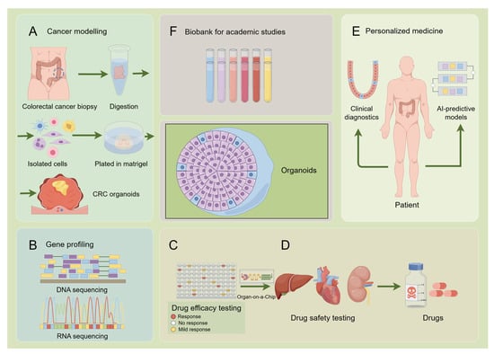
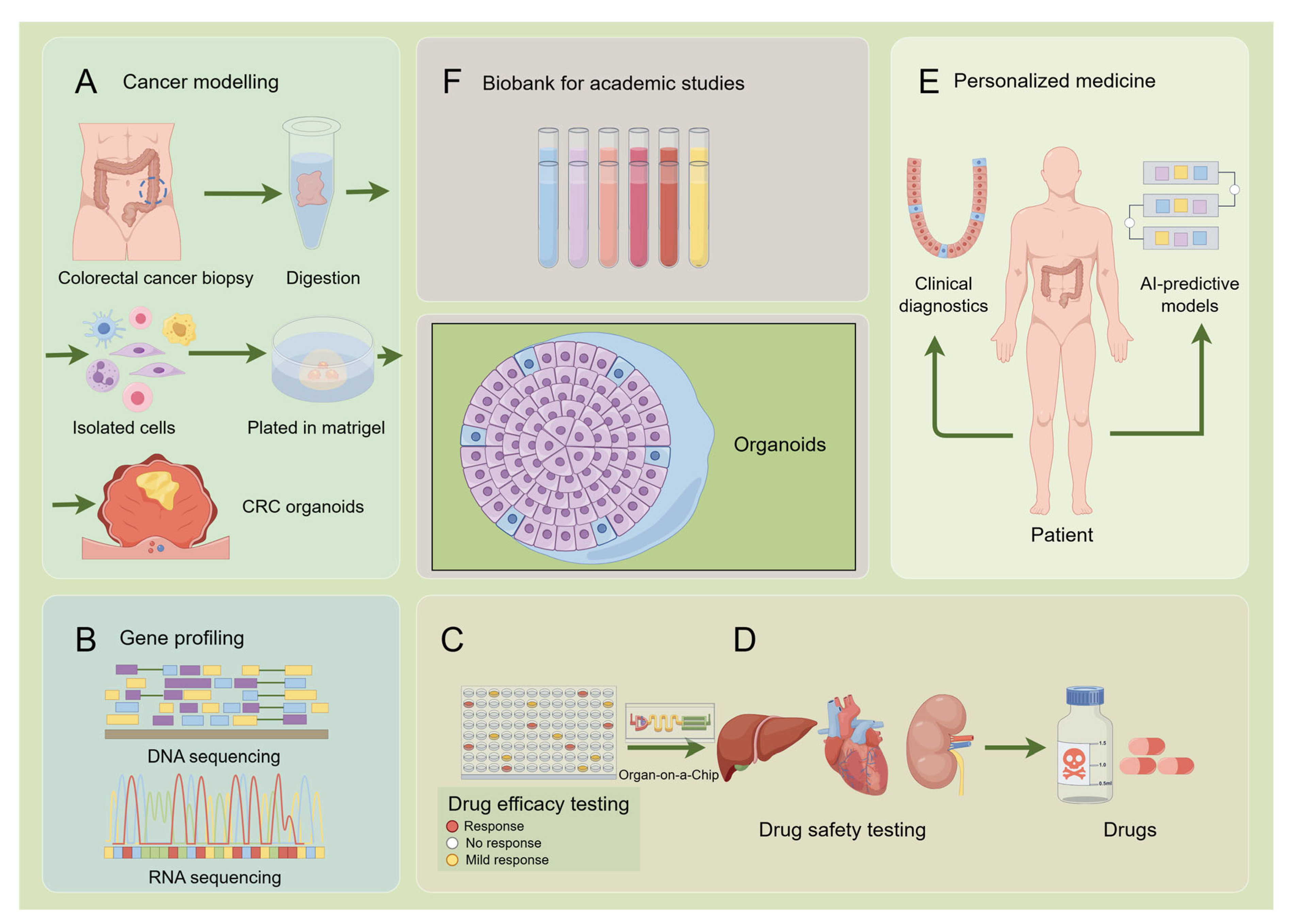
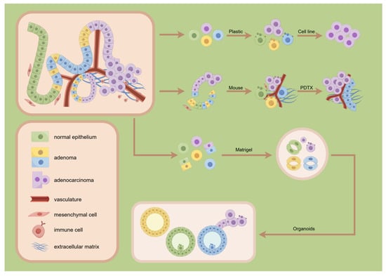
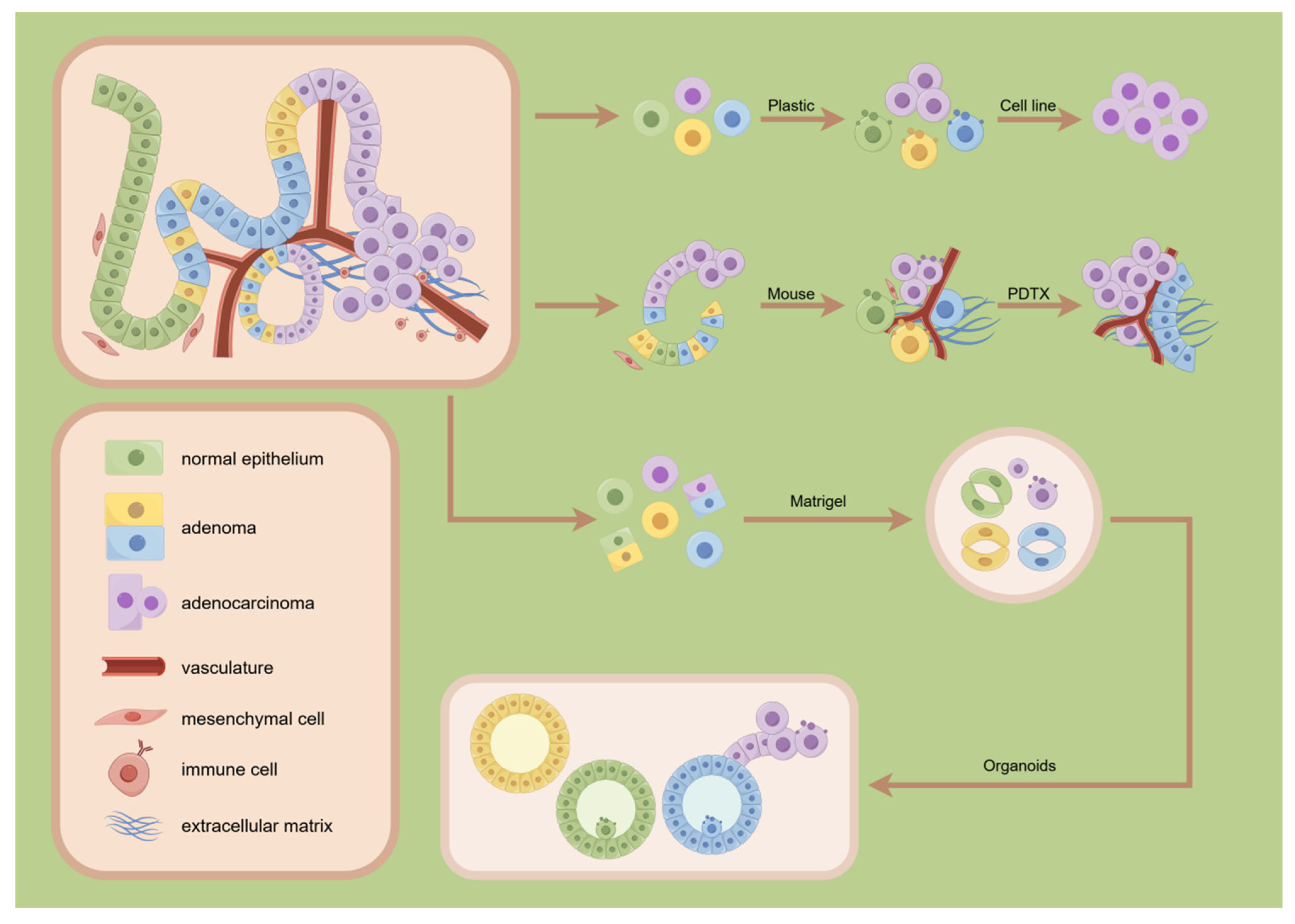
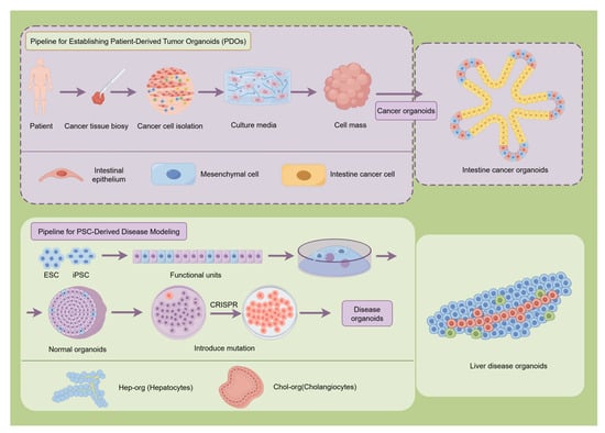
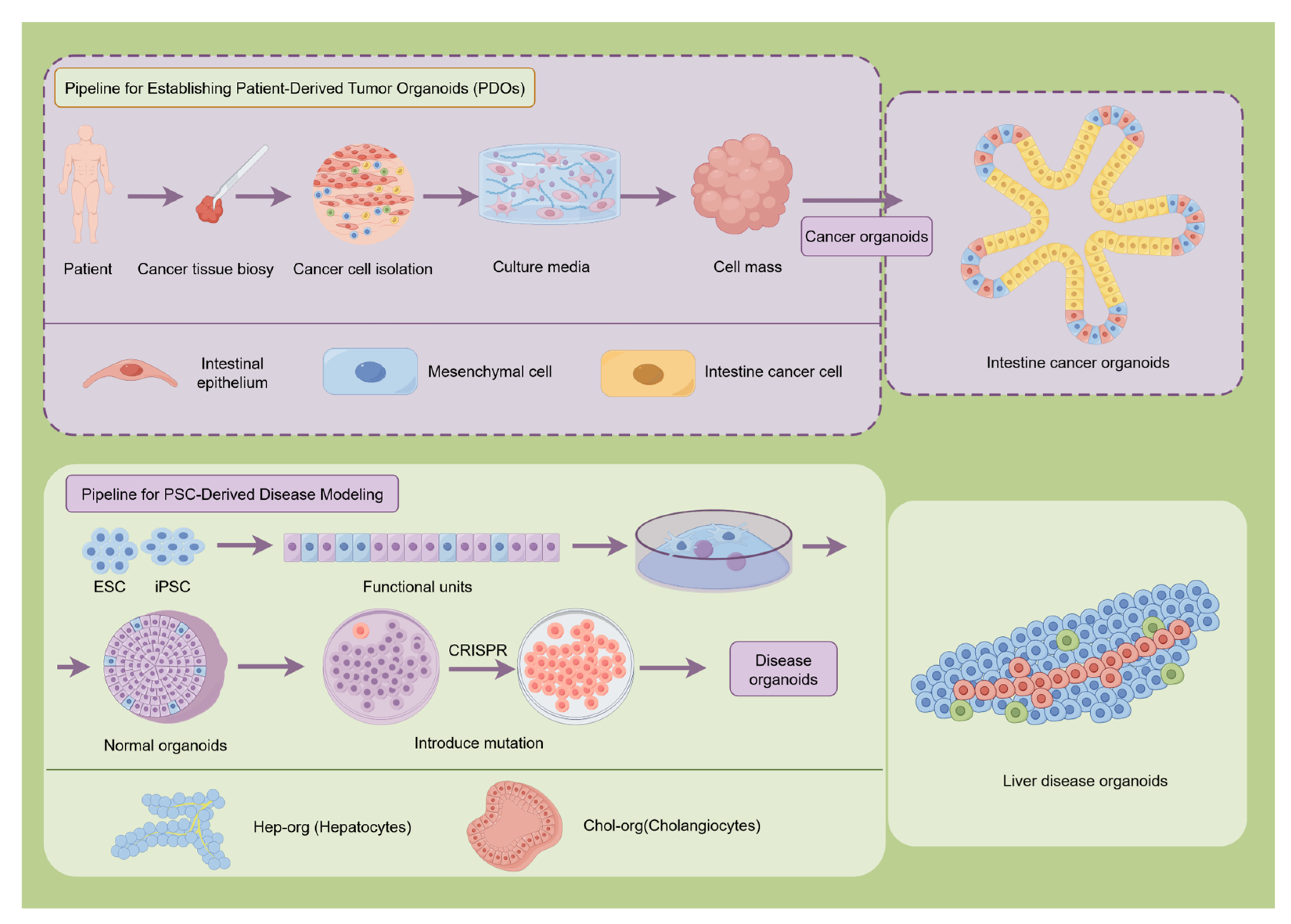

Disclaimer/Publisher’s Note: The statements, opinions and data contained in all publications are solely those of the individual author(s) and contributor(s) and not of MDPI and/or the editor(s). MDPI and/or the editor(s) disclaim responsibility for any injury to people or property resulting from any ideas, methods, instructions or products referred to in the content. |
© 2026 by the authors. Licensee MDPI, Basel, Switzerland. This article is an open access article distributed under the terms and conditions of the Creative Commons Attribution (CC BY) license.