


Abstract
Emergency department providers may be challenged by the presentation of patients who exhibit movement abnormalities that could indicate conditions requiring interventions to reduce the risk of potential morbidity and mortality. Crucially, interventions for potential neurological diseases that require immediate intervention (e.g., stroke) are contraindicated in patients exhibiting movements that may be voluntary (e.g., emotional expressions and fabricated symptoms) or functional (e.g., functional movement disorders such as functional tremors or psychogenic nonepileptic seizures (PNES)). Current motor assessment relies on subjective visual observation by human examiners, which limits the speed and accuracy of this differentiation. We hypothesize that technology measuring the temporal and spatial characteristics of movement can provide quantifiable signatures to differentiate movement subtypes and reflect the distinct neural pathways underlying voluntary and involuntary movement. To assess clinical and commercial interest in such technology, we conducted exploratory interviews with 17 of 56 invited potential customers (neurologists, biomedical engineers, and other clinicians) through a National Science Foundation I-Corps program where interviewees confirmed motor assessments are often performed without instrumentation and expressed willingness to adopt inexpensive, validated technology. The findings support interest in our proposed approach; however, this evidence is still preliminary as the interview sample was a non-random convenience sample and no comparison of respondents and non-respondents was performed. The sensor-based technology also remains conceptual with no current prototype, indicating the need for further studies utilizing more rigorous sampling and prototype development before clinical application.
1. Introduction
The differentiation of subtypes of voluntary movements represents a crucial challenge for observers, particularly providers who face the behaviors in an emergency. A special quandary is the temptation of clinicians to provide interventions required for the imminent benefit of people with organic disorders to individuals with symptoms and signs consistent with organic disorders as well as a spectrum of other conditions, such as functional disorders or voluntary movements, for which treatments for organic disorders may be contraindicated.
We seek to provide a foundation to identify movements that require immediate life-saving intervention from movements that merit observation. We seek to utilize neural networks to generalize schemes for Parkinson’s disease (PD) [1,2,3,4] and functional movement disorders (FMDs) [5] to distinguish among subtypes of voluntary movements. The timing of the activations of neural substrates may proceed through different anatomic pathways for voluntary movements. While voluntary movements may utilize time-consuming movement execution pathways with visuomotor processing through the motor cortex [6], involuntary movements may utilize faster automatic/spontaneous parietal or premotor pathways with less accurate motor behaviors. Thus, voluntary movements may indicate more accurate, time-consuming actions compared to the more spontaneous or automatic involuntary movements. Technology that measures the time course of the three-dimensional space positions of three points on an extremity may provide a foundation to differentiate voluntary movements from one another and from those that are involuntary. Additionally, hardware and software may be constructed to identify key features that uniquely identify the spectrum of voluntary movements. Our format may provide the structure to quickly, accurately, and economically diagnose the spectrum of voluntary movements to institute the appropriate intervention in emergency situations.
Since movement nomenclature is in flux and the trustworthiness of available evidence is questionable [7], we incorporate information from a pilot investigation to examine the need for technology for motor assessment as preliminary data to hypothesize a tool for movement classification. Our hope is that this will facilitate the incorporation of neural underpinnings of motion for further examination in future investigations utilizing rigorous methodology [8].
2. Background
2.1. Movement Execution
Although the primary motor cortex (M1) generates movements [9], the supplementary motor area (SMA) corresponds to awareness of intention to perform actions [10]. The neural underpinnings of voluntary movements provide clues as to how they can be differentiated. While voluntary movements have relatively late time windows of primary motor cortical activation [6], involuntary movements may utilize a spontaneous premotor or parietal mechanism, resulting in quicker but less accurate motor cortex activation [11,12]. Thus, examination of movement accuracy may serve as a valuable differentiation parameter [6,11,12]. For this reason, technology geared to measure temporal and spatial movement accuracy may provide a means to distinguish latent, accurate, voluntary movements from those that may be spontaneous or inaccurate, as seen in involuntary movements.
2.2. Voluntary Movements
Electrophysiological and motion measurements facilitate the identification of voluntary movements. For example, a Bereitschaftspotential, a surface-negative electrical brain potential identified on electroencephalography (EEG) 1–2 s before the movement, can identify self-initiated voluntary acts associated with the awareness of the will to perform the movement 200 milliseconds before the movement itself [6,12,13,14]. Thus, a Bereitschaftspotential characterizes most involuntary FMDs and voluntary movements [6,12,13,14,15].
Voluntary movements are intentionally produced actions [16,17] that include (A) gestures, motions, and utterances deliberately performed by persons to express (1) disagreement and disgust such as cut eye and suck teeth [18,19], (2) negation (“eem”) [20], or (3) exuberance such as applause and zaghrouta (ululation) [15]; and (B) motions deliberately performed by persons to deceive others, such as (a) malingering, the fabrication of actions in order to avoid jail, work, or school, or to obtain financial and other rewards, and (b) factitious disorder (Münchausen syndrome), the fabrication of symptoms and signs to seek medical attention and treatment [21,22].
2.3. Movements of Multiple Etiologies
Individuals may exhibit both organic and functional disorders. When people receive benefits for their condition, they may fabricate symptoms and signs to continue to receive the benefits. Since functional neurological disorders (FNDs) may co-exist with organic disorders, clinicians must be alert to the possible presence or development of organic conditions and psychiatric disorders in individuals with FND [23].
2.4. Motor Assessment
The Movement Disorders Society-sponsored revision of the Unified Parkinson’s Disease Rating Scale (MDS-UPDRS) [24] represents a validated structured interview to generate measurements of dysfunction exhibited by patients with Parkinson’s disease [24]. The MDS-UPDRS was constructed to provide an objective basis for assessments of participants by examiners who are certified in the MDS-UPDRS. The MDS-UPDRS represents a step to improve the measurement of deficits in people with Parkinson’s disease (PD) previously estimated by the Unified Parkinson’s Disease Rating Scale (UPDRS) [25]. The procedure is, however, limited by the ability of human examiners to perceive subtle movement alterations in participants several feet away.
To supplement direct clinical observation, low-cost accelerometer-based protocols have been used to obtain continuous quantitative recordings during standardized extremity tasks. The resulting raw signals, examiner ratings, and derived fast Fourier transform and continuous wavelet transform files have been made publicly available in a reusable dataset [26]. A subsequent study evaluated the reliability of trained raters’ visual classification of continuous wavelet transform images generated from extremity-movement signals [27]. Together, these studies support the feasibility of acquiring and interpreting quantitative representations of movement; however, they do not yet establish differentiation among the voluntary and involuntary movement subtypes proposed in the present article.
3. Our Hypothesis
The standard of care cannot unequivocally differentiate (A) voluntary movements from other movements and (B) among subtypes of voluntary movements. Human error in performing movement ratings represents a barrier to the precise and current movement measurements needed to accurately identify the pathognomonic signatures of a spectrum of voluntary and involuntary movements.
In particular, the current gold standard of motor assessment for Parkinson’s disease [24] is limited by human limitations to (A) distinguish fine motion gradations by visual observation and (B) distinguish finer differences in movement within the indicated scores. The plethora of instrumentation to analyze movements does not yet provide validated methods to accurately distinguish among the wide range of movements.
4. Materials and Methods
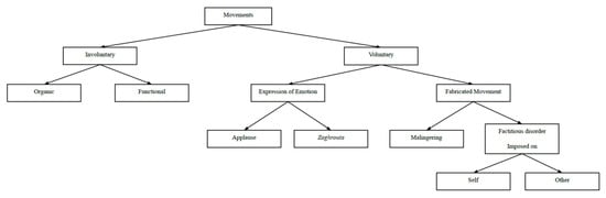
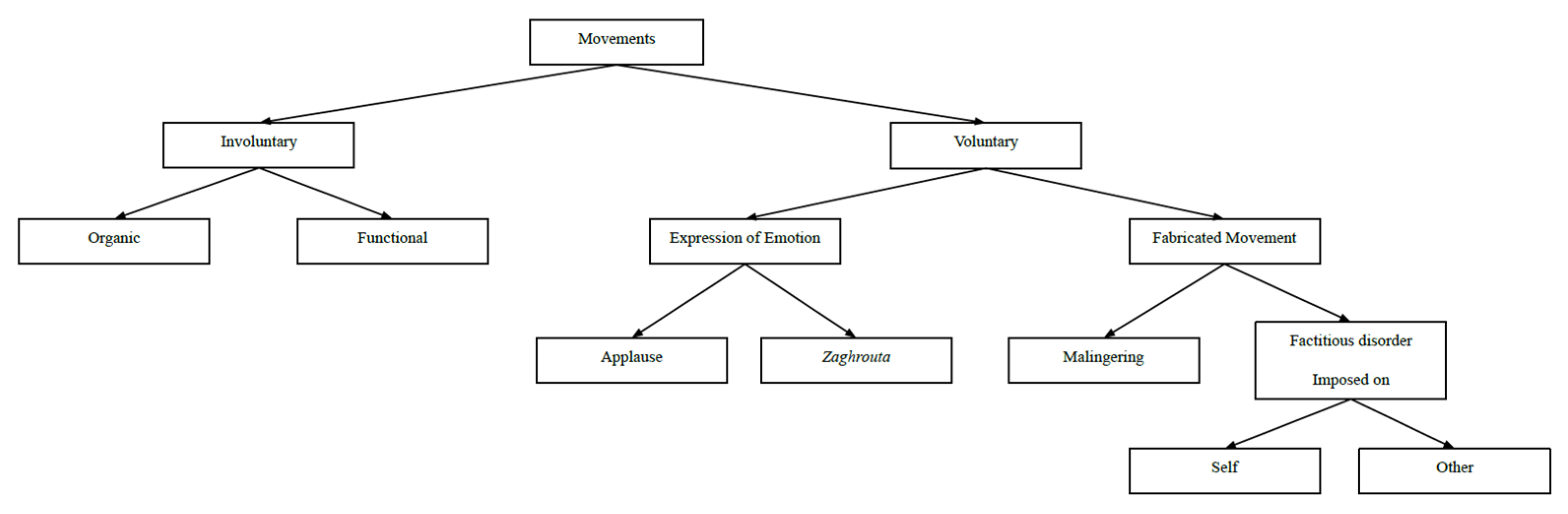
To test our hypothesis, we conducted a pilot study to identify the experiences of colleagues who aim to conduct motor assessments of people with Parkinson’s disease and related conditions. We sought to identify the standard of practice for motor assessments to learn if technology may be utilized to improve the diagnostic process. We aimed to discover customers for the initial draft of our value proposition (The Business Canvas Model, Strategyzer, Switzerland, https://www.strategyzer.com/playbook-library, accessed on 1 July 2026) as outlined in Figure 1, namely that our technology to measure movements [28,29,30,31] could be developed to improve the diagnosis and treatment of Parkinson’s disease and related conditions. We hypothesized a possible classification of movement represented by the schematic diagram in Figure 2.
Figure 1.
The initial draft of our value proposition [28].
Figure 2.
Schematic representation of a hypothesis to differentiate involuntary and voluntary movements. Neurological diseases, organic involuntary conditions, exhibit identified anatomic and physiological abnormalities.
We conducted interviews with potential customers to evaluate the value proposition [32] that technology may improve motor assessments through participation in the Johns Hopkins Technology Ventures Mid-Atlantic Region National Science Foundation I-Corps Hub, Baltimore, Maryland, United States of America, from 7 July to 4 August 2022. We constructed a checklist of items to discuss and record during each interview with potential customers (Table 1). Interview invitations were sent to 56 potential customers and repeated after a week for nonresponse. Although we initially planned to restrict the interviews to movement disorders neurologists, we expanded the network to include others who may participate in motor assessments. Therefore, the specific questions on the checklist were modified to reflect the role of the interviewee.
Table 1.
Checklist of items to discuss during interviews with potential customers regarding a value proposition [32] for technology intended to improve motor assessments.
Open-ended questions were utilized to elicit additional information to follow statements of interviewees.
5. Results
Some individuals who were invited to participate in the discovery interviews declined interviews because they did not perform motor assessments or they were out of office. Most individuals who were invited to participate did not respond to the initial request nor a second request a week later, which was interpreted as declining the interview. Interviews were completed with eight neurologists, three biomedical engineers, a neurosurgical resident, a nuclear physician, a physical therapist, a neuropsychologist, a medical science liaison for medical affairs of a pharmaceutical company, and a patient with Parkinson’s disease. All interviewees agreed to have the interview recorded to be shared with the team except for the medical science liaison due to lack of manager permission.
The results of the interviews provided general information about the conduct of motor assessments in clinical and research settings. Examiners who attained certification in the MDS-UPDRS [24], the current gold standard tool for motor evaluations, are generally considered to be qualified to perform accurate ratings indefinitely without reliability assessments. Providers generally perform the MDS-UPDRS alone without videotaping or instrumentation and record the scores for each item immediately after the item is completed.
Although motor assessments have traditionally been performed in person, recent developments in telemedicine suggest that online assessments yield valuable data [33]. Since any items attached to the participant may alter task performance, the MDS-UPDRS is performed without attaching instrumentation to the participant.
While interviewees provided their publications on motor assessment, they did not share their unpublished ongoing efforts to develop technology for motor assessments. Interviewees pointed out that many other groups are approaching this problem. Interviewees indicated technology is needed to distinguish Parkinson’s disease from related conditions during the first interview. Interviewees indicated their willingness to utilize inexpensive validated technology for motor assessments.
6. Discussion
6.1. Significance of the Evidence
We present a hypothesis that technology for temporal and spatial measurements of extremity movements distinguishes among subtypes of voluntary and involuntary movements to provide inexpensive, accurate output to generate accurate, precise movement signatures for diagnostic and therapeutic purposes.
Precise measurements of the times of activation of structures in the pathways of movements including amygdala, uncinate fasciculus, parietal cortex, premotor cortex, and motor cortex (M1) [6,9,10,11,34,35] may facilitate identification of the timing sequences that differentiate voluntary movements.
We hypothesize that technology [28,29,30,31] for the temporal and spatial measurement of upper extremity movements distinguishes the neural networks underlying subtypes of voluntary movements to provide inexpensive, accurate output that can be used to generate accurate, precise movement signatures for diagnostic and therapeutic purposes.
Accurate diagnosis may be accomplished with the proposed technology so that precision medicine can be applied to each patient. Thus, our proposal may lead to the development of treatment plans tailored to the specific needs of each individual patient. Patients may then benefit from optimal interventions specific to their conditions.
While similar diagnostic procedures may be required to distinguish neurological disorder (involuntary organic movement disorder) from FMDs and voluntary movements, clinicians must exercise caution to identify the anatomic and physiological bases for neurological disorders before instituting interventions with adverse effects. In particular, people with FMDs and voluntary movements do not benefit from the medical and surgical interventions to treat neurological disorders.
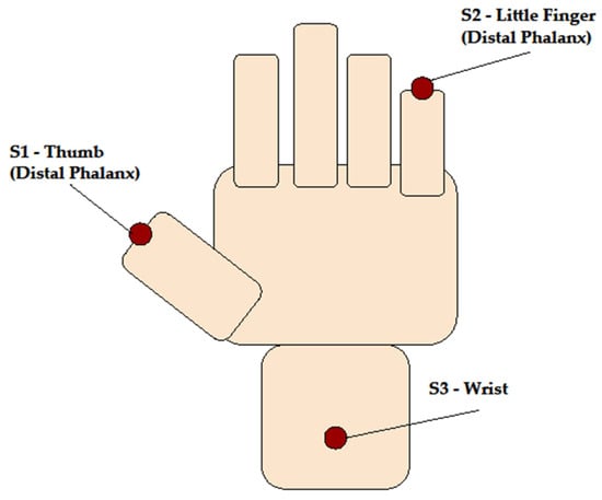
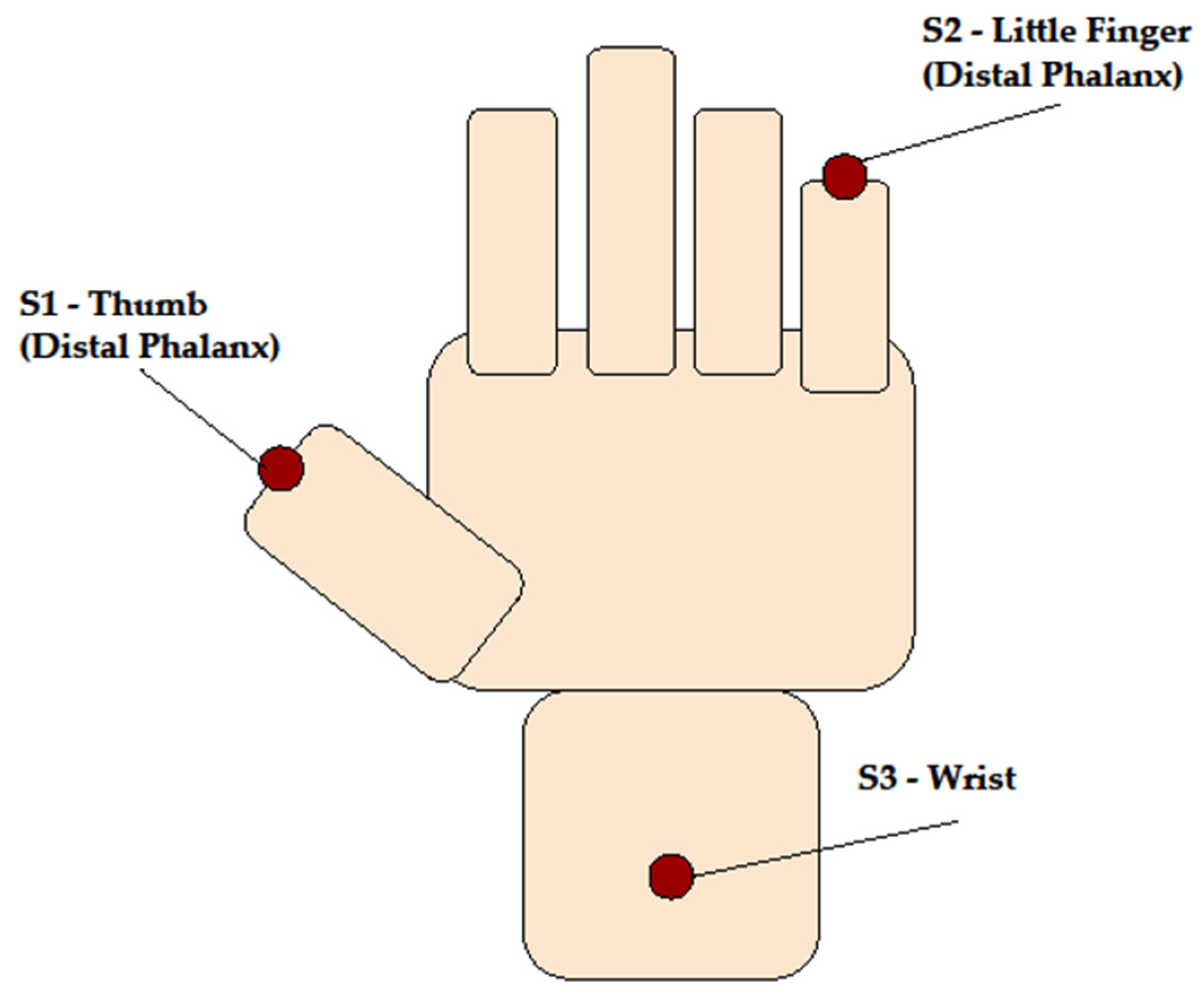
We propose that utilization of three accelerometer sensors (distal phalanx of the thumb, distal phalanx of the little finger, and wrist) on the right hand to document the three-dimensional positions in space on the upper extremity will facilitate the measurement of flexion–extension (finger tapping and hand movements), pronation–supination, and tremors [24,28]. Utilization of these sensors on the right hand will likely be adequate for diagnostic and therapeutic purposes. Use of sensors on the lower extremity is not necessary for the initial evaluation. At present, this sensor configuration is a proposed system with no current integrated hardware prototype being assembled (Figure 3).
Figure 3.
Proposed placement of three accelerometer sensors (S1: distal phalanx of thumb; S2: distal phalanx of little finger; S3: wrist) on the dorsal side of the right hand. Each sensor would record three-dimensional position/acceleration data to characterize flexion–extension, pronation–supination, and tremor. This is a proposed sensor configuration.
Utilization of software to view videos for frame-by-frame analysis to measure the duration of specific movements may provide the basis for more accurate and precise records of the behaviors that distinguish among related disorders and that distinguish the degrees of impairment within disorders. Computational approaches that have been utilized to develop neural networks for social development in children [36] and for automated video-based detection tasks [37] may be utilized to enhance the analysis of signals and videos of individuals performing movements.
We propose that an algorithm for voluntary movements may appear as follows:
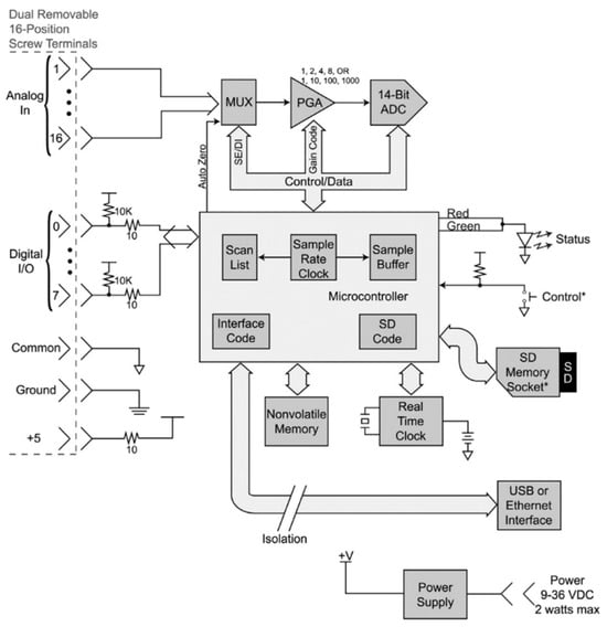
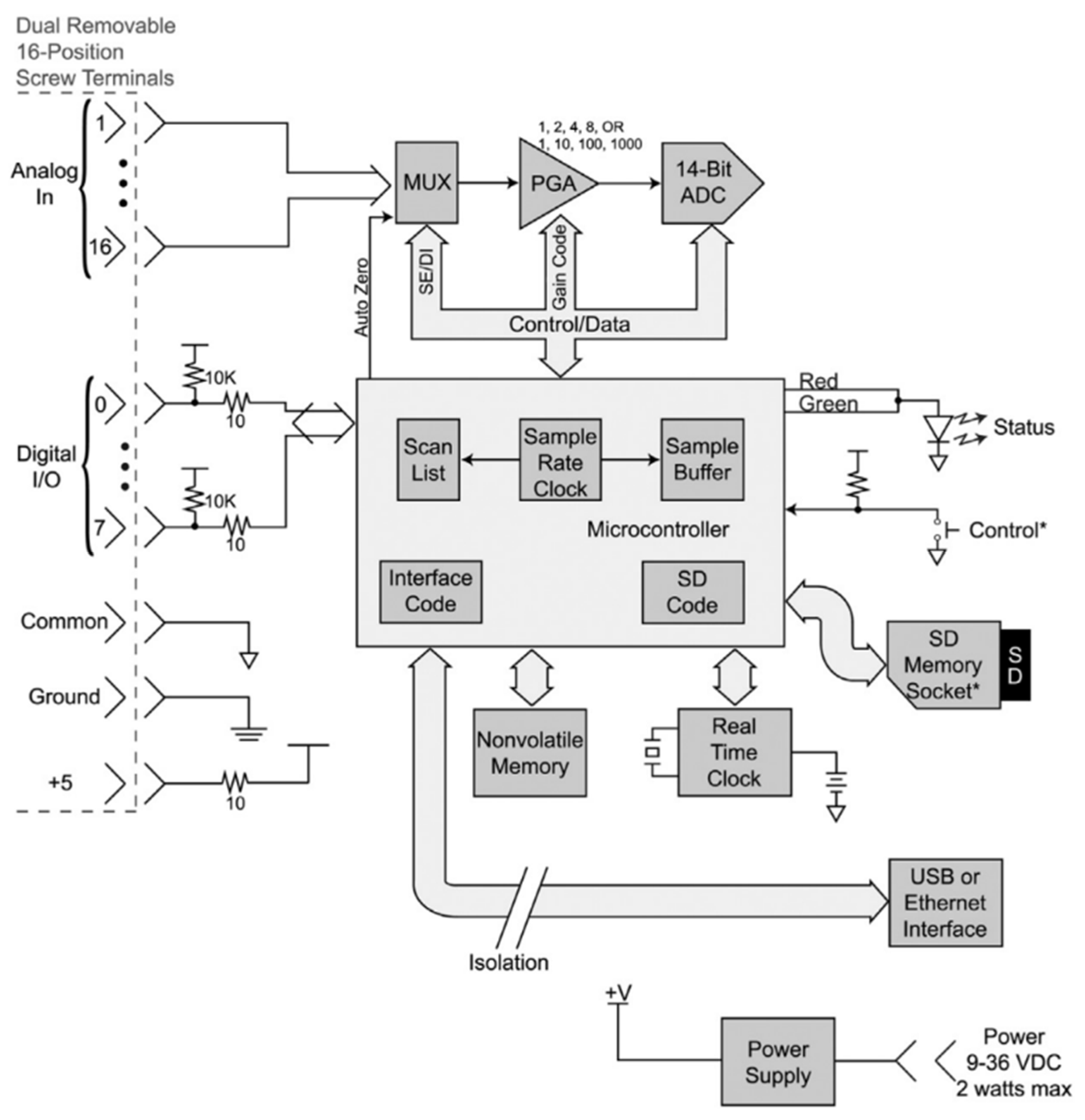
An example of the data-acquisition architecture that could be used to store and analyze signals from the proposed accelerometer sensors is illustrated in Figure 4, adapted from McKay et al. [28]. This example is presented to demonstrate that a similar approach is feasible; however, the present work will not build directly upon the architecture presented itself.
Figure 4.
Block diagram of a low-cost, portable USB/ethernet data logger system (adapted from McKay et al., pages 174–175) of an architecture that could be used to digitize and log accelerometer output from the sensor configuration proposed in Figure 3 [28]. The asterisk in the original diagram denote optional or model-dependent components as specified by the manufacturer (DATAQ Instruments, Akron, OH, USA) indicates the known control output to compare and contrast with the status of the experimental output.
6.2. Limitations
The empirical data of the pilot study are based on a convenience sample of colleagues of the authors and likely contain selection bias. Of the 56 potential invited customers, only 17 completed interviews, and non-respondents were classified as having declined rather than being analyzed as a distinct group. The generalizability and the validity of our conclusions may thus be limited by the low number of completed interviews, low response rate, and the absence of an analysis of the potential differences between respondents and non-respondents. We cannot rule out that individuals with greater interest in or exposure to motor assessment technology were more likely to participate than others. A random process was not performed to generate the data for the pilot investigation, so the assumptions required for statistical analysis are not met. This study does not include specific empirical validation such as simulations or pilot data of the proposed spatiotemporal classification approach, so future work should prioritize a small-scale proof-of-concept study to validate the classification framework proposed here. We included only a small number of relevant studies published in English in our review, which can lead to a reporting bias. Thus, the findings cannot be generalized to the general population. Future investigations utilizing statistical principles to design and analyze the data will generate higher quality evidence [7]. Therefore, the conclusion drawn from these published studies on this hypothesis needs to be verified and revised in the future. While the authors strived to present an accurate summary of the current knowledge, they recognize that this project will be enhanced by systematic reviews incorporating a team of experts to objectively assess information [8].
6.3. Future Directions
The future development of optimal technology for motor assessment [38,39] will be enhanced by conducting a systematic review and meta-analysis of wearable sensors for motor assessment.
Continued discovery of the unique needs of customers in Africa, North America, and other continents for the value proposition [32] that validated, inexpensive technology that is easily administered for the diagnosis of PD and related conditions will advance the construction of an approved device (Figure 1). Expanding the network of collaborators in Africa, North America, and other continents will be key to identifying the unique needs of customers around the world.
7. Conclusions
We present our hypothesis that the unique neural networks underlying the spectrum of subtypes of voluntary and involuntary movements can be identified by precise measurements of alterations in the spatial and temporal performance of movements in a single hand because it expresses concepts that merit consideration by colleagues around the world.
Identification of the correct subtype of voluntary and involuntary movements is crucial to the administration of the appropriate interventions [40]. The differential diagnosis of movements in children can challenge clinicians in emergency rooms. Obtaining a comprehensive history is key to developing an optimal treatment plan [40]. Identifying family members with similar subtypes of FND facilitates the diagnosis. Physical, sexual, and psychological abuse must be ruled out and reported to the authorities if identified.
Providers need inexpensive validated technology for motor assessments. To assess the market for a value proposition of motor assessment technology, interviews are needed from providers, managers, hospitals, institutes, and insurance companies. Future interviews will include managers and purchasing officers and insurance companies to assess potential plans to market the services/products.
Author Contributions
Conceptualization, J.Y.O., T.P.H., M.E.H. and J.R.B.; methodology, T.P.H., M.E.H. and J.R.B.; software, J.R.B.; validation, A.E. and J.R.B.; formal analysis, J.Y.O., A.E. and J.R.B.; investigation, A.E. and J.R.B.; resources, J.R.B.; data curation, J.Y.O., A.E. and J.R.B.; writing—original draft preparation, J.Y.O. and J.R.B.; writing—review and editing, J.Y.O., A.E., T.P.H., M.E.H. and J.R.B.; visualization, J.Y.O., A.E. and J.R.B.; supervision, J.R.B.; project administration, J.Y.O., A.E. and J.R.B.; funding acquisition, J.R.B. All authors have read and agreed to the published version of the manuscript.
Funding
This research was funded by the Johns Hopkins Technology Ventures Mid-Atlantic Region National Science Foundation I-Corps Hub, Baltimore, Maryland, United States of America, from 7 July to 4 August 2022.
Institutional Review Board Statement
Not applicable.
Informed Consent Statement
Not applicable.
Data Availability Statement
All data are available upon reasonable request to the corresponding author.
Acknowledgments
An earlier version of this manuscript was presented at the 2nd International Electronic Conference on Brain Sciences, 15–30 July 2021 (available online: https://sciforum.net/event/IECBS2021, accessed on 2 June 2026) [40].
Conflicts of Interest
The authors declare no conflicts of interest.
Abbreviations
The following abbreviations are used in this manuscript:
| EEG | Electroencephalography |
| FM | Functional Movement Disorder |
| FND | Functional Neurological Disorder |
| MDS-UPDRS | Movement Disorders Society-sponsored revision of the Unified Parkinson’s Disease Rating Scale [24] |
| M1 | Primary Motor Cortex |
| PNES | Psychogenic Nonepileptic Seizures |
| SMA | Supplementary Motor Area |
| UPDRS | Unified Parkinson’s Disease Rating Scale [25] |
References
- Di Battista, M.E.; Giustini, P.; Bernardi, S.; Stirpe, P.; Vanacore, N.; Meco, G. A simplified algorithm may lead to overestimate dementia in PD. A clinical and epidemiological study using criteria for PD-D proposed by the Movement Disorders Task Force. J. Neural Transm. 2011, 118, 1609–1612. [Google Scholar] [CrossRef] [Scilit] [PubMed]
- Dietrichs, E.; Odin, P. Algorithms for the treatment of motor problems in Parkinson’s disease. Acta Neurol. Scand. 2017, 136, 378–385. [Google Scholar] [CrossRef] [Scilit] [PubMed]
- Rao, S.S.; Hofmann, L.A.; Shakil, A. Parkinson’s disease: Diagnosis and treatment. Am. Fam. Physician 2006, 74, 2046–2054. [Google Scholar] [PubMed]
- Schrag, A.; Anastasiou, Z.; Ambler, G.; Noyce, A.; Walters, K. Predicting diagnosis of Parkinson’s disease: A risk algorithm based on primary care presentations. Mov. Disord. 2019, 34, 480–486. [Google Scholar] [CrossRef] [Scilit] [PubMed]
- Morgante, F.; Edwards, M.J.; Espay, A.J. Psychogenic movement disorders. Continuum 2013, 19, 1383–1396. [Google Scholar] [CrossRef] [Scilit] [PubMed]
- Shibasaki, H.; Hallett, M. What is the Bereitschaftspotential? Clin. Neurophysiol. 2006, 117, 2341–2356. [Google Scholar] [CrossRef] [Scilit] [PubMed]
- Hultcrantz, M.; Rind, D.; Akl, E.A.; Treweek, S.; Mustafa, R.A.; Iorio, A.; Alper, B.S.; Meerpohl, J.J.; Murad, M.H.; Ansari, M.T.; et al. The GRADE Working Group clarifies the construct of certainty of evidence. J. Clin. Epidemiol. 2017, 87, 4–13. [Google Scholar] [CrossRef] [Scilit] [PubMed]
- Higgins, J.P.T.; Thomas, J.; Chandler, J.; Cumpston, M.; Li, T.; Page, M.J.; Welch, V.A. (Eds.) Cochrane Handbook for Systematic Reviews of Interventions, 2nd ed.; Wiley: Chichester, UK, 2019. [Google Scholar] [CrossRef] [Scilit]
- Bhattacharjee, S.; Kashyap, R.; Abualait, T.; Chen, S.-H.A.; Yoo, W.-K.; Bashir, S. The role of primary motor cortex: More than movement execution. J. Mot. Behav. 2021, 53, 258–274. [Google Scholar] [CrossRef] [Scilit] [PubMed]
- Sjöberg, R.L. Free will and neurosurgical resections of the supplementary motor area: A critical review. Acta Neurochir. 2021, 163, 1229–1237. [Google Scholar] [CrossRef] [Scilit] [PubMed]
- Hallett, M.; Dworetzky, B.A.; McWhirter, L.; Staab, J.P.; Stone, J. Functional neurological disorder: New subtypes and shared mechanisms. Lancet Neurol. 2022, 21, 537–550. [Google Scholar] [CrossRef] [Scilit] [PubMed]
- Antenucci, P.; Moura, J.; Bhatia, K.P.; Rocchi, L.; Latorre, A. The Bereitschaftspotential in voluntary movement and movement disorders: Potential, limitations and new perspectives. Clin. Neurophysiol. 2026, 187, 2111895. [Google Scholar] [CrossRef] [Scilit] [PubMed]
- Nann, M.; Cohen, L.G.; Deecke, L.; Soekadar, S.R. To jump or not to jump—The Bereitschaftspotential required to jump into a 192-meter abyss. Sci. Rep. 2019, 9, 2243. [Google Scholar] [CrossRef] [Scilit] [PubMed]
- Libet, B.; Gleason, C.A.; Wright, E.W.; Pearl, D.K. Time of conscious intention to act in relation to onset of cerebral activity (readiness-potential): The unconscious initiation of a freely voluntary act. Brain 1983, 106, 623–642. [Google Scholar] [CrossRef] [Scilit] [PubMed]
- Elshourbagy, T.; Mousa, A.; Mohamed, M.A.; Brasic, J.R. Differentiation of zaghrouta, ululation to express joy in the Middle East, from movement disorders and other conditions. Int. J. Health Life Sci. 2021, 7, e106655. [Google Scholar] [CrossRef] [Scilit]
- Espay, A.J.; Aybek, S.; Carson, A.; Edwards, M.J.; Goldstein, L.H.; Hallett, M.; LaFaver, K.; LaFrance, W., Jr.; Lang, A.E.; Nicholson, T.; et al. Current concepts in diagnosis and treatment of functional neurological disorders. JAMA Neurol. 2018, 75, 1132–1141. [Google Scholar] [CrossRef] [Scilit] [PubMed]
- Voon, V.; Gallea, C.; Hattori, N.; Bruno, M.; Ekanayake, V.; Hallett, M. The involuntary nature of conversion disorder. Neurology 2010, 74, 223–228. [Google Scholar] [CrossRef] [Scilit] [PubMed]
- Rickford, J.R.; Rickford, A.E. Cut-eye and suck-teeth: African words and gestures in New World guise. J. Am. Folk. 1976, 89, 294–309. [Google Scholar] [CrossRef] [Scilit]
- Patrick, P.L.; Figueroa, E. Kiss-teeth. Am. Speech 2002, 77, 383–397. [Google Scholar] [CrossRef] [Scilit]
- Jones, T. “Eem” Negation in African American English: A Next Step in Jespersen’s Cycle? Available online: https://repository.upenn.edu/entities/publication/d1fb4279-711a-4a5d-9140-58b5055884c9 (accessed on 2 June 2026).
- Abeln, B.; Love, R. An overview of Munchausen syndrome and Munchausen syndrome by proxy. Nurs. Clin. N. Am. 2018, 53, 375–384. [Google Scholar] [CrossRef] [Scilit] [PubMed]
- American Psychiatric Association. Diagnostic and Statistical Manual of Mental Disorders, 5th ed.; text revision; DSM-5-TR; American Psychiatric Publishing: Washington, DC, USA, 2022. [Google Scholar]
- Epstein, S.A.; Maurer, C.W.; LaFaver, K.; Ameli, R.; Sinclair, S.; Hallett, M. Insights into chronic functional movement disorders: The value of qualitative psychiatric interviews. Psychosomatics 2016, 57, 566–575. [Google Scholar] [CrossRef] [Scilit] [PubMed]
- Goetz, C.G.; Tilley, B.C.; Shaftman, S.R.; Stebbins, G.T.; Fahn, S.; Martinez-Martin, P.; Poewe, W.; Sampaio, C.; Stern, M.B.; Dodel, R.; et al. Movement Disorder Society-sponsored revision of the Unified Parkinson’s Disease Rating Scale (MDS-UPDRS): Scale presentation and clinimetric testing results. Mov. Disord. 2008, 23, 2129–2170. [Google Scholar] [CrossRef] [Scilit] [PubMed]
- Fahn, S.; Elton, R.; Members of the UPDRS Development Committee. The Unified Parkinson’s Disease Rating Scale. In Recent Developments in Parkinson’s Disease, Vol. 2; Fahn, S., Marsden, C.D., Calne, D.B., Goldstein, M., Eds.; McMellam Health Care Information: Florham Park, NJ, USA, 1987; pp. 153–163. [Google Scholar]
- Harrigan, T.; Syed, A.; Hwang, B.; Mathur, A.; Mills, K.; Pantelyat, A.; Bang, J.; Mishra, C.; Vyas, P.; Martin, S.; et al. Quantitative Continuous Measurement of Movements in the Extremities; Mendeley Data, V4; Elsevier: Amsterdam, The Netherlands, 2022. [Google Scholar] [CrossRef]
- Elshourbagy, A.; Mills, K.; Harrigan, T.; Hernandez, M.; Brasic, J. Reliability of visual observation to classify continuous wavelet transforms of signals of extremity movements. Neurology 2025, 104, 2977. [Google Scholar] [CrossRef] [Scilit]
- McKay, G.N.; Harrigan, T.P.; Brašić, J.R. A low-cost quantitative continuous measurement of movements in the extremities of people with Parkinson’s disease. MethodsX 2019, 6, 169–189. [Google Scholar] [CrossRef] [Scilit] [PubMed]
- Harrigan, T.P.; Hwang, B.J.; Mathur, A.K.; Mills, K.A.; Pantelyat, A.Y.; Bang, J.A.; Syed, A.B.; Vyas, P.; Martin, S.D.; Jamal, A.; et al. Dataset of quantitative structured office measurements of movements in the extremities. Data Brief 2020, 31, 105876. [Google Scholar] [CrossRef] [Scilit] [PubMed]
- Hernandez, M.E.H.; Ziegelman, L.; Kosuri, T.; Hakim, H.; Zhao, L.; Mills, K.A.; Brašić, J.R. Classification of extremity movements by visual observation of signals and their transforms. MethodsX 2022, 9, 101739. [Google Scholar] [CrossRef] [Scilit] [PubMed]
- Brasic, J.R.; Ziegelman, L.; Kosuri, T.; Hakim, H.; Zhao, L.; Gokce, A.; Mathur, K.; Hernandez, M.E.; Mills, K.A. Classification of extremity movements by visual observation of signal transforms. Mov. Disord. 2022, 37, S187. [Google Scholar]
- Osterwalder, A.; Pigneur, Y.; Bernarda, G.; Smith, A.; Papadakos, T. Value Proposition Design: How to Create Products and Services Customers Want; Wiley: Hoboken, NJ, USA, 2014. [Google Scholar]
- Elshourbagy, A.; Eltaras, M.M.; Abdelshafy, H.; Javed, S.; Sadaney, A.O.; Harrigan, T.P.; Mills, K.A.; Hernandez, M.E.; Brašić, J.R. Feasibility of virtual low-cost quantitative continuous measurement of movements in the extremities of people with Parkinson’s disease. MethodsX 2023, 11, 102230. [Google Scholar] [CrossRef] [Scilit] [PubMed]
- Grèzes, J.; Valabrègue, R.; Gholipour, B.; Chevallier, C. A direct amygdala-motor pathway for emotional displays to influence action: A diffusion tensor imaging study. Hum. Brain Mapp. 2014, 35, 5974–5983. [Google Scholar] [CrossRef] [Scilit] [PubMed]
- Voon, V.; Brezing, C.; Gallea, C.; Ameli, R.; Roelofs, K.; LaFrance, W.C., Jr.; Hallett, M. Emotional stimuli and motor conversion disorder. Brain 2010, 133, 1526–1536. [Google Scholar] [CrossRef] [Scilit] [PubMed]
- Messinger, D.S.; Perry, L.K.; Mitsven, S.G.; Tao, Y.; Moffitt, J.; Fasano, R.M.; Custode, S.A.; Jerry, C.M. Computational approaches to understanding interaction and development. In New Methods and Approaches for Studying Child Development; Advances in Child Development and Behavior; Gilmore, R.O., Lockman, J.J., Eds.; Elsevier: Amsterdam, The Netherlands, 2022; Volume 62, pp. 191–230. [Google Scholar] [CrossRef] [Scilit] [PubMed]
- Zhang, K.; Zhang, Z.; Li, Z.; Qiao, Y. Joint face detection and alignment using multitask cascaded convolutional networks. IEEE Signal Process. Lett. 2016, 23, 1499–1503. [Google Scholar] [CrossRef] [Scilit]
- Elshourbagy, T.; Brašić, J.R.; Syed, A.B. Guidelines for the diagnosis and treatment of Parkinson’s disease. Biol. Life Sci. Forum 2021, 9, 9. [Google Scholar] [CrossRef] [Scilit]
- Elshourbagy, T.; Syed, A.B.; Amer, M.A.M.; Brasic, J.R. Precision medicine to identify optimal diagnostic and therapeutic interventions for Parkinson’s disease. Med. Sci. Discov. 2021, 8, 514–519. [Google Scholar] [CrossRef] [Scilit]
- Brasic, J.R. Differential of Voluntary and Involuntary Movements. In Proceedings of the 2nd International Electronic Conference on Brain Sciences, Behavioral Neuroscience Session, Online, 15–30 July 2021; MDPI: Basel, Switzerland, 2021; Available online: https://sciforum.net/paper/view/10651 (accessed on 3 June 2026).



Disclaimer/Publisher’s Note: The statements, opinions and data contained in all publications are solely those of the individual author(s) and contributor(s) and not of MDPI and/or the editor(s). MDPI and/or the editor(s) disclaim responsibility for any injury to people or property resulting from any ideas, methods, instructions or products referred to in the content. |
© 2026 by the authors. Licensee MDPI, Basel, Switzerland. This article is an open access article distributed under the terms and conditions of the Creative Commons Attribution (CC BY) license.