


Abstract
Arguably, the most vital function of the cardiovascular system (CVS) is its ability to facilitate the oxygenation of blood in the lungs, and to deliver that oxygen to the rest of the body. This relies on the ability of oxygen to diffuse in and out of the bloodstream, as well as for the CVS to maintain adequate perfusion of body tissues. The CVS also plays a key role in immune responses, particularly in initiating an acute inflammatory reaction in response to infection or injury. Changes in vascular function, such as vasodilation, increased vascular permeability, and initiation of coagulation, are critical elements of this inflammatory response. Normally, the regulated nature of immune responses helps to limit any potential detrimental effects. However, in cases of severe, dysregulated inflammation, as seen in conditions such as sepsis, the vascular responses that form a normal part of the inflammatory reaction may start to impact upon tissue oxygenation. Increased microvascular permeability and the development of edema can impact upon oxygen diffusion, whilst tissue perfusion can be impacted at the level of both the micro- and macrocirculation. In severe cases, impaired perfusion and oxygen delivery to vital organs may lead to multiple organ system failure. An increased understanding of the impact of inflammation on vascular function may help elucidate novel therapeutic approaches to manage these critical care situations.
1. Introduction
By definition, the functions provided by the vital organ systems are essential for life. Given the reliance of the body on a constant, adequate tissue perfusion and oxygenation, the roles played by the cardiovascular and respiratory systems may be considered paramount. The immune system, with its capacity to initiate wound healing and protect the body against infection and cancer, also has a vital role to play. The ability of the immune system to provide its rapid, protective role very much relies on its interaction with the cardiovascular system [1]. The immune system’s initial innate response to any perceived threat is characterized by the initiation of an acute inflammatory response at the site of infection or injury. Inflammation and the innate immune response are very much a function of vascular tissue. Not only are most of the elements required for an inflammatory response associated with either the blood or vascular tissue, but changes in vascular function, such as vasodilation and increased vascular permeability, are also central to the inflammatory reaction [2]. Indeed, three of the cardinal signs of inflammation, namely that the affected area is hot, red and swollen, highlight the involvement of vascular responses [3].
The innate immune response represents a rapid, initial mechanism to deal with invading pathogens, providing a mechanism to limit the spread of an infection, as well as helping to initiate the adaptive immune response. Whilst that acute inflammatory reaction is central to our defense against pathogens, the reaction can be triggered by any noxious stimulus, such as trauma and burns, that result in cell injury [4]. The ability to sense these threats, both pathogenic and non-pathogenic, involves cell-surface pattern-recognition receptors (PRRs). These PRRs are capable of detecting molecular signals associated with either pathogenic organisms (pathogen-associated molecular patterns (PAMPs)) or cell injury (damage-associated molecular patterns (DAMPs)) [4,5]. Activation of PRRs leads to the activation of inflammatory pathways and the release of inflammatory mediators that drive the subsequent inflammatory response [4]. In addition, inflammation can also be triggered by tissue hypoxia through the activation of hypoxia-inducible transcription factors [6,7].
2. Potential Pathological Aspects of Inflammation
Inflammation comprises a powerful range of mechanisms that enable our body to respond to noxious stressors and help restore homeostasis. However, the self-same mechanisms also have the potential to produce detrimental effects within the body, as is clearly evidenced by the wide range of chronic inflammatory conditions that represent such a major burden to society and our healthcare systems [4,8,9,10]. Whilst there is a significant focus on chronic inflammation as a pathological state, it is important to be aware that severe, acute inflammatory responses also pose a major issue in the healthcare setting. The acute inflammatory response can represent a major problem if it is either excessive or inappropriately activated. This is seen all too clearly in situations such as sepsis, severe anaphylactic reactions, major trauma, and severe burns [11,12,13,14,15]. One of the main impacts of the excessive inflammatory response seen in these patients is reduced tissue perfusion and oxygenation of vital organs [13].
Inflammation is no longer viewed as an “on–off” response, nor as a fire that just burns out. Instead, its activity covers a spectrum of responses from physiological to pathological, and it is regulated by a plethora of pro- and anti-inflammatory mediators [9,10]. Viewing the inflammatory response from a homeostatic perspective, infection or trauma represents a noxious stressor that upsets homeostasis, and the goal of the inflammatory/immune response is to restore homeostasis and for the body to return to a healthy state. The inflammatory response involves both positive- and negative-feedback mechanisms to achieve that endpoint. The early pro-inflammatory stage has the hallmark of a positive-feedback response, where there is a rapid elaboration of pro-inflammatory mediators, as seen with the activation of nuclear factor kappa B (NF-κB) transcription factors [9,16]. However, there are also negative-feedback mechanisms that may serve to limit the magnitude of the inflammatory response, for example, through the release of endogenous corticosteroids or expression of inhibitors of κB proteins (IκBs) [17,18,19,20]. These inhibitory mechanisms may play an important role not only in helping to resolve inflammation but also in preventing unwanted activation of the pro-inflammatory pathways [20]. If the inflammatory/immune response is effective in dealing with the infection, or effecting tissue repair, then the stimulus for the inflammatory response will be removed, and homeostasis restored. However, if the attempt to deal with the stressor is unsuccessful, there are two potential pathological outcomes, one chronic and one acute. An unresolved inflammatory response may lead to the establishment of a new homeostatic set-point, and the progression to a chronic inflammatory state [21]. Unfortunately, the potential to progress to chronic inflammation is very common, as evidenced by conditions such as type-2 diabetes and hypertension [21,22,23]. However, the other potential outcome is that the lack of resolution of the inflammatory stimulus can lead to an excessive release of inflammatory mediators and a severe, dysregulated inflammatory reaction referred to as systemic inflammatory response syndrome (SIRS) [24].
It has been estimated that almost 18% of adults presenting at emergency departments are diagnosed with SIRS [25]. The impact of and prognosis associated with SIRS depends upon the nature of the trigger as well as the severity of the response. For example, SIRS associated with an infectious cause, termed sepsis, has a poorer prognosis [26]. A severe SIRS response can lead to multiple organ dysfunction syndrome (MODS), which represents a leading cause of mortality in the intensive care setting [27]. It has been estimated that the mortality rate may range from 7% in patients with simple SIRS through to 46% in patients with MODS resulting from septic shock [28]. That progression from SIRS to MODS is closely linked to the vascular responses and altered tissue perfusion associated with the severe inflammatory reaction [29].
3. Vascular Responses Associated with Acute Inflammation
As indicated, acute inflammation is primarily a response of vascularized tissue; hence, it is associated with a number of specific vascular responses, all of which will have an impact on normal tissue hemodynamics, particularly at the level of the microvasculature. Initially, particularly following trauma, there is a short, transient period of vasoconstriction that may serve to reduce hemorrhage at the site of injury [30]. This is followed by a more sustained, localized vasodilation that increases blood flow to the affected area, leading to the redness and heat that are observed with inflammation. This hyperemia aids in the delivery of leukocytes and plasma proteins to the site of inflammation, helping to drive the immune response.
Perhaps more significant is the altered vascular permeability that allows for not just the extravasation of fluid but also plasma proteins. Normally, most capillary beds have a low permeability to plasma proteins that allows for just a slight leakage into tissue fluid [31]. However, vascular permeability to plasma proteins, particularly in the post-capillary venules, changes significantly with inflammation, allowing for their diffusion into the interstitial space. The diffusion of proteins and fluid from the vasculature is responsible for the edema, or swelling, associated with inflammation. The extravasation of plasma proteins, including complement proteins, and their activation at the site of inflammation is a key component of the innate response to infection [32,33,34,35,36]. However, the activation of complement proteins is not the only coordinated enzymatic cascade associated with inflammation. The coagulation cascade also represents an important component of the innate immune response, where the formation of a fibrin clot at the site of infection may help to limit the spread of invading pathogenic organisms [37,38].
Whilst the most obvious impact of increased vascular permeability is the formation of edema, the increased fluid extravasation associated with inflammation will lead to a local decrease in fluid in the vascular compartment, leading to a local decrease in blood volume. This movement of fluid from the circulation will result in an increase in blood concentration and viscosity, and in turn decreased local blood flow and vascular stasis [39,40]. This localized slowing of blood flow, together with the activation of adhesion molecules, favors leukocyte adhesion to the vascular endothelium, and their subsequent extravasation at the site of inflammation [41,42]. The influx of phagocytic cells, particularly neutrophils and macrophages, to the affected area represents an important aspect of the initial immune response, promoting the clearance of pathogens and cell debris [43]. However, this adhesion of leukocytes in the post-capillary venules at the site of inflammation will lead to a physical narrowing of the microvessel lumen, further reducing local blood flow [44]. These vascular responses highlight the key role that the vascular endothelium plays in inflammation, both regulating vascular permeability to plasma proteins and facilitating leukocyte transmigration [31,45]. In addition, the vascular endothelium also synthesizes and releases a number of potent vasoactive substances that produce either vasodilation (e.g., nitric oxide (NO)) or vasoconstriction (e.g., endothelin) [46,47].
Finally, whilst considering the vascular system’s contribution to the immune response, it is important to remember that lymphatic vessels also have an important role to play. Lymphatic vessels provide a mechanism for the drainage of the extravasated fluid, along with the removal of immune cells, cellular debris, and inflammatory mediators from the affected area [48,49]. The transport of this lymphatic fluid to the lymph nodes is central to the initiation of the adaptive immune response, whilst the removal of inflammatory elements from an affected tissue may also represent a step towards the resolution of inflammation [48]. Dilation of lymphatic vessels during inflammation may aid in the removal of the increased interstitial fluid [48].
4. Hemodynamic Consequences of SIRS
The ability to mount an effective immune and inflammatory response is critical to our survival, both in the face of dealing with invading pathogens as well as enabling tissue repair and wound healing following trauma. However, if those pro-inflammatory mechanisms are not effectively controlled, they have the potential to be not just detrimental, but lethal [50]. Normally, a wide range of pro- and anti-inflammatory mediators ensure that a careful balance is achieved between the beneficial and detrimental aspects of inflammation [9,10]. However, there are a wide range of clinical situations that can lead to a severe, dysregulated inflammatory reaction, or SIRS [24]. Table 1 provides a summary of the range of clinical syndromes that are associated with severe systemic inflammatory reactions.
Table 1.
Clinically significant systemic inflammatory syndromes.
Unsurprisingly, given the role of the vasculature in inflammation, there are a range of hemodynamic consequences associated with SIRS and anaphylaxis, and these contribute both to the progression of and life-threatening nature of these syndromes [50,87,88]. Mechanistically, these are basically the same events that occur during any acute inflammatory response. What changes is the magnitude and dysregulated nature of these events. The key events that can contribute to multiple organ dysfunction are
- Vasodilation;
- Increased vascular permeability;
- Formation of edema;
- Thrombosis.
The impact of these responses can be seen at both the level of the macro- and microvasculature, and they can all impact on tissue perfusion and oxygen delivery.
4.1. Vasodilation and Hypotension
In terms of seeing the impact of severe inflammation on tissue haemodynamics, probably the development of clinical shock is the most graphic example. Shock is defined as a life-threatening condition involving circulatory failure associated with tissue hypoperfusion and hypoxia leading to vital organ dysfunction [89]. From a diagnostic perspective, there is likely to be systemic arterial hypotension, with mean arterial pressure being less than 70 mmHg. The development of shock can be clinically defined as those patients who require vasopressor therapy in order to maintain a mean arterial pressure of 65 mmHg or greater in the absence of, or despite the management of, hypovolemia [26]. There should also be physical signs of tissue hypoperfusion, and this would be associated with raised serum lactate levels.
There are four basic pathophysiological mechanisms that underlie the development of shock, although they are not mutually exclusive:
- Hypovolemic shock (e.g., fluid loss associated with hemorrhage);
- Cardiogenic shock (e.g., following an acute myocardial infarction);
- Obstructive shock (e.g., following a pulmonary embolism);
- Distributive shock (e.g., with SIRS, sepsis and anaphylaxis).
The shock associated with SIRS, sepsis, and anaphylaxis primarily falls into the class of distributive, and to a lesser extent, hypovolemic shock [90]. Distributive shock is by far the most prevalent form of shock seen in the intensive care setting [89]. It is primarily associated with systemic vasodilation, leading to a reduction in systemic vascular resistance and hypotension. Typically, in order to compensate for the vasodilation, there is enhanced sympathetic activity, tachycardia, and increased cardiac output, but that is insufficient to maintain normal levels of tissue perfusion [89]. In the more advanced stages of shock, pro-inflammatory cytokines, potentially through enhancing NO production, can impair myocardial contractility, which will further exacerbate the situation [26,91,92]. Severe inflammation may also lead to hypovolemic shock as increased vascular permeability will lead to a reduction in intravascular fluid volume, which in turn will reduce central venous pressure and cardiac preload [90].
Systemic vasodilation is the key aspect in the development of distributive shock, and there is substantial evidence indicating that NO may be an important mediator of this response [93]. NO is an important physiological mediator of vasodilation, and its physiological synthesis is primarily mediated by the endothelial isoform of the nitric oxide synthase enzyme (eNOS) [94]. However, in contrast to normal NO synthesis, the production of NO in response to endotoxins and cytokines is associated with the inducible form of the synthase enzyme (iNOS), whilst eNOS activity is subject to uncoupling with endothelial dysfunction [95]. In contrast to normal NO synthesis, NO overproduction by iNOS can also lead to free radical overproduction [96]. In addition to NO, other potential mediators include the prostaglandins (PG), particularly PGE2. PGE2 is an inflammatory mediator that can produce marked inflammatory-driven vasodilation, as well as exerting an immunomodulatory action [97]. Expression of the inducible COX enzyme, COX-2, is increased in tissue inflammation, and may be significantly upregulated in response to antigen exposure [98].
4.2. Changes in Vascular Permeability and the Development of Edema
The vascular endothelium plays an important role in health and disease. Rather than being an inert barrier between the blood and surrounding tissues, these cells can regulate vascular tone, regulate vascular permeability and help maintain fluid homeostasis, control hemostasis, regulate leukocyte adhesion and transmigration, influence vascular smooth muscle cell proliferation, and regulate inflammation [99,100,101]. The permeability of the vascular endothelium is closely controlled in order to regulate the extravasation of fluids, proteins, and solutes and maintain organ homeostasis. The integrity of the endothelial barrier is maintained by the gel-like glycocalyx, the extracellular matrix, and the intercellular junctions, including adherens and tight junctions [71]. Endothelial dysfunction is recognized to play an important role in chronic conditions, such as hypertension and atherosclerosis, but it may also play a role in acute conditions [102]. Both external insults, such as exposure to bacterial toxins, and internal perturbations, such as autoimmune reactions, can alter endothelial cell function, induce a pro-inflammatory and pro-coagulant state, and enhance vascular permeability.
Altered endothelial permeability and the formation of edema are a natural component of a localized inflammatory response, where it may help to limit pathogen spread and initiate an adaptive immune response. The decrease in blood flow and vascular stasis associated with fluid extravasation also favors leukocyte extravasation from the post-capillary venules at the site of inflammation. However, leukocyte extravasation is not dependent upon the increased vascular permeability. Indeed, the two can happen in different time frames. Leukocyte extravasation is primarily dependent upon the expression and activation of vascular adhesion molecules [40,42,44,103,104].
In terms of the role of the vascular endothelium in maintaining fluid homeostasis, when an inflammatory reaction becomes systemic, the altered vascular permeability threatens the very integrity of the vascular compartment. That increased permeability can lead to a significant loss of fluid and plasma proteins from the vascular compartment, leading to the development of hypovolemia and hypotension, both of which, as we have discussed, are key contributors to lethal outcomes in SIRS [71]. This phenomenon, which is most commonly observed with sepsis, is referred to as capillary leak syndrome [105,106]. There is evidence to support the theory that this capillary leak may be a key contributor to the transition from SIRS to multiorgan failure [107].
4.3. Organ-Specific Impacts of Edema
The development of edema can have multiple impacts upon tissue oxygenation. Firstly, in tissues where there is capillary leak, there will be hypoperfusion of the tissue as the local blood volume decreases [108]. In addition, with edema there is an increase in the volume of the interstitial fluid, which will increase the diffusion distance across which oxygen has to diffuse. By Fick’s Law of Diffusion, an increase in the diffusion distance will reduce the rate of diffusion. The extent to which this increase in diffusion distance between the capillary and metabolizing cells contributes to tissue hypoxia appears to vary depending upon the organ or tissue being considered. In some peripheral tissues, the impact of decreased diffusion appears to be minimal, whilst in the highly oxygen-dependent brain, it may be a factor that contributes to tissue hypoxia following a brain injury [15,109,110,111]. In addition, because oxygen has a relatively low aqueous solubility, the increase in the volume of interstitial fluid will also have a diluting effect on tissue oxygen levels [110].
The direct impact of edema on oxygen diffusion is probably most marked in the lungs. Acute inflammation in the lungs can lead to the development of acute respiratory distress syndrome (ARDS), which is a major clinical problem, and a significant cause of mortality in intensive care [52,53]. ARDS is characterized by a widespread inflammatory edema in the lungs that leads to severe hypoxemia due to decreased oxygen diffusion into the blood [52,53,54]. As in other organs, pulmonary edema occurs as a result of a significant increase in the permeability of the alveolar capillaries due to the inflammatory reaction. The most common cause of ARDS is pulmonary infection, either bacterial or viral, followed by non-pulmonary sepsis. However, as with other inflammatory reactions, ARDS can be caused by non-infective triggers, such as major trauma, ischemia–reperfusion injury, gastric aspiration, and cancer therapy [112]. In the lungs, alveolar gas exchange, and particularly the diffusion of oxygen into the blood, is dependent upon the alveolar oxygen concentration, the thickness of the respiratory membrane, and hence the diffusion distance, and the flow of blood through the alveolar capillaries [113]. Decreased alveolar oxygen levels, decreased alveolar blood flow, and increased thickness of the respiratory membrane can all lead to hypoxemia [113]. It has long been recognized that pulmonary edema leads to impaired oxygen diffusion in the lungs [114]. Originally, it was thought that the increased diffusion distance across the alveolar wall was responsible for reducing the rate of oxygen diffusion, leading to “alveolar-capillary block syndrome [114]. However, the evidence now supports an alternate mechanism. In the healthy lung, autoregulatory vasoconstriction of arterioles allows for matching between alveolar ventilation and perfusion, ensuring more blood flows to the areas in the lung that are better ventilated [113]. In ARDDS, there is a tendency for ventilation-perfusion matching to fail. This may occur through hyperperfusion of poorly aerated areas, or for vascular injury and microthrombosis to cause hypoperfusion of well-ventilated areas [115,116]. The impaired blood oxygenation and hypoxemia will impact upon oxygenation of systemic tissues, whilst SIRS, and particularly sepsis, can lead to the development of ARDS, leading to a vicious cycle that can exacerbate an already critical clinical situation [55].
One further impact of edema is that it will increase interstitial hydrostatic pressure. The impact of this will depend on the compliance of the tissue in question. In a compliant tissue, it is estimated that interstitial hydrostatic pressure may rise from the normal negative pressure (~−1 mHg) to slightly positive (~5 mmHg) [117]. This rise in hydrostatic pressure may have a small compressive effect on the microvasculature, which may reduce edema formation [117]. However, in situations where the rise in interstitial pressure associated with edema can have severe consequences. This is seen most clearly in the brain, where the rigid nature of the cranial vault means that an increase in the interstitial fluid volume will lead to an increase in intracranial pressure (ICP) [118,119]. As ICP rises, it will reduce cerebral perfusion pressure, reducing tissue perfusion and oxygen concentration [120,121,122]. The rise in ICP with interstitial edema is a major contributor to the morbidity and mortality associated with traumatic brain injuries [15]. Another situation where increased interstitial pressure can have a significant impact is compartment syndrome [123]. Muscle groups, together with the nerves and blood vessels that supply them, are enclosed in a non-elastic connective tissue, or fascia. As with the cranial vault, the non-elastic nature of the compartment means that if inflammation and edema develop following trauma, there will be an increase in pressure within the compartment, impacting on tissue perfusion and oxygen delivery to the nerves and muscles [123]. As such, it too represents a medical emergency.
4.4. Thrombus Formation
Naturally, there is an inextricable link between inflammation and blood coagulation. The ability of blood to clot is a vital mechanism in helping to maintain the integrity of the vascular system. However, the coagulation cascade also represents an important component of the innate immune response, where it may help to limit the spread of invading pathogenic organisms through the formation of a fibrin clot at the site of infection [37,38].
Blood clotting may be activated as a contact-dependent response to vascular injury via what is referred to as the intrinsic pathway [124]. In addition, there is an extrinsic pathway where coagulation is initiated by tissue factor (TF), or coagulation Factor III. TF is normally expressed at low levels by both immune cells and vascular endothelial cells. However, the detection of pathogenic organisms or cell injury by PRRs significantly amplifies the pro-coagulant activity of TF, promoting fibrin clot formation. The inflammatory state and the production of pro-inflammatory cytokines, such as IL-1, increase the activation of the coagulation cascade, whilst also inhibiting natural anticoagulant pathways, such as the Protein C system. Activated neutrophils release neutrophil extracellular traps (NETs) that bind to platelets and coagulation factors to promote fibrin formation [125]. Inflammation also activates platelets, causing them to release procoagulant factors [126]. The formation of thrombin, through the activation of the coagulation cascade, also has a pro-inflammatory feedback action, through increased formation of IL-1 [127]. This interplay between immune responses and the coagulation cascade has been referred to as immunothrombosis [37]. In inflamed tissues, the inflammation-driven reduction in local blood flow, together with the activation of the coagulation cascade, will favor fibrin deposition and clot formation at the site of inflammation [44,103]. In the case of a regulated inflammatory response, this may serve to stop pathogenic spread from the site of the primary infection [128]. However, the formation of microthrombi in inflamed tissues will impact upon tissue perfusion [129]. However, in the dysregulated inflammatory response that occurs with SIRS, microthrombus formation can contribute to MODS, as well as potentially lead to disseminated intravascular coagulation (DIC) [27,130].
4.5. Disseminated Intravascular Coagulation (DIC)
DIC is a life-threatening syndrome that can be associated with SIRS [130,131,132]. The incidence of DIC in trauma patients and those with severe pre-eclampsia may be in the region of 10–15%, whilst this can increase to around 40% in patients with sepsis or eclampsia [131,133,134]. The defining feature of DIC is the loss of the localized nature of the coagulation response, leading to a widespread impact on microcirculatory function that can potentially lead to multiple organ dysfunction [40,135,136]. It is a syndrome that originates from, and causes damage to, the microvasculature [132]. In terms of the relationship between inflammation and thrombosis, it is proposed that there are two separate, but concurrent pathways: an inflammatory pathway, associated with the release of pro-inflammatory cytokines, such as IL-1, IL-6 and TNF-α, and a microthrombotic pathway, associated with endothelial dysfunction [137]. The development of microthrombi will lead to impaired microcirculatory function, leading to tissue injury [129]. Ironically, one of the potential complications of DIC can be hemorrhage. Classically, this has been attributed to the excessive consumption of clotting factors as a result of the pro-coagulant state, although it has been proposed that the associated endotheliopathy may also play a role [138,139].
Particularly with sepsis, early intervention that addresses the primary cause of the inflammation can help prevent the development of this state of pathological coagulation (coagulopathy) [130]. Unfortunately, in cases such as severe trauma, this is not so feasible, so there is a need for interventions that address the complex interactions between the coagulation cascade and innate immune system that lead to coagulopathy, platelet dysfunction, cytokine release, immune cell activation, and endotheliopathy [140]. The prompt correction of coagulopathy and resolution of SIRS is essential to mitigate organ dysfunction and prevent multiorgan failure [140].
In summary, there are a range of mechanisms that contribute to reduced tissue perfusion and hypoxia with severe inflammatory reactions. Whilst the macrocirculation is involved, with systemic vasodilation and hypotension, many of the pathological mechanisms that can lead to vital organ failure are associated with the microcirculation. As such, there is a growing focus on these microcirculatory changes [130,141]. Also, in terms of SIRS, whilst there is a focus on how inflammation may contribute to tissue hypoxia, it should not be overlooked that hypoxia can have a pro-inflammatory effect. Hypoxia-inducible transcription factors are of interest because they are considered to play an important role in tumor progression. The oxygen-sensitive nature of these transcription factors allows for altered gene expression in response to decreased O2 availability [142]. However, their pro-inflammatory activity is known to contribute to edema formation, as evidenced by altitude sickness [6]. Another potential feedback mechanism that is known to exacerbate the clinical picture in SIRS is that of secondary infection. As mentioned, whilst sepsis is a common cause of SIRS, not all SIRS develops as a result of infection. However, secondary infection is a major issue in both SIRS and sepsis. There appear to be two SIRS-related factors that contribute to this. Firstly, there is the immune suppression that is a component of the dysregulated inflammatory response, and this can predispose patients to secondary infection [143,144]. In addition, reduced organ perfusion and hypoxia can lead to barrier dysfunction, particularly in the intestines [145,146]. This intestinal barrier dysfunction can allow for bacterial translocation, leading to secondary infection, which exacerbates sepsis, as well as allows for the transition of non-infective SIRS to sepsis [145]. All these different mechanisms also have the potential to interact with each other, and potentially exacerbate the inflammatory reaction. Figure 1 provides a schematic representation of how these different pathogenic mechanisms may interact in cases of severe inflammation, serving to exacerbate the clinical situation.
Figure 1.
The arrows in this diagram illustrate how, following an initial infection or trauma, and the initiation of inflammation, the different inflammatory mechanisms can influence each other. The development of tissue edema in response to the acute inflammatory response will have a direct effect on microvascular hemodynamics, leading to reduced tissue perfusion. The formation of edema may also reduce oxygen diffusion, adding to tissue hypoxia. In addition, thrombo-inflammation and the development of microthrombi will exacerbate tissue ischemia and hypoxia, which, in turn, can exacerbate the inflammation. Finally, tissue ischemia and hypoxia, along with immune suppression associated with inflammation, can predispose patients to secondary infection, as well as cause tissue trauma.
5. Current Therapeutic Strategies
The COVID-19 pandemic highlighted the need for the effective management of SIRS associated with sepsis, but unfortunately it still represents an ever-present and significant clinical challenge [147]. Superficially, the issue is clear: there is a significant, dysregulated inflammatory response that impacts upon tissue perfusion and oxygenation, leading to multiorgan failure [92]. If the respiratory system is involved, and ARDS develops, the problem is enhanced as hypoxemia and reduced tissue perfusion co-exist [148,149]. For a patient diagnosed with SIRS, early and appropriate intervention is essential. However, while there is full agreement that SIRS represents a dysregulated inflammatory response, how that should be best managed is much less clear [150]. This reflects not just the complexity of immune responses and the myriad of mediators that regulate them, but also the heterogeneous nature of the condition and the fact that there can be competing requirements within the one patient [66].
SIRS can develop in response to any severe noxious event, and the first step in management should be to confirm the diagnosis of SIRS and identify the cause of the response, particularly if infection is suspected. Where bacterial infection is either likely or confirmed, an early, empirical approach to antimicrobial therapy is recommended. In terms of biomarkers, procalcitonin is an approved biomarker of infection for guiding the duration of antimicrobial therapy in sepsis [151,152]. For burn patients, the risk of infection is a major concern, given the loss of the natural protective barrier provided by the skin [153]. However, as with sepsis, the development of SIRS should not be used as an indication of infection, as the inflammatory response is inextricably linked to the injury [154].
Hypovolemia and hypotension, associated with increased vascular permeability, are another major issue, particularly in burns patients. For burns patients, fluid resuscitation is an essential part of patient management to help maintain tissue perfusion [155]. However, the administration of fluids needs to be carefully managed given the significantly increased vascular permeability. The judicious administration of fluids can help maintain perfusion, whilst preventing the exacerbation of edema and development of compartment syndrome [156]. For other patients with SIRS, the use of fluid therapy in the intensive care setting is common, but care must be exercised. By way of example, the lungs are probably the organ that is most sensitive to the effects of fluid overload. The increased vascular permeability associated with SIRS can lead to pulmonary edema and impaired gas exchange if there is fluid overload [157]. As such, a tailored approach to meet an individual patient’s needs may improve outcomes [92,157].
The development of shock as a result of SIRS is primarily associated with intravascular volume depletion together with cardiac dysfunction and peripheral vasodilation [92]. By definition, mean arterial blood pressure (MABP) is low despite adequate fluid resuscitation [26]. As a result, a vasopressor is required to try and maintain MABP at or above 65 mmHg. Noradrenaline (norepinephrine) is generally the agent of choice for its stimulation of both α and β1 adrenergic receptors, providing both vasoconstriction (α1) and cardiac stimulation (β1). Unlike adrenaline (epinephrine), noradrenaline lacks a β2-mediated vasodilatory effect. If additional vasopressor activity is required, vasopressin may be administered along with noradrenaline [147]. When cardiac depression persists following the administration of noradrenaline, then an inotropic agent, such as dobutamine, may be used [147,158]. If the development of shock is associated with anaphylaxis, adrenaline rather than noradrenaline is the agent of choice, primarily because the additional β2-adrenoceptor activity will mediate bronchodilation and help manage respiratory symptoms [85]. One significant change in relation to anaphylaxis is that the routine use of corticosteroids is no longer recommended [85]. However, in terms of septic shock, a recent meta-analysis has indicated that there may be some, albeit small, benefit associated with low-dose corticosteroid administration [159].
These current approaches to managing severe SIRS and the development of shock are primarily focused at the level of the macrocirculation, and try to manage hypotension, hypovolemia, and cardiac suppression in order to improve tissue perfusion. However, despite the best clinical intervention, the mortality rates remain unacceptably high. As such, there is an interest in developing novel therapeutic strategies that could improve circulatory function in SIRS.
6. Discussion and Future Directions
Despite recent advances, the treatment of SIRS and of clinical shock remains a major challenge. Whilst there is a wealth of knowledge about the pathological mechanisms involved in the dysregulated inflammatory response, there is no doubt that the heterogeneous nature of the condition complicates matters [160]. That heterogeneity not only reflects the diverse nature of the stimuli, but also the way the body responds to them, with phases of immune hyperactivation and immune suppression, as well as pro-coagulant and anti-coagulant activity [66,130]. Clinical management is also complicated if SIRS progresses into shock, as both macro- and microcirculatory function is impaired. This change brings conflicting requirements. For example, NO may be a major contributor to the fall in vascular resistance and systemic arterial blood pressure in shock, but at the same time NO is protective at the level of the microcirculation [161]. In terms of future directions, there is obviously significant interest in the nature of the inflammatory response, and of novel approaches to prevent the dysregulation of the innate immune response, such as inhibition of the NLRP3 inflammasome [162,163]. However, the primary aim of this review is to focus on the role of the vasculature in mediating the severe inflammatory response, and how it may contribute to organ dysfunction.
As discussed, irrespective of the underlying cause, SIRS can result in tissue hypoperfusion and hypoxia resulting from increased vascular permeability, hypovolemia, hypotension, and microvascular thrombosis. In particular, it is recognized that the vascular endothelium that plays a major role in the altered vascular function with inflammation. This has led to an attempt to develop a unifying theory that endotheliopathy is a central factor in this pathology [134,137,140,164,165]. Figure 2 is a schematic representation of how inflammation can influence microvascular function, leading to tissue ischemia and hypoxia.
Figure 2.
A schematic representation of how inflammation can influence microvascular function, leading to tissue ischemia and hypoxia. The unbroken arrows represent fluid movement, including flow in the vessel and fluid extravasation. (1) Inflammation leads to an increase in vascular permeability, leading to the extravasation of plasma proteins and fluid. (2) That fluid extravasation leads to an expansion of the interstitial fluid. (3) Fluid extravasation leads to vascular stasis, which together with the activation of adhesion molecules, favors the adhesion and extravasation of leukocytes. (4) The accumulation of interstitial fluid may exert a hydrostatic pressure on the microvasculature, particularly in tissues that have a low compliance. (5) Endothelial damage, together with vascular stasis, will favor thrombus formation and the development of microthrombi. (6) Together, all these factors can produce tissue ischemia and hypoxia, which can lead to the activation of IFs, further promoting the pro-inflammatory state, as indicated by the broken arrows.
The vascular endothelium not only regulates vascular permeability in response to inflammation, but also plays a key role in directing the immune response to the site of infection [166]. However, if the endothelial response becomes dysregulated and systemic in nature, then its pro-coagulant and pro-adhesive activity can become a threat [165]. The luminal surface of the vascular endothelium is naturally coated by a gel-like, glycocalyx layer. The glycocalyx acts as a barrier to prevent the spontaneous adhesion of leukocytes and platelets [71,167]. However, SIRS can lead to the shedding or degradation of the glycocalyx coating of the endothelial surface [45]. This leads to the exposure of the pro-coagulant endothelial surface, which, combined with the increased expression of TF, promotes thrombus formation [71]. In addition, the pro-inflammatory signaling, mediated by NF-κB, leads to the increased expression and/or activation of adhesion molecules, such as ICAM-1 and VCAM-1, enabling the transmigration of leukocytes into healthy tissues, adding to organ damage [165]. In addition to inflammation-mediated endothelial dysfunction, there is evidence that catecholamines may induce endothelial dysfunction and glycocalyx shedding, which may have a bearing upon therapeutic intervention, particularly for patients with shock [134]. Perturbed capillary blood flow, and, in particular, increases in shear stress, may also have adverse effects on endothelial function [164,168].
Nitric oxide (NO), which is synthesized by endothelial cells from L-arginine, is considered to be an important mediator of endothelial function [99]. Endothelial NO is synthesized by a specific isoform of NO synthase, eNOS. The vasodilatory role of NO is well recognized; however, NO has a range of other activities that contribute to vascular function and health [161]. NO inhibits platelet aggregation and adhesion, and may potentially limit pathologic thrombus formation [169]. NO can also decrease leukocyte adhesion and transmigration, both through its actions on the endothelium and leukocytes themselves [161]. The expression of macrophage chemoattractant protein-1, which limits leukocyte trafficking to endothelium. NO also serves as a potent scavenger for ROS [99]. It is proposed that endothelial dysfunction may be associated with impaired eNOS expression and activity, leading to reduced NO synthesis [99]. In conditions of increased oxidative stress, a lack of co-factors drives the production of superoxide instead of NO, a process known as NO uncoupling, promoting endothelial dysfunction [170]. Whilst NO is considered to be a potential mediator of the vasodilation and hypotension associated with the development of shock, that pathological response is considered to be mediated by iNOS rather than eNOS [94,95].
There are a number of natural mediators that play a role in regulating endothelial function, and a number of these are being explored as potential targets for endothelium-targeted therapies. Angiopoietin-1 (Ang-1) is a ligand of the endothelium-specific Tie-2 tyrosine kinase receptor. It is involved in angiogenesis during embryonic development, but it also enhances the integrity of tight junctions between endothelial cells, which also helps maintain the anti-inflammatory endothelial cell phenotype [171]. Vasculotide (VT) is a synthetic peptide agonist of the Tie-2 receptor that has shown efficacy in maintaining endothelial integrity in preclinical models. The use of VT in an animal model of hemorrhagic shock restored microvascular integrity and maintained microcirculatory perfusion [172].
Substance P (SP) is another natural mediator that plays a role in endothelial function. The release of SP is associated with increased vascular permeability and the development of edema [119]. Acting via its NK1 receptor, SP has a range of biological activities, including a pro-inflammatory effect. Activation of the NK1 receptor has direct effects on the vasculature, particularly at the level of the post-capillary venules, where it increases vascular permeability and promotes leukocyte adhesion and transmigration [173,174,175]. Research indicated that the development of cerebral edema following a TBI was associated with the release of SP and its action on the NK1 receptor [176,177]. Importantly, these effects could be attenuated through the post-injury administration of an NK1 receptor antagonist [177,178,179]. It is recognized that SP influences endothelial function and edema formation in a range of tissues, and hence it may represent a broader therapeutic target [174,175,180].
Thromboinflammation, and the potential risk of DIC, is another major factor that impacts upon tissue perfusion in the case of dysregulated inflammation. This again is associated with endothelial cell dysfunction [71]. Sepsis syndrome is usually accompanied by activation of blood coagulation mechanisms, and TF is the prime trigger for sepsis-induced coagulation. TF activity is naturally controlled by tissue factor pathway inhibitor (TFPI). Indeed, inhibition of TFPI represents a target in the treatment of hemophilia [181]. However, whilst they do not provide a direct inhibition of TF, the direct-acting oral anticoagulants, such as apixaban, that inhibit factor Xa, provide downstream inhibition of TF-mediated thrombin formation [182]. As such, there may be therapeutic options to manage the impact of coagulation on tissue perfusion.
7. Conclusions
There has been a significant amount of quality research directed at approaches to improve the management of SIRS and its progression into shock. Unfortunately, despite this, the mortality rates remain unacceptably high; hence, conditions such as sepsis and septic shock remain a global health priority [183]. Whilst there is appropriately a large focus on the nature of the immune and inflammatory response itself, there has also been an attempt to develop a unifying theory that focuses on the role of endothelial dysfunction in these syndromes [74,137]. The vascular endothelium can be seen to play a key role in the vascular responses associated with a severe inflammatory response, including the formation of edema and the development of microthrombi. Given that it is these vascular responses that play a significant role in organ dysfunction, the vascular endothelium also represents an important potential target for novel therapeutic approaches.
Author Contributions
A.N. and A.Y. contributed equally to the development of this manuscript. All authors have read and agreed to the published version of the manuscript.
Funding
This research received no external funding.
Data Availability Statement
No new data were created or analyzed in this study. Data sharing is not applicable to this article.
Conflicts of Interest
The authors declare no conflicts of interest.
Abbreviations
The following abbreviations are used in this manuscript:
| SIRS | Systemic inflammatory response syndrome |
| PRRs | Pattern-recognition receptors |
| PAMPs | Pathogen-associated molecular patterns |
| DAMPs | Damage-associated molecular patterns |
| PG | Prostaglandin |
| IL-1β | Interleukin 1β |
| NF-κB | Nuclear factor κB |
| IκBs | inhibitor of κB proteins |
| DIC | Disseminated intravascular coagulation |
| ARDS | Acute respiratory distress syndrome |
| CRS | Cytokine release syndrome |
| TBI | Traumatic brain injury |
| IRI | Ischemia–reperfusion injury |
| HIFs | Hypoxia-inducible factors |
| CNS | Central nervous system |
| ICP | Intracranial pressure |
| TF | Tissue factor |
| NETs | Neutrophil extracellular traps |
| NO | Nitric oxide |
| eNOS | Endothelial nitric oxide synthase |
| iNOS | Inducible nitric oxide synthase |
| MABP | Mean arterial blood pressure |
| ANG-1 | Angiopoietin-1 |
| VT | Vasculotide |
| VEGF | Vascular endothelial growth factor |
| COX | Cyclooxygenase |
| ROS | Reactive oxygen species |
| SP | Substance P |
| TFPI | Tissue factor pathway inhibitor |
References
- Dal Lin, C.; Tona, F.; Osto, E. The crosstalk between the cardiovascular and the immune system. Vasc. Biol. 2019, 1, H83–H88. [Google Scholar] [CrossRef] [Scilit] [PubMed]
- Zanoli, L.; Briet, M.; Empana, J.P.; Cunha, P.G.; Mäki-Petäjä, K.M.; Protogerou, A.D.; Tedgui, A.; Touyz, R.M.; Schiffrin, E.L.; Spronck, B.; et al. Vascular consequences of inflammation: A position statement from the ESH Working Group on Vascular Structure and Function and the ARTERY Society. J. Hypertens. 2020, 38, 1682–1698. [Google Scholar] [CrossRef] [Scilit] [PubMed]
- van den Tweel, J.G.; Taylor, C.R. A brief history of pathology: Preface to a forthcoming series that highlights milestones in the evolution of pathology as a discipline. Virchows Arch. 2010, 457, 3–10. [Google Scholar] [CrossRef] [Scilit] [PubMed]
- Chen, L.; Deng, H.; Cui, H.; Fang, J.; Zuo, Z.; Deng, J.; Li, Y.; Wang, X.; Zhao, L. Inflammatory responses and inflammation-associated diseases in organs. Oncotarget 2018, 9, 7204–7218. [Google Scholar] [PubMed]
- Takeuchi, O.; Akira, S. Pattern recognition receptors and inflammation. Cell 2010, 140, 805–820. [Google Scholar] [CrossRef] [Scilit] [PubMed]
- Eltzschig, H.K.; Carmeliet, P. Hypoxia and inflammation. N. Engl. J. Med. 2011, 364, 656–665. [Google Scholar] [CrossRef] [Scilit] [PubMed]
- Yu, B.; Wang, X.; Song, Y.; Xie, G.; Jiao, S.; Shi, L.; Cao, X.; Han, X.; Qu, A. The role of hypoxia-inducible factors in cardiovascular diseases. Pharmacol. Ther. 2022, 238, 108186. [Google Scholar] [CrossRef] [Scilit] [PubMed]
- Medzhitov, R. Origin and physiological roles of inflammation. Nature 2008, 454, 428–435. [Google Scholar] [CrossRef] [Scilit] [PubMed]
- Medzhitov, R. The spectrum of inflammatory responses. Science 2021, 374, 1070–1075. [Google Scholar] [CrossRef] [Scilit] [PubMed]
- Fioranelli, M.; Roccia, M.G.; Flavin, D.; Cota, L. Regulation of Inflammatory Reaction in Health and Disease. Int. J. Mol. Sci. 2021, 22, 5277. [Google Scholar] [CrossRef] [Scilit] [PubMed]
- Zilberstein, J.; McCurdy, M.T.; Winters, M.E. Anaphylaxis. J. Emerg. Med. 2014, 47, 182–187. [Google Scholar] [CrossRef] [Scilit] [PubMed]
- Kislitsina, O.N.; Rich, J.D.; Wilcox, J.E.; Pham, D.T.; Churyla, A.; Vorovich, E.B.; Ghafourian, K.; Yancy, C.W. Shock-Classification and Pathophysiological Principles of Therapeutics. Curr. Cardiol. Rev. 2019, 15, 102–113. [Google Scholar] [CrossRef] [Scilit] [PubMed]
- Nedeva, C.; Menassa, J.; Puthalakath, H. Sepsis: Inflammation Is a Necessary Evil. Front. Cell Dev. Biol. 2019, 7, 108. [Google Scholar] [CrossRef] [Scilit] [PubMed]
- Hettiaratchy, S.; Dziewulski, P. ABC of burns: Pathophysiology and types of burns. BMJ 2004, 328, 1427–1429. [Google Scholar] [CrossRef] [Scilit] [PubMed]
- Nimmo, A.; Younsi, A. The Relationship Between Inflammation and the Development of Cerebral Ischaemia and Hypoxia in Traumatic Brain Injury-A Narrative Review. Int. J. Mol. Sci. 2025, 26, 16. [Google Scholar] [CrossRef] [Scilit] [PubMed]
- Mao, H.; Zhao, X.; Sun, S.-C. NF-κB in inflammation and cancer. Cell. Mol. Immunol. 2025, 22, 811–839. [Google Scholar] [CrossRef] [Scilit] [PubMed]
- Shih, V.F.-S.; Kearns, J.D.; Basak, S.; Savinova, O.V.; Ghosh, G.; Hoffmann, A. Kinetic control of negative feedback regulators of NF-κB/RelA determines their pathogen- and cytokine-receptor signaling specificity. Proc. Natl. Acad. Sci. USA 2009, 106, 9619–9624. [Google Scholar] [CrossRef] [Scilit] [PubMed]
- Green, P.G.; Jänig, W.; Levine, J.D. Negative feedback neuroendocrine control of inflammatory response in the rat is dependent on the sympathetic postganglionic neuron. J. Neurosci. 1997, 17, 3234–3238. [Google Scholar] [CrossRef] [Scilit] [PubMed]
- Gamble, C.; McIntosh, K.; Scott, R.; Ho, K.H.; Plevin, R.; Paul, A. Inhibitory kappa B Kinases as targets for pharmacological regulation. Br. J. Pharmacol. 2012, 165, 802–819. [Google Scholar] [CrossRef] [Scilit] [PubMed]
- Pedraza-Alva, G.; Pérez-Martínez, L.; Valdez-Hernández, L.; Meza-Sosa, K.F.; Ando-Kuri, M. Negative regulation of the inflammasome: Keeping inflammation under control. Immunol. Rev. 2015, 265, 231–257. [Google Scholar] [CrossRef] [Scilit] [PubMed]
- Kotas, M.E.; Medzhitov, R. Homeostasis, inflammation, and disease susceptibility. Cell 2015, 160, 816–827. [Google Scholar] [CrossRef] [Scilit] [PubMed]
- Baldeón-Rojas, L.; Alulema, V.; Barrera-Guarderas, F.; Aguirre-Villacís, D.; Cañadas-Herrera, C.; Bedón-Galarza, R.; Pérez-Tasigchana, F.; Pérez-Galarza, J. The Linkage Between Inflammation and the Progression of Type 2 Diabetes Mellitus. Curr. Issues Mol. Biol. 2025, 47, 10. [Google Scholar] [CrossRef] [Scilit] [PubMed]
- Zhang, Z.; Zhao, L.; Zhou, X.; Meng, X.; Zhou, X. Role of inflammation, immunity, and oxidative stress in hypertension: New insights and potential therapeutic targets. Front. Immunol. 2022, 13, 1098725. [Google Scholar] [CrossRef] [Scilit] [PubMed]
- Nie, J.; Zhou, L.; Tian, W.; Liu, X.; Yang, L.; Yang, X.; Zhang, Y.; Wei, S.; Wang, D.W.; Wei, J. Deep insight into cytokine storm: From pathogenesis to treatment. Signal Transduct. Target. Ther. 2025, 10, 112. [Google Scholar] [CrossRef] [Scilit] [PubMed]
- Horeczko, T.; Green, J.P.; Panacek, E.A. Epidemiology of the Systemic Inflammatory Response Syndrome (SIRS) in the emergency department. West. J. Emerg. Med. 2014, 15, 329–336. [Google Scholar] [CrossRef] [Scilit] [PubMed]
- Singer, M.; Deutschman, C.S.; Seymour, C.W.; Shankar-Hari, M.; Annane, D.; Bauer, M.; Bellomo, R.; Bernard, G.R.; Chiche, J.-D.; Coopersmith, C.M.; et al. The Third International Consensus Definitions for Sepsis and Septic Shock (Sepsis-3). JAMA 2016, 315, 801–810. [Google Scholar] [CrossRef] [Scilit] [PubMed]
- Xu, X.; Chen, J.; Feng, Y.; Zou, C.; Peng, X.; Wang, Z.; Ma, Y.; Xie, Y.; Li, H.; Zhang, D. Multiple organ dysfunction syndrome: Molecular mechanisms and therapeutic strategies. Signal Transduct. Target. Ther. 2026, 11, 274. [Google Scholar] [CrossRef] [Scilit] [PubMed]
- Rangel-Frausto, M.S.; Pittet, D.; Costigan, M.; Hwang, T.; Davis, C.S.; Wenzel, R.P. The natural history of the systemic inflammatory response syndrome (SIRS). A prospective study. Jama 1995, 273, 117–123. [Google Scholar] [CrossRef] [Scilit]
- Sistino, J.J.; Acsell, J.R. Systemic inflammatory response syndrome (SIRS) following emergency cardiopulmonary bypass: A case report and literature review. J. Extra Corpor. Technol. 1999, 31, 37–43. [Google Scholar] [CrossRef] [Scilit]
- Lawrence, W.T. Physiology of the Acute Wound. Clin. Plast. Surg. 1998, 25, 321–340. [Google Scholar] [CrossRef] [Scilit]
- Krüger-Genge, A.; Blocki, A.; Franke, R.P.; Jung, F. Vascular Endothelial Cell Biology: An Update. Int. J. Mol. Sci. 2019, 20, 18. [Google Scholar] [CrossRef] [Scilit] [PubMed]
- Nagy, J.A.; Benjamin, L.; Zeng, H.; Dvorak, A.M.; Dvorak, H.F. Vascular permeability, vascular hyperpermeability and angiogenesis. Angiogenesis 2008, 11, 109–119. [Google Scholar] [CrossRef] [Scilit] [PubMed]
- Ryan, G.B.; Majno, G. Acute inflammation. A review. Am. J. Pathol. 1977, 86, 183–276. [Google Scholar] [PubMed]
- Claesson-Welsh, L.; Dejana, E.; McDonald, D.M. Permeability of the Endothelial Barrier: Identifying and Reconciling Controversies. Trends Mol. Med. 2021, 27, 314–331. [Google Scholar] [CrossRef] [Scilit] [PubMed]
- Mastellos, D.C.; Hajishengallis, G.; Lambris, J.D. A guide to complement biology, pathology and therapeutic opportunity. Nat. Rev. Immunol. 2024, 24, 118–141. [Google Scholar] [PubMed]
- Nesargikar, P.N.; Spiller, B.; Chavez, R. The complement system: History, pathways, cascade and inhibitors. Eur. J. Microbiol. Immunol. 2012, 2, 103–111. [Google Scholar] [CrossRef] [Scilit] [PubMed]
- Ryan, T.A.J.; O’Neill, L.A.J. Innate immune signaling and immunothrombosis: New insights and therapeutic opportunities. Eur. J. Immunol. 2022, 52, 1024–1034. [Google Scholar] [CrossRef] [Scilit] [PubMed]
- Esmon, C.T.; Xu, J.; Lupu, F. Innate immunity and coagulation. J. Thromb. Haemost. 2011, 9, 182–188. [Google Scholar] [CrossRef] [Scilit] [PubMed]
- Astiz, M.E.; DeGent, G.E.; Lin, R.Y.; Rackow, E.C. Microvascular function and rheologic changes in hyperdynamic sepsis. Crit. Care Med. 1995, 23, 265–271. [Google Scholar] [CrossRef] [Scilit] [PubMed]
- Spronk, P.E.; Zandstra, D.F.; Ince, C. Bench-to-bedside review: Sepsis is a disease of the microcirculation. Crit. Care 2004, 8, 462. [Google Scholar] [PubMed]
- Filippi, M.D. Neutrophil transendothelial migration: Updates and new perspectives. Blood 2019, 133, 2149–2158. [Google Scholar] [CrossRef] [Scilit] [PubMed]
- Muller, W.A. Getting leukocytes to the site of inflammation. Vet. Pathol. 2013, 50, 7–22. [Google Scholar] [CrossRef] [Scilit] [PubMed]
- Marshall, J.S.; Warrington, R.; Watson, W.; Kim, H.L. An introduction to immunology and immunopathology. Allergy Asthma Clin. Immunol. 2018, 14, 49. [Google Scholar] [CrossRef] [Scilit] [PubMed]
- Benson, B.L.; Li, L.; Myers, J.T.; Dorand, R.D.; Gurkan, U.A.; Huang, A.Y.; Ransohoff, R.M. Biomimetic post-capillary venule expansions for leukocyte adhesion studies. Sci. Rep. 2018, 8, 9328. [Google Scholar] [CrossRef] [Scilit] [PubMed]
- Joffre, J.; Hellman, J.; Ince, C.; Ait-Oufella, H. Endothelial Responses in Sepsis. Am. J. Respir. Crit. Care Med. 2020, 202, 361–370. [Google Scholar] [CrossRef] [Scilit] [PubMed]
- Andrabi, S.M.; Sharma, N.S.; Karan, A.; Shahriar, S.M.S.; Cordon, B.; Ma, B.; Xie, J. Nitric Oxide: Physiological Functions, Delivery, and Biomedical Applications. Adv. Sci. 2023, 10, e2303259. [Google Scholar] [CrossRef] [Scilit] [PubMed]
- Shi, N.; Kong, C.; Yuan, L.; Liu, L.; Zhao, K.; Lü, J.; Wang, X. The bidirectional relationship between periodontitis and diabetes: New prospects for stem cell-derived exosomes. Biomed. Pharmacother. 2023, 165, 115219. [Google Scholar] [CrossRef] [Scilit] [PubMed]
- Schwager, S.; Detmar, M. Inflammation and Lymphatic Function. Front. Immunol. 2019, 10, 308. [Google Scholar] [CrossRef] [Scilit] [PubMed]
- Skobe, M.; Detmar, M. Structure, function, and molecular control of the skin lymphatic system. J. Investig. Dermatol. Symp. Proc. 2000, 5, 14–19. [Google Scholar] [CrossRef] [Scilit] [PubMed]
- Cai, B.; Deitch, E.A.; Ulloa, L. Novel insights for systemic inflammation in sepsis and hemorrhage. Mediat. Inflamm. 2010, 2010, 642462. [Google Scholar] [CrossRef] [Scilit] [PubMed]
- Baddam, S.; Burns, B. Systemic Inflammatory Response Syndrome. In StatPearls; StatPearls Publishing: Treasure Island, FL, USA, 2026. [Google Scholar]
- Meyer, N.J.; Gattinoni, L.; Calfee, C.S. Acute respiratory distress syndrome. Lancet 2021, 398, 622–637. [Google Scholar] [CrossRef] [Scilit] [PubMed]
- Bromley, S.E.; Shakery, K.; Vora, P.; Atabaki, A.; Reimer, T.; McDermott, L.; Hajizadeh, N. Understanding Causes of Death in Patients with Acute Respiratory Distress Syndrome: A Narrative Review. Crit. Care Explor. 2024, 6, e1147. [Google Scholar] [CrossRef] [Scilit] [PubMed]
- Gragossian, A.; Siuba, M.T. Acute Respiratory Distress Syndrome. Emerg. Med. Clin. N. Am. 2022, 40, 459–472. [Google Scholar] [CrossRef] [Scilit] [PubMed]
- Costarino, A. The systemic inflammatory response syndrome and the adult respiratory distress syndrome. Curr. Opin. Anaesthesiol. 1993, 6, 981–992. [Google Scholar] [CrossRef] [Scilit]
- Rudd, K.E.; Johnson, S.C.; Agesa, K.M.; Shackelford, K.A.; Tsoi, D.; Kievlan, D.R.; Colombara, D.V.; Ikuta, K.S.; Kissoon, N.; Finfer, S.; et al. Global, regional, and national sepsis incidence and mortality, 1990–2017: Analysis for the Global Burden of Disease Study. Lancet 2020, 395, 200–211. [Google Scholar] [CrossRef] [Scilit] [PubMed]
- Ramachandran, G. Gram-positive and gram-negative bacterial toxins in sepsis: A brief review. Virulence 2014, 5, 213–218. [Google Scholar] [PubMed]
- Zheng, Y.; He, D.; Zuo, W.; Wang, W.; Wu, K.; Wu, H.; Yuan, Y.; Huang, Y.; Li, H.; Lu, Y.; et al. Influenza A virus dissemination and infection leads to tissue resident cell injury and dysfunction in viral sepsis. EBioMedicine 2025, 116, 105738. [Google Scholar] [CrossRef] [Scilit] [PubMed]
- Rodrigues, R.J.; Wolff, W.I. Fungal septicemia in surgical patients. Ann. Surg. 1974, 180, 741–746. [Google Scholar] [CrossRef] [Scilit] [PubMed]
- Reddy, E.A.; Shaw, A.V.; Crump, J.A. Community-acquired bloodstream infections in Africa: A systematic review and meta-analysis. Lancet Infect. Dis. 2010, 10, 417–432. [Google Scholar] [CrossRef] [Scilit] [PubMed]
- Fajgenbaum, D.C.; June, C.H. Cytokine Storm. N. Engl. J. Med. 2020, 383, 2255–2273. [Google Scholar] [CrossRef] [Scilit] [PubMed]
- Zanza, C.; Romenskaya, T.; Manetti, A.C.; Franceschi, F.; La Russa, R.; Bertozzi, G.; Maiese, A.; Savioli, G.; Volonnino, G.; Longhitano, Y. Cytokine Storm in COVID-19: Immunopathogenesis and Therapy. Medicina 2022, 58, 144. [Google Scholar] [CrossRef] [Scilit] [PubMed]
- Arvanitis, P.; Tziotis, A.; Papadimatos, S.; Farmakiotis, D. Pathogenesis, Diagnosis, and Management of Cytokine Release Syndrome in Patients with Cancer: Focus on Infectious Disease Considerations. Curr. Oncol. 2025, 32, 198. [Google Scholar] [CrossRef] [Scilit] [PubMed]
- Bagaria, D.; Ratnayake, A.S.; Madrid, A.; Worlton, T.J. Trauma systems in Asian countries: Challenges and recommendations. Crit. Care 2024, 28, 47. [Google Scholar] [CrossRef] [Scilit] [PubMed]
- Calfee, C.S.; Matthay, M.A. Clinical immunology: Culprits with evolutionary ties. Nature 2010, 464, 41–42. [Google Scholar] [CrossRef] [Scilit] [PubMed]
- Cao, M.; Wang, G.; Xie, J. Immune dysregulation in sepsis: Experiences, lessons and perspectives. Cell Death Discov. 2023, 9, 465. [Google Scholar] [CrossRef] [Scilit] [PubMed]
- Dobson, G.P.; Morris, J.L.; Letson, H.L. Pathophysiology of Severe Burn Injuries: New Therapeutic Opportunities from a Systems Perspective. J. Burn Care Res. 2024, 45, 1041–1050. [Google Scholar] [CrossRef] [Scilit] [PubMed]
- Porter, C.; Tompkins, R.G.; Finnerty, C.C.; Sidossis, L.S.; Suman, O.E.; Herndon, D.N. The metabolic stress response to burn trauma: Current understanding and therapies. Lancet 2016, 388, 1417–1426. [Google Scholar] [CrossRef] [Scilit] [PubMed]
- McCann, C.; Watson, A.; Barnes, D. Major burns: Part 1. Epidemiology, pathophysiology and initial management. BJA Educ. 2022, 22, 94–103. [Google Scholar] [CrossRef] [Scilit] [PubMed]
- Jackson, D.M. The diagnosis of the depth of burning. Br. J. Surg. 1953, 40, 588–596. [Google Scholar] [CrossRef] [Scilit] [PubMed]
- Hellenthal, K.E.M.; Brabenec, L.; Wagner, N.M. Regulation and Dysregulation of Endothelial Permeability during Systemic Inflammation. Cells 2022, 11, 12. [Google Scholar] [CrossRef] [Scilit] [PubMed]
- Gonzalez Rodriguez, E.; Ostrowski, S.R.; Cardenas, J.C.; Baer, L.A.; Tomasek, J.S.; Henriksen, H.H.; Stensballe, J.; Cotton, B.A.; Holcomb, J.B.; Johansson, P.I.; et al. Syndecan-1: A Quantitative Marker for the Endotheliopathy of Trauma. J. Am. Coll. Surg. 2017, 225, 419–427. [Google Scholar] [CrossRef] [Scilit] [PubMed]
- Welling, H.; Henriksen, H.H.; Gonzalez-Rodriguez, E.R.; Stensballe, J.; Huzar, T.F.; Johansson, P.I.; Wade, C.E. Endothelial glycocalyx shedding in patients with burns. Burns 2020, 46, 386–393. [Google Scholar] [CrossRef] [Scilit] [PubMed]
- Sahib, A.S.; Al-Jawad, F.H.; Al-Kaisy, A.A. Burns, endothelial dysfunction, and oxidative stress: The role of antioxidants. Ann. Burn. Fire Disasters 2009, 22, 6–11. [Google Scholar]
- Chi, Y.; Liu, X.; Chai, J. A narrative review of changes in microvascular permeability after burn. Ann. Transl. Med. 2021, 9, 719. [Google Scholar] [CrossRef] [Scilit] [PubMed]
- Cowled, P.; Fitridge, R. Pathophysiology of Reperfusion Injury. In Mechanisms of Vascular Disease: A Reference Book for Vascular Specialists; Fitridge, R., Thompson, M., Eds.; University of Adelaide Press: Adelaide, Australia, 2011. [Google Scholar]
- Neri, M.; Riezzo, I.; Pascale, N.; Pomara, C.; Turillazzi, E. Ischemia/Reperfusion Injury following Acute Myocardial Infarction: A Critical Issue for Clinicians and Forensic Pathologists. Mediat. Inflamm. 2017, 2017, 7018393. [Google Scholar] [CrossRef] [Scilit] [PubMed]
- Eltzschig, H.K.; Eckle, T. Ischemia and reperfusion-from mechanism to translation. Nat. Med. 2011, 17, 1391–1401. [Google Scholar] [CrossRef] [Scilit] [PubMed]
- Pretzsch, E.; Nieß, H.; Khaled, N.B.; Bösch, F.; Guba, M.; Werner, J.; Angele, M.; Chaudry, I.H. Molecular Mechanisms of Ischaemia-Reperfusion Injury and Regeneration in the Liver-Shock and Surgery-Associated Changes. Int. J. Mol. Sci. 2022, 23, 12942. [Google Scholar] [CrossRef] [Scilit] [PubMed]
- Granger, D.N.; Kvietys, P.R. Reperfusion injury and reactive oxygen species: The evolution of a concept. Redox Biol. 2015, 6, 524–551. [Google Scholar] [CrossRef] [Scilit] [PubMed]
- Nuñez-Borque, E.; Fernandez-Bravo, S.; Yuste-Montalvo, A.; Esteban, V. Pathophysiological, Cellular, and Molecular Events of the Vascular System in Anaphylaxis. Front. Immunol. 2022, 13, 836222. [Google Scholar] [CrossRef] [Scilit] [PubMed]
- Galli, S.J.; Tsai, M.; Piliponsky, A.M. The development of allergic inflammation. Nature 2008, 454, 445–454. [Google Scholar] [CrossRef] [Scilit] [PubMed]
- Dribin, T.E.; Motosue, M.S.; Campbell, R.L. Overview of Allergy and Anaphylaxis. Emerg. Med. Clin. N. Am. 2022, 40, 1–17. [Google Scholar] [CrossRef] [Scilit] [PubMed]
- LoVerde, D.; Iweala, O.I.; Eginli, A.; Krishnaswamy, G. Anaphylaxis. Chest 2018, 153, 528–543. [Google Scholar] [CrossRef] [Scilit] [PubMed]
- Whyte, A.F.; Soar, J.; Dodd, A.; Hughes, A.; Sargant, N.; Turner, P.J. Emergency treatment of anaphylaxis: Concise clinical guidance. Clin. Med. 2022, 22, 332–339. [Google Scholar] [CrossRef] [Scilit] [PubMed]
- Long, B.; Koyfman, A. Clinical Mimics: An Emergency Medicine-Focused Review of Sepsis Mimics. J. Emerg. Med. 2017, 52, 34–42. [Google Scholar] [CrossRef] [Scilit] [PubMed]
- Herget-Rosenthal, S.; Saner, F.; Chawla, L.S. Approach to hemodynamic shock and vasopressors. Clin. J. Am. Soc. Nephrol. 2008, 3, 546–553. [Google Scholar] [CrossRef] [Scilit] [PubMed]
- Bone, R.C.; Balk, R.A.; Cerra, F.B.; Dellinger, R.P.; Fein, A.M.; Knaus, W.A.; Schein, R.M.; Sibbald, W.J. Definitions for sepsis and organ failure and guidelines for the use of innovative therapies in sepsis. The ACCP/SCCM Consensus Conference Committee. American College of Chest Physicians/Society of Critical Care Medicine. Chest 1992, 101, 1644–1655. [Google Scholar]
- Vincent, J.-L.; Backer, D.D. Circulatory Shock. N. Engl. J. Med. 2013, 369, 1726–1734. [Google Scholar] [CrossRef] [Scilit] [PubMed]
- Patel, S.; Holden, K.; Calvin, B.; DiSilvio, B.; Dumont, T. Shock. Crit. Care Nurs. Q. 2022, 45, 225–232. [Google Scholar] [PubMed]
- Schlapbach, L.J.; Watson, R.S.; Sorce, L.R.; Argent, A.C.; Menon, K.; Hall, M.W.; Akech, S.; Albers, D.J.; Alpern, E.R.; Balamuth, F.; et al. International Consensus Criteria for Pediatric Sepsis and Septic Shock. Jama 2024, 331, 665–674. [Google Scholar] [CrossRef] [Scilit] [PubMed]
- Kim, K.; Choi, H.S.; Chung, S.P.; Kwon, W.Y. Septic Shock. In Essentials of Shock Management: A Scenario-Based Approach; Suh, G.J., Ed.; Springer: Singapore, 2018; pp. 55–79. [Google Scholar]
- López, A.; Lorente, J.A.; Steingrub, J.; Bakker, J.; McLuckie, A.; Willatts, S.; Brockway, M.; Anzueto, A.; Holzapfel, L.; Breen, D.; et al. Multiple-center, randomized, placebo-controlled, double-blind study of the nitric oxide synthase inhibitor 546C88: Effect on survival in patients with septic shock. Crit. Care Med. 2004, 32, 21–30. [Google Scholar] [CrossRef] [Scilit] [PubMed]
- Rafikov, R.; Fonseca, F.V.; Kumar, S.; Pardo, D.; Darragh, C.; Elms, S.; Fulton, D.; Black, S.M. eNOS activation and NO function: Structural motifs responsible for the posttranslational control of endothelial nitric oxide synthase activity. J. Endocrinol. 2011, 210, 271–284. [Google Scholar] [CrossRef] [Scilit] [PubMed]
- Janaszak-Jasiecka, A.; Płoska, A.; Wierońska, J.M.; Dobrucki, L.W.; Kalinowski, L. Endothelial dysfunction due to eNOS uncoupling: Molecular mechanisms as potential therapeutic targets. Cell Mol. Biol. Lett. 2023, 28, 21. [Google Scholar] [CrossRef] [Scilit] [PubMed]
- Srdić, T.; Đurašević, S.; Lakić, I.; Ružičić, A.; Vujović, P.; Jevđović, T.; Dakić, T.; Đorđević, J.; Tosti, T.; Glumac, S.; et al. From Molecular Mechanisms to Clinical Therapy: Understanding Sepsis-Induced Multiple Organ Dysfunction. Int. J. Mol. Sci. 2024, 25, 14. [Google Scholar] [CrossRef] [Scilit] [PubMed]
- Wautier, J.L.; Wautier, M.P. Pro- and Anti-Inflammatory Prostaglandins and Cytokines in Humans: A Mini Review. Int. J. Mol. Sci. 2023, 24, 11. [Google Scholar] [CrossRef] [Scilit] [PubMed]
- Rajagopalan, G.; Asmann, Y.W.; Lytle, A.K.; Tilahun, A.Y.; Theuer, J.E.; Smart, M.K.; Patel, R.; David, C.S. Cyclooxygenase 2 pathway and its therapeutic inhibition in superantigen-induced toxic shock. Shock 2008, 30, 721–728. [Google Scholar] [CrossRef] [Scilit] [PubMed]
- Theofilis, P.; Sagris, M.; Oikonomou, E.; Antonopoulos, A.S.; Siasos, G.; Tsioufis, C.; Tousoulis, D. Inflammatory Mechanisms Contributing to Endothelial Dysfunction. Biomedicines 2021, 9, 7. [Google Scholar] [CrossRef] [Scilit] [PubMed]
- Brunn, G.J.; Saadi, S.; Platt, J.L. Differential regulation of endothelial cell activation by complement and interleukin 1alpha. Circ. Res. 2006, 98, 793–800. [Google Scholar] [CrossRef] [Scilit] [PubMed]
- Pluta, W.; Lubkowska, A.; Dudzińska, W. Vascular Endothelium in Health and Disease: Structure, Function, Assessment and Role in Metabolic Disorders. Vasc. Health Risk Manag. 2025, 21, 729–747. [Google Scholar] [CrossRef] [Scilit] [PubMed]
- Ray, A.; Maharana, K.C.; Meenakshi, S.; Singh, S. Endothelial dysfunction and its relation in different disorders: Recent update. Health Sci. Rev. 2023, 7, 100084. [Google Scholar] [CrossRef] [Scilit]
- Díaz, N.L.; Finol, H.J.; Torres, S.H.; Zambrano, C.I.; Adjounian, H. Histochemical and ultrastructural study of skeletal muscle in patients with sepsis and multiple organ failure syndrome (MOFS). Histol. Histopathol. 1998, 13, 121–128. [Google Scholar] [CrossRef] [Scilit] [PubMed]
- Muller, W.A. How endothelial cells regulate transmigration of leukocytes in the inflammatory response. Am. J. Pathol. 2014, 184, 886–896. [Google Scholar] [CrossRef] [Scilit] [PubMed]
- Siddall, E.; Khatri, M.; Radhakrishnan, J. Capillary leak syndrome: Etiologies, pathophysiology, and management. Kidney Int. 2017, 92, 37–46. [Google Scholar] [CrossRef] [Scilit] [PubMed]
- Basile, C. Systemic capillary leak syndrome: A nosological entity that the nephrologist must be aware of. J. Nephrol. 2025, 38, 1533–1540. [Google Scholar] [CrossRef] [Scilit] [PubMed]
- Komara, N.L.; Paragomi, P.; Greer, P.J.; Wilson, A.S.; Breze, C.; Papachristou, G.I.; Whitcomb, D.C. Severe acute pancreatitis: Capillary permeability model linking systemic inflammation to multiorgan failure. Am. J. Physiol. Gastrointest. Liver Physiol. 2020, 319, G573–G583. [Google Scholar] [CrossRef] [Scilit] [PubMed]
- Saravi, B.; Goebel, U.; Hassenzahl, L.O.; Jung, C.; David, S.; Feldheiser, A.; Stopfkuchen-Evans, M.; Wollborn, J. Capillary leak and endothelial permeability in critically ill patients: A current overview. Intensive Care Med. Exp. 2023, 11, 96. [Google Scholar] [CrossRef] [Scilit] [PubMed]
- Menon, D.K.; Coles, J.P.; Gupta, A.K.; Fryer, T.D.; Smielewski, P.; Chatfield, D.A.; Aigbirhio, F.; Skepper, J.N.; Minhas, P.S.; Hutchinson, P.J.; et al. Diffusion limited oxygen delivery following head injury. Crit. Care Med. 2004, 32, 1384–1390. [Google Scholar] [CrossRef] [Scilit] [PubMed]
- Scallan, J.; Korthuis, H.V. Chapter 4, Pathophysiology of Edema Formation. In Capillary Fluid Exchange: Regulation, Functions, and Pathology; Morgan & Claypool Life Sciences: San Rafael, CA, USA, 2010. [Google Scholar]
- Michel, C.C. Oxygen Diffusion in Oedematous Tissue and through Pericapillary Cuffs. Phlebology 1990, 5, 223–230. [Google Scholar] [CrossRef] [Scilit]
- Young, A.Y.; Shannon, V.R. Acute Respiratory Distress Syndrome in Cancer Patients. In Oncologic Critical Care; Nates, J.L., Price, K.J., Eds.; Springer International Publishing: Cham, Switzerland, 2020; pp. 557–582. [Google Scholar]
- Petersson, J.; Glenny, R.W. Gas exchange and ventilation–perfusion relationships in the lung. Eur. Respir. J. 2014, 44, 1023–1041. [Google Scholar] [CrossRef] [Scilit] [PubMed]
- Finley, T.N.; Swenson, E.W.; Comroe, J.H., Jr. The Cause of Arterial Hypoxemia at Rest in Patients with “Alveolar-Capillary Block Syndrome”. J. Clin. Investig. 1962, 41, 618–622. [Google Scholar] [CrossRef] [Scilit] [PubMed]
- Dantzker, D.R.; Brook, C.J.; Dehart, P.; Lynch, J.P.; Weg, J.G. Ventilation-perfusion distributions in the adult respiratory distress syndrome. Am. Rev. Respir. Dis. 1979, 120, 1039–1052. [Google Scholar] [PubMed]
- Schippers, J.R.; Bogaard, H.J.; Aman, J. Measuring and managing pulmonary edema in ARDS: A narrative review. Crit. Care 2026, 30, 258. [Google Scholar] [CrossRef] [Scilit] [PubMed]
- Titze, J. Interstitial fluid homeostasis and pressure: News from the black box. Kidney Int. 2013, 84, 869–871. [Google Scholar] [CrossRef] [Scilit] [PubMed]
- Zusman, B.E.; Kochanek, P.M.; Jha, R.M. Cerebral Edema in Traumatic Brain Injury: A Historical Framework for Current Therapy. Curr. Treat. Options Neurol. 2020, 22, 9. [Google Scholar] [CrossRef] [Scilit] [PubMed]
- Vink, R.; Gabrielian, L.; Thornton, E. The Role of Substance P in Secondary Pathophysiology after Traumatic Brain Injury. Front. Neurol. 2017, 8, 304. [Google Scholar] [CrossRef] [Scilit] [PubMed]
- Kirkpatrick, P.J.; Smielewski, P.; Czosnyka, M.; Pickard, J.D. Continuous monitoring of cortical perfusion by laser Doppler flowmetry in ventilated patients with head injury. J. Neurol. Neurosurg. Psychiatry 1994, 57, 1382–1388. [Google Scholar] [CrossRef] [Scilit] [PubMed]
- Stanimirovic, D.; Satoh, K. Inflammatory mediators of cerebral endothelium: A role in ischemic brain inflammation. Brain Pathol. 2000, 10, 113–126. [Google Scholar] [CrossRef] [Scilit] [PubMed]
- Bragin, D.E.; Peng, Z.; Bragina, O.A.; Statom, G.L.; Kameneva, M.V.; Nemoto, E.M. Improvement of Impaired Cerebral Microcirculation Using Rheological Modulation by Drag-Reducing Polymers. Adv. Exp. Med. Biol. 2016, 923, 239–244. [Google Scholar] [CrossRef] [Scilit] [PubMed]
- Gordon, W.T.; Talbot, M.; Shero, J.C.; Osier, C.J.; Johnson, A.E.; Balsamo, L.H.; Stockinger, Z.T. Acute Extremity Compartment Syndrome and the Role of Fasciotomy in Extremity War Wounds. Mil. Med. 2018, 183, 108–111. [Google Scholar] [CrossRef] [Scilit] [PubMed]
- Gailani, D.; RennÉ, T. The intrinsic pathway of coagulation: A target for treating thromboembolic disease? J. Thromb. Haemost. 2007, 5, 1106–1112. [Google Scholar] [CrossRef] [Scilit] [PubMed]
- Stark, K.; Massberg, S. Interplay between inflammation and thrombosis in cardiovascular pathology. Nat. Rev. Cardiol. 2021, 18, 666–682. [Google Scholar] [CrossRef] [Scilit] [PubMed]
- Müller, F.; Mutch, N.J.; Schenk, W.A.; Smith, S.A.; Esterl, L.; Spronk, H.M.; Schmidbauer, S.; Gahl, W.A.; Morrissey, J.H.; Renné, T. Platelet polyphosphates are proinflammatory and procoagulant mediators in vivo. Cell 2009, 139, 1143–1156. [Google Scholar] [CrossRef] [Scilit] [PubMed]
- Burzynski, L.C.; Humphry, M.; Pyrillou, K.; Wiggins, K.A.; Chan, J.N.E.; Figg, N.; Kitt, L.L.; Summers, C.; Tatham, K.C.; Martin, P.B.; et al. The Coagulation and Immune Systems Are Directly Linked through the Activation of Interleukin-1α by Thrombin. Immunity 2019, 50, 1033–1042.e6. [Google Scholar] [CrossRef] [Scilit] [PubMed]
- Engelmann, B.; Massberg, S. Thrombosis as an intravascular effector of innate immunity. Nat. Rev. Immunol. 2013, 13, 34–45. [Google Scholar] [CrossRef] [Scilit] [PubMed]
- Schwarzmaier, S.M.; de Chaumont, C.; Balbi, M.; Terpolilli, N.A.; Kleinschnitz, C.; Gruber, A.; Plesnila, N. The Formation of Microthrombi in Parenchymal Microvessels after Traumatic Brain Injury Is Independent of Coagulation Factor XI. J. Neurotrauma 2016, 33, 1634–1644. [Google Scholar] [CrossRef] [Scilit] [PubMed]
- Wei, Q.; Wang, M.; Peng, X.; Yang, J.; Niu, T. Comparison of Three Different Disseminated Intravascular Coagulation (DIC) Criteria and Diagnostic and Prognostic Value of Antithrombin Investigation in Patients with Confirmed Sepsis-Induced Coagulopathy (SIC). Clin. Appl. Thromb. Hemost. 2024, 30, 10760296241271334. [Google Scholar] [CrossRef] [Scilit] [PubMed]
- Jackson Chornenki, N.L.; Dwivedi, D.J.; Kwong, A.C.; Zamir, N.; Fox-Robichaud, A.E.; Liaw, P.C.; the Canadian Critical Care Translational Biology Group. Identification of hemostatic markers that define the pre-DIC state: A multi-center observational study. J. Thromb. Haemost. 2020, 18, 2524–2531. [Google Scholar] [CrossRef] [Scilit] [PubMed]
- Papageorgiou, C.; Jourdi, G.; Adjambri, E.; Walborn, A.; Patel, P.; Fareed, J.; Elalamy, I.; Hoppensteadt, D.; Gerotziafas, G.T. Disseminated Intravascular Coagulation: An Update on Pathogenesis, Diagnosis, and Therapeutic Strategies. Clin. Appl. Thromb. Hemost. 2018, 24, 8s–28s. [Google Scholar] [CrossRef] [Scilit] [PubMed]
- Botero, J.P.; McIntosh, J.J. Labor and delivery: DIC, HELLP, preeclampsia. Hematol. Am. Soc. Hematol. Educ. Program 2023, 2023, 737–744. [Google Scholar] [CrossRef] [Scilit] [PubMed]
- Johansson, P.I.; Stensballe, J.; Ostrowski, S.R. Shock induced endotheliopathy (SHINE) in acute critical illness-a unifying pathophysiologic mechanism. Crit. Care 2017, 21, 25. [Google Scholar] [CrossRef] [Scilit] [PubMed]
- Soussi, S.; Legrand, M. Hemodynamic coherence in patients with burns. Best Pract. Res. Clin. Anaesthesiol. 2016, 30, 437–443. [Google Scholar] [CrossRef] [Scilit] [PubMed]
- Arnemann, P.; Seidel, L.; Ertmer, C. Haemodynamic coherence—The relevance of fluid therapy. Best Pract. Res. Clin. Anaesthesiol. 2016, 30, 419–427. [Google Scholar] [CrossRef] [Scilit] [PubMed]
- Chang, J.C. Molecular Pathogenesis of Endotheliopathy and Endotheliopathic Syndromes, Leading to Inflammation and Microthrombosis, and Various Hemostatic Clinical Phenotypes Based on “Two-Activation Theory of the Endothelium” and “Two-Path Unifying Theory” of Hemostasis. Medicina 2022, 58, 9. [Google Scholar] [CrossRef] [Scilit] [PubMed]
- Iba, T.; Maier, C.L.; Scarlatescu, E.; Levy, J.H. Introducing the New Definition and Diagnostic Criteria of Disseminated Intravascular Coagulation Released by the International Society on Thrombosis and Haemostasis in 2025. Semin. Thromb. Hemost. 2026, 52, 10–17. [Google Scholar] [CrossRef] [Scilit] [PubMed]
- Puetz, J. Bleeding during disseminated intravascular coagulation. J. Thromb. Haemost. 2021, 19, 2629. [Google Scholar] [CrossRef] [Scilit] [PubMed]
- Costantini, T.W.; Kornblith, L.Z.; Pritts, T.; Coimbra, R. The intersection of coagulation activation and inflammation after injury: What you need to know. J. Trauma Acute Care Surg. 2024, 96, 347–356. [Google Scholar] [CrossRef] [Scilit] [PubMed]
- De Backer, D.; Ricottilli, F.; Ospina-Tascón, G.A. Septic shock: A microcirculation disease. Curr. Opin. Anaesthesiol. 2021, 34, 85–91. [Google Scholar] [CrossRef] [Scilit] [PubMed]
- Shay, J.E.S.; Celeste Simon, M. Hypoxia-inducible factors: Crosstalk between inflammation and metabolism. Semin. Cell Dev. Biol. 2012, 23, 389–394. [Google Scholar] [CrossRef] [Scilit] [PubMed]
- Liu, D.; Huang, S.Y.; Sun, J.H.; Zhang, H.C.; Cai, Q.L.; Gao, C.; Li, L.; Cao, J.; Xu, F.; Zhou, Y.; et al. Sepsis-induced immunosuppression: Mechanisms, diagnosis and current treatment options. Mil. Med. Res. 2022, 9, 56. [Google Scholar] [CrossRef] [Scilit] [PubMed]
- Hotchkiss, R.S.; Moldawer, L.L.; Opal, S.M.; Reinhart, K.; Turnbull, I.R.; Vincent, J.L. Sepsis and septic shock. Nat. Rev. Dis. Prim. 2016, 2, 16045. [Google Scholar] [PubMed]
- Potruch, A.; Schwartz, A.; Ilan, Y. The role of bacterial translocation in sepsis: A new target for therapy. Ther. Adv. Gastroenterol. 2022, 15, 17562848221094214. [Google Scholar] [CrossRef] [Scilit] [PubMed]
- Chancharoenthana, W.; Kamolratanakul, S.; Schultz, M.J.; Leelahavanichkul, A. The leaky gut and the gut microbiome in sepsis-targets in research and treatment. Clin. Sci. 2023, 137, 645–662. [Google Scholar] [CrossRef] [Scilit] [PubMed]
- Prescott, H.C.; Antonelli, M.; Alhazzani, W.; Møller, M.H.; Alshamsi, F.; Azevedo, L.C.P.; Belley-Cote, E.; De Waele, J.; Derde, L.; Dionne, J.C.; et al. Surviving Sepsis Campaign: International guidelines for management of sepsis and septic shock 2026. Intensive Care Med. 2026, 52, 863–936. [Google Scholar] [CrossRef] [Scilit] [PubMed]
- Hu, Q.; Hao, C.; Tang, S. From sepsis to acute respiratory distress syndrome (ARDS): Emerging preventive strategies based on molecular and genetic researches. Biosci. Rep. 2020, 40, BSR20200830. [Google Scholar] [CrossRef] [Scilit] [PubMed]
- Dellinger, R.P.; Levy, M.M.; Rhodes, A.; Annane, D.; Gerlach, H.; Opal, S.M.; Sevransky, J.E.; Sprung, C.L.; Douglas, I.S.; Jaeschke, R.; et al. Surviving sepsis campaign: International guidelines for management of severe sepsis and septic shock: 2012. Crit. Care Med. 2013, 41, 580–637. [Google Scholar] [PubMed]
- Petitjeans, F.; Geloen, A.; Pichot, C.; Leroy, S.; Ghignone, M.; Quintin, L. Is the Sympathetic System Detrimental in the Setting of Septic Shock, with Antihypertensive Agents as a Counterintuitive Approach? A Clinical Proposition. J. Clin. Med. 2021, 10, 19. [Google Scholar] [CrossRef] [Scilit] [PubMed]
- Annane, D.; Maxime, V.; Faller, J.P.; Mezher, C.; Clec’h, C.; Martel, P.; Gonzales, H.; Feissel, M.; Cohen, Y.; Capellier, G.; et al. Procalcitonin levels to guide antibiotic therapy in adults with non-microbiologically proven apparent severe sepsis: A randomised controlled trial. BMJ Open 2013, 3, e002186. [Google Scholar] [CrossRef] [Scilit] [PubMed]
- Gregoriano, C.; Heilmann, E.; Molitor, A.; Schuetz, P. Role of procalcitonin use in the management of sepsis. J. Thorac. Dis. 2020, 12, S5–S15. [Google Scholar] [CrossRef] [Scilit] [PubMed]
- Greenhalgh, D.G.; Kiley, J.L. Diagnosis and Treatment of Infections in the Burn Patient. Eur. Burn J. 2024, 5, 296–308. [Google Scholar] [CrossRef] [Scilit] [PubMed]
- Greenhalgh, D.G.; Hill, D.M.; Burmeister, D.M.; Gus, E.I.; Cleland, H.; Padiglione, A.; Holden, D.; Huss, F.; Chew, M.S.; Kubasiak, J.C.; et al. Surviving Sepsis After Burn Campaign. Burns 2023, 49, 1487–1524. [Google Scholar] [CrossRef] [Scilit] [PubMed]
- Guilabert, P.; Usúa, G.; Martín, N.; Abarca, L.; Barret, J.P.; Colomina, M.J. Fluid resuscitation management in patients with burns: Update. Br. J. Anaesth. 2016, 117, 284–296. [Google Scholar] [CrossRef] [Scilit] [PubMed]
- Cartotto, R.; Johnson, L.S.; Savetamal, A.; Greenhalgh, D.; Kubasiak, J.C.; Pham, T.N.; Rizzo, J.A.; Sen, S.; Main, E. American Burn Association Clinical Practice Guidelines on Burn Shock Resuscitation. J. Burn Care Res. 2024, 45, 565–589. [Google Scholar] [CrossRef] [Scilit] [PubMed]
- Besen, B.A.; Gobatto, A.L.; Melro, L.M.; Maciel, A.T.; Park, M. Fluid and electrolyte overload in critically ill patients: An overview. World J. Crit. Care Med. 2015, 4, 116–129. [Google Scholar] [CrossRef] [Scilit] [PubMed]
- Vincent, J.L. Current sepsis therapeutics. EBioMedicine 2022, 86, 104318. [Google Scholar] [CrossRef] [Scilit] [PubMed]
- Pitre, T.; Drover, K.; Chaudhuri, D.; Zeraaktkar, D.; Menon, K.; Gershengorn, H.B.; Jayaprakash, N.; Spencer-Segal, J.L.; Pastores, S.M.; Nei, A.M.; et al. Corticosteroids in Sepsis and Septic Shock: A Systematic Review, Pairwise, and Dose-Response Meta-Analysis. Crit. Care Explor. 2024, 6, e1000. [Google Scholar] [CrossRef] [Scilit] [PubMed]
- Nathens, A.B.; Marshall, J.C. Sepsis, SIRS, and MODS: What’s in a name? World J. Surg. 1996, 20, 386–391. [Google Scholar] [CrossRef] [Scilit] [PubMed]
- Cyr, A.R.; Huckaby, L.V.; Shiva, S.S.; Zuckerbraun, B.S. Nitric Oxide and Endothelial Dysfunction. Crit. Care Clin. 2020, 36, 307–321. [Google Scholar] [CrossRef] [Scilit] [PubMed]
- Saavedra-Torres, J.S.; Pinzón-Fernández, M.V.; Ocampo-Posada, M.; Nati-Castillo, H.A.; Jiménez Hincapie, L.A.; Cadrazo-Gil, E.J.; Arias-Intriago, M.; Rojas-Cadena, M.; Tello-De-la-Torre, A.; Osejos, W.; et al. Inflammasomes and Signaling Pathways: Key Mechanisms in the Pathophysiology of Sepsis. Cells 2025, 14, 12. [Google Scholar] [CrossRef] [Scilit] [PubMed]
- Ma, Y.; Meng, J.; Sun, F.; Lei, M. Targeting the NLRP3 inflammasome in sepsis: Molecular mechanisms and therapeutic strategies. Cytokine Growth Factor Rev. 2025, 86, 57–70. [Google Scholar] [CrossRef] [Scilit] [PubMed]
- Bunch, C.M.; Chang, E.; Moore, E.E.; Moore, H.B.; Kwaan, H.C.; Miller, J.B.; Al-Fadhl, M.D.; Thomas, A.V.; Zackariya, N.; Patel, S.S.; et al. SHock-INduced Endotheliopathy (SHINE): A mechanistic justification for viscoelastography-guided resuscitation of traumatic and non-traumatic shock. Front. Physiol. 2023, 14, 1094845. [Google Scholar] [CrossRef] [Scilit] [PubMed]
- Girardis, M.; David, S.; Ferrer, R.; Helms, J.; Juffermans, N.P.; Martin-Loeches, I.; Povoa, P.; Russell, L.; Shankar-Hari, M.; Iba, T.; et al. Understanding, assessing and treating immune, endothelial and haemostasis dysfunctions in bacterial sepsis. Intensive Care Med. 2024, 50, 1580–1592. [Google Scholar] [CrossRef] [Scilit] [PubMed]
- Aird, W.C. The role of the endothelium in severe sepsis and multiple organ dysfunction syndrome. Blood 2003, 101, 3765–3777. [Google Scholar] [CrossRef] [Scilit] [PubMed]
- Rehm, M.; Bruegger, D.; Christ, F.; Conzen, P.; Thiel, M.; Jacob, M.; Chappell, D.; Stoeckelhuber, M.; Welsch, U.; Reichart, B.; et al. Shedding of the endothelial glycocalyx in patients undergoing major vascular surgery with global and regional ischemia. Circulation 2007, 116, 1896–1906. [Google Scholar] [CrossRef] [Scilit] [PubMed]
- Chalkias, A. Shear Stress and Endothelial Mechanotransduction in Trauma Patients with Hemorrhagic Shock: Hidden Coagulopathy Pathways and Novel Therapeutic Strategies. Int. J. Mol. Sci. 2023, 24, 24. [Google Scholar] [CrossRef] [Scilit] [PubMed]
- Gresele, P.; Momi, S.; Guglielmini, G. Nitric oxide-enhancing or -releasing agents as antithrombotic drugs. Biochem. Pharmacol. 2019, 166, 300–312. [Google Scholar] [CrossRef] [Scilit] [PubMed]
- Diers, A.R.; Broniowska, K.A.; Hogg, N. Nitrosative stress and redox-cycling agents synergize to cause mitochondrial dysfunction and cell death in endothelial cells. Redox Biol. 2013, 1, 1–7. [Google Scholar] [CrossRef] [Scilit] [PubMed]
- Gamble, J.R.; Drew, J.; Trezise, L.; Underwood, A.; Parsons, M.; Kasminkas, L.; Rudge, J.; Yancopoulos, G.; Vadas, M.A. Angiopoietin-1 is an antipermeability and anti-inflammatory agent in vitro and targets cell junctions. Circ. Res. 2000, 87, 603–607. [Google Scholar] [CrossRef] [Scilit] [PubMed]
- Trieu, M.; van Meurs, M.; van Leeuwen, A.L.I.; Van Slyke, P.; Hoang, V.; Geeraedts, L.M.G., Jr.; Boer, C.; van den Brom, C.E. Vasculotide, an Angiopoietin-1 Mimetic, Restores Microcirculatory Perfusion and Microvascular Leakage and Decreases Fluid Resuscitation Requirements in Hemorrhagic Shock. Anesthesiology 2018, 128, 361–374. [Google Scholar] [CrossRef] [Scilit] [PubMed]
- Holzer, P. Neurogenic vasodilatation and plasma leakage in the skin. Gen. Pharmacol. 1998, 30, 5–11. [Google Scholar] [CrossRef] [Scilit] [PubMed]
- Mashaghi, A.; Marmalidou, A.; Tehrani, M.; Grace, P.M.; Pothoulakis, C.; Dana, R. Neuropeptide substance P and the immune response. Cell Mol. Life Sci. 2016, 73, 4249–4264. [Google Scholar] [CrossRef] [Scilit] [PubMed]
- Leroux, A.; Paiva Dos Santos, B.; Leng, J.; Oliveira, H.; Amedee, J. Sensory neurons from dorsal root ganglia regulate endothelial cell function in extracellular matrix remodelling. Cell Commun. Signal 2020, 18, 162. [Google Scholar] [CrossRef] [Scilit] [PubMed]
- Nimmo, A.J.; Cernak, I.; Heath, D.L.; Hu, X.; Bennett, C.J.; Vink, R. Neurogenic inflammation is associated with development of edema and functional deficits following traumatic brain injury in rats. Neuropeptides 2004, 38, 40–47. [Google Scholar] [CrossRef] [Scilit] [PubMed]
- Donkin, J.J.; Nimmo, A.J.; Cernak, I.; Blumbergs, P.C.; Vink, R. Substance P is associated with the development of brain edema and functional deficits after traumatic brain injury. J. Cereb. Blood Flow Metab. 2009, 29, 1388–1398. [Google Scholar] [CrossRef] [Scilit] [PubMed]
- Donkin, J.J.; Cernak, I.; Blumbergs, P.C.; Vink, R. A substance P antagonist reduces axonal injury and improves neurologic outcome when administered up to 12 hours after traumatic brain injury. J. Neurotrauma 2011, 28, 217–224. [Google Scholar] [CrossRef] [Scilit] [PubMed]
- Gabrielian, L.; Helps, S.C.; Thornton, E.; Turner, R.J.; Leonard, A.V.; Vink, R. Substance P antagonists as a novel intervention for brain edema and raised intracranial pressure. Acta Neurochir. Suppl. 2013, 118, 201–204. [Google Scholar] [CrossRef] [Scilit] [PubMed]
- Bowden, J.J.; Baluk, P.; Lefevre, P.M.; Vigna, S.R.; McDonald, D.M. Substance P (NK1) receptor immunoreactivity on endothelial cells of the rat tracheal mucosa. Am. J. Physiol. 1996, 270, L404–L414. [Google Scholar] [CrossRef] [Scilit] [PubMed]
- Chowdary, P. Anti-tissue factor pathway inhibitor (TFPI) therapy: A novel approach to the treatment of haemophilia. Int. J. Hematol. 2020, 111, 42–50. [Google Scholar] [CrossRef] [Scilit] [PubMed]
- Lee, C.J.; Ansell, J.E. Direct thrombin inhibitors. Br. J. Clin. Pharmacol. 2011, 72, 581–592. [Google Scholar] [CrossRef] [Scilit] [PubMed]
- Reinhart, K.; Daniels, R.; Kissoon, N.; Machado, F.R.; Schachter, R.D.; Finfer, S. Recognizing Sepsis as a Global Health Priority—A WHO Resolution. N. Engl. J. Med. 2017, 377, 414–417. [Google Scholar] [CrossRef] [Scilit] [PubMed]
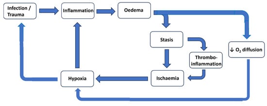
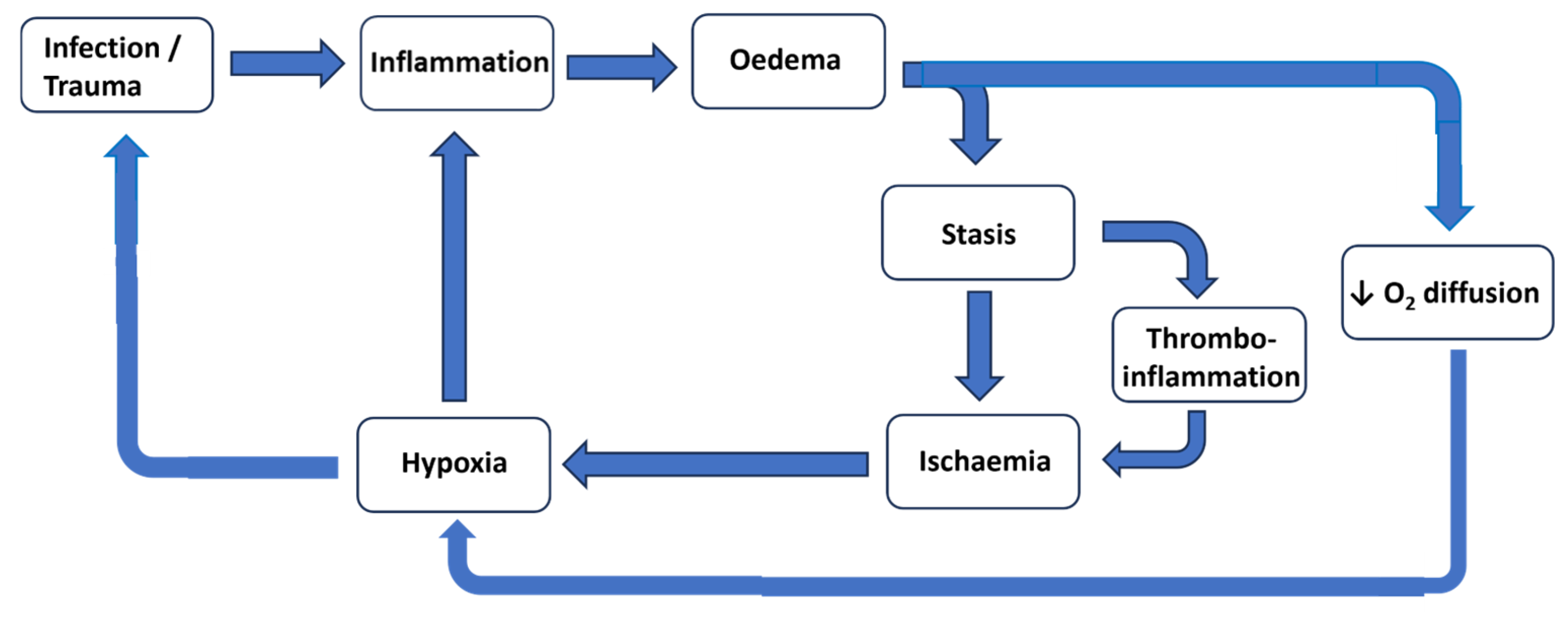
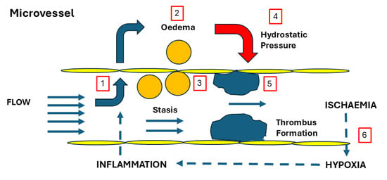
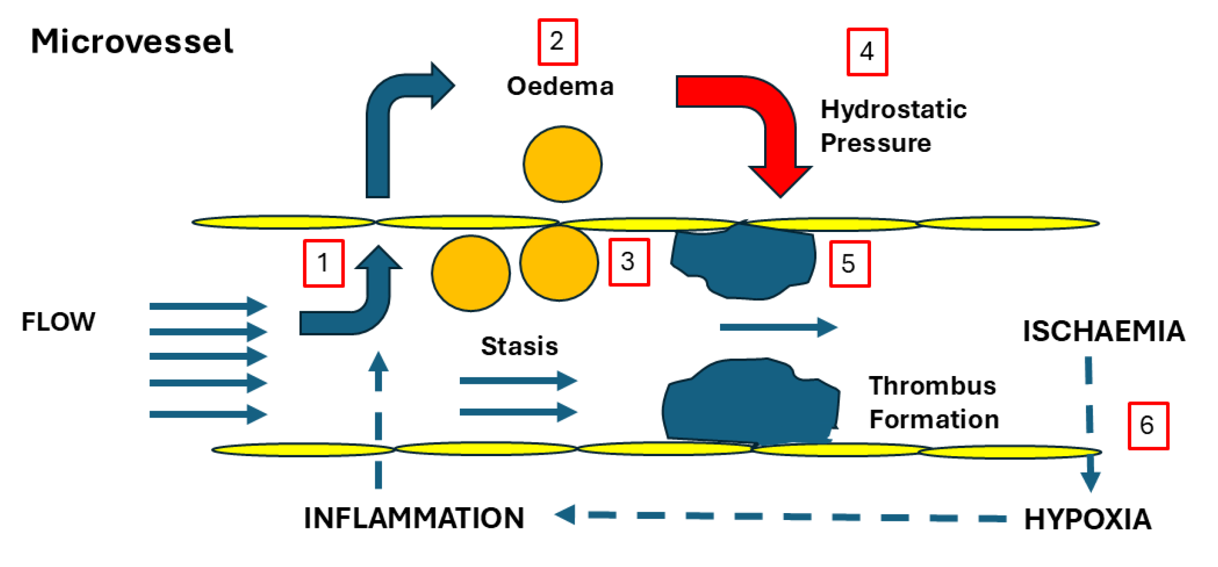

Disclaimer/Publisher’s Note: The statements, opinions and data contained in all publications are solely those of the individual author(s) and contributor(s) and not of MDPI and/or the editor(s). MDPI and/or the editor(s) disclaim responsibility for any injury to people or property resulting from any ideas, methods, instructions or products referred to in the content. |
© 2026 by the authors. Licensee MDPI, Basel, Switzerland. This article is an open access article distributed under the terms and conditions of the Creative Commons Attribution (CC BY) license.