


Abstract
Singlet oxygen is produced by light-dependent excitation of molecular oxygen using photosensitizers. Using blue light and riboflavin (RF) or riboflavin phosphate (RFP) as photosensitizers, we studied the reaction of singlet oxygen with tertiary amines in aqueous solution because many are used as biochemical buffers and as electron donors in photoreduction reactions. The reactions of singlet oxygen with multiple tertiary amines, including ethylenediamine tetraacetic acid (EDTA), bicine, and triethanolamine (TEOA), produced micromolar hydrogen peroxide (H2O2) as the stable end product. For bicine and TEOA, but not EDTA, H2O2 yield increased as pH increased due to their higher amine pKa values. A white LED used in conjunction with riboflavin and tertiary amines also produced H2O2, a contaminant likely to form during tissue culture manipulations. Direct photoreduction of 2,6-dichlorophenolindophenol and 2,3-dimethoxy-5-methyl-p-benzoquinone was achieved using blue light, RF or RFP, and tertiary amines as electron donors. With RF, EDTA was the optimal electron donor for both substrates, whereas with RFP, bicine and TEOA were superior to EDTA. Photochemical redox cycling of both quinones produced H2O2 via singlet oxygen-dependent re-oxidation of reduced quinols. Several amine buffers, including HEPES and PIPES, reacted with singlet oxygen to produce H2O2 but did not function as electron donors in quinone photoreduction assays.
1. Introduction
Visible light and numerous dye photosensitizers produce singlet oxygen (1O2), an excited state of molecular oxygen (O2) [1,2]. Foote and Wexler verified photosensitized production of 1O2, a reactive intermediate, in 1964 [3]. Methylene blue, Rose Bengal and phenosafranine are commonly used photosensitizers that produce 1O2 [1,4,5]. Biological photosensitizers include chlorophyll and its metabolites as well as riboflavin [6,7,8]. Most 1O2 research focuses on its light-dependent generation, with applications in synthetic chemistry, environmental settings, and living organisms [1,9,10].
Chlorophyll is the light harvesting pigment of photosynthesis, a process whereby red light initiates electron transfer from the excited state of chlorophyll to plastoquinone, a benzoquinone embedded in chloroplast membranes [11,12]. When red light is overabundant, the photoexcited state of chlorophyll initiates 1O2 formation [6,7]. To limit cellular damage, plants contain antioxidants that scavenge 1O2 and any resulting reactive oxygen species (ROS) [13]. Thus, the ability of a photosensitizer to initiate photoreduction and photo-oxidation concurrently is well established.
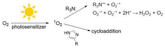
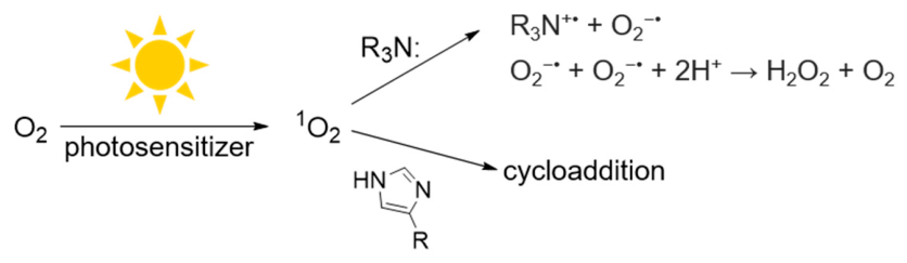
Because our lab employs tertiary amines (R3N) as electron donors in quinone photoreduction reactions, we are also interested in the direct reaction of tertiary amines with 1O2 [14,15]. The initial reaction of 1O2 and R3N produces superoxide anion and an amine radical cation, as shown in Scheme 1 [16]. Two superoxide anions disproportionate to H2O2 and regenerate O2.
Scheme 1.
Photochemical generation of 1O2 and its reaction with tertiary amines.
The reaction of tertiary amines with 1O2 has received little attention, and to our knowledge, only one report determined a rate constant in water for the reaction of 1O2 with EDTA, a commonly used tertiary amine, and with some metal–EDTA complexes [17]. Recently, we reported that the combination of either methylene blue or chlorin e6, multiple tertiary amines, and red light generated H2O2 [14]. Our goal was to determine relative rate constants for the reaction of the amines with 1O2 by using histidine as a competing 1O2 scavenger. Rate constants for the reaction of histidine with 1O2 in aqueous solution at multiple pHs have been reported [18,19,20] (Scheme 1). The histidine concentrations required to scavenge 1O2 varied for each tertiary amine but also depended on the photosensitizer employed [14]. We hypothesized that ionic interactions between the amines, histidine and the photosensitizer were responsible.
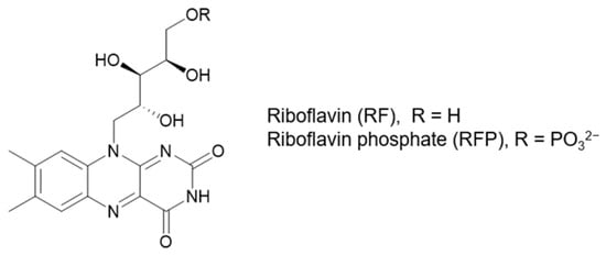
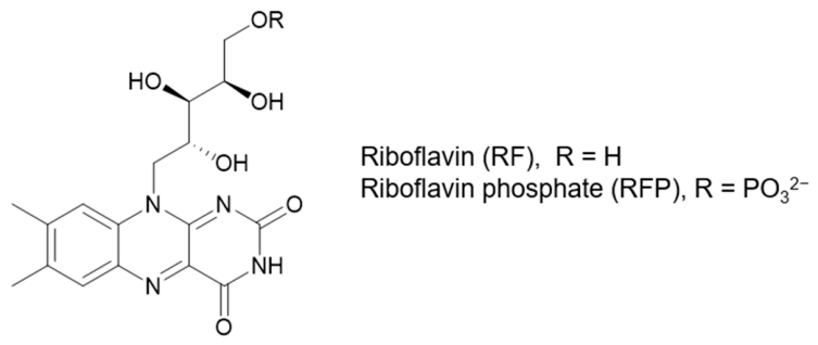
Our interest in riboflavin stems from its essential biological roles as a redox cofactor in enzyme catalyzed reactions and as a photosensor in organisms ranging from bacteria and plants to humans [21,22,23]. Riboflavin is an essential micronutrient synthesized by plants, fungi and many microorganisms that animals and humans obtain from their diet. Riboflavin can be phosphorylated to form riboflavin phosphate (RFP), which is also known as flavin mononucleotide (FMN) (Scheme 2).
Scheme 2.
Structures of riboflavin (RF) and riboflavin phosphate (RFP).
The riboflavin component of the redox cofactor, flavin adenine dinucleotide (FAD), of the Krebs cycle enzyme succinate dehydrogenase is essential for catalysis (light independent) [22]. Riboflavin as RFP cycles between its oxidized and reduced forms in the electron transport chain of mitochondria where ATP is produced. Notable light-dependent riboflavin-containing proteins include DNA photolyase, a bacterial enzyme that repairs thymine photodamage and cryptochromes in plants that detect blue light and initiate phototropism [24,25]. Mammalian cryptochromes that bind flavins also regulate circadian rhythms [26].
Riboflavin and its derivatives are of interest in photodynamic therapy (PDT) because they are nontoxic in biological settings and produce 1O2 and other ROS upon activation by blue light [8]. Ongoing research is evaluating their uses in dermatology, dentistry and ophthalmology [27].
As some of the most abundant dietary photosensitizers that can be excited by visible light, chlorophyll- and riboflavin-based molecules are of interest because they will be photoreactive when consumed [28,29]. Our work with chlorophyll metabolites and tertiary amine electron donors confirmed that they mediate benzo- and naphthoquinone photoreduction [15,30,31]. Only limited research has addressed photoreaction reactions mediated by RF and its derivatives. One notable example is work by Alonso-de Castro et al., where they studied RF-dependent two-electron photoreduction of platinum IV complexes using a tertiary amine as the electron donor [32,33,34].
Herein, our objectives were to measure H2O2 produced photochemically from the reactions of RF and RFP with multiple tertiary amines in aqueous solution. Further, we tested the tertiary amines for their ability to serve as electron donors to RF and RFP during catalytic cycles of quinone photoreduction. Photoreduction of the quinones DCPIP and CoQ0 is readily monitored because the oxidized forms have visible colors, whereas the reduced forms do not.
2. Materials and Methods
Materials: All chemicals were from Fisher Scientific or Sigma (St. Louis, MO, USA) and were of the highest purity available. CoQ0 stock solutions were prepared in DMF and used immediately or stored at −20 °C for up to a month. For RF, 5 mM stock solutions contained up to 15 mM NaOH to ensure solubility. RF and RFP solutions were prepared fresh daily, diluted with water prior to use (to 0.5 or 0.1 mM) and stored in the dark when not in use. All other solutions were prepared in water or 10 mM phosphate buffer (PB) pH 7.4. Horseradish peroxidase (HRP) Type I and bovine liver catalase from Sigma were dissolved to 2 mg/mL in 10 mM PB pH 7.4, and aliquots were stored at −80 °C. HRP concentration was determined from its absorbance at 403 nm (ε = 100,000 M−1 cm−1). Tertiary amine stock solutions in water (100 mM) were adjusted to pH 7.4 with NaOH or HCl. All phosphate buffers were equilibrated to room temperature (20–22 °C) to ensure no variation in dissolved O2.
Concentration measurements: All absorbance scans were performed using a Perkin-Elmer Lambda 35 UV/Visible spectrophotometer (PerkinElmer Life and Analytical Sciences, Inc., Shelton, CT, USA). For CoQ0, absorbance at 405 nm was used to calculate its concentration (740 M−1 cm−1 in 10 mM PB pH 7.4). DCPIP standard curves from 0 to 100 µM in 10 mM PB pH 7.4 or 6.4 were linear. For RF and RFP, absorbance at 450 nm was used to calculate concentrations (12,500 M−1 cm−1 in water or 10 mM PB pH 7.4) [23,35].
Light specifications: For blue light irradiation, a strip of blue LED lights (450 ± 10 nm, 5 W total) was affixed to a foam board mounted on a black foam core box (3 sides). The base of the box was covered in aluminum foil. The distance from the LEDs to the samples was 14 cm. Light was measured in lux with a light meter, and mW/cm2 at 450 nm was measured with a ThorLabs S120C power meter (ThorLabs, Newton, NJ, USA). Samples were positioned at the point of maximum light intensity, where lux readings were 1200–1300 and mW/cm2 was 0.68–0.70. Blue light irradiation times were optimized to limit photobleaching of RF or RFP and did not exceed 10 min. For white light irradiation, a 60-watt equivalent LED bulb was mounted 18 inches above the samples. Typical lux readings were 2350–2400.
Detection of H2O2 with TMB and HRP: For reactions containing only photosensitizers and tertiary amines, TMB and HRP were used to detect H2O2. Aliquots of photochemical reactions (20 µL) were combined with 0.5 mM TMB and 0.5 µM HRP in 10 mM PB pH 7.4 (100 µL total volume). After blue color development, 100 µL 1 M HCl was added [36]. Absorbance was read at 450 nm in a 96-well plate. A standard curve from 0 to 40 µM H2O2 was used to calculate H2O2 concentrations in the photochemical reactions.
DCPIP photoreduction assays: DCPIP (100 µM final) in 10 mM PB of varying pHs was combined with 2.5 µM RF or RFP and varying concentrations of tertiary amines. DCPIP photoreduction was performed in either a semimicro cuvette (1 mL) or a 96-well plate (200 µL). Absorbance (UV/Vis) scans for the cuvette or absorbance at 605 nm in the 96-well plate (200 µL total) was measured prior to irradiation and at time intervals. Dark samples containing all reactants served as controls. Prior to the initial absorbance reading, the cuvette was inverted three times to normalize dissolved O2.
Detection of H2O2 in photoreduction samples: HRP (8 µL, 50 µM) was added directly into the cuvette solution (1 mL) after irradiation and mixed gently to avoid introducing O2. Absorbance increases with HRP (0.4 µM final) indicative of re-oxidation of photoreduced DCPIP were monitored for up to 3 min until no further increase occurred. Controls were performed, where only 8 µL of buffer was added and mixed under identical conditions.
CoQ0 photoreduction assays: CoQ0 (0.5 mM) was combined with RF or RFP and tertiary amines in a semimicro cuvette (1 mL). Typical concentrations of CoQ0, RF or RFP and EDTA were 0.5 mM, 10 µM and 8–12 mM, respectively, in 10 mM PB pH 7.4. Absorbance (full UV/Vis) scans were measured prior to irradiation and at time intervals. Dark samples containing all reactants served as controls. Typical molar ratio of quinone to photosensitizer was 1000:1. Equivalent reactions (200 µL) were also performed in a 96-well plate, and absorbance at 405 nm was measured. As for DCPIP, HRP was added to determine H2O2 produced during irradiation.
Data analysis: For each experiment, at least two–three independent experiments were performed in duplicate or triplicate. Mean values were calculated using all data (typically 6–9 replicates). For figures showing error bars, error is the standard deviation. Details for each are stated in figure legends.
3. Results
3.1. Detection of H2O2
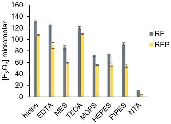
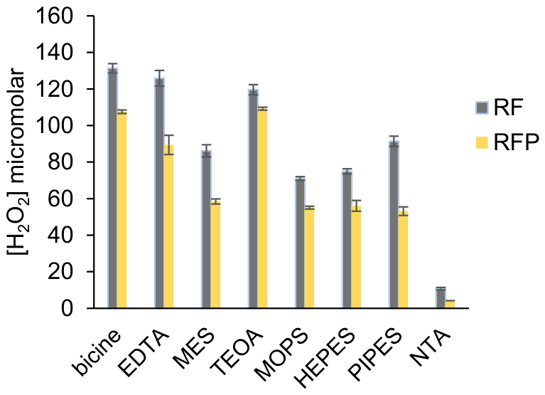
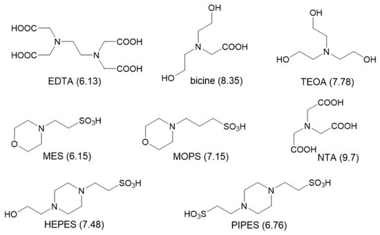
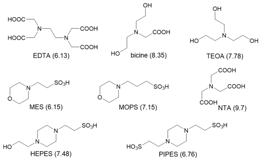
Figure 1 shows that the combination of RF or RFP, tertiary amines and blue light generated H2O2 as the stable end product. The structures of the amines tested are shown in Scheme 3. To detect H2O2, we used horseradish peroxidase (HRP) and a colorimetric co-substrate, tetramethylbenzidine (TMB), that generates a stable yellow product under acidic conditions (Supplemental Figure S1) [36]. In the absence of blue light or photosensitizer, no H2O2 was detected. If catalase, an enzyme that scavenges H2O2 according to Equation (1), was added prior to HRP and TMB, no yellow color was detected.
2H2O2 → 2H2O + O2
Figure 1.
H2O2 yield by RF or RFP, tertiary amines and blue light. Reactions (100 µL) contained 5 µM RF or RFP and 5 mM tertiary amine in 10 mM PB pH 7.4. Samples were irradiated for 5 min. Aliquots (20 µL) were combined with 10 mM PB pH 7.4, 0.5 mM TMB and 0.5 µM HRP (100 µL total). Reactions were quenched with 100 µL 1 M HCl, and absorbance was measured at 450 nm. [H2O2] was determined from a standard curve. These data are the average of three independent experiments performed in duplicate.
Scheme 3.
Structures of tertiary amines and their amine pKa values.
Phosphate buffers (PB) were used because phosphate does not react with 1O2 or other ROS. Two 1O2 are required to generate one H2O2, but because one O2 is regenerated, the stoichiometry is one O2 consumed per H2O2 (Scheme 1). The concentration of dissolved O2 is ~300 µM in 10 mM PB at 20–22 °C (measured with an oxygen electrode). The H2O2 concentrations shown in Figure 1 are all below 300 µM, confirming that dissolved O2 was not limiting.
When RF was used as the photosensitizer, reactions with bicine, EDTA and TEOA and blue light generated the most H2O2, followed by PIPES, MES, HEPES and MOPS (Figure 1). With RFP, all H2O2 yields were lower than RF, and TEOA and bicine generated more H2O2 than EDTA. H2O2 yields with NTA were negligible regardless of photosensitizer, despite similarities in structure to both EDTA and bicine (Scheme 3).
3.2. pH Dependence of H2O2 Yield
To react with 1O2 as shown in Scheme 1, tertiary amines must be deprotonated. Therefore, we examined the pH dependence of H2O2 formation using the most reactive tertiary amines in Figure 1 (pH 7.4). In addition to bicine, EDTA, and TEOA, MES was also tested at multiple pH values because Alonso-de Castro et al. employed it as an electron donor in their work on platinum complex photoreduction at pH 6.0 [32,33,34].
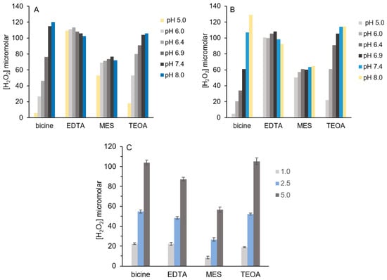
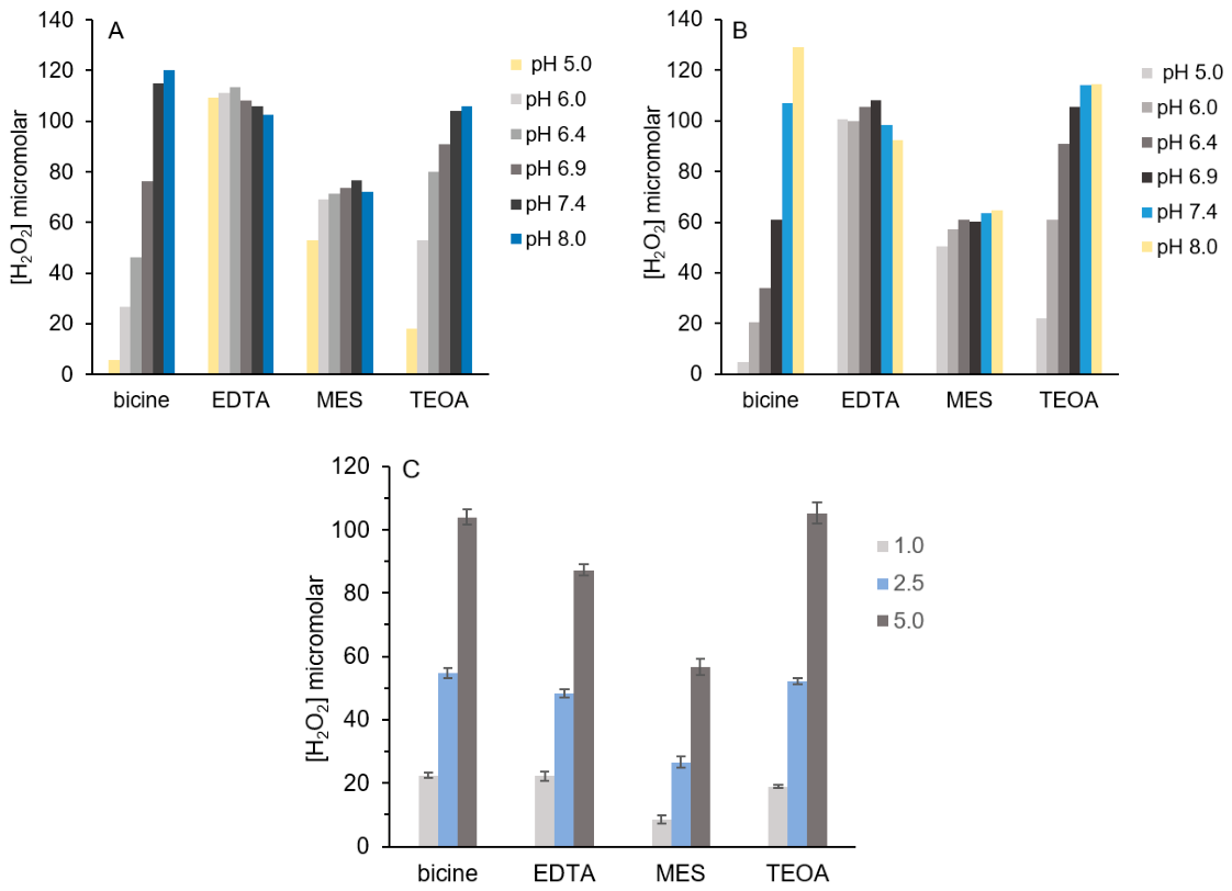
In Figure 2A, using RF as the photosensitizer, bicine showed the greatest pH dependence, with a 20-fold increase in H2O2 as phosphate buffer pH increased from 5 to 8. For TEOA, H2O2 yield increased 6-fold as pH increased. The H2O2 yield did not change when EDTA was assayed across this pH range, and MES showed only a modest increase in H2O2. For MES, once pH was 6, H2O2 yield was constant (Figure 2A).
Figure 2.
pH- and time-dependence of tertiary amine reactions with RF or RFP and blue light. Reactions (100 µL) containing 5 µM RF or RFP and 5 mM tertiary amine in 10 mM PB of varying pHs were irradiated for 5 min. (A) RF. (B) RFP. (C) Reactions contained 5 µM RFP and were irradiated for 1–5 min. These data are the average of three independent experiments performed in duplicate.
All the amines tested with RFP in Figure 2B showed the same pH dependence as for RF. We attribute the results in Figure 2A,B to differences in amine pKa values (given in Scheme 3). Bicine and TEOA have amine pKa values of 8.35 and 7.78, respectively, whereas those of EDTA and MES are 6.13 and 6.15. While EDTA has two amines and therefore two pKa values, the second value of 10.37 renders that amine unreactive in this pH range. Further, it is unlikely that both amines of an EDTA molecule would react with 1O2. For bicine and TEOA, as pH increases, their amines are deprotonated and consequently capable of reacting with 1O2.
Amine pKa values also explain H2O2 yields in Figure 1 for PIPES and HEPES using RF. Although they are similar in structure (Scheme 3), PIPES has a lower pKa of 6.76 vs. 7.48 for HEPES. Consequently, more H2O2 was generated by the PIPES reaction. Likewise, MES and MOPS are similar in structure, but MES, with its lower pKa of 6.15 vs. 7.15 for MOPS, yielded more H2O2. NTA has the highest pKa of the amines tested (9.7) and consequently yielded the least H2O2 at pH 7.4 regardless of photosensitizer.
The difference in HEPES and PIPES reactivity in Figure 1 using RF was not observed with RFP. PIPES has two negative charges at pH 7.4 due to its sulfonates; HEPES has only one. Because RFP is also negatively charged, charge repulsion between it and PIPES is likely responsible for the lower H2O2 yield in Figure 1 relative to HEPES. This observation reinforces that ionic interactions between the photosensitizer and the tertiary amine influence reaction outcome.
H2O2 yields in Figure 1 and Figure 2A,B were measured after 5 min of blue light irradiation. A time course of H2O2 production in Figure 2C shows no noteworthy differences in H2O2 production after 1 or 2.5 min, and results for bicine, EDTA and TEOA are identical. Results for the other amines were all comparable with time-dependent increases in H2O2.
Histidine is a well-characterized 1O2 scavenger that effectively traps O2 via a 4 + 2 cycloaddition reaction [20,37]. This differs from other 1O2 scavengers like azide that regenerates O2 and produces azide-based radicals [38,39]. When millimolar histidine was added to reactions with select tertiary amines as described in Figure 1, H2O2 yields decreased (Supplemental Figure S2). This was observed when both RF and RFP were employed.
Many of the tertiary amines shown in Scheme 3 are biochemical buffers, including HEPES, MES, MOPS, PIPES and TEOA. EDTA is routinely added to buffers to scavenge redox-active metal cations that are required for ROS generation. Further, tissue culture media is supplemented with low micromolar concentrations of RF because it is essential for cell growth and survival. Figure 1 and Figure 2 confirm that blue light irradiation produces up to 120 µM H2O2 in 5 min using only 5 µM RF or RFP. Amines used as buffers are commonly at millimolar concentrations equal to or greater than 5 mM used in Figure 1 and Figure 2.
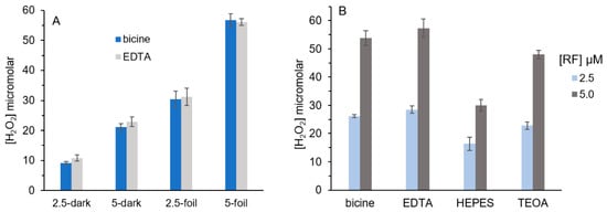
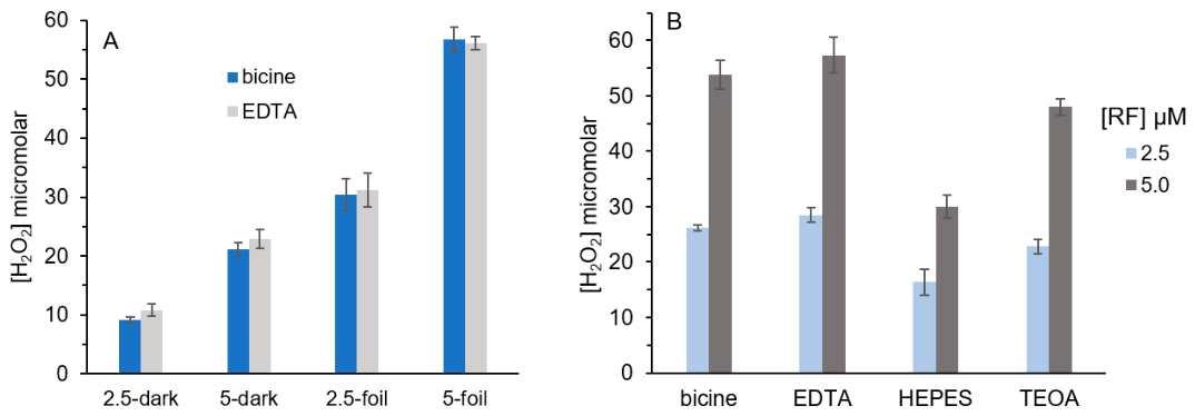
To mimic reaction conditions during tissue culture manipulations, we tested the combinations of RF and tertiary amines for H2O2 production using a 60-watt equivalent white LED bulb. RF concentrations of 2.5 or 5 µM were assayed with 5 mM bicine or EDTA for 10 min. Additionally, samples were either irradiated on a foil-lined surface or a black lab bench. Figure 3A shows that H2O2 was detected under all conditions, and the highest H2O2 was detected with 5 µM RF on the foil-lined surface.
Figure 3.
H2O2 yield by RF and tertiary amines under white LED light. Reactions (100 µL) contained varying RF and 5 mM tertiary amine in 10 mM PB pH 7.4. Samples were irradiated for 10 min. (A) Samples containing 2.5 or 5 µM RF were positioned 18 inches from a white LED on a foil-lined or black surface. (B) All samples were on a foil-lined surface 18 inches from a white LED. These data are the average of two independent experiments performed in triplicate.
When HEPES and TEOA in addition to bicine and EDTA were tested at 2.5 or 5 µM RF under the white light for 10 min, similar micromolar H2O2 was detected (Figure 3B). HEPES is a very common buffer used in biochemical experiments near physiological pH. Further, Zigler et al. reported H2O2 production in tissue culture media that contained riboflavin and HEPES after exposure to visible light. Data in Figure 3A,B supports their findings but also confirms that H2O2 generation is not limited to HEPES. All tertiary amines in Scheme 3 were tested under the white light, and H2O2 was detected with all except NTA.
Our interest in tertiary amine reactivity stems from our quinone photoreduction work. Tertiary amines are routinely used as sacrificial electron donors in photoreduction experiments [40]. In our work on para-benzoquinone and naphthoquinone photoreduction, we routinely use EDTA in combination with methylene blue and red light [15,30,31]. Likewise, we have reported that the primary dietary chlorophyll metabolite pheophorbide A catalyzes quinone photoreduction with TEOA as the best electron donor (red light dependent) [15,41]. In those studies, quinone photoreduction was optimized using different combinations of photosensitizer and electron donor.
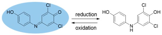
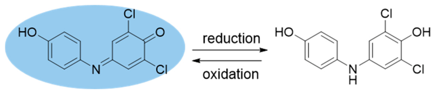
Recently, we reported that DCPIP, a quinoneimine, was readily reduced using methylene blue, EDTA and red light. DCPIP is a well-characterized redox indicator that is blue in its oxidized form and colorless when reduced (Scheme 4) [42]. Oxidized DCPIP absorbs strongly at 605 nm and is water soluble; the molar absorptivity is pH-dependent (Supplemental Figures S3–S6). DCPIP is also used to measure ascorbic acid [43]. In the Hill reaction of chloroplasts, DCPIP serves as the terminal electron acceptor [44].
Scheme 4.
Redox reaction of DCPIP.
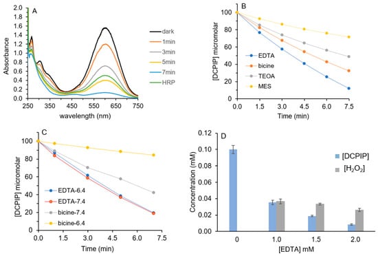
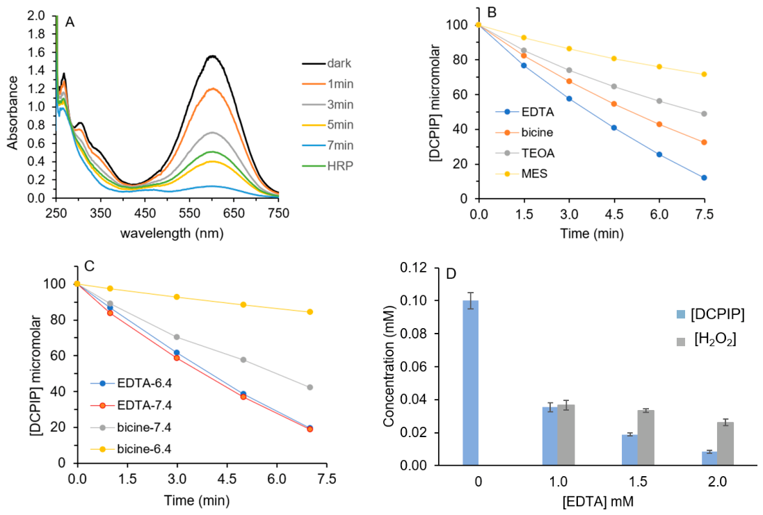
We sought to test RF and RFP as photosensitizers for DCPIP photoreduction in a manner analogous to our prior work. Figure 4A shows that the combination of RF, EDTA and blue light reduced DCPIP. We performed absorbance scans in a 1 mL semimicro cuvette so that it could be irradiated and scanned directly at the time intervals to limit mixing and O2 exposure. After 7 min total of light exposure in the cuvette, 95% of the 100 µM DCPIP was reduced using 2.5 µM RF and 2 mM EDTA. For photocatalysis studies, both the ratio of substrate to photosensitizer (100 µM to 2.5 µM, 40×) and the ratio of electron donor to photosensitizer (2 mM to 2.5 µM; 800:1) are routinely reported.
Figure 4.
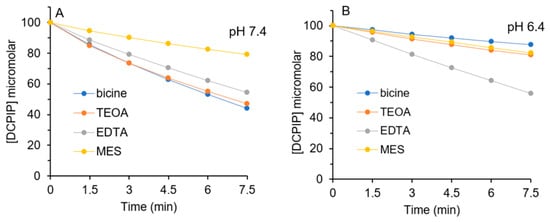
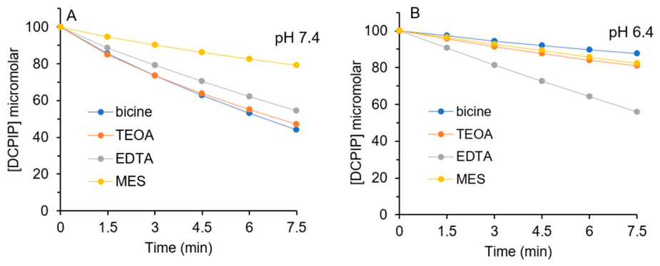
DCPIP photoreduction by RF and tertiary amines. (A) Reactions (1 mL in a quartz cuvette) contained 100 µM DCPIP, 2.5 µM RF and 2 mM EDTA in 10 mM PB pH 7.4 in a quartz cuvette. A blue light source with maximum output at 450 nm was employed, and DCPIP photoreduction was monitored at 605 nm. The increase in A605 after HRP addition (0.4 µM final) was used to calculate [H2O2]. (B) Samples (200 µL) contained 100 µM DCPIP, 2.5 µM RF and 2 mM amine in 10 mM PB pH 7.4. Samples were irradiated in a 96-well plate for up to 7.5 min. The decrease in A605 was used to calculate [DCPIP] at each time. These data are the average of three independent experiments. (C) Samples were prepared as in (B) with 1.5 mM amine and 10 mM PB pH 7.4 or 6.4. These data are the average of two independent experiments performed in duplicate. (D) Samples were prepared as in (A), except samples were irradiated for 7.5 min and [EDTA] varied. These data are the average of three independent experiments.
After irradiation, horseradish peroxidase (HRP) was added, and an increase in DCPIP absorbance was detected as a result of its re-oxidation (Figure 4A). HRP is an enzyme that requires H2O2 to oxidize organic substrates, including catechols and quinols [45,46,47]. Because reduced DCPIP is only oxidized by HRP when H2O2 is available, oxidized, blue DCPIP was regenerated when HRP was added after irradiation (Scheme 5). The increase in DCPIP absorbance in Figure 4A confirms that H2O2 was produced as DCPIP was photoreduced. When an equivalent volume of buffer was added to the cuvette instead of HRP, no increase in DCPIP was observed. If catalase was added prior to HRP, DCPIP absorbance did not increase.
Scheme 5.
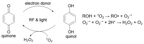
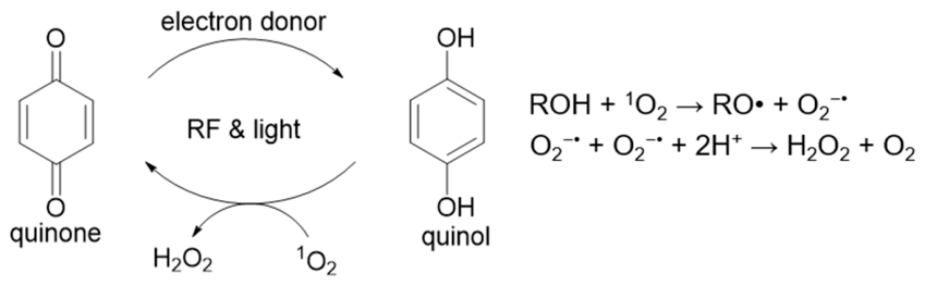
Photochemical redox cycling of quinones to produce H2O2.
There are two possible sources of H2O2 during irradiation of DCPIP, RF and EDTA. The combination of RF and EDTA generates H2O2 from 1O2, as described in Figure 1, Figure 2 and Figure 3. H2O2 will also form if 1O2 reacts with reduced quinol (ROH in Scheme 5) [48]. Hydrogen atom abstraction from the quinol by 1O2 produces a semiquinone and peroxyl radical (•OOH). The pKa of •OOH is 4.8; therefore, it will deprotonate at neutral pH to a superoxide anion that becomes H2O2 [49]. Two semiquinones (RO•) disproportionate rapidly to yield a quinol and regenerate a quinone (blue DCPIP). These results confirm that both photo-oxidation of O2 to 1O2 and photoreduction of DCPIP via an electron transfer mechanism are mediated by the excited state of RF.
All tertiary amines in Scheme 3 were tested for their ability to serve as electron donors for DCPIP photoreduction by RF. Figure 4B shows the time courses of DCPIP photoreduction by RF using EDTA, bicine, TEOA and MES at pH 7.4. Of the other amines in Scheme 3, MOPS and NTA showed minimal DCPIP reduction by RF. No DCPIP photoreduction was detected using HEPES and PIPES under the reaction conditions employed.
Given the pH dependence observed in Figure 2A, EDTA and bicine were also tested at pH 6.4 as electron donors for DCPIP photoreduction (Figure 4C). There was no difference in the rate of DCPIP photoreduction at pH 7.4 vs. pH 6.4 with EDTA. However, for bicine, the rate was ~4× slower at pH 6.4 vs. pH 7.4. We attribute this difference to the amine pKa values of 6.16 and 8.35 because the amines must be deprotonated to serve as electron donors.
Table 1 summarizes DCPIP photoreduction rates mediated by EDTA and MES at pH 7.4 and 6.4 using three different concentrations of each electron donor. Regardless of the amine concentration, reactions with bicine were ~4× slower at pH 6.4 vs. 7.4, consistent with Figure 4C. When additional experiments were performed at pH 8, a further increase in the rate of DCPIP photoreduction was observed with bicine but not with EDTA.
Table 1.
Rates of DCPIP reduction by RF and bicine or EDTA.
As shown in Figure 4A, HRP addition and the subsequent increase in DCPIP absorbance confirmed that H2O2 was produced. Additional samples containing different EDTA concentrations were irradiated and monitored in a cuvette to determine the change in DCPIP absorbance after HRP addition. This increase can be converted to a H2O2 concentration because, for each DCPIP that is re-oxidized, one H2O2 is consumed (Scheme 5).
Figure 4D summarizes the decrease in DCPIP and the increase in H2O2 at three EDTA concentrations. In the absence of EDTA, no DCPIP was photoreduced. The highest concentration of H2O2 detected was 37 µM with 1 mM EDTA, and H2O2 decreased as EDTA increased (Figure 4D). If EDTA were reacting with 1O2, H2O2 would be expected to increase as EDTA increased.
Controls were performed, where equivalent concentrations of RF (2.5 µM) and EDTA (1–2 mM) were irradiated for 7.5 min either in a cuvette or in a plate well (200 µL vol) but without DCPIP. Under these conditions, H2O2 concentrations increased from 61 µM to 79.5 µM as EDTA increased from 1 to 2 mM. H2O2 concentrations in the absence of DCPIP are greater than those in Figure 4D at all EDTA concentrations tested. Increased electron donor favored the photoreduction pathway, thereby limiting 1O2 formation and its reaction with EDTA or quinol, the two possible sources of H2O2.
The rates summarized in Table 1 and Figure 4B,C were measured using 200 µL samples in a 96-well plate, whereas those in Figure 4A,D were obtained in a 1 mL cuvette. The plate format is more amenable to obtaining replicate data, but O2 exposure is greater in the plate well vs. in a narrow microcuvette. For this reason, DCPIP photoreduction rates in Figure 4A–D and Table 1 are not identical.
DCPIP photoreduction was monitored using RFP as the photosensitizer with different amine electron donors. Figure 5A shows that, with RFP, bicine and TEOA were equivalent and more effective than EDTA at pH 7.4. Although reaction conditions were identical to those in Figure 4B, photoreduction rates with RFP were slower than with RF, consistent with the results in Figure 1. Only 56% of the 100 µM DCPIP was photoreduced with RFP and either bicine or TEOA vs. 90% in Figure 4B using RF and EDTA. MES was the least effective of the electron donors in Figure 4B, and only minimal DCPIP photoreduction was detected with equal concentrations of MOPS and NTA. HEPES and PIPES were not effective as electron donors under the reaction conditions used, consistent with the results obtained with RF.
Figure 5.
DCPIP photoreduction by RFP and tertiary amines. Samples (200 µL) contained 100 µM DCPIP, 2.5 µM RFP and 2 mM amine in 10 mM PB. Samples were irradiated in a 96-well plate for up to 7.5 min. The decrease in A605 was used to calculate [DCPIP] at each time. (A) pH 7.4; (B) pH 6.4. These data are the average of three independent experiments. All errors were less than 5%.
When DCPIP photoreduction with RFP and select electron donors was performed at pH 6.4, the pH dependence of bicine and TEOA was evident because the time course of DCPIP photoreduction slowed precipitously. At pH 6.4, EDTA was the best electron donor with RFP (Figure 5B).
In addition to DCPIP, we routinely use 2,3-dimethoxy-5-methyl-p-benzoquinone (CoQ0) as a photoreduction substrate [15,41]. CoQ0 is a more water-soluble analog of CoQ10, the benzoquinone that cycles between its oxidized and reduced forms in the electron transport chain in mitochondria. Oxidized CoQ0 absorbs at 405 nm, whereas its reduced form does not. Because the molar absorptivity of CoQ0 is much lower than that of DCPIP, higher CoQ0 concentrations were necessary for reliable data.
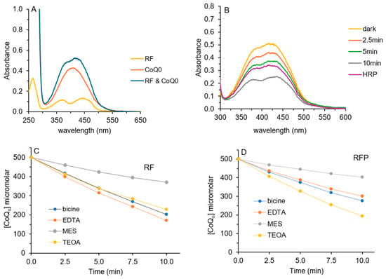
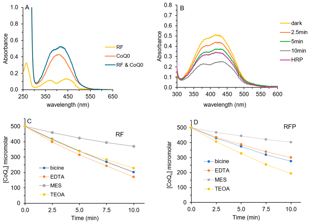
Prior to any photoreduction experiments, UV/visible spectra of RF and CoQ0 and the combination of the two were obtained because of considerable overlap between 350 and 500 nm (Figure 6A). The concentrations of RF (10 µM) and CoQ0 (0.5 mM) were optimized to monitor CoQ0 photoreduction in a 1 mL cuvette.
Figure 6.
CoQ0 photoreduction by RF or RFP and amines. (A) Samples (1 mL) contained 10 µM RF, 0.5 mM CoQ0 or both in 10 mM PB pH 7.4. (B) 10 µM RF, 10 mM EDTA and 0.5 mM CoQ0 in 10 mM PB pH 7.4 (1 mL) were irradiated for up to 10 min. CoQ0 photoreduction was monitored at 405 nm. The increase in A405 after HRP addition was used to calculate [H2O2]. (C) Samples (200 µL) contained 0.5 mM CoQ0, 10 µM RF and 10 mM amine in 10 mM PB pH 7.4. (D) samples were prepared as in (C), except 10 µM RFP was used. Samples were irradiated in a 96-well plate for up to 10 min. The decrease in A405 was used to calculate [CoQ0] at each time. Data in (C,D) are the averages of three independent experiments. Error did not exceed 5% for any sample.
Figure 6B shows that the combination of RF, EDTA and blue light photoreduced CoQ0 because absorbance centered at 405 nm decreased. After 10 min, HRP was added, and oxidized CoQ0 absorbance increased, confirming that H2O2 was produced during CoQ0 photoreduction. Because RF also absorbs at 405 nm, full reduction of 0.5 mM CoQ0 would not decrease absorbance to zero. From the absorbance of RF at 405 nm in Figure 6A, we can estimate that CoQ0 absorbance would decrease to ~0.08 if all CoQ0 were reduced. However, some photobleaching occurred when RF alone was exposed to equivalent blue light over 10 min (Supplemental Figure S7). While photobleaching decreased RF absorbance at 450 nm, absorbance at 403–408 nm did not decrease. It is unclear if the same level of RF photobleaching occurs during photoreduction when electron donors and CoQ0 are present. For this reason, we can only estimate turnover numbers for RF or RFP as photosensitizers using yellow CoQ0 as a photoreduction substrate.
Turnover numbers with CoQ0 (mol quinone reduced/mol photosensitizer) are similar to those observed with DCPIP (Figure 4 and Figure 5). For DCPIP, the maximum turnover number was 40 (100 µM DCPIP and 2.5 µM RF or RFP), whereas with CoQ0, the maximum possible is 50 (0.5 mM CoQ0 and 10 µM RF or RFP). These values are similar to those measured in our photoreduction studies using these quinones and red light with MB or chlorophyll metabolites [15,30,31]. The ratios of amine to photosensitizer were also consistent with our prior work. For DCPIP photoreduction, the highest ratios were 800:1 (2 mM amine and 2.5 µM RF or RFP) and 1200:1 for CoQ0 (12 mM amine and 10 µM RF or RFP).
In Figure 6C, different amine electron donors were tested for their ability to support CoQ0 photoreduction at pH 7.4. Here, only absorbance at 405 nm was measured (96-well plate format). To calculate CoQ0 concentrations, the absorbance of RF at 405 nm was subtracted from that of CoQ0 (dark). EDTA, bicine and TEOA were equally effective. As for DCPIP photoreduction using RF, CoQ0 photoreduction was detected using MES and, to a lesser extent, MOPS and NTA. No photoreduction was detected using HEPES or PIPES. With RFP, TEOA was superior to bicine or EDTA as the electron donor for CoQ0 photoreduction (Figure 6D).
4. Discussion
Our results confirm that the combination of RF or RFP, tertiary amines and blue light produces H2O2 in neutral, aqueous solution. Numerous tertiary amines, including several that are used routinely as biochemical buffers, produced 60–130 µM H2O2 under ambient oxygen conditions (Figure 1). For all amines tested, RF was superior to RFP as the photosensitizer. H2O2 yields in Figure 1 are comparable to those we obtained using red light, tertiary amines and the photosensitizers methylene blue (MB) and chlorin e6 in our prior work [14].
The pH dependence of the photochemical reaction of tertiary amines with 1O2 reinforced the importance of amine pKa values (Figure 2 and Scheme 3). Increased H2O2 yields at higher pH for bicine and TEOA regardless of photosensitizer were evident because those amines, unlike EDTA or MES, have higher pKa values, and only the deprotonated amines react with 1O2.
In our prior work, EDTA showed a modest two-fold increase in H2O2 with MB across the same phosphate buffer range (pH 5–8) but not with chlorin e6 [14]. Although TEOA showed pH-dependent increases in H2O2 with both MB and chlorin e6, neither increase was comparable to the six-fold increase shown in Figure 2A,B with RF or RFP. MB has a positive charge in this pH range, and chlorin e6 has three negative charges [50]. RF is neutral, while RFP has a negatively charged phosphate (Scheme 2). Thus, the identity and chemical properties of the photosensitizer, in addition to the amine pKa values, are likely to influence H2O2 yields.
To determine a rate constant for the reaction of 1O2 and EDTA and its metal complexes, Hessler et al. used Rose Bengal as the photosensitizer, and they measured quenching of 1O2 phosphorescence at 1270 nm [17]. Their reported rate constant on the order of ~105 M−1 s−1 for 1O2 and EDTA is likely dependent on the photosensitizer in addition to the identity of the tertiary amine. It is expected that optimal scavenging of 1O2 by tertiary amines requires 1O2 generation in close proximity to the photosensitizer.
Although histidine scavenged 1O2 and decreased H2O2 yields in reactions of RF and RFP with tertiary amines, higher concentrations were required than in our prior work with MB and chlorin e6 (Supplemental Figure S2), even though 5 mM amine was used in both. Up to 5 mM histidine was used herein, whereas we observed more complete 1O2 scavenging with only 1.0–1.5 mM histidine when MB or chlorin e6 were used as the photosensitizers [14]. As for the different photosensitizer/amine combinations, optimal scavenging of 1O2 by histidine may depend on its interaction with photosensitizers of variable charge and polarity.
Our work using a white LED is important because we detected micromolar H2O2 from the reaction of RF, tertiary amines, and visible light. The stainless steel surfaces of laminar flow hoods are reflective, and exposure to light can extend well beyond the 10 min employed in Figure 3A,B. Zigler et al. reported H2O2 production in tissue culture media that contained RF and HEPES after exposure to visible light using 25 mM HEPES vs. only 5 mM in Figure 3B [51]. Recently, Liu et al. tested some primary, secondary and tertiary amines (all at 25 mM) for H2O2 generation in RPMI 1640, a common tissue culture medium [52]. While they detected micromolar H2O2 with tertiary amines, they did not test bicine, EDTA or TEOA, the most reactive amines at pH 7.4 in Figure 1. Further, none of these studies addressed the pH dependence of the photochemical reaction between RF or RFP and tertiary amines.
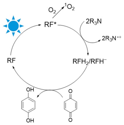
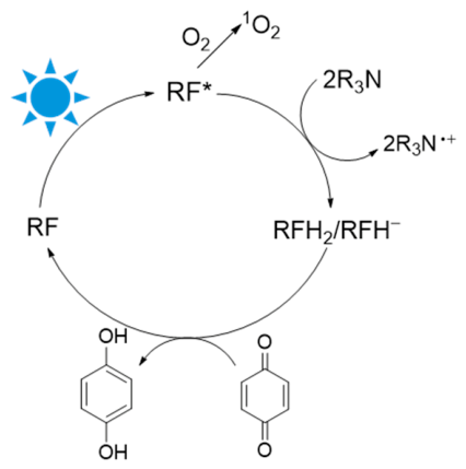
A major goal of this work was to assess the effectiveness of tertiary amines as electron donors in quinone photoreduction reactions mediated by RF and RFP. In our prior work, the parallel reaction of tertiary amines with 1O2 as benzo- and naphthoquinones were photoreduced was observed regardless of the photosensitizer and amine electron donor employed [15,30,31]. The photoexcited, triplet state RF* will either react with O2 to form 1O2 or accept electrons from tertiary amines (Scheme 6). Once reduced to RFH2, electron donation to quinone substrates occurs to regenerate RF. RFH2 has a pKa of 6.7; consequently, some RFH− will be present at neutral pH [53]. Any 1O2 produced has two reaction paths that produce H2O2: reaction with a tertiary amine as shown in Scheme 1 or with the quinol photoreduction product (Scheme 5).
Scheme 6.
Photochemical oxidation and reduction pathways of RF.
Both DCPIP and CoQ0 are excellent model substrates for photoreduction because their oxidized forms have a visible color that is lost upon reduction. Further, their reduced forms are co-substrates for HRP, providing a simple method to quantify H2O2 (Scheme 5). Results in Figure 4A and Figure 6B confirm that H2O2 was generated during photoreduction of both DCPIP and CoQ0. Although both of these experiments used EDTA, H2O2 was detected in all DCPIP and CoQ0 photoreduction experiments with RF and RFP regardless of the tertiary amine used.
When irradiation is first initiated, only the amine can react with 1O2 because no quinol is present. As the reduced quinol forms, it becomes the likely substrate for hydrogen atom abstraction by 1O2. Rate constants for the reaction of 1O2 with quinols in aqueous solution are ~109 M−1 s−1, several orders of magnitude greater than the published rate constant of ~105 M−1 s−1 for 1O2 and EDTA [48]. It is not possible to differentiate the source of H2O2 in our experiments, and amine cation radicals R3N•+ are the initial products of both amine photo-oxidation (Scheme 1) and electron donation in photoreduction (Scheme 6). Further decomposition of R3N•+ to form well-characterized, inert products would be identical [40].
In addition to concurrent quinone photoreduction and 1O2 generation, we determined the best tertiary amine donors for RF and RFP by comparing rates of photoreduction for DCPIP and CoQ0. Figure 4B and Figure 5A show differences in donor compatibility for DCPIP photoreduction at pH 7.4 with RF and RFP, respectively. While EDTA was best with RF, TEOA and bicine were superior to EDTA with RFP. Electrostatics likely play a role because EDTA has four negative charges at pH 7.4, but bicine has only one, and TEOA is uncharged. Any charge repulsion between EDTA and RFP would be expected to diminish electron donation (Scheme 6).
For CoQ0 photoreduction with RF, the same amine ranking as for DCPIP was observed (Figure 4B vs. Figure 6C). This observation supports the catalytic cycle depicted in Scheme 6, where photoexcited RF receives electrons from the amine, followed by electron donation to the quinone substrate. Likewise for RFP, TEOA and bicine were superior to EDTA (Figure 5A vs. Figure 6D).
DCPIP photoreduction rates using RF and either EDTA or bicine at pH 7.4 vs. 6.4 (Figure 4C and Table 1) were consistent with H2O2 yields in Figure 2A at different pH values. As in their reaction with 1O2, amines must be deprotonated to donate electrons to RF*. Bicine showed pH-dependence, but EDTA did not (Figure 4C and Table 1).
EDTA, bicine and TEOA were the most reactive amines with 1O2 (Figure 1) and also the best electron donors at pH 7.4. Although MES, MOPS, HEPES and PIPES reacted with 1O2 to produce H2O2, only modest photoreduction was observed with MES and MOPS. No photoreduction was observed with HEPES or PIPES. All four have sulfonate groups that would be negatively charged, and their pKa values are similar to EDTA, bicine and TEOA. Given that negatively charged amines like EDTA and bicine were very effective electron donors, optimal electron donation requirements are not clear.
Photoreduction turnover numbers (mol DCPIP reduced/mol RF) of ~35 were achieved with 100 µM DCPIP, 2.5 µM RF, and 2 mM EDTA (Figure 4) at pH 7.4. For RFP, turnover numbers were 20–25 with 100 µM DCPIP, 2.5 µM RFP, and 2 mM bicine (Figure 5A). These are comparable to values of 40–45 with MB and EDTA in prior work using DCPIP. Increased electron donor increased photoreduction and decreased H2O2 formation (Figure 4D), which is consistent with Scheme 6. For CoQ0, measurements were complicated by overlapping absorbances of RF or RFP and CoQ0 (Figure 6A,B). Nonetheless, we estimate ~30–35 turnovers in Figure 6B with EDTA as the electron donor. While tertiary amines are not biologically relevant electron donors for photoreduction, it is important to show that photoreduced flavins can donate electrons to substrates like CoQ0, an analog of CoQ10, a key component of the electron transport chain in mitochondria.
The photocatalytic cycle depicted in Scheme 6 mirrors the work of Alonso-de Castro et al., where they used blue light, RF, and the tertiary amine MES at pH 6.0 to photoreduce platinum (IV) prodrugs to activated platinum (II) complexes [32,33,34]. This methodology was used successfully to induce DNA damage in Capan-1 cells because the DNA targeted-platinum (II) drugs were produced, and ROS were generated simultaneously [32]. Their work is one of the only detailed examples of photoreduction using RF or RFP in conjunction with a tertiary amine electron donor. However, they did not evaluate multiple tertiary amines as we have, and their reasoning for performing the reaction at pH 6.0 was not clear.
Understanding RF photoreactivity in a human nutritional context is essential because RF is added to foods, taken as a vitamin supplement, and consumed as a natural food dye. While most RF is consumed as FMN or FAD that are in many of foods, free RF circulates through the blood stream mostly bound to transporter proteins [21]. At least 90 proteins in humans require riboflavin and its derivatives for activity [22]. Blue light penetrates skin, and there are specific photoreceptor proteins in the eye that utilize RF [27].
While photochemical H2O2 generation by RF and amines under cell culture conditions is undesirable, our work suggests a photochemical method to generate H2O2. This work is an example of green chemistry that uses biocompatible RF, common tertiary amines like EDTA, blue light, and ambient O2 in neutral, aqueous solution. While dissolved O2 will become limiting, this can be rectified by bubbling solutions with air or O2 gas. Unlike current H2O2 industrial processes that rely on a Pd catalyst, H2 and O2 gases, anthraquinone as a redox cycler, and a mixture of organic solvents, our reactants are nontoxic, and no organic solvents are required [54,55].
Supplementary Materials
The following supporting information can be downloaded at https://www.mdpi.com/article/10.3390/oxygen6030021/s1, Figure S1: H2O2 standard curve with TMB and HRP—0 to 40 μM linear; Figure S2: Effect of histidine on H2O2 yields in photochemical reactions; Figure S3: UV/Vis scans of DCPIP (20–100 µM) in 10 mM PB pH 7.4; Figure S4: DCPIP standard curve in 10 mM PB pH 7.4; Figure S5: UV/Vis scans of DCPIP (20–100 µM) in 10 mM PB pH 6.4; Figure S6: DCPIP standard curve in 10 mM PB pH 6.4; Figure S7: Photobleaching of RF.
Author Contributions
Conceptualization, L.M.L.; methodology, A.T., H.M., A.M. and L.M.L.; formal analysis, A.T., H.M. and L.M.L.; investigation, A.T., H.M., A.M. and L.M.L.; writing—original draft preparation, L.M.L.; writing—review and editing, L.M.L.; supervision, L.M.L.; project administration, L.M.L. All authors have read and agreed to the published version of the manuscript.
Funding
This research received no external funding.
Institutional Review Board Statement
Not applicable.
Informed Consent Statement
Not applicable.
Data Availability Statement
Raw data, detailed procedures and all Excel files are available upon request.
Acknowledgments
The authors acknowledge the William & Mary Green Fee for internal support.
Conflicts of Interest
The authors declare no conflicts of interest.
Abbreviations
The following abbreviations are used in this manuscript:
| bicine | Bis(2-hydroxyethyl)amino]acetic acid |
| CoQ0 | 2,3-dimethoxy-5-methyl-p-benzoquinone |
| DCPIP | 2,6-dichlorophenolindophenol |
| DMF | dimethylformamide |
| EDTA | ethylenediaminetetraacetic acid |
| HEPES | 4-(2-hydroxyethyl)-1-piperazine ethanesulfonic acid |
| H2O2 | hydrogen peroxide |
| HRP | horseradish peroxidase |
| MES | 2-(N-morpholino)ethanesulfonic acid |
| MOPS | 3-(N-morpholino)propanesulfonic acid |
| NTA | nitrilotriacetic acid |
| PB | phosphate buffer |
| PIPES | piperazine-N,N′-bis(2-ethanesulfonic acid) |
| RF | riboflavin |
| RFP | riboflavin phosphate |
| TEOA | triethanolamine |
| TMB | 3,3′,5,5′-tetramethylbenzidine |
References
- Pibiri, I.; Buscemi, S.; Palumbo Piccionello, A.; Pace, A. Photochemically Produced Singlet Oxygen: Applications and Perspectives. ChemPhotoChem 2018, 2, 535–547. [Google Scholar] [CrossRef] [Scilit]
- Baptista, M.S.; Cadet, J.; Di Mascio, P.; Ghogare, A.A.; Greer, A.; Hamblin, M.R.; Lorente, C.; Nunez, S.C.; Ribeiro, M.S.; Thomas, A.H.; et al. Type I and Type II Photosensitized Oxidation Reactions: Guidelines and Mechanistic Pathways. Photochem. Photobiol. 2017, 93, 912–919. [Google Scholar] [CrossRef] [Scilit] [PubMed]
- Foote, C.S.; Wexler, S. Singlet Oxygen. A Probable Intermediate in Photosensitized Autoxidations. J. Am. Chem. Soc. 1964, 86, 3880–3881. [Google Scholar] [CrossRef] [Scilit]
- Khan, I.; Saeed, K.; Zekker, I.; Zhang, B.; Hendi, A.H.; Ahmad, A.; Ahmad, S.; Zada, N.; Ahmad, H.; Shah, L.A.; et al. Review on Methylene Blue: Its Properties, Uses, Toxicity and Photodegradation. Water 2022, 14, 242. [Google Scholar] [CrossRef] [Scilit]
- Tardivo, J.P.; Del Giglio, A.; De Oliveira, C.S.; Gabrielli, D.S.; Junqueira, H.C.; Tada, D.B.; Severino, D.; De Fátima Turchiello, R.; Baptista, M.S. Methylene Blue in Photodynamic Therapy: From Basic Mechanisms to Clinical Applications. Photodiagnosis Photodyn. Ther. 2005, 2, 175–191. [Google Scholar] [CrossRef] [Scilit] [PubMed]
- Krieger-Liszkay, A. Singlet Oxygen Production in Photosynthesis. J. Exp. Bot. 2005, 56, 337–346. [Google Scholar] [CrossRef] [Scilit] [PubMed]
- Moustaka, J.; Moustakas, M. ROS Generation in the Light Reactions of Photosynthesis Triggers Acclimation Signaling to Environmental Stress. Photochem 2025, 5, 28. [Google Scholar] [CrossRef] [Scilit]
- Bastos, E.L.; Quina, F.H.; Baptista, M.S. Endogenous Photosensitizers in Human Skin. Chem. Rev. 2023, 123, 9720–9785. [Google Scholar] [CrossRef] [Scilit] [PubMed]
- Di Mascio, P.; Martinez, G.R.; Miyamoto, S.; Ronsein, G.E.; Medeiros, M.H.G.; Cadet, J. Singlet Molecular Oxygen Reactions with Nucleic Acids, Lipids, and Proteins. Chem. Rev. 2019, 119, 2043–2086. [Google Scholar] [CrossRef] [Scilit] [PubMed]
- Ogilby, P.R. Singlet Oxygen: There Is Indeed Something New under the Sun. Chem. Soc. Rev. 2010, 39, 3181–3209. [Google Scholar] [CrossRef] [Scilit] [PubMed]
- Scheer, H. Chlorophyll Breakdown in Aquatic Ecosystems. Proc. Natl. Acad. Sci. USA 2012, 109, 17311–17312. [Google Scholar] [CrossRef] [Scilit] [PubMed]
- Havaux, M. Plastoquinone In and Beyond Photosynthesis. Trends Plant Sci. 2020, 25, 1252–1265. [Google Scholar] [CrossRef] [Scilit] [PubMed]
- Pérez-Gálvez, A.; Viera, I.; Roca, M. Carotenoids and Chlorophylls as Antioxidants. Antioxidants 2020, 9, 505. [Google Scholar] [CrossRef] [Scilit] [PubMed]
- Sliwinski, G.; Lessard, E.; Landino, L.M. Structural Effects on the Reaction of Singlet Oxygen with Tertiary Amines in Aqueous Solution. Oxygen 2025, 5, 22. [Google Scholar] [CrossRef] [Scilit]
- Phan, K.; Lessard, E.E.; Reed, J.A.; Warsen, M.G.; Zimmer, S.; Landino, L.M. Concurrent Photooxidation and Photoreduction of Catechols and Para-Quinones by Chlorophyll Metabolites. Photochem 2024, 4, 346–360. [Google Scholar] [CrossRef] [Scilit]
- Ferroud, C.; Rool, P.; Santamaria, J. Singlet Oxygen Mediated Alkaloid Tertiary Amines Oxidation by Single Electron Transfer. Tetrahedron Lett. 1998, 39, 9423–9426. [Google Scholar] [CrossRef] [Scilit]
- Hessler, D.P.; Frimmel, F.H.; Oliveros, E.; Braun, A.M. Solvent Isotope Effect on the Rate Constants of Singlet-Oxygen Quenching by Edta and Its Metal Complexes. Helv. Chim. Acta 1994, 77, 859–868. [Google Scholar] [CrossRef] [Scilit]
- Matheson, I.B.C.; Etheridge, R.D.; Kratowich, N.R.; Lee, J. The Quenching of Singlet Oxygen by Amino Acids and Proteins. Photochem. Photobiol. 1975, 21, 165–171. [Google Scholar] [CrossRef] [Scilit] [PubMed]
- Matheson, I.B.C.; Lee, J. Chemical Reaction Rates of Amino Acids with Singlet Oxygen. Photochem. Photobiol. 1979, 29, 879–881. [Google Scholar] [CrossRef] [Scilit]
- Davies, M.J. Singlet Oxygen-Mediated Damage to Proteins and Its Consequences. Biochem. Biophys. Res. Commun. 2003, 305, 761–770. [Google Scholar] [CrossRef] [Scilit] [PubMed]
- Barile, M.; Giancaspero, T.A.; Leone, P.; Galluccio, M.; Indiveri, C. Riboflavin Transport and Metabolism in Humans. J. Inherit. Metab. Dis. 2016, 39, 545–557. [Google Scholar] [CrossRef] [Scilit] [PubMed]
- Lienhart, W.-D.; Gudipati, V.; Macheroux, P. The Human Flavoproteome. Arch. Biochem. Biophys. 2013, 535, 150–162. [Google Scholar] [CrossRef] [Scilit] [PubMed]
- Losi, A.; Gärtner, W. Old Chromophores, New Photoactivation Paradigms, Trendy Applications: Flavins in Blue Light-Sensing Photoreceptors. Photochem. Photobiol. 2011, 87, 491–510. [Google Scholar] [CrossRef] [Scilit] [PubMed]
- Sancar, A. Structure and Function of DNA Photolyase and Cryptochrome Blue-Light Photoreceptors. Chem. Rev. 2003, 103, 2203–2238. [Google Scholar] [CrossRef] [Scilit] [PubMed]
- Lopez, L.; Fasano, C.; Perrella, G.; Facella, P. Cryptochromes and the Circadian Clock: The Story of a Very Complex Relationship in a Spinning World. Genes 2021, 12, 672. [Google Scholar] [CrossRef] [Scilit] [PubMed]
- Hirano, A.; Braas, D.; Fu, Y.-H.; Ptáček, L.J. FAD Regulates CRYPTOCHROME Protein Stability and Circadian Clock in Mice. Cell Rep. 2017, 19, 255–266. [Google Scholar] [CrossRef] [Scilit] [PubMed]
- Insińska-Rak, M.; Sikorski, M.; Wolnicka-Glubisz, A. Riboflavin and Its Derivates as Potential Photosensitizers in the Photodynamic Treatment of Skin Cancers. Cells 2023, 12, 2304. [Google Scholar] [CrossRef] [Scilit] [PubMed]
- Xu, C.; Zhang, J.; Mihai, D.M.; Washington, I. Light-Harvesting Chlorophyll Pigments Enable Mammalian Mitochondria to Capture Photonic Energy and Produce ATP. J. Cell Sci. 2014, 127, 388–399. [Google Scholar] [CrossRef] [Scilit] [PubMed]
- Zhang, D.; Robinson, K.; Mihai, D.M.; Washington, I. Sequestration of Ubiquitous Dietary Derived Pigments Enables Mitochondrial Light Sensing. Sci. Rep. 2016, 6, 34320. [Google Scholar] [CrossRef] [Scilit] [PubMed]
- Landino, L.M.; Reed, J.A. Photochemical Redox Cycling of Naphthoquinones Mediated by Methylene Blue and Pheophorbide A. Molecules 2025, 30, 1351. [Google Scholar] [CrossRef] [Scilit] [PubMed]
- Warsen, M.G.; Zimmer, S.; Phan, K.; Landino, L.M. Photochemical Redox Reactions of 2,6-Dichlorophenolindophenol and Its Use to Detect Photoreduced Quinones. Photochem 2025, 5, 19. [Google Scholar] [CrossRef] [Scilit]
- Alonso-de Castro, S.; Terenzi, A.; Hager, S.; Englinger, B.; Faraone, A.; Martínez, J.C.; Galanski, M.S.; Keppler, B.K.; Berger, W.; Salassa, L. Biological Activity of PtIV Prodrugs Triggered by Riboflavin-Mediated Bioorthogonal Photocatalysis. Sci. Rep. 2018, 8, 17198. [Google Scholar] [CrossRef] [Scilit] [PubMed]
- Alonso-de Castro, S.; Ruggiero, E.; Ruiz-de-Angulo, A.; Rezabal, E.; Mareque-Rivas, J.C.; Lopez, X.; López-Gallego, F.; Salassa, L. Riboflavin as a Bioorthogonal Photocatalyst for the Activation of a PtIV Prodrug. Chem. Sci. 2017, 8, 4619–4625. [Google Scholar] [CrossRef] [Scilit] [PubMed]
- Alonso-de Castro, S.; Cortajarena, A.L.; López-Gallego, F.; Salassa, L. Bioorthogonal Catalytic Activation of Platinum and Ruthenium Anticancer Complexes by FAD and Flavoproteins. Angew. Chem. Int. Ed. 2018, 57, 3143–3147. [Google Scholar] [CrossRef] [Scilit] [PubMed]
- Losi, A.; Gardner, K.H.; Möglich, A. Blue-Light Receptors for Optogenetics. Chem. Rev. 2018, 118, 10659–10709. [Google Scholar] [CrossRef] [Scilit] [PubMed]
- Zhang, X.; Yang, Q.; Lang, Y.; Jiang, X.; Wu, P. Rationale of 3,3′,5,5′-Tetramethylbenzidine as the Chromogenic Substrate in Colorimetric Analysis. Anal. Chem. 2020, 92, 12400–12406. [Google Scholar] [CrossRef] [Scilit] [PubMed]
- Davies, M.J. Reactive Species Formed on Proteins Exposed to Singlet Oxygen. Photochem. Photobiol. Sci. 2004, 3, 17–25. [Google Scholar] [CrossRef] [Scilit] [PubMed]
- Hasty, N.; Merkel, P.B.; Radlick, P.; Kearns, D.R. Role of Azide in Singlet Oxygen Reactions: Reaction of Azide with Singlet Oxygen. Tetrahedron Lett. 1972, 13, 49–52. [Google Scholar] [CrossRef] [Scilit]
- Harbour, J.R.; Issler, S.L. Involvement of the Azide Radical in the Quenching of Singlet Oxygen by Azide Anion in Water. J. Am. Chem. Soc. 1982, 104, 903–905. [Google Scholar] [CrossRef] [Scilit]
- Pellegrin, Y.; Odobel, F. Les Donneurs d’électron Sacrificiels Pour La Production de Combustible Solaire. Comptes Rendus Chim. 2017, 20, 283–295. [Google Scholar] [CrossRef] [Scilit]
- Landino, L.M.; Shuckrow, Z.T.; Mooney, A.S.; Lauderback, C.O.; Lorenzi, K.E. Photo-Oxidation and Photoreduction of Catechols by Chlorophyll Metabolites and Methylene Blue. Chem. Res. Toxicol. 2022, 35, 1851–1862. [Google Scholar] [CrossRef] [Scilit] [PubMed]
- Jahn, B.; Jonasson, N.S.W.; Hu, H.; Singer, H.; Pol, A.; Good, N.M.; den Camp, H.J.M.O.; Martinez-Gomez, N.C.; Daumann, L.J. Understanding the Chemistry of the Artificial Electron Acceptors PES, PMS, DCPIP and Wurster’s Blue in Methanol Dehydrogenase Assays. JBIC J. Biol. Inorg. Chem. 2020, 25, 199–212. [Google Scholar] [CrossRef] [Scilit] [PubMed]
- Elbehery, N.H.A.; Amr, A.E.-G.E.; Kamel, A.H.; Elsayed, E.A.; Hassan, S.S.M. Novel Potentiometric 2,6-Dichlorophenolindo-Phenolate (DCPIP) Membrane-Based Sensors: Assessment of Their Input in the Determination of Total Phenolics and Ascorbic Acid in Beverages. Sensors 2019, 19, 2058. [Google Scholar] [CrossRef] [Scilit] [PubMed]
- Walker, D.A. ‘And Whose Bright Presence’—An Appreciation of Robert Hill and His Reaction. Photosynth. Res. 2002, 73, 51–54. [Google Scholar] [CrossRef] [Scilit] [PubMed]
- Conyers, S.M.; Kidwell, D.A. Chromogenic Substrates for Horseradish Peroxidase. Anal. Biochem. 1991, 192, 207–211. [Google Scholar] [CrossRef] [Scilit] [PubMed]
- Lopes, G.R.; Pinto, D.C.G.A.; Silva, A.M.S. Horseradish Peroxidase (HRP) as a Tool in Green Chemistry. RSC Adv. 2014, 4, 37244–37265. [Google Scholar] [CrossRef] [Scilit]
- Veitch, N.C. Horseradish Peroxidase: A Modern View of a Classic Enzyme. Phytochemistry 2004, 65, 249–259. [Google Scholar] [CrossRef] [Scilit] [PubMed]
- Mukai, K.; Morimoto, H.; Kikuchi, S.; Nagaoka, S. Kinetic Study of Free-Radical-Scavenging Action of Biological Hydroquinones (Reduced Forms of Ubiquinone, Vitamin K and Tocopherol Quinone) in Solution. Biochim. Biophys. Acta (BBA)—Gen. Subj. 1993, 1157, 313–317. [Google Scholar] [CrossRef] [Scilit] [PubMed]
- de Grey, A.D.N.J. HO2•: The Forgotten Radical. DNA Cell Biol. 2002, 21, 251–257. [Google Scholar] [CrossRef] [Scilit] [PubMed]
- Čunderlíková, B.; Gangeskar, L.; Moan, J. Acid–Base Properties of Chlorin E6: Relation to Cellular Uptake. J. Photochem. Photobiol. B Biol. 1999, 53, 81–90. [Google Scholar] [CrossRef] [Scilit] [PubMed]
- Zigler, J.S.; Lepe-Zuniga, J.L.; Vistica, B.; Gery, I. Analysis of the Cytotoxic Effects of Light-Exposed Hepes-Containing Culture Medium. In Vitro Cell. Dev. Biol. 1985, 21, 282–287. [Google Scholar] [CrossRef] [Scilit] [PubMed]
- Sun, J.; Peng, W.; Fan, B.; Gan, D.; Li, L.; Liu, P.; Shen, J. Tertiary Amines Convert 1O2 to H2O2 with Enhanced Photodynamic Antibacterial Efficiency. J. Hazard. Mater. 2022, 435, 128948. [Google Scholar] [CrossRef] [Scilit] [PubMed]
- McBride, R.A.; Barnard, D.T.; Jacoby-Morris, K.; Harun-Or-Rashid, M.; Stanley, R.J. Reduced Flavin in Aqueous Solution Is Nonfluorescent. Biochemistry 2023, 62, 759–769. [Google Scholar] [CrossRef] [Scilit] [PubMed]
- Campos-Martin, J.M.; Blanco-Brieva, G.; Fierro, J.L.G. Hydrogen Peroxide Synthesis: An Outlook beyond the Anthraquinone Process. Angew. Chem. Int. Ed. 2006, 45, 6962–6984. [Google Scholar] [CrossRef] [Scilit] [PubMed]
- Hou, H.; Zeng, X.; Zhang, X. Production of Hydrogen Peroxide by Photocatalytic Processes. Angew. Chem. Int. Ed. 2020, 59, 17356–17376. [Google Scholar] [CrossRef] [Scilit] [PubMed]

Disclaimer/Publisher’s Note: The statements, opinions and data contained in all publications are solely those of the individual author(s) and contributor(s) and not of MDPI and/or the editor(s). MDPI and/or the editor(s) disclaim responsibility for any injury to people or property resulting from any ideas, methods, instructions or products referred to in the content. |
© 2026 by the authors. Licensee MDPI, Basel, Switzerland. This article is an open access article distributed under the terms and conditions of the Creative Commons Attribution (CC BY) license.