


Abstract
Background/Objectives: Inter-brain synchronization (IBS) has emerged as a key indicator of social cognition, reflecting the temporal alignment of neural activities across individuals. This systematic review aimed to determine the neurophysiological markers of inter-brain synchronization and the modulation of Frontoparietal Networks during social cooperation tasks in healthy adults. Methods: Following PRISMA 2020 guidelines, a search was executed across Scopus, Web of Science, PubMed, and APA PsycInfo. Hyperscanning studies using EEG, fNIRS, or dual fMRI that evaluated dyads in cooperative contexts were selected based on the PICO framework, with methodological quality rigorously assessed through the MMAT. Results: Eleven studies were included, predominantly utilizing fNIRS (72.7%) and undirected statistical coupling metrics. The data reveal a recurring spatial convergence in key nodes of the Frontoparietal Network (FPN), the Mirror Neuron System (MNS), and the Theory of Mind (ToM) network (IFG, dlPFC, rTPJ, and precuneus) during social cooperation conditions. Although evidence suggests preliminary inter-brain coupling within these networks during social cooperation, the high methodological heterogeneity of the studies precludes the establishment of a definitive causal mechanism. Conclusions: Therefore, inter-brain synchronization should be interpreted as an associative correlate and shared computational task state that requires greater experimental and instrumental standardization in future research.
1. Introduction
Social cooperation is one of the most dynamic and complex human behaviors, as it facilitates the coordination of actions, the exchange of information, and the achievement of common goals [1,2]. From an evolutionary perspective, the capacity for cooperation among individuals has driven the construction of complex social structures and the adaptation of human groups to changing environments [3,4]. Therefore, due to its critical role in social functioning and cohesion, this phenomenon has been a subject of study for social neuroscience, psychology, and cognitive sciences [5,6].
For decades, much of the research adopted a “single-brain” or “single-person” approach, as it was implicitly assumed that the complexity of social processes could be understood through simple observation and the summation of individual brain responses [3,7,8]. However, to overcome these limitations of traditional research, social neuroscience has adopted an approach where participants are able to interact while performing a task together to measure neural activity simultaneously [1,6,9].
Through “second-person neuroscience” it is proposed that the study of brain activity must be developed within a bidirectional interaction space in real time [10,11]. Additionally, methodological advances and the development of hyperscanning techniques such as functional magnetic resonance imaging (fMRI), functional near-infrared spectroscopy (fNIRS), and electroencephalography (EEG) have been used to record neural activity during participation in interactive social processes, thereby delving into the study of inter-brain synchronization [4,9,12].
According to several authors, IBS is understood as the temporal coupling of activity between two or more brains during a shared social interaction [11,13,14]. In recent years, there has been a trend in the application of hyperscanning techniques with fNIRS and EEG to capture IBS, as both possess particularities regarding their portability, affordability, and ease of implementation [1,2,15]. The fNIRS neuroimaging technique is attractive due to its mobility and resistance to motion, while the EEG technique has an advantage of high temporal resolution that allows for more precise analysis [15].
Based on these neuroimaging techniques, social neuroscience has managed to identify that brain regions in the prefrontal cortex (PFC), dorsolateral prefrontal cortex (dlPFC), and the temporoparietal junction (rTPJ) play an important role in processes of joint attention, communication, interpersonal coordination, cooperation, and decision-making [15,16]. Especially, scientific evidence has supported the activation of a robust pattern in rTPJ regions and PFC zones during the development of cooperative tasks, with lower prevalence in competitive tasks [2,10,15].
Studies such as that of Cui et al. [17], which used fNIRS to record neural dynamics during a cooperative video game, revealed in their results that IBS in the right superior frontal brain region was even more pronounced than synchrony during competitive conditions. Similarly, the studies by Osaka et al. [18] and Liu et al. [19], point out that the PFC, specifically the inferior frontal gyrus (IFG) and the middle frontal gyrus (MFG), reveal significant IBS when gamified tasks (tangram puzzle, jenga, and cooperative singing) were performed. Likewise, other studies reveal IBS in the rTPJ when participants tested their ability to solve cooperative or creativity problems (alternative uses tasks, object feature tasks, and turn-taking dynamics) [20,21].
Following this same line, studies on EEG demonstrated that IBS is observed through an increase in theta bands, associated with both cooperative and competitive interactions, in comparison with the increase in the gamma band, which stands out as a neurophysiological marker specifically during cooperative tasks (signal-target visual task) [22]. On the other hand, the study by Vicente et al. [23] and Chuang et al. [24], who applied a cooperative task for decision-making and an ecological jigsaw puzzle game, concluded that IBS was driven mostly by oscillations in the alpha-beta bands, located in the fronto-central, parietal, and occipital sensors of the brain. In this way, scientific literature reflects a variety of experimental paradigms within studies that have recorded IBS during cooperation in adults.
From a conceptual point of view, this study is based on the integration of Theory of Mind (ToM) and the emerging approach of second-person neuroscience [25]. Traditional ToM explains how individuals attribute mental states, intentions, and beliefs to others from the position of a passive observer. In contrast, interactive neuroscience suggests that these cognitive processes are transformed qualitatively when subjects participate in a dynamic, direct, and bidirectional social exchange [25,26]. Under this conceptual framework, cooperation is not reduced to a succession of individual and separate actions, but rather acts as a “system-matching” mechanism, in which the minds of the participants are actively intertwined.
This real-time interaction requires a continuous updating of the other’s intentions and a co-construction of shared meanings that depend on the alignment of specific neural networks [10]. Thus, IBS acts as the neurobiological expression of this coupling, allowing the cognition of both agents to function as a unified dynamic system during interaction processes [6]. Nevertheless, although its study has positioned itself as a central axis for deepening the understanding of social cooperation, findings often differ across studies due to the heterogeneity of methodological designs and the variability of the experimental paradigms applied. Therefore, there arises a need for a systematic review of the literature that synthesizes the available evidence regarding this marker in healthy adults to establish a clear baseline of human neural coupling, with an emphasis on the FPN.
In this sense, the objective of the study was to determine the neurophysiological markers of inter-brain synchronization and the modulation of Frontoparietal Networks during social cooperation tasks in healthy adults. To address the general objective, the following specific objectives were formulated: (1) To identify experimental paradigms of social cooperation in hyperscanning studies utilizing electroencephalography (EEG), functional near-infrared spectroscopy (fNIRS), and Dual-fMRI. (2) To compare inter-brain synchronization indices across cooperation, competition, and resting-state conditions. And (3) to synthesize the neuroanatomical evidence regarding the recruitment of Frontoparietal Networks in interactive social cognition.
2. Results
2.1. Study Selection
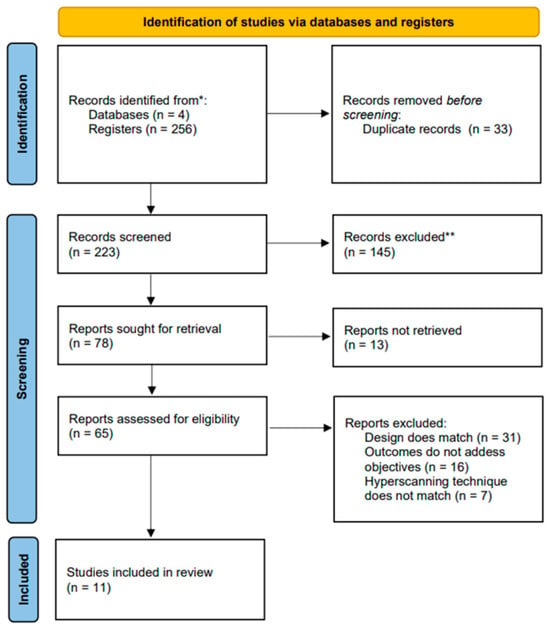
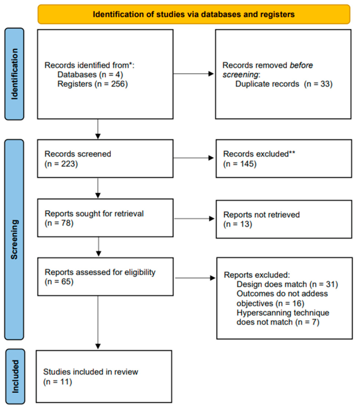
As shown in Figure 1, a total of 256 records were identified through database literature searches (n = 4). Of the initial documents, 33 duplicate records were removed prior to the screening phase. The remaining 223 records underwent title and abstract screening, leading to the exclusion of 145 of them. Subsequently, an attempt was made to retrieve the 78 resulting reports, successfully obtaining only 65 full-text articles and omitting 13 because they were not available for reading. Following a detailed evaluation of the full texts based on eligibility criteria, a total of 54 reports were excluded for the following reasons: inappropriate study design (n = 31), outcomes that did not address the established objectives (n = 16), and hyperscanning technique that did not meet the inclusion criteria (n = 7). After this refinement, 11 studies were selected for the final systematic review sample.
Figure 1.
PRISMA 2020 flow diagram. Note. * Records identified through database searches (n = 4) and registers (n = 256). ** Records excluded through preliminary selection criteria prior to full-text assessment (n = 145).
2.2. Systematization of Included Studies
Across the 11 primary studies included in this review (Table 1 and Table 2), total sample sizes were modest, ranging from 18 to 104 participants (9 to 52 dyads per study; total aggregate n = 506 individuals/253 dyads). Methodologically, functional near-infrared spectroscopy (fNIRS) was the predominant imaging technique (n = 8, 72.7%), followed by dual-fMRI (n = 2, 18.2%) and high-density EEG hyperscanning (n = 1, 9.1%). Experimental tasks relied on dyadic cooperation paradigms spanning rhythmic or verbal coordination, joint gaming (e.g., Jenga, Tetris, tile assembly), creative problem-solving, and economic decision-making.
Table 1.
Systematization of included studies: study characteristics and experimental paradigm.
Table 2.
Systematization of included studies: analytical metrics and neuroanatomical findings.
To evaluate inter-brain synchronization, studies applied analytical metrics such as Wavelet Transform Coherence (WTC; n = 45.4%), Inter-Subject Correlation (ISC; n = 9.1%), Partial Directed Coherence (PDC; n = 9.1%), and general signal coherence or latency measures (n = 36.4%). While a majority of the reviewed reports describe higher statistical coupling during joint cooperative conditions compared to independent, competitive, or resting baselines, these observations stem from small, methodologically diverse cohorts. Consequently, these pattern variations represent preliminary associative trends rather than conclusive evidence of a uniform neurobiological mechanism.
At the neuroanatomical level, the compiled data indicate frequent spatial convergence across cortical regions embedded within the Frontoparietal Network (FPN), the Mentalizing Network (MENT), and the Mirror Neuron System (MNS). Specifically, localized increases in neural synchrony were repeatedly observed in the inferior frontal cortex/gyrus (IFG; n = 5 studies), dorsolateral prefrontal cortex (dlPFC; n = 4 studies), right temporoparietal junction (rTPJ; n = 5 studies), and precuneus (n = 2 studies). Given the limited pool of only 11 studies and their substantial methodological heterogeneity, the current evidence is not sufficiently robust to establish definitive claims; consequently, the results synthesized below must be strictly interpreted as preliminary observations of statistical association rather than causal neurophysiological mechanisms.
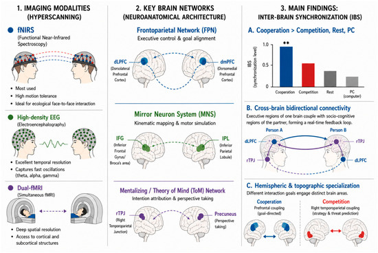
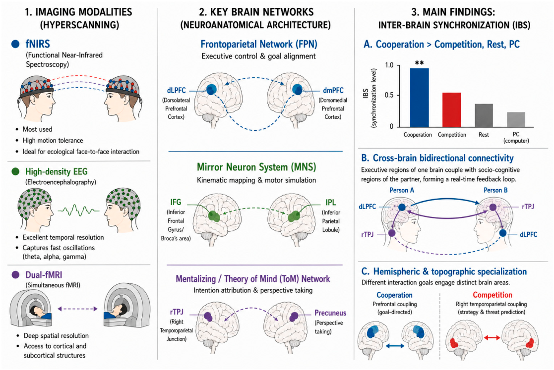
Based on the general mapping of evidence presented in the tables above and considering the preliminary scope of the retrieved literature, the data indicate that the dynamics of inter-brain synchronization may depend not only on analytical metrics but also on a complex methodological and neuroanatomical interaction. As schematically represented in Figure 2, these indications can be structured around three analytical axes: hyperscanning imaging modalities, cross-sectional convergence in the activation of key brain networks, and preliminary synchrony findings.
Figure 2.
Overview of research in inter-brain synchronization: hyperscanning modalities, neuroanatomical components, and preliminary findings of social connectivity. Note. ** indicates statistically significant differences compared to other conditions (p < 0.01). Dashed and solid-colored arrows represent directional functional coupling and cross-brain synchronization loops between partners. Colors (blue, green, purple, and red) distinguish specific neuroanatomical networks, functional regions, and inter-individual roles (Person A vs. Person B) during cooperative and competitive paradigms.
2.3. Experimental Paradigms of Social Cooperation and Hyperscanning Modalities
The synthesis of the selected literature revealed substantial methodological heterogeneity, making direct quantitative comparisons across studies difficult. Experimental paradigms and hyperscanning technologies varied significantly, introducing distinct task demands and signal characteristics.
The predominant modality, fNIRS, was utilized in 8 out of 11 studies due to its tolerance to movement artifacts in face-to-face setups. Experimental designs within fNIRS investigations varied widely, ranging from unison vocalization tasks [18] to physical interactive games like Jenga [19]. Computerized environments were also evaluated using Tetris [27], turn-based matrix games [30], and digital jigsaw puzzles [31]. Furthermore, fNIRS examined sociocognitive interactions through structured brainstorming sessions [20,21] and virtual economic exchanges [32].
Dual-fMRI was employed in two investigations to assess blind fine-motor synchronization [29] and real-time geometric figure recreation [33]. Lastly, a single study integrated high-density EEG to examine continuous motor coordination without linguistic mediation during a bar-balancing game [28]. This extensive variation in both task design and imaging modality significantly limits the generalizability of the findings across the review.
To provide a structured overview of the trade-offs inherent to each imaging technique utilized across the included literature, Table 3 summarizes the primary technical specifications, practical advantages, and methodological limitations of high-density EEG, fNIRS, and dual-fMRI in hyperscanning contexts.
Table 3.
Comparative overview of hyperscanning modalities: technical features, advantages, and limitations.
2.4. Comparison of Inter-Brain Synchronization Indices
Across the reviewed studies, inter-brain statistical associations were primarily evaluated through Wavelet Transform Coherence (WTC), Inter-Subject Correlation (ISC), and Partial Directed Coherence (PDC). It is critical to emphasize that these methods do not measure identical neurophysiological phenomena and should not be treated as fully interchangeable. Furthermore, most metrics employed (e.g., WTC, ISC) capture undirected statistical dependence rather than directional, causal neural communication. Only a minority of studies utilized directed approaches like PDC [28].
When comparing cooperation against baseline conditions, several studies reported statistically significant increases in undirected IBS during goal-oriented joint tasks [18,19,20,21,27,31]. In one instance, higher synchronization levels statistically correlated with task success [31]. Another paradigm validated IBS by comparing real interacting dyads against computationally generated random pseudo-pairs, controlling for common input confounds [32].
Contrasting cooperation with competition revealed distinct topographical patterns. Cooperative conditions in divergent thinking tasks were associated with higher prefrontal coherence [20]; whereas, turn-based games linked cooperation to bilateral inferior and prefrontal channels [30]. Dual-fMRI data indicated higher interpersonal statistical coupling during simultaneous joint actions compared to sequential turns [33]. When compared to individual execution (Solo) or computer-controlled conditions (PC), human-to-human interaction was consistently associated with parallel signal fluctuations [27,28,29]. However, because these data are predominantly correlational, these findings indicate shared functional task dynamics rather than verified interpersonal information transmission.
2.5. Synthesis of Neuroanatomical Evidence: Frontoparietal Network Recruitment
The neuroanatomical synthesis tentatively points toward the involvement of cortical regions broadly corresponding to the Frontoparietal Network (FPN), the Mentalizing Network, and the Mirror Neuron System (MNS). However, conclusions regarding complete network recruitment must be tempered. Many included studies—particularly those using fNIRS—measured only superficial cortical regions, sampled restricted prefrontal areas, and utilized varying channel montages. Consequently, evidence for full FPN recruitment remains indirect.
2.5.1. Frontal and Prefrontal Regions
Increased inter-brain statistical coherence in dorsolateral prefrontal cortex (dlPFC) channels was reported during tasks requiring joint cognitive control and shared goals [29,33]. Higher rDLPFC synchrony was observed during creative brainstorming [20] and in dyads with lower individual baseline creativity [21]. Channels overlying the Inferior Frontal Gyrus (IFG/Broca’s area) showed synchronized signal changes during motor imitation and joint action understanding [18,31], with bilateral IFG involvement noted in turn-based tasks [18,30]. Statistical associations in superior prefrontal channels (dmPFC/BA 9/BA 10) were observed during decision-making [19] and puzzle-solving [31], while the Superior and Middle Frontal Gyri exhibited coupling during economic transactions [32].
2.5.2. Parietal and Temporoparietal Regions
Elevated signal coherence in posterior channels overlying the right temporoparietal junction (rTPJ) and Angular Gyrus was documented during joint attention and social inference tasks [20,21,27,31]. Notably, applying inhibitory stimulation to the rTPJ did not significantly affect inter-brain synchronization, suggesting that its activation may not be strictly required to sustain statistical coupling in all contexts [27]. Areas corresponding to the left Inferior Parietal Lobule (IPL) and precuneus demonstrated elevated coherence during simultaneous pattern reproduction [33], fine temporal motor coordination [29], and non-verbal fluid joint action [28,30].
2.5.3. Inter-Regional Correlates and Cross-Brain Associations
Some studies explored cross-regional statistical dependencies across paired brains. When directional modeling (PDC) was applied, exploratory analyses suggested statistical associations between frontal and temporoparietal nodes across dyads [20,28]. While these preliminary patterns are consistent with theoretical models proposing concurrent recruitment of action-execution and mentalizing systems, the available evidence remains largely correlational. These data currently preclude concluding that inter-brain synchronization acts as a direct, causal mechanism for cooperative success.
2.5.4. Integrative Framework of the Frontoparietal, Mirror, and Mentalizing Networks in Social Cognition
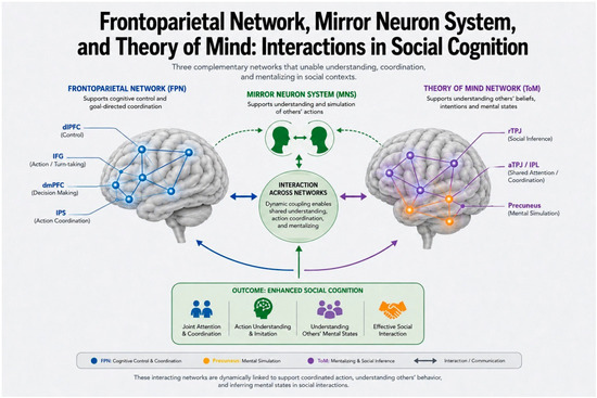
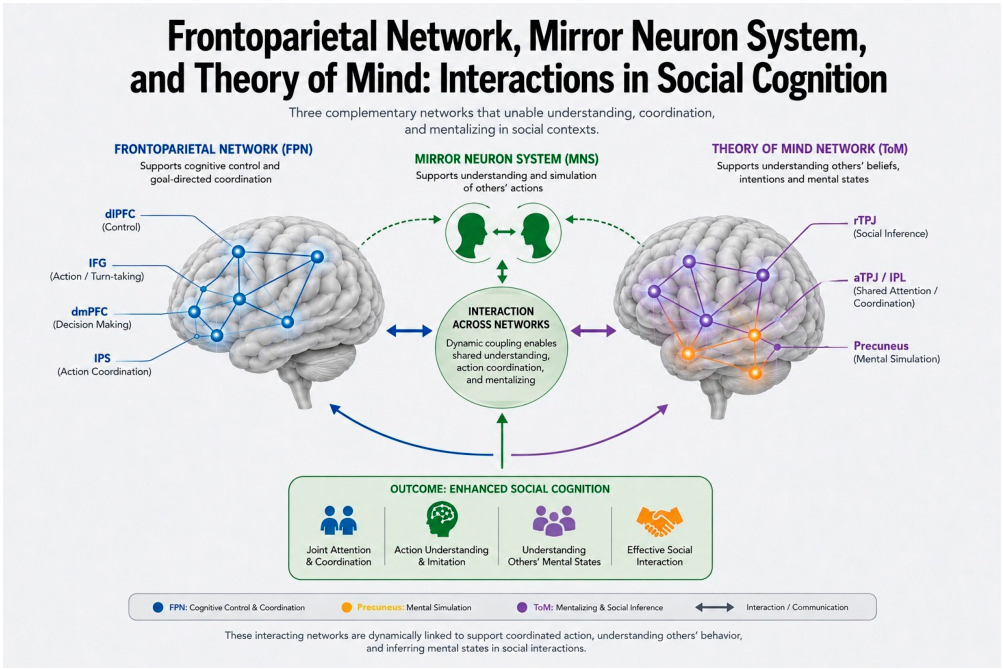
The empirical evidence reviewed in the previous subsections tentatively suggests that social interaction cannot be explained solely by the isolated functioning of individual neural systems. Indeed, flexible cognitive control and real-time adaptation appear to depend on a continuous tripartite dialog among the Frontoparietal Network (FPN), the Mirror Neuron System (MNS), and the Theory of Mind (ToM) network. To conceptualize this dynamic architecture, Figure 3 presents a conceptual framework proposed by the authors.
Figure 3.
Conceptual framework proposed by the authors. Note. This diagram represents an interpretative theoretical framework proposed by the authors to integrate heterogeneous findings across different studies and imaging modalities, rather than an experimentally established network or proven direct biological pathway. The yellow nodes are part of the Theory of Mind Network (ToM) and specifically represent the precuneus, which supports mental simulation within social cognition. Abbreviations: dlPFC, dorsolateral prefrontal cortex; IFG, inferior frontal gyrus; dmPFC, dorsomedial prefrontal cortex; IPS, intraparietal sulcus; rTPJ, right temporoparietal junction; aTPJ/IPL, anterior temporoparietal junction/Inferior Parietal Lobule; MNS, Mirror Neuron System; ToM, Theory of Mind network.
As illustrated in Figure 3, executive control nodes within the FPN (dlPFC, IFG, dmPFC, IPS) are hypothesized to support goal-directed coordination and cognitive flexibility. The MNS appears to support kinematic simulation and real-time action mirroring; whereas, the ToM network (anchored in the rTPJ, aTPJ/IPL, and precuneus) is positioned to mediate mental state attribution and social inference. Readers are explicitly reminded that this integrated schema represents a heuristic, interpretative theoretical synthesis rather than a direct empirical finding uniformly demonstrated across all reviewed primary studies.
3. Discussion
This systematic review synthesizes the current empirical evidence regarding inter-brain synchronization (IBS) and Frontoparietal Network (FPN) modulation during social cooperation tasks in healthy adults. By evaluating 11 hyperscanning studies across fNIRS, EEG, and dual-fMRI, this review addresses a specific gap in the literature: assessing how regional cortical coupling varies across task structures and recording modalities. However, given the limited number of eligible studies and their substantial methodological heterogeneity, the synthesis presented here must be interpreted as preliminary and observational rather than definitive.
3.1. Methodological Considerations and the Interpretation of Inter-Brain Synchronization
A critical requirement in evaluating the reviewed literature lies in explicitly distinguishing empirical evidence from theoretical interpretation. Most included studies utilized undirected statistical measures—such as Wavelet Transform Coherence (WTC), Inter-Subject Correlation (ISC), or phase coherence—which capture co-varying signal fluctuations rather than direct causal neural communication. Consequently, observed increases in IBS during cooperative tasks must be interpreted strictly as empirical associative correlates rather than a proven causal driver or biological mechanism of cooperative success.
Furthermore, a major methodological debate in hyperscanning concerns whether elevated IBS stems from active social cognition or merely from shared sensory inputs, identical task timing, motor co-regulation, or joint attention. Although several included studies implemented control conditions—such as scrambled pseudo-pairs, solo executions, or human–computer (PC) interactions—surrogate pair methodologies remain variable across studies. The absence of elevated coherence in PC or solo conditions suggests a potential degree of social specificity, yet the influence of common external stimulation cannot be completely ruled out. Therefore, claims suggesting that cooperation creates an integrated single cognitive entity or acts as an absolute biological glue are conceptually overstated; current evidence supports IBS strictly as a correlational neurophysiological correlate associated with joint behavioral engagement.
3.2. Topographical Trends, Modality Differences, and Network Interpretations
Across the reviewed studies, a recurring pattern involves the engagement of prefrontal, inferior frontal, and temporoparietal regions during cooperative paradigms. The literature suggests a potential functional specialization, where cooperative goals frequently coincide with prefrontal/frontopolar alignment [34,35]; whereas, competitive interactions tend to recruit right temporoparietal structures associated with strategic monitoring and threat prediction [17,36,37].
However, general conclusions regarding the Frontoparietal Network (FPN) as an executive macrostructure must be tempered. The vast majority of included studies relied on functional near-infrared spectroscopy (fNIRS) due to its motion tolerance and suitability for face-to-face paradigms [38,39], aligning with the principles of “second-person neuroscience” [40]. Nevertheless, fNIRS optode montages in these studies were often restricted to superficial cortical channels (predominantly prefrontal or temporoparietal areas) without full-brain coverage. Thus, direct evidence for full FPN recruitment remains limited.
Moreover, cross-study integration is constrained by substantial technological and analytical heterogeneity:
fNIRS vs. EEG vs. dual-fMRI: fNIRS suffers from limited spatial depth (precluding subcortical assessments of structures like the amygdala or striatum), signal contamination from extracerebral scalp blood flow, and systemic hemodynamic noise. EEG offers millisecond temporal precision but is susceptible to movement artifacts and volume conduction. Dual-fMRI provides subcortical spatial resolution but severely compromises ecological validity due to constrained physical environments.
Analytical Discrepancies: Variations in frequency bands (e.g., mu/theta in EEG vs. low-frequency hemodynamic fluctuations in fNIRS), preprocessing pipelines, filter thresholds, and channel montages mean that different studies measure distinct neurophysiological phenomena that are not fully interchangeable.
3.3. Theoretical Models and Functional Hypotheses
Theoretical frameworks integrating the Mirror Neuron System (MNS; e.g., IFG, IPL) and the Theory of Mind network (ToM; e.g., dlPFC, dmPFC, rTPJ) offer valuable interpretative lenses [41]. It has been hypothesized that early sensorimotor mapping via the MNS may provide low-level kinematic alignment upon which higher-level mentalizing processes via ToM are integrated. Similarly, observations of cross-brain node coupling (e.g., one participant’s rDLPFC synchronizing with their partner’s rTPJ) [42] or resilience to focal disruption are intriguing.
However, these conceptual models—including proposed sender-receiver dynamics, executive-mentalizing loops, and adaptive neural compensation—must be explicitly categorized as interpretative hypotheses rather than experimentally proven conclusions. Because the majority of studies used undirected metrics (rather than directed connectivity approaches like Partial Directed Coherence or Dynamic Causal Modeling), directional information flow between partners cannot be mathematically confirmed.
3.4. Limitations of the Reviewed Literature and Review Methodology
The interpretations drawn in this review are bounded by several methodological limitations across the primary literature and the review process itself:
Absence of Quantitative Meta-Analysis: Due to extreme heterogeneity in experimental paradigms, imaging modalities, signal metrics, and statistical reporting, a quantitative meta-analysis was not feasible. This review is strictly narrative, which limits the ability to quantify effect sizes or systematically test for publication bias.
Small Cumulative Sample Size: The inclusion of only 11 primary studies severely limits the generalizability of these findings, reinforcing that all conclusions are preliminary.
Primary Focus on fNIRS and Cortical Surfaces: The heavy reliance on fNIRS leaves major blind spots regarding subcortical contributions and fine-grained electrophysiological dynamics.
Dyadic and Demographic Constraints: The primary studies focused overwhelmingly on healthy, young adult dyads in artificial laboratory environments. It remains unknown how these neural correlates translate to larger group dynamics (triads, teams), lifespan aging, or clinically impaired populations.
3.5. Future Directions
To move the field from correlational observations toward mechanistic clarity, future hyperscanning research should prioritize:
Directional Connectivity Analysis: Applying directed effective connectivity models (e.g., Granger Causality, Dynamic Causal Modeling) to test hypotheses regarding neural leadership, information flow, and asymmetric dyadic roles.
Rigorous Control Paradigms: Systematically incorporating surrogate pair analyses, baseline task-timing controls, and physiological artifact removal (e.g., short-distance channels in fNIRS, systemic physiology monitoring) to isolate genuine neural coupling from common environmental input.
Preregistration, Standardized Pipelines, and Open Data Sharing: Encouraging the systematic adoption of preregistered hyperscanning protocols (e.g., via OSF or PROSPERO), the implementation of standardized preprocessing pipelines (such as MNE-Python, Homer3, or FieldTrip), and the open sharing of raw hyperscanning datasets in public repositories (e.g., OpenNeuro). These developments are becoming increasingly vital to enhance methodological transparency, minimize publication bias, and foster global reproducibility in interactive social neuroscience.
Multimodal and Multi-Agent Approaches: Combining simultaneous fNIRS-EEG recordings to bridge temporal and spatial gaps, as well as expanding paradigms beyond isolated dyads to larger collectives and real-world ecological settings.
Clinical Translation: Investigating whether baseline inter-brain coupling metrics can serve as exploratory markers in social interaction disorders, provided that standardized and reproducible analytical pipelines are established.
4. Materials and Methods
4.1. Protocol Registration and Compliance
This systematic review was conducted in strict accordance with the PRISMA 2020 (Preferred Reporting Items for Systematic Reviews and Meta-Analyses) guidelines to ensure a transparent, rigorous, and replicable reporting structure [43]. The PRISMA 2020 checklists [43] are provided in Supplementary Table S1.
The review protocol was registered in the International Prospective Register of Systematic Reviews (PROSPERO, Centre for Reviews and Dissemination, University of York, York, United Kingdom), registration number: CRD420261428714), ensuring methodological transparency and minimizing the risk of reporting bias.
4.2. Methodological Framework (PICO)
To address the research questions with the specificity required for high-impact literature, the PICO (Population, Intervention/Exposure, Comparison, Outcome) framework was selected as the structural gold standard. Consequently, alternative qualitative frameworks, such as SPICE or SPIDER, were explicitly rejected as they lack the clinical and objective parameters necessary to isolate empirical neurophysiological data. The PICO components were defined as follows:
- Population (P): Dyads or groups of healthy adult subjects (≥18 years old) with no history or current diagnosis of neurological, psychiatric, or neurodevelopmental disorders.
- Intervention/Exposure (I): Experimental tasks involving interactive social cooperation and interpersonal behavioral or neural synchronization, simultaneously assessed using hyperscanning setups (electroencephalography [EEG], functional near-infrared spectroscopy [fNIRS], or dual functional magnetic resonance imaging [fMRI]).
- Comparison (C): Control conditions consisting of competitive social interaction paradigms, asynchronous tasks, or resting-state (baseline) conditions.
- Outcomes (O): Inter-brain synchronization (IBS) indices and differential activation/connectivity metrics within the Frontoparietal Networks (FPN).
4.3. Eligibility Criteria
Studies meeting the following eligibility criteria were included in this systematic review:
- Study Design: Original empirical research published in peer-reviewed, indexed journals. Review articles, book chapters, conference abstracts lacking full text, and editorials were excluded to ensure data robustness and methodological comparability.
- Participants: Samples composed entirely of healthy adults meeting the population criteria. Studies focusing on pediatric populations, clinical cohorts, or individual, non-interactive settings were excluded.
- Methodology: Experimental design; explicit use of hyperscanning (EEG, fNIRS, or dual fMRI) during social interaction tasks. Studies evaluating solely single-subject paradigms or non-social cognitive tasks were omitted.
- Language: Articles published exclusively in English or Spanish were included, reflecting the working languages of the review team and encompassing the majority of indexed literature in this domain. However, restricting inclusion to these languages may introduce language bias by potentially omitting relevant studies published in other languages.
- Timeline: The search frame was set from 1 January 2010 to 16 June 2026. The 2010 cutoff was selected because it marks a pivotal surge in hyperscanning methodology, characterized by the technical consolidation of multi-channel fNIRS, high-density wireless EEG systems, and standardized dual-fMRI pipelines for interactive paradigms. Nevertheless, excluding studies published before 2010 is acknowledged as a potential source of publication and temporal bias, as foundational early-stage multi-brain recording studies were excluded.
4.4. Information Sources and Search Strategy
To ensure a comprehensive, impartial, and highly reproducible bibliographic retrieval, a systematic electronic search was conducted in four top-tier, high-impact databases, beginning and ending on 16 June 2026. No searches for subsequent citations were performed. The rationale for including each source is detailed below in Table 4.
Table 4.
Databases and Search Strings.
4.5. Study Selection and Data Extraction
Following data retrieval, all identified records were exported to an Excel (Microsoft 365, Microsoft Corporation, Redmond, United States) spreadsheet, and duplicates were manually removed. Subsequently, two reviewers independently screened the titles, abstracts, and full texts of the remaining documents against the predefined inclusion and exclusion criteria. To rigorously assess the consistency of the screening process, inter-rater reliability was calculated using Cohen’s kappa coefficient. The actual agreement between reviewers was high (k = 0.87), indicating a strong level of agreement. Any initial conflicts or discrepancies regarding study eligibility were resolved through a consensus-based discussion between the two reviewers.
To capture the relevant parameters of the final selected articles, the following metadata were extracted within the same matrix:
- Study Characteristics: Author, year of publication, and specific neuroimaging modality (EEG, fNIRS, dual-fMRI).
- Sample Metadata: Number of dyads/groups, age distribution, and gender breakdown.
- Experimental Design: Type of social interaction paradigm (e.g., economic games, motor synchronization, verbal communication) and control conditions.
- Neurophysiological Variables: Specific IBS metrics, localized activation maps, and FPN functional connectivity values.
- Main Outcomes: Statistical values detailing the specific neurofunctional changes associated with cooperative states.
4.6. Quality and Risk of Bias Assessment
To comply with the PRISMA 2020 statement (Item 11) and evaluate the risk of bias of the included studies, the Mixed Methods Appraisal Tool (MMAT, 2018 version, McGill University, Montreal, Canada) was used. The suitability of the MMAT lies in its capability to examine the internal validity and rigor of heterogeneous methodologies within a single analytical framework using standardized criteria [44]. Its systematic application facilitates the critical synthesis of evidence by categorizing each study according to its design, thereby ensuring the robustness and reliability of the incorporated findings. The appraisal procedure is structured initially with two general screening questions followed by specific criteria corresponding to each study design.
In the particular case of non-randomized quantitative studies (Category 3), the tool evaluates the risk of bias across five essential domains. First, the representativeness of the participants (Criterion 3.1) is examined to confirm that the sample accurately reflects the target population; second, it is verified whether the measurements of outcome and exposure variables are appropriate (Criterion 3.2), ensuring the use of valid and standardized instruments; third, the completeness of outcome data is analyzed (Criterion 3.3), assessing the absence of attrition bias or missing data; fourth, the control of confounding factors is judged (Criterion 3.4) in both the design phase and statistical analysis; and for the final criterion, it is confirmed that the intervention or study condition was administered or applied as intended (Criterion 3.5), guaranteeing procedural fidelity throughout the experimental development.
The analysis of the matrix, as presented in Table 5, demonstrated an overall high level of methodological quality across all evaluated studies. Specifically, the studies conducted by Osaka et al. [18], Liu et al. [19], Tan et al. [27], Ciaramidaro et al. [28], Xue et al. [21], Lu et al. [20], Sijben et al. [29], Liu et al. [30], Zhou et al. [31], Kikuchi et al. [32], and Špiláková et al. [33] satisfactorily met the required non-randomized quantitative standards. Although minor variations were identified in some specific criteria related to control or measurement, none of the studies exhibited critical deficiencies that compromised their validity, thus confirming the structural robustness of the analyzed body of evidence.
Table 5.
Methodological quality assessment of the included studies using the MMAT tool.
It is important to emphasize that the MMAT is designed as a qualitative assessment tool rather than a numerical scoring system; therefore, the quality categorization (‘High’) was based on strict adherence to the specific criteria for non-randomized designs proposed by Hong et al. [44]. No predefined arbitrary thresholds were applied; instead, a study was established to achieve a robust quality level by fulfilling the criteria for bias control, representativeness, and measurement validity. To ensure the rigor of the process, each study was independently appraised by two blinded reviewers. Initial discrepancies in the appraisal were resolved through consensus-based discussion, with a third reviewer acting as a final arbitrator if disagreements persisted. This ensured a uniform interpretation of the domains prior to assigning the final quality rating.
5. Conclusions
The preliminary evidence synthesized from the included studies suggests that IBS may serve as a plausible neurophysiological correlate—rather than a proven causal mechanism—of interpersonal alignment during social interaction. Regarding the general architecture of interactive social cognition, the available literature tentatively links FPN regions, particularly anterior executive nodes such as the dorsolateral prefrontal cortex (dlPFC) and posterior socio-cognitive hubs like the right temporoparietal junction (rTPJ), to cooperative states. However, given the reliance on predominantly undirected synchrony metrics (e.g., Wavelet Transform Coherence) and the presence of shared sensory-motor confounds, assertions of cross-brain causal communication or integrated mirror-Theory of Mind networks remain working hypotheses that require validation through rigorous experimental controls (such as surrogate/pseudo-pair analyses).
Addressing the first specific objective, experimental paradigms of social cooperation across hyperscanning modalities display substantial methodological diversity. The reviewed literature is heavily dominated by functional near-infrared spectroscopy (fNIRS) owing to its practical utility in ecologically valid, face-to-face setups (e.g., joint problem-solving, economic negotiations, and creative tasks). Conversely, high-density electroencephalography (EEG) and dual-fMRI remain underrepresented in cooperative paradigms, being reserved primarily for constrained motor-rhythmic synchronization tasks. This reliance on fNIRS limits inferences strictly to superficial cortical channels, making it difficult to claim full-brain network recruitment without accounting for motion artifacts, extracerebral systemic contamination, and heterogeneous channel montages across studies.
In response to the second specific objective, comparing inter-brain synchronization indices indicates a general trend where cooperative tasks elicit higher statistical co-registration than resting states, individual controls, or interaction with non-human agents. However, claims that cooperation consistently exceeds competition must be interpreted with caution: while prefrontal coupling is frequently reported during goal-aligned tasks, competition often shifts statistical synchrony toward temporoparietal regions involved in strategic monitoring. Because most primary studies report statistical dependencies without establishing directionality or controlling fully for shared external pacing, increased synchrony across these conditions reflects parallel neural engagement or mutual task constraints as much as genuine interpersonal neural coupling.
Finally, concerning the third specific objective, the neuroanatomical synthesis indicates tentative co-activation patterns involving prefrontal regions (dlPFC, dmPFC, IFG) and posterior structures (rTPJ, precuneus). While it is conceptually appealing to propose an integrative executive-mentalizing framework combining mirror neuron and Theory of Mind systems, current evidence across these 11 studies does not formally prove hierarchical inter-brain coupling. Future research must address these preliminary bounds by implementing standardized multi-modal designs (simultaneous fNIRS-EEG), adopting directed connectivity metrics (e.g., Partial Directed Coherence) to resolve directional information flow, routinely incorporating pseudo-pair surrogate controls, and evaluating these paradigms in larger group structures and clinical samples.
Supplementary Materials
The following supporting information can be downloaded at: https://www.mdpi.com/article/10.3390/physiologia6030051/s1, Table S1: PRISMA 2020 checklist.
Author Contributions
Conceptualization, W.A.Z.V. and P.G.M.M.; methodology, W.A.Z.V., P.G.M.M. and Á.S.F.B.; software, J.C.R.R. and C.A.L.M.; validation, W.A.Z.V., P.G.M.M., Á.S.F.B. and Y.E.Z.C.; formal analysis, W.A.Z.V., Á.S.F.B. and J.C.R.R.; investigation, W.A.Z.V., P.G.M.M., Á.S.F.B., J.C.R.R., C.A.L.M. and Y.E.Z.C.; resources, W.A.Z.V. and C.A.L.M.; data curation, Á.S.F.B., J.C.R.R. and Y.E.Z.C.; writing—original draft preparation, W.A.Z.V., P.G.M.M. and Á.S.F.B.; writing—review and editing, W.A.Z.V., P.G.M.M., J.C.R.R., C.A.L.M. and Y.E.Z.C.; visualization, J.C.R.R. and C.A.L.M.; supervision, W.A.Z.V.; project administration, W.A.Z.V. and P.G.M.M.; funding acquisition, W.A.Z.V. All authors have read and agreed to the published version of the manuscript.
Funding
This research was funded by Universidad Estatal Península de Santa Elena, code AbcPsic.
Institutional Review Board Statement
As this is a systematic review of public, peer-reviewed literature, this study did not involve direct interventions with human or animal subjects. Therefore, direct institutional ethical approval was not required.
Informed Consent Statement
Not applicable.
Data Availability Statement
In accordance with MDPI’s open science policies, all protocols, data extraction sheets, search records, and analysis codes associated with this manuscript have been made publicly available. The data validating the findings of this systematic review are documented within the study protocol registered in PROSPERO under registration number [CRD420261428714]. No new primary datasets were generated or analyzed during this study, and no restrictions apply to the availability of materials or information for this submission.
Acknowledgments
The authors express their sincere gratitude to the research team for their outstanding commitment and dedication to this systematic review. Special thanks are extended to the Universidad Estatal Península de Santa Elena (UPSE) for the institutional funding and support provided for this study. During the preparation of this manuscript, the authors used Gemini AI (Gemini 1.5 Pro, Google LLC, Mountain View, United States) to assist in structuring the PICO criteria, refining the database-specific search syntax, and supporting academic translation and text drafting; all AI-generated outputs were thoroughly reviewed, verified, and edited by the human authors to ensure scientific accuracy and compliance with PRISMA guidelines. Generative AI tools were not used for data extraction, quality assessment, or interpretation of results. The authors take full responsibility for the final content of this publication.
Conflicts of Interest
The authors declare no conflicts of interest.
References
- Berni, T.; Sacheli, L.M.; Cicirello, M.; Tettamanti, M.; Paulesu, E.; Zapparoli, L. Together we sync: A systematic qualitative and quantitative review of fMRI hyperscanning studies. Soc. Cogn. Affect. Neurosci. 2026, 21, nsaf122. [Google Scholar] [CrossRef] [Scilit] [PubMed]
- Li, R.; Mayseless, N.; Balters, S.; Reiss, A.L. Dynamic inter-brain synchrony in real-life inter-personal cooperation: A functional near-infrared spectroscopy hyperscanning study. NeuroImage 2021, 238, 118263. [Google Scholar] [CrossRef] [Scilit] [PubMed]
- Czeszumski, A.; Liang, S.H.-Y.; Dikker, S.; König, P.; Lee, C.-P.; Koole, S.L.; Kelsen, B. Cooperative Behavior Evokes Interbrain Synchrony in the Prefrontal and Temporoparietal Cortex: A Systematic Review and Meta-Analysis of fNIRS Hyperscanning Studies. eNeuro 2022, 9, ENEURO.0268-21.2022. [Google Scholar] [CrossRef] [Scilit] [PubMed]
- Misaki, M.; Kerr, K.L.; Ratliff, E.L.; Cosgrove, K.T.; Simmons, W.K.; Morris, A.S.; Bodurka, J. Beyond synchrony: The capacity of fMRI hyperscanning for the study of human social interaction. Soc. Cogn. Affect. Neurosci. 2021, 16, 84–92. [Google Scholar] [CrossRef] [Scilit] [PubMed]
- Balconi, M.; Angioletti, L. Dyadic inter-brain EEG coherence induced by interoceptive hyperscanning. Sci. Rep. 2023, 13, 4344. [Google Scholar] [CrossRef] [Scilit] [PubMed]
- Nam, C.S.; Choo, S.; Huang, J.; Park, J. Brain-to-Brain Neural Synchrony During Social Interactions: A Systematic Review on Hyperscanning Studies. Appl. Sci. 2020, 10, 6669. [Google Scholar] [CrossRef] [Scilit]
- Schurz, M.; Radua, J.; Tholen, M.G.; Maliske, L.; Margulies, D.S.; Mars, R.B.; Sallet, J.; Kanske, P. Toward a hierarchical model of social cognition: A neuroimaging meta-analysis and integrative review of empathy and theory of mind. Psychol. Bull. 2021, 147, 293–327. [Google Scholar] [CrossRef] [Scilit] [PubMed]
- Todorov, A.B. Social Neuroscience: Toward Understanding the Underpinnings of the Social Mind; Social Cognition and Social Neuroscience Ser; Oxford University Press, Incorporated: Cary, NC, USA, 2011; 301p. [Google Scholar]
- Hakim, U.; De Felice, S.; Pinti, P.; Zhang, X.; Noah, J.; Ono, Y.; Burgess, P.W.; Hamilton, A.; Hirsch, J.; Tachtsidis, I. Quantification of inter-brain coupling: A review of current methods used in haemodynamic and electrophysiological hyperscanning studies. NeuroImage 2023, 280, 120354. [Google Scholar] [CrossRef] [Scilit] [PubMed]
- Carollo, A.; Bizzego, A.; Schäfer, V.; Pletti, C.; Hoehl, S.; Esposito, G. Interpersonal Neural Synchrony Across Levels of Interpersonal Closeness and Social Interactivity. NeuroImage 2025, 322, 121532. [Google Scholar] [CrossRef] [Scilit]
- Redcay, E.; Schilbach, L. Using second-person neuroscience to elucidate the mechanisms of social interaction. Nat. Rev. Neurosci. 2019, 20, 495–505. [Google Scholar] [CrossRef] [Scilit] [PubMed]
- Li, Y.; Si, Y.; Pang, X.; Li, S.; Jiang, L.; Yi, C.; Yao, D.; Li, F.; Xu, P. EEG hyperscanning reveals dynamic interbrain network patterns during interactive social decision-making. Commun. Biol. 2026, 9, 595. [Google Scholar] [CrossRef] [Scilit] [PubMed]
- Lotter, L.D.; Kohl, S.H.; Gerloff, C.; Bell, L.; Niephaus, A.; Kruppa, J.A.; Dukart, J.; Schulte-Rüther, M.; Reindl, V.; Konrad, K. Revealing the neurobiology underlying interpersonal neural synchronization with multimodal data fusion. Neurosci. Biobehav. Rev. 2023, 146, 105042. [Google Scholar] [CrossRef] [Scilit] [PubMed]
- Schilbach, L.; Redcay, E. Synchrony Across Brains. Annu. Rev. Psychol. 2025, 76, 883–911. [Google Scholar] [CrossRef] [Scilit] [PubMed]
- Czeszumski, A.; Eustergerling, S.; Lang, A.; Menrath, D.; Gerstenberger, M.; Schuberth, S.; Schreiber, F.; Rendon, Z.Z.; König, P. Hyperscanning: A Valid Method to Study Neural Inter-brain Underpinnings of Social Interaction. Front. Hum. Neurosci. 2020, 14, 39. [Google Scholar] [CrossRef] [Scilit] [PubMed]
- Zhao, Q.; Zhao, W.; Lu, C.; Du, H.; Chi, P. Interpersonal neural synchronization during social interactions in close relationships: A systematic review and meta-analysis of fNIRS hyperscanning studies. Neurosci. Biobehav. Rev. 2024, 158, 105565. [Google Scholar] [CrossRef] [Scilit] [PubMed]
- Cui, X.; Bryant, D.M.; Reiss, A.L. NIRS-based hyperscanning reveals increased interpersonal coherence in superior frontal cortex during cooperation. NeuroImage 2012, 59, 2430–2437. [Google Scholar] [CrossRef] [Scilit] [PubMed]
- Osaka, N.; Minamoto, T.; Yaoi, K.; Azuma, M.; Shimada, Y.M.; Osaka, M. How Two Brains Make One Synchronized Mind in the Inferior Frontal Cortex: fNIRS-Based Hyperscanning During Cooperative Singing. Front. Psychol. 2015, 6, 1811. [Google Scholar] [CrossRef] [Scilit] [PubMed]
- Liu, N.; Mok, C.; Witt, E.E.; Pradhan, A.H.; Chen, J.E.; Reiss, A.L. NIRS-Based Hyperscanning Reveals Inter-brain Neural Synchronization during Cooperative Jenga Game with Face-to-Face Communication. Front. Hum. Neurosci. 2016, 10, 82. [Google Scholar] [CrossRef] [Scilit] [PubMed]
- Lu, K.; Xue, H.; Nozawa, T.; Hao, N. Cooperation Makes a Group be More Creative. Cereb. Cortex 2019, 29, 3457–3470. [Google Scholar] [CrossRef] [Scilit] [PubMed]
- Xue, H.; Lu, K.; Hao, N. Cooperation makes two less-creative individuals turn into a highly-creative pair. NeuroImage 2018, 172, 527–537. [Google Scholar] [CrossRef] [Scilit] [PubMed]
- Barraza, P.; Pérez, A.; Rodríguez, E. Brain-to-Brain Coupling in the Gamma-Band as a Marker of Shared Intentionality. Front. Hum. Neurosci. 2020, 14, 295. [Google Scholar] [CrossRef] [Scilit] [PubMed]
- Vicente, U.; Ara, A.; Marco-Pallarés, J. Intra- and inter-brain synchrony oscillations underlying social adjustment. Sci. Rep. 2023, 13, 11211. [Google Scholar] [CrossRef] [Scilit] [PubMed]
- Chuang, T.-M.; Peng, P.-C.; Su, Y.-K.; Lin, S.-H.; Tseng, Y.-L. Exploring Inter-Brain Electroencephalogram Patterns for Social Cognitive Assessment During Jigsaw Puzzle Solving. IEEE Trans. Neural Syst. Rehabil. Eng. 2024, 32, 422–430. [Google Scholar] [CrossRef] [Scilit] [PubMed]
- Paal, T.; Bereczkei, T. Adult theory of mind, cooperation, Machiavellianism: The effect of mindreading on social relations. Personal. Individ. Differ. 2007, 43, 541–551. [Google Scholar] [CrossRef] [Scilit]
- Ridinger, G.; McBride, M. Theory-of-mind ability and cooperation. J. Econ. Behav. Organ. 2025, 230, 106895. [Google Scholar] [CrossRef] [Scilit]
- Tan, S.H.J.; Leuk, S.P.J.; Teo, W.-P. Inter-brain synchrony during collaborative gaming: An investigation using theta-burst stimulation at the right temporal-parietal junction. NeuroImage 2026, 332, 121906. [Google Scholar] [CrossRef] [Scilit] [PubMed]
- Ciaramidaro, A.; Toppi, J.; Vogel, P.; Freitag, C.M.; Siniatchkin, M.; Astolfi, L. Synergy of the mirror neuron system and the mentalizing system in a single brain and between brains during joint actions. NeuroImage 2024, 299, 120783. [Google Scholar] [CrossRef] [Scilit] [PubMed]
- Sijben, R.; Friedmann, R.; Hernandez-Pena, L.; Rodriguez-Raecke, R. Wavelet Analysis of Dual-fMRI-Hyperscanning Reveals Cooperation and Communication Dependent Effects on Interbrain Neuronal Coherence. Hum. Brain Mapp. 2025, 46, e70355. [Google Scholar] [CrossRef] [Scilit] [PubMed]
- Liu, T.; Saito, G.; Lin, C.; Saito, H. Inter-brain network underlying turn-based cooperation and competition: A hyperscanning study using near-infrared spectroscopy. Sci. Rep. 2017, 7, 8684. [Google Scholar] [CrossRef] [Scilit] [PubMed]
- Zhou, S.; Zhang, Y.; Fu, Y.; Wu, L.; Li, X.; Zhu, N.; Li, D.; Zhang, M. The Effect of Task Performance and Partnership on Interpersonal Brain Synchrony during Cooperation. Brain Sci. 2022, 12, 635. [Google Scholar] [CrossRef] [Scilit] [PubMed]
- Kikuchi, Y.; Tanioka, K.; Hiroyasu, T.; Hiwa, S. Interpersonal brain synchronization during face-to-face economic exchange between acquainted dyads. Oxf. Open Neurosci. 2023, 2, kvad007. [Google Scholar] [CrossRef] [Scilit] [PubMed]
- Špiláková, B.; Shaw, D.J.; Czekóová, K.; Mareček, R.; Brázdil, M. Getting into sync: Data-driven analyses reveal patterns of neural coupling that distinguish among different social exchanges. Hum. Brain Mapp. 2020, 41, 1072–1083. [Google Scholar] [CrossRef] [Scilit] [PubMed]
- Knyazev, G.G.; Savostyanov, A.N.; Bocharov, A.V.; Rudych, P.D.; Saprigyn, A.E. Multivariate pattern analysis of cooperation and competition in constructive action. Neuropsychologia 2024, 202, 108956. [Google Scholar] [CrossRef] [Scilit] [PubMed]
- Léné, P.; Karran, A.J.; Labonté-Lemoyne, E.; Sénécal, S.; Fredette, M.; Johnson, K.J.; Léger, P. Is there collaboration specific neurophysiological activation during collaborative task activity? An analysis of brain responses using electroencephalography and hyperscanning. Brain Behav. 2021, 11, e2270. [Google Scholar] [CrossRef] [Scilit] [PubMed]
- Decety, J.; Jackson, P.L.; Sommerville, J.A.; Chaminade, T.; Meltzoff, A.N. The neural bases of cooperation and competition: An fMRI investigation. NeuroImage 2004, 23, 744–751. [Google Scholar] [CrossRef] [Scilit] [PubMed]
- Zhang, H.; Liu, H.; Li, Z.; Zhang, D. Distinct fNIRS Inter-Brain Coupling Patterns for Cooperation versus Competition in a Tennis Game. Soc. Cogn. Affect. Neurosci. 2025, 20, nsaf060. [Google Scholar] [CrossRef] [Scilit] [PubMed]
- Gonçalves Da Cruz Monteiro, V.; Antunes Nascimento, J.; Bazán, P.R.; Silva Lacerda, S.; Bisol Balardin, J. Group Synchronization during Collaborative Drawing using Functional Near-Infrared Spectroscopy. JoVE 2022, e63675. [Google Scholar] [CrossRef] [Scilit] [PubMed]
- Wang, Y.; Dong, Y.; Leuk, J.S.-P.; Zhai, X.; Xu, C.; Fu, Y.; Teo, W.-P. The Role of Functional Near-Infrared Spectroscopy in Collaborative Research: A systematic Review. Educ. Psychol. Rev. 2024, 36, 1. [Google Scholar] [CrossRef] [Scilit]
- Schilbach, L.; Timmermans, B.; Reddy, V.; Costall, A.; Bente, G.; Schlicht, T.; Vogeley, K. Toward a second-person neuroscience. Behav. Brain Sci. 2013, 36, 393–414. [Google Scholar] [CrossRef] [Scilit] [PubMed]
- Van Overwalle, F.; Baetens, K. Understanding others’ actions and goals by mirror and mentalizing systems: A meta-analysis. NeuroImage 2009, 48, 564–584. [Google Scholar] [CrossRef] [Scilit] [PubMed]
- Tian, Y.; Tan, C.; Tan, J.; Yang, L.; Tang, Y. Top-down modulation of DLPFC in visual search: A study based on fMRI and TMS. Cereb. Cortex 2024, 34, bhad540. [Google Scholar] [CrossRef] [Scilit] [PubMed]
- Page, M.J.; McKenzie, J.E.; Bossuyt, P.M.; Boutron, I.; Hoffmann, T.C.; Mulrow, C.D.; Shamseer, L.; Tetzlaff, J.M.; Akl, E.A.; Brennan, S.E.; et al. The PRISMA 2020 statement: An updated guideline for reporting systematic reviews. BMJ 2021, 372, n71. [Google Scholar] [CrossRef] [Scilit] [PubMed]
- Hong, Q.N.; Fàbregues, S.; Bartlett, G.; Boardman, F.; Cargo, M.; Dagenais, P.; Gagnon, M.-P.; Griffiths, F.; Nicolau, B.; O’Cathain, A.; et al. The Mixed Methods Appraisal Tool (MMAT) version 2018 for information professionals and researchers. Educ. Inf. 2018, 34, 285–291. [Google Scholar] [CrossRef] [Scilit]

Disclaimer/Publisher’s Note: The statements, opinions and data contained in all publications are solely those of the individual author(s) and contributor(s) and not of MDPI and/or the editor(s). MDPI and/or the editor(s) disclaim responsibility for any injury to people or property resulting from any ideas, methods, instructions or products referred to in the content. |
© 2026 by the authors. Licensee MDPI, Basel, Switzerland. This article is an open access article distributed under the terms and conditions of the Creative Commons Attribution (CC BY) license.