


Abstract
Background: Rickettsioses are emerging diseases worldwide caused by intracellular bacteria of the genus Rickettsia. In recent years, these diseases have caused deaths in northwestern Mexico, particularly cases of Rocky Mountain spotted fever (RMSF). While ticks are considered the primary vector for RMSF, other possible vectors of rickettsioses, such as fleas, lice, and mosquitoes, coexist with reservoirs, including dogs, cats, and other domestic animals that may interact with humans. This study aimed to detect the presence of Rickettsia spp., including spotted fever group (SFG) Rickettsia, in arthropod vectors not previously reported in northwestern Mexico using real-time polymerase chain reaction (qPCR). Methods: Ticks, fleas, lice, and mosquitoes were collected from dogs, cats, guinea pigs, and humans. qPCR was used to detect Rickettsia spp. and SFG Rickettsia. Results: Rickettsia spp. were detected in dog ticks (Rhipicephalus sanguineus) and cat fleas (Ctenocephalides felis), with pooled positivity rates of 13.5% and 22.2%, respectively. Conclusions: These results suggest that ticks and fleas are the main arthropod vectors involved in transmitting Rickettsia spp. in northwestern Mexico.
1. Introduction
Rickettsioses are recognized as an important group of emerging diseases caused by intracellular bacteria from the genus Rickettsia. These pathogens are transmitted by arthropod vectors such as ticks, fleas, lice, and mites [1]. In addition, Rickettsia DNA has been detected in mosquitoes as possible transmitters; however, more studies on vector capacity are needed [2]. Studies based on phylogenetic analyses have classified Rickettsia species into four groups: the spotted fever group (SFG), the typhus group (TG), the transitional group (TRG), and the “Ancestral group” (AG) [3]. The TG comprises only two species, R. typhi and R. prowazekii, which are transmitted by fleas and lice, respectively. In contrast, the SFG includes more than 20 species; among them R. rickettsii and most of these species are primarily transmitted by ticks [4].
The emergence and spread of new arthropod vectors are major public health concerns worldwide, especially regarding rickettsioses. Ticks are among the most important vectors of these diseases and include species in the genera Dermacentor [5], Rhipicephalus [6], and Ixodes [7]. Similarly, the human head louse (Pediculus humanus corporis) and fleas of the genera Xenopsylla and Ctenocephalides are recognized vectors of several Rickettsia species worldwide. The occurrence of rickettsioses is closely associated with the natural distribution of these vectors and their reservoirs, as well as environmental factors such as temperature and humidity, which influence vector abundance and pathogen transmission dynamics [8,9].
Several Rickettsia species have been identified in arthropod vectors or reservoirs from different regions of northern and southern Mexico. For the case of arthropod vectors, R. typhi has been reported in Rh. sanguineus and Amblyomma mixtum [10], while R. parkeri has been reported in D. parumapertus [11]. In addition, R. felis has been reported in Polygenis odiosus and C. felis; it has also been identified in R. typhi and in the rodent Rattus rattus [12]. Additionally, R. felis [13] and R. rickettsii [14] have been reported in humans. These findings demonstrate that the epidemiology of rickettsioses in Mexico involves multiple Rickettsia species and vectors. Furthermore, rickettsioses commonly occur in children and in rural communities, where socioeconomic and environmental conditions may favor contact between humans, reservoirs, and arthropod vectors [15]. One of the most important rickettsioses reported in northwestern Mexico is Rocky Mountain spotted fever (RMSF), which is caused by R. rickettsii and transmitted by Rh. sanguineus [15]. There are reports of other species involved in other spotted fevers. These include R. parkeri and Candidatus R. andeanae [16].
On the other hand, it is important to determine the distribution of arthropod vectors that transmit Rickettsia and to identify the pathogen. For instance, methods exist for diagnosing rickettsioses in animals or humans based on serology using antigen–antibody agglutination. Examples include the Weil–Felix test and the indirect immunofluorescence assay (IFA). However, these methods may exhibit low sensitivity and generate false positives among Rickettsia species [17]. Real-time polymerase chain reaction (qPCR) is a useful and effective tool for diagnosing and identifying this pathogen in arthropod vectors. qPCR has high sensitivity, provides results in less time than nested PCR and DNA sequencing, and can differentiate between SFG and TG Rickettsia [18]. Therefore, the objective of this study was to detect Rickettsia spp. and SFG Rickettsia from possible arthropod vectors collected in northwestern Mexico using qPCR to assess potential transmission risks in the local population.
2. Materials and Methods
2.1. Collection and Identification of Samples
Arthropod vector samples were collected in Ciudad Obregón (27°29′11″ N, 109°56′27″ W), Sonora, Mexico. The specimens included were ticks, fleas, lice, and mosquitoes. These possible vectors were obtained from various reservoirs (except mosquitoes), including dogs, cats, guinea pigs, and humans. Local veterinary clinics supplied the ticks, lice, and fleas. The human lice were supplied by specialists who treat pediculosis in children. The mosquitoes were collected directly from vegetation. Arthropod vector samples were placed in 1.5 mL conical tubes containing 0.5 mL of 70% ethanol for preservation and transport. Samples were stored at 4 °C or under. Randomized sampling was performed, and a total of 83 samples were collected. The samples (S) were processed as “pools” where each pool contained between 1 and 10 specimens. Thus, a single sample represented several arthropods collected from the same reservoir and collection site. This information is visualized in Table 1. Arthropods were identified using taxonomic keys from the National Communicable Disease Center [19].
Table 1.
Information on the arthropod vectors collected with their respective quantity of samples, genus, species, and reservoirs.
2.2. Extraction and Quantification of Genomic DNA from Arthropod Vectors
Genomic DNA (gDNA) extraction from arthropod vector samples was performed according to the method by Green and Sambrook [20] with modifications. Samples were allowed to air-dry at room temperature, and approximately 100 mg of the sample (or all specimens within each sample/pool) was transferred into 1.5 mL tubes containing 400 µL of lysis buffer (100 mM NaCl, 10 mM Tris-HCl, 25 mM EDTA, and 1% SDS) and 20 µL of proteinase K. The tubes were incubated at 65 °C for 30 min, then macerated with a sterile plastic pestle, with shaking every 5 min (modifications). After incubation, 200 µL of a saturated NaCl solution was added, and the tubes were centrifuged at 12,000 rpm for 5 min. Next, 250 µL of the aqueous phase was transferred to a tube containing 400 µL of chloroform and isoamyl alcohol (24:1). The mixture was then centrifuged at 12,000 rpm for 10 min. Then, 250 µL of the resulting supernatant was transferred to a tube containing 400 µL of cold isopropyl alcohol and mixed thoroughly. This mixture was then centrifuged again at 12,000 rpm for 5 min. The supernatant was discarded, and the gDNA pellet was washed with 400 µL of cold 70% ethanol and centrifuged at 12,000 rpm for 5 min. After removing the ethanol, the gDNA pellet was air-dried for 2 min and resuspended in 100 µL of sterile Milli-Q deionized water. The gDNA samples were stored at −20 °C until further analysis. The gDNA obtained from the samples was quantified in a UV–visible spectrophotometer (Multiskan Go Thermo Scientific, Waltham, MA, USA) with a Nanodrop microplate, where 2 µL of gDNA was placed and its absorbance was measured at 260 nm and 280 nm (A260/A280) to calculate its concentration (ng/µL) and purity.
2.3. Molecular Detection of Rickettsia spp. and Sfg Rickettsia
The molecular identification of Rickettsia spp. and SFG Rickettsia in arthropod vectors was performed using qPCR in a StepOne thermocycler (Applied Biosystems, Foster City, CA, USA). The qPCR technique was used for absolute quantification of Rickettsia DNA in the positive samples. For this purpose, a Pan-Rickettsia quantification kit (BEI resources, Manassas, VA, USA, No. NR-35520) was used, which contained primers targeting the 16S ribosomal RNA gene to generate a 135 bp amplicon, a probe labeled with 6-carboxyfluorescein (6-FAM) at the 5’ end, and a minor groove binding moiety (MGB) at both ends, as well as a non-fluorescent quencher (NFQ) at the 3’ end. Each reaction was performed in optical tubes with a final volume of 20 µL, comprising 10 µL of Biotium (Fremont, CA, USA) Forget-Me-Not™ Universal Probe qPCR Master Mix; 1 µL of the forward primer and 1 µL of the reverse primer, both at 10 µM; 0.5 µL of the probe (5 µM); 1 µL of the ROX reference dye; 5.5 µL of water; and 1 µL of template gDNA.
The process was standardized to a gDNA concentration of 20 ng/µL per sample to obtain defined CT (cycle threshold) values. A standard plasmid containing the 1 kb fragment of the 16S ribosomal RNA from the R. prowazekii strain Madrid E (BEI resources, Manassas, VA, USA, No. NR-35520) was used as a positive control; sterile Milli-Q water was used as the negative control. Thermal cycling conditions were Taq polymerase activation at 95 °C for 2 min, denaturation at 95 °C for 5 s, and annealing/extension at 60.8 °C for 20 s for a total of 40 cycles. To determine amplification efficiency and absolute quantification, serial dilutions (1:10) of the standard plasmid were performed from 108 copy numbers to 104 copy numbers (dynamic range). A result was considered positive when the CT value fell within the dynamic range of the standard curve. Quantification results were reported as the number of copies per reaction. The reactions were performed in duplicate. Efficiency (E) was calculated with the equation E = [10−1/m − 1] × 100, where m represents the slope of the standard curve.
Samples positive for Rickettsia spp. were subsequently analyzed using a second qPCR assay with primers reported to target the gene encoding outer membrane protein A (ompA) specific to R. rickettsii and SFG Rickettsia. The primers used were RR190.547F (5’-CCTGCCGATAATTATACAGGTTTA-3’) and RR190.701R (5’-GTTCCGTTAATGGCAGCAT-3’), which produce a 154 bp amplicon, as reported by Eremeeva et al. [21]. Primers were synthesized by IDT (Integrated DNA Technologies, Inc., Coralville, IA, USA). The reaction was performed in optical tubes with a final volume of 20 µL using Fast-Plus EvaGreen Master Mix High ROX (Biotium, Fremont, CA, USA) according to the manufacturer’s instructions, with 1 µL of forward and reverse primers (0.5 µM final concentration) and 1 µL of gDNA (20 ng/µL). R. rickettsii Bitterroot strain gDNA was used as the positive control (BEI Resources, Manassas, VA, USA, No. NR-48826), and sterile Milli-Q water was used as the negative control. Thermal cycling conditions were Taq polymerase activation at 95 °C for 2 min, denaturation at 95 °C for 30 s, annealing at 57 °C for 20 s, extension at 65 °C for 1 min, and final extension at 72 °C for 5 min, for a total of 40 cycles. CT values and melting curves (Tm) were obtained for the amplicons generated using the ompA gene. A positive result for the ompA gene was considered to be a CT value between 16 and 35. The dissociation curve ranged from 55 °C to 95 °C with reading intervals of 0.3 °C.
2.4. Geospatial Information
To develop geospatial information, spatial distribution maps of the collected arthropod vectors and those in which Rickettsia spp. were found were created. These maps were generated using ArcGIS Version 10.2.1 software (Environmental Systems Research Institute, Redlands, CA, USA). A satellite image of Ciudad Obregón (Sentinel Online, www.sentinels.copernicus.eu, accessed on 8 October 2025) was used as the geographic base layer. The coordinates of the sampling points for each arthropod vector, corresponding to its place of origin, were plotted on this image. Inverse Distance Weighting (IDW) interpolation was performed on each map to reflect the number of specimens collected by each arthropod vector and the presence of Rickettsia spp. in different areas of the urban area. Spatial variation was illustrated using color gradients, with red representing the highest values and blue representing the lowest values.
2.5. Statistical Analysis
The pooled positivity rate was calculated as the number of positive simples divided by the total number of samples analyzed and expressed as a percentage. The pooled positivity rate of Rickettsia spp. in each arthropod vector type was statistically compared in a 2 × 2 contingency table using chi-square (X2) tests (p < 0.05) to demonstrate independence from the presence of Rickettsia in different arthropod vector types using Statgraphics Centurion XV Version 15.2.06 (StatPoint Inc., Warrenton, VA, USA). The minimum infection rate (MIR) was calculated as the number of positive pools divided by the total number of individual specimens analyzed and expressed per 100 specimens.
3. Results
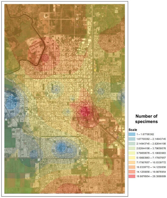
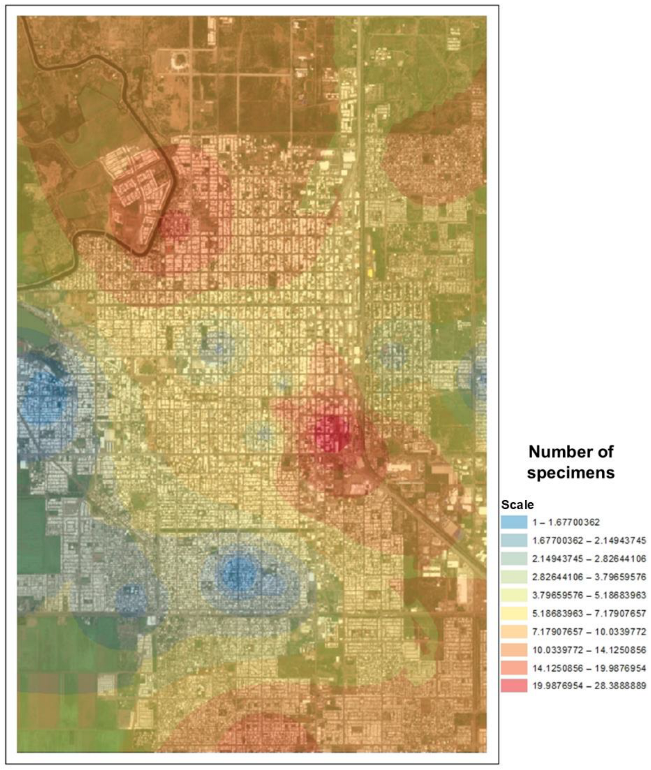
The collected arthropod vectors were identified as dog ticks (Rhipicephalus sanguineus), cat fleas (Ctenocephalides felis), dog lice (Heterodoxus spiniger), human head lice (Pediculus humanus capitis), and mosquitoes (Culex sp.). A total of 83 pooled samples were collected (n = 83), containing 820 arthropod specimens distributed among different arthropod vectors and reservoirs (Table 1). The spatial distribution and collection sites of the arthropod vectors are shown in Figure 1. The urban area of Ciudad Obregón exhibited a higher concentration of arthropod vector specimens than the peripheral areas. Specimens were distributed throughout the central, northern, southern, eastern, and western zones of the city.
Figure 1.
Interpolation of sampling points with respect to the number of specimens obtained from all arthropod vectors.
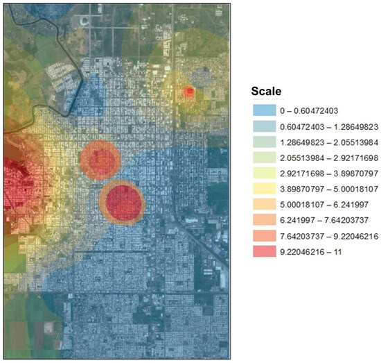
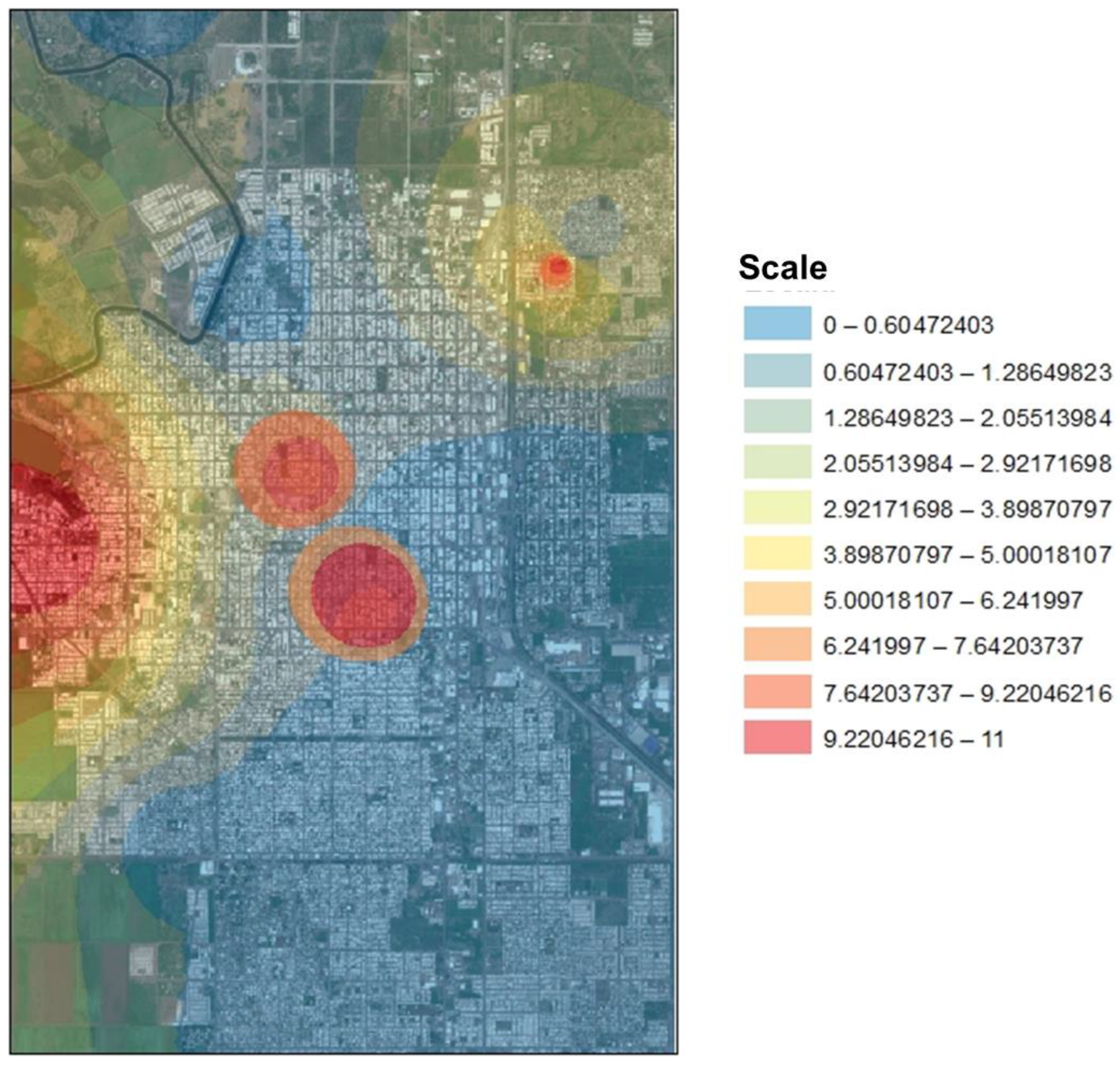
Of the 83 samples analyzed, 11 tested positive for Rickettsia spp., dog ticks (Rh. sanguineus), and cat fleas (C. felis) and were the only arthropod vectors that proved positive results. Based on this, the pooled positivity rate of Rickettsia spp. in ticks was 13.51% (5/37), and for fleas, it was 22.22% (6/27). The spatial distribution of positive samples was interpolated based on the locations where Rickettsia spp. were detected (Figure 2). The presence of Rickettsia spp. DNA was independent of the arthropod vector type and was not statistically significant (χ2 = 0.83; p > 0.05). The overall pooled positivity rate of Rickettsia spp. among all arthropod vectors analyzed was 13.25% (11/83). The MIRs for ticks and fleas were 3.57% (5/140) and 2.22% (6/270), respectively. The overall MIR obtained for Rickettsia spp. was 1.34% (11/820).
Figure 2.
Interpolation of sampling points with the presence of Rickettsia spp. from all arthropod vectors.
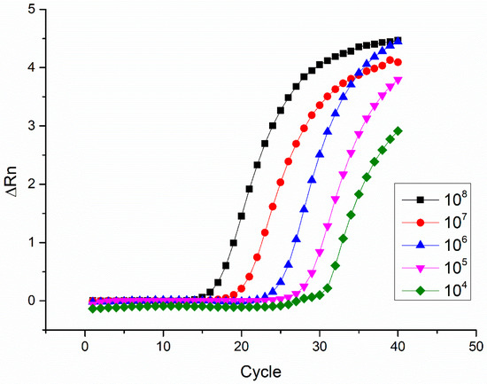
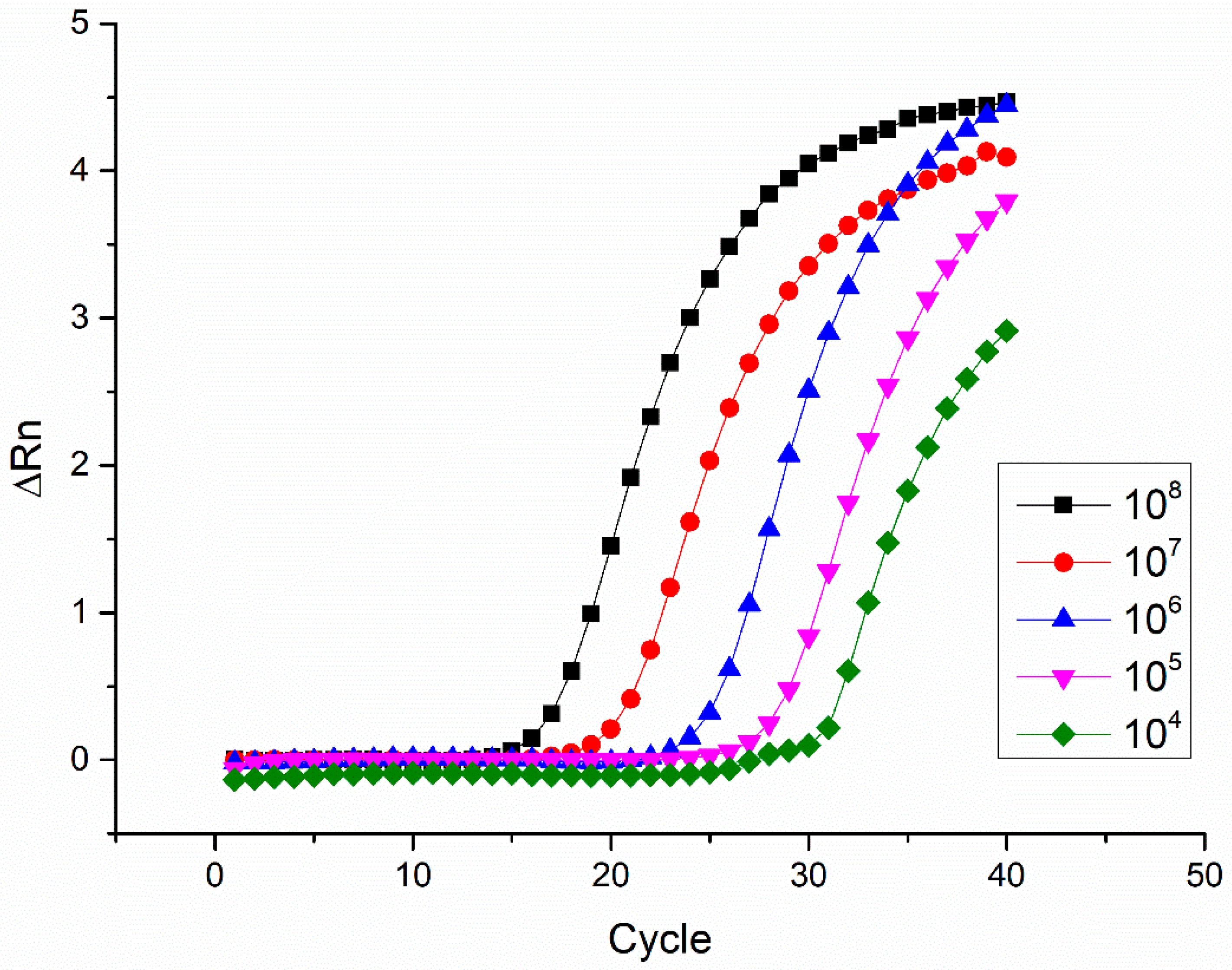
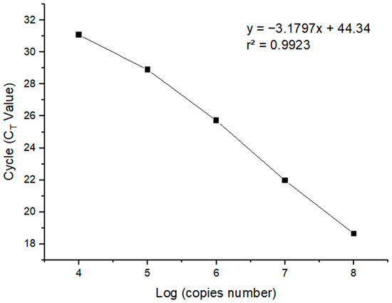
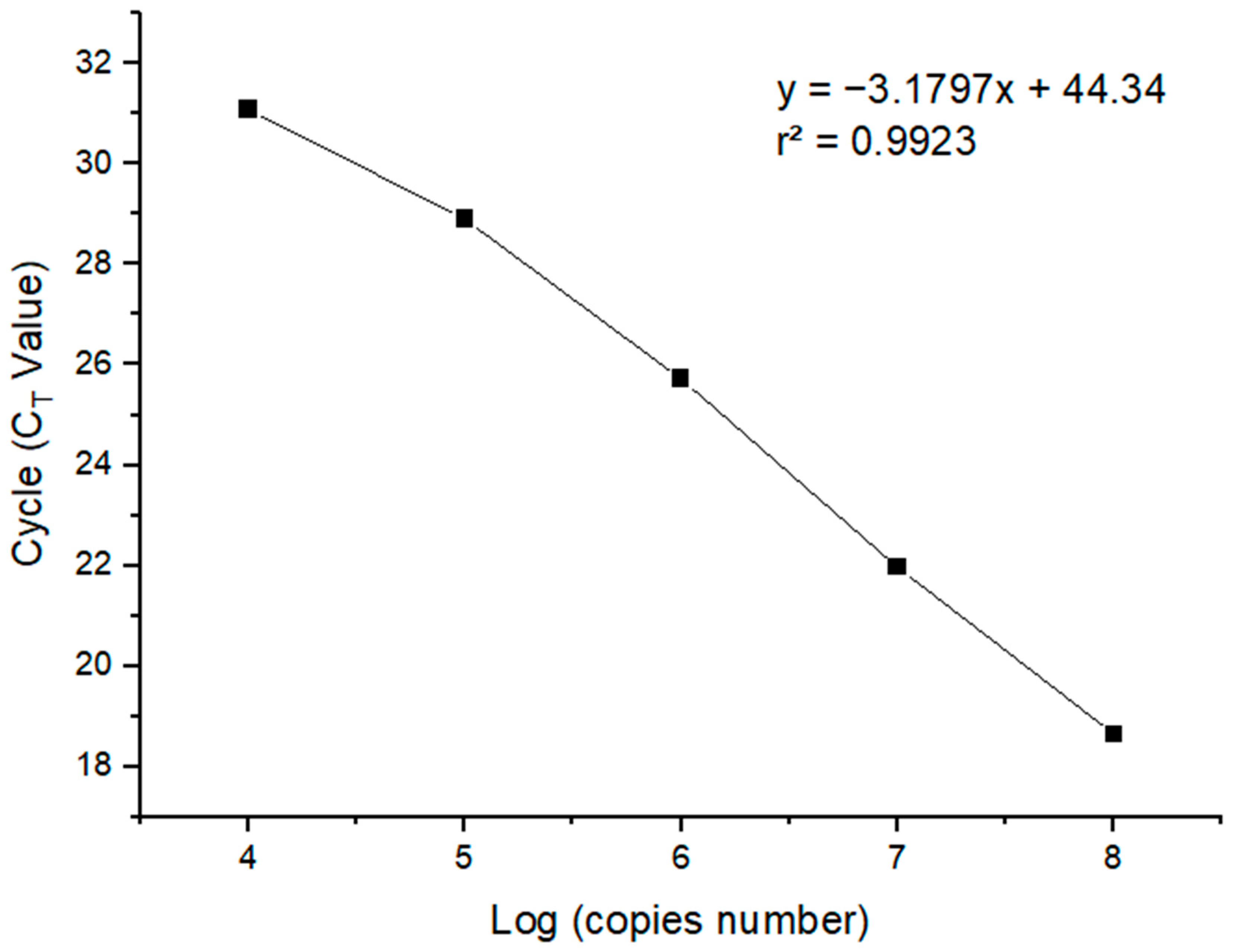
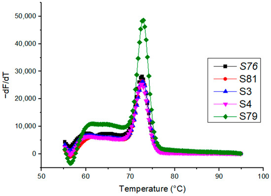
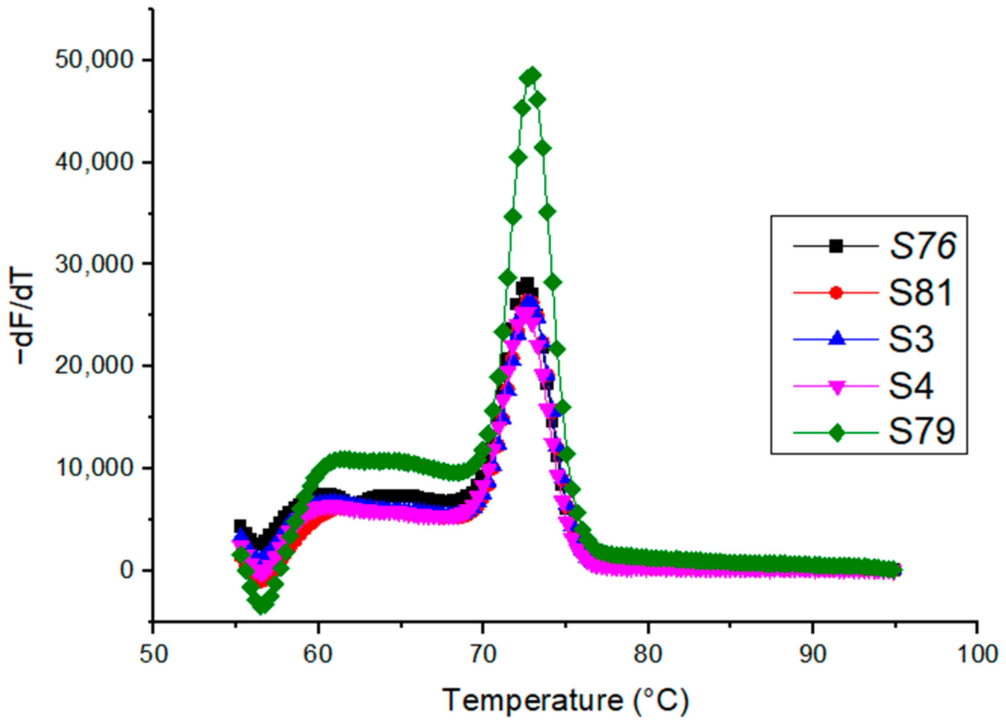
All positive samples for Rickettsia spp. by qPCR showed similar CT and copy number values regardless of the arthropod vector type (Table 2). Amplifications within the dynamic range of 104–108 copy numbers per reaction were satisfactory (Figure 3). After linearizing the amplifications, the standard curve demonstrated high linearity (r2 = 0.9923) and an efficiency of 106% (Figure 4). Absolute quantification was thus achieved, with a detection limit of 104 Rickettsia spp. gDNA copy numbers per reaction. Five samples (S3, S4, S76, S79, and S81) tested positive for SFG Rickettsia species; they showed amplification with the ompA gene primers. It is noteworthy that samples S3 and S4 are from fleas (C. felis), and samples S76, S79, and S81 are from ticks (Rh. sanguineus). The analyzed samples positive for the ompA gene revealed a melting temperature close to 73 °C (Figure 5).
Table 2.
Number of samples positive for Rickettsia spp. with their corresponding CT value and copy numbers per reaction obtained by qPCR.
Figure 3.
qPCR amplification to different quantities of copy numbers per reaction (108–104) of the plasmid 16S ribosomal RNA gene.
Figure 4.
Standard curve for quantification: Log (copy number) vs. CT.
Figure 5.
Dissociation curves from samples S3, S4, S76, S79, and S81 of the ompA gene.
4. Discussion
In Sonora, Mexico, including Ciudad Obregón, arthropods such as lice, mites, fleas, mosquitoes, and especially ticks are common [22]. These arthropods frequently interact with reservoirs, such as dogs, cats, guinea pigs, hamsters, rabbits, rats, and even humans. Additionally, these arthropods are particularly prevalent in areas where domestic or wild animals are concentrated and not protected from exposure to these ectoparasites. Interpolation shows that most arthropod vectors are concentrated in the central, northern, and southern parts of the city (areas with higher population density).
The detection of Rickettsia spp. in arthropod vectors associated with reservoirs in Ciudad Obregón, particularly in the central, western, and northeastern areas, was recorded. The spatial interpolation maps provide valuable information about potential areas of pathogen/arthropod circulation and may support epidemiological surveillance programs aimed at rickettsioses and other vector-borne diseases. In this case, the presence of Rickettsia spp. in Rh. sanguineus ticks obtained from dogs and C. felis fleas obtained from cats are reported, which may contribute to the prevalence of rickettsioses in the region. Moreover, characteristics such as sex, age, life stage, mode of transmission, and the environment are directly associated with the presence of Rickettsia spp. in these arthropod vectors [23]. Environmental conditions may also contribute to the maintenance of arthropod populations and pathogen transmission. The arid climate of Sonora, characterized by high temperatures and limited rainfall, may influence the abundance and seasonal dynamics of arthropod vectors [24].
The distribution of Rickettsia species in different arthropod vectors and reservoirs is important for the epidemiological surveillance of rickettsioses. Several studies have reported the presence of Rickettsia spp. in ticks worldwide. This is the case in Colombia, where Rickettsia species were found in ticks from different wildlife species [25]. SFG Rickettsia species were also reported in ticks collected in Romania [26]. Similarly, Rickettsia species were found in ticks collected from camels in Pakistan [27].
Specifically, there is information regarding the epidemiology of Rickettsia in Mexico [9,10,11,12,13,16,17,28]; however, there are few studies like ours that focus on arthropod vectors in Sonora, Mexico. Sánchez-Montes et al. [28] reported that different species of Rickettsia have been detected in Sonora, such as R. parkeri, R. massiliae, and Rickettsia sp. in Rh. sanguineus, but there are no reports or analyses of Rickettsia in other types of arthropod vectors (ej. C. felis), as we report in this study. Additionally, in Coahuila, México, the presence of Rickettsia spp. en Rh. sanguineus with a prevalence of 6.3% was reported [29]. In this study, a pooled positivity rate of 13.51% was found, which is considered higher than that in another Mexican state with high rates of rickettsioses. Likewise, the MIR observed in this study (3.57%) is higher than that reported in Coahuila (3.3%) [29] and other places in Central America, such as Costa Rica (2.3%) and Nicaragua (0.1%) [30]. Therefore, there is a possible risk of pathogen transmission through this vector in the study region compared to others.
In addition, several studies on the detection of Rickettsia in C. felis fleas have also been reported. For example, the prevalence reported in Kenya (6.7%) [31] is lower than that of the pooled positivity rate in the present study (22.22%); however, higher values have been reported, such as those from Israel (39.5%) [32] and the United States (28%) [33]. It should be noted that these studies identified R. felis as the primary host species of C. felis fleas, and R. felis is a globally recognized pathogen that causes spotted fevers [34].
The qPCR parameters, such as linearity and efficiency, were satisfactory. Good efficiency for a qPCR method should be between 90 and 110%. Values outside this range may be due to the presence of inhibitors, amplicon size, and the formation of secondary structures [35]. The reported efficiency value (106%) in this study falls within this range. However, an efficiency > 100% can result in inefficient amplification in dilute samples, inhibition of concentrated standards, or atypical CT values [36]. The efficiency was adequate compared with other studies reporting 128% [37] and 86.98% [38]. However, the latter study also reported an efficiency of 99.76% for the prsA gene, whereas this study used the 16S rRNA gene, which may have influenced the result due to amplicon size.
On the other hand, five samples (S3, S4, S76, S79, and S81) amplified the ompA gene and were classified as belonging to SFG Rickettsia. These samples showed Tm values of ~73 °C. In this regard, the ompA gene shows 55.4–98.8% similarity in SFG Rickettsia, demonstrating that we may be dealing with other SFG species, such as R. parkeri, R. rhipicephali, R. massiliae [39], and even R. felis; however, a sequencing assay is necessary to confirm this. The remaining samples (S11, S17, S40, S58, S59, and S67) were positive for Rickettsia spp. They did not show amplification for the ompA gene. This indicates that they may be Rickettsia species that do not belong to the SFG or that they may be species from the TG, such as R. prowazekii and R. typhi, or some species from other groups [40]. Therefore, to confirm these species, more than one gene (16S rRNA, ompB, gltA, ompA, htrA, and sca4) must be sequenced, followed by a phylogenetic analysis [41], to differentiate these highly related species.
A limitation of the present study is that positive samples were not confirmed by sequencing; therefore, species-level identification of the detected Rickettsia species could not be achieved. The qPCR method provides a rapid, sensitive, and reliable tool for epidemiological surveillance. Future studies should incorporate sequencing and phylogenetic analysis of genes, such as gltA, ompB, or htrA, to achieve more detailed taxonomic characterization. In regions with rickettsioses, the timely detection of Rickettsia spp. in arthropod vectors may be more relevant for public health than immediate species-level characterization as in our study.
5. Conclusions
In conclusion, the qPCR method developed in this study successfully detected Rickettsia spp. and SFG Rickettsia in arthropod vectors, such as dog ticks (Rh. sanguineus) and cat fleas (C. felis). The findings demonstrate the circulation of the DNA of Rickettsia spp. and SFG Rickettsia in arthropod vectors associated with domestic reservoirs and highlight the importance of considering vectors other than ticks in epidemiological surveillance programs. The study shows that this methodology could be used for early diagnosis of rickettsioses and for identifying geographic areas at higher risk of acquiring rickettsioses through different arthropod vectors and reservoirs affecting the population.
Author Contributions
Conceptualization, S.L.V.-S., R.R.-R. and L.A.Á.-V.; Methodology, S.L.V.-S., R.R.-R., L.A.Á.-V. and R.M.M.-B.; Software, S.L.V.-S., B.A.M.-N. and V.G.-G.; Validation, S.L.V.-S., R.R.-R., L.A.Á.-V. and A.G.V.-L.; Formal Analysis, S.L.V.-S., R.R.-R., L.A.Á.-V. and A.G.V.-L.; Investigation, S.L.V.-S., R.R.-R., L.A.Á.-V. and R.M.M.-B.; Resources, R.R.-R., L.A.Á.-V. and A.G.V.-L.; Data Curation, S.L.V.-S., R.R.-R., L.A.Á.-V. and A.G.V.-L.; Writing—Original Draft Preparation, S.L.V.-S., R.R.-R. and A.G.V.-L.; Writing—Review and Editing, S.L.V.-S., R.R.-R. and A.G.V.-L.; Visualization, S.L.V.-S., R.R.-R., B.A.M.-N. and V.G.-G.; Supervision, R.R.-R. and L.A.Á.-V.; Project Administration, R.R.-R., L.A.Á.-V. and A.G.V.-L. All authors have read and agreed to the published version of the manuscript.
Funding
This research received no external funding.
Institutional Review Board Statement
The animal study protocol was approved by the ethics committee of Instituto Tecnológico de Sonora (protocol code No. 2020-06 on 22 June 2020).
Informed Consent Statement
Not applicable.
Data Availability Statement
The original contributions presented in this study are included in the article. Further inquiries can be directed to the corresponding authors.
Acknowledgments
We extend our gratitude to BEI Resources for donation of materials (No. NR-35520 and No. NR-35520) and the Programa de Fomento y Apoyo a Proyectos de Investigación (PROFAPI-2025).
Conflicts of Interest
The authors declare no conflicts of interest.
References
- Miranda, J.; Mattar, S. Molecular detection of Rickettsia bellii and Rickettsia sp. strain Colombianensi in ticks from Cordoba, Colombia. Ticks Tick-Borne Dis. 2014, 5, 208–212. [Google Scholar] [CrossRef] [Scilit] [PubMed]
- Zhang, J.; Wang, J.; Chen, L.; Yassin, A.K.; Kelly, P.; Butaye, P.; Li, J.; Gong, J.; Cattley, R.; Qi, K.; et al. Housefly (Musca domestica) and Blowfly (Protophormia terraenovae) as vectors of bacteria carrying colistin resistance genes. Appl. Environ. Microbiol. 2018, 84, e01736-17. [Google Scholar] [CrossRef] [Scilit] [PubMed]
- Estrada-Peña, A.; Binder, L.C.; Nava, S.; Szabó, M.P.J.; Labruna, M.B. Exploring the ecological and evolutionary relationships between Rickettsia. Ticks Tick-Borne Dis. 2021, 12, 101754. [Google Scholar] [CrossRef] [Scilit] [PubMed]
- Pacheco, R.C.; Moraes-Filho, J.; Marcili, A.; Richtzenhain, L.J.; Szabó, M.P.J.; Catroxo, M.H.B.; Bouyer, D.H.; Labruna, M.B. Rickettsia monteiroi sp. nov., Infecting the Tick Amblyomma incisum in Brazil. Appl. Environ. Microbiol. 2011, 77, 5207–5211. [Google Scholar] [CrossRef] [Scilit] [PubMed]
- Zemtsova, G.E.; Killmaster, L.F.; Montgomery, M.; Schumacher, L.; Burrows, M.; Levin, M.L. First report of Rickettsia identical to R. slovaca in colony-originated D. variabilis in the United States: Detection, laboratory animal model, and vector competence of ticks. Vector Borne Zoonotic Dis. 2016, 16, 77–84. [Google Scholar] [CrossRef] [Scilit] [PubMed]
- Eremeeva, M.E.; Bosserman, E.A.; Demma, L.J.; Zambrano, M.L.; Blau, D.M.; Dasch, G.A. Isolation and identification of Rickettsia massiliae from Rhipicephalus sanguineus ticks collected in Arizona. Appl. Environ. Microbiol. 2006, 72, 5569–5577. [Google Scholar] [CrossRef] [Scilit] [PubMed]
- Boretti, F.S.; Perreten, A.; Meli, M.L.; Cattori, V.; Willi, B.; Wengi, N.; Hornok, S.; Honegger, H.; Hegglin, D.; Woelfel, R.; et al. Molecular investigations of Rickettsia helvetica infection in dogs, foxes, humans, and Ixodes ticks. Appl. Environ. Microbiol. 2009, 75, 3230–3237. [Google Scholar] [CrossRef] [Scilit] [PubMed]
- Torres-Castro, M.; Reyes-Novelo, E.; Noh-Pech, H.; Tello-Martín, R.; Lugo-Caballero, C.; Dzul-Rosado, K.; Puerto-Manzano, F.; Rodríguez-Vivas, R.G. Personal and household factors involved in recent Rickettsia exposure in a rural population from Yucatán, Mexico. Zoonoses Public Health 2020, 67, 506–515. [Google Scholar] [CrossRef] [Scilit] [PubMed]
- Herrero, J.A.; García-Vázquez, E.; Hernández, A.; Gómez, J. Infecciones por rickettsias y fiebre Q. Medicine. Programa Form. Médica Contin. Acreditado 2010, 10, 3881–3888. [Google Scholar] [CrossRef] [Scilit]
- Ulloa-García, A.; Dzul-Rosado, K.; Bermúdez-Castillero, S.E.; López-López, N.; Torres-Monzón, J.A. Detección de Rickettsia typhi en Rhipicephalus sanguineus s.l. y Amblyomma mixtum en el sur de México. Salud Pública México 2020, 62, 358–363. [Google Scholar] [CrossRef] [Scilit] [PubMed]
- Sánchez-Montes, S.; López-Pérez, A.M.; Guzmán-Cornejo, C.; Colunga-Salas, P.; Becker, I.; Delgado-de la Mora, J.; Licona-Enríquez, J.D.; la Mora, D.D.-D.; Karpathy, S.E.; Paddock, C.D.; et al. Rickettsia parkeri in Dermacentor parumapertus ticks, Mexico. Emerg. Infect. Dis. 2018, 24, 1108. [Google Scholar] [CrossRef] [Scilit] [PubMed]
- Peniche-Lara, G.; Dzul-Rosado, K.; Perez-Osorio, C.; Zavala-Castro, J. Rickettsia typhi in rodents and R. felis in fleas in Yucatán as a possible causal agent of undefined febrile cases. Rev. Inst. Med. Trop. São Paulo 2015, 57, 129–132. [Google Scholar] [CrossRef] [Scilit] [PubMed]
- Zavala-Castro, J.; Zavala-Velázquez, J.; Walker, D.; Pérez-Osorio, J.; Peniche-Lara, G. Severe human infection with Rickettsia felis associated with hepatitis in Yucatan, Mexico. Int. J. Med. Microbiol. 2009, 299, 529–533. [Google Scholar] [CrossRef] [Scilit] [PubMed]
- Zavala-Castro, J.E.; Zavala-Velázquez, J.E.; Walker, D.H.; Arcila, E.E.R.; Laviada-Molina, H.; Olano, J.P.; Ruiz-Sosa, J.A.; Small, M.A.; Dzul-Rosado, K.R. Fatal human infection with Rickettsia rickettsii, Yucatan, Mexico. Emerg. Infect. Dis. 2006, 12, 672. [Google Scholar] [CrossRef] [Scilit] [PubMed]
- Álvarez-Hernández, G.; Roldán, J.F.G.; Milan, N.S.H.; Lash, R.R.; Behravesh, C.B.; Paddock, C.D. Rocky Mountain spotted fever in Mexico: Past, present, and future. Lancet Infect. Dis. 2017, 17, e189–e196. [Google Scholar] [CrossRef] [Scilit] [PubMed]
- Delgado-De la Mora, J.; Sánchez-Montes, S.; Licona-Enríquez, J.D.; Delgado-De la Mora, D.; Paddock, C.D.; Beati, L.; Colunga-Salas, P.; Guzmán-Cornejo, C.; Zambrano, M.L.; Karpathy, S.E.; et al. Rickettsia parkeri and Candidatus Rickettsia andeanae in tick of the Amblyomma maculatum group, Mexico. Emerg. Infect. Dis. 2019, 25, 836. [Google Scholar] [CrossRef] [Scilit] [PubMed]
- Stewart, A.G.; Stewart, A.G.A. An update on the laboratory diagnosis of Rickettsia spp. infection. Pathogens 2020, 10, 1319. [Google Scholar]
- Husin, N.A.; AbuBakar, S.; Khoo, J.J. Current tools for the diagnosis and detection of spotted fever group Rickettsia. Acta Trop. 2021, 218, 105887. [Google Scholar] [CrossRef] [Scilit] [PubMed]
- National Communicable Disease Center (US). Pictorial Keys to Arthropods, Reptiles, Birds, and Mammals of Public Health Significance (No. 1955); US Government Printing Office: Washington, DC, USA, 1969.
- Green, M.R.; Sambrook, J. Molecular Cloning: A Laboratory Manual; Cold Springer Harbor Laboratory Press: Woodbury, NY, USA, 2012. [Google Scholar]
- Eremeeva, M.E.; Dasch, G.A.; Silverman, D.J. Evaluation of a PCR Assay for Quantitation of Rickettsia rickettsii and Closely Related Spotted Fever Group Rickettsiae. J. Clin. Microbiol. 2003, 41, 5466–5472. [Google Scholar] [CrossRef] [Scilit] [PubMed]
- Castrezana, S.J. Artrópodos terrestres no-hexápodos. In Diversidad Biológica de Sonora, 1st ed.; Molina-Freaner, F.E., Van Devender, T.R., Eds.; UNAM: Ciudad de México, Mexico, 2010; pp. 293–314. [Google Scholar]
- Azad, A.F.; Beard, C.B. Rickettsial pathogens and their arthropod vectors. Emerg. Infect. Dis. 1998, 4, 179. [Google Scholar] [CrossRef] [Scilit] [PubMed]
- Buechler, S. Gender, water, and climate change in Sonora, Mexico: Implications for policies and programmes on agricultural income-generation. G&D 2009, 17, 51–66. [Google Scholar] [CrossRef] [Scilit]
- Álvarez-Londoño, J.; Giraldo-Noreña, D.F.; Martínez-Sánchez, E.T.; Rivera-Páez, F.A.; Matta, N.E. Molecular survey of Rickettsia spp. in ticks infesting wild animals in six departments in Colombia. Ticks Tick-Borne Dis. 2024, 15, 102381. [Google Scholar] [CrossRef] [Scilit] [PubMed]
- Ivan, T.; Matei, I.A.; Novac, C.S.; Kalmár, Z.; Borșan, S.-D.; Panait, L.-C.; Gherman, C.M.; Ionică, A.M.; Papuc, I.; Mihalca, A.D. Spotted Fever Group Rickettsia spp. Diversity in Ticks and the First Report of Rickettsia hoogstraalii in Romania. Vet. Sci. 2022, 9, 343. [Google Scholar] [CrossRef] [Scilit] [PubMed]
- Ullah, S.; Alouffi, A.; Khan, M.; Khan, M.; Tila, H.; Tanaka, T.; da Silva Vaz, I.; Almutairi, M.M.; Ali, A. Molecular detection of Rickettsia aeschlimannii, Candidatus Rickettsia shennongii, Rickettsia sp. and Coxiella burnetii in ticks collected from camels. Sci. Rep. 2024, 14, 22129. [Google Scholar] [CrossRef] [Scilit] [PubMed]
- Sánchez-Montes, S.; Colunga-Salas, P.; Lozano-Sardaneta, Y.N.; Zazueta-Islas, H.M.; Ballados-González, G.G.; Salceda-Sánchez, B.; Huerta-Jiménez, H.; Torres-Castro, M.; Panti-May, J.F.; Peniche-Lara, G.; et al. The genus Rickettsia in Mexico: Current knowledge and perspectives. Ticks Tick-Borne Dis. 2021, 12, 101633. [Google Scholar] [CrossRef] [Scilit] [PubMed]
- Ortega-Morales, A.I.; Nava-Reyna, E.; Ávila-Rodríguez, V.; González-Álvarez, V.H.; Castillo-Martínez, A.; Siller-Rodríguez, Q.K.; Cabezas-Cruz, A.; Dantas-Torres, F.; Almazán, C. Detection of Rickettsia spp. in Rhipicephalus sanguineus (sensu lato) collected from free roaming dogs in Coahuila state, northern Mexico. Parasit. Vectors. 2019, 12, 1–7. [Google Scholar] [CrossRef] [Scilit]
- Springer, A.; Montenegro, V.M.; Schicht, S.; Wolfel, S.; Schaper, S.R.; Chitimia-Dobler, L.; Siebert, S.; Strube, C. Detection of Rickettsia monacensis and Rickettsia amblyommatis in ticks collected from dogs in Costa Rica and Nicaragua. Ticks Tick-Borne Dis. 2018, 9, 1565–1572. [Google Scholar] [CrossRef] [Scilit] [PubMed]
- Jiang, J.; Maina, A.N.; Knobel, D.L.; Cleaveland, S.; Laudisoit, A.; Wamburu, K.; Ogola, E.; Parola, P.; Breiman, R.F.; Njenga, M.K.; et al. Molecular detection of Rickettsia felis and Candidatus Rickettsia Asemboensis in Fleas from Human Habitats, Asembo, Kenya. Vector Borne Zoonotic Dis. 2013, 13, 550–558. [Google Scholar] [CrossRef] [Scilit] [PubMed]
- Hornok, S.; Baneth, G.; Grima, A.; Takáes, N.; Kontschán, J.; Meli, M.L.; Suter, V.; Salant, H.; Farkas, R.; Hofmann-Lehmann, R. Molecular investigations of cat fleas (Ctenocephalides felis) provide the first evidence of Rickettsia felis in Malta and Candidatus Rickettsia senegalensis in Israel. New Microbe New Infect. 2018, 25, 3–6. [Google Scholar] [CrossRef] [Scilit]
- Maina, A.N.; Fogarty, C.; Krueger, L.; Macaluso, K.R.; Odhiambo, A.; Nguyen, K.; Farris, C.M.; Luce-Fedrow, A.; Bennett, S.; Jiang, J.; et al. Rickettsial Infections among Ctenocephalides felis and Host Animals during a Flea-Borne Rickettsioses Outbreak in Orange County, California. PLoS ONE 2016, 11, e0160604. [Google Scholar] [CrossRef] [Scilit] [PubMed]
- Brown, L.D.; Macaluso, K.R. Rickettsia felis, an Emerging Flea-Borne Rickettsiosis. Curr. Trop. Med. Rep. 2013, 3, 27–39. [Google Scholar]
- Kubista, M.; Andrade, J.M.; Bengtsson, M.; Forootan, A.; Jonák, J.; Lind, K.; Sindelka, R.; Sjöback, R.; Sjögreen, B.; Strömbom, L.; et al. The real-time polymerase chain reaction. Mol. Asp. Med. 2006, 27, 95–125. [Google Scholar] [CrossRef] [Scilit]
- Claas, E.C.J.; Melchers, W.J.G.; Van den Brule, A.J.C. The Role of Real-time PCR in Routine Microbial Diagnostics. In Real-Time PCR in Microbiology From Diagnosis to Characterization; Mackay, I.M., Ed.; Caister Academic Press: Poole, UK, 2007; pp. 232–267. [Google Scholar]
- Stenos, J.; Graves, S.R.; Unsworth, N.B. A highly sensitive and specific real-time PCR assay for the detection of spotted fever and typhus group Rickettsiae. Am. J. Trop. Med. Hyg. 2005, 73, 1083–1085. [Google Scholar] [CrossRef] [Scilit] [PubMed]
- Papp, S.; Rauch, J.; Kuehl, S.; Richardt, U.; Keller, C.; Osterloh, A. Comparative evaluation of two Rickettsia typhi-specific quantitative real-time PCRs for research and diagnostic purposes. Med. Microbiol. Immunol. 2016, 206, 41–51. [Google Scholar] [CrossRef] [Scilit] [PubMed]
- Eremeeva, M.E.; Dasch, G.A. The Genus Rickettsia: Microbiology, Diversity and Taxonomy. Rev. Tunis. D’infectiologie 2011, 5, S7–S14. [Google Scholar]
- Roux, V.; Raoult, D. Phylogenetic analysis of members of the genus Rickettsia using the gene encoding the outer-membrane protein rOmpB (ompB). Int. J. Syst. Evol. Microbiol. 2000, 50, 1449–1455. [Google Scholar] [CrossRef] [Scilit] [PubMed]
- Oteo, J.A.; Nava, S.; De Sousa, R.; Mattar, S.; Venzal, J.M.; Abarca, K. Guías Latinoamericanas de la RIICER para el diagnóstico de las rickettsiosis transmitidas por garrapatas. Rev. Chil. Infectología 2014, 31, 54–65. [Google Scholar] [CrossRef] [Scilit]

Disclaimer/Publisher’s Note: The statements, opinions and data contained in all publications are solely those of the individual author(s) and contributor(s) and not of MDPI and/or the editor(s). MDPI and/or the editor(s) disclaim responsibility for any injury to people or property resulting from any ideas, methods, instructions or products referred to in the content. |
© 2026 by the authors. Licensee MDPI, Basel, Switzerland. This article is an open access article distributed under the terms and conditions of the Creative Commons Attribution (CC BY) license.