


Abstract
This is an investigation of molecular phylogeny and morphology of the genus Cymbosellaphora (Bacillariophyceae, Cymbellales). For this study, a strain of Cymbosellaphora geisslerae isolated from the Plotnikova River (Kamchatka Territory, Russia) was studied using light, scanning, and transmission electron microscopy, as well as molecular methods. Phylogenetic analysis based on 18S rDNA and rbcL gene sequences revealed that Cymbosellaphora geisslerae belongs to the order Cymbellales and forms an alliance with representatives of genera Gomphonella and Reimeria. The results of molecular study are supported by morphology. In the course of molecular analysis, we discuss the diversity of valve morphology across Cymbosellaphora, Gomphonella, Reimeria and related genera. As a result, a new type of pore occlusions, typical for Cymbosellaphora, is proposed, the diagnoses of the genus Cymbosellaphora and the species Cymbosellaphora geisslerae are emended, and the epitypification of this species is made. Most importantly, our data indicates that the concepts of areolae morphology and pore occlusions structure in the order Cymbellales might require critical evaluation.
1. Introduction
Diatoms of the order Cymbellales D.G. Mann constitute a diverse, globally distributed taxonomic group [1], which currently includes six families, more than 60 genera and more than 2300 species [2]. The order has a long history of study, with the first representatives described back in the 1830s [3,4,5]. In the course of the past two centuries, the taxonomic concept of the order experienced a number of re-evaluations. The earliest studies on Cymbellales were devoted to descriptions of new species and genera, e.g., Anomoeoneis Pfitzer, Cymbella C. Agardh, Didymosphenia M. Schmidt, Encyonema Kützing, Gomphonema Ehrenberg and Rhoicosphenia Grunow [3,4,5,6,7,8]. At the time, new taxa were erected on the basis of overall morphology of valves, chloroplast and protoplast [9,10,11,12,13,14]. Later on, methods of light microscopy and diatom identification were improved, and a number of floristic studies on Cymbellales were carried out in Europe [15], North America [16] and Asia, primarily USSR [17,18].
Since the 1980s, when the methods of electron microscopy (SEM and TEM) became widespread in diatom science, the knowledge on taxonomy, ecology and biology of Cymbellales has expanded significantly. In particular, the new approach offered the basis for taxonomic revisions in the two biggest genera of the order: studies by Krammer [19,20] aimed to reassess the concept of Cymbella, while the research of Kociolek & Stoermer [21,22] focused on Gomphonema and related genera. Morphological features of the valve—type of symmetry, raphe structure, morphology of striae and areolae—became crucial for taxonomic identification and delimitation. Therefore, several new genera were split off Cymbella and Gomphonema: Afrocymbella Krammer, Cymbopleura (Krammer) Krammer, Delicata Krammer, Encyonopsis Krammer, Gomphocymbellopsis Krammer and Reimeria Kociolek & Stoermer [21,23,24]. Notably, preliminary studies on the phylogeny and evolution of Cymbellales were performed [25,26]; the results of cladistic analyses of valve and protoplast characters indicated the necessity for further revisions and descriptions of new taxa. At the time, the order itself was formally described [1], and its classic system—with families Anomoeoneidaceae D.G. Mann, Cymbellaceae Kützing, Gomphonemataceae Kützing and Rhoicospheniaceae J.Y. Chen & H.Z. Zhu—was established.
In the recent studies, the “narrow” concept of species and genera [27,28] was adopted in diatom science, and morphological analysis of valve ultrastructure gained a key role. Thus, numerous genera were described de novo, e.g., Oricymba Jüttner, Krammer, E.J. Cox, Van de Vijver & Tuji—from the Nepalese Himalaya [29]; Kozhowia Kulikovskiy & Lange-Bertalot, Skvortzowia Kulikovskiy, Lange-Bertalot & Metzeltin, Paraplaconeis Kulikovskiy, Lange-Bertalot & Metzeltin, Ochigma Kulikovskiy, Lange-Bertalot & Metzeltin—from Lake Baikal, Russia [30]; Celebesia Kapustin, Kulikovskiy & Kociolek—from Sulawesi Island, Indonesia [31]; Vladinikolaevia Kulikovskiy, Glushchenko, Y. Liu & Kociolek—from Mongolia [32]; Qinia Y. Liu, Kociolek & Kulikovskiy—from China [33], etc. In the course of morphological revisions, new genera were split off: Rexlowea Kociolek & E.W. Thomas—from Naviculadicta Lange-Bertalot [34], Gomphosinica J.P. Kociolek, Q.-M. You, Q.-X. Wang & Q. Liu—from Gomphoneis Cleve [35]; Indiconema B. Karthick & Kociolek—from Gomphonema [36].
In the 2000s, molecular phylogenetics was introduced into the taxonomy and systematics of Cymbellales. The first molecular data on the order was acquired by Bruder & Medlin [37]. Their SSU rRNA, LSU rRNA and rbcL-based phylogeny supported the monophyly of this group and also approved the affinity of Placoneis Mereschkowsky to Cymbellales. Afterwards, molecular phylogeny was reconstructed for Didymosphenia—its close relationship to Cymbella tumida (Brébisson) Van Heurck group was determined by Kermarrec et al. [38] and Nakov et al. [39]. Another molecular study [40] helped ascertain the connections between Placoneis and Geissleria Lange-Bertalot & Metzeltin, another supposed member of Cymbellales. In the following years, the reference molecular database for the representatives of Cymbellales was greatly expanded [41,42,43,44,45,46,47].
The mentioned morphological and molecular studies highlighted the omissions of the original system of the order [1]. In fact, alternative systems have been suggested [48,49], but they have not been widely embraced in the future. Modern studies on Cymbellales incorporate an integrative approach to phylogeny, evolution and systematics. A recent example of that is the re-assessment of the genus Gomphoneis [42,44] which involved molecular analysis and investigation of raphe, striae and areolae morphology. The studies resulted in the separation of new genera—Gomphonella Rabenhorst and Gomphadelpha R. Jahn & N. Abarca. A similar route was adopted by Mironov et al. [50], whose integrative study focused on Placoneis and its allies. The authors investigated the ultrastructural morphological features of Placoneis, Paraplaconeis, Geissleria, etc., and re-emphasized the evolutionary significance of pore occlusions, following the ideas of Mann [51] and Cox [52]. Based on those findings, a new system of Cymbellales was instituted and two new families—Encyonemataceae Kulikovskiy, Mironov, Maltsev & Kociolek and Witkowskiaceae Kulikovskiy, Mironov, Maltsev & Kociolek—were established [53].
Despite the development of the new system, some issues of systematics and phylogeny still remain unresolved. First of all, studies repeatedly indicate the polyphylies of large genera of Cymbellales, e.g., Cymbella and Cymbopleura, which is why further taxonomic revisions are needed. Secondly, many genera of Cymbellales lack reference molecular data, which limits the possibility of conducting molecular phylogenetic analyses to understand the phylogeny of these genera. Thirdly, there is still no consensus on the importance of various morphological features for taxonomy and evolution, which makes it difficult to define the boundaries of taxa and develop effective systems. And lastly, our knowledge on diatom diversity in some regions is still limited.
The species diversity of diatoms on the Kamchatka Peninsula has been studied unevenly, which is explained by the peninsula’s unique geographic location and topography that influence the accessibility of certain aquatic ecosystems for researchers. The Kamchatka Peninsula is located in northeastern Russia, bordered to the west by the Sea of Okhotsk and to the east by the Bering Sea and the Pacific Ocean. The peninsula’s aquatic ecosystem is quite rich, with over 140,000 large and small flowing waterbodies located within its territory [54].
The region is known for the natural spawning of Pacific salmon which occurs in its rivers and lakes, where fish undergo important stages of ontogenesis, from the development of eggs to the formation of smolt [55,56]. Because of this, the greatest number of studies on freshwater ecosystems of Kamchatka are devoted to the research of phytoplankton communities in lakes, of which the Kuril Lake is the most well studied. A large array of data on phytoplankton (collections for the period from 1980 to 2000) was processed and summarized in the dissertation of E.V. Lepskaya [57] and the article by Lepskaya and Bonk [58]. Furthermore, the phytoplankton of the lakes Dalneye [59], Kronotskoye [60], Talovskoye [61] and others has been investigated in considerable detail. Some works were dedicated exclusively to individual taxa, for example, the morphology and seasonal abundance dynamics of the centric diatom Aulacoseira subarctica (O. Müller) E.Y. Hawort [62] or the morphological variability of the species Gomphonema ventricosum Gregory [63]. In addition, paleoecological studies of diatom communities in lake sediments have been conducted [64], and recently the terrestrial algae of the peninsula, volcanic substrates, lava tubes and ecotone zones have been studied [63,64,65,66,67,68,69]. Concerning the issue of the current work, only a small number of studies investigated the diversity of Cymbellales in Kamchatka Peninsula [70,71,72].
In the current work, we address the problem of phylogeny of Cymbosellaphora Kulikovskiy, Glushchenko, Genkal & Kociolek, which was originally designated as a member of Cymbellales. We provide the first molecular data on the genus to determine its affinity to the order. The phylogenetic position of the genus is discussed in detail, and proposals on its relationships with other genera are made. Using the results of our extensive morphological analysis of Cymbosellaphora and its allies, we review the currently accepted concept of areolae morphology in Cymbellales and propose a new type of pore occlusions. As our data demonstrates, the widely understood concept of pore occlusions in Cymbellales might require critical re-evaluation. Additionally, we emend the diagnoses of Cymbosellaphora geisslerae (Jahn) Kulikovskiy, Glushchenko, Genkal & Kociolek and the genus Cymbosellaphora itself, and discuss the distribution of Cymbosellaphora geisslerae in waterbodies throughout the world.
2. Materials and Methods
2.1. Sampling, Culturing and Slide Preparation
The sample used for this study was collected on 10 July 2021, in Kamchatka Peninsula, Yelizovsky District, Nachiki village, Plotnikova River, periphyton (53°7.162′ N, 157°44.708′ E). The sample was designated no. 12. The water temperature during sampling was 8.8 °C, pH 7.45 and conductivity 271 μS/cm, determined using a Hanna Combo (HI 98129) multiparameter probe (Hanna Instruments Ltd., Inc., New York, NY, USA).
For culturing, part of the sample was transferred to a Petri dish, and some WC liquid culturing medium [73] was added. Monoclonal strains were established by micropipetting a single cell under an inverted microscope Axio Vert. A1 (Zeiss, Oberkochen, Germany). Non-axenic unialgal culture was maintained in WC liquid medium at 22–25 °C in a growth container with a 12:12 h light:dark photoperiod. The acquired strain was analyzed after approximately 30 days of culturing. The strain was assigned the number CBMCkam360 and was deposited in the Culture and Barcode Collection of Microalgae and Cyanobacteria “Algabank” (CBMC) at K.A. Timiryazev Institute of Plant Physiology RAS [74].
For light microscope (LM) and scanning and transmission electron microscope (SEM, TEM) investigations the diatom culture was processed using a standard procedure that entails boiling it with concentrated hydrogen peroxide. Afterwards, the material was washed with distilled water. Light microscopic (LM) observations were made with the use of the AxioScope A1 microscope (Zeiss, Oberkochen, Germany) equipped with an Axiocam Erc 5s camera (Zeiss, Oberkochen, Germany), an oil immersion EC Plan-NEOFLU-AR objective (×100/n.a. 1.3) for epifluorescent microscopy (EFM) of live material and an oil immersion objective (×100/n.a. 1.4, Nomarski differential interference contrast) for examination of cleaned material. Permanent diatom preparations were mounted in Naphrax® (Brunel Microscopes Ltd., Chippenham, UK; refractive index = 1.73). Valve ultrastructure was investigated using the scanning electron microscopes JSM-6510LV at working distance 11 mm, accelerating voltage 15kV (JEOL, Tokyo, Japan) and TESCAN Vega III at working distance 7 mm, accelerating voltage 10kV (TESCAN, Brno, Czech Republic), and transmission electron microscope JEOL JEM-1011 (JEOL, Tokyo, Japan; accelerating voltage 80 kV). For the SEM examination, part of the cleaned material was spread onto aluminum stubs and air-dried at room temperature for 24 h. The stubs were sputter-coated with 50 nm Au in an Eiko IB-3 (Eiko Engineering, Ltd., Hitachinaka, Japan). For TEM studies, the culture suspension was applied to standard copper support grids coated with formvar film (Tescan, Brno, Czech Republic) and dried at room temperature. The suspension and slides prepared from the strain CBMCkam360 were assigned no. 08192. All material of the strain was deposited in the Herbarium of K.A. Timiryazev Institute of Plant Physiology, Russian Academy of Sciences (HD), Moscow, Russia [75].
2.2. Molecular Analysis
After a month of cultivation, procedures of DNA extraction, polymerase chain reaction (PCR) and phylogenetic analysis were performed. For each strain, part of cultured material was centrifuged in 1.5 mL eppendorfs for 5 min at 6000 rpm. From this material, DNA was extracted using the Chelex100 Chelating Resin (Bio-Rad Laboratories, Hercules, CA, USA), following the 2.2. manufacturer protocol. The V4 region of 18S rDNA was amplified using D512for and D978rev primers [76]. The plastid rbcL gene was amplified with rbcL404+ and rbcL1444- primers [77]. The amplification was performed with a pre-made mastermix ScreenMix (Evrogen, Moscow, Russia). For the amplification of 18S rDNA, the following program was chosen: initial denaturation (95 °C, 5 min), 35 cycles of denaturation (94 °C, 30 s), annealing (52 °C, 30 s), elongation (72 °C, 50 s), final extension (72 °C, 7 min) and maintenance (12 °C, ∞). For the amplification of the rbcL gene: initial denaturation (94 °C, 5 min), 44 cycles of denaturation (94 °C, 50 s), annealing (53 °C, 50 s), elongation (72 °C, 80 s), final extension (72 °C, 10 min) and maintenance (12 °C, ∞). PCR products were visualized on a 1.0% agarose gel with the SYBRTM Safe stain (Life Technologies, Carlsbad, CA, USA), then purified with a mix of sterile water, FastAP, 10× FastAP Buffer and Exonuclease I (Thermo Fisher Scientific, Waltham, MA, USA). The acquired genetic material was sequenced using the Genetic Analyzer 3500 (Applied Biosystems, Waltham, MA, USA).
Raw data sequences were viewed and edited in Ridom TraceEdit (Ridom© GmbH, Münster, Germany). Forward-read and reverse-read sequences were united using MEGA7.1 (Pennsylvania State University, Pennsylvania, PA, USA; [78]). 18S rDNA and rbcL matrices were constructed from the newly acquired sequences of strain CBMCkam360 and 169 reference sequences selected from NCBI GenBank nucleotide database. Every strain was represented in each matrix. A total of 4 strains of Stephanodiscus Ehrenberg were chosen as outgroup, and other reference sequences were selected to represent the different lineages of Cymbellales. In addition, the matrices were supplemented with several sequences of monoraphid diatoms, due to their similarity to the sequences of the new strain according to Standart Nucleotide BLAST (https://blast.ncbi.nlm.nih.gov/Blast.cgi?PROGRAM=blastn&PAGE_TYPE=BlastSearch&LINK_LOC=blasthome, accessed on 25 November 2025). The matrices were then aligned separately. rbcL matrix was aligned in Mafft ver. 7 (RIMD, Osaka, Japan; [79]) with the G-INS-I algorithm. 18S rDNA matrix was aligned by T-Coffee online aligner [80]. Aligned matrices were edited in MEGA7.1 again: the sequences were trimmed and unpaired regions were removed. The rbcL matrix was translated in order to ensure that the first nucleotide in the dataset corresponds to the first nucleotide of the codon. Afterwards, a single concatenated matrix was constructed (170 sequences × 1251 nt [318 18S rDNA + 933 rbcL]).
Bayesian inference (BI) analysis was carried out using the BEAST ver.1.10.1 software (BEAST Developers, Auckland, New Zealand; [81]). For BI analysis, specifications were determined by the Bayesian information criterion (BIC) in the jModelTest ver.2.1.10 program (Vigo, Spain; [82]). GTR+G+I substitution model was selected, shape parameter α = 0.2112, and a proportion of invariable sites (pinvar) = 0.6125. During BI, Yule process tree prior speciation model was applied, 10 MCMC analyses were employed for 10 million generations (burn-in 1 million generations). Resulting data was analyzed in Tracer ver. 1.7.1 software (MCMC Trace Analysis Tool, Edinburgh, UK; [83]) and initial 10% of trees were removed. Rapid bootstrapping and a subsequent ML search (RAxML) were conducted to estimate the tree topology robustness. For RAxML, raxmlGUI 2.0 software was employed [83]. RAxML was performed with 1000 replicas, GTR substitution matrix and gamma substitution rates. Best resulting phylogram was viewed in FigTree ver. 1.4.4 (University of Edinburgh, Edinburgh, UK). The final tree was manually edited in Adobe Photoshop CC ver.19.0 (Adobe, San Jose, CA, USA).
Input and resulting data of molecular analyses can be accessed in Supplementary files (Files S1–S3).
2.3. Terminology
The terminology of the valve morphology follows [19,20,24,52,84].
3. Results
3.1. Molecular Phylogeny
The results of the conducted two-gene (18S rRNA and rbcL) molecular analysis demonstrate the phylogenetic relationships between different lineages of the order Cymbellales (Figure 1). The data from our analysis is generally consistent with the system of the order proposed in Mironov et al. [53]. In our phylogram, families Rhoicospheniaceae and Anomoeoneidaceae are represented by their type genera—Rhoicosphenia and Anomoeoneis, respectively.
Figure 1.
Molecular phylogeny of the order Cymbellales (18S rRNA and rbcL genes). Values of Bayesian Posterior Probabilities (PP) below 0.90 and values of likelihood bootstrap (LB) from RAxML analyses below 50 are not shown. Available strain IDs are indicated for all sequences (* indicates a strain with no ID available). GenBank accession numbers for all sequences are provided in Supplementary files (File S1). The grey background shows clade GRC—Gomphonella + Reimeria + Cymbosellaphora. The family Gomphonemataceae is marked with a question mark (“?”) since our analysis shows it as polyphyletic.
The position of these two clades relative to the rest of the tree remains disputable: both clades are located basally in relation to other branches of Cymbellales, but the nodes of Rhoicospheniaceae and Anomoeoneidaceae are unsupported. At the same time, each clade itself branches out with maximum statistical support (PP = 1; LB = 100).
According to the phylogram, the strains representing monoraphid diatoms branch together within Cymbellales, sister to the clade of Anomoeoneidaceae. These strains demonstrated high similarity to the newly obtained strain CBMCkam360 according to 18S rDNA BLAST comparison. However, our two-gene analysis reveals that these strains are neither closely related to C. geisslerae CBMCkam360 nor associated with any other lineage of Cymbellales. Their placement alongside Anomoeoneidaceae is unsupported, as well.
The recently established families Encyonemataceae and Witkowskiaceae are well-supported by our analysis, comprising separate independent clades. The clade of Encyonemataceae (PP = 1; LB = 88) includes 19 strains of Encyonema. The clade of Witkowskiaceae (PP = 0.98) includes a total of 18 strains belonging to Witkowskia Kulikovskiy, Mironov, Glushchenko & Kociolek, Geissleria and Paraplaconeis. In the current phylogram, the family Cymbellaceae is represented by a monophyletic clade; however, the statistical support is lacking. Unfortunately, the topology demonstrated herein cannot be interpreted as proof of Cymbellaceae’s monophyly without robust evidence from BI or RAxML. It is highly likely that the expansion of the reference molecular database for the largest genera of Cymbellaceae—Cymbella, Cymbopleura and Encyonopsis—would help ascertain the monophyly of the family and resolve the phylogenetic relationships between the cymbelloid genera and species groups.
Unlike previous studies, our analysis reveals the polyphyly of Gomphonemataceae, a family traditionally understood as monophyletic [37,39,50]. According to our phylogram, species of Gomphonema and Gomphadelpha comprise a strongly supported clade (PP = 1; LB = 94), while two other gomphonemoid genera—Gomphonella and Reimeria—branch out separately, together with Cymbosellaphora geisslerae. Clade GRC (Gomphonella + Reimeria + Cymbosellaphora) is resolved only by BI (PP = 1) and is located in a sister position in relation to the clade of Encyonemataceae. However, the common node of the two clades is not supported by BI or RAxML. Thus, the discrepancy between Gomphonella + Reimeria and other gomphonemoid genera is obvious from our analysis and may potentially indicate the need for a critical approach to the phylogeny of Gomphonemataceae. Undoubtedly, obtaining additional molecular data on representatives of Gomphonemataceae could be an effective tool for studying the phylogeny and systematics of this group. In our study, we propose explanations for the mismatch between the clade GRC and the genera Gomphonema and Gomphadelpha, both in terms of molecular data and morphology.
Most importantly, our molecular investigation determines the independence of Cymbosellaphora and ascertains its affinity to Cymbellales. Phylogenetic analysis suggests that this genus is a close ally of Gomphonella and Reimeria. This connection is also discussed below, with particular reference to ultrastructural morphological features of the three genera.
3.2. Morphology of Cymbosellaphora geisslerae
As a result of the study of the morphology of Cymbosellaphora species, begun by us earlier [84] and continued in the current work, below we propose an emended diagnosis for the genus Cymbosellaphora and the species Cymbosellaphora geisslerae.
Cymbosellaphora Kulikovskiy, Glushchenko, Genkal & Kociolek emend. Glushchenko, Tseplik, Mironov, Genkal & Kulikovskiy.
Generitype: Cymbosellaphora vietnamensis Glushchenko, Kulikovskiy & Kociolek in Kulikovskiy et al. 2023.
Diagnosis.
LM. Cells are solitary. The single plastid consists of four lobes connected by an isthmus with an invagination. Each of the two adjacent lobes is pressed against the valve mantle and face. The lobes have processes and notches which are especially noticeable in girdle view. Frustule is rectangular in girdle view. It is represented by an epivalve, a hypovalve, two valvocopulae, and at least one girdle band. The valve apices have a characteristic notch, so the valve mantle is smaller at the apices than in the central part of the valve. Valves are nearly linear-elliptic to almost rhombic-lanceolate, naviculoid or slightly dorsiventral. Ends are broadly protracted and broadly rounded or obtusely rounded, sometimes rostrate or sub-capitate. Raphe is straight and filiform. Axial area is narrow,straight. Central area is variable, from small to medium size, transversely widened and irregularly delimited by shortened striae.
SEM, externally. The valve face is flat. The raphe is positioned in the middle of the valve. Proximal raphe ends are more or less expanded, slightly bent in the direction opposite the distal ends; they may lie either on the surface of the valve or in an elongated, teardrop-shaped depression. Distal raphe ends are hooked, bent to one side, and extended onto the valve mantle. The central nodule is broad. Striae are uniseriate, parallel or slightly radiate, becoming radiate in the center, convergent near the ends, with puncta usually resolvable. At the apices, shortened parallel striae are also present as well as individual areolae. The areolae are covered by structureless, rounded or slightly transversely elongated silica plaques positioned slightly below the surface of the valve; these closures are termed “plomba” (rus. ”плoмбa”—dental filling; pl. plombas). The areolar openings have various shapes: from round to elongated, from zigzag to dumbbell-shaped, which is explained by the unevenness of the corrosion of the openings and the exposure of their internal structure. During processing, the plomba first begins to dissolve in the center, then a part of it falls out, forming a zigzag-shaped or dumbbell-shaped lumen, and then it is dissolved completely, following the shape of the internal opening of the areola. Apical pore fields are absent. The mantle is very wide with striae and areolae continuing from valve face. Girdle bands are open, with one row of rounded or transversely elongated perforations.
SEM, internally. Axial area is straight with a narrow sternum evident or slightly evident. The valve mantle is low. Proximal raphe ends are small and bent to one side. Distal raphe ends terminate in small, well-developed helictoglossae. The helictoglossae are located on a hyaline area. The central nodule is broad. Interstriae are not elevated, but they are wide and broader than striae. Striae lie in a small depression. Areolae are covered with a thin, imperforate, flat silica layer. This silica layer covers the areolae at one level, lying flush with the inner surface of the valve. When this layer is destroyed, the internal structure of the areola becomes visible. Areolae are square to elongated rectangular, separated by well-defined struts, which are destroyed during further processing of the material, and as a result, two small remnants of strut remain lying opposite each other. Plombas are visible at the bottom of each areola; they dissolve starting from the center of the closure, subsequently taking the shape of the areolar lumen. Voigt discontinuity is present on the secondary side of the valves.
Cymbosellaphora geisslerae (Jahn) Kulikovskiy, Glushchenko, Genkal & Kociolek 2023 emend. Glushchenko, Tseplik, Mironov, Genkal & Kulikovskiy.
Epitype (here designated). Strain CBMCkam360 from the Culture and Barcode Collection of Microalgae and Cyanobacteria “Algabank” (CBMC), isolated from sample no. 12; oxidized culture of strain CBMCkam360 prepared on slide no. 08192, deposited in the Herbarium of K.A. Timiryazev Institute of Plant Physiology, Russian Academy of Sciences (HD), Moscow, Russia.
Locus epitypicus. Kamchatka Peninsula, Yelizovsky District, Nachiki village, Plotnikova River (53°7.162′ N, 157°44.708′ E), periphyton, 10.07.2021, leg. M.S. Kulikovskiy.
Epitype DNA sequences. Available at GenBank with the following accession numbers: PZ158815 for rbcL and PZ161853 for 18S rDNA.
Description.LM, live cells (Figure 2). Cells are free-living and motile, with a length of 13.8–26.2 µm and a width of 6.1–6.6 µm. During mass development in culture, they form clusters (Figure 2A,B). In valve view, cells are linear-lanceolate to linear-elliptical (Figure 2A, black arrows; Figure 2B–O). In girdle view, cells are rectangular (Figure 2A, white arrows).
Figure 2.
Cymbosellaphora geisslerae. Live culture, strain CBMCkam360. (A,C–E,H,I,K–M,O–R) Light microscopy (LM). (B,F,G,H,J,N) Epifluorescent microscopy (EFM). (A,B) Mass of cells in culture. Valve and girdle views. The photographs demonstrate the chloroplast structure. The equals sign (=) marks photographs taken of the same groups of cells or specimens with different focal length and using different methods (LM and EFM). Black arrows indicate cells in valve view. White arrows indicate cells in girdle view. (P–R) Auxospores in different stages. (P) Auxospore beginning to form. (Q) Growing auxospore pushing the gametangium (parent cell) walls apart; they stay attached to the auxospore for some time. (R) Further growth of the auxospore; one of the gametangium walls begins to detach. Scale bar = 10 μm.
The single plastid consists of four lobes connected by an isthmus with an invagination. Each of the two adjacent lobes is pressed against the valve mantle and face. The lobes have processes and notches, which are especially noticeable in girdle view (Figure 2A, white arrows). The auxospore is located in the parent cell (Figure 2P). Growing auxospores push the walls of the gametangium (the epitheca and hypotheca of the parent cell frustule) apart. The gametangium length is 9.7–9.9 µm (Figure 2Q,R). The auxospores are irregularly shaped, sometimes swollen in the central part, length 24.2–27.2 µm, width 6.7–6.8 µm (Figure 2Q,R).
LM, cleaned material (Figure 3). Frustules are rectangular in girdle view (Figure 3AI,AJ). Valves are linear-lanceolate to rhombic-lanceolate, naviculoid, often weakly dorsiventral (Figure 3A–AH), with a length of 8.7–24.6 µm and width of 3.6–5.4 µm. Valve ends are broadly rounded, sometimes weakly protracted. The axial area is narrow and linear, the central area is bowtie-shaped. The central area is bordered by shortened striae, 1–4 on each side of the valve. The raphe is narrow and linear. Proximal raphe ends are slightly expanded; distal raphe ends extend onto the valve mantle. Striae are radiate, becoming subparallel closer to the valve apices, convergent at the apices themselves, numbering 20–23 in 10 µm. Areolae are not resolvable in LM; individual areolae are visible on the striae located closer to the central area of the valve (Figure 3A).
Figure 3.
Cymbosellaphora geisslerae. Light microscopy, Nomarski differential interference contrast (LM, DIC). Slide No. 08192 from oxidized culture strain CBMCkam360. Size diminution series. (A–AH) Valve view. (AI,AJ). Frustules in girdle view. Scale bar = 10 μm.
SEM, external views.
Frustule (Figure 4A). Frustule is rectangular in girdle view. It is represented by an epivalve, a hypovalve, two valvocopulae and at least one girdle band. The striae extend from the valve face onto the mantle. The valve apices have a characteristic notch, so the valve mantle is smaller at the apices than in the central part of the valve (Figure 4A, black arrows).
Figure 4.
Cymbosellaphora geisslerae. Scanning electron microscopy (SEM). Oxidized culture strain CBMCkam360. (A). Frustule, girdle view. EV—epivalve; VC—valvocopula (girdle band adjacent to the valve); B—girdle band; HV—hypovalve. Black arrows indicate a characteristic notch in the apical part of the valves, which is typical for cymbelloid diatoms. (B) Whole post-initial frustule, external view. Black arrow indicates a rudiment of an additional raphe branch. White arrow indicates part of the frustule covered by the remnant of the perizonium. Black arrowheads indicate perizonium rings. White arrowheads indicate rows of apically elongated slit-like poroids. (C) Post-initial valve, internal view. Black arrows indicate helictoglossae that are noticeably distant from the valve apices. White arrow indicates an intercalary stria. Scale bar = 5 μm (A–C).
Post-initial valve (Figure 4B,C). Post-initial valve is weakly fusiform, swollen in the central portion, and has a convex surface.
Raphe branches are poorly visible; rudiments of additional raphe branches are present (Figure 4B, black arrow). Remnants of the perizonium are present at one of the apices of the post-initial valve (Figure 4B, white arrow). The perizonium has transverse rings, approximately 18 rings in 10 µm (Figure 4B, black arrowheads). Apically elongated slit-like poroids are present between the perizonium rings, approximately 75 in 10 µm (Figure 4B, white arrowheads). The areolae are small, round, and form transverse striae.
Valves (Figure 5). Valve face is flat. Proximal raphe ends are expanded, curved, and lie at the bottom of a teardrop-shaped depression (Figure 5B,C, black arrows). Distal raphe ends are curved towards the valve margin, to the other side than the central ends, and are positioned fairly close to one of the striae (Figure 5A,D, black arrow). Striae are uniseriate, radiate, becoming subparallel closer to valve apices, and convergent at the apices themselves. At the apices there are also present shortened parallel striae as well as individual areolae (Figure 5D, white arrow).
Figure 5.
Cymbosellaphora geisslerae. External views, scanning electron microscopy (SEM). Oxidized culture strain CBMCkam360. (A) Whole valve. Black arrow indicates the distal raphe end. (B) Whole valve. Black arrows indicate the proximal raphe ends. (C) Central area. Black arrows indicate the proximal raphe ends positioned in elongated teardrop-shaped depressions. Black arrowhead indicated the areola occlusion, represented by a plomba, in a slight depression. White arrohead indicates the beginning of corrosion of the plomba in its central part. White arrow indicates corrosion of the closure into a dumbbell-shaped opening. (D). Valve end. Black arrow indicates the distal raphe end. White arrow indicates the densely positioned striae on the valve apex. Black arrowhead indicates the areola occlusion, represented by a plomba, in a slight depression. Scale bar = 5 μm (A,B), 1 μm (C,D).
In the central part of the areola lumen there is a plomba—a plate-like silica closure. The plomba is an occlusion represented by an unperforated silica flap. It is located in the middle part of the areola, not on the valve surface (Figure 5C,D, black arrowheads). Boiling of the material destroys the areola, revealing the structure of the areolae. During processing, the plomba first begins to dissolve in the center (Figure 5C, white arrowhead), then a part of it falls out, forming a dumbbell-shaped lumen (Figure 5C, white arrow), and then it is dissolved completely [85] (Figure 9G,H). Areolae density is approximately 40–45 in 10 µm.
SEM, internal views.
Post-initial valve (Figure 4C). The post-initial valve is irregularly shaped, slightly swollen in the central part, and its inner surface is concave. The raphe is filiform and straight. Proximal raphe ends are not visible. Distal raphe ends are unilaterally bent and terminate in helictoglossae. Helictoglossae are located at a greater distance from the valve apices than in normally formed valves (Figure 4C, black arrows). Striae are radiate, with intercalary striae present (Figure 4C, white arrow).
Valves (Figure 6). The valve mantle is low. The raphe lies on a slightly elevated sternum. Proximal raphe ends are small and bent to one side (Figure 6D, black arrows). Distal raphe ends terminate in small, well-developed helictoglossae (Figure 6B,C, black arrows). The helictoglossae are located on a hyaline area. Striae are significantly narrower than interstriae. Striae lie in a small depression. Areolae are apically elongated and rectangular, closer to the central part of the valve, but square on the valve apices, separated by well-developed struts located on the vimines (Figure 6D, white arrows), which are destroyed during further processing of the material; as a result, two small remnants of the struts remain positioned opposite each other [84] (Figure 3C–F). Plombas are visible at the bottom of each areola (Figure 6E, black arrows); they dissolve starting from the center of the closure (Figure 6E, white arrows), subsequently taking the shape of the areolae lumen [84] (Figure 9L). Shortened striae consisting of 2–3 areolae are visible at valve apices. The frequent alternation of interstriae and struts resembles a lattice (Figure 6A, black arrow; Figure 6E). Intercalary striae are present on both sides of the valve (Figure 6A, white arrows).
Figure 6.
Cymbosellaphora geisslerae. Internal views, scanning electron microscopy (SEM). Oxidized culture strain CBMCkam360. (A) Whole valve. Black arrows indicate shortened striae consisting of 2–3 areolae. The frequent alternation of interstriae and struts resembles a lattice. White arrows indicate shortened striae bordering the central area on both sides. (B,C) Whole valve. Black arrows indicate distal raphe ends that terminate in helictoglossae. (D) Central area. Black arrows indicate the proximal raphe ends that are hooked to one side. (E). Valve end. Black arrows indicate the occlusion represented by a plomba that covers the areola in a depression. White arrows indicate different stages of corrosion of the plomba, starting from its central part. Scale bar = 5 μm (A,B), 2 μm (C–E).
TEM (Figure 7 and Figure 8). Large valves are nearly naviculoid (Figure 7A); the smaller valve is distinctly dorsiventral (Figure 7B). Both the external and internal raphe branches are clearly visible (Figure 8B). The external proximal raphe ends are expanded, elongated and drop-shaped (Figure 8B, black arrow); the internal ones are hooked (Figure 8B, white arrow). Areolae are separated by vimines (Figure 7B, black arrows). Areolae have square to rectangular internal openings; in the central part the lumen of the areola is closed by a plomba (Figure 8F, black arrow). Destruction of the plombas begins from the central part (Figure 8D, black arrows); afterwards, two opposite remnants of the closure remain in the lumen of the areola (Figure 8B,C, white arrowheads). The plombas have no ordered structure (Figure 8F). The plomba is more rigid and denser in the center and thinner on the periphery. The boundaries between individual areolae are visible due to denser struts (Figure 8E, black arrows). At the valve apex, hyaline areas (Figure 7B and Figure 8A, white arrowheads) and individual shortened striae (Figure 7B, black arrows) are clearly visible. The girdle band is open, narrowed at one end (Figure 7C, white arrow), and bears a single row of small poroids (Figure 8C, black arrow).
Figure 7.
Cymbosellaphora geisslerae. Transmission electron microscopy (TEM). Oxidized culture strain CBMCkam360. (A,B). Whole valve. Larger valves are almost naviculoid in shape, the small valve is distinctly dorsiventral. (B) Whole valve. Black arrows indicate shortened striae near the valve apices. White arrows indicate vimines. White arrowheads indicate hyaline areas on the valve apices. (C) Open girdle band. White arrow indicates the narrowing of the girdle band on the apex. Black arrow indicates the single row of round poroids perforating the band. Scale bar = 5 μm (A,C), 2 μm (B).
Figure 8.
Cymbosellaphora geisslerae. Transmission electron microscopy (TEM). Oxidized culture strain CBMCkam360. (A) Valve end. White arrowhead indicates the hyaline area on the valve apex. (B) Central area. Black arrow indicates the external central raphe end. White arrow indicates the internal hooked central raphe end. Black arrowhead indicates the vimen. White arrowheads indicate the remnants of the plomba in the shape of two tongues located opposite each other. (C) Areolae. White arrowheads indicate the remnants of the plomba in the shape of two tongues located opposite each other. (D) Detail of areolae. Black arrows indicate the start of corrosion of the plombas in their central part. (E) Detail of areolae. Black arrows indicate thickened partitions that appear darker because of their increased thickness. (F) Areolae. Black arrow indicates the plomba that is thicker in the central part and thinner on the periphery. Scale bar = 1 μm (A,B,D,E), 0.25 μm (C,F).
4. Discussion
4.1. Molecular Phylogeny and Morphology of Cymbosellaphora and Related Genera
Cymbosellaphora was introduced by Kulikovskiy et al. [84] as a genus belonging to the order Cymbellales. At the time, it was assumed that this genus should be classified into the family Cymbellaceae. The assumption was justified by the morphological similarities of Cymbosellaphora and other cymbelloid genera—slight dorsiventrality of valves, perforated girdle bands and occlusions represented by tectula [84]. The authors especially noted the structure of pore occlusions in Cymbosellaphora. It was described as follows: “areolae are covered internally by a nonperforated silica layer with presence of rectangular struts that divide every areola. During the cleaning of the valves, these rectangular struts can be destroyed, revealing two outgrowths situated parallel to the valve level and opposite one another”. Therefore, the structure of pore occlusions was interpreted as tectulum sensu Cox [52], i.e., similar to that of Placoneis (sensu Cox [52]).
An important advantage of the initial study on Cymbosellaphora was the determination of the significance of pore occlusions for phylogeny of the genus. In fact, Kulikovskiy et al. [84] anticipated the boom of cymbelloid taxonomy, which began shortly after Cymbosellaphora was described and continues to this day. As a result of this boom, the structure of pore occlusions is gaining a crucial role for phylogeny and systematics of diatoms; new classifications of pore occlusions are being introduced and new genera are being described based on the unique structure of pore occlusions (e.g., [50,53,85]). In particular, a new classification was introduced for the pore occlusions of Placoneis sensu Cox and related genera of Cymbellales with naviculoid symmetry (Paraplaconeis, Geissleria, Rexlowea, etc.) [50]. In that study, tectulum s. s. was characterized as a type of pore occlusion represented by “several regularly arranged small struts, extending in the areolar opening perpendicular to the valve surface” and seven new types of occlusions were described (paratectulum, oculus, pseudovola, etc.). Hence, according to the modern concept of tectulum (sensu Mironov et al. [50]), this type of pore occlusion cannot be treated as typical for Cymbosellaphora. The major disparity is the absence of perpendicularly arranged struts in areolae of Cymbosellaphora. Instead of that, the vimines of Cymbosellaphora are furnished with struts [84] (Figure 8B–D) (Figure 8E, black arrows) which appear as oppositely positioned stubs [84] (Figures 3C–F, 7A and 11F) during corrosion and can sometimes deteriorate completely [84]. Such structures on vimines are quite usual for cymbelloid diatoms, e.g., typical for Encyonema [23] (Plate 3, Figures 2–4; Plate 23: Figure 1; Plate 62: Figure 1) and Cymbopleura [24] (Plate 1, Figures 2 and 4B; Plate 18: Figure 5; Plate 21: Figures 3–5).
Using the new SEM and TEM data on valve ultrastructure of Cymbosellaphora, we characterize a type of pore occlusions which has not been shown in representatives of Cymbellales before. We hereby suggest a new term plomba (rus. “плoмбa”—dental filling; pl. plombas) to describe the pore occlusions of Cymbosellaphora (see description of genus and species of this study). The plomba is a delicate internal occlusion represented by an unperforated silica flap, denser in the center and thinner on the periphery (Figure 8F). In the process of corrosion, the plomba starts degrading in the central region (Figure 5C, white arrowhead), while two oppositely positioned remnants persist (Figure 8B,C, white arrowheads) and further deteriorate completely.
Obviously, the presence of plombas in Cymbosellaphora vs. tectula s. l. in Placoneis and its allies from the family Witkowskiaceae is a convincing proof of phylogenetic discrepancy between these taxa. Another evidence of this is molecular data presented in this study. Our phylogram (Figure 1) reveals that the newly obtained strain of Cymbosellaphora geisslerae is not allied with the clade of Witkowskiaceae, but instead clusters together with Gomphonella and Reimeria. This association can be explained both from a molecular and morphological points of view, which is discussed below.
The results of our molecular analysis indicate that Gomphonella and Reimeria (clade GRC, Figure 1) are isolated from the diatoms of Gomphonemataceae—Gomphonema and Gomphadelpha. The current study is not the first one to demonstrate that Gomphonemataceae in its modern understanding [1,53] is polyphyletic. For instance, the 18S rDNA-based molecular study by Kermarrec et al. [38] revealed that Reimeria is closely related to Gomphonema micropus Kützing, but not to the species of Gomphonema’s “core group” [43]—Gomphonema acuminatum Ehrenberg (generitype), Gomphonema truncatum Ehrenberg, etc. Cladistic, valve morphology-based analysis provided similar outcomes, as Reimeria + G. micropus-clade branched out independently from Gomphonema and Gomphoneis [38]. Subsequently, the genus Gomphonella was molecularly typified [42]. Single-gene (rbcL and 18S rDNA) phylogenies illustrated that Gomphonella olivacea (Hornemann) Rabenhorst is not allied with species of Gomphonema. In fact, the authors stated that Gomphonella exhibits dissimilarities in valve structure with Gomphonema and “does not even belong to Gomphonemataceae” from the molecular point of view. The research of Jahn et al. [42] aimed to resolve the polyphyly of Gomphoneis by reinstating the genus Gomphonella. Thus, a bigger problem—polyphyly of Gomphonemataceae—was noted, but not approached. In their recent studies Abarca et al. [43,44] delivered more molecular data on gomphonemoid diatoms and, as a result, more evidence of polyphyly in Gomphonematacae. As can be concluded from the discussed studies, the taxonomic entity of Gomphonemataceae is not so unambiguous, at least from the perspective of molecular phylogeny.
Moreover, morphology is consistent with molecular data when it comes to the relationship between Gomphonella and Gomphonema. In fact, our morphological comparison reveals that representatives of the GRC alliance exhibit similarities of valve structure. In addition, the data of morphology indicates that neither Cymbosellaphora nor Reimeria and Gomphonella are related to the Gomphonema + Gomphadelpha complex. Some of the morphological features of these genera were already compared in refs. [38] and [42]. Kermarrec et al. [38] noted that G. micropus and several species of Reimeria have small and round areolae, rather than C-shaped, reniform areolae typical for Gomphonema s. s. Jahn et al. [42] mentioned that Gomphonella differs from Gomphonema s. s. by lacking stigmoids, straight and filiform raphe, undifferentiated apical pore fields and round, not reniform, areolae in striae. It is worth mentioning that both studies underscore the disparities in areolae morphology of Gomphonella + Reimeria vs. Gomphonema s. s. Our study supplements these findings: we demonstrate that Cymbosellaphora, another genus with small, round areolae (Figure 5, Figure 6, Figure 7 and Figure 8) is genetically related to Gomphonella and Reimeria. Most importantly, the naturalness of the GRC union is warranted by the structure of pore occlusions.
Our research with the use of genetic methods allowed us to determine the phylogenetic position of Cymbosellaphora, which was not clear from the initial description of this genus. We have shown its separate position from other genera, which supports the independence of Cymbosellaphora, and its close relation to Gomphonella and Reimeria (Figure 1). The morphological analysis of pore occlusions in the studied group agrees with these results. The establishment of an epitype for C. geissleri that is complete with molecular data is an important step in the study of cymbelloid taxonomy, since it is a valuable reference point for future research.
Furthermore, our data supports the previously made conclusion that the family Gomphonemataceae is polyphyletic. We can assume that this issue, which is so far not completely resolved, can be addressed in following studies and solved with the help of future molecular research as well as SEM and TEM-based analyses, with use of more monoclonal cultures. In addition, phylogeny of Reimeria certainly requires further investigation, as molecular data for the genus is still scarce, with eight sequences available in NCBI GenBank database. The integrative approach, involving SEM, TEM and molecular analysis, could provide resolutions for greater issues—polyphyly of Gomphonemataceae and taxonomic entity of the GRC clade. Until then, we suggest that the data presented in this research is sufficient to answer the question of molecular phylogeny of Cymbosellaphora and explain the peculiarities of its morphology.
4.2. Areolae Morphology vs. Structure of Pore Occlusions
Since Cox [52] revised the classification of pore occlusions for raphid diatoms, it has traditionally been accepted that foriculum (pl. foricula) is a structure, representing typical pore occlusions in the order Cymbellales. Particularly, Cox [52] distinguished variations in foricula—symmetrical (as in Cymbella s. s.), uneven (as in Delicata), tri-/quadriradiate (as in Cymbopleura inaequalis (Ehrenberg) Krammer), dendritic (as in Didymosphenia), unilateral (as in Gomphonema s. s.) and round (as in Gomphoneis and Reimeria). The two latter types of foricula are of the biggest interest: unilateral foricula, which form reniform or C-shaped openings of areolae, were well-defined on the example of Gomphonema species by Cox [52] (Figures 7–9, 18 and 24). By contrast, round foricula were not illustrated, i.e., it is unclear what kind of ultrastructure Cox [52] understood by this term. Also, it is unclear which morphological group of Gomphoneis was meant by Cox [52] when describing round foricula. However, representatives of Gomphadelpha [44] (formerly Gomphoneis herculeana (Ehrenberg) Cleve group) possess areolae with irregular or angular, rather than rounded openings [44] (Figures 14–18, 32–34, 38–40 and 59–64), while species of Gomphonella [42] (formerly Gomphoneis olivacea (Hornemann) P.A. Dawson ex R. Ross & P.A. Sims group) exhibit a type of structure that could be treated as a round foriculum sensu Cox [52] (see [42] (Figures 5, 8–10, 12 and 13)).
As the term “round foriculum” was neither effectively illustrated nor adopted in diatom science, we believe that it should not be used when describing the pore occlusions of Gomphonella or Reimeria. Instead of that, we suggest that diatoms of the three genera (Cymbosellaphora, Gomphonella, Reimeria) should be characterized by a common type of pore occlusions—plomba—which is described and illustrated herein on the example of C. geisslerae (Figure 5, Figure 6, Figure 7 and Figure 8). Previously, plombas were demonstrated for other species of Cymbosellaphora [84] (Figures 3C–F, 8A,D and 9I–L); furthermore, the pore occlusions illustrated for some species of Gomphonella [42] (Figures 8D–F,J–L, 9D–F,J–L, 10D–F, 12D–F and 12J–L), in our opinion, also resemble plombas. In the case of Reimeria, the structure of areolae seems to be heterogenous, because round areolae with plombas were illustrated for Reimeria fontinalis Levkov & Ector [86] (Figure 47) and Reimeria sinuata (W. Gregory) Kociolek & Stoermer [86] (Figures 57 and 58), [87] (Figure 4D–F), while C-shaped and reniform areolae with plombas—for Reimeria ovata (Hustedt) Levkov & Ector [86] (Figures 48–53), [88] (Figure 3) and Reimeria uniseriata Sala, Guerrero & Ferrario [86] (Figures 59–64), [87] (Figure 2), [88] (Figure 4).
Notably, Cymbosellaphora shares similarities in the structure of areolae and pore occlusions with the recently discovered genus Liuyania Kulikovskiy, Mironov & Glushchenko [89]. Areolae in Liuyania are closed by a silica plate from the inside, which is clearly visible both from the external [89] (Figure 4F) and from the internal surface [89] (Figures 5C–D and 6F). One important difference is that the silica plate in Liuyania often begins to corrode from the edge, rather than from the central part as in representatives of Cymbosellaphora. Another important difference in the areolae structure is the absence of stubs and struts on vimines in Liuyania [89] (Figures 5 and 6). Nevertheless, as noted earlier, the features of areolae occlusions in cymbelloid diatoms are quite diverse, and their classification and re-evaluation is still being carried out. Probably, morphological similarities between the two genera may be ascertained with the help of molecular methods in the future.
Our morphological data leads to the idea that another issue was not discussed in ref. [52] when Cox proposed the new types of pore occlusions. The study of Cox [52] implied that diatoms of Cymbellales are characterized by varying morphology of external openings of areolae, i.e., those “are formed similarly by the outgrowth of thin flanges of silica”. However, Cox [52] stated that “the final slit shape is dependent on the primary direction(s) of growth”, so that different types of pore occlusions can be detected. Thus, according to the definition of Cox [52], presence of differently structured external openings of areolae indicates the presence of different types of occlusions on the inner valve side. However, this concept conflicts with two features. Firstly, areolae are delicate structures, easily corroded during sample preparation for SEM and TEM, which complicates actual perception of their morphology. Secondly, external openings are often uniform with the silica layer of the external valve surface. The typical example for that is Gomphonema: C-shaped apertures are visible from the outside as a single-layer, homogenic formation together with the rest of the valve [43] (Figures 28–42). This peculiarity is quite evident when the valves are more corroded [43] (Figures 49–52). The misconception becomes clear in the case of Delicata—a genus originally established based on the unique undulate structure of striae [24]. Cox linked the presence of undulate striae to “uneven foricula” [52], but the consequent studies revealed species of Delicata with roundish, not undulate external openings of areolae (see discussion in ref. [90] and Figures 13–19 in it). The analysis of remaining valve features indicates the monophyly of the genus, which means that the definition of pore occlusions according to Cox is not applicable. Hence, we suggest that the term “foriculum” is not just unsuitable for Delicata or, as discussed in Section 4.1, for Gomphonella and Reimeria, but also for Cymbellales in general.
Our analysis concludes with the identification of a new, previously undescribed type of pore occlusions—plomba—typical of Cymbosellaphora and, presumably, Gomphonella and Reimeria. This discovery helped us highlight the problem of foriculum, a term that was originally suggested to describe pore occlusions in Cymbellales [52]. As we found out, this term has to be used to describe only the morphology of external openings of areolae in Cymbellales. For example, externally, in SEM, areolae in Gomphonema s.s. can be described as unilateral foricula, while areolae in Delicata can be described as uneven foricula. In the discussion, we prove that the actual structure of pore occlusions in Cymbellales is different, resembling plombas of Cymbosellaphora. Based on these findings, we suppose that with the help of additional data, the concepts of “areolae morphology” and “type of pore occlusions” can be formally delimited. Achieving this aim would ensure the accurate study of phylogeny, evolution and systematics of the order.
4.3. Comments on Cymbosellaphora geisslerae
Cymbosellaphora geisslerae was described as Navicula geisslerae from the Spree River, Berlin [91]. This species was also reported from James Lake, Canada, Central Europe [92], Weinfelder Maar, Germany [93] and from Lake Glubokoe, Russia [94]. We recorded this species from Mongolia, from benthos of Lake Khövsgöl (Khiger Bay), as well as from an unnamed lake in the Lake Khövsgöl region, separated by a sandbar [84]. Our finding of C. geisslerae in Kamchatka is the first record of this genus and species for the region.
The material of C. geisslerae from Kamchatka is morphologically consistent with the material studied by R. Jahn [91], Chudaev and Gololobova [94], as well as the material from Mongolia [84]. The analysis of quantitative traits of different populations of C. geisslerae shows the similarity of the studied populations (see Table 1). In the current work we have demonstrated a fairly detailed morphological variation in the species in culture, starting from post-initial valves and ending with auxospores (Figure 2P–R). The closest values for the range of valve length were shown by Chudaev and Gololobova from Lake Glubokoe, Moscow Region [94] (p. 398, Table 229, Figures 1–32). The maximum valve length of their material reaches 27.6 µm, while our largest valves reach 26.2 µm (see Table 1).
Table 1.
Comparison of quantitative morphological features of different populations of Cymbosellaphora geisslerae.
The formation and growth of auxospores in this genus and species is demonstrated here for the first time. The chloroplast is also demonstrated and studied for the first time. The chloroplast structure we observed is characteristic of cymbelloid diatoms [96]. The structure consists of a single chloroplast with four lobes connected by an isthmus with an invagination, further confirming the classification of Cymbosellaphora as a cymbelloid diatom.
The ultrastructure of our cultured material of C. geisslerae is identical to that of the species studied by various authors (see Table 1).
The process of molecular epitypification of previously described species is gaining importance due to the expanding use of the integrative approach to diatom taxonomy [97,98]. It provides the basis for further research. Well-documented biodiversity and community structure provide a basis for verifying species richness, assessing changes in communities under anthropogenic pressure and climate change, harmonizing existing and future monitoring data, and comparing data obtained using modern metabarcoding methods [99,100,101,102,103,104]. Further morphological analysis, culture, and molecular data on representatives of the genus Cymbosellaphora will allow for a broader understanding of the phylogeny of cymbelloid diatoms.
Supplementary Materials
The following supporting information can be downloaded at https://www.mdpi.com/article/10.3390/phycology6020034/s1. File S1: Alignment of the SSU rRNA genes used for phylogenetic analysis in this study; File S2: The Bayesian phylogenetic topology for the constructed phylogenetic tree; File S3: Alignment of the rbcL rRNA and 18S rRNA genes used for phylogenetic analysis in this study.
Author Contributions
Conceptualization, M.K., A.G. and A.M.; methodology, M.K., A.G., A.M., Y.M. and S.G.; validation, M.K., N.T. and A.G.; investigation, A.M. and A.G.; resources, M.K.; writing—original draft preparation, A.M., A.G. and M.K.; writing—review and editing, N.T. and M.K.; visualization, N.T., S.G. and A.G.; supervision, A.G. and M.K.; funding acquisition, A.G. and M.K. All authors have read and agreed to the published version of the manuscript.
Funding
Publication is based on research carried out with financial support by the Russian Science Foundation (25-14-00286, https://rscf.ru/project/25-14-00286/ accessed on 23 March 2026) for molecular investigation, LM and SEM, the percentage contribution is 90%; by the framework of state assignment of the Ministry of Science and Higher Education of the Russian Federation (theme 126012015839-6) for culturing and finishing the manuscript, the percentage contribution is 10%.
Data Availability Statement
The samples collected during this study and prepared samples were deposited into the Diatom Herbarium (HD) at K.A. Timiryazev Institute of Plant Physiology, RAS, Moscow, Russia. The analyzed strains are housed at the Culture and Barcode Collection of Microalgae and Cyanobacteria “Algabank” (CBMC) at K.A. Timiryazev Institute of Plant Physiology, RAS, Moscow, Russia. The sequences obtained during the current study will be available in the NCBI SRA database upon the publication of this article. Until then, sequence data for the strain CBMCkam360 should be requested from A. Mironov. The molecular datasets and results of the molecular analyses performed can be accessed in the Supplementary files.
Acknowledgments
The authors express their gratitude to R.A. Rakitov (from the instrument analytics room of the Borissiak Paleontological Institute of the Russian Academy of Science, PIN RAS) for assistance in working with the scanning electron microscope. The authors are also grateful to the anonymous reviewers for their valuable input and contributions to the significant improvement of the current manuscript.
Conflicts of Interest
The authors declare no conflicts of interest.
Abbreviations
The following abbreviations are used in this manuscript:
| LM | Light microscopy |
| EFM | Epifluorescent microscopy |
| SEM | Scanning electron microscopy |
| TEM | Transmission electron microscopy |
| rbcL | Gene-encoding large subunit of ribulose-1,5-bisphosphate carboxylase/oxygenase |
| 18S rDNA | Small subunit of the ribosomal deoxyribonucleic acid |
| SSU rRNA | Small subunit ribosomal ribonucleic acid |
| SSU rRNA | Large subunit ribosomal ribonucleic acid |
| DIC | Differential interference contrast |
| PCR | Polymerase chain reaction |
| BI | Bayesian inference |
| RAxML | Randomized accelerated maximum likelihood |
| GTR | General time-reversible |
| LB | Likelihood bootstrap |
| PP | Posterior probability |
References
- Round, F.E.; Crawford, R.M.; Mann, D.G. The Diatoms. Biology and Morphology of the Genera; Cambridge University Press: Cambridge, UK, 1990; pp. 1–747. [Google Scholar]
- Guiry, M.D.; Guiry, G.M. AlgaeBase. World-Wide Electronic Publication, University of Galway, Ireland. Available online: https://www.algaebase.org (accessed on 11 December 2025).
- Agardh, C.A. Conspectus criticus Diatomacearum. Part 1; Literis Berlingianus: Lundae, Sweden, 1830; pp. 1–16. [Google Scholar]
- Ehrenberg, C.G. Über die Entwickelung und Lebensdauer der Infusionsthiere; nebst ferneren Beiträgen zu einer Vergleichung ihrer organischen Systeme. Abh. Der Königlichen Akad. Wiss. Zu Berl. Phys. Kl. 1832, 1–154. [Google Scholar]
- Kützing, F.T. Synopsis diatomearum oder Versuch einer systematischen Zusammenstellung der Diatomeen. Linnaea 1834, 8, 529–620. [Google Scholar] [CrossRef] [Scilit]
- Grunow, A. Über neue oder ungenügend gekannte Algen. Erste Folge, Diatomeen, Familie Naviculaceen. Verhandlungen Der Kais.-Königlichen Zool.-Bot. Ges. Wien 1860, 10, 503–582. [Google Scholar]
- Pfitzer, E. Untersuchungen über Bau und Entwicklung der Bacillariaceen (Diatomaceen). In Botanische Abhandlungen aus dem Gebiet der Morphologie und Physiologie Bd 2; bei Adolph Marcus: Bonn, Germany, 1871; pp. 1–189. [Google Scholar]
- Schmidt, A.W.F. Atlas der Diatomaceen-Kunde; Series V: Heft 54; O.R. Reisland: Leipzig, Germany, 1899; pp. 213–216. [Google Scholar]
- Agardh, C.A. Conspectus criticus Diatomacearum. Part 3; Literis Berlingianus: Lundae, Sweden, 1831; pp. 33–48. [Google Scholar]
- Greville, R.K. Div. IV. Diatomaceae. In The English Flora of Sir James Edward Smith. Class XXIV. Cryptogamia. Vol. V. Part I. Comprising the Mosses, Hepaticae, Lichens, Characeae and Algae; Hooker, W.J., Ed.; Longman, Brown, Green & Longmans Paternoster-Row: London, UK, 1833; pp. 401–415. [Google Scholar]
- Ehrenberg, C.G. Verbreitung und Einfluss des mikroskopischen Lebens in Süd- und Nord-Amerika. Abh. Der Königlichen Akad. Der Wiss. Zu Berl. 1843, 1841, 291–445. [Google Scholar]
- Kützing, F.T. Species algarum; F.A. Brockhaus: Leipzig, Germany, 1849; pp. 1–922. [Google Scholar]
- Heiberg, P.A.C. Conspectus criticus Diatomacearum Danicarum. Kritisk Oversigt over de Danske Diatomeer; Wilhelm Priors Forlag: Kjøbenhavn, Denmark, 1863; pp. 1–135. [Google Scholar] [CrossRef] [Scilit]
- Cleve, P.T. Synopsis of the naviculoid diatoms. Part I. In Kongliga Svenska Vetenskapsakademiens Handlingar; Series 4; Norstedt: Stockholm, Sweden, 1894; Volume 26, pp. 1–194. [Google Scholar]
- Hustedt, F. Bacillariophyta (Diatomeae) Zweite Auflage. In Die Süsswasser-Flora Mitteleuropas. Heft 10; Pascher, A., Ed.; Verlag von Gustav Fischer: Jena, Germany, 1930; pp. 1–466. [Google Scholar]
- Patrick, R.; Reimer, C.W. The Diatoms of the United States Exclusive of Alaska and Hawaii. Vol. 2, Part 1. Entomoneidaceae, Cymbellaceae, Gomphonemaceae, Epithemiaceae; The Academy of Natural Sciences of Philadelphia: Philadelphia, PA, USA, 1975; pp. 1–213. [Google Scholar]
- Zabelina, M.M.; Kiselev, I.A.; Proshkina-Lavrenko, A.I.; Sheshukova, V.S. Diatom Analysis. Volume 3. Definition of Fossil and Modern Diatoms. Order Pennales; State Publishing House of Geological Literature: Moscow, Russia, 1950; pp. 1–633. [Google Scholar]
- Zabelina, M.M.; Kiselev, I.A.; Proshkina-Lavrenko, A.I.; Sheshukova, V.S. Identification Key of Freshwater Algae of the USSR, Vol. 4. Diatoms; Sovetskaya Nauka: Moscow, Russia, 1951; pp. 1–619. [Google Scholar]
- Krammer, K. Valve morphology in the genus Cymbella C.A. Agardh. In Micromorphology of Diatom Valves. Vol. 11; Helmcke, J.-G., Krammer, K., Eds.; J. Camer: Vaduz, Liechtenstein, 1982; pp. 1–299. [Google Scholar]
- Krammer, K. Cymbella. In Diatoms of Europe, Diatoms of the European Inland Waters and Comparable Habitats, Vol. 3; Lange-Bertalot, H., Ed.; A.R.G. Gantner Verlag K.G.: Ruggell, Liechtenstein, 2002; pp. 1–584. [Google Scholar]
- Kociolek, J.P.; Stoermer, E.F. Ultrastructure of Cymbella sinuata and its allies (Bacillariophyceae), and their transfer to Reimeria, gen. nov. Syst. Bot. 1987, 12, 451–459. [Google Scholar] [CrossRef] [Scilit]
- Kociolek, J.P.; Stoermer, E.F. The diatom genus Gomphocymbella O. Müller: Taxonomy, ultrastructure and phylogenetic relationships. Beih. Nova Hedwigia 1993, 106, 71–91. [Google Scholar]
- Krammer, K. Die cymbelloiden Diatomeen. Eine Monographie der weltweit bekannten Taxa. Teil 1. Allgemeines und Encyonema Part. Bibl. Diatomol. 1997, 36, 1–382. [Google Scholar]
- Krammer, K. Cymbopleura, Delicata, Navicymbula, Gomphocymbellopsis, Afrocymbella. In Diatoms of Europe, Diatoms of the European Inland Waters and Comparable Habitats. Vol. 4; Lange-Bertalot, H., Ed.; A.R.G. Gantner Verlag K.G.: Ruggell, Liechtenstein, 2003; pp. 1–529. [Google Scholar]
- Kociolek, J.P.; Stoermer, E.F. A preliminary investigation of the phylogenetic relationships among the freshwater, apical pore field-bearing cymbelloid and gomphonemoid diatoms (Bacillariophyceae). J. Phycol. 1988, 24, 377–385. [Google Scholar] [CrossRef]
- Kociolek, J.P.; Stoermer, E.F. Phylogenetic relationships and evolutionary history of the diatom genus Gomphoneis. Phycologia 1989, 28, 438–454. [Google Scholar] [CrossRef] [Scilit]
- Mann, D.G. Sieves and flaps: Siliceous minutiae in the pores of raphid diatoms. In Proceedings of the 6th Symposium on Recent and Fossil Diatoms, Budapest, Hungary, 1–5 September 1980. [Google Scholar]
- Mann, D.G. The species concept in diatoms. Phycologia 1999, 38, 437–495. [Google Scholar] [CrossRef] [Scilit]
- Jüttner, I.; Krammer, K.; Van de Vijver, B.; Tuji, A.; Simkhada, B.; Gurung, S.; Sharma, S.; Sharma, C.; Cox, E.J. Oricymba (Cymbellales, Bacillariophyceae), a new cymbelloid genus and three new species from the Nepalese Himalaya. Phycologia 2010, 49, 407–423. [Google Scholar] [CrossRef] [Scilit]
- Kulikovskiy, M.S.; Lange-Bertalot, H.; Metzeltin, D.; Witkowski, A. Lake Baikal: Hotspot of endemic diatoms I. Iconogr. Diatomol. 2012, 23, 1–607. [Google Scholar]
- Kapustin, D.A.; Kulikovskiy, M.; Kociolek, J.P. Celebesia gen. nov., a new cymbelloid diatom genus from the ancient lake Matano (Sulawesi Island, Indonesia). Beih. Nova Hedwig. 2017, 146, 147–155. [Google Scholar] [CrossRef] [Scilit]
- Kulikovskiy, M.; Kociolek, J.P.; Liu, Y.; Kuznetsova, I.; Glushchenko, A. Vladinikolaevia, gen. nov.—A new enigmatic freshwater diatom genus (Cymbellaceae, Bacillariophyceae) from Mongolia. Fottea 2022, 22, 204–210. [Google Scholar] [CrossRef] [Scilit]
- Liu, Y.; Kociolek, J.P.; Kulikovskiy, M.; Glushchenko, A.; Yu, P.; Wang, Q.X.; Lu, X.X.; Fan, Y.W. Qinia gen. nov. (Bacillariophyceae: Cymbellales) from Yunnan Province, China. J. Oceanol. Limnol. 2023, 41, 1965–1977. [Google Scholar] [CrossRef] [Scilit]
- Kociolek, J.P.; Thomas, E.W. Taxonomy and ultrastructure of five naviculoid diatoms (class Bacillariophyceae) from the Rocky Mountains of Colorado (USA), with the description of a new genus and four new species. Nova Hedwig. 2010, 90, 195–214. [Google Scholar] [CrossRef] [Scilit]
- Kociolek, J.P.; You, Q.-M.; Wang, Q.-X.; Liu, Q. Consideration of some interesting freshwater gomphonemoid diatoms from North America and China, and the description of Gomphosinica, gen. nov. Beih. Nova Hedwig. 2015, 144, 175–198. [Google Scholar]
- Karthick, B.; Yogeshwaran, M.; Kociolek, J.P. A new freshwater gomphonemoid diatom genus from India, with the description of a new species from the Eastern Ghats. Phycologia 2023, 62, 499–511. [Google Scholar] [CrossRef] [Scilit]
- Bruder, K.; Medlin, L.K. Morphological and molecular investigations of naviculoid diatoms. II. Selected genera and families. Diatom Res. 2008, 23, 283–329. [Google Scholar] [CrossRef] [Scilit]
- Kermarrec, L.; Ector, L.; Bouchez, A.; Rimet, F.; Hoffmann, L. A preliminary phylogenetic analysis of the Cymbellales based on 18S rDNA gene sequencing. Diatom Res. 2011, 26, 305–315. [Google Scholar] [CrossRef] [Scilit]
- Nakov, T.; Ruck, E.C.; Galachyants, Y.; Spaulding, S.A.; Theriot, E.C. Molecular phylogeny of the Cymbellales (Bacillariophyceae, Heterokontophyta) with a comparison of models for accommodating rate variation across sites. Phycologia 2014, 53, 359–373. [Google Scholar] [CrossRef] [Scilit]
- Kulikovskiy, M.S.; Gusev, E.; Andreeva, S.; Annenkova, N. Phylogenetic position of the diatom genus Geissleria Lange-Bertalot & Metzeltin and description of two new species from Siberian mountain lakes. Phytotaxa 2014, 177, 249–260. [Google Scholar] [CrossRef] [Scilit]
- Skibbe, O.; Zimmermann, J.; Kusber, W.-H.; Abarca, N.; Buczkó, K.; Jahn, R. Gomphoneis tegelensis sp. nov. (Bacillariophyceae): A morphological and molecular investigation based on selected single cells. Diatom Res. 2018, 33, 251–262. [Google Scholar] [CrossRef] [Scilit]
- Jahn, R.; Kusber, W.-H.; Skibbe, O.; Zimmermann, J.; Van, A.T.; Buczkó, K.; Abarca, N. Gomphonella olivacea (Bacillariophyceae)—A new phylogenetic position for a well-known taxon, its typification, new species and combinations. Pl. Ecol. Evol. 2019, 152, 219–247. [Google Scholar] [CrossRef] [Scilit]
- Abarca, N.; Zimmermann, J.; Kusber, W.-H.; Mora, D.; Van, A.T.; Skibbe, O.; Jahn, R. Defining the core group of the genus Gomphonema Ehrenberg with molecular and morphological methods. Bot. Lett. 2020, 167, 114–159. [Google Scholar] [CrossRef] [Scilit]
- Abarca, N.; Stancheva, R.; Skibbe, O.; Schimani, K.; Kusber, W.-H.; Zimmermann, J.; Jahn, R. Gomphadelpha (Bacillariophyceae)—A new genus name for taxa formerly subsumed in the Gomphoneis herculeana-group. Nova Hedwig. 2023, 117, 213–254. [Google Scholar] [CrossRef] [Scilit]
- Glushchenko, A.M.; Maltsev, Y.I.; Kociolek, J.P.; Kuznetsova, I.V.; Kulikovskiy, M.S. Molecular and morphological investigations of two giant diatom Cymbella species from the Transbaikal Area (Russia, Siberia) with comments on their distributions. Plants 2022, 11, 2445. [Google Scholar] [CrossRef] [Scilit]
- Li, J.S.; Mironov, A.; Jiang, Y.T.; Liang, J.Y.; Kociolek, J.P.; Maltsev, Y.; Fan, J.W.; Liu, Y.; Kulikovskiy, M. Molecular and morphological investigations of two new species in Qinia and Cymbella (Bacillariophyceae: Cymbellales) from China. PLoS ONE 2024, 19, e0314880. [Google Scholar] [CrossRef] [Scilit]
- Mironov, A.; Glushchenko, A.; Maltsev, Y.; Nergui, S.; Chebotaryova, S.; Kulikovskiy, M. Paraplaconeis dorofeyukae sp. nov. (Cymbellales, Bacillariophyceae)—A new species from Mongolia, described based on molecular and morphological investigations. Phytotaxa 2025, 681, 277–289. [Google Scholar] [CrossRef] [Scilit]
- Cox, E.J. Coscinodiscophyceae, Mediophyceae, Fragilariophyceae, Bacillariophyceae (Diatoms). In Syllabus of Plant Families. Adolf Engler’s Syllabus der Pflanzenfamilien. Part 2/1. Photoautotrophic eukaryotic Algae: Glaucocystophyta, Cryptophyta, Dinophyta/Dinozoa, Haptophyta, Heterokontophyta/Ochrophyta, Chlorarachniophyta/Cercozoa, Euglenophyta/Euglenozoa, Chlorophyta, Streptophyta; Jaklitsch, W., Baral, H.O., Lücking, R., Lumbsch, H.T., Frey, W., Eds.; Borntraeger Verlagsbuchhandlung: Stuttgart, Germany, 2015; pp. 64–103. [Google Scholar]
- Kulikovskiy, M.S.; Glushchenko, A.M.; Genkal, S.I.; Kuznetsova, I.V. Identification Book of Diatoms from Russia; Filigran: Yaroslavl, Russia, 2016; pp. 1–804. [Google Scholar]
- Mironov, A.; Glushchenko, A.; Maltsev, Y.; Genkal, S.; Kuznetsova, I.; Kociolek, J.P.; Liu, Y.; Kulikovskiy, M. Reassessment of pore occlusion in some diatom taxa with re-evaluation of Placoneis Mereschkowsky (Bacillariophyceae: Cymbellales) and description of two new genera. PeerJ 2024, 12, e17278. [Google Scholar] [CrossRef] [Scilit]
- Mann, D.G. An ontogenetic approach to diatom systematics. In Proceedings of the 7th International Diatom Symposium, Philadelphia, PA, USA, 22–27 August 1982. [Google Scholar]
- Cox, E.J. Pore occlusions in raphid diatoms—A reassessment of their structure and terminology, with particular reference to members of the Cymbellales. Diatom 2004, 20, 33–46. [Google Scholar]
- Mironov, A.; Maltsev, Y.; Kociolek, J.P.; Kezlya, E.; Liu, Y.; Kulikovskiy, M. Description of Encyonemataceae fam. nov. and Witkowskiaceae fam. nov. (Bacillariophyceae, Cymbellales) based on molecular and morphological analyses. Sci. Rep. 2024, 14, 31045. [Google Scholar] [CrossRef] [Scilit] [PubMed]
- Vvedenskaya, T.L. Fishery importance of watercourses of Petropavlovsk-Kamchatsky. Res. Aquat. Biol. Resour. Kamchatka North-West Part Pac. Ocean 2011, 23, 88–101. [Google Scholar]
- Zaporozhets, G.V.; Zaporozhets, O.M. Kamchatka fish hatcheries: Some consequences of Pacific salmon reproduction. In Proceedings of the Preservation of Biodiversity of Kamchatka and Adjacent Seas, XI International Scientific Conference Dedicated to the 100th Anniversary of the Birth of Outstanding Russian Ichthyologists A.P. Andryashev and A.Ya. Taranets, Petropavlovsk-Kamchatsky, Russian, 24–25 November 2010; pp. 186–189. [Google Scholar]
- Vvedenskaya, T.L.; Ulatov, A.V. Results of monitoring of small rives withing the basin of Avacha River, situated in the area of anthropogenic influence. Res. Aquat. Biol. Resour. Kamchatka North-West Part Pac. Ocean 2012, 26, 124–136. [Google Scholar]
- Lepskaya, E.V. Phytoplankton in the Ecosystem of Lake Kurilskoye. Ph.D. Thesis, Institute of Marine Biology, Far Eastern Branch of the RAS, Vladivostok, Russia, 2004. [Google Scholar]
- Lepskaya, E.V.; Bonk, T.V. Specifics of pelagic feeding by Daphnia longiremis Sars in the Kurile Lake in terms of structural transformation of phytoplankton. Res. Aquat. Biol. Resour. Kamchatka North-West Part Pac. Ocean 2021, 63, 50–58. [Google Scholar] [CrossRef] [Scilit]
- Sorokin, Y.I.; Paveljeva, E.B. On the quantitative characteristics of the pelagic ecosystem of Dalnee Lake (Kamchatka). Hydrobiologia 1972, 40, 519–552. [Google Scholar] [CrossRef] [Scilit]
- Lepskaya, E.V.; Koval, M.V.; Bazarkina, L.A.; Bonk, T.V.; Bochkova, E.V.; Bugaev, V.F.; Vinogradova, D.S.; Losenkova, K.V.; Gavruseva, T.V.; Sviridenko, V.D.; et al. Formation and modern state of ecosystem in Tolmachevskoye reservoir (Kamchatka) and the acclimatized there population of kokanee (Oncorhynchus nerka kennerlyi). Izv. TINRO 2014, 178, 95–115. [Google Scholar] [CrossRef] [Scilit]
- Lepskaya, E.V.; Bonk, T.V.; Bekker, E.I. Freshwater microalgae and invertebrates in the basin of Talovskoye Lake (Koryak Reserve, Kamchatka). Res. Aquat. Biol. Resour. Kamchatka North-West Part Pac. Ocean 2019, 52, 108–119. [Google Scholar] [CrossRef] [Scilit]
- Lepskaya, E.V.; Jewson, D.H.; Usoltseva, M.V. Aulacoseira subarctica in Kurilskoye Lake, Kamchatka: A deep, oligotrophic lake and important Pacific salmon nursery. Diatom Res. 2010, 25, 323–335. [Google Scholar] [CrossRef] [Scilit]
- Yoshitake, S.; Fukushima, H.; Kimura, T.; Lepskaya, E.V.; Ko-Bayashi, T. Variability of the pennatae diatom Gomphonema ventricosum Gregory from far eastern lakes. Acta Bot. Croat. 2009, 68, 421–430. [Google Scholar]
- Solovieva, N.; Klimaschewski, A.; Self, A.E.; Jones, V.J.; Andrén, E.; Andreev, A.A.; Hammarlund, D.; Lepskaya, E.V.; Nazarova, L. The Holocene environmental history of a small coastal lake on the north-eastern Kamchatka Peninsula. Glob. Planet. Change 2015, 134, 55–66. [Google Scholar] [CrossRef] [Scilit]
- Kuzyakina, T.I. Transformation of volcanic ash by microorganisms. In Volcanism and Associated Processes; Petropavlovsk-Kamchatski: Dalnauka, Russia, 1985; pp. 232–234. [Google Scholar]
- Fazlutdinova, A.I.; Allaguvatova, R.Z.; Gaysina, L.A. Ecotonic Communities of Diatoms in the Southeastern Part of the Kamchatka Peninsula. Earth 2023, 4, 209–222. [Google Scholar] [CrossRef] [Scilit]
- Allaguvatova, R.Z.; Nikulin, A.Y.; Nikulin, V.Y.; Bagmet, V.B.; Gaysina, L.A. Study of Biodiversity of Algae and Cyanobacteria of Mutnovsky and Gorely Volcanoes Soils (Kamchatka Peninsula) Using a Polyphasic Approach. Diversity 2022, 14, 375. [Google Scholar] [CrossRef] [Scilit]
- Abdullin, S.R.; Bagmet, V.B.; Nikulin, A.Y.; Nikulin, V.Y.; Gorpenchenko, T.Y.; Grishin, S.Y.; Allaguvatova, R.Z.; Gontcharov, A.A. Emended description of the genus Eremochloris (Trebouxiophyceae, Chlorophyta), with Eremochloris kamchatica sp. nov. from Kamchatka, Russia. Phycologia 2022, 61, 175–183. [Google Scholar] [CrossRef] [Scilit]
- Abdullin, S. Cyanobacteriae and algae of lava tubes in Kamchatka, Russia. Cave Karst Sci. 2013, 40, 141–144. [Google Scholar]
- Potapova, M. Diatoms of Bering Island, Kamchatka, Russia. Nova Hedwig. 2014, 143, 63–102. [Google Scholar] [CrossRef]
- Kezlya, E.M.; Glushchenko, A.M.; Kulikovskiy, M.S. Diatom diversity from watercourses of North-Eastern Kamchatka with description of one new species. Diversity 2024, 16, 592. [Google Scholar] [CrossRef] [Scilit]
- Mironov, A.; Glushchenko, A.; Kezlya, E.; Kulikovskiy, M. Species composition of the diatom communities in the watercourses of Northeastern Kamchatka with comments on abundant taxa and description of Gomphonema lepskayae sp. nov. Phytotaxa 2025, 726, 1–19. [Google Scholar] [CrossRef] [Scilit]
- Guillard, R.R.L.; Lorenzen, C.J. Yellow-green algae with chlorophyllide c1,2. J. Phycol. 1972, 8, 10–14. [Google Scholar] [CrossRef] [Scilit]
- AlgaBank. Culture and Barcode Collection of Microalgae and Cyanobacteria. Available online: https://algabank.ru/ (accessed on 11 December 2025).
- Herbarium of K.A. Timiryazev Institute of Plant Physiology in Index Herbariorum. Available online: https://sweetgum.nybg.org/science/ih/herbarium-details/?irn=267855 (accessed on 11 December 2025).
- Zimmermann, J.; Jahn, R.; Gemeinholzer, B. Barcoding diatoms: Evaluation of the V4 subregion on the 18S rRNA gene, including new primers and protocols. Org. Divers. Evol. 2011, 11, 173–192. [Google Scholar] [CrossRef] [Scilit]
- Ruck, E.C.; Theriot, E.C. Origin and evolution of the canal raphe system in diatoms. Protist 2011, 162, 723–737. [Google Scholar] [CrossRef] [Scilit]
- Kumar, S.; Stecher, G.; Tamura, K. MEGA7: Molecular evolutionary genetics analysis version 7.0 for bigger datasets. Mol. Biol. Evol. 2016, 33, 1870–1874. [Google Scholar] [CrossRef] [Scilit] [PubMed]
- Katoh, K.; Toh, H. Parallelization of the MAFFT multiple sequence alignment program. Bioinformatics 2010, 26, 1899–1900. [Google Scholar] [CrossRef] [Scilit] [PubMed]
- Notredame, C.; Higgins, D.G.; Heringa, J. T-Coffee: A novel method for fast and accurate multiple sequence alignment. J. Mol. Biol. 2000, 302, 205–217. [Google Scholar] [CrossRef] [Scilit] [PubMed]
- Drummond, A.J.; Rambaut, A. BEAST: Bayesian evolutionary analysis by sampling trees. BMC Evol. Biol. 2007, 7, 214. [Google Scholar] [CrossRef] [Scilit]
- Darriba, D.; Taboada, G.L.; Doallo, R.; Posada, D. ModelTest 2: More models, new heuristics and parallel computing. Nat. Methods 2012, 9, 772. [Google Scholar] [CrossRef] [Scilit]
- Edler, D.; Klein, J.; Antonelli, A.; Silvestro, D. raxmlGUI 2.0: A graphical interface and toolkit for phylogenetic analyses using RAxML. Methods Ecol. Evol. 2021, 12, 373–377. [Google Scholar] [CrossRef] [Scilit]
- Kulikovskiy, M.S.; Glushchenko, A.M.; Genkal, S.I.; Kuznetsova, I.V.; Maltsev, Y.I.; Kociolek, J.P. Is Sellaphora the new Navicula? Cymbosellaphora (Cymbellales), a new genus based on taxa previously assigned to Sellaphora. Plants 2023, 12, 3890. [Google Scholar] [CrossRef] [Scilit]
- Mironov, A.; Glushchenko, A.; Genkal, S.; Kezlya, E.; Maltsev, Y.; Nergui, S.; Kulikovskiy, M. Members of the order Mastogloiales sensu Cox belong to the different evolutionary lineages of diatoms: Phylogenetic resolutions and descriptions of new types of pore occlusions. Phycology 2025, 5, 68. [Google Scholar] [CrossRef] [Scilit]
- Levkov, Z.; Ector, L. A comparative study of Reimeria species (Bacillariophyceae). Nova Hedwig. 2010, 90, 469–489. [Google Scholar] [CrossRef] [Scilit]
- Glushchenko, A.M.; Kociolek, J.P.; Kuznetsova, I.V.; Kulikovskiy, M.S. The genus Reimeria Kociolek and Stoermer (Bacillariophyceae) in water ecosystems of Vietnam (Southeast Asia). Inland Water Biol. 2020, 13, 147–154. [Google Scholar] [CrossRef] [Scilit]
- Noga, T.; Stanek-Tarkowska, J.; Rybak, M.; Kochman-Kędziora, N. Morphology of Reimeria ovata (Hust.) Levkov & Ector in comparison with similar Reimeria species. Oceanol. Hydrobiol. Stud. 2017, 46, 123–131. [Google Scholar] [CrossRef] [Scilit]
- Mironov, A.; Glushchenko, A.; Tseplik, N.; Kuznetsova, I.; Kulikovskiy, M. Liuyania gen. nov.—A new freshwater diatom genus described from Lake Baikal. Diatom Res. 2025, 40, 271–283. [Google Scholar] [CrossRef] [Scilit]
- Liu, J.; Mironov, A.; Asimtul, P.; Jv, H.; Tan, X.; Kulikovskiy, M.; Kociolek, J.P.; Fan, Y.; Liu, Y. Morphological analysis of the genus Delicata (Bacillariophyta), with description of D. zhangii from China. Phycologia 2025, 64, 404–412. [Google Scholar] [CrossRef] [Scilit]
- Jahn, R. Navicula geisslerae sp. nov.—A small species from the River Spree (Berlin, Germany). Diatom Res. 1992, 7, 69–75. [Google Scholar] [CrossRef] [Scilit]
- Lange-Bertalot, H.; Moser, G. Brachysira. Monographie der Gattung und Naviculadicta nov. gen. Bibl. Diatomol. 1994, 29, 1–212. [Google Scholar]
- Lange-Bertalot, H.; Metzeltin, D. Indicators of oligotrophy. 800 taxa representative of three ecologically distinct lake types, carbonate buffered-Oligodystrophic-weakly buffered soft water with 2428 figures on 125 plates. Oligotrophie-Indikatoren. 800 Taxa repräsentativ für drei diverse Seen-Typen: Kalkreich-Oligodystroph-Schwach gepuffertes Weichwasser mit 2428 Figuren auf 125 Tafeln. Iconogr. Diatomol. 1996, 2, 1–390. [Google Scholar]
- Chudaev, D.A.; Gololobova, M.A. Diatoms of the Glubokoe Lake (Moscow Region); KMK: Moscow, Russia, 2016; pp. 1–446. (In Russian) [Google Scholar]
- Lange-Bertalot, H.; Hofmann, G.; Werum, M.; Cantonati, M. Freshwater Benthic Diatoms of Central Europe: Over 800 Common Species Used in Ecological Assessment. English edition with Updated Taxonomy and Added Species; Koeltz Botanical Books: Schmitten-Oberreifenberg, Germany, 2017; pp. 1–942. [Google Scholar]
- Cox, E.J. Identification of Freshwater Diatoms from Live Material; Chapman and Hall: London, UK, 1996; pp. 1–158. [Google Scholar]
- Jahn, R.; Mann, D.G.; Evans, K.M.; Poulíčková, A. The identity of Sellaphora bacillum (Ehrenberg) D.G. Mann. Fottea 2008, 8, 121–124. [Google Scholar] [CrossRef] [Scilit]
- Jahn, R.; Kusber, W.-H.; Lange-Bertalot, H. Typification and taxonomy of Hantzschia amphioxys (Ehrenberg) Grunow (Bacillariophyta): Type of the genus name Hantzschia Grunow. Beih. Nova Hedwig. 2014, 143, 103–110. [Google Scholar]
- Bishop, I.W.; Esposito, R.M.; Tyree, M.; Spaulding, S.A. A diatom voucher flora from selected southeast rivers (USA). Phytotaxa 2017, 332, 101–140. [Google Scholar] [CrossRef] [Scilit]
- Alers-García, J.; Lee, S.S.; Spaulding, S.A. Resources and practices to improve diatom data quality. Limnol. Oceanogr. Bull. 2021, 30, 48–53. [Google Scholar] [CrossRef] [Scilit]
- Noble, P.J.; Seitz, C.; Lee, S.S.; Manoylov, K.M.; Chandra, S. Characterization of algal community composition and structure from the nearshore environment, Lake Tahoe (United States). Front. Ecol. Evol. 2023, 10, 1053499. [Google Scholar] [CrossRef] [Scilit]
- Hamilton, V.A.; Lee, S.S.; Rober, A.R.; Furey, P.C.; Manoylov, K.M.; Wyatt, K.H. A Voucher Flora of Diatoms from Fens in the Tanana River Floodplain, Alaska. Water 2023, 15, 2803. [Google Scholar] [CrossRef] [Scilit]
- Johnson, K.M.; Manoylov, K.M. Living Algal Community Voucher and Taxonomy in Dickinson County Iowa: A Case for Collection Based Research. Diversity 2024, 16, 21. [Google Scholar] [CrossRef] [Scilit]
- Potapova, M.G.; Lee, S.S.; Spaulding, S.A.; Schulte, N.O. A harmonized dataset of sediment diatoms from hundreds of lakes in the northeastern United States. Sci. Data 2022, 9, 540. [Google Scholar] [CrossRef] [Scilit]
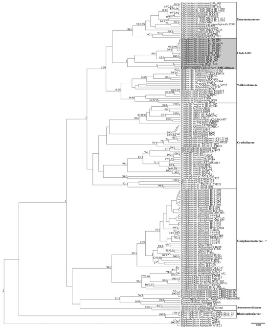
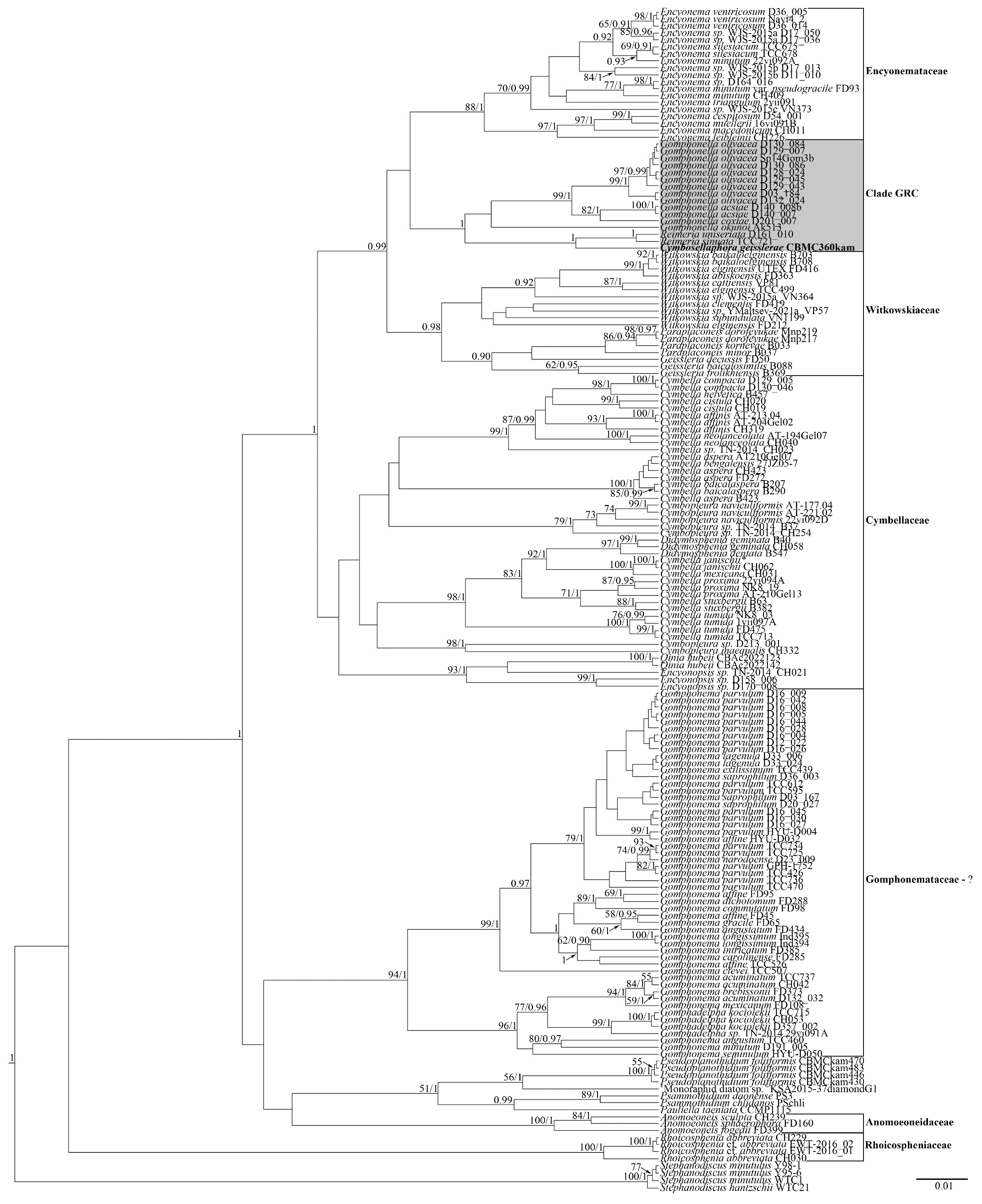
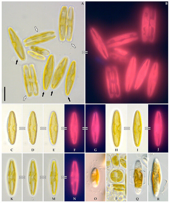
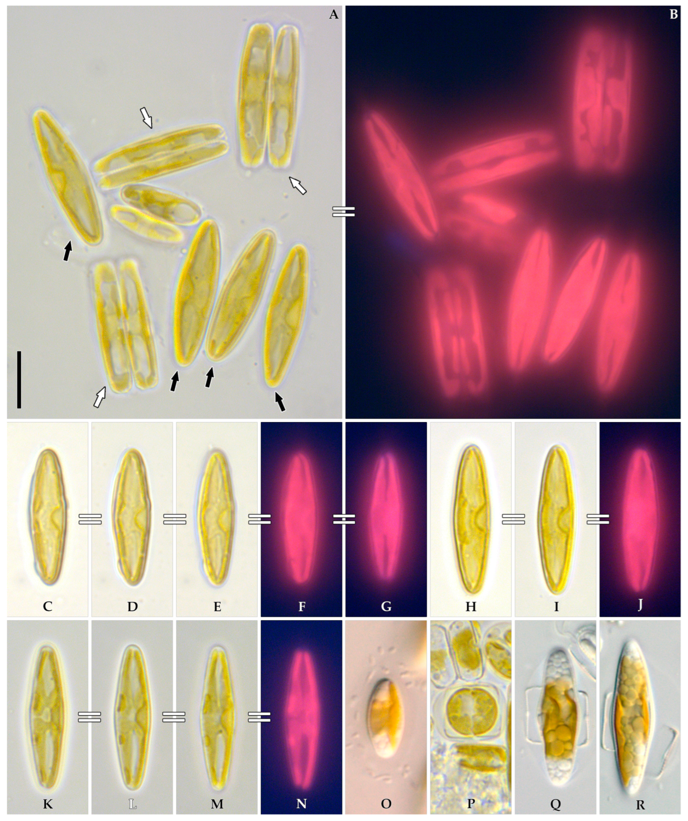
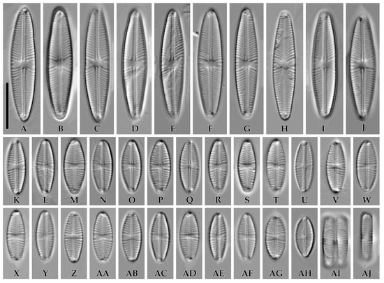
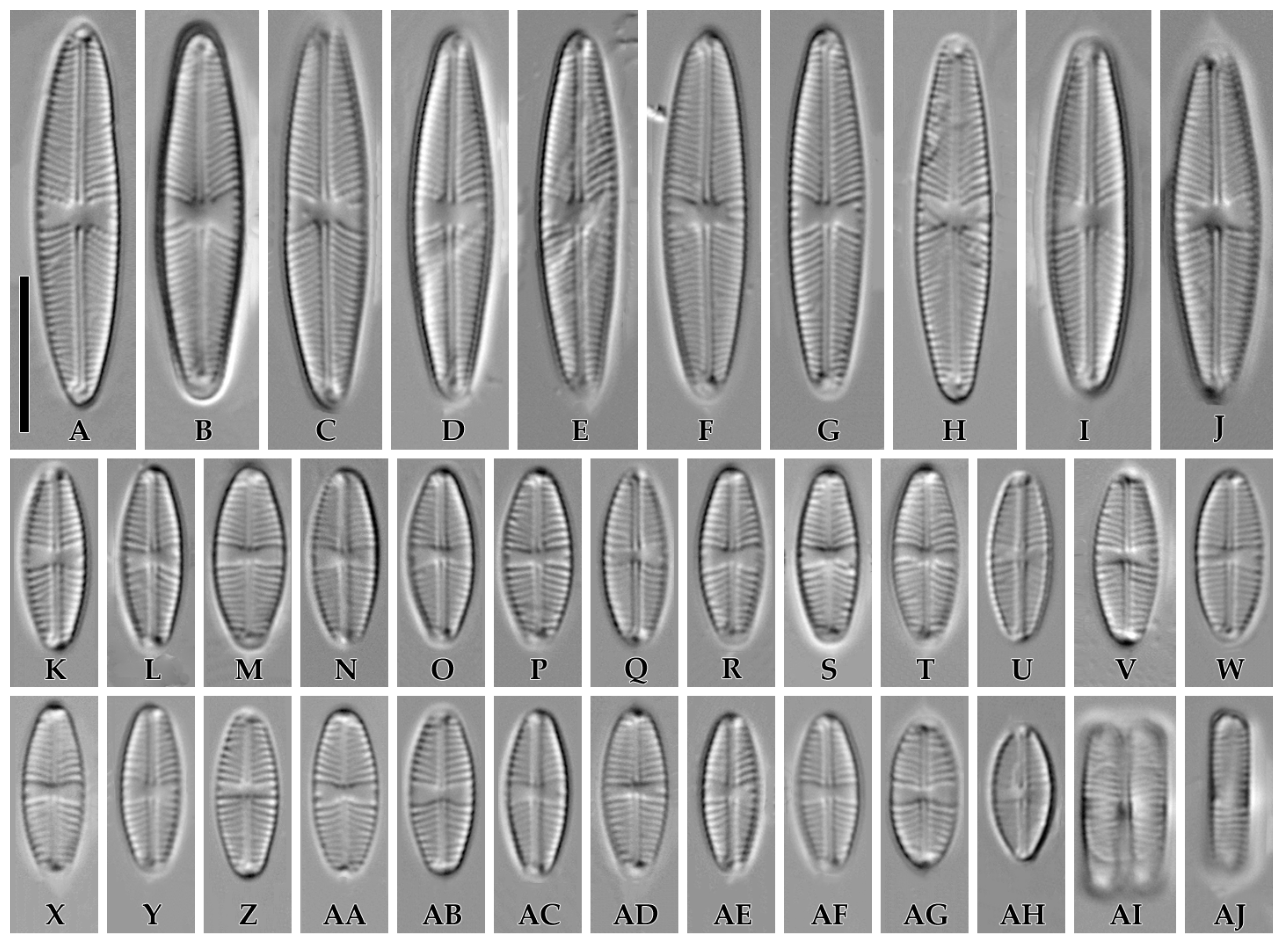
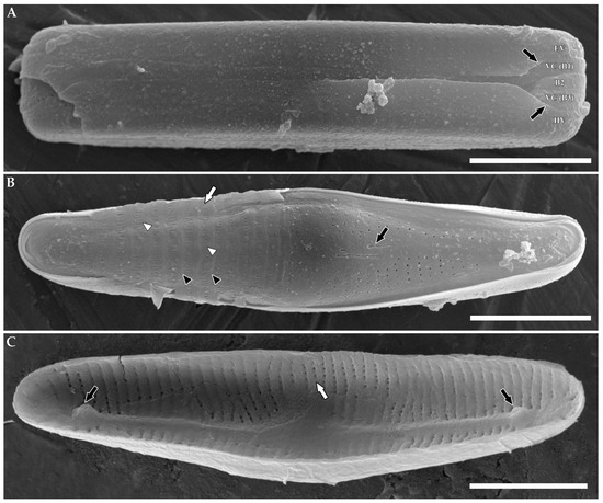
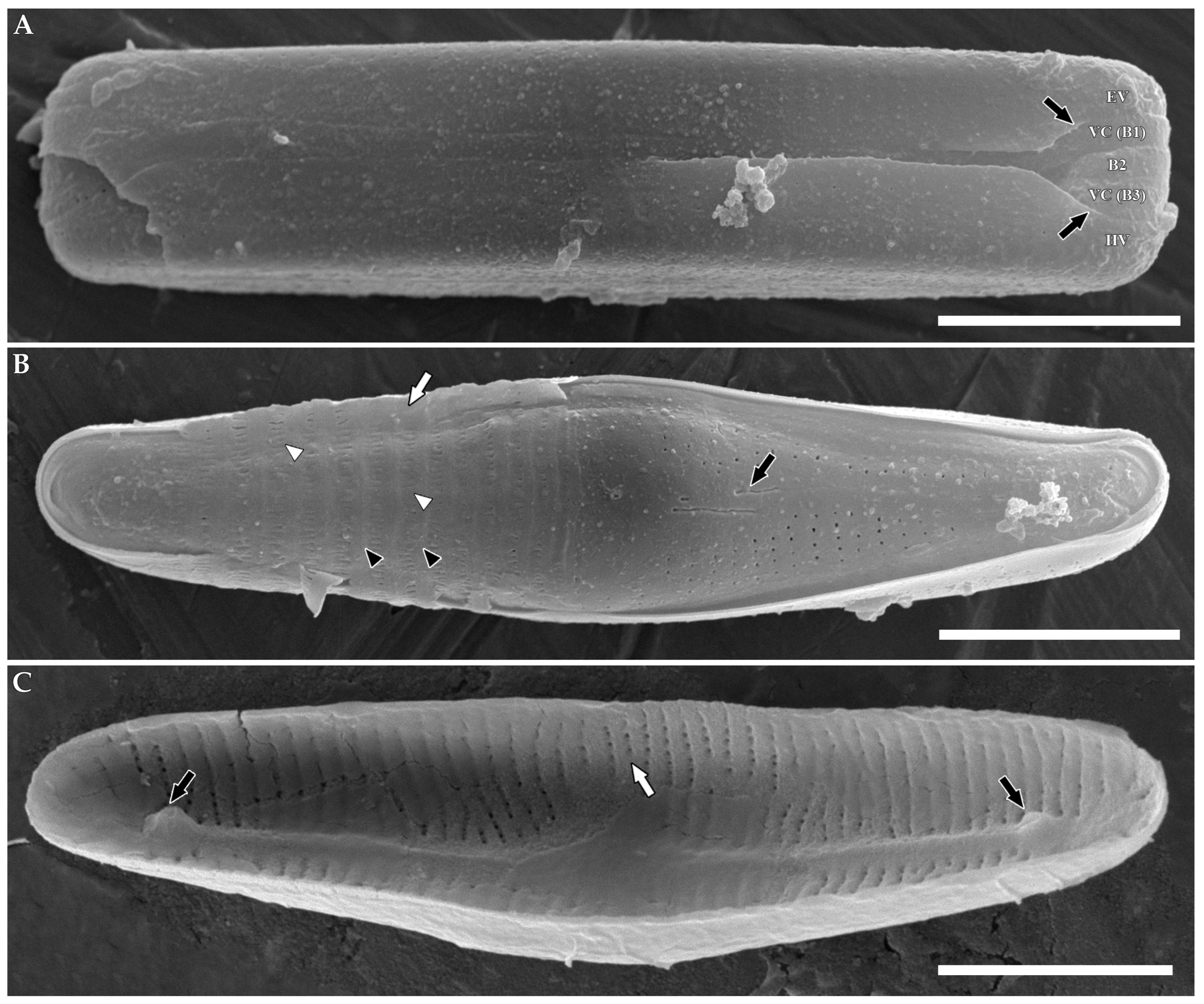
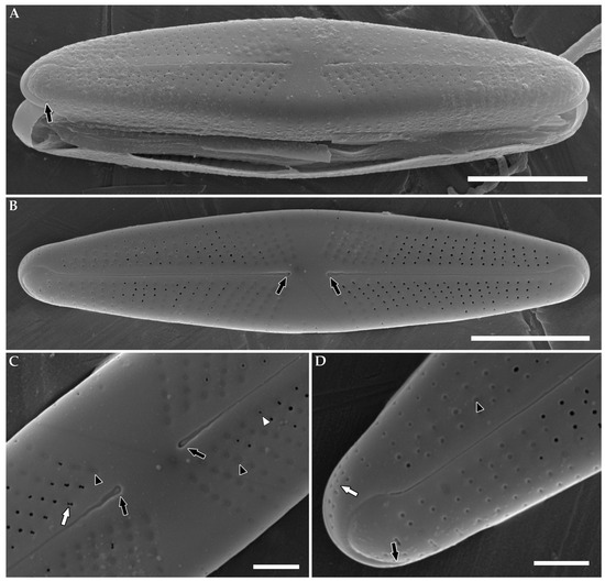
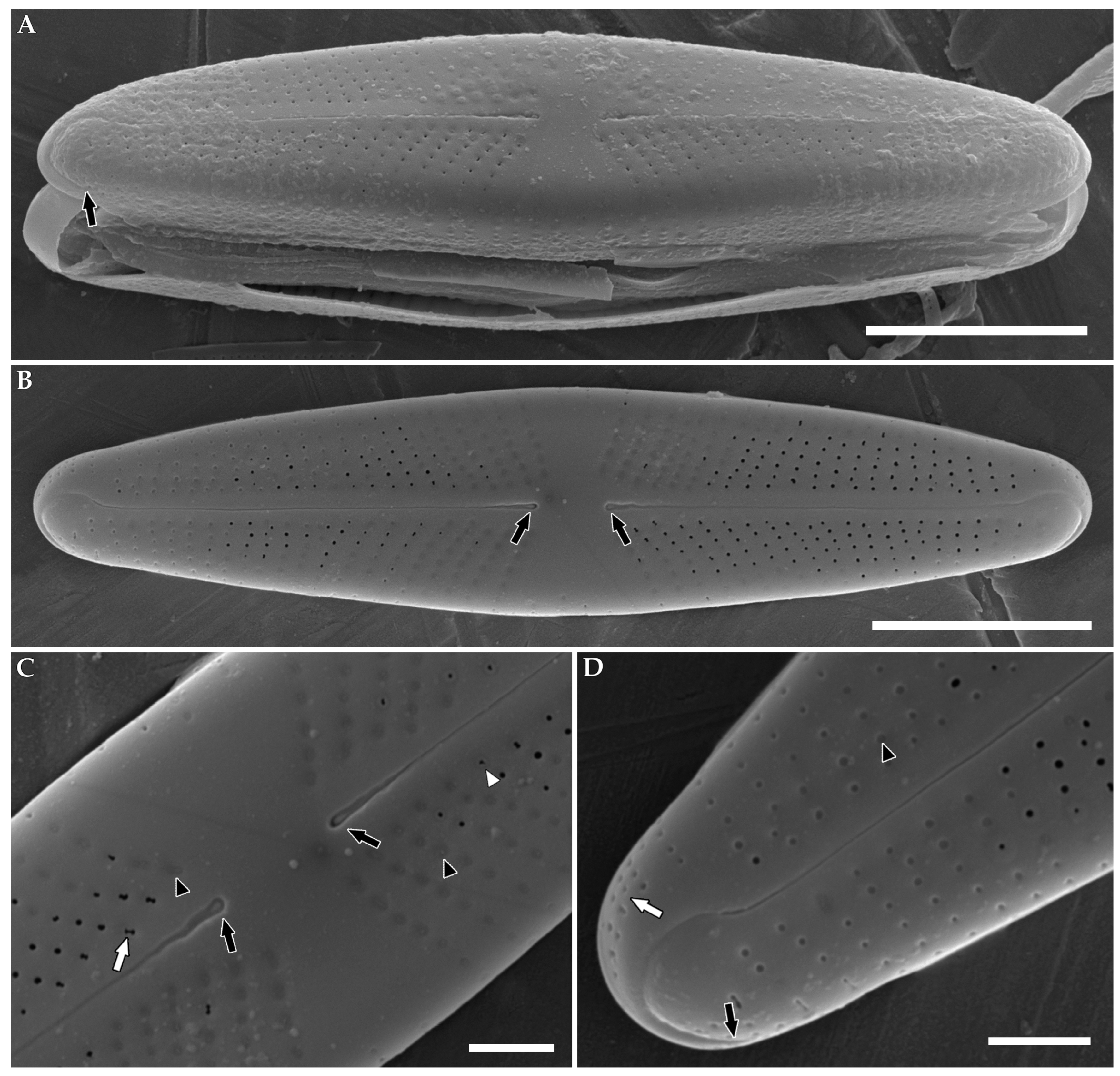
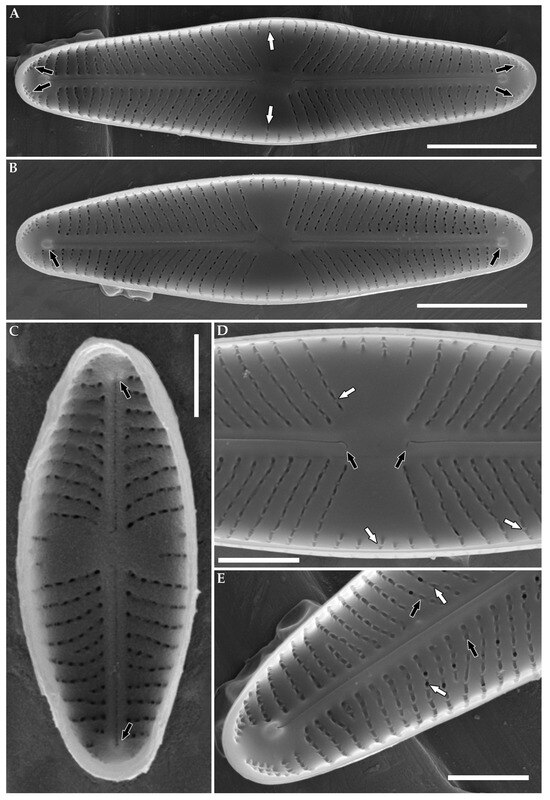
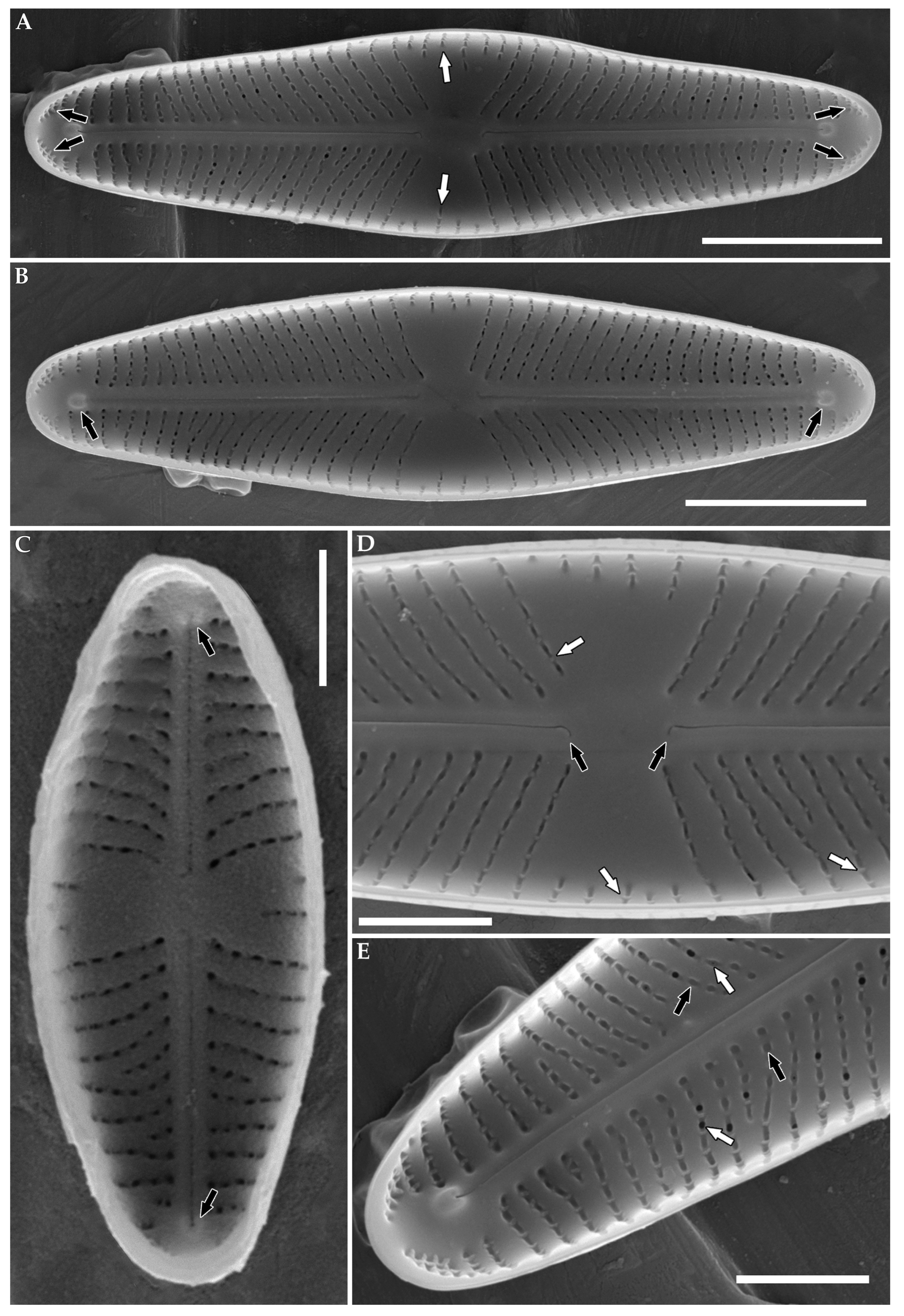
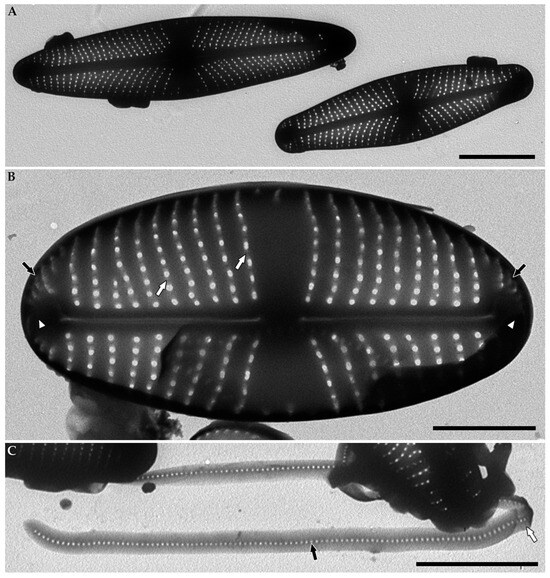
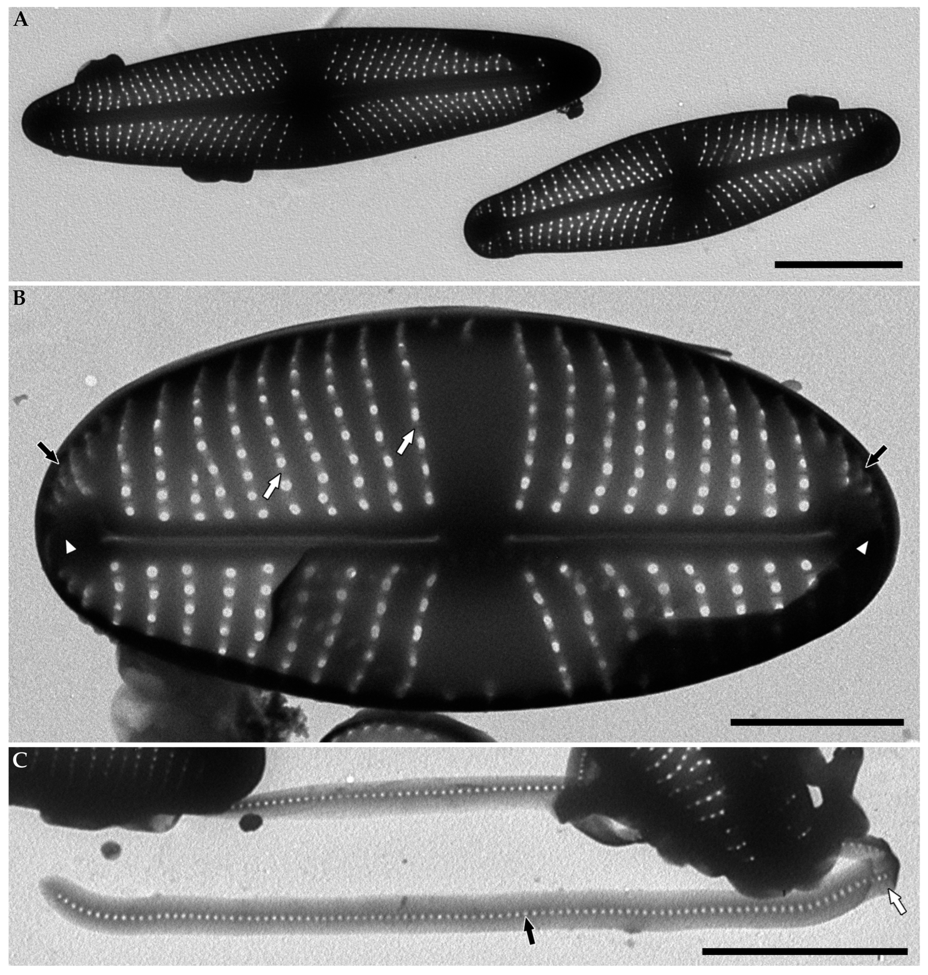
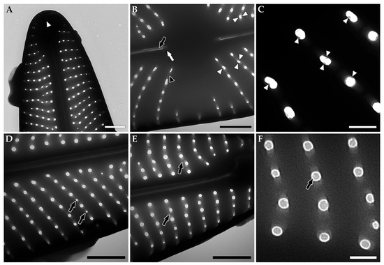
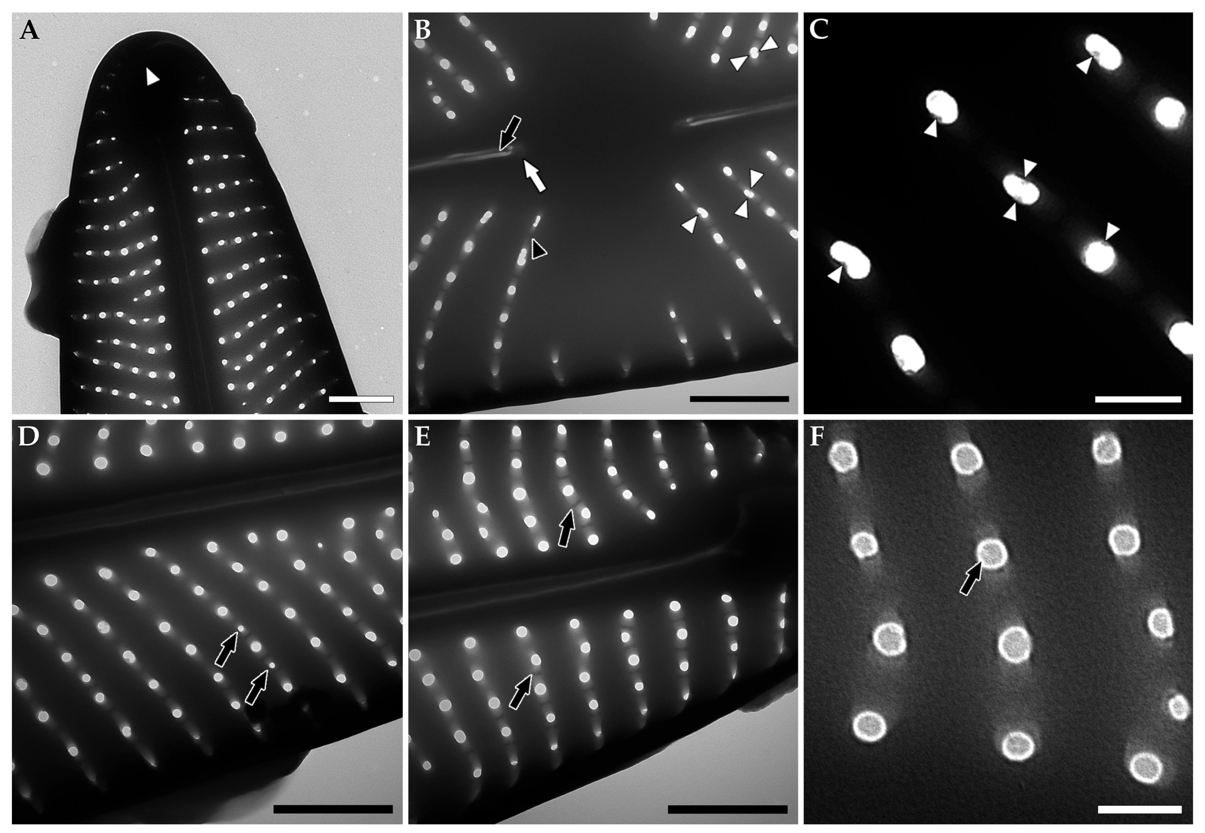

Disclaimer/Publisher’s Note: The statements, opinions and data contained in all publications are solely those of the individual author(s) and contributor(s) and not of MDPI and/or the editor(s). MDPI and/or the editor(s) disclaim responsibility for any injury to people or property resulting from any ideas, methods, instructions or products referred to in the content. |
© 2026 by the authors. Licensee MDPI, Basel, Switzerland. This article is an open access article distributed under the terms and conditions of the Creative Commons Attribution (CC BY) license.