


Abstract
The present study investigated the community composition and dynamics of freshwater biofouling on fiberglass inland waterway vessel (IWV) models coated with two commercial antifouling paints deployed in the Danube River (Serbia) for a total of five months. Biofouling was characterized using visual observations, in situ optical microscopy, the rapid ATP bioluminescence method, dry biomass measurements, and analyses of phototrophic and fungal communities. Based on the results, Hard Racing TecCel demonstrated the highest suppression of biofouling, with the lowest biomass accumulation and reduced algal diversity. At all stages of biofouling, diatoms dominated the phototrophic community, comprising 123 taxa. Achnanthidium minutissimum and Gomphonella olivacea were shown to be persistent hull colonizers, while Cyanobacteriophyta and Chlorophyta had reduced presence. Overall, the results highlight a slower progression of freshwater biofouling compared to marine systems and emphasize the need for the development of tailored antifouling strategies for IWVs to reduce environmental impact and operational costs.
1. Introduction
In recent years, water transport has emerged as a key participant in the global transport sector, offering a sustainable alternative to traditional road and rail transport [1]. It has 39% and 371% lower CO2 emissions compared to road and rail transport, respectively, which is why today approximately 80% of international trade is performed by the world’s fleet. However, the estimated 300 million tonnes of carbon-based fuel consumed annually by the world’s fleet emphasizes the need for further improvements to shipping’s environmental footprint, as current emissions still fall short of developing global standards. To better utilize the potential of water transport, one of its top environmental challenges must be addressed, i.e., the phenomenon of hull colonization by aquatic organisms, mainly bacteria, algae, protozoa, and invertebrates—biofouling [2]. These biofoulers modify surface contour, complexity, and heterogeneity, typically through mass accumulation, thereby increasing surface friction and significantly impacting ship performance and fuel consumption. Estimates suggest that a 10 μm increase in surface roughness, due to biofouling, can lead to a 0.3% to 1.0% rise in fuel consumption, an annual fuel consumption increase of up to 10%, and, in extreme events, a speed reduction of up to 80% [3,4]. Average fuel consumption, per Howell and Behrends [5], increases 6% for every 100 μm increase in the average hull roughness induced by biofouling.
Because of their high energy density per unit of mass and volume, established infrastructure for extraction, processing, transportation, and distribution, and cost-effectiveness and compatibility with already in-use internal combustion engines, carbon-based fossil fuels remain the primary energy source in the water transport industry. However, fossil fuel combustion significantly contributes to air pollution through the emission of greenhouse gases (GHG), namely CO2, SOx, NOx, and particulate matter [6]. Amid the growing concern about the greenhouse effect and climate change, the reduction in GHG emissions has become a critical priority. Despite ongoing efforts, the shipping industry’s contribution to global anthropogenic GHG emissions rose from 2.76% in 2012 to 2.89% in 2018 [7].
According to Gavurova et al. [8], CO2 holds a dominant position among GHGs, with its reduction having the most far-reaching effects. To date, shipowners and operators have adopted numerous strategies to combat CO2 emissions, including the use of alternative energy sources, engine/vessel design modifications, and antifouling coatings [9]. While biofouling is most often addressed utilizing periodical manual or mechanized cleaning, it is an extremely demanding, time-consuming, and cost-ineffective process. Thus, the focus is placed on different types of coatings, with antifouling coatings being the most commonly used. Coatings serve as the primary barrier, preventing the attachment and growth of aquatic organisms on ship hulls. By 2011, more than 90% of coatings in use were antifouling [10]. However, they are specifically designed and optimized for marine environments, while antifouling coatings adapted for freshwater biofouling on inland waterway vessels (IWVs) remain undeveloped. To the best of our knowledge, no peer-reviewed scientific data or systematic evaluations for freshwater-designed antifouling agents currently exist. This issue is in part due to data on fouling in freshwaters being very limited and often site-specific, presumably as a result of ecosystem heterogeneity making data difficult to generalize, and a research lag due to the weaker ecological pressure of freshwater biofouling and relatively smaller economic impact, making it under-recognized compared to its marine counterpart. The extent and type of fouling in freshwater is dependent on eutrophication, and is influenced by water chemistry and local control measures that differ markedly with the geographic distribution of freshwater bodies [11]. The most significant part of the issue, however, relates to the fact that IWVs are excluded from International Maritime Organization (IMO) regulations and are not required to regulate CO2 emissions [12], thus reducing the incentives for operators to implement state-of-the-art antifouling measures or adopt environmentally sustainable practices. However, with over 15,000 IWVs in operation in Europe and annual CO2 emissions from a typical European vessel exceeding three million tonnes, there is an evident need for innovation in the inland waterway transport sector [1,13]. Furthermore, aside from being less studied and pronounced, compared to its marine counterpart, freshwater fouling must still be considered economically important, with an estimated annual global cost of approximately 277 million USD and a tendency of worsening in the coming decades due to unintentional human-mediated dispersal of invasive species, eutrophication, rising global freshwater demand, and climate change [14].
In addition to its operational relevance, freshwater biofouling is also ecologically important. Early stage biofilms developing on submerged artificial surfaces represent the first step in colonization and strongly influence the subsequent establishment, composition, and succession of fouling communities. In inland waterways, such biofilms may also contribute to the persistence and dispersal of microorganisms and other fouling-associated taxa between habitats via vessel movement. Therefore, investigating freshwater biofouling is important not only for coating performance and vessel efficiency, but also for understanding the ecological dynamics of colonization on man-made surfaces in riverine systems.
Given the general lack of data regarding freshwater biofouling on IWVs and the absence of optimized antifouling coatings for freshwater environments, the primary aim of this study was to assess early community composition and dynamics of biofouling on coated fiberglass IWV models in the environment of the Danube River in Serbia. Taking into account that turbulent flow determines structure, composition and biofouling mass, and considering the 5 month time frame of the study, realistic data obtained from the field will provide valuable insights into the unique characteristics of the early-to-intermediate stages of biofouling in freshwater ecosystems necessary for evaluation of antifouling performance and successional dynamics. This valuable data will provide IWV owners and operators with recommendations for selecting the most suitable coating for inland waterway use, thereby reducing environmental impacts associated with IWVs.
2. Materials and Methods
2.1. Study Site and Sampling Dynamics
Sampling for quantitative and qualitative biofouling characterization was performed on three experimental models of IWVs placed in the Danube River, at the Nautec port (Zemun Quay, Belgrade, Serbia) (44°50′15″ N 20°25′09″ E). Sampling was performed on predesignated 2.5 × 2.5 cm areas of IWV models at constant depth with collections made at regular intervals over a 5 month period, from October 2023 to March 2024. This time frame was selected as the 3–6 month period is considered the critical window for comparing materials, antifouling performance, and successional dynamics since most major colonizers have already settled and the community is stabilized. Sampling was performed every 7 days during the first month, followed by monthly sample collection thereafter.
2.2. Experimental Setup and Model Design
The IWV models were designed as simplified representations of typical inland waterway vessel hulls, constructed as quadrilateral prism shapes with trapezoidal cross-sections made of glass-reinforced plastic (GRP), hand-laminated in a mold. This approach was selected due to its fabrication simplicity and cost-effectiveness, which were prioritized given the exploratory nature of this pilot study as a methodological foundation for a larger-scale research initiative. The IWV models were designed in such a way that 300 mm of the 500 mm depth would be submerged in water during the experiment. After construction, models were coated with an epoxy primer (SeaJet, Heijningen, The Netherlands). Two out of three constructed models were coated with selected antifouling coatings: Hard Racing TecCel (Hempel, Lyngby, Denmark) for the H model and Self Polishing Antifouling Black, Emperor 034 (SeaJet) for the S model. The coatings were applied manually, with a brush, until the surface was black, uniform, and consistent. The third model served as a negative control with no coating applied. The selected coatings represent two of the most widely used antifouling products available in the Serbian market. Hard Racing TecCel (Hempel) is more commonly applied on large commercial and industrial vessels, while Emperor 034 (SeaJet) is frequently used for recreational and smaller crafts. However, both coatings are applied across vessel types depending on user preference. Although neither product is explicitly formulated for freshwater environments, they are widely employed in practice. In this study, both coatings were applied in accordance with the manufacturers’ technical specifications. Following application of the coatings, all models were dried for 24 h, mounted onto steel supports, and launched into the Danube River (Supplementary Figure S1). During this initial study, the models were rigidly fixed to the side of a large pontoon at the marina and positioned transversely to the river flow; only pitch motion was free, providing two different exposure surfaces—one facing the current and the other shielded. The draft of each model was proportionally derived from its height to reflect typical inland vessel submersion. Although the flow velocity of the river was not measured in this phase, the variation in current speed throughout the observation period was considered an adequate analog for the dynamic conditions experienced by operating vessels.
2.3. Measurement of Physico-Chemical Water Parameters
The temperature, pH, conductivity, saturation, and dissolved oxygen (DO) concentration of water were measured near the submerged IWV models during each sampling period in situ using a WTW Multi 3620 IDS (Weilheim, Germany) portable instrument.
2.4. Characterization of Biofouling Morphology
Direct observation of biofouling morphology, as well as microscopic impairments of coated IWV model surfaces, was performed using a Dino-Lite Edge AM7915MZTL digital microscope with N3C-L Long Cap (New Taipei City, Taiwan). For image processing, DinoCapture 2.0 v1.5.39. Software was used.
2.5. Quantification of Biofouling
2.5.1. Determination of Level of Fouling (LoF) Ranks
The level of fouling of IWV models was determined according to the six categories (0–5) LoF scale of Davidson et al. [15] modified for a freshwater environment, mainly taking into account percent cover (proportion of a visible surface that is covered in biofouling), as well as micro- and macro-fouling morphology.
2.5.2. Determination of the Degree of Surface Contamination
To determine the total contamination of IWV models by microorganisms and organic residues during the beginning stages of biofouling development, a rapid ATP bioluminescence method was used in situ [16]. ATP swabs in triplicate and a Lightning MVP portable luminometer (BioControl Systems, Bellevue, WA, USA) were used for this purpose on predesignated same-depth areas of defined surface (2.5 × 2.5 cm) on all three models (18 samples per IWV model; 54 samples in total). To enable consistency during all sampling periods, the sampled surface areas were premarked and standardized using a sterile template to maintain identical swabbing dimensions (6.25 cm2) at each time point. Sampled ATP swabs were dipped into the kit-provided luciferin/luciferase reagent, processed by a luminometer, and results were expressed as “Zones of Cleanliness” using a logarithmic approximation of relative light units (RLUs). The intensity of emitted light is proportional to the ATP concentration on the surfaces and provides an assessment of the overall levels of surface contamination [16]. Results were compared to the manufacturer-provided guideline reference scale and sorted into three “Zones of Cleanliness” categories: clean zone (0.0–2.5), caution zone (2.5–3.0), and danger zone (3.0–7.5).
2.5.3. Measurement of Dry Biomass per Unit Area
Samples for dry biomass determination were taken from each IWV model in triplicate by carefully scraping the biofouling into sterile vials with dH2O (three samples per IWV model per sampling period; 45 samples in total). Collected samples were frozen at −20 °C and lyophilized in a cold drying system (Christ Alpha 2-4 LSCplus) at a −70 °C desublimator temperature and the working temperature of the material from −35 °C. After lyophilization, samples were weighed on an analytical balance AS 220.R2 (Radwag, Radom, Poland).
2.6. Determination of Biofouling Constituents
2.6.1. Analysis of the Phototrophic Community
Samples for the analysis of the phototrophic biofouling community were collected by scraping the biofouling into sterile vials containing dH2O. All samples were preserved and fixed with formaldehyde to a final concentration of 4%. To analyze the presence of cyanobacteria and non-diatoms, temporary slides were prepared, while diatoms were observed on permanent slides prepared according to Taylor et al. [17]. Algae were analyzed using an Axio Imager M1 (Carl Zeiss, Oberkochen, Germany) optical microscope with differential interference contrast, an AxioCam MRc5 digital camera, and AxioVision 4.9 software. The relative abundance of diatoms was determined by counting 400 valves, with a 10% threshold for dominant taxa. Identification of cyanobacteria and algae was carried out based on morphology to species or genus level using the following identification keys found in [18,19,20,21,22,23,24,25,26,27,28,29]. Due to differences in preparation and counting procedures, diatoms were analyzed quantitatively, while cyanobacteria and other non-diatom algae were assessed qualitatively based on their presence in the samples. Therefore, comparisons among phototrophic groups were based on presence/absence data rather than direct abundance estimates. Spatial and temporal overlap of phototrophic biofouling constituents is summarized in three-way and five-way Venn diagrams constructed using the web-based program ^InteractiVenn^ (https://www.interactivenn.net/, accessed on 11 February 2026) [30].
2.6.2. Analysis of Actively Growing Heterotrophic Community
To determine the presence of actively growing fungi in biofouling, samples were collected by scraping biofouling into sterile vials. Samples were stained with Lactophenol Cotton Blue and analyzed with a Zeiss Axio Imager M1 microscope using AxioVision Release 4.9 software.
2.6.3. Analysis of Culturable Mycobiome
Sterile swabs were used to collect biofouling samples from the IWV models. In sterile conditions, samples were spread onto potato dextrose agar plates (PDA, TM Media, Belgrade, Serbia), supplemented with 500 mg L−1 Streptomycin sulfate salt (Sigma Aldrich, St. Louis, MI, USA) to suppress bacterial growth. The inoculated plates were incubated in a UE 500 thermostat chamber (Memmert, Schwabach, Germany) at 25 ± 2 °C for 7 days. Following the incubation period, morphologically different colonies were reinoculated on PDA and incubated under the same conditions to obtain axenic cultures. The isolated fungi were identified on the basis of colony morphology, observed with a Stemi DV4 stereomicroscope (Carl Zeiss), and the micromorphological characteristics of reproductive structures were analyzed with the optical microscope Zeiss Axio Imager M1 (Carl Zeiss), using AxioVision Release 4.9 software. The following identification keys were used [31,32,33,34,35].
For molecular identification of fungi, total genomic DNA was extracted by harvesting 50–100 mg of marginal mycelia from 7-day-old fungal cultures using the ZR Fungal/Bacterial DNA Mini Prep KIT, according to the manufacturer’s protocol (Zymo Research, Irvine, CA, USA). The ITS II gene region (internal transcribed spacer 1, partial sequence; 5.8S ribosomal RNA gene and internal transcribed spacer 2, complete sequence; and large subunit ribosomal RNA gene) was amplified via ITS1-F (5′-TCCGTAGGTGAACCTGCGG-3′) and ITS4-R (5′-TCCTCCGCTTATTGATATG-3′) primers [36]. The PCR reaction was carried out according to the protocol of Six et al. [37], and the PCR products were purified using the EXTRACTME DNA CLEAN-UP KIT according to the manufacturer’s protocol (BLIRT, Gdańsk, Poland). Purified PCR products were sequenced as a commercial service in Eurofins Scientific (Hamburg, Germany). The obtained sequences were searched for homology with sequenced genes in the GenBank database using the BLASTN program at the National Centre for Biotechnology Information (NCBI), and all chromatograms were manually checked for nucleotide positions. The edited and reference strain sequences from the GenBank database were aligned using CLUSTAL W implemented in BioEdit 7.2.5 software. A phylogenetic tree was constructed in MEGA 11.0.13 software using the neighbor-joining method based on a pairwise distance matrix obtained by the Kimura two-parameter nucleotide substitution model. The tree’s topology was evaluated using the bootstrap resampling method with 1000 replicates.
2.7. Statistical and Multivariate Analyses
Statistical analyses were conducted using Microsoft Excel and the XLSTAT statistical software package [38]. To assess differences between certain groups for quantitative data, both parametric and non-parametric statistical tests were applied. Prior to the analyses, the assumptions of normality and homogeneity of variance were evaluated using the Shapiro–Wilk test and Levene’s test. When both assumptions were met, Student’s t-test was applied; if the data did not meet the assumptions of normality or homogeneity of variance, the non-parametric Mann–Whitney U test was used. A significance level of p < 0.05 was applied to all statistical tests.
Multivariate analyses were conducted using Canoco version 5 [39]. Presence–absence datasets for algae and fungi were imported into the software. The recorded algal and fungal taxa were used as response variables, while the sampling period (months) was included as a nominal explanatory variable. The relationships between algae or fungi and sampling period were examined using canonical correspondence analysis (CCA), as the gradient length exceeded 3.5 standard deviations units (SD). Due to the higher number of algal taxa, only a subset of the best-fitting taxa was shown in the ordination diagram.
3. Results and Discussion
3.1. Physico-Chemical Water Parameters
The results of the physico-chemical water parameters are presented in Supplementary Table S1. The lowest water temperature was recorded in January (3.1 °C), and the highest at the end of October (17.2 °C). pH was lowest in October (7.943), and highest at the end of March (8.331). Conductivity peaked in January and March (459 μS/cm) and was lowest in mid-November (391 μS/cm). Dissolved oxygen was highest in January (12.70 mg/L), and lowest at the end of October (8.88 mg/L). Saturation was lowest in early November (92.6%), and highest in March (105.7%) Based on the results of surface water quality testing for 2022 by the Environmental Protection Agency (Ministry of Environmental Protection, 2023), recorded physico-chemical parameters of the Danube River measured in the vicinity of the submerged IWV models at the Zemun station showed no significant deviation during the study period.
3.2. Biofouling Development on IWV Models
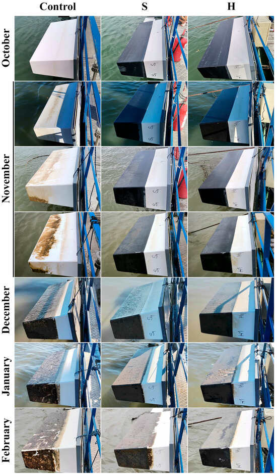
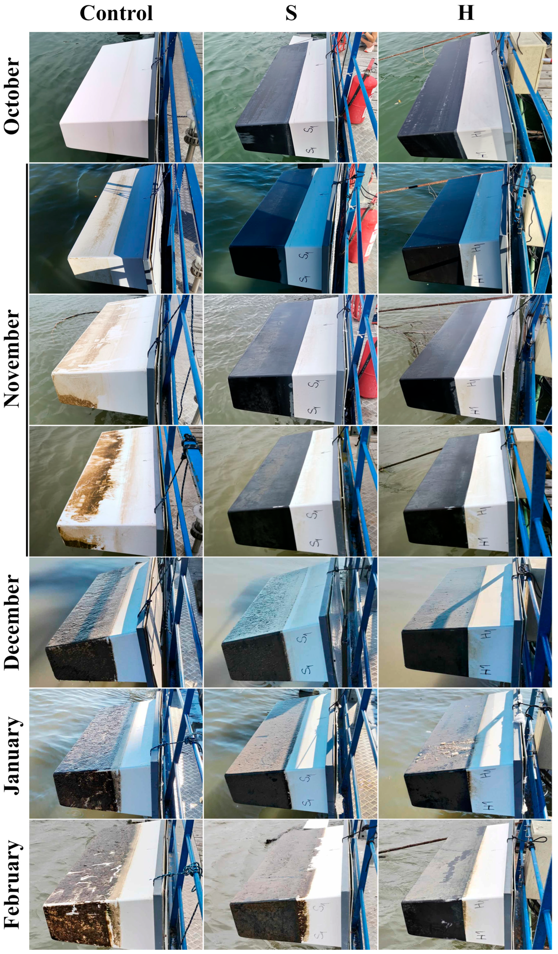
Visual observation of biofouling formation on IWV models confirmed the considerable effect of selected antifouling coatings in the suppression of fouling organisms’ attachments to the surface of the experimental models (Figure 1). Initial stages of biofouling formation, i.e., adsorption of dissolved proteins and polysaccharides and formation of conditioning film, and bacterial adhesion, were observed on the control IWV model as early as 24 h after the launch of the model into the river. Developed film covering large areas of the model was documented one month after launch, while complete biofouling coverage of submerged parts of the control model was observed by the end of the second month. However, on the S and H models, biofouling establishment was markedly slower, with only a thin layer of organic deposits accumulating locally in the first month. From the third month onward, the submerged areas of the S and H models were completely covered, albeit with finer biofouling and notably less thickness. As no macrofouling was documented on any of the experimental models, based on the modified LoF scale of Davidson et al. [15], observed biofouling did not progress beyond the primary stage. In the studied freshwater environment of the Danube River, community composition does not influence the percent cover as the main criterion for a higher LoF rank assignment. Based solely on the percent cover, the highest level (level five) of the LoF scale, corresponding to 41–100% coverage of the submerged area, was reached by all three models at different intervals: the control model by the end of month 2, and the S and H models within month 4. Given that no macroalgae colonized any of the IWVs following first week of the submersion, as was to be expected per successional fouling hypothesis of marine biofouling [2], and the fact that no macroalgae were observed at all during the five-month study period, it can be assumed that the biofouling process in freshwater ecosystems occurs at a slower rate compared to marine environments, or possibly indicates a lack of community stability or early-colonizer equilibrium preventing further succession. Moreover, this finding is consistent with the expectation that only limited macroalgal diversity is generally observed in the Serbian sector of the Danube River.
Figure 1.
Biofouling formation on the surface of the control and IWV models S and H coated with selected antifouling coatings.
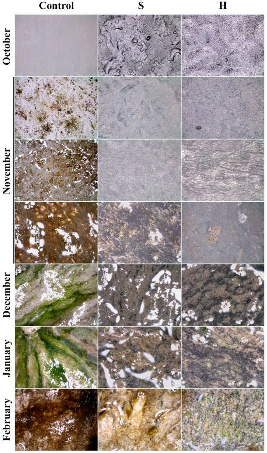
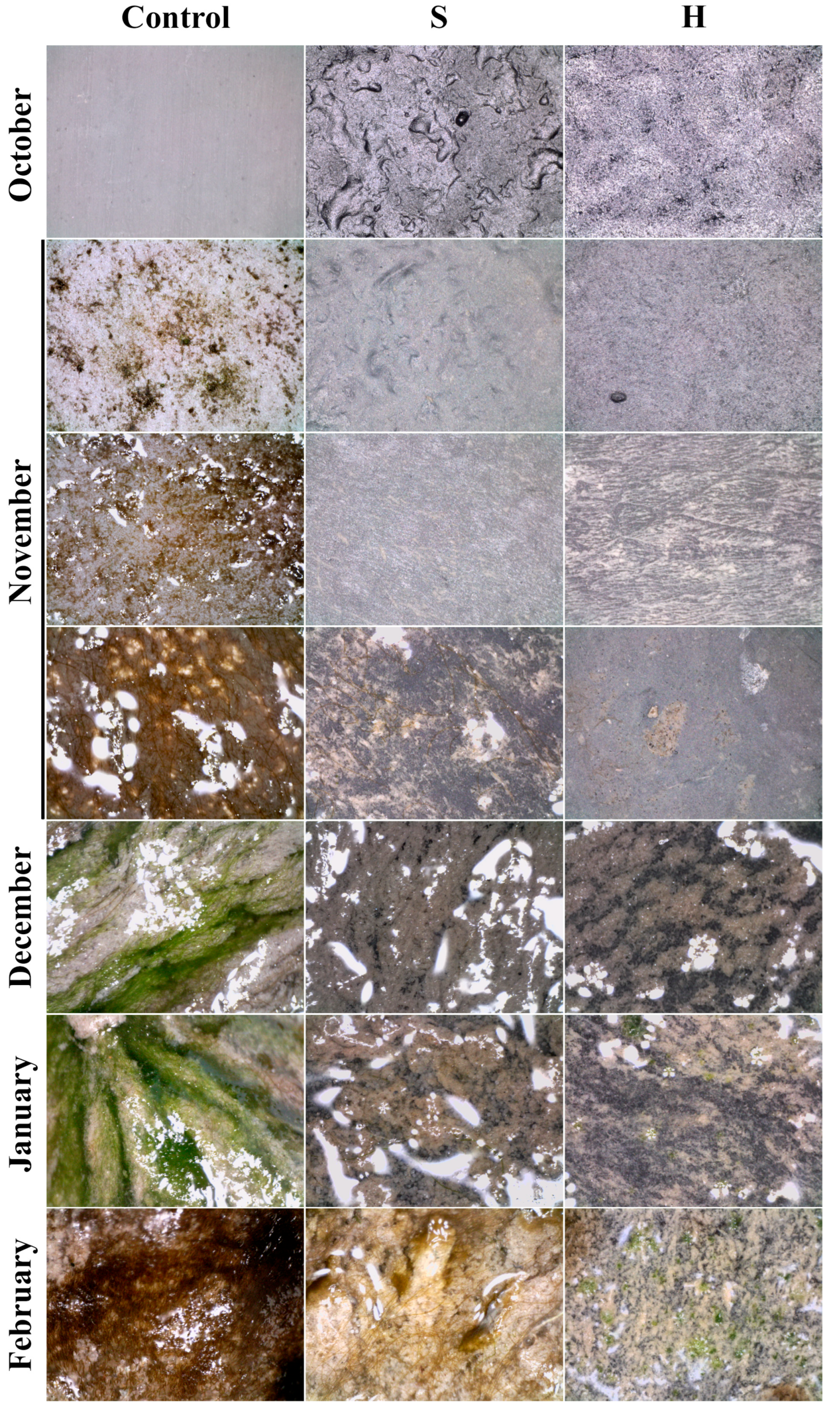
Observation of the IWV models’ surface, via a portable in situ microscope, revealed a gradual loss of coating roughness within the first two weeks, as well as providing a more detailed insight into the successive phases of biofouling development (Figure 2). The first sign of colonization by primary colonizers was observed in the control model within 24 h, while a developed, dense mat made of filamentous algae, specifically trichal cyanobacteria, formed by the end of the first month. On the S and H models, however, loose algal filaments appeared sporadically from the third month onward, with no well-developed mat, suggesting that surface colonization remained in its early stages.
Figure 2.
In situ micrographs of biofouling formation on the surface of the control and IWV models S and H coated with selected antifouling coatings (scale bar: 500 μm; magnification range: 60–70).
3.3. Dynamics of Freshwater Biofouling on IWV Models
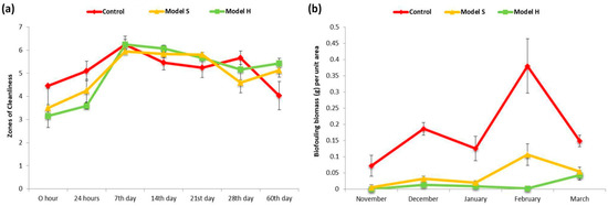
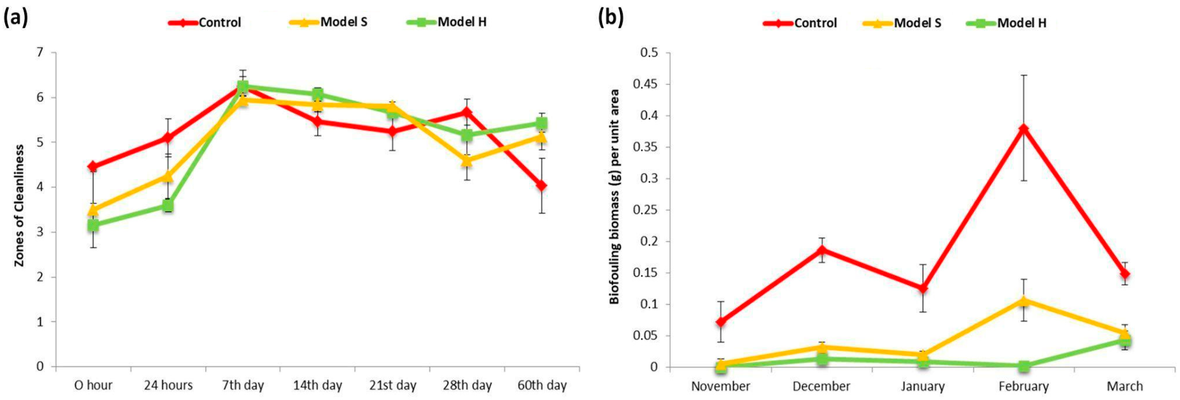
Using the rapid ATP bioluminescence method, the total degree of the contamination of surfaces of the IWV models by microorganisms and organic residues was determined to be above 3.0—a value that corresponded to the danger zone even before the launch of experimental models into the Danube River. This implies the presence of bacteria and/or microbial propagules that originate from contamination during the production and coating of the IWV models. During the initial 7 days, “Zones of Cleanliness” values for all IWV models were established around 6.0, with minimal fluctuations of mean values most often in the range of 5.13 to 6.25 throughout the rest of the experiment (Figure 3a). Observation of temporal changes in total ATP, as an indicator of microbial cell presence/activity, and in biomass content in general, proved an effective method for monitoring biofouling development in the early phases of colonization. A steady increase of “Zones of Cleanliness” values (log approximation of relative light units, i.e., the amount of light generated from one fmol of ATP) was observed 24 h after launch and in the time span from the first to the seventh day. However, the method’s sensitivity proved insufficient in the later stages of biofouling, as despite a visibly increasing, thickening film, a plateau was reached. Since ATP is present in all living cells, its measurement has been hypothesized to indicate biomass content and has been used in numerous studies, most often in research on marine biofouling on reverse osmosis membranes for seawater desalination [40,41]. This method was also used to study the antimicrobial performance of two fouling-release coating systems applied to glass slides deployed in a marine environment [42]. Based on the observations, the authors concluded that the amount of ATP molecules does not depend on the number of bacteria and diatoms in the microfouling, possibly due to the presence of luminescence-inhibiting salts and non-ionic chemicals in the seawater, making this useful and rapid method unsuitable for the quantification of marine biofouling. In the similar research to ours performed by Kojima et al. [43], ATP measurements performed on coated flat panels immersed in seawater revealed an increase in the amount of ATP as the leaching rate of applied coatings decreased; an increase in ATP with the increase in immersion period, and accelerated biofouling possibly due to the stationary nature of the experiment; and saturation of the ATP in biofouling at the end of the third month. Even though freshwater biofouling occurred at a much slower rate in our study compared to the data on marine biofouling, saturation of ATP happened much quicker, i.e., in the span of 7 days. This observation is most likely attributed to the characteristics of the freshwater environment and the local site conditions at the Zemun quay. The relatively low diversity and microbial activity in the early stages of biofouling, coupled with the physico-chemical properties of the Danube water at this site, may have resulted in a more rapid saturation of the ATP signal despite slower visible biomass accumulation. The material of the substrate, i.e., GRP, may also contribute to a more uniform initial colonization, but this remains to be explored in future work.
Figure 3.
Quantification of biofouling on the control and IWV models S and H coated with selected antifouling coatings. (a) “Zones of Cleanliness” values. (b) Dry biomass per unit area. Results are presented as mean values of three measurements ± standard deviation.
The highest biofouling biomass, in the range of 0.07 to 0.31 g per unit area, was measured on the control IWV model (Figure 3b). Both tested antifouling coatings reduced fouling by up to 35 times, with the H model being fouling-free during the first month and having measured dry biomass in the range of 0.01 (January) to 0.04 g (March) per unit area. The same range of values was documented for the S model; however, fouling started earlier and the lowest measured values of 0.01 g of dry biomass per unit area were documented in December. Statistical analyses revealed significant differences in biofouling biomass between the control IWV model and the H model (Student’s t-test, p = 0.006), and between the control IWV model and S model (Mann–Whitney U test, p = 0.016). The best results for the H model are expected, as it was coated with a material suitable for vessels constructed from steel, aluminum, fiberglass, plywood, and ferrocement [44]. A coating applied to the S model, by contrast, exhibits excellent algicidal properties, reduces biofouling, and improves hydrodynamic efficiency [45].
3.4. The Phototrophic Component of the Biofilm
3.4.1. General Overview of the Phototrophic Component
All identified algal taxa are presented in Supplementary Table S2 (the “+” sign indicates the presence of a taxon on the IWVs, while numbers represent the percentage abundance of diatoms). Throughout the study period, taxa from the phyla Cyanobacteriophyta, Chlorophyta, Euglenophyta, and Bacillariophyta were recorded. Diatoms (Bacillariophyta) clearly dominated, with a total of 123 taxa recorded. They were followed by Chlorophyta (10 taxa), Cyanobacteriophyta (nine taxa), and Euglenophyta (two taxa). These findings are consistent with observations by Callow [10], who stated that diatoms, cyanobacteria, and filamentous green algae are typically the most dominant components of algal biofilms. Macroalgae were not recorded during this study. However, these organisms, particularly representatives of Rhodophyta, Chlorophyta, and Phaeophyta, are very common in marine ecosystems [46]. Given that the colonization of models was observed only during the initial five months, macroalgae are likely to appear in later stages of biofouling, with their presence being strongly conditioned by biofilm-induced changes in physical and chemical properties of a substratum [47,48]. A study by Ács et al. [49] on periphyton colonization of glass surfaces in the Danube River revealed species overlap, particularly among representatives of the phyllum Bacillariophyta. The most common diatoms were of the genera Amphora, Cocconeis, Diatoma, Encyonema, Gomphonema, Navicula, Nitzschia, and Surirella. Among Cyanobacteriophyta, the species of the genera Heteroleibleinia, Leptolyngbya, Oscillatoria, and Pseudoanabaena were documented. One species dominated in particular—Leptolyngbya notata, and was recorded in every month of the study. According to research from Ukraine, this species is widespread across a variety of freshwater ecosystems, including rivers, ponds, and lakes [50]. The vessel models also hosted typical planktonic algal taxa, such as members of the genera Scenedesmus, Desmococcus, and Coelastrum (Chlorophyta), suggesting that these species originated in the surrounding water and subsequently became incorporated into the biofilm [51]. From the phyllum Euglenophyta, only two species from the genus Trachelomonas were recorded.
Unlike marine biofouling, data on freshwater biofouling and its impact on antifouling coatings remain scarce. Although biofilms in freshwater and marine environments are highly dynamic and complex systems, their respective ecological dynamics can differ substantially [52]. Species from some genera, such as Amphora and Navicula, are common to both systems; however, species-level interactions and community structures vary due to differences in physico-chemical conditions between marine and freshwater habitats. The development of biofilms on vessels in freshwater ecosystems is strongly influenced by light availability and nutrient concentrations in the surrounding water. Light conditions directly affect the metabolic activity of microorganisms, particularly algae, which play a key role in biofilm dynamics. Under high light, increased photosynthetic activity promotes algal biomass accumulation, in turn enhancing bacterial productivity and extracellular enzyme activity. In contrast, under low light conditions, biofilms tend to be thinner, with reduced algal biomass, and rely primarily on dissolved and particulate organic matter from the water column [53]. Nutrient availability, particularly nitrogen and phosphorus, plays a crucial role in biofilm formation. In oligotrophic waters, limited resources can slow down biofouling, whereas an increased nutrient supply, especially during spring and summer, can significantly enhance algal and bacterial growth [53]. In the Danube River, seasonal fluctuations in nutrient availability and light conditions can strongly influence the intensity of biofouling on vessel surfaces. In areas with high nutrient concentrations and sufficient illumination, biofilms are expected to develop rapidly and accumulate greater biomass, whereas in poorly lit and nutrient-limited zones, biofilm is less pronounced. These variations greatly influence the effectiveness of antifouling coatings in freshwater ecosystems, as sufficient light and nutrient availability, also expected in the Danube, directly promote increased biofouling intensity.
3.4.2. Phototrophic Component Comparison Between Models
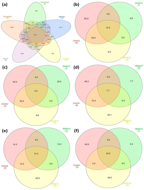
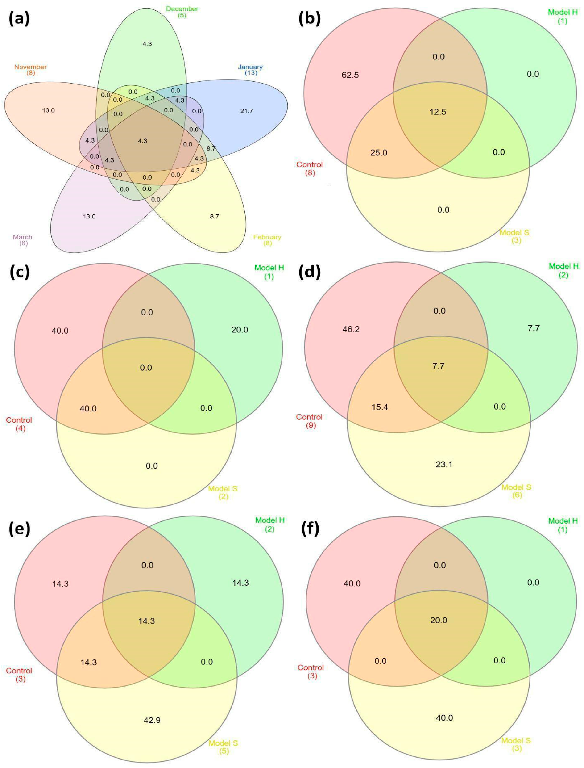
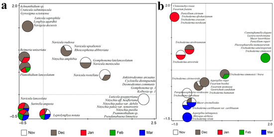
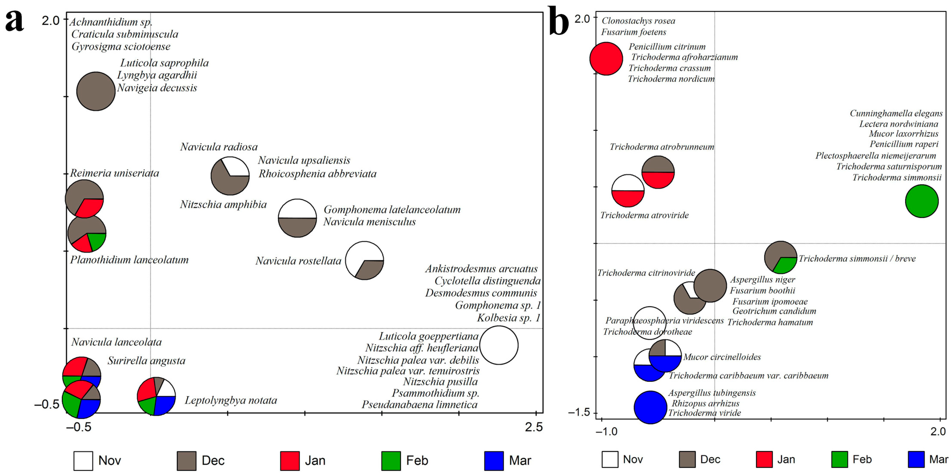
The greatest temporal overlap of 8.7% in the algal community was observed between January and February (Figure 4). Furthermore, 4.3% of identified algal taxa were consistently present throughout the study period, indicating a small but stable core group of species contributing to biofilm development across all sampling intervals. Figure 5a (CCA, F = 1.2, p = 0.03), which represents the 30 best-fitted taxa, revealed that many recorded algal taxa were associated with only one sampling month, with November and December being the most prominent in this respect. Taxa associated exclusively with December are located in the upper left part of the ordination diagram, while those associated exclusively with November are positioned in the lower right part. During the five-month study, differences in algal colonization were observed across the three vessel models: control model, model S, and model H (Figure 4).
Figure 4.
Temporal (a) and spatial (b–f) overlap percentage (%) of algal taxa documented on biofouled IWV models: (b) November; (c) December; (d) January; (e) February; (f) March.
Figure 5.
CCA showing (a) algae and (b) fungi in relation to sampling months.
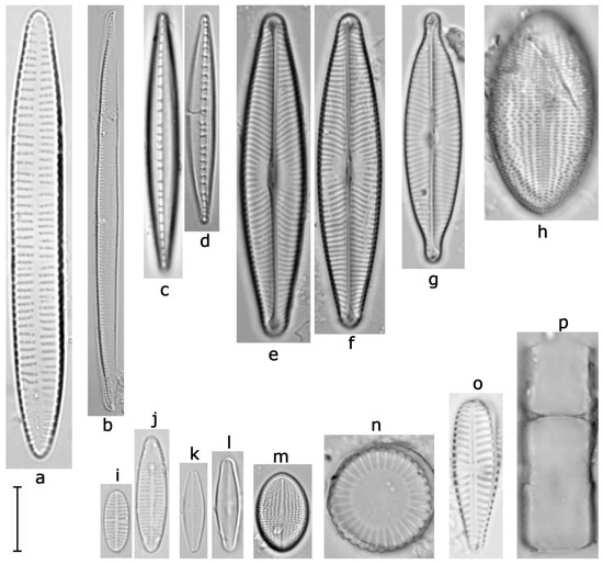
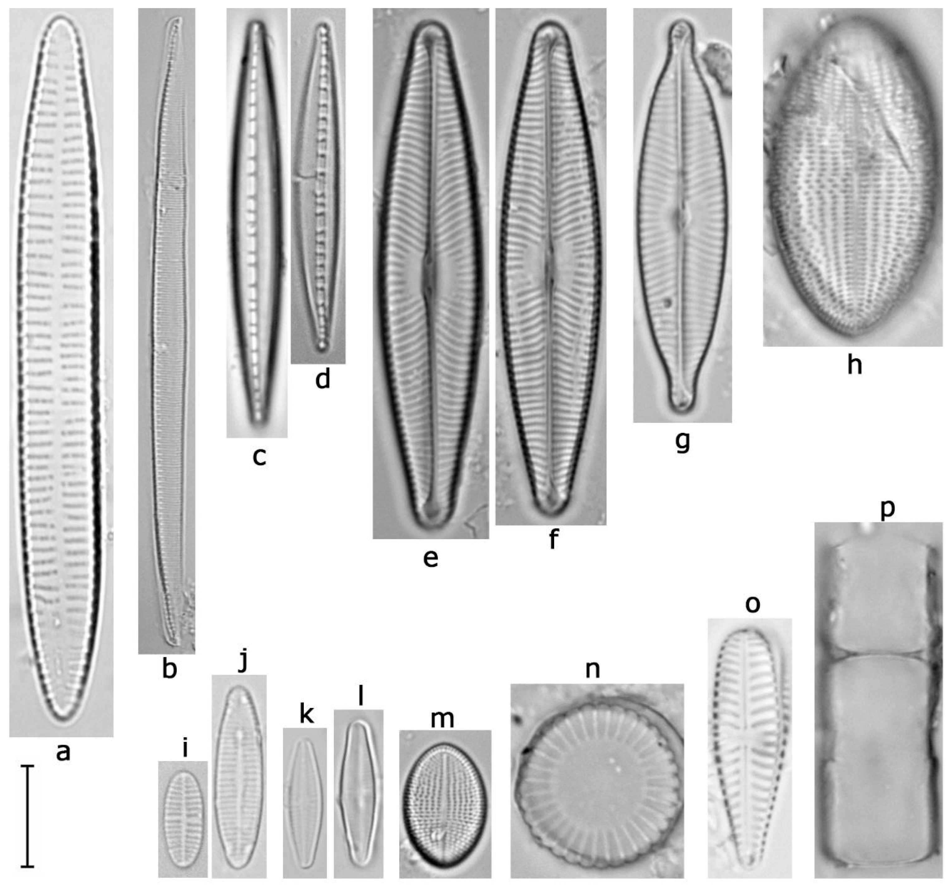
The control model, which was not coated with antifouling paint, exhibited the highest number of algal taxa. As expected, this surface facilitated algal attachment, contributing to its high diversity. Most taxa belonged to the Bacillariophyta, with dominant species in the initial months including: Achnanthidium minutissimum, A. pyrenaicum, Cocconeis euglypta, Melosira varians, Nitzschia aff. heufleriana, N. dissipata and Navicula rostellata (Figure 6) (Bacillariophyta). Toward the end of the study, Gomphonella olivacea and Navicula lanceolata (Bacillariophyta) became dominant. Taxa from the genus Achnanthidium, particularly A. minutissimum, were present during all five months. This taxon indicates the rapid, continuous development of biofilm on the unprotected surface, as it is known to colonize early-stage biofilms and adapt to a wide range of conditions [54]. In addition to diatoms, several taxa of Cyanobacteriophyta and Chlorophyta were also recorded on the control model. Cyanobacteriophyta included Heteroleibleinia rigidula, Leptolyngbya notata, Oscillatoria limosa, and Pseudanabaena limnetica. Representatives of Chlorophyta comprised Ankistrodesmus arcuatus, Coelastrum microporum, Desmodesmus communis, and Stigeoclonium farctum. As these encompass both the benthic and planktonic taxa, such a finding further confirms that the unprotected surface supports a diverse phototrophic community.
Figure 6.
Dominant diatom taxa on inland vessel models. (a)—Ulnaria ulna, (b)—Nitzschia aff. heufleriana, (c,d)—N. dissipata, (e,f)—Navicula lanceolata, (g)—N. rostellata, (h)—Cocconeis euglypta, (i,j)—Achnanthidium pyrenaicum, (k,l)—A. minutissimum, (m)—Cocconeis lineata, (n)—Stephanocyclus meneghinianus, (o)—Gomphonella olivacea, (p)—Melosira varians. Scale bar 10 µm.
On model H, an increased relative abundance of algae was observed in the last two months. Notably, Achnanthidium minutissimum, a species typically associated with the early stages of biofilm formation, became dominant only in the final month, suggesting that the antifouling coating applied to this model was particularly effective in delaying colonization. A notable finding of Gomphonema latelanceolatum (formerly Gomphonema lanceolatum) (Bacillariophyta) on model H during the first month marks a new record for the algal flora of Serbia. This species had previously been recorded only at its type locality, the Bregalnica River in North Macedonia, a site known for high concentrations of electrolytes and pesticides [55]. Regarding Cyanobacteriophyta on model H, Heteroleibleinia rigidula and Leptolyngbya notata were consistently present throughout the entire study period. Other taxa, e.g., Pseudoanabaena galeata and Oscillatoria limosa, were recorded only sporadically.
In the early stages, model S was dominated by Cocconeis lineata, Melosira varians, Ulnaria ulna, and Stephanocyclus meneghinianus (Figure 6). However, toward the end of the research, Gomphonella olivacea and Nitzschia dissipata became the prevailing species. In contrast to model H, in model S, A. minutissimum was replaced in the final month by Gomphonella olivacea. This species is characteristic of later successional stages, as it colonizes more stable biofilms by attaching with mucilage and stalks it secretes [56]. Of particular note, a new record for the algal flora of Serbia, Paraplanothidium rostratoholarcticum (formerly Planothidium rostratoholarcticum) (Bacillariophyta) was documented on the model S. This species is typically found in eutrophic rivers and lakes with moderate to high concentrations of electrolytes [57]. From Cyanobacteriophyta, Heteroleibleinia rigidula and Leptolyngbya notata were present throughout the entire study, while Pseudoanabaena galeata and P. limnetica appeared sporadically.
3.4.3. Fungal Community Composition
The total culturable mycobiome, isolated from the surfaces of IWV models, was composed of 30 taxa from 15 genera (64 isolates in total) (Supplementary Figure S2, Supplementary Table S3; the “+” sign indicates the presence of a taxon on the IWVs). The Ascomycota phylum dominated with 25 species, while five species were identified from the Zygomycota phylum. Within Ascomycota, genus Trichoderma dominated with 13 species, of which T. citrinoviride and T. simmonsii/T. breve were the most frequently isolated in all sampling periods. Fusarium and Penicillium genera were represented with four species each, while genera Clonostachys, Geotrichium, Lectera, and Plectosphaerella were represented by only one species throughout the study. From the Zygomycota phylum, genus Mucor dominated with M. circinelloides as the most often isolated zygomycete. Other zygomycetes included Cunninghamella elegans, Mortierella alpina, Mucor laxorrhizus, and Rhizopus arrhizus. The fungal community was also related to the sampling month, as shown in Figure 5b (CCA, F = 1.4, p = 0.006), with all fungal taxa plotted in the ordination diagram. Many taxa were associated with a single month, while some occurred in two or more months. These included Trichoderma atrobrunneum (December and January), Trichoderma atroviride (November and January), Trichoderma simmonsii/breve (December and February), Trichoderma citrinoviride (November and December), Trichoderma caribbaeum var. caribbaeum (November and March), and Mucor circinelloides, which was observed in November, December, and March. The highest total number of isolates was obtained from the S model, followed by the H model. Interestingly, the control model, which visually showed the most developed biofouling, had the lowest culturable diversity. A similar composition of the culturable mycobiome was also documented in biofouling on a cargo ship sailing the Sava River in a study by Šovran et al. [58]. All the documented fungi are saprotrophic terrestrial fungi, and their presence in aquatic ecosystems as allochthone transients, and on the surface of IWV models, is due to washing of the surrounding soil and transmission of fungal propagules via river current. As such, their presence was only observed in the form of viable propagules, while microscopic analysis revealed only sporadic presence of hyphal fragments, either thin-walled, hyaline, and coenocytic, or more frequently thick-walled, melanized, and septate. As these fragments are not part of the actively growing biofouling community, and rather a surface deposit, it thereby confirms the general notion of fungi being less prominent than other fouling organisms—less than 1% according to Romani [53]. Furthermore, absence of fungal growth on the moist, nutrient-rich environment of the models’ surface is also probably due to the presence of antagonistic fouling bacteria, as well as a lack of carbon in the extracellular polymeric substances secreted by bacteria that prevent hyphal penetration through the biofilm [59]. Competitive interactions between fouling bacteria and fungi that affect composition and growth of biofouling in marine environments was also researched by Li and Qian [60].
4. Conclusions
The emission of GHGs from water transport significantly impacts global warming and poses a serious threat to the survival of life on the planet. Unfortunately, research on biofouling in freshwater ecosystems remains limited, even though inland shipping’s environmental footprint is far from being addressed by developing global standards. Present data suggest much slower biofouling dynamics in the early-to-intermediate stages compared with similar data in marine ecosystems, with diatoms as the dominant fouling community and the absence of active fungal and macrofouler growth up to the fifth-month mark, possibly indicating a lack of community stabilization or a dominant early-colonizer equilibrium. Although tailored antifouling paint for IWVs is rarely available, the application of commercially available paints designed for marine use has been shown to be effective in suppressing freshwater biofouling. In the next phase of research, methodology will be expanded to include scaled or full-size inland waterway vessels with realistic hull geometries and a longer study period, allowing the evaluation of late stages of freshwater biofouling and summer months to capture full seasonal succession. Furthermore, CFD simulations, in situ resistance measurements, and real-time flow-velocity monitoring will be used to establish a quantitative relationship between biofouling and increased hydrodynamic resistance. This will enable the development of predictive models linking biofouling to fuel consumption and CO2 emissions under varying environmental conditions, and contribute to improved coating selection and operational strategies for the inland fleet.
Supplementary Materials
The following supporting information can be downloaded at: https://www.mdpi.com/article/10.3390/phycology6010033/s1, Figure S1: The IWV models coated with selected antifouling coatings and launched into the Danube River at the Nautec port (Zemun Quay, Belgrade, Serbia); Figure S2: Neighbor-joining phylogenetic tree of fungi isolated from biofilm samples collected from the surface of the control and IWV models S and H coated with selected antifouling coatings; Table S1: Physico-chemical parameters of water in the vicinity of the submerged IWV models measured during the study period; Table S2: Identified algal taxa and their relative abundance by month and vessel models; Table S3: Micromycetes isolated from biofilm samples on the surfaces of vessel models coated with antifouling paints.
Author Contributions
Conceptualization, S.Š., N.U. and M.K.; methodology, M.K., N.U. and S.Š.; software, S.P.; validation, N.U., S.Š. and D.V.; formal analysis, A.K.; investigation, S.Š., N.U., A.K., D.V., S.P. and M.K.; resources, M.K. and S.Š.; data curation, A.K. and S.P.; writing—original draft preparation, A.K.; writing—review and editing, S.Š., N.U., A.K., D.V., S.P. and M.K.; visualization, N.U.; supervision, S.Š. and N.U.; project administration, N.U. and S.Š.; funding acquisition, M.K., S.Š. and N.U. All authors have read and agreed to the published version of the manuscript.
Funding
This research was financially supported by the Ministry of Education, Science, and Technological Development of the Republic of Serbia (Grant numbers: 451-03-34/2026-03/200178, 451-03-136/2025-03/200178, 451-03-1041/2024-03/2/3322, 451-03-65/2024-03/200105, and 451-03-33/2026-03/200026).
Data Availability Statement
The raw data supporting the conclusions of this article will be made available by the authors on request.
Acknowledgments
The authors gratefully acknowledge Marina Nautec at Zemun Quay for permitting the deployment of our vessel models and supporting the experimental setup.
Conflicts of Interest
The authors declare no conflicts of interest.
Abbreviations
| IWV | Inland waterway vessels |
| GHGs | Greenhouse gases |
| LoF | Level of fouling |
| NCBI | National Centre for Biotechnology Information |
References
- Central Commission for the Navigation of the Rhine. Annual Report 2022: Overview of Market Developments in Inland Navigation in Europe; Central Commission for the Navigation of the Rhine: Strasbourg, France, 2022. [Google Scholar]
- Weber, F.; Esmaeili, N. Marine biofouling and the role of biocidal coatings in balancing environmental impacts. Biofouling 2023, 39, 661–681. [Google Scholar] [CrossRef] [Scilit]
- Yebra, D.M.; Kiil, S.; Dam-Johansen, K. Antifouling technology—Past, present, and future steps towards efficient and environmentally friendly antifouling coatings. Prog. Org. Coat. 2004, 50, 75–104. [Google Scholar] [CrossRef] [Scilit]
- Coley, K.; Manock, J.; Brooks, G.A.; Peterson, I.; Moniz, R. No More Foul Play: Time to Tackle Biofouling. Ocean News & Technology. 2020. Available online: https://oceannews.com/featured-stories/no-more-foul-play-time-to-tackle-biofouling/ (accessed on 11 February 2026).
- Howell, D.; Behrends, B. Consequences of antifouling coatings—The chemist’s perspective. In Biofouling; Wiley-Blackwell: Oxford, UK, 2010; pp. 226–242. [Google Scholar] [CrossRef] [Scilit]
- Bixler, G.D.; Bhushan, B. Biofouling: Lessons from nature. Philos. Trans. R. Soc. A 2012, 370, 2381–2417. [Google Scholar] [CrossRef] [Scilit]
- International Maritime Organization (IMO). Fourth IMO Greenhouse Gas Study 2020; IMO: London, UK, 2021. [Google Scholar]
- Gavurova, B.; Rigelsky, M.; Ivankova, V. Greenhouse gas emissions and health in the countries of the European Union. Front. Public Health 2021, 9, 756652. [Google Scholar] [CrossRef] [Scilit]
- Gunbeyaz, S.A.; Giagloglou, E.; Kurt, R.E.; Rogge, K.G.; Alkaner, S.; McKenna, S.A.; Turan, O.; Lord, R. Workers’ exposure to dust and potentially toxic elements during steel cutting in two ship dismantling cases. Ocean Eng. 2023, 270, 113628. [Google Scholar] [CrossRef] [Scilit]
- Lindholdt, A.; Dam-Johansen, K.; Olsen, S.M.; Yebra, D.M.; Kiil, S. Effects of biofouling development on drag forces of hull coatings for ocean-going ships: A review. J. Coat. Technol. Res. 2015, 12, 415–444. [Google Scholar] [CrossRef] [Scilit]
- Callow, M.E. A review of fouling in freshwaters. Biofouling 1993, 7, 313–327. [Google Scholar] [CrossRef] [Scilit]
- Schultz, M.P.; Swain, G.W. The influence of biofilms on skin friction drag. Biofouling 2020, 15, 129–139. [Google Scholar] [CrossRef] [Scilit] [PubMed]
- Kalajdžić, M.; Vasilev, M.; Momčilović, M. Evaluating an inland waterway cargo vessel’s energy efficiency indices. Pol. Marit. Res. 2022, 29, 27–34. [Google Scholar] [CrossRef] [Scilit]
- Nakano, D.; Strayer, D.L. Biofouling animals in freshwater: Biology, impacts, and ecosystem engineering. Front. Ecol. Environ. 2014, 12, 167–175. [Google Scholar] [CrossRef] [Scilit]
- Davidson, I.; Floerl, O.; Fletcher, L.; Cahill, P. Level of Fouling Rank Scale—An Updated Guideline; Prepared for Auckland Council: Auckland, New Zealand, 2019; p. 14. [Google Scholar]
- Unković, N.; Ljaljević-Grbić, M.; Stupar, M.; Vukojević, J.; Subakov-Simić, G.; Jelikić, A.; Stanojević, D. ATP bioluminescence method: Tool for rapid screening of organic and microbial contaminants on deteriorated mural paintings. Nat. Prod. Res. 2019, 33, 1061–1069. [Google Scholar] [CrossRef] [Scilit]
- Taylor, J.C.; De La Rey, P.A.; Van Rensburg, L. Recommendations for the collection, preparation and enumeration of diatoms from riverine habitats for water quality monitoring in South Africa. Afr. J. Aquat. Sci. 2005, 30, 65–75. [Google Scholar] [CrossRef] [Scilit]
- Jüttner, I.; Williams, D.M.; Levkov, Z.; Falasco, E.; Battegazzore, M.; Cantonati, M.; Van de Vijver, B.; Angele, C.; Ector, L. Reinvestigation of the type material for Odontidium hyemale (Roth) Kützing and related species, with description of four new species in the genus Odontidium (Fragilariaceae, Bacillariophyta). Phytotaxa 2015, 234, 1–36. [Google Scholar] [CrossRef] [Scilit]
- Komárek, J.; Anagnostidis, K. Cyanoprokaryota. 2. Teil: Oscillatoriales. In Süßwasserflora von Mitteleuropa; Büdel, B., Gärtner, G., Krienitz, L., Schagerl, M., Eds.; Spektrum Akademischer Verlag: Heidelberg, Germany, 2005. [Google Scholar]
- Komárek, J. Cyanoprokaryota. 3. Teil: Heterocytous Genera. In Süßwasserflora von Mitteleuropa; Büdel, B., Gärtner, G., Krienitz, L., Schagerl, M., Eds.; Spektrum Akademischer Verlag: Heidelberg, Germany, 2013. [Google Scholar]
- Krammer, K. The Genus Pinnularia. In Diatoms of Europe. Diatoms of the European Inland Waters and Comparable Habitats; Lange-Bertalot, H., Ed.; ARG Gantner Verlag K.G.: Rugell, Liechtenstein, 2000; Volume 1, p. 703. [Google Scholar]
- Krammer, K. The Genus Cymbella. In Diatoms of Europe: Diatoms of European Inland Waters and Comparable Habitats; Lange-Bertalot, H., Ed.; Gantner Verlag: Ruggell, Liechtenstein, 2002; Volume 3, p. 584. [Google Scholar]
- Krammer, K. Cymbopleura, Delicata, Navicymbula, Gomphocymbellopsis, Afrocymbella. In Diatoms of Europe: Diatoms of the European Inland Waters and Comparable Habitats; Lange-Bertalot, H., Ed.; A.R.G. Gantner Verlag K.G.: Ruggell, Liechtenstein, 2003; Volume 4, p. 529. [Google Scholar]
- Lange-Bertalot, H. Navicula sensu stricto. 10 Genera Separated from Navicula sensu lato. Frustulia. In Diatoms of Europe: Diatoms of European Inland Waters and Comparable Habitats; Lange-Bertalot, H., Ed.; Gantner Verlag: Ruggell, Liechtenstein, 2001; Volume 2, p. 526. [Google Scholar]
- Lange-Bertalot, H.; Bąk, M.; Witkowski, A.; Tagliaventi, N. Eunotia and Some Related Genera. In Diatoms of Europe: Diatoms of European Inland Waters and Comparable Habitats; Lange-Bertalot, H., Ed.; A.R.G. Gantner Verlag K.G.: Ruggell, Liechtenstein, 2011; Volume 6, p. 747. [Google Scholar]
- Lange-Bertalot, H.; Hofmann, G.; Werum, M.; Cantonati, M. Freshwater Benthic Diatoms of Central Europe: Over 800 Common Species Used in Ecological Assessment. English Edition with Updated Taxonomy and Added Species; Koeltz Botanical Books: Schmitten-Oberreifenberg, Germany, 2017; p. 942. [Google Scholar]
- Lange-Bertalot, H.; Fuhrmann, A.; Werum, M.; Jovanovska, E.; Levkov, Z. Freshwater Diploneis. Two Studies. In Diatoms of Europe: Diatoms of European Inland Waters and Comparable Habitats; Lange-Bertalot, H., Ed.; Koeltz Botanical Books: Königstein, Germany, 2020; Volume 9, p. 700. [Google Scholar]
- Subakov-Simić, G. Floristička, Taksonomska i Ekološka Istraživanja Pigmentisanih Euglenophyta Srbije. Doctoral Dissertation, Biološki Fakultet, Univerzitet u Beogradu, Belgrade, Serbia, 2006. [Google Scholar]
- Werum, M.; Lange-Bertalot, H. Diatoms in springs from Central Europe and elsewhere under the influence of hydrogeology and anthropogenic impacts. In Iconographia Diatomologica; Lange-Bertalot, H., Ed.; A.R.G. Gantner Verlag K.G.: Ruggell, Liechtenstein, 2004; Volume 13, 480p. [Google Scholar]
- Heberle, H.; Meirelles, G.V.; da Silva, F.R.; Telles, G.P.; Minghim, R. InteractiVenn: A web-based tool for the analysis of sets through Venn diagrams. BMC Bioinform. 2015, 16, 169. [Google Scholar] [CrossRef] [Scilit]
- Ellis, M.B.; Ellis, J.P. Microfungi on Land Plants: An Identification Handbook; Cambridge University Press: Cambridge, UK, 1985; p. 818. [Google Scholar]
- Pitt, J.I. The Genus Penicillium and Its Teleomorphic States Eupenicillium and Talaromyces; Academic Press: London, UK, 1979. [Google Scholar]
- Raper, B.K.; Fennell, D.I. The Genus Aspergillus; The Williams & Wilkins Company: Baltimore, MD, USA, 1965. [Google Scholar]
- Samson, R.A.; Houbraken, J.; Thrane, U.; Frisvad, J.C.; Andersen, B. Food and Indoor Fungi, 1st ed.; CBS-KNAW Fungal Biodiversity Centre: Utrecht, The Netherlands, 2010. [Google Scholar]
- Watanabe, T. Soil and Seed Fungi: Morphologies of Cultured Fungi and Key to Species; CRC Press: London, UK, 2002. [Google Scholar] [CrossRef] [Scilit]
- White, T.J.; Bruns, T.D.; Lee, S.; Taylor, J.W. Amplification and direct sequencing of fungal ribosomal RNA genes for phylogenetics. In PCR Protocols: A Guide to Methods and Applications; Innis, M.A., Gelfand, D.H., Sninsky, J.J., White, T.J., Eds.; Academic Press: San Diego, CA, USA, 1990; p. 482. [Google Scholar] [CrossRef] [Scilit]
- Six, D.L.; de Beer, Z.W.; Duong, T.A.; Carroll, A.L.; Wingfield, M.J. Fungal associates of the lodgepole pine beetle, Dendroctonus murrayanae. Antonie Van Leeuwenhoek 2011, 100, 231–244. [Google Scholar] [CrossRef] [Scilit]
- Lumivero. XLSTAT Statistical and Data Analysis Solution; Lumivero: New York, NY, USA, 2026. [Google Scholar]
- Ter Braak, C.J.F.; Šmilauer, P. Canoco Reference Manual and User’s Guide: Software for Ordination, version 5.10; Wageningen University & Research: Wageningen, The Netherlands, 2018. [Google Scholar]
- Veza, J.M.; Ortiz, M.; Sadhwani, J.J.; Gonzalez, J.E.; Santana, F.J. Measurement of biofouling in seawater: Some practical tests. Desalination 2008, 220, 326–334. [Google Scholar] [CrossRef] [Scilit]
- Nakaya, S.; Yamamoto, A.; Kawanishi, T.; Toya, N.; Miyakawa, H.; Takeuchi, K.; Endo, M. Detection of dynamic biofouling from adenosine triphosphate measurements in water concentrated from reverse osmosis desalination of seawater. Desalination 2021, 518, 115286. [Google Scholar] [CrossRef] [Scilit]
- Butler, A.J.; Canning-Clode, J.; Coutts, A.D.; Cowie, P.R.; Dobretsov, S.; Dürr, S.; Thomason, J.C. Techniques for the Quantification of Biofouling. In Biofouling; Dürr, S., Thomason, J.C., Eds.; Wiley-Blackwell: Oxford, UK, 2010; pp. 319–332. [Google Scholar] [CrossRef] [Scilit]
- Kojima, R.; Imai, S.; Shibata, T.; Ueda, K. Assessment of biofouling using leaching rate of antifouling agents and bioluminescent assay. J. Shipp. Ocean Eng. 2014, 4, 214–224. [Google Scholar]
- Linak, E.; Kishi, A.; Wang, V. High-Performance Anticorrosion Coatings; Specialty Chemicals, SRI Consulting: Menlo Park, CA, USA, 2008. [Google Scholar]
- Katsuno, E.T. Topology Optimization of the Superficial Painting Pattern on Model Scale Propellers Applied for Hydrodynamic Performance. Master’s Thesis, Universidade de São Paulo, São Paulo, Brazil, 2020. [Google Scholar]
- Dahms, H.U.; Dobretsov, S. Antifouling compounds from marine macroalgae. Mar. Drugs 2017, 15, 265. [Google Scholar] [CrossRef] [Scilit] [PubMed]
- Becker, K.; Siriratanachai, S.; Hormchong, T. Influence of initial substratum surface tension on marine micro- and macro-fouling in the Gulf of Thailand. Helgoländer Meeresunters. 1998, 51, 445–461. [Google Scholar] [CrossRef] [Scilit]
- Huggett, M.J.; Nedved, B.T.; Hadfield, M.G. Effects of initial surface wettability on biofilm formation and subsequent settlement of Hydroides elegans. Biofouling 2009, 25, 387–399. [Google Scholar] [CrossRef] [Scilit] [PubMed]
- Ács, É.; Kiss, K.T.; Szabó-Taylor, K.; Makk, J. Short-term colonization sequence of periphyton on glass slides in a large river (River Danube, near Budapest). Acta Bot. Croat. 2000, 100, 135–156. [Google Scholar] [CrossRef] [Scilit]
- Vinogradova, O.N.; Kovalenko, O.V. The subfamily Leptolyngbyoideae Anagn. et Komárek (Cyanoprokaryota) in the flora of Ukraine. Algologia 2012, 22, 316–330. [Google Scholar]
- Krizmanić, J.; Šovran, S.; Subakov-Simić, G. Praktikum iz Algologije; Biološki fakultet, Univerzitet u Beogradu: Belgrade, Serbia, 2012; pp. 54–57. [Google Scholar]
- Dobretsov, S.; Thomason, J.C. The development of marine biofilms on two commercial non-biocidal coatings: A comparison between silicone and fluoropolymer technologies. Biofouling 2011, 27, 869–880. [Google Scholar] [CrossRef] [Scilit]
- Romaní, A.M. Freshwater biofilms. In Biofouling; Wiley-Blackwell: Oxford, UK, 2010; pp. 137–153. [Google Scholar]
- Ács, É.; Borsodi, A.K.; Kröpfl, K.; Vladár, P.; Záray, G. Changes in the algal composition, bacterial metabolic activity and element content of biofilms developed on artificial substrata in the early phase of colonization. Acta Bot. Croat. 2007, 66, 89–100. [Google Scholar]
- Levkov, Z.; Pfiester, L.A.; Tison, J. The diatom genus Gomphonema in the Republic of Macedonia. In Diatoms of Europe: Diatoms of the European Inland Waters and Comparable Habitats; Biopress: Sofia, Bulgaria, 2016; Volume 8, pp. 71–100. [Google Scholar]
- Kociolek, P.; Spaulding, S. Gomphonella olivacea. In Diatoms of North America. Available online: https://diatoms.org/genera/gomphonella (accessed on 11 February 2026).
- Bąk, M.; Lange-Bertalot, H. New and Rare Diatoms from European Springs with an Enlarged Iconotype and Bibliography for the Hustedt Diatom Collection; Koeltz Scientific Books: Königstein, Germany, 2015; Volume 62, pp. 1–412. [Google Scholar]
- Šovran, S.; Unković, N.; Knežević, A.; Kalajdžić, M. Diversity of fouling organisms on a cargo ship sailing the Sava River, Serbia. In Proceedings of the 3rd International Conference “Conference on Advances in Science and Technology” Coast 2024; Faculty of Management: Herceg Novi, Montenegro, 2024. [Google Scholar]
- Chlebicki, A.; Zielenkiewicz, U.; Wilczek, A.M. Fungi are not involved in biofilm formation on rock wall in subterranean arsenic mine in Poland. Nova Hedwig. 2014, 99, 255–269. [Google Scholar] [CrossRef] [Scilit]
- Li, M.; Qian, P.Y. Antagonistic antimicrobial activity of marine fungi and bacteria isolated from marine biofilm and seawaters of Hong Kong. Aquat. Microb. Ecol. 2005, 38, 231–238. [Google Scholar] [CrossRef] [Scilit]
Disclaimer/Publisher’s Note: The statements, opinions and data contained in all publications are solely those of the individual author(s) and contributor(s) and not of MDPI and/or the editor(s). MDPI and/or the editor(s) disclaim responsibility for any injury to people or property resulting from any ideas, methods, instructions or products referred to in the content. |
© 2026 by the authors. Licensee MDPI, Basel, Switzerland. This article is an open access article distributed under the terms and conditions of the Creative Commons Attribution (CC BY) license.